Embed Size (px)
Citation preview
BioMol Concepts 2020; 11: 182–200
Review Article Open Access
Masomeh Maleki†, Reza Zarezadeh†, Mohammad Nouri, Aydin Raei Sadigh, Farhad Pouremamali, Zatollah Asemi, Hossein Samadi Kafil, Forough Alemi*, Bahman Yousefi*
Graphene Oxide: A Promising Material for Regenerative Medicine and Tissue Engineeringhttps://doi.org/10.1515/bmc-2020-0017received October 7, 2020; accepted November 6, 2020.
Abstract: Regenerative medicine and tissue engineering have been considered pioneer fields in the life sciences, with an ultimate goal of restoring or switching lost or impaired body parts. Graphene oxide (GO) is the product of graphene oxidation and presents a great opportunity to make substantial progress in the field of regenerative medicine; for example, it supports the possibility of creating a cellular niche for stem cells on a nanoparticle surface. GO creates a fascinating structure for regulating stem cell behavior, as it can potentially applied to the non-invasive chase of stem cells in vivo, the liberation of active biological factors from stem cell-containing delivery systems, and the intracellular delivery of factors such as growth factors, DNA, or synthetic proteins in order to modulate stem cell differentiation and proliferation. Due to the interesting physicochemical properties of GO and its possible usage in tissue engineering approaches, the present review aims to elaborate on the ways in which GO can improve current regenerative strategies. In this respect, the applicability of GO to the repair and regeneration of various tissues and organs, including
cardiac muscle, skeletal muscle, and nervous, bone, cartilage, adipose, and skin tissues, is discussed.
Keywords: Graphene oxide; Regenerative medicine; Tissue Engineering.
IntroductionRegenerative medicine (RM) and tissue engineering (TE) have been considered pioneer fields to the life sciences, with the ultimate goal being the restoration or switching of lost or impaired body parts through tissue transplantation assisted by supportive scaffolds and biomolecules. Technological advances derived from RM and TE have continued to contribute to biomedical technology, including in the fields of personalized medicine, cell-based therapeutics, cancer biology, and developmental biology [1]. Interdisciplinary methods play a fundamental role in the achievements of these fields. In the last few years, scientists in the field of tissue regeneration have centralized their attention on the formulation of biomaterials that can substitute, regrow or restore injured cells or tissues by providing adequate support and by acting as a network for cellular attachment, displacement, and tissue ingrowth. Nanoparticles are colloidal solid-phase materials with sizes ranging from 10 and 200 nm. They are associated with an increased surface-to-volume ratio and offer tremendous multi-functionality with respect to their size, surface constituents, and chemical properties. Many nanoparticles have been developed which could be used for this purpose, including dendrimers, liposomes, polymer-based nanoparticles, micelles, carbon nanotubes, and graphene derivatives [2]. Graphene oxide (GO) is the product of graphene oxidation, and it contains numerous hydroxyl and carboxylic acid functional groups. Recently, the use of GO as a biomaterial for tissue regeneration and drug delivery has drawn interest from researchers, particularly due to its bio-adaptability and privileged electroconductivity [3].
*Corresponding authors: Forough Alemi, Bahman Yousefi, Molecular Medicine Research Center, Tabriz University of Medical Sciences, Tabriz, Iran, E-mails: [email protected]; [email protected] Zatollah Asemi, Research Center for Biochemistry and Nutrition in Metabolic Diseases, Kashan University of Medical Sciences, Kashan, Iran Hossein Samadi Kafil, Drug Applied Research Center, Tabriz University of Medical Sciences, Tabriz, Iran Masomeh Maleki, Reza Zarezadeh, Mohammad Nouri, Aydin Raei Sadigh, Department of Biochemistry and Clinical Laboratories, Faculty of Medicine, Tabriz University of Medical Sciences, Tabriz, Iran Masomeh Maleki, Student’s Research committee, Tabriz University of Medical Sciences, Tabriz, Iran Farhad Pouremamali, Department of Medical Biotechnology, Faculty of Advanced Medical Sciences, Tabriz University of Medical Sciences, Tabriz, Iran † These authors have contributed equally
Open Access. © 2020 Masomeh Maleki et al., published by De Gruyter. This work is licensed under the Creative Commons Attribution 4.0 International License.
Masomeh Maleki et al.: Graphene Oxide: A Promising Material for Regenerative Medicine and Tissue Engineering 183
GO presents a great opportunity to make substantial progress in the RM field. One advantage of using GO in TE and RM is the ability to create a cellular niche for stem cells on the nanoparticle surface [4]. In turn, stem cells make a significant contribution to RM, as they are capable of self-renewing, proliferating, and differentiating into particular cell types under appropriate conditions. On the other hand, TE aims to inspire specific differentiation into stem cells by designing and engineering specific niches in terms of constituents, topographical features, and growth factor involvement. In this respect, GO creates a fascinating structure for the regulation of stem cell behavior, as it can potentially be used in the non-invasive chase of stem cells in vivo, the liberation of active biological (e.g. pro-survival and anti-inflammatory) factors from stem cell-containing delivery systems, and the intracellular delivery of factors like growth factors, DNA, and synthetic proteins in order to modulate stem cell differentiation and proliferation [5], among other applications. Furthermore, because of features such as surface chemistry and size, GO can be utilized to transport growth factors (GFs), which play pivotal roles in migration, maturation, and proliferation, as well as the differentiation of immature precursors into functional tissues [6].
As an alternative approach, therapeutic factors can be directly employed to improve tissue ingrowth and recovery. Nevertheless, the applicability of the latter approach is restricted by susceptibility to degeneration, as well as insufficient and non-specific cellular uptake. This constraint gives rise to the need for higher doses, which is expensive and heightens the risk of adverse side effects. However, nanoparticle-based vehicles can improve the pharmacokinetics of these therapeutic factors by facilitating high factor concentration while preventing degeneration. GO has a great capacity for chemical functionalization, making it suitable for drug delivery and tissue targeting purposes. There has been growing interest in the utilization of GFs and other bioactive compounds in TE approaches. Another strategy for increasing the local bioavailability of GFs is the upregulation of their expression via gene delivery and transfection [7]. Gene transfection may be more beneficial than direct delivery, as it guarantees the persistent accessibility of GFs. Moreover, gene therapy makes it possible to promote cell proliferation by upregulating relevant genes. Nevertheless, the ability to maintain the stability and integrity of exogenous DNA remains a major limitation of the latter approach. To achieve efficient gene delivery, multiple nanoparticle-based vectors have been designed [8], of which GO has proven to be one of the most efficient.
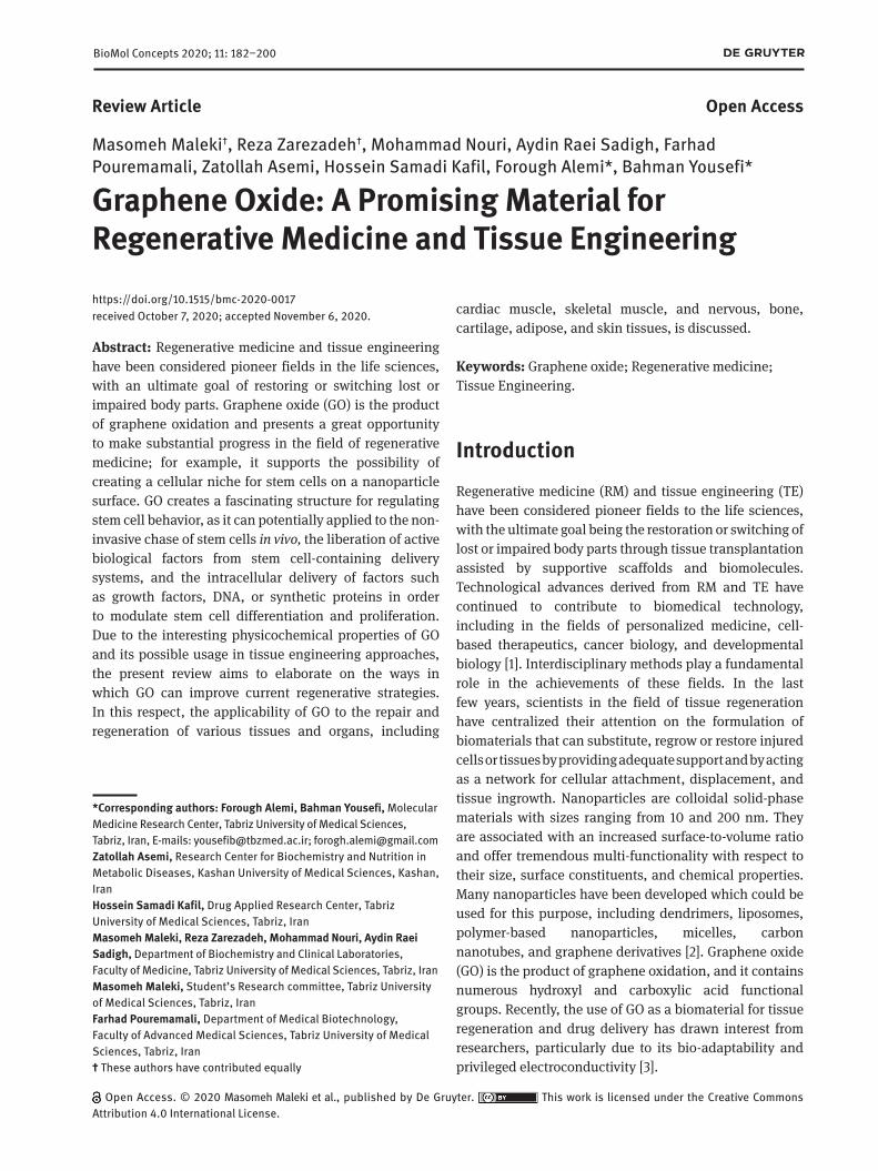
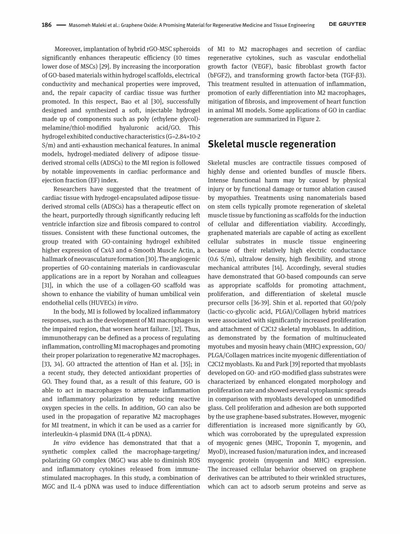
In light of the interesting physicochemical properties of GO and its possible utility in the aforementioned applications, the present review aims to elaborate on ways in which GO can improve current strategies for tissue and organ regeneration (figure 1).
GO properties and biomedical applicationsRecently, the use of GO as a biomaterial for tissue regeneration and drug delivery has become fascinating for researchers, as the compound has several favorable features, including excellent bio-adaptability, high drug loading capacity, privileged electroconductivity, and controllable size and shape [9].
GO contains numerous hydroxyl and carboxylic acid functional groups. The existence of oxygen-containing groups, including carboxyl (−COOH), hydroxyl (−OH), and epoxy (−O−) moieties, endows GO with hydrophilic properties, increasing its bioavailability [10]. These functional groups also facilitate conjugation of GO to other molecules and polymers, allowing it to be used in sophisticated drug delivery applications [11].
Due to GO’s large surface area (890 m2/g) and large π-conjugated structure, it has the potential to adsorb various proteins and adhere to cells, making it a good candidate for use in connective tissue regeneration. Its vast surface area also endows GO with a large capacity for growth factor loading and delivery to the injury site. Moreover, due to maintenance of the sp2 bonding lattice in GO, it has a high electrical conductivity appropriate for use in cardiac and nerve regeneration [12]. Furthermore, due to its mechanical strength, GO could be used as a high mechanical resistance matrix in bone and cartilage regeneration [13].
Cardiac tissue engineering and regenerationDamage to cardiac tissue as a result of coronary artery disease or myocardial infarction is one of the leading causes of morbidity and mortality around the world. At present, the approach for heart injury control is reliant on heart transplantation. However, the number of donors is much lower than the number of prospective patients, limiting the practical use of transplantation in
184 Masomeh Maleki et al.: Graphene Oxide: A Promising Material for Regenerative Medicine and Tissue Engineering
the greater population. Consequently, researchers have focused on the development of methods to restore the biological and physical properties of damaged tissues [14]. Features like anisotropic microarchitecture and a high level of organization endow specific contractile properties to cardiac tissue [15]. In addition, heart muscles have electrical conductivity (0.005 (transverse) ~ 0.1 (longitudinal) S/m). The exclusive mechanical features of myocardium result from the presence of highly aligned collagen nanofibers with diameters of 10-100 nm [16, 17]. Investigators have doped biomaterials with electrically conductive nanomaterials (e.g., gold- and carbon-based nanoparticles) to engineer materials with electrical
conductivity [17]. The conductivity of these materials derives from their particular structure, which consists of a conjugated backbone that permits electrons to move in polymeric chains [18]. Investigators have used these materials to generate 2D and 3D scaffolds for cardiac tissue engineering, resulting in improved cellular function.
In this regard, the addition of 150 mg/L (0.015%) GO to the chitosan-based scaffold was found to markedly increase its conductivity compared with a non-GO scaffold (0.134 S/m vs.1.63 × 10−3 S/m, respectively) [19]. Likewise, Saravanan et al. [20] revealed that, while there was an increasing trend in scaffold conductivity with the inclusion of GO at a concentration of 0.5%, the addition
Figure 1: Applications of graphene oxide s for tissue regeneration.
Masomeh Maleki et al.: Graphene Oxide: A Promising Material for Regenerative Medicine and Tissue Engineering 185
of 0.5% GO-Au terminated considerable amplifications in the conductivity of the chitosan scaffold. For the first time, researchers investigated whether the use of a chitosan-GO-Au scaffold affects conduction velocity, contractility, or electrical circulation in the infarcted myocardium. To this end, they implanted scaffolds into the infarcted hearts of rat models, after which marked improvements in these parameters were observed. Additional ex vivo studies further support these findings and include reports of increased ventricular contractility.
In this study, improved conduction velocity and cardiac contraction after scaffold implantation was attributed to upregulation of Connexin 43 (Cx43 as there was a significant increase in Cx43 expression in the infarcted myocardium after conductive polymer scaffold implantation [20]. It is worth noting that Cx43 is the predominant ventricular gap junction protein and is primarily responsible for electrical conduction in the myocardium [21]. A number of investigators have proposed that conduction of the scaffold is capable of inducing cell alignment, elongation, functional maturation, and anisotropy of myocardial cells between differing layers [22]. Additionally, it has been previously established that GO flakes influence heart repair through enhancing the release of reparation paracrine factors and reducing apoptotic heart tissue [23]. Taken together, these results corroborate the idea that the introduction of GO-containing polymeric scaffolds to the infarcted heart can restore cardiac conductivity, contractility, and ventricular function, in addition to potentially stimulating healing of the scar area. However, an apparent inconsistency exists across studies in terms of the concentrations of GO incorporated into the structures of corresponding scaffolds. This parameter is of critical importance with regard to the reduction in scaffold porosity observed with increasing GO concentration [19], which can limit cell infiltration and overall tissue maturation [24]. Therefore, additional studies will be required to determine an optimal GO concentration that balances conductivity and synthetic scaffold porosity. Primary studies have indicated that impaired myocardium can be repaired or regenerated by the therapeutic application of stem cells, which exert their impact via differentiation of injured cells into cardiomyocytes or vascular-specific cellular phenotypes. Consequently, they may boost contractile functionality, as well as myocardial perfusion. Nevertheless, stem cell treatment faces challenges, including invasive isolation of autologous cells, protracted and costly methods of manipulating cells in vitro, poor efficacy of cell grafting, and post-implantation electrophysiological challenges [25]. However, graphene and its derivatives can be used
to affect the fate of cells in vivo by increasing the electrical conductivity of the matrix and providing morphological signs, subsequently eliminating the limitations of stem cell therapies for MI [14].
It has been suggested that GO and its derivatives have a stimulatory effect on the differentiation of MSCs into cardiomyogenic cells when cells are cultured on a graphene monolayer [26, 27]. Accordingly, Stone and colleagues [24] recently reported upregulated expression of early cardiac differentiation markers (e.g., GATA-4 and Nkx2.5) in iPSC-derived MSCs cultured on rGO-containing scaffolds.
These finding suggest that rGO may have some effect on cardiac differentiation. Similarly, experiments in which heart H9C2 cells were seeded in GO-chitosan scaffolds confirmed that these scaffolds exhibited good cell viability, promoted of cell attachment and intercellular network formation and upregulated the expression of cardiac-specific genes and proteins involved in muscle conduction of electrical signals (e.g., Cx43). These data confirmed that the presence of GO in chitosan scaffolds enhances the cardiogenic phenotype and promotes expression of cardiac markers without external electrical stimulation [19].
Shin et al. introduced a new approach for the engineering of 3D cardiac tissue multi-arrays. This technique involves applying a systematic layer-by-layer (LBL) assembly of cells in which GO-based thin films were used as an adhesive layer to guarantee strong adhesion among sheets [28]. Using this approach, three cell types of hMSCs, cardiomyocytes, and endothelial cells were incorporated into the construct. Researchers observed the up-regulation of cardiac indicators, including sarcomeric α-actinin, and harmonized pulsing of the construct under low external electric fields, confirming that the presence of GO-based film plays a pivotal role in the maturation of cardiac cells, improving cell-cell electrical coupling and organization. Since GO acts as a shield for MSCs against reactive oxygen species, MSC survival rate was shown to improve notably after in vivo co-implantation of GO flakes [23]. Altered expression of cell signaling molecules and improved expression of cardiac-specific markers have been reported after the incorporation of rGO flakes into hMSC spheroids. In addition to high conduction, the high affinity of rGO flakes towards fibronectin might have improved the function of cardiac spheroids [29]. Compared to the implantation of either hMSCs or rGO, implantation of hybrid rGO-hMSC spheroids into a murine model of myocardial infarction (MI) was associated with enhanced cardiac repair and functionality.
186 Masomeh Maleki et al.: Graphene Oxide: A Promising Material for Regenerative Medicine and Tissue Engineering
Moreover, implantation of hybrid rGO-MSC spheroids significantly enhances therapeutic efficiency (10 times lower dose of MSCs) [29]. By increasing the incorporation of GO-based materials within hydrogel scaffolds, electrical conductivity and mechanical properties were improved, and, the repair capacity of cardiac tissue was further promoted. In this respect, Bao et al [30], successfully designed and synthesized a soft, injectable hydrogel made up of components such as poly (ethylene glycol)-melamine/thiol-modified hyaluronic acid/GO. This hydrogel exhibited conductive characteristics (G=2.84×10⁻² S/m) and anti-exhaustion mechanical features. In animal models, hydrogel-mediated delivery of adipose tissue-derived stromal cells (ADSCs) to the MI region is followed by notable improvements in cardiac performance and ejection fraction (EF) index.
Researchers have suggested that the treatment of cardiac tissue with hydrogel-encapsulated adipose tissue-derived stromal cells (ADSCs) has a therapeutic effect on the heart, purportedly through significantly reducing left ventricle infarction size and fibrosis compared to control tissues. Consistent with these functional outcomes, the group treated with GO-containing hydrogel exhibited higher expression of Cx43 and α-Smooth Muscle Actin, a hallmark of neovasculature formation [30]. The angiogenic properties of GO-containing materials in cardiovascular applications are in a report by Norahan and colleagues [31], in which the use of a collagen-GO scaffold was shown to enhance the viability of human umbilical vein endothelial cells (HUVECs) in vitro.
In the body, MI is followed by localized inflammatory responses, such as the development of M1 macrophages in the impaired region, that worsen heart failure. [32]. Thus, immunotherapy can be defined as a process of regulating inflammation, controlling M1 macrophages and promoting their proper polarization to regenerative M2 macrophages. [33, 34]. GO attracted the attention of Han et al. [35]; in a recent study, they detected antioxidant properties of GO. They found that, as a result of this feature, GO is able to act in macrophages to attenuate inflammation and inflammatory polarization by reducing reactive oxygen species in the cells. In addition, GO can also be used in the propagation of reparative M2 macrophages for MI treatment, in which it can be used as a carrier for interleukin-4 plasmid DNA (IL-4 pDNA).
In vitro evidence has demonstrated that that a synthetic complex called the macrophage-targeting/polarizing GO complex (MGC) was able to diminish ROS and inflammatory cytokines released from immune-stimulated macrophages. In this study, a combination of MGC and IL-4 pDNA was used to induce differentiation
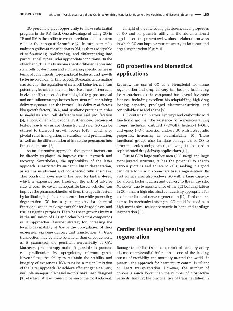
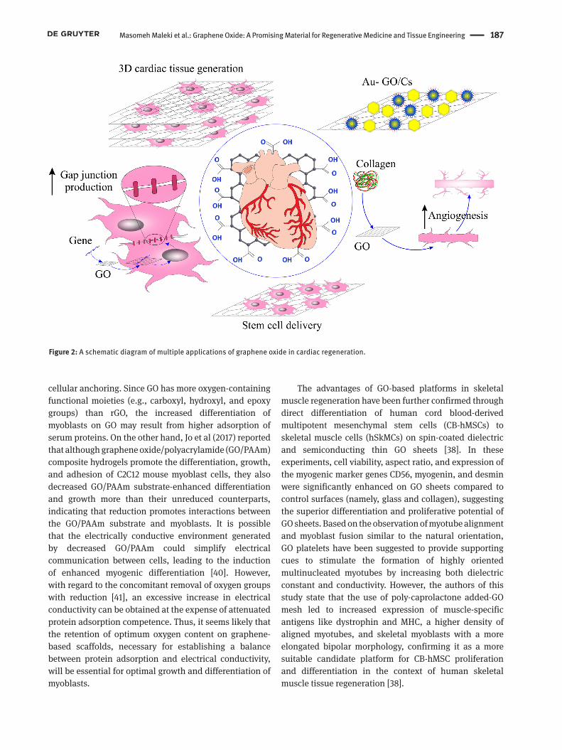
of M1 to M2 macrophages and secretion of cardiac regenerative cytokines, such as vascular endothelial growth factor (VEGF), basic fibroblast growth factor (bFGF2), and transforming growth factor-beta (TGF-β3). This treatment resulted in attenuation of inflammation, promotion of early differentiation into M2 macrophages, mitigation of fibrosis, and improvement of heart function in animal MI models. Some applications of GO in cardiac regeneration are summarized in Figure 2.
Skeletal muscle regenerationSkeletal muscles are contractile tissues composed of highly dense and oriented bundles of muscle fibers. Intense functional harm may by caused by physical injury or by functional damage or tumor ablation caused by myopathies. Treatments using nanomaterials based on stem cells typically promote regeneration of skeletal muscle tissue by functioning as scaffolds for the induction of cellular and differentiation viability. Accordingly, graphenated materials are capable of acting as excellent cellular substrates in muscle tissue engineering because of their relatively high electric conductance (0.6 S/m), ultralow density, high flexibility, and strong mechanical attributes [14]. Accordingly, several studies have demonstrated that GO-based compounds can serve as appropriate scaffolds for promoting attachment, proliferation, and differentiation of skeletal muscle precursor cells [36-39]. Shin et al. reported that GO/poly (lactic-co-glycolic acid, PLGA)/Collagen hybrid matrices were associated with significantly increased proliferation and attachment of C2C12 skeletal myoblasts. In addition, as demonstrated by the formation of multinucleated myotubes and myosin heavy chain (MHC) expression, GO/PLGA/Collagen matrices incite myogenic differentiation of C2C12 myoblasts. Ku and Park [39] reported that myoblasts developed on GO- and rGO-modified glass substrates were characterized by enhanced elongated morphology and proliferation rate and showed several cytoplasmic spreads in comparison with myoblasts developed on unmodified glass. Cell proliferation and adhesion are both supported by the use graphene-based substrates. However, myogenic differentiation is increased more significantly by GO, which was corroborated by the upregulated expression of myogenic genes (MHC, Troponin T, myogenin, and MyoD), increased fusion/maturation index, and increased myogenic protein (myogenin and MHC) expression. The increased cellular behavior observed on graphene derivatives can be attributed to their wrinkled structures, which can act to adsorb serum proteins and serve as
Masomeh Maleki et al.: Graphene Oxide: A Promising Material for Regenerative Medicine and Tissue Engineering 187
cellular anchoring. Since GO has more oxygen-containing functional moieties (e.g., carboxyl, hydroxyl, and epoxy groups) than rGO, the increased differentiation of myoblasts on GO may result from higher adsorption of serum proteins. On the other hand, Jo et al (2017) reported that although graphene oxide/polyacrylamide (GO/PAAm) composite hydrogels promote the differentiation, growth, and adhesion of C2C12 mouse myoblast cells, they also decreased GO/PAAm substrate-enhanced differentiation and growth more than their unreduced counterparts, indicating that reduction promotes interactions between the GO/PAAm substrate and myoblasts. It is possible that the electrically conductive environment generated by decreased GO/PAAm could simplify electrical communication between cells, leading to the induction of enhanced myogenic differentiation [40]. However, with regard to the concomitant removal of oxygen groups with reduction [41], an excessive increase in electrical conductivity can be obtained at the expense of attenuated protein adsorption competence. Thus, it seems likely that the retention of optimum oxygen content on graphene-based scaffolds, necessary for establishing a balance between protein adsorption and electrical conductivity, will be essential for optimal growth and differentiation of myoblasts.
The advantages of GO-based platforms in skeletal muscle regeneration have been further confirmed through direct differentiation of human cord blood-derived multipotent mesenchymal stem cells (CB-hMSCs) to skeletal muscle cells (hSkMCs) on spin-coated dielectric and semiconducting thin GO sheets [38]. In these experiments, cell viability, aspect ratio, and expression of the myogenic marker genes CD56, myogenin, and desmin were significantly enhanced on GO sheets compared to control surfaces (namely, glass and collagen), suggesting the superior differentiation and proliferative potential of GO sheets. Based on the observation of myotube alignment and myoblast fusion similar to the natural orientation, GO platelets have been suggested to provide supporting cues to stimulate the formation of highly oriented multinucleated myotubes by increasing both dielectric constant and conductivity. However, the authors of this study state that the use of poly-caprolactone added-GO mesh led to increased expression of muscle-specific antigens like dystrophin and MHC, a higher density of aligned myotubes, and skeletal myoblasts with a more elongated bipolar morphology, confirming it as a more suitable candidate platform for CB-hMSC proliferation and differentiation in the context of human skeletal muscle tissue regeneration [38].
Figure 2: A schematic diagram of multiple applications of graphene oxide in cardiac regeneration.
188 Masomeh Maleki et al.: Graphene Oxide: A Promising Material for Regenerative Medicine and Tissue Engineering
Nerve regenerationGraphene and its derivatives, particularly GO, are currently regarded as the next generation of tissue engineering scaffolds, capable of promoting the regenerative potential of neural cells and thereby revitalizing their functional activities following nervous system damage. In vitro use of graphene in the structures of scaffolds and neural substrates has been shown to confer advantages in terms of neural cell viability, proliferation, differentiation, and network signal transduction [42, 43]. To assess inflammation caused by graphene in the central nervous system (CNS), graphene-functionalized scaffolds were implanted into the striatum of adult rats. Observation of the inflammatory responses of microglia and astrocytes demonstrated that the implanted scaffolds did not trigger a significant immune response. Furthermore, the scaffold was shown to suppress inflammatory responses of microglia, astrocytes, and infiltrated macrophages. Implantation of these graphene scaffolds into the intercepted subventricular zone (SVZ) of rat models was also investigated in this study, and it was demonstrated that the scaffold influenced the growth direction of permeated astrocytes and stimulated proliferation of SVZ neuroblasts in a scaffold-oriented direction [44]. Recently, Verre et al. studied the effect of GO/rGO-coated coverslips on phenotype maintenance in differentiated adipose stem cells (dASCs), which can differentiate to Schwann-like cells and subsequently reform peripheral neural gaps. The authors noticed significant upregulation in the expression of neurotrophin and filament proteins, particularly on the GO/rGO surface, indicating that glial differentiation of dASCs can be promoted by the use of GO/rGO-coated culture media [45]. Moreover, it has been reported that the stabilization of nerve regenerative GFs, such as insulin-like growth factor 1 (IGF-1) and brain-derived neurotrophic factor (BDNF), with PLGA/GO electrospun nanofibers not only rescued neural stem cells (NSCs) from H2O2-induced oxidative damages, but also ameliorated defects to proliferation and differentiation potential of NSCs in vitro. Additionally, targeted delivery of IGF-1 and BDNF using PLGA/GO nanofibers resulted in enhanced restoration of locomotor activity, decreased cavity foundation, and improved neural cell proliferation at the damaged spot [46]. In addition, GO treatment in form of quantum dots has been shown to counteract the neurotoxic effects substances such as 1-methyl-4-phenyl-pyridinium (MPP) through reducing reactive oxygen species (ROS), cell cytotoxicity, apoptosis, and α-synuclein expression [47]. Combined administration of graphene quantum dots (GQDs) and neuroprotective peptide glycine-proline-
glutamate (GQDG) was found to prevent deterioration of Aβ1-42 plaques in transgenic mice models of Alzheimer’s disease [48]. Graphene and its derivatives have been also found to reinforce the functional activity of neurons by enhancing protein-protein adhesions. Indeed, Gonzalez-Mayorga and colleagues demonstrated that coating rGO microfibers with adhesive proteins (e.g. poly-lysine and N-cadherin) promoted neural cells growth in damaged regions of central neural tissue [49]. Increased adhesion of glial cells can induce secretion of neurotransmitters and other neurochemical signals, thereby ameliorating intracellular transportation and ultimately leading to improved neural network formation. Recently, it has been shown that the extracellular domains of the N-cadherin molecules that have been physically adsorbed on the GO/rGO-covered glass surface exhibit stronger adhesive properties than those directly adsorbed on the sole glass. Increased cohesion of N-cadherin, in turn, enhances neuron distribution on the substrate, intracellular mass transportation, and neuronal process, and thereby assisting to revitalize functional neural tissues [50]. From the viewpoint of peripheral nerve regeneration, graphene oxide/polyacrylamide (GO/PAM) hydrogels have been found to increase secretion of biochemical factors from Schwann cells, as well as to promote their adhesion to matrix compared to other compounds [51]. Applications of GO derivatives in nerve regeneration are summarized in Table 1.
Bone regeneration Nowadays, bone repair/regeneration is primarily limited to transplantation and use of titanium implants. Despite desperate attempts, researchers have so far been unable to fabricate an artificial platform that provides the essential features of an optimum bone scaffold and the ideal milieu for bone regeneration. The development of a scaffold with suitable degradation ability that is able to stimulate the body’s intrinsic healing mechanisms without requiring transplantation and/or undegradable prosthetics is of remarkable significance and is considered to be the aim of regenerative engineering. Multiple studies have demonstrated that GO and its derivatives can assist these efforts through different mechanisms (Table 2).
While stem cell therapies have excellent potential to induce innate regenerative pathways, the incorporation, conservation, and differentiation of these cells at the damaged site has remained challenging [52]. One use of GO in bone regeneration has been the manufacture of an osteoconductive platform to increase cell adhesion and
Masomeh Maleki et al.: Graphene Oxide: A Promising Material for Regenerative Medicine and Tissue Engineering 189
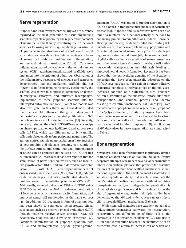
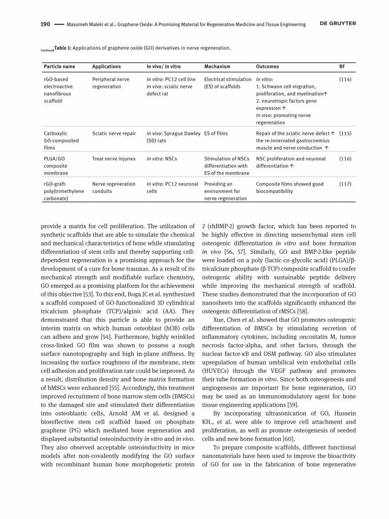
Table 1: Applications of graphene oxide (GO) derivatives in nerve regeneration.
Particle name Applications In vivo/ in vitro Mechanism Outcomes RF
self-assembled graphene implants
Implantation of scaffold into the striatum and into the subventricular zone (SVZ) of adult rats.
In vivo: adult rats Direct electrical coupling to neural cells
The implanted scaffolds did not trigger a significant immune responsescaffold stimulated proliferation of neuroblasts in SVZ
(44)
of GO/ reduced graphene oxide (rGO)-coated coverslips
Reform peripheral neural gaps
In vitro: Adipose stem cells (ASCs)
Stimulation of ASC differentiation
Promoting glial differentiation of ASCs
(45)
PLGA/GO Spinal cord injury in vitro: Neural stem cells (NSCs)in vivo: T9 spinal cord hemisection rat model
Stabilization of growth factors
in vitro: Ameliorate proliferation and differentiation of NSCs in vivo: 1. functional locomotor recovery ↑2. cavity formation ↓3. the number of neurons at the injury site ↑
(46)
GOQDs Neurotoxic injuries caused by 1‐methyl‐4‐phenyl‐pyridinium ion (MPP+)
In vitro: MPP+‐induced PC12 cellsIn vivo: zebrafish treated with MPP+
Amelioration of MPP+‐induced neurotoxicity
In vitro: 1. apoptosis ↓2. α‐synuclein ↓3. ROS ↓In vivo:1. ROS ↓2. mitochondrial damage ↓3. locomotive activity ↑ 4. Nissl bodies in the brain ↑
(47)
GQDG Alzheimer’s disease (AD)
In vitro: Aβ1-42 samplesIn vivo: APP/PS1 transgenic mice
GQDs conjugated to neuroprotective peptide glycine-proline-glutamate (GPE)
In vitro: Aβ1-42 aggregation ↓In vivo:1. pro-inflammatory cytokines↓2. microglial activation↓3. NGF ↑4. BDNF ↑5. density of dendritic spines in the brain ↑6. neurogenesis ↑
(48)
rGO microfibers Damaged regions of CNS
In vitro: embryonic neural progenitor cells In vivo: spinal cord injured rat
Coating rGO microfibers with adhesive proteins to enhance protein-protein adhesions
In vitro: Highly interconnected cultures of cells were formed on the rGO microfibers surface.In vivo: implantation of rGO microfibers in the injured rat spinal cord showed a capacity to be colonized by cells without subacute local toxicity.
(49)
N-cadherin- GO/rGO-glass
Recreating active neural tissues
In vitro: Hippocampal neurons were obtained from neonatal (P1-P2) Long Evans BluGill rat brains
Increase neuronal and glial cell adhesion.
N-cadherin extracellular domains retain greater adhesive function when physisorbed to GO or rGO coated glass.Enhancement of intracellular mass transport along neurites relative to N-cadherin on glass
(50)
190 Masomeh Maleki et al.: Graphene Oxide: A Promising Material for Regenerative Medicine and Tissue Engineering
provide a matrix for cell proliferation. The utilization of synthetic scaffolds that are able to simulate the chemical and mechanical characteristics of bone while stimulating differentiation of stem cells and thereby supporting cell-dependent regeneration is a promising approach for the development of a cure for bone traumas. As a result of its mechanical strength and modifiable surface chemistry, GO emerged as a promising platform for the achievement of this objective [53]. To this end, Boga JC et al. synthesized a scaffold composed of GO-functionalized 3D cylindrical tricalcium phosphate (TCP)/alginic acid (AA). They demonstrated that this particle is able to provide an interim matrix on which human osteoblast (hOB) cells can adhere and grow [54]. Furthermore, highly wrinkled cross-linked GO film was shown to possess a rough surface nanotopography and high in-plane stiffness. By increasing the surface roughness of the membrane, stem cell adhesion and proliferation rate could be improved. As a result, distribution density and bone matrix formation of hMSCs were enhanced [55]. Accordingly, this treatment improved recruitment of bone marrow stem cells (BMSCs) to the damaged site and stimulated their differentiation into osteoblastic cells, Arnold AM et al. designed a bioreflective stem cell scaffold based on phosphate graphene (PG) which mediated bone regeneration and displayed substantial osteoinductivity in vitro and in vivo. They also observed acceptable osteoinductivity in mice models after non-covalently modifying the GO surface with recombinant human bone morphogenetic protein
2 (rhBMP-2) growth factor, which has been reported to be highly effective in directing mesenchymal stem cell osteogenic differentiation in vitro and bone formation in vivo [56, 57]. Similarly, GO and BMP-2-like peptide were loaded on a poly (lactic-co-glycolic acid) (PLGA)/β-tricalcium phosphate (β-TCP) composite scaffold to confer osteogenic ability with sustainable peptide delivery while improving the mechanical strength of scaffold. These studies demonstrated that the incorporation of GO nanosheets into the scaffolds significantly enhanced the osteogenic differentiation of rMSCs [58].
Xue, Chen et al. showed that GO promotes osteogenic differentiation of BMSCs by stimulating secretion of inflammatory cytokines, including oncostatin M, tumor necrosis factor-alpha, and other factors, through the nuclear factor-κB and OSM pathway. GO also stimulates upregulation of human umbilical vein endothelial cells (HUVECs) through the VEGF pathway and promotes their tube formation in vitro. Since both osteogenesis and angiogenesis are important for bone regeneration, GO may be used as an immunomodulatory agent for bone tissue engineering applications [59].
By incorporating ultrasonication of GO, Hussein KH., et al. were able to improve cell attachment and proliferation, as well as promote osteogenesis of seeded cells and new bone formation [60].
To prepare composite scaffolds, different functional nanomaterials have been used to improve the bioactivity of GO for use in the fabrication of bone regenerative
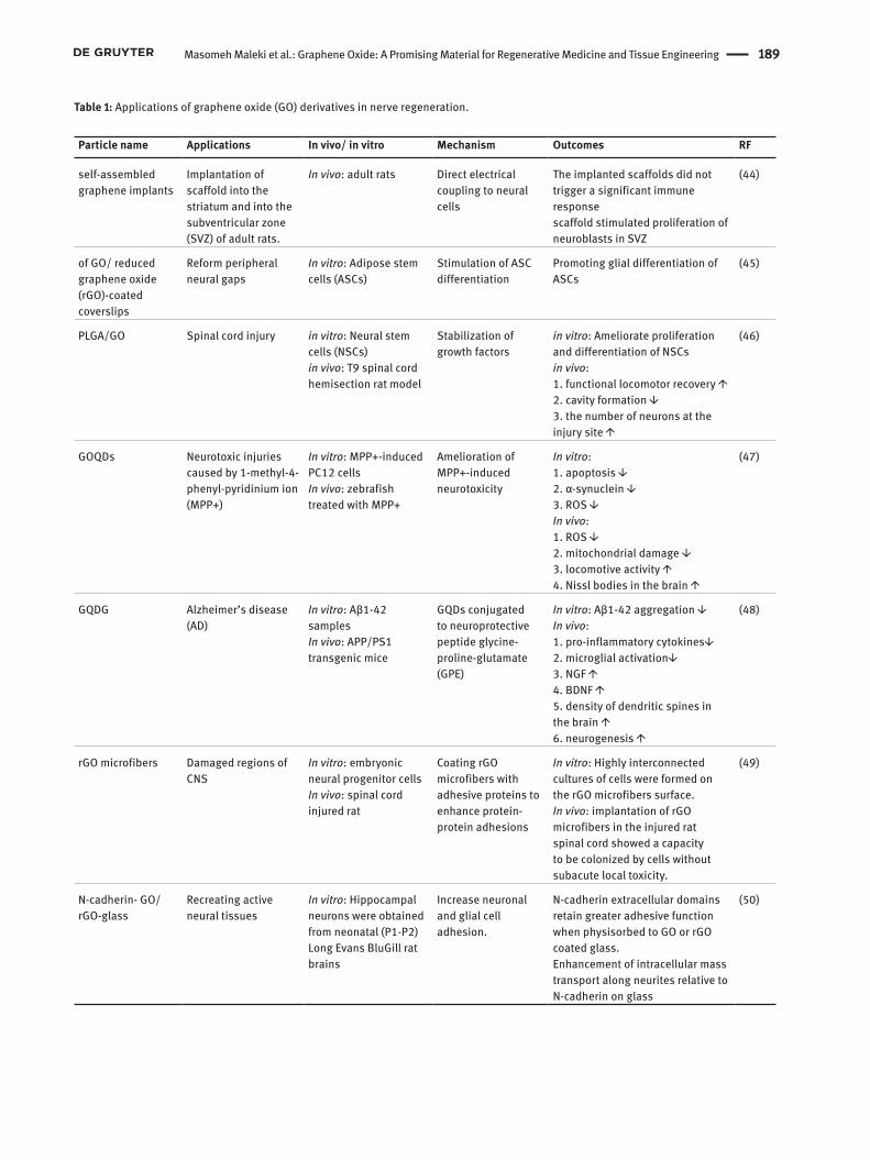
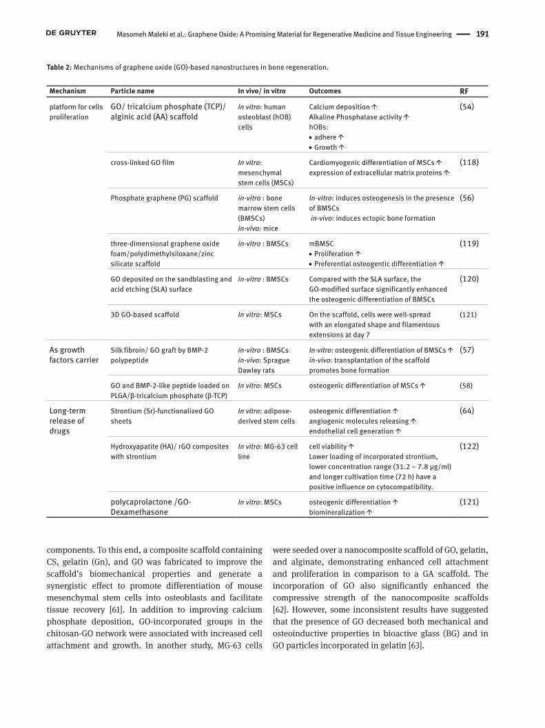
Particle name Applications In vivo/ in vitro Mechanism Outcomes RF
rGO-based electroactive nanofibrous scaffold
Peripheral nerve regeneration
In vitro: PC12 cell lineIn vivo: sciatic nerve defect rat
Electrical stimulation (ES) of scaffolds
In vitro: 1. Schwann cell migration, proliferation, and myelination↑ 2. neurotropic factors gene expression ↑In vivo: promoting nerve regeneration
(114)
Carboxylic GO-composited films
Sciatic nerve repair In vivo: Sprague Dawley (SD) rats
ES of films Repair of the sciatic nerve defect ↑the re-innervated gastrocnemius muscle and nerve conduction ↑
(115)
PLGA/GO composite membrane
Treat nerve injuries In vitro: NSCs Stimulation of NSCs differentiation with ES of the membrane
NSC proliferation and neuronal differentiation ↑
(116)
rGO-graft-poly(trimethylene carbonate)
Nerve regeneration conduits
In vitro: PC12 neuronal cells
Providing an environment for nerve regeneration
Composite films showed good biocompatibility
(117)
ContinuedTable 1: Applications of graphene oxide (GO) derivatives in nerve regeneration.
Masomeh Maleki et al.: Graphene Oxide: A Promising Material for Regenerative Medicine and Tissue Engineering 191
components. To this end, a composite scaffold containing CS, gelatin (Gn), and GO was fabricated to improve the scaffold’s biomechanical properties and generate a synergistic effect to promote differentiation of mouse mesenchymal stem cells into osteoblasts and facilitate tissue recovery [61]. In addition to improving calcium phosphate deposition, GO-incorporated groups in the chitosan-GO network were associated with increased cell attachment and growth. In another study, MG-63 cells
were seeded over a nanocomposite scaffold of GO, gelatin, and alginate, demonstrating enhanced cell attachment and proliferation in comparison to a GA scaffold. The incorporation of GO also significantly enhanced the compressive strength of the nanocomposite scaffolds [62]. However, some inconsistent results have suggested that the presence of GO decreased both mechanical and osteoinductive properties in bioactive glass (BG) and in GO particles incorporated in gelatin [63].
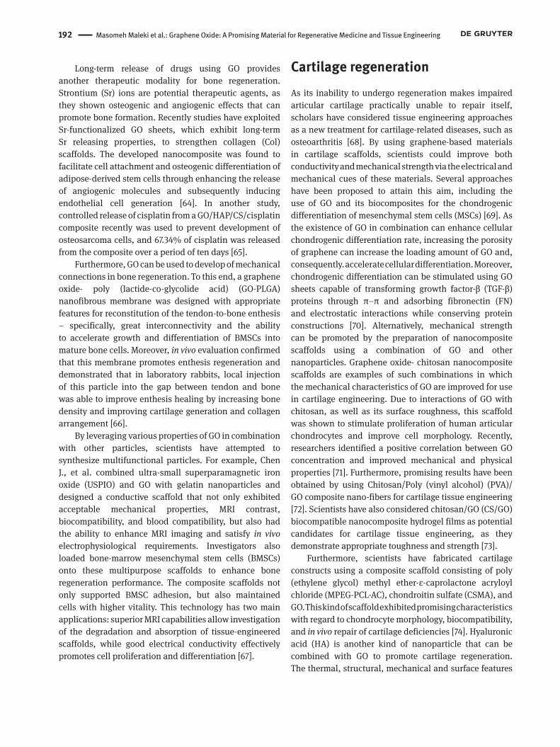
Table 2: Mechanisms of graphene oxide (GO)-based nanostructures in bone regeneration.
Mechanism Particle name In vivo/ in vitro Outcomes RF
platform for cells proliferation
GO/ tricalcium phosphate (TCP)/alginic acid (AA) scaffold
In vitro: human osteoblast (hOB) cells
Calcium deposition ↑Alkaline Phosphatase activity ↑hOBs: • adhere ↑• Growth ↑
(54)
cross-linked GO film In vitro: mesenchymalstem cells (MSCs)
Cardiomyogenic differentiation of MSCs ↑expression of extracellular matrix proteins ↑
(118)
Phosphate graphene (PG) scaffold in-vitro : bone marrow stem cells (BMSCs)in-vivo: mice
In-vitro: induces osteogenesis in the presence of BMSCs in-vivo: induces ectopic bone formation
(56)
three-dimensional graphene oxide foam/polydimethylsiloxane/zinc silicate scaffold
in-vitro : BMSCs mBMSC • Proliferation ↑• Preferential osteogentic differentiation ↑
(119)
GO deposited on the sandblasting and acid etching (SLA) surface
in-vitro : BMSCs Compared with the SLA surface, the GO-modified surface significantly enhanced the osteogenic differentiation of BMSCs
(120)
3D GO-based scaffold In vitro: MSCs On the scaffold, cells were well-spread with an elongated shape and filamentous extensions at day 7
(121)
As growth factors carrier
Silk fibroin/ GO graft by BMP-2 polypeptide
in-vitro : BMSCsin-vivo: Sprague Dawley rats
In-vitro: osteogenic differentiation of BMSCs ↑in-vivo: transplantation of the scaffold promotes bone formation
(57)
GO and BMP-2-like peptide loaded on PLGA/β-tricalcium phosphate (β-TCP)
In vitro: MSCs osteogenic differentiation of MSCs ↑ (58)
Long-term release of drugs
Strontium (Sr)-functionalized GO sheets
In vitro: adipose-derived stem cells
osteogenic differentiation ↑angiogenic molecules releasing ↑endothelial cell generation ↑
(64)
Hydroxyapatite (HA)/ rGO composites with strontium
In vitro: MG-63 cell line
cell viability ↑Lower loading of incorporated strontium, lower concentration range (31.2 – 7.8 µg/ml) and longer cultivation time (72 h) have apositive influence on cytocompatibility.
(122)
polycaprolactone /GO- Dexamethasone
In vitro: MSCs osteogenic differentiation ↑biomineralization ↑
(121)
192 Masomeh Maleki et al.: Graphene Oxide: A Promising Material for Regenerative Medicine and Tissue Engineering
Long-term release of drugs using GO provides another therapeutic modality for bone regeneration. Strontium (Sr) ions are potential therapeutic agents, as they shown osteogenic and angiogenic effects that can promote bone formation. Recently studies have exploited Sr-functionalized GO sheets, which exhibit long-term Sr releasing properties, to strengthen collagen (Col) scaffolds. The developed nanocomposite was found to facilitate cell attachment and osteogenic differentiation of adipose-derived stem cells through enhancing the release of angiogenic molecules and subsequently inducing endothelial cell generation [64]. In another study, controlled release of cisplatin from a GO/HAP/CS/cisplatin composite recently was used to prevent development of osteosarcoma cells, and 67.34% of cisplatin was released from the composite over a period of ten days [65].
Furthermore, GO can be used to develop of mechanical connections in bone regeneration. To this end, a graphene oxide- poly (lactide-co-glycolide acid) (GO-PLGA) nanofibrous membrane was designed with appropriate features for reconstitution of the tendon-to-bone enthesis – specifically, great interconnectivity and the ability to accelerate growth and differentiation of BMSCs into mature bone cells. Moreover, in vivo evaluation confirmed that this membrane promotes enthesis regeneration and demonstrated that in laboratory rabbits, local injection of this particle into the gap between tendon and bone was able to improve enthesis healing by increasing bone density and improving cartilage generation and collagen arrangement [66].
By leveraging various properties of GO in combination with other particles, scientists have attempted to synthesize multifunctional particles. For example, Chen J., et al. combined ultra-small superparamagnetic iron oxide (USPIO) and GO with gelatin nanoparticles and designed a conductive scaffold that not only exhibited acceptable mechanical properties, MRI contrast, biocompatibility, and blood compatibility, but also had the ability to enhance MRI imaging and satisfy in vivo electrophysiological requirements. Investigators also loaded bone-marrow mesenchymal stem cells (BMSCs) onto these multipurpose scaffolds to enhance bone regeneration performance. The composite scaffolds not only supported BMSC adhesion, but also maintained cells with higher vitality. This technology has two main applications: superior MRI capabilities allow investigation of the degradation and absorption of tissue-engineered scaffolds, while good electrical conductivity effectively promotes cell proliferation and differentiation [67].
Cartilage regenerationAs its inability to undergo regeneration makes impaired articular cartilage practically unable to repair itself, scholars have considered tissue engineering approaches as a new treatment for cartilage-related diseases, such as osteoarthritis [68]. By using graphene-based materials in cartilage scaffolds, scientists could improve both conductivity and mechanical strength via the electrical and mechanical cues of these materials. Several approaches have been proposed to attain this aim, including the use of GO and its biocomposites for the chondrogenic differentiation of mesenchymal stem cells (MSCs) [69]. As the existence of GO in combination can enhance cellular chondrogenic differentiation rate, increasing the porosity of graphene can increase the loading amount of GO and, consequently. accelerate cellular differentiation. Moreover, chondrogenic differentiation can be stimulated using GO sheets capable of transforming growth factor-β (TGF-β) proteins through π–π and adsorbing fibronectin (FN) and electrostatic interactions while conserving protein constructions [70]. Alternatively, mechanical strength can be promoted by the preparation of nanocomposite scaffolds using a combination of GO and other nanoparticles. Graphene oxide- chitosan nanocomposite scaffolds are examples of such combinations in which the mechanical characteristics of GO are improved for use in cartilage engineering. Due to interactions of GO with chitosan, as well as its surface roughness, this scaffold was shown to stimulate proliferation of human articular chondrocytes and improve cell morphology. Recently, researchers identified a positive correlation between GO concentration and improved mechanical and physical properties [71]. Furthermore, promising results have been obtained by using Chitosan/Poly (vinyl alcohol) (PVA)/ GO composite nano-fibers for cartilage tissue engineering [72]. Scientists have also considered chitosan/GO (CS/GO) biocompatible nanocomposite hydrogel films as potential candidates for cartilage tissue engineering, as they demonstrate appropriate toughness and strength [73].
Furthermore, scientists have fabricated cartilage constructs using a composite scaffold consisting of poly (ethylene glycol) methyl ether-ε-caprolactone acryloyl chloride (MPEG-PCL-AC), chondroitin sulfate (CSMA), and GO. This kind of scaffold exhibited promising characteristics with regard to chondrocyte morphology, biocompatibility, and in vivo repair of cartilage deficiencies [74]. Hyaluronic acid (HA) is another kind of nanoparticle that can be combined with GO to promote cartilage regeneration. The thermal, structural, mechanical and surface features
Masomeh Maleki et al.: Graphene Oxide: A Promising Material for Regenerative Medicine and Tissue Engineering 193
of polysaccharide-based combinations can be improved significantly by the addition of HA Recently, Hunger M et al. combined the advantageous properties of tannic acid, iron chloride (II), sodium hyaluronate, and GO to engineer a multiphase hydrogel system that generates a hierarchical structure to facilitate cartilage tissue regeneration. In this complex, GO improved mechanical and porosity properties. A higher concentration of GO was associated with higher film tensile strength, as well as higher compressive strength and a higher Young’s modulus of the porous scaffolds [75]. HA-strengthened GO can be used to provide lubrication during cartilage restoration and plays an important regulatory role in the joint cavity microenvironment, exerting an obvious antifriction influence on the joint surface and preparing an appropriate environment for cartilage restoration [76].
The potential dental applications of GOOral tissues are critical for the performance of certain functions in humans. Oral tissues may be destroyed during life by common diseases, such as periodontitis and dental caries [77]. Regeneration of various hard and soft dental tissues using stem cells has been performed successfully in vitro, representing a very promising development in the engineering of dental tissues [78]. Many researchers have recently endeavored to promote regeneration through the use of different scaffolding materials, such as natural silk, ceramics [hydroxyapatite (HA) and tri-calcium phosphate], natural polymers (collagen, polysaccharides, or fibrin) and synthetic polymers [poly (lactic acid) or poly (glycolic acid) [79, 80]. Tissue engineering approaches have been effectively used in a wide range of dental applications, including guided tissue regeneration (GTR), pulp dentin complex regeneration, and regeneration of salivary glands [81]. However, a number of concerns have been raised regarding the use of bio-dental materials. For example, exposure to body fluids can lead to the destruction or corrosion of materials within the oral cavity, and the oral environment may react with materials to produce harmful or cytotoxic components [78, 82]. Graphene oxide and graphene nanomaterials are of great interest in the design of composite materials, as they are believed to be a promising new material for dental material production [83, 84] Studies of graphene toxicity in vitro have demonstrated that the material is not cytotoxic [85]. In a study by Dreanca et al. researchers evaluated the compatibility of dental materials, including a dental
cement and a restorative composite, with graphene after bone implantation in a rat model of non-mandibular diagnosis violation. Chronic kidney and liver toxicity was assessed by interpreting organ-specific isoenzymes, and enzyme levels were found to be within the normal range. Histopathological evaluation was used determine the ratio of limbs and showed no differences in relative limb weight, confirming the lack of systemic toxicity of graphene dental materials [86].
Graphene and its derivatives can be combined with bioceramics to form composites with advanced mechanical properties for use both in vitro and in vivo. For example, the addition of 1% graphene nanosheets (GNS) by weight to HAp increased fracture toughness from 0.5 to 1.0 MPa / m 2 and increased hardness from 5.5 to 7.2 GPa [87]. Furthermore, the incorporation of GO nanoflakes in gelatin/HAp scaffolds increased compressive strength and resulted in a higher yield compared to the original gelatin/Hap [88]. Chitosan is a natural polymer with hemostatic, analgesic, antibacterial, and antifungal potential, leading to its use in tissue engineering of wound dressings and implants [89]. The addition of 3 wt% GO to chitosan scaffolds was shown to enhance the modulus of elasticity from 2.6 to 6.7 GPa and increase hard-ness from 0.3 to 1.1 GPa [90].
The combination of graphene and polymer, in addition to imbuing advanced mechanical and physical properties, can improve and enhance stem cell differentiation, bioactivity, and osteogenic potential [91]. For instance, the combination of rGO with polydopamine (PDA) lead to enhanced osteoblastic cell proliferation and higher adhesion compared to glass alone. In addition, pre-osteoblasts cultured in rGO / PDA substrate show higher Alkaline phosphate (ALP) activity, indicating increased osteogenic differentiation compared with controls Alkaline phosphatase (ALP) has been identified as a primary marker of osteoblastic differentiation [92].
Because the sheet structure of graphene provides a large surface area for drug loading, it also increases uptake of dexamethasone (Dex) into GO for electrospun polylactic-co-glycolic acid (PLGA) nanofibrous mats compared to unmodified mats. Dexamethasone, an osteogenic inducer, increases expression of ALP, OCN, collagen I in MSCs. Even in the absence of dexamethasone, cells in the GO/PLGA scaffold exhibited more OCN protein than cells in the control scaffold [93]. One unexpected results of restorative composite graphene is the formation of activated osteoblasts that increase activity of calcium and alkaline phosphate. This result is in good agreement with previous findings [86, 94]. Another study confirmed the role of GO in inducing bone formation. A Ti alloy was
194 Masomeh Maleki et al.: Graphene Oxide: A Promising Material for Regenerative Medicine and Tissue Engineering
modified with RGO, and a DEX estrogenic chemical drug was loaded on it. The findings showed differentiation of MC3T3-E1 pre-osteoblasts into osteoblasts, which increased ALP and calcium nodule deposition, as well as markers of osteogenic expression (Col-1, OPN, Runx2, and OCN) [95].
When used in nanocomposite coating, graphene-based materials (GBMs) not only improve mechanical and surface properties, but also have anti-corrosion properties that can be utilized in prosthetic implants and in endodontic dentistry instruments [96]. For example, Nickel-titanium (NiTi) alloy has properties of anti-corrosion, shape memory, and superelasticity. However, due to the significant release of Ni and Ti ions in the physiological environment, it is attacked in the human body, causing allergies, foreign body reactions, and side effect such as loss of taste, angular cheilitis, and a burning sensation [97]. GO-coated NiTi and GO/Ag coated NiTi have better corrosion resistance, lower corrosion resistance, and higher protection performance than the bare NiTi alloy. Also, both NiTi variants were compatible with GO coating and NiTi GO / Ag coating of human pulp fibroblasts and induced upregulation of IL-6 and IL-8 levels. Therefore, GBMs may cause changes in the levels of cytokines released in the body [98].
GO nano-plates are a promising nanomaterial for use in dental treatment and care. Periodontal disease and tooth decay are closely related to germs. For example, Spectrocomotance (S. mutans), which decreases pH in the microenvironment, Porphyromonas gingivalis, and Fusobacterium nucleatum have all been associated with root canal infection and periodontitis [99, 100]. GO nanoparticles destroy bacterial membrane integrity, causing intracellular contents to leak into the plasma [101]. Furthermore, Zisheng Tan et al. ascertained that in early stages, GO nanoparticles prevent activity and adhesion of S.mutans biofilms. EPS is an extracellular polymer secreted by bacteria that plays a critical role in resistance and protection against antimicrobial agents. GO can affect EPS levels by combining with the cell to create a non-encapsulated functional layer, and researchers found that GO was powerful in killing S.mutans bacteria [102].
Graphene can also be used in teeth whitening. When used with hydrogen peroxide (H2O2), cobalt Tetraphenylporphyrin (CoTPP) and RGO nanocomposites reduce treatment time, promote ease of whitening, increase whitening rate and effect, retain the bleach qualities of H2O2, and reduce side effects such as dental sensitivity and gingival irritation. Combining graphene with appropriate ingredients improves tooth whitening [103, 104].
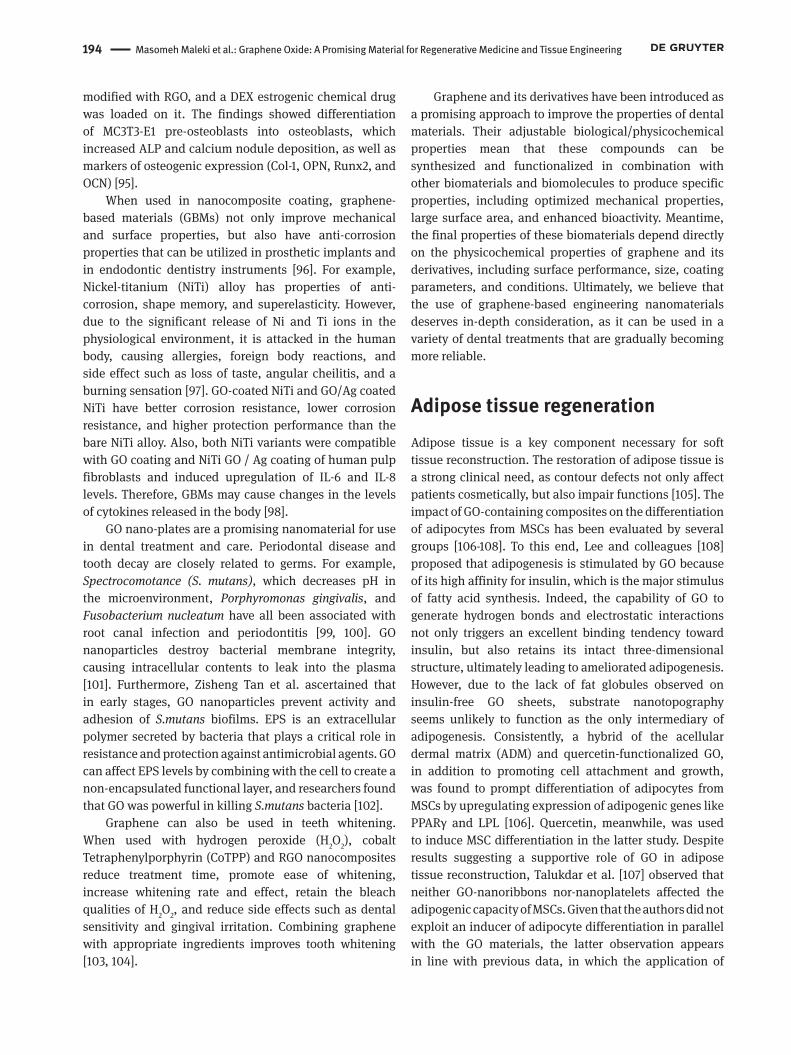
Graphene and its derivatives have been introduced as a promising approach to improve the properties of dental materials. Their adjustable biological/physicochemical properties mean that these compounds can be synthesized and functionalized in combination with other biomaterials and biomolecules to produce specific properties, including optimized mechanical properties, large surface area, and enhanced bioactivity. Meantime, the final properties of these biomaterials depend directly on the physicochemical properties of graphene and its derivatives, including surface performance, size, coating parameters, and conditions. Ultimately, we believe that the use of graphene-based engineering nanomaterials deserves in-depth consideration, as it can be used in a variety of dental treatments that are gradually becoming more reliable.
Adipose tissue regenerationAdipose tissue is a key component necessary for soft tissue reconstruction. The restoration of adipose tissue is a strong clinical need, as contour defects not only affect patients cosmetically, but also impair functions [105]. The impact of GO-containing composites on the differentiation of adipocytes from MSCs has been evaluated by several groups [106-108]. To this end, Lee and colleagues [108] proposed that adipogenesis is stimulated by GO because of its high affinity for insulin, which is the major stimulus of fatty acid synthesis. Indeed, the capability of GO to generate hydrogen bonds and electrostatic interactions not only triggers an excellent binding tendency toward insulin, but also retains its intact three-dimensional structure, ultimately leading to ameliorated adipogenesis. However, due to the lack of fat globules observed on insulin-free GO sheets, substrate nanotopography seems unlikely to function as the only intermediary of adipogenesis. Consistently, a hybrid of the acellular dermal matrix (ADM) and quercetin-functionalized GO, in addition to promoting cell attachment and growth, was found to prompt differentiation of adipocytes from MSCs by upregulating expression of adipogenic genes like PPARγ and LPL [106]. Quercetin, meanwhile, was used to induce MSC differentiation in the latter study. Despite results suggesting a supportive role of GO in adipose tissue reconstruction, Talukdar et al. [107] observed that neither GO-nanoribbons nor-nanoplatelets affected the adipogenic capacity of MSCs. Given that the authors did not exploit an inducer of adipocyte differentiation in parallel with the GO materials, the latter observation appears in line with previous data, in which the application of
Masomeh Maleki et al.: Graphene Oxide: A Promising Material for Regenerative Medicine and Tissue Engineering 195
GO-based compounds in adipose tissue regeneration is merely restricted to the delivery of a differentiation factor to MSCs.
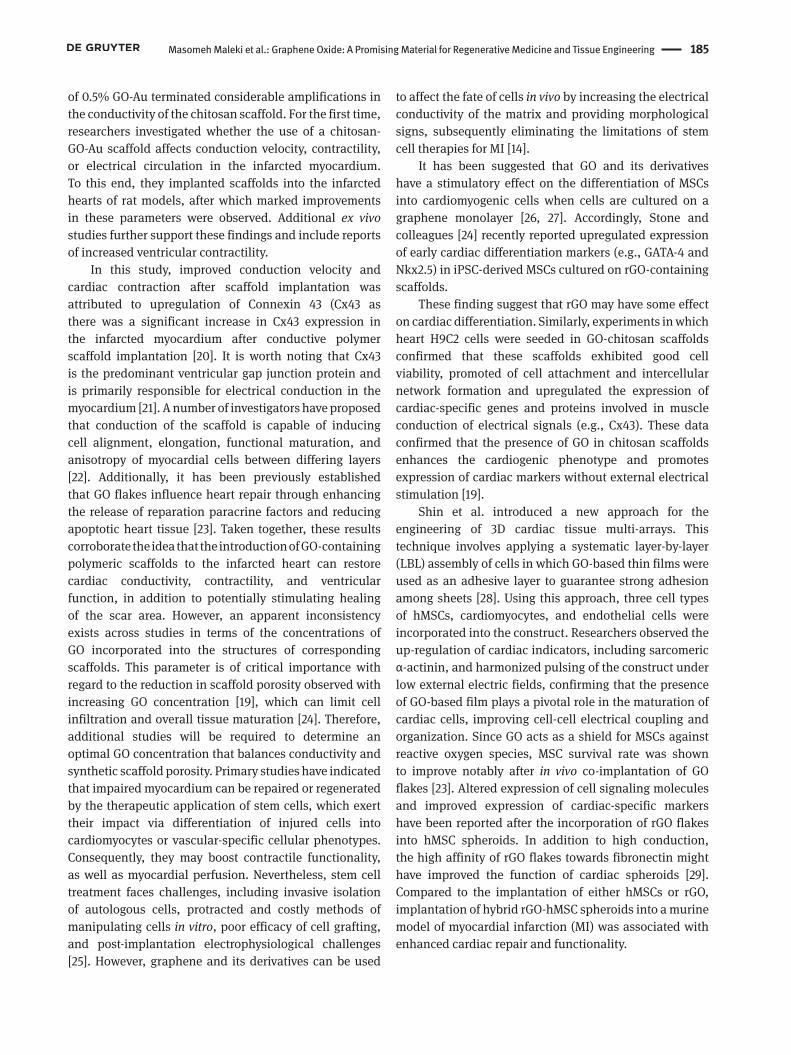
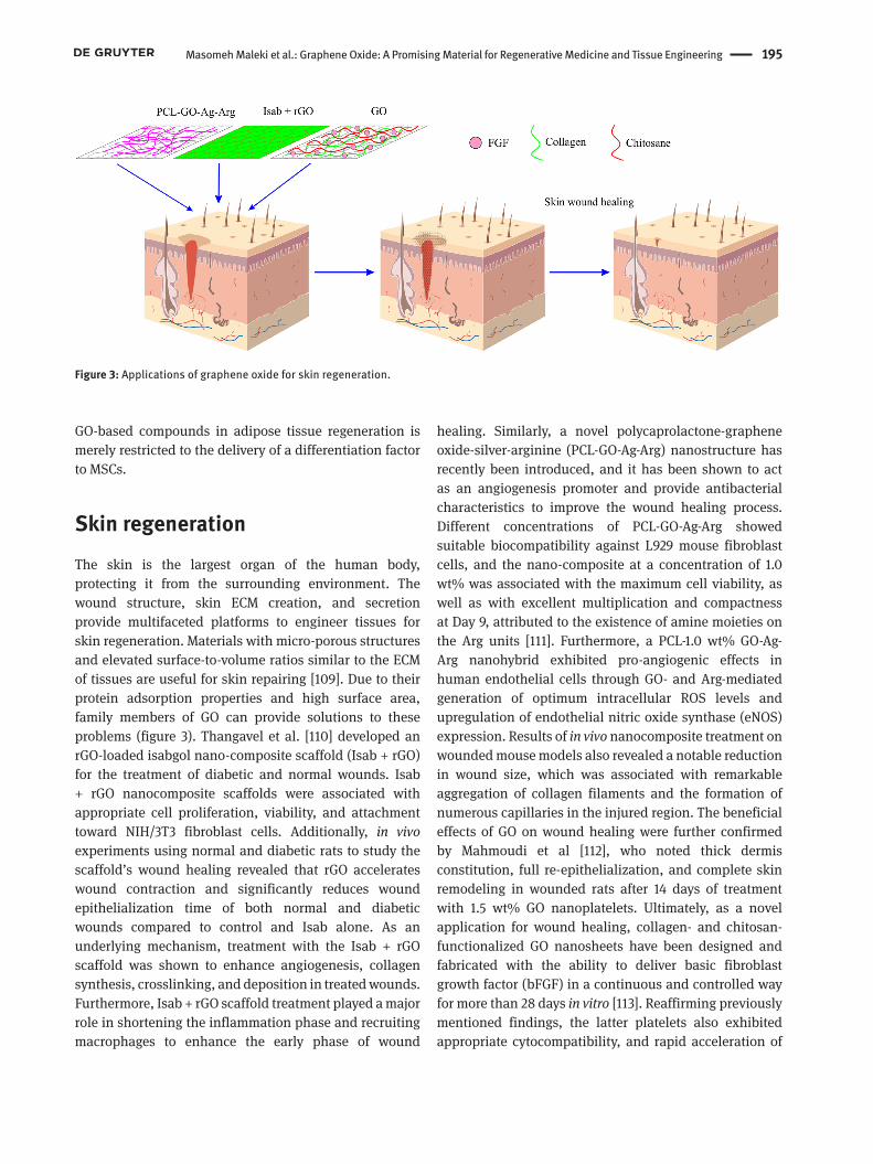
Skin regenerationThe skin is the largest organ of the human body, protecting it from the surrounding environment. The wound structure, skin ECM creation, and secretion provide multifaceted platforms to engineer tissues for skin regeneration. Materials with micro-porous structures and elevated surface-to-volume ratios similar to the ECM of tissues are useful for skin repairing [109]. Due to their protein adsorption properties and high surface area, family members of GO can provide solutions to these problems (figure 3). Thangavel et al. [110] developed an rGO-loaded isabgol nano-composite scaffold (Isab + rGO) for the treatment of diabetic and normal wounds. Isab + rGO nanocomposite scaffolds were associated with appropriate cell proliferation, viability, and attachment toward NIH/3T3 fibroblast cells. Additionally, in vivo experiments using normal and diabetic rats to study the scaffold’s wound healing revealed that rGO accelerates wound contraction and significantly reduces wound epithelialization time of both normal and diabetic wounds compared to control and Isab alone. As an underlying mechanism, treatment with the Isab + rGO scaffold was shown to enhance angiogenesis, collagen synthesis, crosslinking, and deposition in treated wounds. Furthermore, Isab + rGO scaffold treatment played a major role in shortening the inflammation phase and recruiting macrophages to enhance the early phase of wound
healing. Similarly, a novel polycaprolactone-graphene oxide-silver-arginine (PCL-GO-Ag-Arg) nanostructure has recently been introduced, and it has been shown to act as an angiogenesis promoter and provide antibacterial characteristics to improve the wound healing process. Different concentrations of PCL-GO-Ag-Arg showed suitable biocompatibility against L929 mouse fibroblast cells, and the nano-composite at a concentration of 1.0 wt% was associated with the maximum cell viability, as well as with excellent multiplication and compactness at Day 9, attributed to the existence of amine moieties on the Arg units [111]. Furthermore, a PCL-1.0 wt% GO-Ag-Arg nanohybrid exhibited pro-angiogenic effects in human endothelial cells through GO- and Arg-mediated generation of optimum intracellular ROS levels and upregulation of endothelial nitric oxide synthase (eNOS) expression. Results of in vivo nanocomposite treatment on wounded mouse models also revealed a notable reduction in wound size, which was associated with remarkable aggregation of collagen filaments and the formation of numerous capillaries in the injured region. The beneficial effects of GO on wound healing were further confirmed by Mahmoudi et al [112], who noted thick dermis constitution, full re-epithelialization, and complete skin remodeling in wounded rats after 14 days of treatment with 1.5 wt% GO nanoplatelets. Ultimately, as a novel application for wound healing, collagen- and chitosan-functionalized GO nanosheets have been designed and fabricated with the ability to deliver basic fibroblast growth factor (bFGF) in a continuous and controlled way for more than 28 days in vitro [113]. Reaffirming previously mentioned findings, the latter platelets also exhibited appropriate cytocompatibility, and rapid acceleration of
Figure 3: Applications of graphene oxide for skin regeneration.
196 Masomeh Maleki et al.: Graphene Oxide: A Promising Material for Regenerative Medicine and Tissue Engineering
skin wound regeneration was observed upon culture of L929 fibroblastic cells and implantation onto the injured areas in animal models.
ConclusionMany articles describing the use of GO and its derivatives in RM have been published in recent years, and there are both advantages and disadvantages to the use of these compounds to increase regeneration efficiency. Several unique features of GO make it a suitable tool for RM. For example, electric conductivity is necessary for cardiac and nerve regeneration, mechanical strength is necessary for bone and cartilage regeneration, and a vast surface is required as a stem cell milieu during regeneration of most tissues. GO is also an ideal platform to increase cell adhesion, as its protein adsorption properties and high surface area promote connective tissue regeneration and wound healing. Because GO can incorporate all of these features, it is considered a powerful tool by scientists in the RM field. Overall, GO presents outstanding capabilities in terms of RM, and it could represent a valuable solution for patients who have lost part body tissues . However, despite promising results from s biocompatibility studies, more studies are needed on the toxicity and side effects of GO.
Funding: This work was not supported by any grant funding.
Conflict of interest: The authors declare no conflict of interest.
References1. Pina S, Ribeiro VP, Marques CF, Maia FR, Silva TH, Reis RL, et al.
Scaffolding strategies for tissue engineering and regenerative medicine applications. Materials (Basel). 2019 Jun;12(11):1824.
2. Tan HL, Teow SY, Pushpamalar J. Application of metal nanoparticle–hydrogel composites in tissue regeneration. Bioengineering (Basel). 2019 Feb;6(1):17.
3. Alemi F, Zarezadeh R, Sadigh AR, Hamishehkar H, Rahimi M, Majidinia M, et al. Graphene Oxide and Reduced Graphene Oxide: Efficient Cargo Platforms for Cancer Theranostics. J Drug Deliv Sci Technol. 2020;60:101974.
4. Ramos T, Moroni L. Tissue Engineering and Regenerative Medicine 2019: The Role of Biofabrication-A Year in Review. Tissue Eng Part C Methods. 2020 Feb;26(2):91–106.
5. Lee WC, Lim CH, Shi H, Tang LA, Wang Y, Lim CT, et al. Origin of enhanced stem cell growth and differentiation on graphene and graphene oxide. ACS Nano. 2011 Sep;5(9):7334–41.
6. Zhou M, Lozano N, Wychowaniec JK, Hodgkinson T, Richardson SM, Kostarelos K, et al. Graphene oxide: A growth factor delivery carrier to enhance chondrogenic differentiation of human mesenchymal stem cells in 3D hydrogels. Acta Biomater. 2019 Sep;96:271–80.
7. Han XM, Zheng KW, Wang RL, Yue SF, Chen J, Zhao ZW, et al. Functionalization and optimization-strategy of graphene oxide-based nanomaterials for gene and drug delivery. Am J Transl Res. 2020 May;12(5):1515–34.
8. Amreddy N, Babu A, Muralidharan R, Panneerselvam J, Srivastava A, Ahmed R, et al. Recent advances in nanoparticle-based cancer drug and gene delivery. Advances in cancer research. 137. Elsevier; 2018. pp. 115–70.
9. Chen X, Hai X, Wang J. Graphene/graphene oxide and their derivatives in the separation/isolation and preconcentration of protein species: A review. Anal Chim Acta. 2016 May;922:1–10.
10. Kim J, Jeon JH, Kim HJ, Lim H, Oh IK. Durable and water-floatable ionic polymer actuator with hydrophobic and asymmetrically laser-scribed reduced graphene oxide paper electrodes. ACS Nano. 2014 Mar;8(3):2986–97.
11. Gao W. Graphene oxide: reduction recipes, spectroscopy, and applications. Springer; 2015. https://doi.org/10.1007/978-3-319-15500-5.
12. Stobinski L, Lesiak B, Malolepszy A, Mazurkiewicz M, Mierzwa B, Zemek J, et al. Graphene oxide and reduced graphene oxide studied by the XRD, TEM and electron spectroscopy methods. J Electron Spectrosc Relat Phenom. 2014;195:145–54.
13. Feng P, Jia J, Peng S, Yang W, Bin S, Shuai C. Graphene oxide-driven interfacial coupling in laser 3D printed PEEK/PVA scaffolds for bone regeneration. Virtual Phys Prototyp. 2020;15(2):211–26.
14. Shin SR, Li YC, Jang HL, Khoshakhlagh P, Akbari M, Nasajpour A, et al. Graphene-based materials for tissue engineering. Advanced drug delivery reviews. 2016;105(Pt B):255-74. https://doi.org/10.1016/j.addr.2016.03.007.
15. You JO, Rafat M, Ye GJ, Auguste DT. Nanoengineering the heart: conductive scaffolds enhance connexin 43 expression. Nano Lett. 2011 Sep;11(9):3643–8.
16. Liau B, Zhang D, Bursac N. Functional cardiac tissue engineering. Regen Med. 2012 Mar;7(2):187–206.
17. Dvir T, Timko BP, Brigham MD, Naik SR, Karajanagi SS, Levy O, et al. Nanowired three-dimensional cardiac patches. Nat Nanotechnol. 2011 Sep;6(11):720–5.
18. Guiseppi-Elie A. Electroconductive hydrogels: synthesis, characterization and biomedical applications. Biomaterials. 2010 Apr;31(10):2701–16.
19. Jiang L, Chen D, Wang Z, Zhang Z, Xia Y, Xue H, et al. Preparation of an Electrically Conductive Graphene Oxide/Chitosan Scaffold for Cardiac Tissue Engineering. Appl Biochem Biotechnol. 2019 Aug;188(4):952–64.
20. Saravanan S, Sareen N, Abu-El-Rub E, Ashour H, Sequiera GL, Ammar HI, et al. Graphene Oxide-Gold Nanosheets Containing Chitosan Scaffold Improves Ventricular Contractility and Function After Implantation into Infarcted Heart. Sci Rep. 2018 Oct;8(1):15069.
Masomeh Maleki et al.: Graphene Oxide: A Promising Material for Regenerative Medicine and Tissue Engineering 197
21. Boengler K, Schulz R. Connexin 43 and Mitochondria in Cardiovascular Health and Disease. Adv Exp Med Biol. 2017;982:227–46.
22. Wu Y, Wang L, Guo B, Ma PX. Interwoven aligned conductive nanofiber yarn/hydrogel composite scaffolds for engineered 3D cardiac anisotropy. ACS Nano. 2017 Jun;11(6):5646–59.
23. Park J, Kim B, Han J, Oh J, Park S, Ryu S, et al. Graphene oxide flakes as a cellular adhesive: prevention of reactive oxygen species mediated death of implanted cells for cardiac repair. ACS Nano. 2015 May;9(5):4987–99.
24. Stone H, Lin S, Mequanint K. Preparation and characterization of electrospun rGO-poly(ester amide) conductive scaffolds. Mater Sci Eng C. 2019 May;98:324–32.
25. Satessa G, Lenjisa J, Gebremariam E, Woldu M. Stem cell therapy for myocardial infarction: challenges and prospects. J Stem Cell Res Ther. 2015;5(270):2.
26. Bressan E, Ferroni L, Gardin C, Sbricoli L, Gobbato L, Ludovichetti FS, et al. Graphene based scaffolds effects on stem cells commitment. J Transl Med. 2014 Oct;12(1):296.
27. Park J, Park S, Ryu S, Bhang SH, Kim J, Yoon JK, et al. Graphene-regulated cardiomyogenic differentiation process of mesenchymal stem cells by enhancing the expression of extracellular matrix proteins and cell signaling molecules. Adv Healthc Mater. 2014 Feb;3(2):176–81.
28. Shin SR, Aghaei-Ghareh-Bolagh B, Gao X, Nikkhah M, Jung SM, Dolatshahi-Pirouz A, et al. Layer-by-layer assembly of 3D tissue constructs with functionalized graphene. Adv Funct Mater. 2014 Oct;24(39):6136–44.
29. Park J, Kim YS, Ryu S, Kang WS, Park S, Han J, et al. Graphene potentiates the myocardial repair efficacy of mesenchymal stem cells by stimulating the expression of angiogenic growth factors and gap junction protein. Adv Funct Mater. 2015;25(17):2590–600.
30. Bao R, Tan B, Liang S, Zhang N, Wang W, Liu W. A π-π conjugation-containing soft and conductive injectable polymer hydrogel highly efficiently rebuilds cardiac function after myocardial infarction. Biomaterials. 2017 Apr;122:63–71.
31. Norahan MH, Amroon M, Ghahremanzadeh R, Mahmoodi M, Baheiraei N. Electroactive graphene oxide-incorporated collagen assisting vascularization for cardiac tissue engineering. J Biomed Mater Res A. 2019 Jan;107(1):204–19.
32. Frantz S, Nahrendorf M. Cardiac macrophages and their role in ischaemic heart disease. Cardiovasc Res. 2014 May;102(2):240–8.
33. Choo EH, Lee JH, Park EH, Park HE, Jung NC, Kim TH, et al. Infarcted myocardium-primed dendritic cells improve remodeling and cardiac function after myocardial infarction by modulating the regulatory T cell and macrophage polarization. Circulation. 2017 Apr;135(15):1444–57.
34. Gombozhapova A, Rogovskaya Y, Shurupov V, Rebenkova M, Kzhyshkowska J, Popov SV, et al. Macrophage activation and polarization in post-infarction cardiac remodeling. J Biomed Sci. 2017 Feb;24(1):13.
35. Han J, Kim YS, Lim MY, Kim HY, Kong S, Kang M, et al. Dual Roles of Graphene Oxide To Attenuate Inflammation and Elicit Timely Polarization of Macrophage Phenotypes for Cardiac Repair. ACS Nano. 2018 Feb;12(2):1959–77.
36. Shin YC, Lee JH, Jin L, Kim MJ, Kim YJ, Hyun JK, et al. Stimulated myoblast differentiation on graphene oxide-impregnated PLGA-
collagen hybrid fibre matrices. J Nanobiotechnology. 2015 Mar;13(1):21.
37. Jo H, Sim M, Kim S, Yang S, Yoo Y, Park JH, et al. Electrically conductive graphene/polyacrylamide hydrogels produced by mild chemical reduction for enhanced myoblast growth and differentiation. Acta Biomater. 2017 Jan;48:100–9.
38. Chaudhuri B, Bhadra D, Moroni L, Pramanik K. Myoblast differentiation of human mesenchymal stem cells on graphene oxide and electrospun graphene oxide-polymer composite fibrous meshes: importance of graphene oxide conductivity and dielectric constant on their biocompatibility. Biofabrication. 2015 Feb;7(1):015009.
39. Ku SH, Park CB. Myoblast differentiation on graphene oxide. Biomaterials. 2013 Mar;34(8):2017–23.
40. Thrivikraman G, Mallik PK, Basu B. Substrate conductivity dependent modulation of cell proliferation and differentiation in vitro. Biomaterials. 2013 Sep;34(29):7073–85.
41. Dong L, Yang J, Chhowalla M, Loh KP. Synthesis and reduction of large sized graphene oxide sheets. Chem Soc Rev. 2017 Nov;46(23):7306–16.
42. Park SY, Park J, Sim SH, Sung MG, Kim KS, Hong BH, et al. Enhanced differentiation of human neural stem cells into neurons on graphene. Adv Mater. 2011 Sep;23(36):H263–7.
43. Tang M, Song Q, Li N, Jiang Z, Huang R, Cheng G. Enhancement of electrical signaling in neural networks on graphene films. Biomaterials. 2013 Sep;34(27):6402–11.
44. Zhou K, Motamed S, Thouas GA, Bernard CC, Li D, Parkington HC, et al. Graphene functionalized scaffolds reduce the inflammatory response and supports endogenous neuroblast migration when implanted in the adult brain. PLoS One. 2016 Mar;11(3):e0151589.
45. Verre AF, Faroni A, Iliut M, Silva C, Muryn C, Reid AJ, et al. Improving the glial differentiation of human Schwann-like adipose-derived stem cells with graphene oxide substrates. Interface Focus. 2018 Jun;8(3):20180002.
46. Pan S, Qi Z, Li Q, Ma Y, Fu C, Zheng S, et al. Graphene oxide-PLGA hybrid nanofibres for the local delivery of IGF-1 and BDNF in spinal cord repair. Artif Cells Nanomed Biotechnol. 2019 Dec;47(1):651–64.
47. Ren C, Hu X, Zhou Q. Graphene oxide quantum dots reduce oxidative stress and inhibit neurotoxicity in vitro and in vivo through catalase‐like activity and metabolic regulation. Adv Sci (Weinh). 2018 Mar;5(5):1700595.
48. Xiao S, Zhou D, Luan P, Gu B, Feng L, Fan S, et al. Graphene quantum dots conjugated neuroprotective peptide improve learning and memory capability. Biomaterials. 2016 Nov;106:98–110.
49. González-Mayorga A, López-Dolado E, Gutiérrez MC, Collazos-Castro JE, Ferrer ML, Del Monte F, et al. Favorable biological responses of neural cells and tissue interacting with graphene oxide microfibers. ACS Omega. 2017 Nov;2(11):8253–63.
50. Qin EC, Kandel ME, Liamas E, Shah TB, Kim C, Kaufman CD, et al. Graphene oxide substrates with N-cadherin stimulates neuronal growth and intracellular transport. Acta Biomater. 2019 May;90:412–23.
51. Li G, Zhao Y, Zhang L, Gao M, Kong Y, Yang Y. Preparation of graphene oxide/polyacrylamide composite hydrogel and its effect on Schwann cells attachment and proliferation. Colloids Surf B Biointerfaces. 2016 Jul;143:547–56.
198 Masomeh Maleki et al.: Graphene Oxide: A Promising Material for Regenerative Medicine and Tissue Engineering
52. Vining KH, Mooney DJ. Mechanical forces direct stem cell behaviour in development and regeneration. Nat Rev Mol Cell Biol. 2017 Dec;18(12):728–42.
53. Kostarelos K, Novoselov KS. Materials science. Exploring the interface of graphene and biology. Science. 2014 Apr;344(6181):261–3.
54. Boga JC, Miguel SP, de Melo-Diogo D, Mendonça AG, Louro RO, Correia IJ. In vitro characterization of 3D printed scaffolds aimed at bone tissue regeneration. Colloids Surf B Biointerfaces. 2018 May;165:207–18.
55. Kenry, Lee WC, Loh KP, Lim CT. When stem cells meet graphene: opportunities and challenges in regenerative medicine. Biomaterials. 2018 Feb;155:236–50.
56. Arnold AM, Holt BD, Daneshmandi L, Laurencin CT, Sydlik SA. Phosphate graphene as an intrinsically osteoinductive scaffold for stem cell-driven bone regeneration. Proc Natl Acad Sci USA. 2019 Mar;116(11):4855–60.
57. Wu J, Zheng A, Liu Y, Jiao D, Zeng D, Wang X, et al. Enhanced bone regeneration of the silk fibroin electrospun scaffolds through the modification of the graphene oxide functionalized by BMP-2 peptide. Int J Nanomedicine. 2019 Jan;14:733–51.
58. Zhang Y, Wang C, Fu L, Ye S, Wang M, Zhou Y. Fabrication and Application of Novel Porous Scaffold in Situ-Loaded Graphene Oxide and Osteogenic Peptide by Cryogenic 3D Printing for Repairing Critical-Sized Bone Defect. Molecules. 2019 Apr;24(9):1669.
59. Xue D, Chen E, Zhong H, Zhang W, Wang S, Joomun MU, et al. Immunomodulatory properties of graphene oxide for osteogenesis and angiogenesis. Int J Nanomedicine. 2018 Sep;13:5799–810.
60. Hussein KH, Abdelhamid HN, Zou X, Woo HM. Ultrasonicated graphene oxide enhances bone and skin wound regeneration. Mater Sci Eng C. 2019 Jan;94:484–92.
61. Saravanan S, Chawla A, Vairamani M, Sastry TP, Subramanian KS, Selvamurugan N. Scaffolds containing chitosan, gelatin and graphene oxide for bone tissue regeneration in vitro and in vivo. Int J Biol Macromol. 2017 Nov;104 Pt B:1975–85.
62. Purohit SD, Bhaskar R, Singh H, Yadav I, Gupta MK, Mishra NC. Development of a nanocomposite scaffold of gelatin-alginate-graphene oxide for bone tissue engineering. Int J Biol Macromol. 2019 Jul;133:592–602.
63. Pazarçeviren AE, Evis Z, Keskin D, Tezcaner A. Resorbable PCEC/gelatin-bismuth doped bioglass-graphene oxide bilayer membranes for guided bone regeneration. Biomed Mater. 2019 Apr;14(3):035018.
64. Chen Y, Zheng Z, Zhou R, Zhang H, Chen C, Xiong Z, et al. Developing a Strontium-Releasing Graphene Oxide-/Collagen-Based Organic-Inorganic Nanobiocomposite for Large Bone Defect Regeneration via MAPK Signaling Pathway. ACS Appl Mater Interfaces. 2019 May;11(17):15986–97.
65. Sumathra M, Sadasivuni KK, Kumar SS, Rajan M. Cisplatin-Loaded graphene oxide/chitosan/hydroxyapatite composite as a promising tool for osteosarcoma-affected bone regeneration. ACS Omega. 2018 Nov;3(11):14620–33.
66. Su W, Wang Z, Jiang J, Liu X, Zhao J, Zhang Z. Promoting tendon to bone integration using graphene oxide-doped electrospun poly(lactic-co-glycolic acid) nanofibrous membrane. Int J Nanomedicine. 2019 Mar;14:1835–47.
67. Chen J, Hu H, Feng L, Zhu Q, Hancharou A, Liu B, et al. Preparation and characterization of 3D porous conductive scaffolds with
magnetic resonance enhancement in tissue engineering. Biomed Mater. 2019 May;14(4):045013.
68. Marchiori G, Berni M, Boi M, Filardo G, Filardo G. Cartilage mechanical tests: evolution of current standards for cartilage repair and tissue engineering. A literature review. Clin Biomech (Bristol, Avon). 2019 Aug;68:58–72.
69. Lee WC, Lim CH, Kenry, Su C, Loh KP, Lim CT. Cell-assembled graphene biocomposite for enhanced chondrogenic differentiation. Small. 2015 Feb;11(8):963–9.
70. Yoon HH, Bhang SH, Kim T, Yu T, Hyeon T, Kim BS. Dual Roles of Graphene Oxide in Chondrogenic Differentiation of Adult Stem Cells: Cell‐Adhesion Substrate and Growth Factor‐Delivery Carrier. Adv Funct Mater. 2014;24(41):6455–64.
71. Shamekhi MA, Mirzadeh H, Mahdavi H, Rabiee A, Mohebbi-Kalhori D, Baghaban Eslaminejad M. Graphene oxide containing chitosan scaffolds for cartilage tissue engineering. Int J Biol Macromol. 2019 Apr;127:396–405.
72. Cao L, Zhang F, Wang Q, Wu X. Fabrication of chitosan/graphene oxide polymer nanofiber and its biocompatibility for cartilage tissue engineering. Mater Sci Eng C. 2017 Oct;79:697–701.
73. Zhang Y, Zhang M, Jiang H, Shi J, Li F, Xia Y, et al. Bio-inspired layered chitosan/graphene oxide nanocomposite hydrogels with high strength and pH-driven shape memory effect. Carbohydr Polym. 2017 Dec;177:116–25.
74. Liao J, Qu Y, Chu B, Zhang X, Qian Z. Biodegradable CSMA/PECA/graphene porous hybrid scaffold for cartilage tissue engineering. Sci Rep. 2015 May;5(1):9879.
75. Hunger M, Domalik-Pyzik P, Chłopek J. Sodium hyaluronate/graphene oxide hydrogels for cartilage tissue engineering. Engineering of Biomaterials. 2018;21.
76. Liu A, Wang P, Zhang J, Ye W, Wei Q. Restoration Effect and Tribological Behavior of Hyaluronic Acid Reinforced with Graphene Oxide in Osteoarthritis. J Nanosci Nanotechnol. 2019 Jan;19(1):91–7.
77. Jones F. Teeth and bones: applications of surface science to dental materials and related biomaterials. Surf Sci Rep. 2001;42(3-5):75–205.
78. Zafar MS, Khurshid Z, Almas K. Oral tissue engineering progress and challenges. Tissue Eng Regen Med. 2015;12(6):387–97.
79. Huang GT. Pulp and dentin tissue engineering and regeneration: current progress. Regen Med. 2009 Sep;4(5):697–707.
80. Zafar MS, Al-Samadani KH. Potential use of natural silk for bio-dental applications. J Taibah Univ Med Sci. 2014;9(3):171–7.
81. Goyal B, Tewari S, Duhan J, Sehgal PK. Comparative evaluation of platelet-rich plasma and guided tissue regeneration membrane in the healing of apicomarginal defects: a clinical study. J Endod. 2011 Jun;37(6):773–80.
82. Ahmed N, Zafar M. Effects of wear on hardness and stiffness of restorative dental materials. Life Sci J. 2014;11:11–8.
83. Ohara T, Itaya T, Usami K, Ando Y, Sakurai H, Honda MJ, et al. Evaluation of scaffold materials for tooth tissue engineering. J Biomed Mater Res A. 2010 Sep;94(3):800–5.
84. Langer R, Vacanti JP. Tissue engineering. Science. 1993 May;260(5110):920–6.
85. Hu W, Peng C, Luo W, Lv M, Li X, Li D, et al. Graphene-based antibacterial paper. ACS Nano. 2010 Jul;4(7):4317–23.
86. Dreanca A, Sarosi C, Parvu AE, Blidaru M, Enacrachi G, Purdoiu R, et al. Systemic and Local Biocompatibility Assessment of Graphene Composite Dental Materials in
Masomeh Maleki et al.: Graphene Oxide: A Promising Material for Regenerative Medicine and Tissue Engineering 199
Experimental Mandibular Bone Defect. Materials (Basel). 2020 May;13(11):2511.
87. Nair M, Nancy D, Krishnan AG, Anjusree GS, Vadukumpully S, Nair SV. Graphene oxide nanoflakes incorporated gelatin-hydroxyapatite scaffolds enhance osteogenic differentiation of human mesenchymal stem cells. Nanotechnology. 2015 Apr;26(16):161001.
88. Baradaran S, Moghaddam E, Basirun WJ, Mehrali M, Sookhakian M, Hamdi M, et al. Mechanical properties and biomedical applications of a nanotube hydroxyapatite-reduced graphene oxide composite. Carbon. 2014;69:32–45.
89. Croisier F, Jérôme C. Chitosan-based biomaterials for tissue engineering. Eur Polym J. 2013;49(4):780–92.
90. Depan D, Girase B, Shah JS, Misra RD. Structure-process-property relationship of the polar graphene oxide-mediated cellular response and stimulated growth of osteoblasts on hybrid chitosan network structure nanocomposite scaffolds. Acta Biomater. 2011 Sep;7(9):3432–45.
91. Xie H, Cao T, Rodríguez-Lozano FJ, Luong-Van EK, Rosa V. Graphene for the development of the next-generation of biocomposites for dental and medical applications. Dent Mater. 2017 Jul;33(7):765–74.
92. Cheng J, Liu H, Zhao B, Shen R, Liu D, Hong J, et al. MC3T3-E1 preosteoblast cell-mediated mineralization of hydroxyapatite by poly-dopamine-functionalized graphene oxide. J Bioact Compat Polym. 2015;30(3):289–301.
93. Luo Y, Shen H, Fang Y, Cao Y, Huang J, Zhang M, et al. Enhanced proliferation and osteogenic differentiation of mesenchymal stem cells on graphene oxide-incorporated electrospun poly(lactic-co-glycolic acid) nanofibrous mats. ACS Appl Mater Interfaces. 2015 Mar;7(11):6331–9.
94. Yang K, Wan J, Zhang S, Zhang Y, Lee ST, Liu Z. In vivo pharmacokinetics, long-term biodistribution, and toxicology of PEGylated graphene in mice. ACS Nano. 2011 Jan;5(1):516–22.
95. 95. Jung HS, Lee T, Kwon IK, Kim HS, Hahn SK, Lee CS. Surface modification of multipass caliber-rolled Ti alloy with dexamethasone-loaded graphene for dental applications. ACS Appl Mater Interfaces. 2015 May;7(18):9598–607.
96. Petoumenou E, Arndt M, Keilig L, Reimann S, Hoederath H, Eliades T, et al. Nickel concentration in the saliva of patients with nickel-titanium orthodontic appliances. Am J Orthod Dentofacial Orthop. 2009 Jan;135(1):59–65.
97. Chakravarthi S, Padmanabhan S, Chitharanjan AB. Allergy and orthodontics. J Orthod Sci. 2012 Oct;1(4):83–7.
98. Srimaneepong V, Rokaya D, Thunyakitpisal P, Qin J, Saengkiettiyut K. corrosion Resistance of Graphene oxide/Silver coatings on ni–ti alloy and Expression of IL-6 and IL-8 in Human oral fibroblasts. Sci Rep. 2020 Feb;10(1):3247.
99. Wade WG. The oral microbiome in health and disease. Pharmacol Res. 2013 Mar;69(1):137–43.
100. Loesche WJ. Role of Streptococcus mutans in human dental decay. Microbiol Rev. 1986 Dec;50(4):353–80.
101. He J, Zhu X, Qi Z, Wang C, Mao X, Zhu C, et al. Killing dental pathogens using antibacterial graphene oxide. ACS Appl Mater Interfaces. 2015 Mar;7(9):5605–11.
102. Tahriri M, Del Monico M, Moghanian A, Tavakkoli Yaraki M, Torres R, Yadegari A, et al. Graphene and its derivatives: opportunities and challenges in dentistry. Mater Sci Eng C. 2019 Sep;102:171–85.
103. Donlan RM. Biofilm elimination on intravascular catheters: important considerations for the infectious disease practitioner. Clin Infect Dis. 2011 Apr;52(8):1038–45.
104. Guazzo R, Gardin C, Bellin G, Sbricoli L, Ferroni L, Ludovichetti FS, et al. Graphene-based nanomaterials for tissue engineering in the dental field. Nanomaterials (Basel). 2018 May;8(5):349.
105. Casadei A, Epis R, Ferroni L, Tocco I, Gardin C, Bressan E, et al. Adipose tissue regeneration: a state of the art. BioMed research international. 2012;2012. https://doi.org/10.1155/2012/462543.
106. Chu J, Shi P, Yan W, Fu J, Yang Z, He C, et al. PEGylated graphene oxide-mediated quercetin-modified collagen hybrid scaffold for enhancement of MSCs differentiation potential and diabetic wound healing. Nanoscale. 2018 May;10(20):9547–60.
107. Talukdar Y, Rashkow J, Lalwani G, Kanakia S, Sitharaman B. The effects of graphene nanostructures on mesenchymal stem cells. Biomaterials. 2014 Jun;35(18):4863–77.
108. Lee WC, Lim CH, Shi H, Tang LA, Wang Y, Lim CT, et al. Origin of enhanced stem cell growth and differentiation on graphene and graphene oxide. ACS Nano. 2011 Sep;5(9):7334–41.
109. Lee EJ, Lee JH, Shin YC, Hwang DG, Kim JS, Jin OS, et al. Graphene oxide-decorated PLGA/collagen hybrid fiber sheets for application to tissue engineering scaffolds. Biomater Res. 2014;18(1):18–24.
110. Thangavel P, Kannan R, Ramachandran B, Moorthy G, Suguna L, Muthuvijayan V. Development of reduced graphene oxide (rGO)-isabgol nanocomposite dressings for enhanced vascularization and accelerated wound healing in normal and diabetic rats. J Colloid Interface Sci. 2018 May;517:251–64.
111. Shahmoradi S, Golzar H, Hashemi M, Mansouri V, Omidi M, Yazdian F, et al. Optimizing the nanostructure of graphene oxide/silver/arginine for effective wound healing. Nanotechnology. 2018 Nov;29(47):475101.
112. Mahmoudi N, Eslahi N, Mehdipour A, Mohammadi M, Akbari M, Samadikuchaksaraei A, et al. Temporary skin grafts based on hybrid graphene oxide-natural biopolymer nanofibers as effective wound healing substitutes: pre-clinical and pathological studies in animal models. J Mater Sci Mater Med. 2017 May;28(5):73.
113. Liu T, Dan W, Dan N, Liu X, Liu X, Peng X. A novel grapheme oxide-modified collagen-chitosan bio-film for controlled growth factor release in wound healing applications. Mater Sci Eng C. 2017 Aug;77:202–11.
114. Wang J, Cheng Y, Chen L, Zhu T, Ye K, Jia C, et al. In vitro and in vivo studies of electroactive reduced graphene oxide-modified nanofiber scaffolds for peripheral nerve regeneration. Acta Biomater. 2019 Jan;84:98–113.
115. Chen X, Liu C, Huang Z, Pu X, Shang L, Yin G, et al. Preparation of carboxylic graphene oxide-composited polypyrrole conduits and their effect on sciatic nerve repair under electrical stimulation. J Biomed Mater Res A. 2019 Dec;107(12):2784–95.
116. Fu C, Pan S, Ma Y, Kong W, Qi Z, Yang X. Effect of electrical stimulation combined with graphene-oxide-based membranes on neural stem cell proliferation and differentiation. Artif Cells Nanomed Biotechnol. 2019 Dec;47(1):1867–76.
117. Guo Z, Kofink S, Chen H, Liang J, Grijpma DW, Poot AA. Synthesis and characterization of rGO-graft-poly(trimethylene carbonate) for nerve regeneration conduits. Biomed Mater. 2019 Mar;14(3):034101.
200 Masomeh Maleki et al.: Graphene Oxide: A Promising Material for Regenerative Medicine and Tissue Engineering
118. Park J, Park S, Ryu S, Bhang SH, Kim J, Yoon JK, et al. Graphene-regulated cardiomyogenic differentiation process of mesenchymal stem cells by enhancing the expression of extracellular matrix proteins and cell signaling molecules. Adv Healthc Mater. 2014 Feb;3(2):176–81.
119. Li Y, Zhang X, Dai C, Yin Y, Gong L, Pan W, et al. Bioactive Three-Dimensional Graphene Oxide Foam/Polydimethylsiloxane/Zinc Silicate Scaffolds with Enhanced Osteoinductivity for Bone Regeneration. ACS Biomater Sci Eng. 2020;6(5):3015–25.
120. Li Q, Shen A, Wang Z. Enhanced osteogenic differentiation of BMSCs and M2-phenotype polarization of macrophages on a titanium surface modified with graphene oxide for potential implant applications. RSC Advances. 2020;10(28):16537–50.
121. Chen Y, Su X, Esmail D, Buck E, Tran S, Szkopek T, et al. Dual Template, Three-Dimensional Hierarchical Porous Scaffolds Based on Graphene Oxide for Bone Tissue Engineering.
122. Edwin N, Saranya S, Wilson P. Strontium incorporated hydroxyapatite/hydrothermally reduced graphene oxide nanocomposite as a cytocompatible material. Ceram Int. 2019;45(5):5475–85.


![[Chondrocytes application in regenerative medicine]](https://img.dokumen.tips/doc/110x75/63498ce52cd4c1a3540d90c3/chondrocytes-application-in-regenerative-medicine.jpg)


