Embed Size (px)
Citation preview

Free Radical Biology & Medicine 46 (2009) 884–892
Contents lists available at ScienceDirect
Free Radical Biology & Medicine
j ourna l homepage: www.e lsev ie r.com/ locate / f reeradb iomed
Original Contribution
Glucose-mediated tyrosine nitration in adipocytes: Targets and consequences
Thomas Koeck a,⁎, Belinda Willard b, John W. Crabb c, Mike Kinter b,d, Dennis J. Stuehr a, Kulwant S. Aulak a,⁎a Department of Pathobiology, Lerner Research Institute, Cleveland Clinic Foundation, 9500 Euclid Avenue, Cleveland, OH 44118, USAb Department of Cell Biology, Lerner Research Institute, Cleveland Clinic Foundation, Cleveland, OH, USAc Departments of Ophthalmic Research and Cell Biology, Cole Eye Institute and Lerner Research Institute, Cleveland Clinic Foundation, Cleveland, OH, USAd Department of Physiology and Biophysics, Case Western Reserve University School of Medicine, Cleveland, OH, USA
Abbreviations: FABP4, fatty acid binding protein 4; Rinhibitor 2; NDK-B, nucleoside-diphosphate kinaseglucose transporter protein-4; PPAR-γ, peroxisome-proTCA, tricarboxylic acid; ER, endoplasmic reticulum; IGIL-1β, interleukin-1β; ROS, reactive oxygen species; NOsynthase; SOD, superoxide dismutase; MALDI, mationization/time-of-flight mass spectrometry; CID, collisi⁎ Corresponding authors. Fax: +1 216 444 8372.
E-mail addresses: [email protected] (T. Koeck), aulakk@
0891-5849/$ – see front matter © 2008 Published by Edoi:10.1016/j.freeradbiomed.2008.12.010
a b s t r a c t
a r t i c l e i n f oArticle history:
Hyperglycemia, a key factor Received 28 July 2008Revised 3 December 2008Accepted 11 December 2008Available online 24 December 2008Keywords:AdipocytesNitric oxideOxidative stressProtein tyrosine nitrationGlucoseObesityInsulin resistanceDiabetes
in insulin resistance and diabetic pathology, is associated with cellular oxidativestress that promotes oxidative protein modifications. We report that protein nitration is responsive tochanges in glucose concentrations in 3T3-L1 adipocytes. Alterations in the extent of tyrosine nitration as wellas the cellular nitroproteome profile correlated tightly with changing glucose concentrations. The targetproteins we identified are involved in fatty acid binding, cell signaling, protein folding, energy metabolism,antioxidant capacity, and membrane permeability. The nitration of adipocyte fatty acid binding protein(FABP4) at Tyr19 decreases, similar to phosphorylation, the binding of palmitic acid to the fatty acid-freeprotein. This potentially alters intracellular fatty acid transport, nuclear translocation of FABP4, and agonismof PPAR gamma. Our results suggest that protein tyrosine nitration may be a factor in obesity, insulinresistance, and the pathogenesis of diabetes.
© 2008 Published by Elsevier Inc.
Introduction
The prevalence of obesity and one of its foremost comorbidities,type 2 diabetes [1], is increasing worldwide in epidemic proportions[2–4]. A key link between both metabolic diseases is the chronicsubacute inflammatory status that is characteristic for obesity.Together other factors like endoplasmic reticulum (ER) stress andchronic inflammation may impair the insulin-stimulated glucoseuptake in insulin-sensing tissues like liver, muscles, and adipose tissue[5]. The resulting insulin resistance, a primary condition in obesity, is acrucial step in the pathogenesis of type 2 diabetes and the lead causeof impaired glucose tolerance (IGT) [6]. However, limited glycemiccontrol is maintained by compensatory increases in β-cells insulinsecretion, resulting in hyperinsulinemia [7]. The full transition to type2 diabetes is triggered by β-cell failure [1].
Regulation and control of the systemic metabolic homeostasis andenergy storage by interorgan communication networks are critical for
hoGDI2, Rho GDP-dissociationB; GLUT4, insulin-responsiveliferator activated receptor γ;T, impaired glucose tolerance;, nitric oxide; NOS, nitric oxiderix-assisted laser desorptionon-induced dissociation.
ccf.org (K.S. Aulak).
lsevier Inc.
this process. Adipocytes of the white adipose tissue are an importantpart of this network due to their endocrine and secretory function aswell as the capacity to store and release lipids [8]. Alterations in themetabolic state of hypertrophic adipocytes and the recruitment ofimmune cells like macrophages, especially in the obese visceraladipose tissue, are now thought to play an important regulatory rolein the obesity-associated pathological processes [1,8,9]. This includesthe accumulation and redistribution of potentially toxic metabolic by-products like nonesterified fatty acids as well as the altered release ofpeptide hormones (adipokines) and expression of proinflammatorycytokines (e.g., interleukins IL-1 and IL-6; tumor necrosis factor-α,TNFα; interferon-γ, IFNγ) [1,6,8–11]. These factors are detrimental forinsulin signaling and glucose homeostasis in liver, skeletalmuscle, andadipose tissue itself [1,8]. They also affect the expression of insulin andmetabolic enzymes in β-cells [9]. Thus, the regulation of themetabolicstate of adipocytes is highly relevant for the onset of insulin resistanceand type 2 diabetes.
At physiological levels, nitric oxide (NO) acts as a signalingmolecule regulating energy homeostasis in adipose tissue bystimulating glucose uptake and insulin-responsive glucose transpor-ter protein-4 (GLUT4) translocation along with increasing glucose andfatty acid metabolism [10,11]. In adipocytes NO is generated byendothelial (eNOS) and inducible (iNOS) NO synthase [12]. Adipogenicdifferentiation and obesity increase the expression of iNOS leading toan augmented generation of NO. Since insulin increases NO generationin human preadipocytes [12], one of the contributing factors might be
885T. Koeck et al. / Free Radical Biology & Medicine 46 (2009) 884–892
the increased insulin secretion by β-cells due to insulin resistance.Glycemic dysregulation leading to a proinflammatory response andthe augmentation of reactive oxygen species (ROS) [13] could furthermodulate NO bioavailability in adipocytes. Conditions characterizedby the simultaneous generation of increased amounts of NO and ROSlike superoxide are prone to oxidative protein modifications, particu-larly protein tyrosine nitration [14].
Protein tyrosine nitration can be part of a transient adaptiveresponse based on regulated nitration/denitration or have detrimentaleffects on excessive and potentially accumulative modification due tooverwhelmed cellular response mechanisms [14–17]. Thus, proteinnitration in adipocytes could be a crucial factor in adipose dysfunctionand therefore obesity-related pathologies. However, the effects ofelevated glucose or lipid levels on protein tyrosine nitration inadipocytes have not been studied. In the present study, we thereforeidentified the target proteins for tyrosine nitration in 3T3-L1adipocytes under different hyperglycemic conditions. The resultsprovide insights into the cellular effects of protein nitration inadipocytes.
Materials and methods
Cell culture
Mouse 3T3-L1 cells (ATCC, CRL-173) were used as a model forwhite adipose tissue adipocytes. Cells were grown at 37 °C underisobaric conditions (5% CO2, 95% air) in humidified atmosphereusing Corning CellBIND culture material. Dulbecco`s modified Eagle'smedium (DMEM) supplemented with 5 mM D-glucose (normogly-cemic standard), 10% fetal bovine serum, 2 mM L-glutamine, 100units/ml penicillin, and 100 μg/ml streptomycin was used topropagate the cells. The media were changed every day and cellswere maintained at b60% confluence prior to diffentiation. Foradipocyte differentiation 3T3-L1 cells were grown to confluency(Day 0), and then stimulated for 2 days with 1 μM dexamethasone,0.5 mM isobutyl-methylxanthine, and 2 μg/ml insulin (Day 2),followed by another 2 days with 2 μg/ml insulin alone (Day 4). ByDay 4 media were changed to regular DMEM and fully differentiatedphenotype, including the accumulation of lipid droplets, reached atDays 8 to 10, which was monitored by light microscopy. At this timeexperiments were performed. Differentiated 3T3-L1 cells expressadipokines like leptin and adiponectin [18].
Conditions of elevated glucose levels
To simulate in vivo conditions of normal fasting glucose (NFG),which is now linked to plasma glucose levels of less than 5.2 mM, cellswere constantly cultured at 5mMD-glucose andmedia changed every24 h. As IFG is related to glucose levels between 5.6 and 6.9 mM andIGT is associated with a postprandial hyperglycemia marked byD-glucose levels of 7.8–11mM [6,19], 3T3-L1 cells were exposed to 6.5,8, and 11mMD-glucose in DMEM in the presence of 2 μg/ml insulin tosupport glucose uptake. To exclude the influence of osmotic changes,variable amounts of L-glucose were added resulting in 11 mM totalglucose. The time of exposure was 24 or 12 h with intermittent phasesof 5 and 11 mM glucose. The intermittent exposure was used tosimulate physiological fluctuations in glucose levels according to thefact that postprandial blood glucose level regularly peaks approxi-mately 30–120 min after the start of a meal [6].
Cell lysis
After removing culture media cells were washed three timeswith PBS and lysed by adding lysis buffer (7.8 M urea, 2.2 Mthiourea, and 1% Triton X-100). For two-dimensional electrophoreses1% 3-[(3-cholamidopropyl)dimethylammonio]-1-propanesulfonate
(CHAPS), 1% dithiothreitol (DTT), and 1% IPG ampholytes (Bio-Lyte3/10) were added immediately before isoelectric focusing [15,20].
Two-dimensional gel electrophoresis
Two-dimensional gel electrophoresis was performed with the IEF/Criterion gel system (Bio-Rad, Hercules, CA) [16,20]. The firstdimension used lysis buffer (above) and 11-cm nonlinear pH 3–10immobilized pH gradient (IPG) strips. IPG strips were rehydrated withsample at 50 V/14 h, and then isoelectric focusing was performed by alinear increase to 250 V over 20 min followed by a linear increase to8000 V over 170 min and then held at 8000 V until a total of 45 kV h isreached. For the second dimension, the IPG strips were equilibratedfor 12 min in 50 mM Tris/HCl, pH 8.8, 6 M urea, 30% glycerol, 2% SDS,1% DTT, and bromophenol blue, and then 15 min in 50 mM Tris/HCl,pH 8.8, 6 M urea, 30% glycerol, 2% SDS, 2% iodoacetamide, andbromophenol blue. The strips then were embedded in 1% (wt/vol)agarose on the top of 12.5% acrylamide gels containing 4% stacking gel(Criterion gel). The second dimension SDS/PAGE was performedessentially according to Laemmli. After completion acrylamide gelswere soaked 20 min in transfer buffer (25 mM Tris/HCl, 192 mMglycine, pH 8.3, and 20% methanol) and then partially electro-transferred to a Hybond-P PVDF membrane (Bio-Rad) using a semidrytransfer apparatus. The gels then were stained with colloidal Coo-massie blue (GelCode blue stain).
Western analysis
PVDF Membranes were blocked for 60 min by using blockingbuffer (25 mM Tris, 150 mM NaCl, pH 7.5, 0.2% Tween 20, and 1.5%BSA). Membranes were then probed for 60 min at 25 °C with amonoclonal antibody against 3-nitrotyrosine (1:5000; clone 1A6,Upstate Biotechnology) in blocking buffer. The membranes were thenwashed four times in washing buffer (20 mM Tris, 150 mM NaCl, pH7.5, and 0.2% Tween 20), probed 60 min at 25 °C with a goat anti-mouse antibody (horseradish peroxidase conjugate,1:3.000, Bio-Rad),and finally washed again four times in washing buffer. Immunopo-sitive spots were visualized by chemiluminescence using ECL-Plusreagent (Amersham Biosciences, Little Chalfont, Buckinghamshire,England) according to the manufacturer. The nitrotyrosine immunor-eactivity results were verified by reduction of nitrotyrosine toaminotyrosine with sodium hydrosulfite followed by determinationof remaining nitrotyrosine immunoreactivity using anti-nitrotyrosineantibody [16,20].
Protein identification by MALDI-TOF mass spectrometry
In-gel spots matching spots immunopositive for 3-nitrotyrosineon immunoblots were subjected to in-gel tryptic digestion. Thetryptic peptide mixtures were analyzed by matrix-assisted laserdesorption ionization/time-of-flight mass spectrometry (MALDI-TOF/TOF PE Biosystems Model 4800) as primarily described in detail[20,21]. Measured peptide masses were used to search the Swiss-Prot, TrEMBL, and NCBI sequence databases using Mascot (http://www.matrixscience.com). However, these proteins should be con-sidered putatively nitrated until the nitrated sites have beenidentified by sequence analysis. All searches were performed witha mass tolerance of 0.005% error (50 ppm). Only proteins thatreproducibly showed positive nitrotyrosine immunoreactivity in allsamples from each experimental condition were included in theanalysis.
FABP4-GST expression
GST (glutathione S transferase)-tagged FABP4 was expressed inBL21 Escherichia coli cells transformed with the plasmid pGEX3-
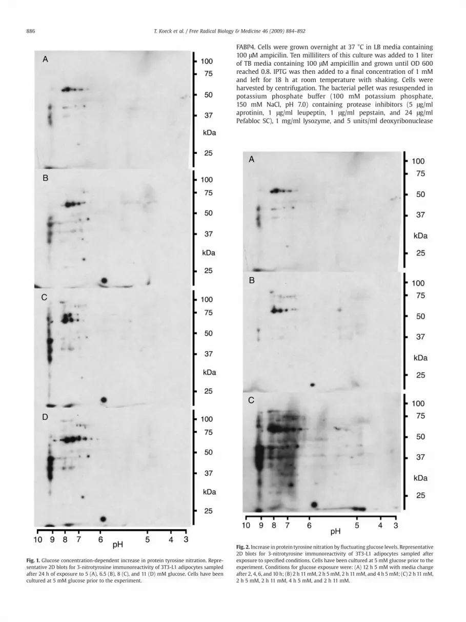
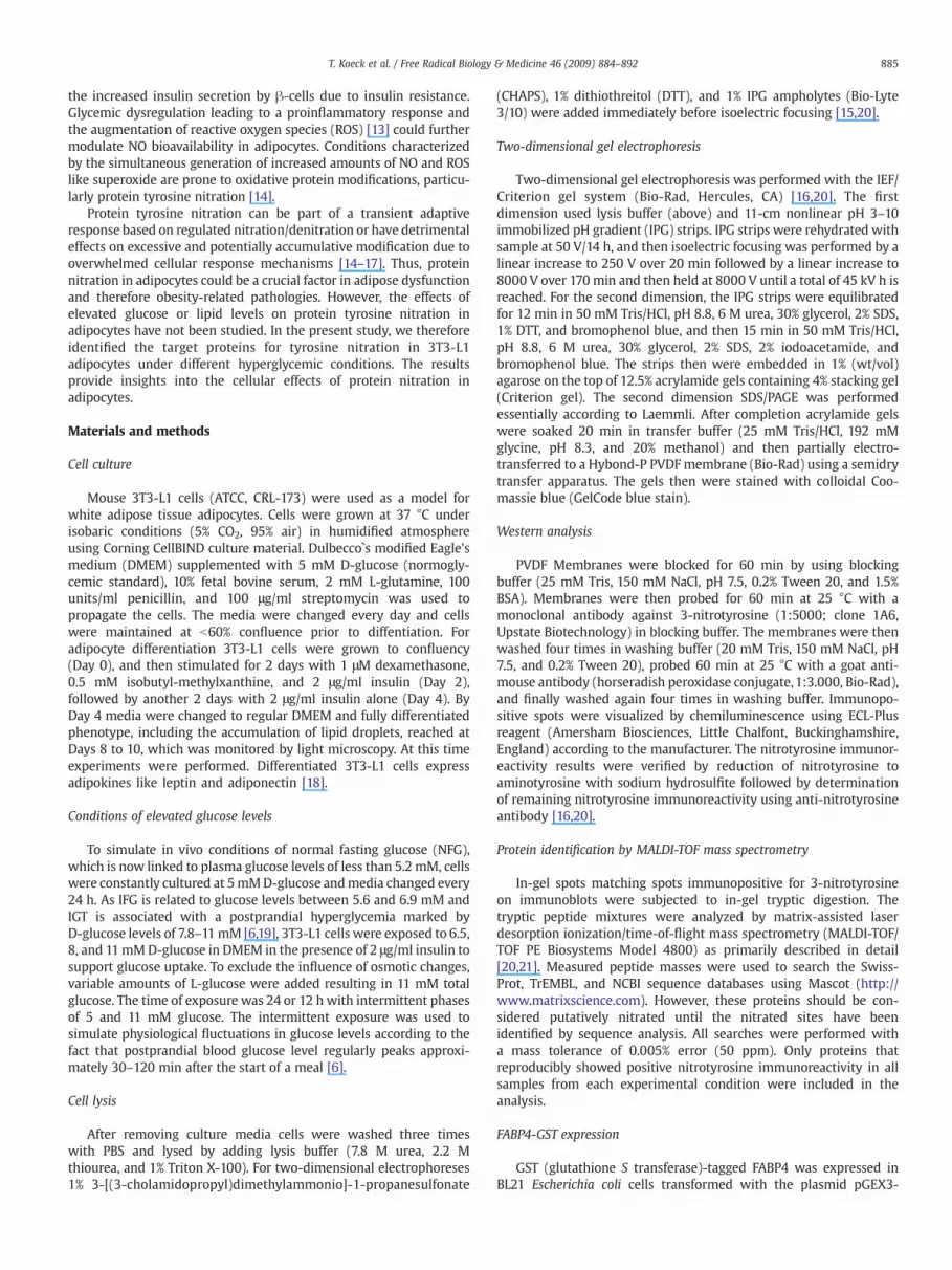
Fig. 1. Glucose concentration-dependent increase in protein tyrosine nitration. Repre-sentative 2D blots for 3-nitrotyrosine immunoreactivity of 3T3-L1 adipocytes sampledafter 24 h of exposure to 5 (A), 6.5 (B), 8 (C), and 11 (D) mM glucose. Cells have beencultured at 5 mM glucose prior to the experiment.
886 T. Koeck et al. / Free Radical Biology & Medicine 46 (2009) 884–892
FABP4. Cells were grown overnight at 37 °C in LB media containing100 μM ampicilin. Ten milliliters of this culture was added to 1 literof TB media containing 100 μM ampicillin and grown until OD 600reached 0.8. IPTG was then added to a final concentration of 1 mMand left for 18 h at room temperature with shaking. Cells wereharvested by centrifugation. The bacterial pellet was resuspended inpotassium phosphate buffer (100 mM potassium phosphate,150 mM NaCl, pH 7.0) containing protease inhibitors (5 μg/mlaprotinin, 1 μg/ml leupeptin, 1 μg/ml pepstain, and 24 μg/mlPefabloc SC), 1 mg/ml lysozyme, and 5 units/ml deoxyribonuclease
Fig. 2. Increase in protein tyrosine nitration by fluctuating glucose levels. Representative2D blots for 3-nitrotyrosine immunoreactivity of 3T3-L1 adipocytes sampled afterexposure to specified conditions. Cells have been cultured at 5 mM glucose prior to theexperiment. Conditions for glucose exposure were: (A) 12 h 5 mM with media changeafter 2, 4, 6, and 10 h; (B) 2 h 11mM, 2 h 5mM, 2 h 11mM, and 4 h 5mM; (C) 2 h 11mM,2 h 5 mM, 2 h 11 mM, 4 h 5 mM, and 2 h 11 mM.

Table 1Identification of nitrated proteins
Symbol Protein % Sequence coverage Peptide matches
AC2 Aconitase 2, mitochondrial 27 16FABP4 Adipocyte fatty acid binding protein (aP2/FABP4) 38 6ALDA Aldolase A 52 12CAT Catalase 17 7G3PDH Glyceraldehyde-3-phosphate dehydrogenase (G3PDH) 15 3RNP Heterogeneous nuclear ribonucleoprotein A2/B1 (hnRNP-A2/B1) 23 6HSP60 Heat shock protein 60 (HSP60) 26 8IVDH Isovaleryl-CoA dehydrogenase 19 7MDH Malate dehydrogenase 36 9NDKB Nucleoside-diphosphate kinase B (NDK-B) 59 9PGK Phosphoglycerate kinase 24 8ER60 Protein disulfide isomerase A3 (ERp60, Erp57, ER-60) 25 11PK Pyruvate kinase 31 12GDI Rho GDP-dissociation inhibitor 2 (RhoGDI2) 50 7TP Trifunctional protein, mitochondrial, α-chain 12 7VDAC Voltage-dependent anion-selective channel protein 1 (VDAC-1) 40 7
Proteins immunopositive for 3-nitrotyrosine from 3T3-L1 adipocytes were identified by mass spectrometry. Values for sequence coverage and peptide matches originate from aMascot search using MALDI-TOF mass spectrometry peptide map data. Only proteins reproducibly present in the 2D nitroproteome profiles of all samples from each experimentalcondition using 11 mM glucose are listed.
887T. Koeck et al. / Free Radical Biology & Medicine 46 (2009) 884–892
1. Cells were then sonicated on ice and centrifuged at 16,000 g for30 min at 4°C. The supernatant was loaded onto a columncontaining 5 ml glutathione-Sepharose preequilibrated with potas-sium phosphate buffer. After washing with 100 ml of potassiumphosphate buffer, the protein was eluted with potassium phosphatebuffer containing 20 mM reduced glutathione. FABP4 was thendialyzed with phosphate buffer overnight at 4°C and concentratedwith Amicon ultra-4 centrifugal filter devices (Millipore) with a 3-kDA molecular weight cutoff. Protein yield was 6.5 mg per liter ofbacterial culture.
FABP4 purification
The FABP4-GST protein construct, which contains a thrombincleavage site between the GST and the mouse FABP4, wasincubated with 10 U/mg thrombin (7.5 U/λ) for 20 min at 37°Cin phosphate buffer. The reaction was stopped with serineprotease inhibitors (5 μg/ml aprotinin, 24 μg/ml Pefabloc SC).FABP4 was separated from GST and thrombin by FPLC at 4°C usinga Sephacryl S-200 column and potassium phosphate buffer at aflow rate of 0.5 ml/min. Purification was verified by SDS-PAGE andsolely FABP4 containing eluate concentrated to 1 mg/ml withAmicon ultra-4 centrifugal filter devices with 3 kDA molecularweight cutoff.
Biotinylation of FABP4
GST-free FABP4 was reacted with EZ-link Sulfo-NHS-LC-Biotin(Pierce) at a molar ratio of 1:20 in potassium phosphate buffer, pH 7.0,for 2h at 4°C and 1h at room temperature according to themanufacturer.
Nitration of FABP4
GST-free FABP4 and biotinylated FABP4 were nitrated withhydrogen peroxide-free peroxynitrite (Calbiochem) at molar ratiosof 1:10 and 1:50 as reported recently [22]. Briefly, FABP4 was exposedto peroxynitrite for 30 s at 25 °C in potassium phosphate buffer, pH 7,with 25 mM sodium bicarbonate by rapid mixing. Thereby equalamounts of 0.1 M NaOHwere added (with and without peroxynitrite),which changed the pH of the protein solution from 7 to 7.1. Thisexcludes any nitration of tyrosine residues by acidic reactions ofdecomposition products like nitrate. Nitration in the presence of fattyacid was done at amolar ratio of 1:1 between FABP4 and palmitic acid.
After nitration the protein solutions were stored on ice until used.Aliquots were taken for Western analysis. The concentration of theperoxynitrite stock solution was UV spectrophotometrically deter-mined in 0.1 M sodium hydroxide using ɛ=1670 M-1cm-1 at 302 nm.
Identification of FABP4 nitration sites by LC-MS/MS
Nonnitrated and nitrated FABP4 were subjected to SDS/PAGEaccording to Laemmli and the resulting protein bands excised andtreated with trypsin [22]. The resulting extracts were mixed 1:11% (v/v) acetic acid and 20 μl was transferred to the autosampler of theThermoScientific LTQ ion trap mass spectrometer system. For analysisa volume of 10 μl was injected onto a 8 cm×75 μmPhenomenex JupiterC18 reversed-phase capillary chromatography column using anEksigent splitless nanoflow LC system. The peptides were elutedfrom the column by an acetonitrile/0.1% (v/v) formic acid gradient at250 nl/min. The microelectrospray ion source was operated at 2.5 kV.Full-scan mass spectra were acquired to determine peptide molecularweights and product ion spectra to determine amino acid sequences insuccessive instrument scans. The data were analyzed by using allcollisionally induced dissociation spectra (CID) collected in theexperiment to search the NCBI sequence databases using Mascot.Each identification was verified by manual inspection of severalmatching spectra. The potential nitration sites were interrogated bymanual inspection of the spectra and through the use of the programSequest.
Fatty acid binding to FABP4
Nitrated and nonnitrated biotinylated FABP4 were incubated withequimolar amounts of [9,10-3H]palmitic acid (50 Ci/mmol; Sigma;final ethanol concentration 1% v/v) in potassium phosphate buffer for2 h at room temperature. Then the biotinylated FABP4 with boundpalmitic acid was separated using UltraLink Immobilized NeutrAvidinProtein (Pierce) according to the manufacturer. Washed ImmobilizedNeutrAvidin Protein with bound FABP4 was quantatively transferredto scintillation vials and activity measured using a Beckman CoulterLS6500 Multipurpose scintillation counter.
Statistics and data analysis
Presented 2D blots are representative for three independentexperiments. All other data are presented as mean±SEM. Unpairedtwo-tailed Student's t tests were performed using the Prism software

888 T. Koeck et al. / Free Radical Biology & Medicine 46 (2009) 884–892

Fig. 4. Fatty acid binding to FABP4. The binding of [9,10-3H]palmitic acidto nonnitratedand nitrated FABP4 is shown dependent on the applied peroxynitrite concentration. Thedata are represented as mean±SEM of 4 independent experiments (n=4). Therebyasterisks represent alterations with Pb0.02 and were therefore considered significant.
889T. Koeck et al. / Free Radical Biology & Medicine 46 (2009) 884–892
package version 4 (GraphPad). Differences were considered significantat Pb0.02.
Results
Glucose concentration-dependent protein tyrosine nitration
Our established proteomic method [16,20] was used to examinethe effects of glucose on the nitroproteome of differentiated mouse3T3-L1 adipocytes to reveal the potential involvement of NO-mediated oxidative protein modifications in the pathology of obesityand insulin resistance. To avoid premature oxidative stress, excessivetriglyceride accumulation, and deregulated basal glucose uptake aswell as the onset of insulin resistance, preadipocytes were diffe-rentiated under normoglycemic conditions, hence in the presence of5 mM glucose [13]. The increase in glucose concentration from 5 mMto physiologically/pathologically relevant concentrations of 6.5, 8, and11 mM resulted in a general and concentration-dependent increase in3-nitrotyrosine immunoreactivity (Figs. 1A–D). These results areconsistent with previous studies showing increased glucose-mediatedgeneration of nitrating reagents fueled by elevated generation of NOand ROS like superoxide in adipocytes [13] and retinal Müller cells[23,24]. However, this regimen also caused alterations of thenitroproteome that are highly target selective as a subset of theprotein targets had a decrease or loss of immunoreactivity withincreasing glucose concentrations rather than an increase.
Effect of fluctuating glucose concentrations on protein tyrosine nitration
To test how physiological glucose concentration fluctuationsinfluence protein nitration in 3T3-L1 adipocytes, physiological condi-tions were mimicked by switching the cell culture four to five timesbetween media containing 5 or 11 mM glucose over a period of 12 h(Figs. 2A–C). This approach revealed thatfluctuatingglucose levels resultin a pronounced increase in protein tyrosine nitration after repeated 2-h11mMglucose phases (Fig. 2C). Despite thewidely similar alterations inthe 2D nitroproteome profiles, the increase in 3-nitrotyrosine immu-
Fig. 3. Identification of tyrosine 19 as a nitration site in FABP4 using capillary column LC-peptide peptide 10-LVSSENFDDYMK-21 from FABP4 (NCBI Accession No. 14149635). Represepeptide (LVSSENFDDYMoK), and tyrosine nitrated-methionine oxidized peptide (LVSSENFDDinterpretation of each spectrum is inset.
noreactivity resulting from 12 h of intermittent exposure to 11 mMglucose notably exceeded the one resulting from 24 h of continuousexposure (compare Figs. 1D and 2C). However, a comparison with cellsgrown at 5 mM glucose showed that a period of 4 h at 5 mM glucosefollowing two intermittent 2-h 11 mM glucose phases resulted in thereturn to the base tyrosine nitration level (compare Figs. 2B with A andC). This points toward cellular mechanisms that remove nitratedproteins by increased turnover [25] and/or denitration of modifiedtarget proteins [15,17]. In case of a regulated denitration, proteinnitration could be part of a cellular response to minimize glucotoxicitythat includes NO-dependent glucose uptake [26].
Identities of the nitrated proteins
The identification of the tyrosine nitrated proteins is a prerequisitefor the approximation of the potential (patho)physiological impact oftheir modification. Thus, following 2D SDS-PAGE, immunoblotting,and in-gel tryptic digestion, product peptides from nitrotyrosine-immunoreactive protein spots were subjected to mass spectrometricanalysis. The modified proteins identified in samples from 3T3-L1adipocytes that were exposed to 11 mM glucose are listed in Table 1.They participate in a variety of physiological processes essential forthe maintenance of normal metabolic homeostasis and paracrinefunction. The affected processes comprise glycolysis, fatty acidbinding, mitochondrial metabolism, mitochondrial permeability,protein folding, protein translation, antioxidant defense, and signaltransduction.
Nitration of FABP4
FABP4 contains two tyrosine residues and both have beendemonstrated to be critical for function. To identify the site of tyrosinemodification in FABP4, we used capillary column LC-tandem massspectrometry. The analysis confirmed the protein identity (NCBIAccession No. 14149635). Successive analysis showed that exposure ofFABP4 to peroxynitrite at molar ratios of 1:10 as well as 1:50 leads tonitration of tyrosine 19 (Tyr19) of the peptide LVSSENFDDYMK (Fig. 3).In all cases in which Tyr19 was modified, methionine 20 (Met20) wasalso found oxidized. This observation is consistent with the prefer-ential oxidation of the methionine prior to the tyrosine nitrationunder the conditions used. Thereby the increase of the molar ratiofrom 1:10 to 1:50 led to a several-fold increase in the amount ofpeptides with oxidized Met20 as well as the comodification in theform of nitrated Tyr19 and oxidized Met20 relative to unmodifiedpeptide. This increase was more pronounced for the double-modifiedpeptide.
Fatty acid binding to nitrated FABP4
To determine the potential functional impact of the nitration ofTyr19 and oxidation of Met20 the affect of peroxynitrite exposure onthe fatty acid binding ability of FABP4 was determined. Depending onthe molar ratio of FAPB4 and peroxynitrite, the exposure toperoxynitrite was associated with a significant decrease in the bindingof palmitic acid to FABP4 (Fig. 4). However, treatment of FABP4 withdecomposed peroxynitrate had no effect on the fatty acid binding. Thisdecrease in binding occurs likely due to the alteration of the pKa of thehydroxyl group and therefore the polarity of the tyrosine residue bynitration [27]. In the case of FABP4 the change in polarity probablymimics phosphorylation [23,28], which has been shown to greatlyinhibit lipid binding and release [29].
tandem mass spectrometry. Collision-induced dissociation (CID) spectra of the trypticntative CID spectra of the unmodified peptide (LVSSENFDDYMK), methionine oxidizedYNO2MoK) clearly showing the site of the modified amino acids Tyr19 and Met20. The

890 T. Koeck et al. / Free Radical Biology & Medicine 46 (2009) 884–892
Discussion
Continuous consumption of surplus lipids and carbohydrates is amajor causative factor for obesity. The disproportionate or excessivegain of white adipose tissue mass affects the systemic energy balanceas well as glucose and lipid metabolism [8], which is correlated withthe development of comorbidities like type 2 diabetes, hepatostea-tosis, and atherosclerosis [5]. Thereby different molecular factors likenonesterified fatty acids and cytokines, originating from the dysregu-lation of metabolic and secretory/endocrine functions of adipocytes aswell as the chronic subacute inflammation, contribute to an inade-quate control of blood glucose due to insulin resistance. Acute orchronically elevated glucose can cause toxicity that correlates withoxidative stress, favoring oxidative protein modifications like proteinnitration [14]. Insulin resistance is also associated with an increasedgeneration of reactive species like peroxynitrite [30]. The variouspotential (patho)physiological effects of protein nitration under theseconditions will depend on target proteins and the extent ofmodification [17]. In this context our study represents the firstsystematic investigation that correlates glucose and protein tyrosinenitration in adipocytes. Our data show that the extent of cellulartyrosine protein nitration, along with an expansion of the nitropro-teome, mirrored rises in the glucose concentration during continuousexposure as well as during the simulation of a more physiologicallyrelevant periodic fluctuation in the glucose concentration. Thus, ourstudy adds greatly to the understanding of the effects of oxidants likeperoxynitrite in insulin resistance [30].
The largest functional group of target proteins comprised meta-bolic enzymes including enzymes of the glycolytic pathway, tricar-boxylic acid (TCA) cycle, and fatty acid β-oxidation. Among theproteins identified, the glycolytic pathway was most extensivelymodified. We have recently shown that nitration of aldolase A inhibitsenzyme activity [22]. G3PDH and phosphoglycerate kinase were alsofound to be sensitive to protein nitration and/or thiol oxidation[31,32]. Thus, the simultaneous nitration of aldolase A, G3PDH,phosphoglycerate kinase, and pyruvate kinase suggests that elevatedglucose levels can decrease the glycolytic activity in adipocytes. Themetabolism of glucose carbons could be suppressed further, as themitochondrial TCA cycle enzymes malate dehydrogenase [33] andaconitase [34,35] become inactivated by oxidative modifications.Tyrosine nitration acts probably as markers for these oxidativemodifications. These metabolic alterations potentially lead to adecreased generation of ATP, GTP, NADHs and FADH2, a decrease inmitochondrial membrane potential as well as a depletion orquantitative shift of essential metabolic intermediates like oxaloace-tate. A depletion or shift in these intermediates has the potential todecrease mitochondrial fatty acid β-oxidation, whichmight be furtheraffected by the nitration of the α-chain of the trifunctional protein. Incombination with the potential impairment of integrity and bioge-nesis as well as the altered permeability of mitochondria through thenitration of heat shock protein 60 [36–38] and voltage-dependentanion-selective channel protein 1 [39,40], this could result inmitochondrial dysfunction accompanied by altered fuel metabolismand lipid homeostasis. Elevated nonesterified fatty acids and meta-bolic lipid products transmit stress responses through activation ofseveral kinases, including c-jun NH2-terminal kinase (JNK) andinhibitor of kappa kinase (IκK) [41].
In adipocytes lipid trafficking/metabolism and hormone actionare integrated with stress and inflammatory responses by aP2/FABP4, the predominant fatty acid binding protein, which modulatesthe systemic glucose and lipid metabolism [41,42]. All FABPs (1–9) inmammals share the same general tertiary structure. It consists of aβ-barrel around the ligand-binding cavity covered with a helix-turn-helix cap and contains only two tyrosine residues, Tyr19 andTyr128, which are highly conserved [43]. Both tyrosine residues arefunctionally important. The carboxylate group of a lipid ligand
directly interacts with Tyr128 through hydrogen bonding [43,44]and phosphorylation of Tyr19 by insulin receptor tyrosine kinase[43,45] regulates lipid binding and release [29]. Nitration of Tyr19mimicking and modulating phosphorylation [23,28] could there-fore represent an additional regulatory mechanism especiallyunder stress conditions. This, in turn, could further affect theregulatory interaction of FABP4 with hormone-sensitive lipase [46],nuclear translocation of FABP4, and agonist supply for peroxisome-proliferator activated receptor γ (PPAR-γ) [47–49] as well as theactivation of JNK [41,42]. Alterations in PPAR-γ agonism further alterFABP4 expression, lipolysis, fatty acid β-oxidation, and mitochon-drial biogenesis [50]. Therefore, the nitration of FABP4 could be animportant factor for the adipose tissue and systemic response tohigh metabolic load in regard of adipocytokines as well as glucoseand lipid metabolism.
The (patho)physiological effects resulting from the high metabolicload and inflammatory responses also depend on the status of the ERand cellular antioxidant capacity. In adipocytes under hyperglycemicconditions this capacity is likely weakened by the tyrosine nitration ofthe antioxidant enzyme catalase, which is associated with inactivation[51]. Moreover, alterations in the agonist stimulation of PPAR-γ coulddecrease the expression of catalase [52]. The potential effects of thenitration protein disulfide isomerase A3 are unknown, but theoxidative folding of glycoproteins in the ER and/or the interactionwith calnexin and calreticulin could be affected [53].
The nitration of Rho GDP-dissociation inhibitor 2 (RhoGDI2) in3T3-L1 adipocytes has the potential to further alter metabolic stressresponse. RhoGDI2, whose abundance decreases during differentia-tion [54], has a relatively narrow specificity for Cdc42, Rac1, andRhoA of the Rho family of small GTP-binding proteins [55]. Itcontains a tyrosine phosphorylation site at Tyr24 [56], whichdecreases the ability for complex formation with target proteins.Thus, tyrosine nitration has the potential to alter the physiologicalfunction of Cdc42, Rac1, and/or RhoA and therefore stress fiberformation, adipocyte morphology [57,58], and maybe even glucose[59] and GLUT4 trafficking [57,58,60]. Nitration of nucleoside-diphosphate kinase B (NDK-B) could contribute to these effects asit can exist in two distinct pools in 3T3 fibroblasts: one populationis transiently translocated to the cell periphery on activation ofreceptor tyrosine kinases and G-protein-coupled receptors, whereasa second pool binds constitutively to microtubule-associatedvesicles [61]. The association with vesicles might provide aregulated source of GTP for GTPases that control intracellulartrafficking, while the potential interaction with integrin cytoplasmicdomain-associated protein 1α (ICAP-1α) as a consequence of Racactivation might affect cell morphology and behavior duringadhesion [61]. NDK-B further binds polyunsaturated fatty acids[62], interacts with heterotrimeric G protein βγ dimers [63],facilitates coat protein complex II assembly [64], and inducesc-myc [65] in various cell types. These functions of NDK-B could,if present in adipocytes, provide additional mechanisms by whichNDK-B nitration might alter adipocyte biology.
Conclusion
The understanding is incomplete of the molecular mechanismslinking obesity to insulin resistance, type 2 diabetes, and othercomorbidities. However, in recent years it has become clear thatinflammation and adverse metabolic alterations are key factors.Many of these factors are associated with oxidative stress, andprotein tyrosine nitration is an important (patho)physiologicalmechanism by which oxidative stress is manifested in cells. Thephysiological functions of the proteins identified in this studyimplicate tyrosine nitration as a potentially significant mechanismfor the development of insulin resistance itself and the associatedcomplications in obesity.

891T. Koeck et al. / Free Radical Biology & Medicine 46 (2009) 884–892
Acknowledgments
The work was supported by National Institute of Health Grant NIHP01 HL076491. The pGEX3-FABP4 plasmid was generously providedby Dr. Noa Noy.
References
[1] Muoio, D. M.; Newgard, C. B. Mechanisms of disease: molecular and metabolicmechanisms of insulin resistance and beta-cell failure in type 2 diabetes. Nat. Rev.Mol. Cell. Biol.; 2008.
[2] Cara, J. F.; Chaiken, R. L. Type 2 diabetes and the metabolic syndrome in childrenand adolescents. Curr. Diabetes Rep. 6:241–250; 2006.
[3] Wild, S.; Roglic, G.; Green, A.; Sicree, R.; King, H. Global prevalence of diabetes:estimates for the year 2000 and projections for 2030. Diabetes Care 27:1047–1053;2004.
[4] Jones, K. L. Role of obesity in complicating and confusing the diagnosis andtreatment of diabetes in children. Pediatrics 121:361–368; 2008.
[5] Guilherme, A.; Virbasius, J. V.; Puri, V.; Czech, M. P. Adipocyte dysfunctions linkingobesity to insulin resistance and type 2 diabetes. Nat. Rev. Mol. Cell. Biol. 9:367–377; 2008.
[6] Bock, G.; Dalla Man, C.; Campioni, M.; Chittilapilly, E.; Basu, R.; Toffolo, G.; Cobelli,C.; Rizza, R. Pathogenesis of pre-diabetes: mechanisms of fasting and postprandialhyperglycemia in people with impaired fasting glucose and/or impaired glucosetolerance. Diabetes 55:3536–3549; 2006.
[7] Weir, G. C.; Bonner-Weir, S. A dominant role for glucose in beta cell compensationof insulin resistance. J. Clin. Invest. 117:81–83; 2007.
[8] Rosen, E. D.; Spiegelman, B. M. Adipocytes as regulators of energy balance andglucose homeostasis. Nature 444:847–853; 2006.
[9] Zhao, Y. F.; Feng, D. D.; Hernandez, M.; Chen, C. 3T3-L1 adipocytes inducedysfunction of MIN6 insulin-secreting cells via multiple pathways mediated bysecretory factors in a co-culture system. Endocrine 31:52–60; 2007.
[10] Jobgen, W. S.; Fried, S. K.; Fu, W. J.; Meininger, C. J.; Wu, G. Regulatory role for thearginine-nitric oxide pathway in metabolism of energy substrates. J. Nutr.Biochem. 17:571–588; 2006.
[11] Tanaka, T.; Nakatani, K.; Morioka, K.; Urakawa, H.; Maruyama, N.; Kitagawa, N.;Katsuki, A.; Araki-Sasaki, R.; Hori, Y.; Gabazza, E. C.; Yano, Y.; Wada, H.; Nobori, T.;Sumida, Y.; Adachi, Y. Nitric oxide stimulates glucose transport through insulin-independent GLUT4 translocation in 3T3-L1 adipocytes. Eur. J. Endocrinol./Eur. Fed.Endocr. Soc. 149:61–67; 2003.
[12] Engeli, S.; Janke, J.; Gorzelniak, K.; Bohnke, J.; Ghose, N.; Lindschau, C.; Luft, F. C.;Sharma, A. M. Regulation of the nitric oxide system in human adipose tissue.J. Lipid Res. 45:1640–1648; 2004.
[13] Lin, Y.; Berg, A. H.; Iyengar, P.; Lam, T. K.; Giacca, A.; Combs, T. P.; Rajala, M. W.; Du,X.; Rollman, B.; Li, W.; Hawkins, M.; Barzilai, N.; Rhodes, C. J.; Fantus, I. G.;Brownlee, M.; Scherer, P. E. The hyperglycemia-induced inflammatory response inadipocytes: the role of reactive oxygen species. J. Biol. Chem. 280:4617–4626; 2005.
[14] Pacher, P.; Beckman, J. S.; Liaudet, L. Nitric oxide and peroxynitrite in health anddisease. Physiol. Rev. 87:315–424; 2007.
[15] Koeck, T.; Fu, X.; Hazen, S. L.; Crabb, J. W.; Stuehr, D. J.; Aulak, K. S. Rapid andselective oxygen-regulated protein tyrosine denitration and nitration in mito-chondria. J. Biol. Chem. 279:27257–27262; 2004.
[16] Aulak, K. S.; Miyagi, M.; Yan, L.; West, K. A.; Massillon, D.; Crabb, J. W.; Stuehr, D. J.Proteomic method identifies proteins nitrated in vivo during inflammatorychallenge. Proc. Natl. Acad. Sci. U. S. A. 98:12056–12061; 2001.
[17] Koeck, T.; Stuehr, D. J.; Aulak, K. S. Mitochondria and regulated tyrosine nitration.Biochem. Soc. Trans. 33:1399–1403; 2005.
[18] Fischer-Posovszky, P.; Wabitsch, M.; Hochberg, Z. Endocrinology of adiposetissue—an update. Horm. Metab. Res. 39:314–321; 2007.
[19] Sorkin, J. D.; Muller, D. C.; Fleg, J. L.; Andres, R. The relation of fasting and 2-hpostchallenge plasma glucose concentrations to mortality: data from the BaltimoreLongitudinal Study of Aging with a critical review of the literature. Diabetes Care28:2626–2632; 2005.
[20] Aulak, K. S.; Koeck, T.; Crabb, J. W.; Stuehr, D. J. Proteomicmethod for identificationof tyrosine-nitrated proteins. Methods Mol. Biol. 279:151–165; 2004.
[21] Miyagi, M.; Sakaguchi, H.; Darrow, R. M.; Yan, L.; West, K. A.; Aulak, K. S.; Stuehr,D. J.; Hollyfield, J. G.; Organisciak, D. T.; Crabb, J. W. Evidence that light modulatesprotein nitration in rat retina. Mol. Cell. Proteomics 1:293–303; 2002.
[22] Koeck, T.; Levison, B.; Hazen, S. L.; Crabb, J. W.; Stuehr, D. J.; Aulak, K. S. Tyrosinenitration impairs mammalian aldolase A activity. Mol. Cell. Proteomics 3:548–557;2004.
[23] Zhan, X.; Du, Y.; Crabb, J. S.; Gu, X.; Kern, T. S.; Crabb, J. W. Targets of tyrosinenitration in diabetic rat retina. Mol. Cell. Proteomics 7:864–874; 2008.
[24] Du, Y.; Smith, M. A.; Miller, C. M.; Kern, T. S. Diabetes-induced nitrative stress inthe retina, and correction by aminoguanidine. J. Neurochem. 80:771–779; 2002.
[25] Elfering, S. L.; Haynes, V. L.; Traaseth,N. J.; Ettl, A.; Giulivi, C. Aspects,mechanism, andbiological relevance of mitochondrial protein nitration sustained by mitochondrialnitric oxide synthase. Am. J. Physiol. Heart Circ. Physiol. 286:H22–H29; 2004.
[26] McGrowder, D.; Ragoobirsingh, D.; Brown, P. Modulation of glucose uptake inadipose tissue by nitric oxide-generating compounds. J. Biosci. 31:347–354; 2006.
[27] Sokolovsky, M.; Riordan, J. F.; Vallee, B. L. Tetranitromethane. A reagent for thenitration of tyrosyl residues in proteins. Biochemistry 5:3582–3589; 1966.
[28] Mallozzi, C.; Di Stasi, A. M.; Minetti, M. Nitrotyrosine mimics phosphotyrosinebinding to the SH2 domain of the src family tyrosine kinase lyn. FEBS Lett. 503:189–195; 2001.
[29] Buelt, M. K.; Xu, Z.; Banaszak, L. J.; Bernlohr, D. A. Structural and functionalcharacterization of the phosphorylated adipocyte lipid-binding protein (pp.15).Biochemistry 31:3493–3499; 1992.
[30] Duplain, H.; Sartori, C.; Dessen, P.; Jayet, P. Y.; Schwab, M.; Bloch, J.; Nicod, P.;Scherrer, U. Stimulation of peroxynitrite catalysis improves insulin sensitivity inhigh fat diet-fed mice. J. Physiol. 586:4011–4016; 2008.
[31] Buchczyk, D. P.; Grune, T.; Sies, H.; Klotz, L. O. Modifications of glyceralde-hyde-3-phosphate dehydrogenase induced by increasing concentrations ofperoxynitrite: early recognition by 20S proteasome. Biol. Chem. 384:237–241;2003.
[32] Markland, F. S.; Bacharach, A. D.; Weber, B. H.; O'Grady, T. C.; Saunders, G. C.;Umemura, N. Chemical modification of yeast 3-phosphoglycerate kinase. J. Biol.Chem. 250:1301–1310; 1975.
[33] Varrone, S.; Consiglio, E.; Covelli, I. The nature of inhibition of mitochondrialmalate dehydrogenase by thyroxine, iodine cyanide and molecular iodine. Eur. J.Biochem./FEBS 13:305–312; 1970.
[34] Tortora, V.; Quijano, C.; Freeman, B.; Radi, R.; Castro, L. Mitochondrial aconitasereaction with nitric oxide, S-nitrosoglutathione, and peroxynitrite: mechanismsand relative contributions to aconitase inactivation. Free Radic. Biol. Med. 42:1075–1088; 2007.
[35] Han, D.; Canali, R.; Garcia, J.; Aguilera, R.; Gallaher, T. K.; Cadenas, E. Sites andmechanisms of aconitase inactivation by peroxynitrite: modulation by citrate andglutathione. Biochemistry 44:11986–11996; 2005.
[36] Khor, H. K.; Fisher, M. T.; Schoneich, C. Potential role of methionine sulfoxide in theinactivation of the chaperone GroEL by hypochlorous acid (HOCl) and peroxyni-trite (ONOO-). J. Biol. Chem. 279:19486–19493; 2004.
[37] Voos, W.; Rottgers, K. Molecular chaperones as essential mediators of mitochon-drial biogenesis. Biochim. Biophys. Acta 1592:51–62; 2002.
[38] Deocaris, C. C.; Kaul, S. C.; Wadhwa, R. On the brotherhood of the mitochondrialchaperones mortalin and heat shock protein 60. Cell Stress Chaperones 11:116–128; 2006.
[39] O'Rourke, B. Mitochondrial ion channels. Annu. Rev. Physiol. 69:19–49; 2007.[40] Lemasters, J. J. Modulation of mitochondrial membrane permeability in
pathogenesis, autophagy and control of metabolism. J. Gastroenterol. Hepatol. 22(Suppl. 1):S31–S37; 2007.
[41] Erbay, E.; Cao, H.; Hotamisligil, G. S. Adipocyte/macrophage fatty acid bindingproteins in metabolic syndrome. Curr. Atheroscler. Rep. 9:222–229; 2007.
[42] Hotamisligil, G. S. Inflammation and metabolic disorders. Nature 444:860–867;2006.
[43] Xu, Z.; Bernlohr, D. A.; Banaszak, L. J. Crystal structure of recombinant murineadipocyte lipid-binding protein. Biochemistry 31:3484–3492; 1992.
[44] Sha, R. S.; Kane, C. D.; Xu, Z.; Banaszak, L. J.; Bernlohr, D. A. Modulation of ligandbinding affinity of the adipocyte lipid-binding protein by selective mutation.Analysis in vitro and in situ. J. Biol. Chem. 268:7885–7892; 1993.
[45] Hresko, R. C.; Hoffman, R. D.; Flores-Riveros, J. R.; Lane, M. D. Insulin receptortyrosine kinase-catalyzed phosphorylation of 422(aP2) protein. Substrate activa-tion by long-chain fatty acid. J. Biol. Chem. 265:21075–21085; 1990.
[46] Smith, A. J.; Thompson, B. R.; Sanders, M. A.; Bernlohr, D. A. Interaction of theadipocyte fatty acid-binding protein with the hormone-sensitive lipase:regulation by fatty acids and phosphorylation. J. Biol. Chem. 282:32424–32432;2007.
[47] Adida, A.; Spener, F. Adipocyte-type fatty acid-binding protein as inter-compartmental shuttle for peroxisome proliferator activated receptor gammaagonists in cultured cell. Biochim. Biophys. Acta 1761:172–181; 2006.
[48] Ayers, S. D.; Nedrow, K. L.; Gillilan, R. E.; Noy, N. Continuous nucleocytoplasmicshuttling underlies transcriptional activation of PPARgammaby FABP4.Biochemistry46:6744–6752; 2007.
[49] Wolfrum, C.; Borrmann, C. M.; Borchers, T.; Spener, F. Fatty acids and hypoli-pidemic drugs regulate peroxisome proliferator-activated receptors alpha- andgamma-mediated gene expression via liver fatty acid binding protein: asignaling path to the nucleus. Proc. Natl. Acad. Sci. U. S. A. 98:2323–2328;2001.
[50] Laplante, M.; Festuccia, W. T.; Soucy, G.; Gelinas, Y.; Lalonde, J.; Berger, J. P.;Deshaies, Y. Mechanisms of the depot specificity of peroxisome proliferator-activated receptor gamma action on adipose tissue metabolism. Diabetes55:2771–2778; 2006.
[51] Ghosh, S.; Janocha, A. J.; Aronica, M. A.; Swaidani, S.; Comhair, S. A.; Xu, W.; Zheng,L.; Kaveti, S.; Kinter, M.; Hazen, S. L.; Erzurum, S. C. Nitrotyrosine proteome surveyin asthma identifies oxidative mechanism of catalase inactivation. J. Immunol.176:5587–5597; 2006.
[52] Okuno, Y.; Matsuda, M.; Kobayashi, H.; Morita, K.; Suzuki, E.; Fukuhara, A.;Komuro, R.; Shimabukuro, M.; Shimomura, I. Adipose expression of catalase isregulated via a novel remote PPARgamma-responsive region. Biochem. Biophys.Res. Commun. 366:698–704; 2008.
[53] Hatahet, F.; Ruddock, L. W. Substrate recognition by the protein disulfideisomerases. FEBS J. 274:5223–5234; 2007.
[54] Welsh, G. I.; Griffiths, M. R.; Webster, K. J.; Page, M. J.; Tavare, J. M. Proteomeanalysis of adipogenesis. Proteomics 4:1042–1051; 2004.
[55] Takai, Y.; Sasaki, T.; Matozaki, T. Small GTP-binding proteins. Physiol. Rev. 81:153–208; 2001.
[56] Rush, J.; Moritz, A.; Lee, K. A.; Guo, A.; Goss, V. L.; Spek, E. J.; Zhang, H.; Zha, X. M.;Polakiewicz, R. D.; Comb, M. J. Immunoaffinity profiling of tyrosine phosphoryla-tion in cancer cells. Nat. Biotechnol. 23:94–101; 2005.

892 T. Koeck et al. / Free Radical Biology & Medicine 46 (2009) 884–892
[57] Bar-Sagi, D.; Hall, A. Ras and Rho GTPases: a family reunion. Cell 103:227–238;2000.
[58] DerMardirossian, C.; Bokoch, G. M. GDIs: central regulatory molecules in RhoGTPase activation. Trends Cell Biol. 15:356–363; 2005.
[59] Usui, I.; Imamura, T.; Huang, J.; Satoh, H.; Olefsky, J. M. Cdc42 is a Rho GTPasefamily member that can mediate insulin signaling to glucose transport in 3T3-L1adipocytes. J. Biol. Chem. 278:13765–13774; 2003.
[60] Hou, J. C.; Shigematsu, S.; Crawford, H. C.; Anastasiadis, P. Z.; Pessin, J. E. Dualregulation of Rho and Rac by p120 catenin controls adipocyte plasma membranetrafficking. J. Biol. Chem. 281:23307–23312; 2006.
[61] Gallagher, B. C.; Parrott, K. A.; Szabo, G.; de, S. O. A. Receptor activation regulatescortical, but not vesicular localization of NDP kinase. J. Cell Sci. 116:3239–3250;2003.
[62] Brock, T. G. Capturing proteins that bind polyunsaturated fatty acids: demonstra-tion using arachidonic acid and eicosanoids. Lipids 43:161–169; 2008.
[63] Wieland, T. Interaction of nucleoside diphosphate kinase B with heterotrimeric Gprotein betagamma dimers: consequences on G protein activation and stability.Naunyn-Schmiedeberg's Arch. Pharmacol. 374:373–383; 2007.
[64] Kapetanovich, L.; Baughman, C.; Lee, T. H. Nm23H2 facilitates coat proteincomplex II assembly and endoplasmic reticulum export in mammalian cells. Mol.Biol. Cell 16:835–848; 2005.
[65] Arnaud-Dabernat, S.; Masse, K.; Smani, M.; Peuchant, E.; Landry, M.; Bourbon,P. M.; Le Floch, R.; Daniel, J. Y.; Larou, M. Nm23-M2/NDP kinase B inducesendogenous c-myc and nm23-M1/NDP kinase A overexpression in BAF3 cells.Both NDP kinases protect the cells from oxidative stress-induced death. Exp. CellRes. 301:293–304; 2004.


