Embed Size (px)
Citation preview
Gender Differences in Leg Stiffness and Stiffness RecruitmentStrategy During Two-Legged Hopping
Darin A. PaduaDepartment of Exercise and Sport Science University of North Carolina Chapel Hill
Brent L. ArnoldDepartment of Exercise Science Virginia Commonwealth University Richmond
Christopher R. CarciaDepartment of Physical Therapy Duquesne University Pittsburgh, PA
Kevin P. GranataDepartment of Engineering Science and Mechanics Virginia Polytechnic Institute and StateUniversity Blacksburg
AbstractThe authors compared leg stiffness (KVERT), muscle activation, and joint movement patterns between11 men and 10 women during hopping. Physically active and healthy men and women performedcontinuous 2-legged hopping at their preferred rate and at 3.0 Hz. Compared with men, womendemonstrated decreased KVERT; however, after the authors normalized for body mass, genderdifferences in KVERT were eliminated. In comparison with men, women also demonstrated increasedquadriceps and soleus activity, as well as greater quadriceps-to-hamstrings coactivation ratios. Therewere no significant gender differences for joint movement patterns (p > .05). The relationshipbetween the observed gender differences in muscle recruitment and the increased risk of anteriorcruciate ligament injury in women requires further study.
KeywordsACL; quadriceps; coactivation; hop; jump
Females' anterior cruciate ligament (ACL) injury rates are 2.0 to 9.7 times greater than aremales' ACL injury rates (Arendt, Agel, & Dick, 1999; Bjordal, Arnly, Hannestad, & Strand,1997; Cox & Lenz, 1984; Gomez, DeLee, & Farney, 1996; Malone, Hardaker, Garrett, Feagin,& Bassett, 1993; Messina, Farney, & DeLee, 1999). That difference has been demonstrated inseveral comparable sports and activity levels (Arendt et al.; Gomez et al.; Messina et al.) andin nonathletic populations of similarly trained backgrounds (e.g., military-related training; Cox& Lenz; Gwinn, Wilckens, McDevitt, Ross, & Kao, 2000). Biomechanical factors are believedto partially explain the gender bias in ACL injury rates (L. Y. Griffin et al., 2000), and stiffnessof the musculoskeletal system is one such biomechanical factor that may influence the genderbias in ACL injury rates.
Correspondence address: Darin A. Padua, Department of Exercise and Sport Science, 216 Fetzer CB#8700, University of North Carolina,Chapel Hill, NC 27599-8700, USA. E-mail address: [email protected] of Journal of Motor Behavior is the property of Heldref Publications and its content may not be copied or emailed to multiplesites or posted to a listserv without the copyright holder's express written permission. However, users may print, download, or emailarticles for individual use.
NIH Public AccessAuthor ManuscriptJ Mot Behav. Author manuscript; available in PMC 2006 October 26.
Published in final edited form as:J Mot Behav. 2005 March ; 37(2): 111–125.
NIH
-PA Author Manuscript
NIH
-PA Author Manuscript
NIH
-PA Author Manuscript
The term stiffness describes the force response that results from and resists mechanical stretch.Stability requires active muscle stiffness (Duan, Allen, & Sun, 1997; Wagner & Blickhan,1999) and may ultimately influence musculoskeletal injury (McGill, 2001). However, thecontribution of gender differences in stiffness properties to the increased ACL injury ratesobserved in women has been largely overlooked. During controlled open-chain measurementsof the isolated in vivo knee, women demonstrate less active muscle stiffness than men do(Blackburn, Riemann, Padua, & Guskiewicz, 2004; Granata, Wilson, & Padua, 2002). Granata,Padua, and Wilson (2002) observed similar findings during closed-chain, functional tasks suchas two-legged hopping. Reduced stiffness properties in women may result in decreased stabilityand may potentially influence their elevated risk of ACL injury.
The stiffness behavior of the lower extremity during functional loading conditions is complex.Lower extremity stiffness during functional tasks represents the average stiffness of themusculoskeletal system and thus depends on the torsional stiffness of the joints (torsional jointstiffness) during ground contact (Arampatzis, Bruggemann, & Klapsing, 2001; Arampatzis,Bruggemann, & Metzler, 1999; Farley, Houdikj, Strien, & Louie, 1998; Farley & Morgenroth,1999; Greene & McMahon, 1979; McMahon, Valiant, & Frederick, 1987). Torsional jointstiffness is controlled by several biomechanical factors, including muscle activation and force(Hunter & Kearney, 1982; Julian & Sollins, 1975; Lacquanti, Licata, & Soechting, 1982;Weiss, Hunter, & Kearney, 1988; Zhang, Nuber, Butler, Bowen, & Rymer, 1998), reflexes(Houk, 1979; Kearney, Stein, & Parameswaran, 1997; Nichols & Houk, 1976), antagonistmuscle coactivation (Agarwal & Gottlieb, 1977; Cannon & Zahalak, 1982; Lacquanti et al.),and lower extremity kinematics during ground contact (Farley & Morgenroth; Greene &McMahon; McMahon et al.; Zhang et al.). As such, one can modulate lower extremity stiffnessduring functional loading conditions through different muscle activation and movementstrategies. In a multijoint system with several strategies available to modulate torsional jointstiffness, the potential stiffness recruitment strategies available to modulate lower extremitystiffness are limitless (Farley et al.; Farley & Morgenroth). Stiffness recruitment strategy maybe operationally defined as the multijoint coordination (joint kinematics) and muscularrecruitment plan (muscle activation) an individual executes to modulate joint torsional stiffnessand lower extremity stiffness and, hence, to satisfy the objectives of the functional task (Farleyet al.; Hortobagyi & DeVita, 1999, 2000). Women may use altered stiffness recruitmentstrategies (muscle activation, movement strategies, or both) to compensate for inherentreductions in stiffness properties during functional loading conditions.
Investigations of gender differences in muscle activation and movement strategies haverevealed that women repeatedly demonstrate greater reliance on their quadriceps muscles andmove in a more erect posture (increased knee and hip extension) than do their male counterparts(Decker, Torry, Wyland, Sterett, & Steadman, 2003; Hewett, Stroupe, Nance, & Noyes,1996; Lephart, Ferris, Riemann, Myers, & Fu, 2002; Malinzak, Colby, Kirkendall, Yu, &Garrett, 2001; Wojtys, Huston, Taylor, & Bastian, 1996). Quadriceps and gastrocnemiuscontractions increase anterior tibial shear forces that magnify ACL strain (Beynnon et al.,1995; Durselen, Claes, & Kiefer, 1995; Fleming et al., 2001; Hirokawa, Solomonow, Lu, Lou,& D'Ambrosia, 1992; Li, Sakane, Kanamori, Ma, & Woo, 1999; K. L. Markolf, Gorek, Kabo,& Shapirt, 1990; Renstrom, Arms, Stanwyck, Johnson, & Pope, 1986). Rotary stresses at theknee are also known to facilitate ACL strain (Arms et al., 1984; K. Markolf et al., 1995). Assuch, imbalanced recruitment between the medial and lateral muscles crossing the knee (e.g.,quadriceps, hamstrings, and gastrocnemius) may influence the magnitude of the rotary stressesat the knee joint (Arms et al.; K. Markolf et al.). Because excessive strain is the ultimate causeof ACL injury, greater quadriceps and gastrocnemius activation or imbalanced activation ofmedial and lateral knee musculature may increase ACL injury risk. Quadriceps- andgastrocnemius-induced ACL strain is amplified when the knee joint is in a more extendedposition and the hamstrings are unable to counteract the force generated by those muscles
Padua et al. Page 2
J Mot Behav. Author manuscript; available in PMC 2006 October 26.
NIH
-PA Author Manuscript
NIH
-PA Author Manuscript
NIH
-PA Author Manuscript
(Beynnon et al.; Renstrom et al.). Increased quadriceps and gastrocnemius activity with theknee in more extended postures may be an effective stiffness recruitment strategy for increasinglower extremity stiffness during functional tasks. However, such stiffness recruitmentstrategies may increase ACL loading and strain and, hence, possibly facilitate increased ACLinjury risk.
The previously identified gender differences in kinematics (Malinzak et al., 2001; McNitt-Gray, Yokoi, & Millward, 1993) and muscular recruitment (Hewett et al., 1996; Huston &Wojtys, 1996; Malinzak et al.) may represent women's use of altered stiffness recruitmentstrategies to modulate or compensate for reduced stiffness properties when performingfunctional loading tasks. Unfortunately, we have not been able to find reports of specificexaminations of gender differences in lower extremity stiffness and stiffness recruitmentstrategies during functional loading conditions.
Gender differences in lower extremity stiffness, stiffness recruitment strategies, or both, duringfunctional loading conditions may play a role in the elevated ACL injury rates observed inwomen. One can assess lower extremity stiffness and the associated stiffness recruitmentstrategies during functional loading conditions such as hopping. Thus, we hypothesized thatwomen would demonstrate less lower extremity stiffness and altered stiffness recruitmentstrategies than would men during a functional, closed-chain task (two-legged hopping).Specifically, we hypothesized that women would demonstrate greater quadriceps andgastrocnemius activity, reduced hamstrings activity, an increased coactivation ratio of thequadriceps and hamstrings, and less knee flexion and ankle plantarflexion in comparison withthose of men. We further hypothesized that gender differences in quadriceps, hamstrings, andgastrocnemius muscle activation would be influenced by muscle side because we believed thatwomen may use an imbalanced muscle recruitment strategy between the medial and lateralmuscles within a muscle group.
MethodParticipants
Physically active men (n = 11, age = 27.81 ± 4.35 years [M ± SD], height = 176.54 ± 7.54 cm,weight = 80.11 ± 9.21 kg) and women (n = 10, age = 24.10 ± 3.75 years, height = 168.50 ±5.91 cm, weight = 66.92 ± 12.39 kg) volunteered to participate in this study. All participantshad previous recreational experience in jumping and landing sports (basketball, volleyball, andsoccer) as documented through a questionnaire. No participants enrolled in the study hadprevious history of significant knee ligament trauma. In addition, no participant reported anytype of vestibular disorder. Written informed consent approved by the university's HumanInvestigations Committee was obtained from all participants.
Testing ProceduresUpon arrival at the laboratory, participants completed a verbal questionnaire so that we couldensure compliance with the inclusion criteria. Before testing, participants received anexplanation of all testing procedures and were allowed practice trials to become acquaintedwith the testing procedures. The dominant lower extremity limb served as the test limb for allmuscle activity and kinematic data. We determined limb dominance by having participantsperform a single-leg landing from a 30-cm-high box. We defined the dominant limb as the self-selected lower extremity limb on which the participant landed. We performed all testing duringa single session in the Motion Analysis and Motor Performance Laboratory.
Padua et al. Page 3
J Mot Behav. Author manuscript; available in PMC 2006 October 26.
NIH
-PA Author Manuscript
NIH
-PA Author Manuscript
NIH
-PA Author Manuscript
Two-Legged HoppingTo enable us to assess lower extremity stiffness and stiffness recruitment strategies, participantsperformed repetitive two-legged jumping in place during two different conditions. To beconsistent with previous investigations of near identical tasks, we refer herein to repetitive two-legged jumping as hopping. Because participants performed two-legged hopping for all trials,lower extremity stiffness was equivalent to the combined stiffness for both legs. Given the lackof studies of gender differences in lower extremity stiffness and stiffness recruitment strategies,we felt it important to investigate a highly controlled and well-documented functional task suchas two-legged hopping.
Participants performed two separate hopping frequency conditions on the same occasion.During all hopping trials, participants maintained their trunk in an upright position, with theirhands on their hips, and wore no shoes. Participants were allowed to self-select their preferredknee and ankle movement patterns during the hopping trials. They hopped at their preferred,self-selected rate (FREQPREF) and at a controlled hopping rate of 3.0 Hz (FREQ3.0). We chosethose hopping frequencies to investigate lower extremity stiffness and stiffness recruitmentstrategies at two different hopping frequencies. The results of previous research havedemonstrated that individuals' preferred hopping frequency is approximately 2.2 ± .07 hops/s(M ± SE; Farley, Blickhan, Saito, & Taylor, 1991). In addition, lower extremity stiffnessincreases in proportion to hopping frequency (Farley et al.). In the current study, men andwomen displayed near identical preferred hopping frequencies (men, M = 2.30 ± .35 Hz;women, M = 2.30 ± .35 Hz), which are comparable with previous results. In addition, themagnitude of change in hopping frequency from FREQPREF to FREQ3.0 hopping conditionswas also similar for men (M = 22% ± 11%) and women (M = 22% ± 11%). Assessment of theFREQPREF hopping condition allowed us to investigate gender differences in lower extremitystiffness and stiffness recruitment strategies during conditions in which participants were ableto self-select their lower extremity stiffness behavior and corresponding preferred hoppingfrequency. We were also able to investigate whether the men and women adjusted their lowerextremity stiffness and stiffness recruitment strategies similarly during the faster FREQ3.0hopping conditions. We achieved controlled frequency hopping trials (FREQ3.0) by havingparticipants hop in time with a digital metronome. They were instructed that each hop must bea continuous motion and were allowed as much practice as needed until they felt comfortableperforming each of the hopping conditions.
We determined during preliminary testing that use of the digital metronome during FREQ3.0hopping influenced the preferred hopping rates (FREQPREF). When participants performedFREQ3.0 hopping before FREQPREF conditions, their FREQPREF was increased compared withtheir initial FREQPREF. Therefore, participants performed FREQPREF hopping conditionsfollowed by FREQ3.0 hopping conditions—approximately 45 continuous hops in each of thehopping conditions.
Data Processing and AnalysisWe acquired all data by using the Datapac III Version 2000 data-collection hardware andsoftware systems (Run Technologies; Laguna Hills, CA), and we stored the data in a personalcomputer for later analysis by using customized software developed in MATLAB Version 6.1(The Math-Works, Natick, MA). We sampled all data at 1000 Hz; thus, all electromyographic(EMG), force plate, and electrogoniometer data were synchronized.
Data SelectionWe used the first 10 acceptable hopping trials from each of the hopping conditions for analysis.We determined hopping trials to be acceptable on the basis of two criteria. First, we acceptedfor analysis only those trials in which participants' hopping frequency was within 5% of the
Padua et al. Page 4
J Mot Behav. Author manuscript; available in PMC 2006 October 26.
NIH
-PA Author Manuscript
NIH
-PA Author Manuscript
NIH
-PA Author Manuscript
designated metronome frequency (FREQ3.0) or average self-selected hopping rate(FREQPREF). We selected the 5% criterion on the basis of previous research resultsdemonstrating vertical leg stiffness to be directly related to hopping frequency (Farley &Morgenroth, 1999; Granata, Padua et al., 2002). Hopping frequencies slower or faster than 5%of the designated or self-selected hopping rate would likely result in significantly differentvertical leg stiffness values. We used vertical ground reaction force profiles to determine whichtrials were within the acceptable hopping frequency range. Second, the linear correlationbetween vertical center of mass (COM) displacement and vertical ground reaction force duringthe ground-contact phases of hopping had to be greater than r = .80 to be accepted for analysis.It is assumed in the vertical leg stiffness term, KVERT, that the lower extremity behaves like asimple spring-mass system. To evaluate that assumption, we calculated the linear relationshipbetween vertical ground reaction force and vertical COM displacement during the ground-contact phase of the hopping trials. Because we accepted only trials in which the correlationbetween vertical ground reaction force and vertical COM displacement was r > .80, weexamined only those trials in which the lower extremity behaved like a simple spring-masssystem (Farley & Morgenroth; Granata, Padua, et al.). We did not use for data analysis hoppingtrials that were unable to meet those specified criteria.
We used the aforementioned data-selection criteria to ensure that gender comparisons weremade across similar loading conditions. We also investigated the flight time and the duty cycleof the acceptable hopping trials to further ensure similar loading conditions across genders.Similar flight times would indicate equivalent hopping heights for men and women. We definedduty cycle as the ground-contact time divided by the total hop time (sum of ground-contacttime and flight time as determined from force plate data).
Vertical Leg Stiffness CalculationTo measure KVERT, we modeled the lower extremity as a simple spring-mass system asparticipants performed continuous two-legged hopping on a force platform (Kistler/Bertec6700, natural frequency 400 Hz, linearity ± 0.2% full scale), sampling at 1000 Hz. Wecalculated KVERT during each hop from the regression slope of the profile when vertical groundreaction was plotted versus the vertical displacement of the individual's COM during theground-contact phase in kN/m (see Figure 1; McMahon & Cheng, 1990). Briefly, wedetermined vertical acceleration of the COM from the ground reaction force and theparticipant's body mass measured during static calibration trials (Cavagna, 1975). Wecalculated vertical displacement of the COM during ground-contact periods from numericaldouble integration in the time domain of the acceleration-time data. The acceleration-timecurve was generated from the vertical ground reaction force. We based the integration constantsfor velocity upon steady-state performance criteria in which the mean vertical COM velocitywas zero. Because our goal was to determine COM displacement, we set the integrationconstant for position arbitrarily to zero. We assumed that the vertical velocity of the COM waszero at the time when the COM reached its peak downward displacement.
Previous research results have demonstrated lower extremity stiffness to be strongly related toparticipant size (Farley, Glasheen, & McMahon, 1993). On the basis of that knowledge andthe knowledge that the men were significantly taller, F(1, 19) = 7.294, p = .014, and heavier,F(1, 19) = 7.708, p = .012, than were the women in this study, we normalized each participant's KVERT values by dividing by the respective body weight (KVERT-NORM [N]).KVERT-NORM values for each participant were averaged across the acceptable trials for eachhopping frequency.
Padua et al. Page 5
J Mot Behav. Author manuscript; available in PMC 2006 October 26.
NIH
-PA Author Manuscript
NIH
-PA Author Manuscript
NIH
-PA Author Manuscript
Muscle Activity and CoactivationWe used an eight-channel, telemetry EMG system (Noraxon, Scottsdale, AZ) sampling at 1000Hz to record peak muscle activity and coactivation. Unit specifications included a differentialamplifier gain of 1,000 fixed, a frequency bandwidth of 16–500 Hz, a common mode rejectionratio = 114 dB, and input resistance from 20 MΩ to 1 GΩ. We placed bipolar silver/silver-chloride surface electrodes (Medicotest, Rolling Meadows, IL) measuring 10 mm in diameterwith a center-to-center distance of approximately 2.0 cm in parallel arrangement over themuscle bellies of the rectus femoris (RF), vastus medialis (VM), medial hamstring (MH), lateralhamstring (LH), medial gastrocnemius (MG), lateral gastrocnemius (LG), soleus (SO), andanterior tibialis (AT) according to Cram and Kasman (1998). The participant's skin was shavedand cleaned with isopropyl alcohol before we applied surface electrodes. We confirmed allelectrode placements with manual muscle testing and checked for cross-talk. We checkedcross-talk through visual inspection of the EMG data collected during manual muscle testingto ensure that no cross-talk occurred between antagonist muscle groups (D. Winter, Fuglevand,& Archer, 1994). We further secured the surface electrodes with an elastic bandage to preventcable tensioning and movement artifact during hopping. Muscle activity was collected fromsurface electrodes via a battery-operated FM transmitter/amplifier (Noraxon, Scottsdale, AZ)worn by the participant. From the transmitter, the signal was telemetered to the computer wherethe raw EMG data were stored for later analysis. Postacquisition, we low-pass filtered at 250Hz, high-pass filtered at 30 Hz, rectified, and smoothed all EMG data by using a Hanningintegrator set to 20 points.
Once we had achieved proper placement of surface electrodes, participants sat on a commercialisokinetic dynamo-meter (Biodex Medical Systems, Shirley, NY) and we asked them toperform maximal voluntary isometric contractions (MVIC) for each of the muscles tested(Yang & Winter, 1984). All MVIC testing was performed in standardized joint positions forthe specific muscle group. We established MVIC levels for the eight muscles tested for eachparticipant by collecting three maximal 5-s trials. We removed the first and last second of theMVIC trials from the data to assure only steady-state results during MVIC trials. We averagedthe peak activity across the three trials for each muscle and then used the average peak muscleactivity during MVIC trials to normalize all EMG data collected during hopping. Thus, EMGdata are expressed as a percentage of MVIC (% MVIC).
We assessed muscle activity by averaging the peak muscle activation amplitude during thepreparatory response (PR) and loading response (LR) phases from the first 10 acceptablehopping trials for each muscle tested. Similarly, we recorded coactivation ratios during thesame phases while participants were hopping. We defined the PR phase as the 50 ms precedingthe instant of ground contact, as determined from the vertical ground reaction force (Figure 2).PR-phase muscle activation is believed to represent the individual's preprogrammed musclerecruitment strategy for modulating lower extremity stiffness and joint stability during groundcontact. In previous investigations of preparatory muscle activation during jumping tasks froma fixed height, time windows of 100–200 ms before ground contact were found (Horita, Komi,Nicol, & Kyrolainen, 1999;Hortobagyi & DeVita, 2000;Mortiani, Oddson, & Thorstensson,1990). In the current study, we were unable to use such a long time window to determinepreparatory activation, given the participants' flight time between successive hops(FREQPREF, M = 146.42 ± 17.57 ms; FREQ3.0, M = 106.76 ± 9.10 ms). The use of longer timewindows to calculate preparatory muscle activation would have resulted in our collecting dataduring the ground-contact phase or the upward movement of the individual's COM during theflight phase of the previous hop. We defined the LR phase as the 50-ms interval immediatelyfollowing ground contact (Figure 2). We selected that time interval in an attempt to assess themuscle activation response immediately following perturbation from ground contact and to beconsistent with previous research investigations of muscle response activation following
Padua et al. Page 6
J Mot Behav. Author manuscript; available in PMC 2006 October 26.
NIH
-PA Author Manuscript
NIH
-PA Author Manuscript
NIH
-PA Author Manuscript
landing (Avela & Komi, 1998;Avela, Kyrolainen, Komi, & Rama, 1999;Maton & Pellec,2001;Nicol, Komi, Horita, Kyrolainen, & Takala, 1996). In addition, Boden and colleagues(Boden, Dean, Feagin, & Garrett, 2000;Boden, Griffin, & Garrett, 2000) indicated that legcollapse following ACL injury occurs near foot strike; however, they did not describe a specifictemporal window. Leg collapse following ACL injury likely results from a painful responsecaused by the injury. Although the exact moment of injury following foot strike has not beendetermined, the temporal window for ACL injury following foot strike appears to be quitesmall. We believe that assessment of muscle activation during the 50-ms time windowfollowing foot strike (LR phase) may offer insight into the muscle recruitment strategy usedto stiffen and stabilize the lower extremity during periods when ACL injury is estimated tooccur.
We determined coactivation ratios for the quadriceps and hamstrings (Q:H) as well as thetriceps surae (MG, LG, and SO muscles) and anterior tibialis (TS:AT) muscle groups. For ourpurposes in this article, we considered a muscle according to the role it plays during theexecution of motion (Hortobagyi & DeVita, 2000). Thus, during hopping, the Q and TS areagonists undergoing eccentric contraction and absorbing energy during the loading phase, andthe antagonists H and AT stabilize the knee and ankle joints (Hortobagyi & DeVita). Wecomputed Q:H coactivation as the sum of quadriceps (RF and VM) activity divided by the sumof hamstrings (MH and LH) activity. We computed the TS:AT coactivation ratio as the sumof TS activity divided by AT activity.
Knee and Ankle KinematicsWe assessed knee (flexion and extension) and ankle (plan-tarflexion and dorsiflexion) motionby using electrogoniometers (Penny and Giles Biometrics Ltd, Cwmfelinfach, Gwent, UK).Electrogoniometer specifications included an accuracy of ±2° over a 90° range of motion. Theelectrogoniometers weighed 19 g and 17 g for the knee and ankle, respectively. For assessmentof knee motion, we placed the electrogoniometer over the lateral aspect of the dominant leg,using the joint line as the axis of rotation and lines drawn from the greater trochanter to thelateral femoral condyle, and from the head of the fibula to the lateral malleolus. We assessedankle motion by placing the electrogoniometer over the dorsum of the foot in line with the thirdmetatarsal and along the anterior shaft of the tibia. We attached electrogoniometers to theparticipant's skin with double-sided medical tape and positioned them over the lateral aspectof the knee to measure knee flexion and extension and over the dorsum of the foot and anterioraspect of the tibia for ankle plantarflexion and dorsiflexion measures. We further secured theelectrogoniometers by using an elastic bandage to prevent movement artifact during hopping.
We computed knee (ANGKNEE) and ankle (ANGANKLE) joint positions at the moment of initialground contact. Joint position at initial ground contact has been revealed to influence legstiffness as well as ground reaction forces (DeVita & Skelly, 1992; McMahon et al., 1987). Inaddition, we assessed knee (ROMKNEE) and ankle (ROMANKLE) joint excursion. We definedROMKNEE and ROMANKLE as the range of joint motion occurring from the time of initial groundcontact until reaching the position of maximal joint flexion during the ground-contact portionof hopping. That definition of joint excursion has been used in previous studies (Hortobagyi& DeVita, 1999; Lephart et al., 2002). Knee and ankle joint excursions have been found toinfluence leg stiffness and ground reaction forces during hopping and jumping maneuvers(Arampatzis et al., 1999; Farley & Morgenroth, 1999; Hortobagyi & DeVita).
Statistical AnalysesThe basic research design was multivariate repeated measures analyses of variance(ANOVAs). In all analyses, gender was the only between and independent variable (two levels:men and women), whereas the number of within and repeated variables differed depending on
Padua et al. Page 7
J Mot Behav. Author manuscript; available in PMC 2006 October 26.
NIH
-PA Author Manuscript
NIH
-PA Author Manuscript
NIH
-PA Author Manuscript
the parameters tested. Analyses of KVERT, ANGKNEE, ANGANKLE, ROMKNEE, andROMANKLE involved a 2 × 2 (Gender × Hopping Frequency) multivariate repeated measuresANOVA with only one within and repeated variable: hopping frequency (FREQPREF andFREQ3.0). Muscle activation amplitude of the SO and AT, as well as Q:H and TS:ATcoactivation ratios, involved a 2 × 2 × 2 (Gender × Hopping Frequency × Phase) multivariaterepeated measures ANOVA with two within and repeated variables, including hoppingfrequency and phase (PR and LR). Muscle activation amplitude of the VM and RF, MH andLH, and MG and LG involved a 2 × 2 × 2 × 2 (Gender × Hopping Frequency × Phase × Side)multivariate repeated measures ANOVA with three within and repeated variables, includinghopping frequency, phase, and muscle side (M and L). We used Box's M test to check forhomogeneity of the covariance matrices of the dependent variables. When Box's M test wassignificant, we adjusted the significance level by using the Hyunh-Feldt technique. Aspreviously indicated, we also performed statistical analyses, using independent t tests, todetermine if there were gender differences in height and weight. Statistical significance wasset a priori at α < .05 for all analyses. To investigate significant main effects and interactions,we performed Tukey's post hoc analyses. SPSS for Windows software (Version 10.0, SPSSInc., Chicago, IL) was used for all statistical analyses.
ResultsWe removed 13 of the 420 trials from the data set before analysis (8 at FREQPREF and 5 atFREQ3.0 hopping conditions). Trials were removed from the data set when the participant'smeasured hopping frequency was not within 5% of specified hopping frequency or when thecorrelation (r) between vertical COM displacement and vertical ground reaction force duringthe ground-contact phases of hopping was less than .80. Average correlations between verticalCOM displacement and vertical ground reaction for the accepted trials were high for each ofthe hopping conditions (FREQPREF, M = .99 ± .02; FREQ3.0, M = .99 ± .01). That highly linearrelationship demonstrated that the lower extremity behaved like a simple spring-mass systemduring the acceptable hopping trials.
Means and standard deviations for the dependent measures are presented by gender during theFREQPREF and FREQ3.0 hopping conditions in Tables 1 through 4. Specifically, the followingdependent measures are presented: (a) KVERT and KVERT-NORM, (b) knee and ankle jointlanding angles and excursions, (c) EMG activity during PR and LR phases for thigh and anklemusculature, and (d) coactivation ratios during PR and LR phases.
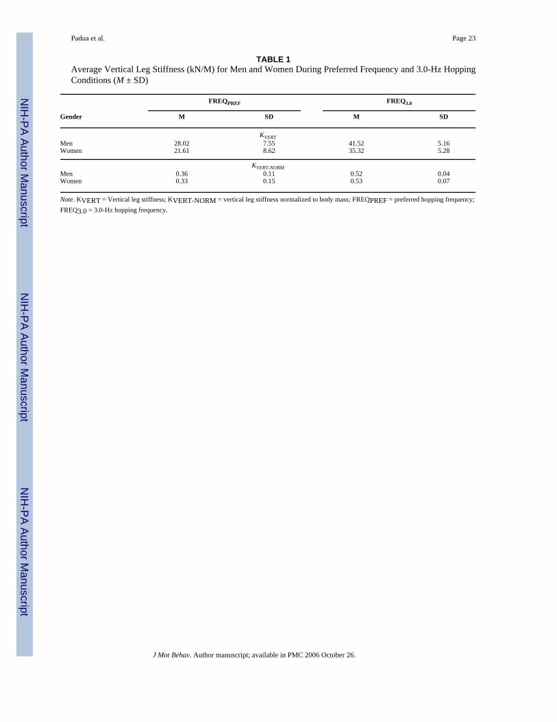
Vertical Leg Stiffness (KVERT)Statistical analysis revealed significant main effects for gender, F(1, 19) = 7.875, p = .011, andhopping frequency, F(1, 19) = 48.968, p = .001, but no significant Gender × Hopping Frequencyinteraction, F(1, 19) = .003, p = .960. On average, the women's KVERT values were 18% lessthan were those of the men across both hopping conditions (Figure 3). The main effect forgender was not influenced by preferred hopping rate, flight time, or duty cycle because thosevariables were equivalent between men and women (p > .05). Once normalized for body mass,KVERT-NORM values were nearly identical between genders, and the gender differences wereno longer apparent (Table 1), F(1, 18) = .002, p = .962.
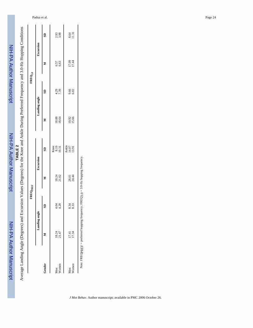
Joint Position and Range of MotionThere were no significant main effects or interactions involving gender for ANGKNEE,ANGANKLE,ROMKNEE, and ROMANKLE. Those findings indicate that joint angles at initialground contact as well as joint excursions of the knee and ankle joints were similar betweengenders during FREQPREF and FREQ3.0 hopping conditions (Table 2). There were significantmain effects involving hopping frequency for both ROMKNEE, F(1, 19) = 41.766, p = .001,
Padua et al. Page 8
J Mot Behav. Author manuscript; available in PMC 2006 October 26.
NIH
-PA Author Manuscript
NIH
-PA Author Manuscript
NIH
-PA Author Manuscript
and ROMANKLE, F(1, 19) = 30.626, p = .001. Those findings indicated that joint excursionsduring FREQ3.0 were significantly decreased in comparison with their values duringFREQPREF hopping conditions by 66% and 35% at the knee and ankle joints, respectively(Table 2).
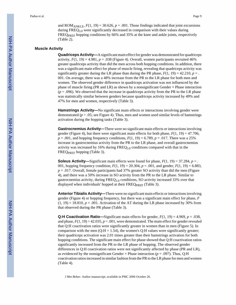
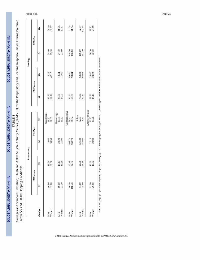
Muscle ActivityQuadriceps Activity—A significant main effect for gender was demonstrated for quadricepsactivity, F(1, 19) = 4.981, p = .038 (Figure 4). Overall, women participants recruited 46%greater quadriceps activity than did the men across both hopping conditions. In addition, therewas a significant main effect for phase of muscle firing, revealing that quadriceps activity wassignificantly greater during the LR phase than during the PR phase, F(1, 19) = 42.210, p = .001. On average, there was a 48% increase from the PR to the LR phase for both men andwomen. The observed gender difference in quadriceps activation was not influenced by thephase of muscle firing (PR and LR) as shown by a nonsignificant Gender × Phase interaction(p = .096). We observed that the increase in quadriceps activity from the PR to the LR phasewas statistically similar between genders because quadriceps activity increased by 49% and47% for men and women, respectively (Table 3).
Hamstrings Activity—No significant main effects or interactions involving gender weredemonstrated (p > .05; see Figure 4). Thus, men and women used similar levels of hamstringsactivation during the hopping tasks (Table 3).
Gastrocnemius Activity—There were no significant main effects or interactions involvinggender (Figure 4), but there were significant main effects for both phase, F(1, 19) = 47.706,p = .001, and hopping frequency conditions, F(1, 19) = 6.789, p = .017. There was a 25%increase in gastrocnemius activity from the PR to the LR phase, and overall gastrocnemiusactivity was increased by 16% during FREQ3.0 conditions compared with that in theFREQPREF hopping (Table 3).
Soleus Activity—Significant main effects were found for phase, F(1, 19) = 37.284, p = .001, hopping frequency condition, F(1, 19) = 20.304, p = .001, and gender, F(1, 19) = 6.883,p = .017. Overall, female participants had 37% greater SO activity than did the men (Figure4), and there was a 50% increase in SO activity from the PR to the LR phase. Similar togastrocnemius activity, during FREQ3.0 conditions, SO activity increased 33% over thatdisplayed when individuals' hopped at their FREQPREF (Table 3).
Anterior Tibialis Activity—There were no significant main effects or interactions involvinggender (Figure 4) or hopping frequency, but there was a significant main effect for phase, F(1, 19) = 18.810, p = .001. Activation of the AT during the LR phase increased by 30% fromthat observed during the PR phase (Table 3).
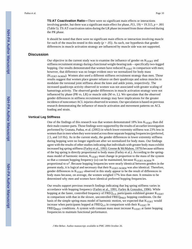
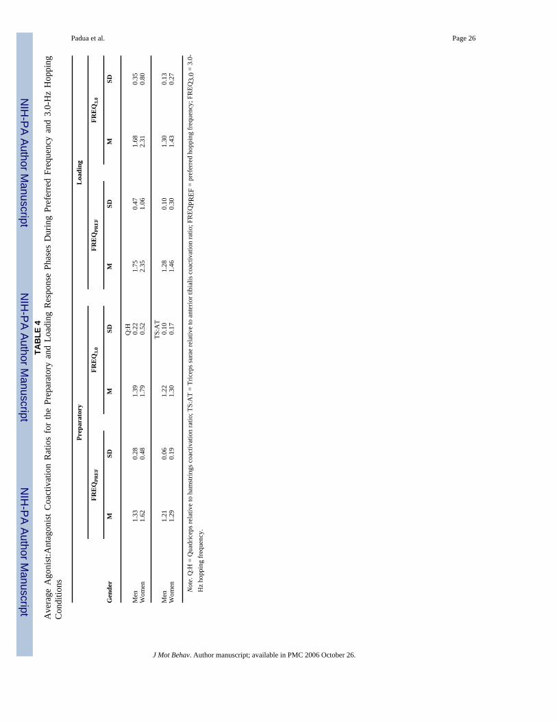
Q:H Coactivation Ratio—Significant main effects for gender, F(1, 19) = 4.969, p = .038,and phase, F(1, 19) = 42.035, p = .001, were demonstrated. The main effect for gender revealedthat Q:H coactivation ratios were significantly greater in women than in men (Figure 5). Incomparison with the men (Q:H = 1.54), the women's Q:H values were significantly greater;their quadriceps activation was 2.01 times greater than their hamstrings activation for bothhopping conditions. The significant main effect for phase showed that Q:H coactivation ratiossignificantly increased from the PR to the LR phase of hopping. The observed genderdifferences in Q:H coactivation ratios were not significantly affected by phase (PR and LR),as evidenced by the nonsignificant Gender × Phase interaction (p = .097). Thus, Q:Hcoactivation ratios increased in similar fashion from the PR to the LR phase for men and women(Table 4).
Padua et al. Page 9
J Mot Behav. Author manuscript; available in PMC 2006 October 26.
NIH
-PA Author Manuscript
NIH
-PA Author Manuscript
NIH
-PA Author Manuscript
TS:AT Coactivation Ratio—There were no significant main effects or interactionsinvolving gender, but there was a significant main effect for phase, F(1, 19) = 19.315, p = .001(Table 5). TS:AT coactivation ratios during the LR phase increased from those observed duringthe PR phase.
It should be noted that there were no significant main effects or interaction involving musclefor each of the muscles tested in this study (p > .05). As such, our hypothesis that genderdifferences in muscle activation strategy are influenced by muscle side was not supported.
DiscussionOur objective in the current study was to examine the influence of gender on KVERT andstiffness recruitment strategy during a functional weight-bearing task—specifically two-leggedhopping. Our results demonstrated that women have reduced KVERT in comparison with men;however, that difference was no longer evident once we normalized for body mass(KVERT-NORM). Women also used a different stiffness recruitment strategy than men. Thoseresults suggest that women place greater reliance on their quadriceps and soleus muscles tomodulate the torsional joint stiffness about the knee and ankle joints, respectively. Theincreased quadriceps activity observed in women was not associated with greater scaling ofhamstrings activity. The observed gender differences in muscle activation strategy were notinfluenced by phase (PR vs. LR) or muscle side (M vs. L). We speculate that the observedgender differences in stiffness recruitment strategy may have implications for the greaterincidence of noncontact ACL injuries observed in women. Our speculation is based on previousresearch demonstrating the influence of muscle activation and movement patterns on ACLloading and strain.
Vertical Leg StiffnessOne of the findings of this research was that women demonstrated 18% less KVERT than didtheir male counter-parts. Those findings were supported by the results of an earlier investigationperformed by Granata, Padua, et al. (2002) in which lower extremity stiffness was 23% less inwomen than in men when they were tested across three separate hopping frequencies (preferred,2.5, and 3.0 Hz). As in the current study, the gender differences in lower extremity stiffnessduring hopping were no longer significant after we normalized for body mass. Our findingsagree with the results of other studies indicating that individuals with greater body mass exhibitincreased leg spring stiffness (Farley et al., 1993; Greene & McMahon, 1979) because stiffnessof the leg spring is directly proportional to body mass (Farley et al.). According to the spring-mass model of harmonic motion, KVERT must change in proportion to the mass of the systemso that a constant hopping frequency (ω) can be maintained, because KVERT-NORM isproportional to ω2. Because hopping frequencies were nearly identical between genders in thepresent study, it is logical and necessary that their KVERT-NORM was also identical. Therefore,gender differences in KVERT observed in this study appear to be the result of differences inbody mass because, on average, the women weighed 17% less than men. It remains to bedetermined why men and women have identical preferred hopping frequencies.
Our results support previous research findings indicating that leg spring stiffness varies inaccordance with hopping frequency (Farley et al., 1991; Farley & Gonzalez, 1996). Whilehopping at the faster, controlled frequency of FREQ3.0, participants exhibited greater KVERTin comparison with that in the slower, uncontrolled FREQPREF hopping conditions. On thebasis of the simple spring-mass model of harmonic motion, we expected that KVERT wouldincrease when participants hopped at FREQ3.0 in comparison with their KVERT inFREQPREF conditions. A system with constant mass must increase KVERT at faster hoppingfrequencies to maintain functional performance.
Padua et al. Page 10
J Mot Behav. Author manuscript; available in PMC 2006 October 26.
NIH
-PA Author Manuscript
NIH
-PA Author Manuscript
NIH
-PA Author Manuscript
Those results suggest that the basic mechanism for increasing KVERT at faster hoppingfrequencies was to increase the torsional stiffness about the knee and ankle joints. Thatconclusion is based on our findings, which demonstrated significant decreases in bothROMKNEE and ROMANKLE from the slower FREQPREF to the faster FREQ3.0 hoppingconditions (Table 2). Although we did not specifically measure torsional joint stiffnessproperties, a reduction in total knee and ankle joint excursion during ground contact suggestsan increase in torsional knee and ankle joint stiffness, assuming similar joint moments betweenFREQPREF and FREQ3.0 hopping conditions.
Stiffness Recruitment StrategyKnee and Ankle Kinematics—The absence of a kinematic strategy difference betweengenders was somewhat of a surprise because previous researchers have demonstrated moreextended knee angles at ground contact when women performed cutting maneuvers or landedfrom a jump (Malinzak et al., 2001; McNitt-Gray et al., 1993). The previously identifiedkinematic differences between genders were observed during physical tasks that involved rapiddeceleration as the individual attempted to halt motion. Deceleration during two-leggedhopping is probably much less demanding than cutting and landing maneuvers, hencepotentially explaining the difference between current and previous research findings. Webelieve that the previously reported gender differences in lower extremity kinematics mayrepresent an altered strategy used to modulate KVERT in women during more strenuousfunctional tasks (cutting and landing from a jump). Performing those functional tasks withgreater knee extension would serve as an effective mechanism to increase KVERT with littleextra work from surrounding knee musculature. However, that type of movement strategyduring strenuous physical tasks may compromise overall knee stability because greater ground-reaction and resultant knee joint forces accompany that type of functional posturing (DeVita& Skelly, 1992; McMahon et al., 1987). The absence of a gender difference in kinematicstrategies suggests that during the less demanding hopping tasks, the primary distinguishingfactor in stiffness recruitment strategy between genders lies within the muscle activationstrategies they use to modulate KVERT.
Muscle Activity and Coactivation—A major finding of the present work was that althoughthere were no gender differences in KVERT once we normalized it for body mass, womenperformed two-legged hopping with substantially greater quadriceps and soleus muscle activitythan did the men. It is interesting that although the women were of lesser mass, they stillrecruited greater quadriceps activity than the men because they scaled their preparatoryquadriceps activity in anticipation for ground contact to a larger extent (∼2 times greater) thanthe men did. The greater reliance on quadriceps activation displayed by women was furtheremphasized during the LR phase because the absolute increase in quadriceps activation duringthe LR phase was 31% in women but was only 17% in men (Table 3). Those data suggest thatwomen attempt to modify KVERT in part through greater recruitment of the quadriceps muscles.That type of quadriceps-dominant recruitment strategy has been previously identified inwomen during controlled (Huston & Wojtys, 1996) and functional (jumping and cutting) tasks(Hewett et al., 1996;Malinzak et al., 2001). This is the first report of a quadriceps-dominantstrategy in women during a hopping task. A quadriceps-dominant profile for women has nowbeen identified in several different research studies that incorporated different measurementtechniques and physical tasks. We believe the quadriceps-dominant profile in women mayinfluence the gender bias in noncontact ACL injury rates.
Although greater quadriceps activation may serve as an effective mechanism for modulatingKVERT during two-legged hopping, its effects on knee joint stability are potentially injurious.Large quadriceps forces result in increased anterior tibial shear forces that cause anteriortranslation of the tibia with respect to the femur, placing increased forces and strain on the
Padua et al. Page 11
J Mot Behav. Author manuscript; available in PMC 2006 October 26.
NIH
-PA Author Manuscript
NIH
-PA Author Manuscript
NIH
-PA Author Manuscript
ACL (Beynnon et al., 1995; Durselen et al., 1995; Fleming et al., 2001; Hirokawa et al.,1992; Li et al., 1999; K. L. Markolf et al., 1990; Renstrom et al., 1986). Excessive strain is theultimate cause of ACL injury; thus, a quadriceps-dominant recruitment strategy may placewomen closer to their injury threshold by facilitating quadriceps-induced ACL strain.
Minimizing ACL strain and injury risk requires an increase in antagonist hamstrings activationto counteract quadriceps muscle contraction (Draganich, Jaeger, & Kralj, 1989; Li et al.,1999; Pandy & Shelburne, 1997; Renstrom et al., 1986; Shelburne & Pandy, 1998). In thisstudy, women used an imbalanced recruitment between the quadriceps and hamstrings (Q:H= 1.7). In contrast, men demonstrated a relatively balanced recruitment strategy (Q:H = 1.3;Figure 5). During the LR phase, both genders exhibited significantly increased Q:H ratiosbecause quadriceps activity increased (47% increase from PR to LR) with no significant changein hamstrings activation (PR = 23.0%, LR = 23.8%) to stiffen the knee joint and support theexternal knee flexion moment. During the LR phase, the quadriceps activity of women was2.3 times greater than that of their hamstrings, whereas in men, quadriceps activity was only1.7 times that of their hamstrings. Thus, the Q:H ratio in men during the weight-bearing LRphase was essentially identical to that of women during the non–weight-bearing PR phase. Wewere unable to find previous research investigations of gender differences in Q:H coactivation;thus, we were unable to compare our findings with those of previous research.
Overall, soleus muscle activation was 38% greater in women than in men. Greater reliance onsoleus muscle activity is an efficient strategy for controlling KVERT. However, it is assumedthat the soleus muscle is unable to protect the ACL because of its anatomical location. Womenalso demonstrated a trend for greater gastrocnemius activity than men (25% greater in women),but that difference was not significant (p = .277). We calculated mean overall effect size of0.64 for that comparison, indicating that, although not statistically significant, the greatergastrocnemius activity in women may be of clinical significance. Recent researchers havedemonstrated that the gastrocnemius is an antagonist to the ACL by increasing ACL strain(Fleming et al., 2001). That research revealed that the greatest ACL strain was produced duringsimultaneous activation of the gastrocnemius and quadriceps muscles (Fleming et al.). Theexact mechanism by which gastrocnemius contraction facilitates ACL strain is unknown. It isspeculated, however, that because of its anatomical attachment the gastrocnemius creates aposterior shear force on the femur, which may result in posterior translation of the femurrelative to the tibia. Essentially, because that creates anterior translation of the tibia relative tothe femur, it is known to increase ACL strain. Women, who recruit significantly greaterquadriceps activation and tend to recruit greater gastrocnemius activation in comparison withmen, may use a muscle recruitment strategy that facilitates ACL strain and places them at riskfor ACL injury.
Therefore, women appear to use a different stiffness recruitment strategy than men duringhopping. In comparison with men, women use a quadriceps-dominant and an ankle-dominantstiffness recruitment strategy that involves significantly greater quadriceps and soleus activity,a tendency for greater gastrocnemius activity, and minimal coactivation of the hamstrings. Aquadriceps- and ankle-dominant stiffness recruitment strategy will efficiently modulateKVERT so that the functional demands of the physical task can be met and sustained. Weobserved that strategy because, once we accounted for gender differences in body mass, thewomen displayed equivalent lower extremity stiffness values. However, we speculate that thestiffness recruitment strategy observed in women may potentially influence the gender biasassociated with ACL injury risk.
Potential explanations for gender differences in muscle activation include gender differencesin knee joint moment during hopping, neuromuscular control, strength, rate of force production,and active muscle stiffness. We do not feel that gender differences in knee joint moment can
Padua et al. Page 12
J Mot Behav. Author manuscript; available in PMC 2006 October 26.
NIH
-PA Author Manuscript
NIH
-PA Author Manuscript
NIH
-PA Author Manuscript
explain the differences in muscle activation because there were no differences between menand women in joint angles or duration of activity (hopping frequencies) and the data werescaled to anthropometrics. The reduced muscular strength of women has been documented (J.Griffin, Tooms, Zwaag, Bertorini, & O'Toole, 1993; Hakkinen, Kraemer, & Newton, 1997;Huston & Wojtys, 1996; Kanehisa, Okuyama, Ikegawa, & Fukunaga, 1996; Miller,MacDougall, Tarnopolsky, & Sale, 1993), as have their reduced rate of muscular forceproduction (Bell & Jacobs, 1986; Hakkinen & Hakkinen, 1991; Komi & Bosco, 1978; Viitasalo& Komi, 1978; E. M. Winter & Brookes, 1991) and active muscle stiffness compared withthose of men (Blackburn et al., 2004; Granata, Wilson, et al., 2002). Although women usegreater quadriceps activation, it is possible that the relative force acting at the knee and anklemay not differ between men and women. The observed gender differences in muscle activationmay represent a feedforward neuromuscular control strategy whereby women compensate fordecreased muscular strength, rate of force production, and active muscle stiffness by increasingactivation of the quadriceps and soleus muscles. It is important to note that speculationconcerning muscle force solely on the basis of muscle activity during dynamic motion istenable. Although muscle activity level is an important factor in determining a muscle's output,the resultant contractile force is also influenced by muscle length, contractile velocity, andcontraction mode (isometric, concentric, and eccentric). Therefore, we do not suggest thatmuscle activity level is a direct representation of the resultant muscle force at the joint. Thatlimitation should be considered when interpreting the results of this study. We are unable todefinitively explain the quadriceps- and ankle-dominant stiffness recruitment strategy used bywomen during hopping.
ConclusionsWomen demonstrated reduced KVERT during the functional hopping tasks in comparison withthat of men. We attribute the gender difference in KVERT to the lighter body mass observed inwomen because once we normalized for body mass there were no significant differences inKVERT-NORM between men and women. That result indicates that the gender differences inKVERT during a functional hopping task are likely functions of anthropometric differences.
When comparing the stiffness recruitment strategy between genders during a functionalhopping task, we revealed that female participants recruited significantly greater quadricepsand soleus activity that was not associated with increased hamstrings activity. In theory, therecruitment strategy may efficiently modulate KVERT. However, it may compromise stabilityat the knee joint. We are unable to explain why the women used a different stiffness recruitmentstrategy than men did, yet demonstrated equivalent lower extremity stiffness once we hadnormalized for body mass. However, similar gender differences in muscle activation strategieshave been reported previously. Future research is necessary to determine the factorscontributing to the observed gender differences in stiffness recruitment strategy. In addition,whether the quadriceps-dominant and ankle-dominant strategies used by women actually placethe ACL at greater risk for injury is still unknown and requires further study.
REFERENCESAgarwal GC, Gottlieb GL. Oscillation of the human ankle joint in response to applied sinusoidal torque
at the foot. Journal of Physiology 1977;268:151–176. [PubMed: 874886]Arampatzis A, Bruggemann GP, Klapsing GM. Leg stiffness and mechanical energetic processes during
jumping on a sprung surface. Medicine and Science in Sports and Exercise 2001;33(6):923–937.[PubMed: 11404657]
Arampatzis A, Bruggemann G, Metzler V. The effect of speed on leg stiffness and joint kinetics in humanrunning. Journal of Biomechanics 1999;32:1349–1353. [PubMed: 10569714]
Padua et al. Page 13
J Mot Behav. Author manuscript; available in PMC 2006 October 26.
NIH
-PA Author Manuscript
NIH
-PA Author Manuscript
NIH
-PA Author Manuscript
Arendt EA, Agel J, Dick R. Anterior cruciate ligament injury patterns among collegiate men and women.Journal of Athletic Training 1999;34(2):86–92. [PubMed: 16558564]
Arms S, Pope M, Johnson R, Fischer R, Arvidsson I, Eriksson E. The biomechanics of anterior cruciateligament rehabilitation and reconstruction. American Journal of Sports Medicine 1984;12:8–18.[PubMed: 6703185]
Avela J, Komi PV. Reduced stretch reflex sensitivity and muscle stiffness after long-lasting stretch-shortening cycle exercise in humans. European Journal of Applied Physiology 1998;78(5):403–410.
Avela J, Kyrolainen H, Komi PV, Rama D. Reduced reflex sensitivity persists several days after long-lasting stretch-shortening cycle exercise. Journal of Applied Physiology 1999;86(4):1292–1300.[PubMed: 10194215]
Bell DG, Jacobs I. Electro-mechanical response times and rate of force development in males and females.Medicine and Science in Sports and Exercise 1986;18:31–36. [PubMed: 3959861]
Beynnon BD, Fleming BC, Johnson RJ, Nichols CE, Renstrom PA, Pope MH. Anterior cruciate ligamentstrain behavior during rehabilitation exercises in vivo. American Journal of Sports Medicine 1995;23(1):24–34. [PubMed: 7726347]
Bjordal JM, Arnly F, Hannestad B, Strand T. Epidemiology of anterior cruciate ligament injuries insoccer. American Journal of Sports Medicine 1997;25:341–345. [PubMed: 9167814]
Blackburn JT, Riemann BL, Padua DA, Guskiewicz KM. Sex comparison of active extensibility, andactive and passive stiffness of the knee flexors. Clinical Biomechanics 2004;19:36–43. [PubMed:14659928]
Boden BP, Dean GS, Feagin JA, Garrett WE. Mechanisms of anterior cruciate ligament injury.Orthopedics 2000;23(6):573–578. [PubMed: 10875418]
Boden BP, Griffin LY, Garrett WE. Etiology and prevention of noncontact ACL injury. Physician andSportsmedicine 2000;28(4):53–60.
Cannon SC, Zahalak GI. The mechanical behavior of active human skeletal muscle in small oscillations.Journal of Biomechanics 1982;15(2):111–121. [PubMed: 7076686]
Cavagna GA. Force platforms as ergometers. Journal of Applied Physiology 1975;39(1):174–179.[PubMed: 1150585]
Cox JS, Lenz HW. Women midshipmen in sports. American Journal of Sports Medicine 1984;12(3):241–243. [PubMed: 6742310]
Cram, J.; Kasman, G. Introduction to surface electromyography. Aspen; Gaithersburg, MD: 1998.Decker MJ, Torry MR, Wyland DJ, Sterett WI, Steadman JR. Gender differences in lower extremity
kinematics, kinetics and energy absorption during landing. Clinical Biomechanics 2003;18:662–669.[PubMed: 12880714]
DeVita P, Skelly WA. Effect of landing stiffness on joint kinetics and energetics in the lower extremity.Medicine and Science in Sports and Exercise 1992;24(1):108–115. [PubMed: 1548984]
Draganich LF, Jaeger RJ, Kralj AR. Coactivation of the hamstrings and quadriceps during extension ofthe knee. Journal of Bone and Joint Surgery 1989;71:1075–1081. [PubMed: 2760083]
Duan XH, Allen RH, Sun JQ. A stiffness-varying model of human gait. Medical Engineering and Physics1997;19(6):518–524. [PubMed: 9394899]
Durselen L, Claes L, Kiefer H. The influence of muscle forces and external loads on cruciate ligamentstrain. American Journal of Sports Medicine 1995;23(1):129–136. [PubMed: 7726343]
Farley CT, Blickhan R, Saito J, Taylor R. Hopping frequency in humans: A test of how springs set stridefrequency in bouncing gaits. Journal of Applied Physiology 1991;71(6):2127–2132. [PubMed:1778902]
Farley CT, Glasheen J, McMahon TA. Running springs: Speed and animal size. Journal of ExperimentalBiology 1993;185:71–86. [PubMed: 8294853]
Farley CT, Gonzalez O. Leg stiffness and stride frequency in human running. Journal of Biomechanics1996;29(2):181–186. [PubMed: 8849811]
Farley CT, Houdikj HHP, Strien CV, Louie M. Mechanism of leg stiffness adjustment for hopping onsurfaces of different stiffnesses. Journal of Applied Physiology 1998;85(3):1044–1055. [PubMed:9729582]
Padua et al. Page 14
J Mot Behav. Author manuscript; available in PMC 2006 October 26.
NIH
-PA Author Manuscript
NIH
-PA Author Manuscript
NIH
-PA Author Manuscript
Farley CT, Morgenroth DC. Leg stiffness primarily depends on ankle stiffness during human hopping.Journal of Biomechanics 1999;32(3):267–273. [PubMed: 10093026]
Fleming BC, Renstrom PA, Ohlen G, Johnson RJ, Peura GD, Beynnon BD, et al. The gastrocnemius isan antagonist of the anterior cruciate ligament. Journal of Orthopaedic Research 2001;19:1178–1184.[PubMed: 11781021]
Gomez E, DeLee JC, Farney WC. Incidence of injury in Texas girls' high school basketball. AmericanJournal of Sports Medicine 1996;24(5):684–687. [PubMed: 8883693]
Granata KP, Padua DA, Wilson SE. Gender differences in active musculoskeletal stiffness. Part II.Quantification of leg stiffness during functional hopping tasks. Journal of Electromyography andKinesiology 2002;12:127–135. [PubMed: 11955985]
Granata KP, Wilson SE, Padua DA. Gender differences in active musculoskeletal stiffness. Part I.Quantification in controlled measurements of knee joint dynamics. Journal of Electromyography andKinesiology 2002;12:119–126. [PubMed: 11955984]
Greene P, McMahon T. Reflex stiffness of man's antigravity muscles during kneebends while carryingextra weights. Journal of Biomechanics 1979;12:881–891. [PubMed: 528546]
Griffin J, Tooms R, Zwaag R, Bertorini TE, O'Toole M. Eccentric muscle performance of elbow andknee muscle groups in untrained men and women. Medicine and Science in Sports and Exercise1993;25:936–944. [PubMed: 8371655]
Griffin LY, Agel JA, Albohm MJ, Arendt EA, Dick RW, Garrett WE, et al. Noncontact anterior cruciateligament injuries: Risk factors and prevention strategies. Journal of the American Academy ofOrthopaedic Sugeons 2000;8(3):141–150.
Gwinn DE, Wilckens JH, McDevitt ER, Ross G, Kao T. The relative incidence of anterior cruciateligament injury in men and women at the United States Naval Academy. American Journal of SportsMedicine 2000;28(1):98–102. [PubMed: 10653551]
Hakkinen K, Hakkinen A. Muscle cross-sectional area, force production and relaxation characteristics inwomen at different ages. European Journal of Applied Physiology 1991;62(6):410–414.
Hakkinen K, Kraemer WJ, Newton RU. Muscle activation and force production during bilateral andunilateral concentric and isometric contractions of the knee extensors in men and women at differentages. Electromyography and Clinical Neurophysiology 1997;37:131–142. [PubMed: 9187864]
Hewett TE, Stroupe AL, Nance TA, Noyes FR. Plyometric training in female athletes. Decreased impactforces and increased hamstring torques. American Journal of Sports Medicine 1996;24(6):765–773.[PubMed: 8947398]
Hirokawa S, Solomonow M, Lu Y, Lou Z, D'Ambrosia R. Anterior-posterior and rotational displacementof the tibia elicited by quadriceps contraction. American Journal of Sports Medicine 1992;20(3):299–306. [PubMed: 1636861]
Horita T, Komi P, Nicol C, Kyrolainen H. Effect of exhausting stretch-shortening cycle exercise on thetime course of mechanical behaviour in the drop jump: Possible role of muscle damage. EuropeanJournal of Applied Physiology 1999;79(2):160–167.
Hortobagyi T, DeVita P. Altered movement strategy increases lower extremity stiffness during steppingdown in the aged. Journal of Gerontology 1999;54A(2):B63–B70.
Hortobagyi T, DeVita P. Muscle pre- and coactivity during downward stepping are associated with legstiffness in aging. Journal of Electromyography and Kinesiology 2000;10:117–126. [PubMed:10699559]
Houk J. Regulation of stiffness by skeletomotor reflexes. Annual Review of Physiology 1979;41:99–114.Hunter IW, Kearney RE. Dynamics of human ankle stiffness: Variation with mean ankle torque. Journal
of Biomechanics 1982;15(10):747–752. [PubMed: 7153227]Huston LJ, Wojtys EM. Neuromuscular performance characteristics in elite female athletes. American
Journal of Sports Medicine 1996;18:31–36.Julian FJ, Sollins MR. Variation of muscle stiffness with force at increasing speeds of shortening. Journal
of General Physiology 1975;66:287–302.Kanehisa H, Okuyama H, Ikegawa S, Fukunaga T. Sex difference in force generation capacity during
repeated maximal knee extensions. European Journal of Applied Physiology 1996;73:557–562.Kearney R, Stein R, Parameswaran L. Identification of intrinsic and reflex contributions to human ankle
stiffness dynamics. IEEE Transactions of Biomedical Engineering 1997;44(6):493–504.
Padua et al. Page 15
J Mot Behav. Author manuscript; available in PMC 2006 October 26.
NIH
-PA Author Manuscript
NIH
-PA Author Manuscript
NIH
-PA Author Manuscript
Komi PV, Bosco C. Utilization of stored elastic energy in leg extensor muscles by men and women.Medicine and Science in Sports 1978;10(4):261–265. [PubMed: 750844]
Lacquanti F, Licata F, Soechting JF. The mechanical behavior of the human forearm in response totransient perturbations. Biological Cybernetics 1982;44:35–46. [PubMed: 7093368]
Lephart SM, Ferris CM, Riemann BL, Myers JB, Fu FH. Gender differences in strength and lowerextremity kinematics during landing. Clinical Orthopaedics and Related Research 2002;401:162–169. [PubMed: 12151893]
Li G, Sakane R, Kanamori A, Ma C, Woo S. The importance of quadriceps and hamstring muscle loadingon knee kinematics and in-situ forces in the ACL. Journal of Bio-mechanics 1999;32:395–400.
Malinzak RA, Colby SM, Kirkendall DT, Yu B, Garrett WE. A comparison of knee joint motion patternsbetween men and women in selected athletic tasks. Clinical Biomechanics 2001;16:438–445.[PubMed: 11390052]
Malone TR, Hardaker WT, Garrett WE, Feagin JA, Bassett FH. Relationship of gender to anterior cruciateligament injuries in intercollegiate basketball players. Journal of the Southern OrthopaedicAssociation 1993;2(1):36–39.
Markolf K, Burchfield D, Shapiro M, Shepard M, Finerman G, Slauterbeck J. Combined knee loadingstates that generate high anterior cruciate ligament forces. Journal of Orthopaedic Research1995;13:930–935. [PubMed: 8544031]
Markolf KL, Gorek JF, Kabo M, Shapirt MS. Direct measurement of resultant forces in the anteriorcruciate ligament. Journal of Bone and Joint Surgery 1990;72A(4):557–567. [PubMed: 2324143]
Maton B, Pellec AL. Adaptation of the short latency component of the stretch reflex plays only a minorrole in compensating for muscle fatigue induced by spontaneous hopping in humans. EuropeanJournal of Applied Physiology 2001;84:26–35. [PubMed: 11394250]
McGill SM. Low back stability: From formal description to issues for performance and rehabilitation.Exercise and Sport Sciences Reviews 2001;29(1):26–31. [PubMed: 11210443]
McMahon TA, Cheng GC. The mechanics of running: How does stiffness couple with speed? Journal ofBiomechanics 1990;23(Suppl 1):65–78. [PubMed: 2081746]
McMahon TA, Valiant G, Frederick EC. Groucho running. Journal of Applied Physiology 1987;62(6):2326–2337. [PubMed: 3610929]
McNitt-Gray JL, Yokoi T, Millward C. Landing strategy adjustments made by female gymnasts inresponse to drop height and mat composition. Journal of Applied Biomechanics 1993;9:173–190.
Messina DF, Farney WC, DeLee JC. The incidence of injury in Texas high school basketball. Aprospective study among male and female athletes. American Journal of Sports Medicine 1999;27(3):294–299. [PubMed: 10352762]
Miller A, MacDougall J, Tarnopolsky M, Sale D. Gender differences in strength and muscle fibercharacteristics. European Journal of Applied Physiology 1993;66:254–262.
Mortiani T, Oddson L, Thorstensson A. Electromyographic evidence of selective fatigue during theeccentric phase of stretch/shortening cycles in man. European Journal of Applied Physiology1990;60:425–429.
Nichols T, Houk J. Improvement in linearity and regulation of stiffness that results from actions of thestretch reflex. Journal of Neurophysiology 1976;39:119–142. [PubMed: 1249597]
Nicol C, Komi PV, Horita T, Kyrolainen H, Takala TE. Reduced stretch-reflex sensitivity after exhaustingstretch-shortening cycle exercise. European Journal of Applied Physiology 1996;72(5–6):401–409.
Pandy MG, Shelburne KB. Dependence of cruciate-ligament loading on muscle forces and external load.Journal of Biomechanics 1997;30(10):1015–1024. [PubMed: 9391868]
Renstrom P, Arms SW, Stanwyck TS, Johnson RJ, Pope MH. Strain within the anterior cruciate ligamentduring hamstring and quadriceps activity. American Journal of Sports Medicine 1986;14(1):83–87.[PubMed: 3752352]
Shelburne KB, Pandy MG. Determinants of cruciate-ligament loading during rehabilitation exercise.Clinical Biomechanics 1998;13:403–413. [PubMed: 11415815]
Viitasalo JT, Komi PV. Force-time characteristics and fiber composition in human leg extensor muscles.European Journal of Applied Physiology 1978;40(1):7–15.
Padua et al. Page 16
J Mot Behav. Author manuscript; available in PMC 2006 October 26.
NIH
-PA Author Manuscript
NIH
-PA Author Manuscript
NIH
-PA Author Manuscript
Wagner H, Blickhan R. Stabilizing function of skeletal muscles: An analytical investigation. Journal ofTheoretical Biology 1999;199:163–179. [PubMed: 10395812]
Weiss PL, Hunter IW, Kearney RE. Human ankle joint stiffness over the full range of muscle activationlevels. Journal of Biomechanics 1988;21(7):539–544. [PubMed: 3410857]
Winter D, Fuglevand A, Archer S. Crosstalk in surface electromyography: Theoretical and practicalestimates. Journal of Electromyography and Kinesiology 1994;4:15–26.
Winter EM, Brookes FB. Electromechanical response times and muscle elasticity in men and women.European Journal of Applied Physiology 1991;63:124–128.
Wojtys EM, Huston LJ, Taylor PD, Bastian SD. Neuromuscular adaptations in isokinetic, isotonic, andagility training programs. American Journal of Sports Medicine 1996;24(2):187–192. [PubMed:8775118]
Yang JF, Winter DA. Electromyographic amplitude normalization methods: Improving their sensitivityas diagnostic tools in gait analysis. Archives of Physical Medicine and Rehabilitation 1984;65:517–521. [PubMed: 6477083]
Zhang LQ, Nuber G, Butler J, Bowen M, Rymer WZ. In vivo human knee joint dynamic properties asfunctions of muscle contraction and joint position. Journal of Bio-mechanics 1998;31:71–76.
Padua et al. Page 17
J Mot Behav. Author manuscript; available in PMC 2006 October 26.
NIH
-PA Author Manuscript
NIH
-PA Author Manuscript
NIH
-PA Author Manuscript
FIGURE 1.Lower extremity spring force (vertical ground reaction force) plotted as a function of verticalcenter of mass (COM) displacement during the ground-contact phase of a single trial at thepreferred (FREQPREF) and 3.0-Hz (FREQ3.0) hopping frequencies. The linear relationship isan indicator that the lower extremity behaved like a simple spring-mass system at both hoppingfrequencies. The slope (dashed line) of the force versus displacement curves represents thevertical leg stiffness during hopping. As hopping frequency increased, the slope, that is, verticalleg stiffness (KVERT), also increased. The graph represents the exemplar records from a singleparticipant.
Padua et al. Page 18
J Mot Behav. Author manuscript; available in PMC 2006 October 26.
NIH
-PA Author Manuscript
NIH
-PA Author Manuscript
NIH
-PA Author Manuscript
FIGURE 2.Representation of vertical ground reaction force (VGRF) and electromyographic (EMG)activity normalized to maximal voluntary isometric contraction (MVIC) for the quadriceps,hamstrings, gastrocnemius, and soleus muscles for 1 male and 1 female participant during thepreparatory (PR) and loading (LR) response phases. The typical EMG records for allparticipants are shown.
Padua et al. Page 19
J Mot Behav. Author manuscript; available in PMC 2006 October 26.
NIH
-PA Author Manuscript
NIH
-PA Author Manuscript
NIH
-PA Author Manuscript
FIGURE 3.Effect of gender on vertical leg stiffness (KVERT). Data were pooled across the preferred(FREQPREF) and 3.0-Hz (FREQ3.0) hopping frequency conditions. The men's KVERT wasincreased in comparison with the women's. When KVERT was normalized for body mass(KVERT-NORM), gender differences were no longer significantly different. Asterisk (*)indicates significantly greater KVERT (p < .05).
Padua et al. Page 20
J Mot Behav. Author manuscript; available in PMC 2006 October 26.
NIH
-PA Author Manuscript
NIH
-PA Author Manuscript
NIH
-PA Author Manuscript
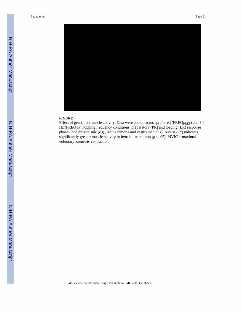
FIGURE 4.Effect of gender on muscle activity. Data were pooled across preferred (FREQPREF) and 3.0-Hz (FREQ3.0) hopping frequency conditions, preparatory (PR) and loading (LR) responsephases, and muscle side (e.g., rectus femoris and vastus medialis). Asterisk (*) indicatessignificantly greater muscle activity in female participants (p < .05). MVIC = maximalvoluntary isometric contraction.
Padua et al. Page 21
J Mot Behav. Author manuscript; available in PMC 2006 October 26.
NIH
-PA Author Manuscript
NIH
-PA Author Manuscript
NIH
-PA Author Manuscript

FIGURE 5.Effect of gender on quadriceps:hamstrings coactivation ratios (Q:H). Data were pooled acrosspreferred (FREQPREF) and 3.0-Hz (FREQ3.0) hopping frequency conditions and preparatory(PR) and loading (LR) response phases. Asterisk (*) indicates significantly greater Q:H inwomen than in men (p < .05).
Padua et al. Page 22
J Mot Behav. Author manuscript; available in PMC 2006 October 26.
NIH
-PA Author Manuscript
NIH
-PA Author Manuscript
NIH
-PA Author Manuscript
NIH
-PA Author Manuscript
NIH
-PA Author Manuscript
NIH
-PA Author Manuscript
Padua et al. Page 23
TABLE 1Average Vertical Leg Stiffness (kN/M) for Men and Women During Preferred Frequency and 3.0-Hz HoppingConditions (M ± SD)
FREQPREF FREQ3.0
Gender M SD M SD
KVERTMen 28.02 7.55 41.52 5.16Women 21.61 8.62 35.32 5.28
KVERT-NORMMen 0.36 0.11 0.52 0.04Women 0.33 0.15 0.53 0.07
Note. KVERT = Vertical leg stiffness; KVERT-NORM = vertical leg stiffness normalized to body mass; FREQPREF = preferred hopping frequency;FREQ3.0 = 3.0-Hz hopping frequency.
J Mot Behav. Author manuscript; available in PMC 2006 October 26.
NIH
-PA Author Manuscript
NIH
-PA Author Manuscript
NIH
-PA Author Manuscript
Padua et al. Page 24TA
BLE
2A
vera
ge L
andi
ng A
ngle
(Deg
rees
) and
Exc
ursi
on V
alue
s (D
egre
es) f
or th
e K
nee
and
Ank
le D
urin
g Pr
efer
red
Freq
uenc
y an
d 3.
0-H
z H
oppi
ng C
ondi
tions
FRE
QPR
EF
FRE
Q3.
0
Lan
ding
ang
leE
xcur
sion
Lan
ding
ang
leE
xcur
sion
Gen
der
MSD
MSD
MSD
MSD
Kne
eM
en18
.24
4.04
18.2
4 8
.19
18.8
84.
296.
572.
93W
omen
21.4
74.
2821
.02
10.3
118
.64
7.39
6.63
3.98
Ank
leM
en17
.41
8.34
28.0
114
.07
19.9
29.
6617
.09
8.64
Wom
en17
.34
8.33
24.4
413
.91
15.6
66.
0217
.44
11.3
1
Not
e. F
REQ
PREF
= p
refe
rred
hop
ping
freq
uenc
y; F
REQ
3.0
= 3.
0-H
z ho
ppin
g fr
eque
ncy.
J Mot Behav. Author manuscript; available in PMC 2006 October 26.
NIH
-PA Author Manuscript
NIH
-PA Author Manuscript
NIH
-PA Author Manuscript
Padua et al. Page 25TA
BLE
3A
vera
ge (a
nd S
tand
ard
Dev
iatio
n) T
high
and
Ank
le M
uscl
e A
ctiv
ity V
alue
s (%
MV
IC) f
or th
e Pr
epar
ator
y an
d Lo
adin
g R
espo
nse
Phas
es D
urin
g Pr
efer
red
Freq
uenc
y an
d 3.
0-H
z H
oppi
ng C
ondi
tions
Prep
arat
ory
Loa
ding
FRE
QPR
EF
FRE
Q3.
0FR
EQ
PRE
FFR
EQ
3.0
Gen
der
MSD
MSD
MSD
MSD
Qua
dric
eps
Men
16.
8018
.66
19.
6018
.97
37.
70 8
.26
34.
40 2
9.09
Wom
en 3
1.00
20.5
6 3
9.50
20.8
9 6
7.10
42.1
2 6
5.30
32.
17
Ham
strin
gsM
en 1
9.00
10.4
4 2
3.20
14.5
5 2
5.80
10.4
4 2
7.30
10.
75W
omen
23.
3011
.28
21.
9015
.92
24.
0011
.61
22.
60 1
1.61
Gas
trocn
emiu
sM
en 8
6.10
67.9
910
4.70
89.8
113
5.50
89.8
114
4.50
71.
78W
omen
118.
2074
.62
160.
7098
.84
160.
0098
.84
186.
60 7
8.94
Sole
usM
en 4
4.60
26.5
612
1.30
72.7
3 7
5.80
62.3
016
6.00
92.
97W
omen
74.
6029
.19
185.
00 9
.93
160.
2068
.65
232.
3010
2.48
Ant
erio
r tib
ialis
Men
21.
1013
.60
22.
1012
.97
28.
8020
.87
30.
10 1
9.92
Wom
en 2
9.80
14.9
2 2
9.90
14.2
6 4
5.40
23.2
2 4
2.70
21.
89
Not
e. F
REQ
PREF
= p
refe
rred
hop
ping
freq
uenc
y; F
REQ
3.0
= 3.
0-H
z ho
ppin
g fr
eque
ncy;
% M
VIC
= p
erce
ntag
e of
max
imal
vol
unta
ry is
omet
ric c
ontra
ctio
n.
J Mot Behav. Author manuscript; available in PMC 2006 October 26.
NIH
-PA Author Manuscript
NIH
-PA Author Manuscript
NIH
-PA Author Manuscript
Padua et al. Page 26TA
BLE
4A
vera
ge A
goni
st:A
ntag
onis
t C
oact
ivat
ion
Rat
ios
for
the
Prep
arat
ory
and
Load
ing
Res
pons
e Ph
ases
Dur
ing
Pref
erre
d Fr
eque
ncy
and
3.0-
Hz
Hop
ping
Con
ditio
ns
Prep
arat
ory
Loa
ding
FRE
QPR
EF
FRE
Q3.
0FR
EQ
PRE
FFR
EQ
3.0
Gen
der
MSD
MSD
MSD
MSD
Q:H
Men
1.33
0.28
1.39
0.22
1.75
0.47
1.68
0.35
Wom
en1.
620.
481.
790.
522.
351.
062.
310.
80
TS:A
TM
en1.
210.
061.
220.
101.
280.
101.
300.
13W
omen
1.29
0.19
1.30
0.17
1.46
0.30
1.43
0.27
Not
e. Q
:H =
Qua
dric
eps r
elat
ive
to h
amst
rings
coa
ctiv
atio
n ra
tio; T
S:A
T =
Tric
eps s
urae
rela
tive
to a
nter
ior t
ibia
lis c
oact
ivat
ion
ratio
; FR
EQPR
EF =
pre
ferr
ed h
oppi
ng fr
eque
ncy;
FR
EQ3.
0 =
3.0-
Hz
hopp
ing
freq
uenc
y.
J Mot Behav. Author manuscript; available in PMC 2006 October 26.


