Embed Size (px)
Citation preview
Biochimica et Biophysica Acta 1828 (2013) 382–390
Contents lists available at SciVerse ScienceDirect
Biochimica et Biophysica Acta
j ourna l homepage: www.e lsev ie r .com/ locate /bbamem
Fusion of gemini based cationic liposomes with cell membrane models:implications for their biological activity
Simone Aleandri a, Cecilia Bombelli b, Maria Grazia Bonicelli c, Federico Bordi d,e, Luisa Giansanti b,⁎,Giovanna Mancini b, Marco Ierino c, Simona Sennato d,e,⁎a Dipartimento di Chimica Università degli Studi di Roma “Sapienza”, P.le A. Moro 5, 00185 Roma, Italyb CNR, Istituto di Metodologie Chimiche and Dipartimento di Chimica Università degli Studi di Roma “Sapienza”, P.le A. Moro 5, 00185 Roma, Italyc Dipartimento di Ingegneria Chimica, dei Materiali e dell'Ambiente (ICMA), Università degli Studi di Roma “Sapienza”, via del Castro Laurenziano 7, 00161 Roma, Italyd Dipartimento di Fisica, Università degli Studi di Roma “Sapienza”, P.le A. Moro 2, 00185 Roma, Italye CNR-IPCF, Università degli Studi di Roma “Sapienza”, P.le A. Moro 2, 00185 Roma, Italy
⁎ Corresponding authors. Tel.: +39 06 49913681; +3490421.
E-mail addresses: [email protected] (L. [email protected] (S. Sennato).
0005-2736/$ – see front matter © 2012 Elsevier B.V. Alhttp://dx.doi.org/10.1016/j.bbamem.2012.10.001
a b s t r a c t
a r t i c l e i n f oArticle history:Received 13 July 2012Received in revised form 25 September 2012Accepted 3 October 2012Available online 7 October 2012
Keywords:Mixed cationic liposomesStereochemistry of geminiDSCFluorescenceDLSFusion
The interaction of neutral and anionic phospholipid liposomes, used as cell models, with cationic liposomesformulated with 1,2-dimyristoyl-sn-glicero-3-phosphocholine and stereomeric cationic gemini surfactantswas investigated by differential scanning calorimetry, fluorescence experiments and dynamic laser lightscattering. This study was aimed at rationalizing the different biological features shown by liposomesbased on different gemini stereoisomers observed in previous investigations. In fact, to correlate the observedbiological activity of liposomes with the molecular structure of their components is critical for a rational andsystematic approach to the design of new carriers for drug delivery. The obtained results show that thedifferent stereochemistry of the gemini surfactant controls the interaction and the extent of fusion withdifferent cell models.
© 2012 Elsevier B.V. All rights reserved.
1. Introduction
Liposomes can be used as drug carriers to transport a variety ofbiologically active molecules into cells in vitro and in vivo [1], includ-ing those that would not ordinarily be taken up by the cells. There isnow a large body of evidence, including pre-clinical and clinical stud-ies, showing that many different drugs packaged in liposomes exhibitsignificantly reduced toxicity, while retaining or even gaining efficacy[2], so that liposomes are regarded as most promising in the pano-rama of drug delivery systems. However, in most studies the empha-sis has been focused on demonstrating that the material encapsulatedwithin the vesicles can affect the cell activity and functioning, and lessattention has been paid to the mechanisms of liposome internali-zation. A deeper understanding of the mechanisms involved inthe drug uptake mediated by lipid vesicles, together with a morecomplete information on the fate of the vesicles inside the cell and
9 06 49913503 fax: +39 06
nsanti),
l rights reserved.
on the intracellular distribution of the vesicles content, is necessaryto the best design of new lipid based drug delivery systems.
We previously reported detailed investigations on the effi-cacy of liposomes formulated with 1,2-dimyristoyl-sn-glycero-3-phosphocholine (DMPC) and the cationic gemini surfactants (S,S)-2,3-dimethoxy-1,4-bis(N-hexadecyl-N,N-dimethylammonium)butane bromide 1 or its stereoisomer (S,R)-2,3-dimethoxy-1,4-bis(N-hexadecyl-N,N-dimethylammonium)butane bromide, 2(Chart 1) as drug delivery systems. It was shown that the stereo-chemistry of the gemini components significantly affects impor-tant features of the mixed liposomes as carriers: the efficiencyof the delivery, the intracellular distribution of the drug, andthe DNA condensation and transfection in gene delivery [3–8].Moreover, the delivery efficacy was shown to depend on cell lines[6,7]. The DNA-cationic liposome aggregates (lipoplexes) employedfor gene delivery are known to form complex structures [9–11]. Whilesimple liposomes cannot be considered adequate models to studythe interaction of these structures with cell membranes, neverthe-less also in this case differences in the molecular structure of lipidmolecules apparently play an important role [12]. On these premises,we investigated by differential scanning calorimetry (DSC), fluores-cence and dynamic light scattering (DLS) experiments the influenceof the stereochemistry on the interaction of the cationic formulationswith different liposomes employed as cell membrane models,

Chart 1. Structure of the stereomeric cationic gemini surfactants 1 and 2.
383S. Aleandri et al. / Biochimica et Biophysica Acta 1828 (2013) 382–390
namely DMPC, 1,2-dipalmitoyl-sn-glycero-3-phosphatidylcoline(DPPC) and 1,2-dipalmitoyl-sn-glycero-3-phospho-(1′-rac-glycerol)(sodium salt) (DPPG) liposomes.
While liposomes, due to the complete absence of protein compo-nents, can be righty considered an excessively oversimplified modelof cell membrane under several aspects, studies of the molecularmechanism of membrane fusion and/or lipid exchange in simple, pro-tein free, model systems provide fundamental insights into the fusionprocess in biomembranes, where complex protein machines promotethe lipid molecule rearrangements required for fusion [13–15]. In-deed, the study of the interaction between different bilayer structuressuch as liposomes, black lipid membranes and supported bilayers hasbeen instrumental not only in defining the sequences of the interme-diate structures formed in the course of bilayer merger, but also in de-termining the properties of the lipid bilayers and the characteristics ofthe individual lipid molecules that control the propensity of mem-branes to fuse [16].
The data presented here give clear evidence that the presenceof the cationic gemini in the bilayer composition promotes anextensive exchange of lipids between the liposomes, probably asa consequence of “hemifusion”. Hemifusion is the partial and re-versible fusion of the external leaflet of two bilayers in close con-tact (see for example Ref. [17]). This process, which is sometimescolorfully described as “kiss-and-run”, does not imply a “full fu-sion” of the liposomes, with the mixing of their internal aqueouscores. In what follows, since our experimental approach focuseson the lipid exchange between the gemini based cationic lipo-somes and the cell models, and we cannot discriminate betweenhemifusion and full fusion, we will use the term “fusion” in thegeneral sense, meaning a significant degree of lipid-transfer andmixing of the bilayers.
The cationic liposomes employed in this study were formulatedat 6/4 DMPC/gemini molar ratio, because these formulations hadshown in previous studies the highest efficiency of delivery [4–8],leading to different intracellular distribution of the transporteddrug [8].
2. Materials
DMPC, DPPC, DPPG, (purity>99%) 1,2-dioleoyl-sn-glycero-3-phosphoethanolamine-N-(7-nitro-2-1,3-benzoxadiazol-4-yl) (N-NBD-PE) and 1,2-dioleoyl-sn-glycero-3-phosphoethanolamine-N-(Lissamine Rhodamine B Sulfonyl) (N-Rh-PE) were purchasedfrom Avanti Polar Lipids (Alabaster, AL) and used without furtherpurification. Calcein (Bis[N,N-bis(carboxymethyl)aminomethyl]fluorescein), Sephadex® G-50 (20–80 μm, CAS 9048-71-9) andPBS buffer solution (0.01 M phosphate buffer, 0.0027 M KCl, 0.137 MNaCl, pH 7.4 at 25 °C) were purchased from Sigma Aldrich. Laurdan(6-dodecanoyldimethylaminonaphthalene) and Triton® X-100 werepurchased from Fluka. Gemini surfactants 1 and 2 were prepared andpurified as reported previously [18].
3. Methods
As already specified in the introduction, in all the experiments,the gemini liposomes were formulated at 6/4 DMPC/gemini molarratio. The interaction of the DMPC/gemini liposomes with the phos-pholipid membrane models was evaluated on multilamellar vesicles(MLVs) in DSC experiments, while large unilamellar vesicles (LUVs)were employed in fluorescence, DLS and Zeta potential experiments.
DSC allowed us to study the interaction of gemini based cationicliposomes with cell models (DMPC or DPPG liposomes) by analyzingthe thermal behavior of the interacting bilayers.
The fluorescence investigation involved different experiments.The extent of the interaction of the cationic liposomes with the
DMPC cell model was evaluated by using a membrane associatedfluorescent probe, 4-heptadecyl-7-hydroxycoumarin (HC), common-ly used to study the electrostatic properties of liposomes at water–lipid interface. Here HC was exploited to monitor the change of theelectrostatic surface potential of the neutral DMPC liposomes uponthe interaction with the gemini cationic liposomes: a significant in-crease of the electrostatic surface potential would be a clear indica-tion of lipid (gemini) transfer.
The influence of the gemini sterochemistry on the interaction withthe cell models was also investigated by fluorescence resonance ener-gy transfer (FRET) experiments, following a procedure described inthe literature [19,20]. Briefly, the fusion of the cationic liposomeswith the cell models, containing both a donor and an acceptor fluo-rescent phospholipid analogue, by diluting the fluorescent probesinduces a decrease of the energy transfer efficiency.
Finally, because the fusion of liposomes is usually accompanied byleakage of the solution from the internal liposome pool into theenvironment [21], the leakage of the internal water pool during theinteraction of the cationic liposomes with the cell models was alsoinvestigated. To this aim we evaluated the fluorescence of calcein, afluorescent hydrophilic probe which is self-quenched when confinedat high concentration in the liposomes internal water pool, and fluo-resces when, as a consequence of leakages, is diluted in the bulk.
The size, size-distribution and electrophoretic mobility of theDMPC or DPPG cell models and of the cationic liposomes wereanalyzed by dynamic light-scattering (DLS) and zeta potential mea-surements before and after the mixing.
3.1. Sample preparation
3.1.1. Preparation of MLVsA lipid film was prepared on the inside wall of a round-bottom
flask by evaporation of CHCl3 solutions containing the proper amountof lipids to obtain either the cationic liposomes or the cell models. Theobtained films were stored overnight under reduced pressure(0.4 mbar), then a PBS buffer solution (Aldrich, 0.01 M phosphatebuffer, 0.0027 M KCl, 0.137 M NaCl, pH 7.4 at 25 °C) was added to ob-tain a lipid dispersion of the desired concentration for the preparationof the LUVs (see below), or a concentration of 1 mg total lipids/10 μL

384 S. Aleandri et al. / Biochimica et Biophysica Acta 1828 (2013) 382–390
for the DSC measurements. The solutions were then heated at 45 °Cand vortex-mixed.
3.1.2. Preparation of LUVsThe MLV obtained as described above were freeze–thawed six
times from liquid nitrogen to 45 °C. The dispersions were thenextruded (10 times) through a 100 nm polycarbonate membrane(Whatman Nucleopore). The extrusions were carried out at 45 °C,i.e. above Tm [22] using a 2.5 mL extruder (Lipex Biomembranes,Vancouver, Canada).
3.2. DSC measurements
DSC measurements were carried out on mixtures, at a 1:1 ratio, ofDMPC or DPPG MLVs and cationic MLVs. Each sample, kept undercontinuous stirring, was incubated at 45 °C, and 35 μL aliquots ofthe suspension were examined every 2 h. The calorimetric experi-ments were performed using an adiabatic differential scanning calo-rimeter Pyris1 (Perkin Elmer). The cells were pressurized withnitrogen to 2.7 mbar to prevent bubbling when heating, and theloss of solvent by evaporation. All heating scans were recorded at a5 °C/min rate. A scan rate of 1 °C/min was occasionally used to verifyif the shape of the heat capacity curves could depend on the scan rate,and based on these experiments, it was ascertained that the phasetransitions under consideration were not influenced by the scanrate [23]. Each sample was heated several times, up to the achieve-ment of reproducible thermograms. All the experiments were repeat-ed several times (three at least) at the same incubation time and thereproducibility was excellent.
3.3. Fluorescence measurements
Steady state emission or excitation spectra were obtained using aFluoroMax-4 Horiba Jobin Yvon spectrofluorimeter. The excitationand emission slits were 2 nm. Suspensions of LUVs composed ofphospholipids including the fluorescent probe and cationic liposomeformulations in a 1:1 ratio were incubated at 45 °C and kept undercontinuous stirring. Samples were analyzed soon after mixing andthen every 2 h. The extent of the lipid exchange between the bilayerswas evaluated by monitoring the changes in the fluorescence inten-sity at the proper wavelength as a function of time.
3.3.1. Evaluation of the extent of bilayer fusion by changes in the surfacepotential
HC-containing DMPC liposomes were prepared as described above,and adding the proper amount of a HC stock solution (5×10⁻⁴ M intetrahydrofuran) to the lipid chloroform solution, to obtain, after hydra-tion, a 5 mM final lipid concentration. In cell model samples, the molarratio lipid/HC was 375:1. The preparation of HC-containing liposomesand all the experiments dealing with this fluorophore were performedin the dark to avoid photodegradation. The HC fluorescence wasmeasured by scanning the excitation wavelength between 300 and400 nm and collecting at 450 nm. The extent and the rate of fusionbetween the cationic formulations and the DMPC cell model wereestimated from the increase of the HC fluorescence intensity at380 nm observed as a function of time after the mixing, and comparingthis fluorescence to that of the same amount of HC included in aformulation mimicking 100% fusion. To this aim, liposomes wereprepared at 16/4 DMPC/gemini molar ratio (10/0 in cell model+6/4in cationic liposomes) and 375:0.5 lipid/HC molar ratio (375/1 in cellmodel+375/0 in gemini liposomes).
3.3.2. Evaluation of the extent of bilayer fusion by FRETFluorescence (or Förster) resonance energy transfer, FRET, is the
non-radiative or resonant transfer of energy [24] where a donor chro-mophore (N-NBD-PE in our case), initially in its electronic excited
state, may transfer energy to an acceptor chromophore (N-Rh-PE)through non-radiative dipole–dipole coupling. This transfer occursover very short distances, typically a few nanometers, and since theFRET efficiency decays very rapidly with the distance d between theacceptor and the donor (≈d−6) a measure of this quantity allows avery effective evaluation of the dilution of a lipid bilayer where thedonor and the acceptor are included at an appropriate surface density[25].
1% molar N-NBD-PE and of N-Rh-PE were incorporated in DMPC orDPPG liposomes 2.5 mM prepared as described above. The rate of thebilayer fusion between the cationic formulations and the phospholip-id cell models was evaluated by monitoring the changes of the fluo-rescence intensity at 530 nm and 585 nm (λex=450 nm) as afunction of time. Finally, the vesicles were disrupted with TritonX-100 (10% final concentration) to completely eliminate the energytransfer, thus obtaining only the donor fluorescence (100% fluores-cence of the donor, at 530 nm). Then, the extent of the bilayer fusionwas estimated from the fluorescence emission intensity of N-NBD-PEat 530 nm in the absence (F) and in the presence (F0) of Triton X-100,i.e. (F/F0) ∙100. Values obtained in the presence of Triton X-100 werecorrected for sample dilution and for the effect of Triton X-100 on thequantum yield of N-NBD-PE (1.39) [26].
3.3.3. Evaluation of liposome leakageDPPC and DPPG liposomes were hydrated with 3 mL of calcein
solution (80 mM in PBS buffer at pH 7.4) in order to obtain a12.5 mM lipid dispersion. The non-entrapped calcein was separatedfrom the liposomes on a Sephadex G-50® gel column (100 μL of lipo-some solution on 2.5 mL of gel), equilibrated in a PBS buffer solutionenriched in NaCl (0.2 M). Calcein-loaded liposomes were added, afterfiltration, with equimolar (in total lipids) cationic liposomes and theobtained suspension was diluted with PBS buffer in order to achievea final concentration of ≈1 μM calcein. Calcein fluorescence wasmeasured at λex=490 and λem=515 nm. To calibrate the assay, a100% release was obtained by adding Triton X-100 (10% final concen-tration). The percentage of calcein release was calculated according toEq. (1):
%Fi ¼ It–I0ð Þ= If–I0ð Þ � 100 ð1Þ
where I0 is the initial fluorescence, It is the fluorescence at the varioustimes and If is the total fluorescence observed after the addition ofTriton X-100, corrected for dilution and for the variation of thequantum-yield of calcein due to the presence of Triton X-100 [27,28].In fact, the presence of Triton X-100 affects the calcein fluorescence ina way that is a function of their relative concentrations. Briefly, the var-iation of quantumyieldwas evaluated bymeasuring the fluorescence at515 nmof a 1 μMsolution of calcein in PBS before and after the additionof Triton (1.25 mM final concentration). The resulting correction factorwas 1.22.
3.4. DLS measurements
Theparticle size and their size-distribution in the LUV co-suspensionsof cell models and cationic liposome formulations at a ratio 1:1(1.25 mM in total lipids, 15 mM PBS buffer) were analyzed by DLSmeasurements. The sampleswere incubated at 45 °C, kept under contin-uous stirring and examined soon after mixing and then every 2 h. Forall light-scattering measurements, a MALVERN Zetasizer apparatusequipped with a 5 mW HeNe laser was employed. This instrumentemploys backscatter detection, i.e. the scattered light is collected at anangle of 173°. The main advantage of this detection geometry, whencompared to the more conventional 90°, is that it is less sensitive tomultiple scattering effects [29]. Intuitively, since nor the illuminatinglaser beam, nor the detected scattered light need to travel through theentire sample, the chance that incident and scattered photons will
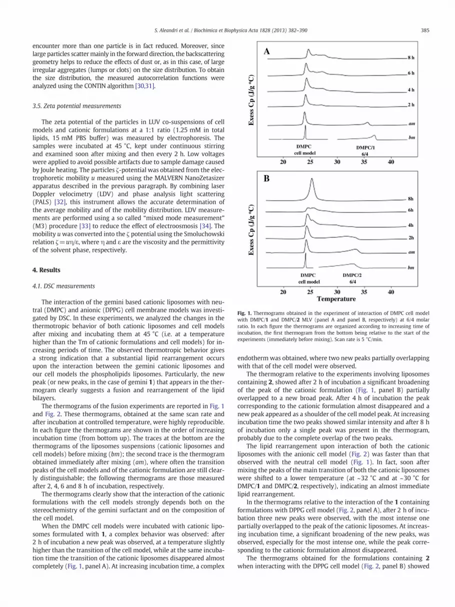
Fig. 1. Thermograms obtained in the experiment of interaction of DMPC cell modelwith DMPC/1 and DMPC/2 MLV (panel A and panel B, respectively) at 6/4 molarratio. In each figure the thermograms are organized according to increasing time ofincubation, the first thermogram from the bottom being relative to the start of theexperiments (immediately before mixing). Scan rate is 5 °C/min.
385S. Aleandri et al. / Biochimica et Biophysica Acta 1828 (2013) 382–390
encounter more than one particle is in fact reduced. Moreover, sincelarge particles scattermainly in the forward direction, the backscatteringgeometry helps to reduce the effects of dust or, as in this case, of largeirregular aggregates (lumps or clots) on the size distribution. To obtainthe size distribution, the measured autocorrelation functions wereanalyzed using the CONTIN algorithm [30,31].
3.5. Zeta potential measurements
The zeta potential of the particles in LUV co-suspensions of cellmodels and cationic formulations at a 1:1 ratio (1.25 mM in totallipids, 15 mM PBS buffer) was measured by electrophoresis. Thesamples were incubated at 45 °C, kept under continuous stirringand examined soon after mixing and then every 2 h. Low voltageswere applied to avoid possible artifacts due to sample damage causedby Joule heating. The particles ζ-potential was obtained from the elec-trophoretic mobility u measured using the MALVERN NanoZetasizerapparatus described in the previous paragraph. By combining laserDoppler velocimetry (LDV) and phase analysis light scattering(PALS) [32], this instrument allows the accurate determination ofthe average mobility and of the mobility distribution. LDV measure-ments are performed using a so called “mixed mode measurement”(M3) procedure [33] to reduce the effect of electroosmosis [34]. Themobility uwas converted into the ζ potential using the Smoluchowskirelation ζ=uη/ε, where η and ε are the viscosity and the permittivityof the solvent phase, respectively.
4. Results
4.1. DSC measurements
The interaction of the gemini based cationic liposomes with neu-tral (DMPC) and anionic (DPPG) cell membrane models was investi-gated by DSC. In these experiments, we analyzed the changes in thethermotropic behavior of both cationic liposomes and cell modelsafter mixing and incubating them at 45 °C (i.e. at a temperaturehigher than the Tm of cationic formulations and cell models) for in-creasing periods of time. The observed thermotropic behavior givesa strong indication that a substantial lipid rearrangement occursupon the interaction between the gemini cationic liposomes andour cell models the phospholipids liposomes. Particularly, the newpeak (or new peaks, in the case of gemini 1) that appears in the ther-mogram clearly suggests a fusion and rearrangement of the lipidbilayers.
The thermograms of the fusion experiments are reported in Fig. 1and Fig. 2. These thermograms, obtained at the same scan rate andafter incubation at controlled temperature, were highly reproducible.In each figure the thermograms are shown in the order of increasingincubation time (from bottom up). The traces at the bottom are thethermograms of the liposomes suspensions (cationic liposomes andcell models) before mixing (bm); the second trace is the thermogramobtained immediately after mixing (am), where often the transitionpeaks of the cell models and of the cationic formulation are still clear-ly distinguishable; the following thermograms are those measuredafter 2, 4, 6 and 8 h of incubation, respectively.
The thermograms clearly show that the interaction of the cationicformulations with the cell models strongly depends both on thestereochemistry of the gemini surfactant and on the composition ofthe cell model.
When the DMPC cell models were incubated with cationic lipo-somes formulated with 1, a complex behavior was observed: after2 h of incubation a new peak was observed, at a temperature slightlyhigher than the transition of the cell model, while at the same incuba-tion time the transition of the cationic liposomes disappeared almostcompletely (Fig. 1, panel A). At increasing incubation time, a complex
endotherm was obtained, where two new peaks partially overlappingwith that of the cell model were observed.
The thermogram relative to the experiments involving liposomescontaining 2, showed after 2 h of incubation a significant broadeningof the peak of the cationic formulation (Fig. 1, panel B) partiallyoverlapped to a new broad peak. After 4 h of incubation the peakcorresponding to the cationic formulation almost disappeared and anew peak appeared as a shoulder of the cell model peak. At increasingincubation time the two peaks showed similar intensity and after 8 hof incubation only a single peak was present in the thermogram,probably due to the complete overlap of the two peaks.
The lipid rearrangement upon interaction of both the cationicliposomes with the anionic cell model (Fig. 2) was faster than thatobserved with the neutral cell model (Fig. 1). In fact, soon aftermixing the peaks of the main transition of both the cationic liposomeswere shifted to a lower temperature (at ~32 °C and at ~30 °C forDMPC/1 and DMPC/2, respectively), indicating an almost immediatelipid rearrangement.
In the thermograms relative to the interaction of the 1 containingformulations with DPPG cell model (Fig. 2, panel A), after 2 h of incu-bation three new peaks were observed, with the most intense onepartially overlapped to the peak of the cationic liposomes. At increas-ing incubation time, a significant broadening of the new peaks, wasobserved, especially for the most intense one, while the peak corre-sponding to the cationic formulation almost disappeared.
The thermograms obtained for the formulations containing 2when interacting with the DPPG cell model (Fig. 2, panel B) showed

Fig. 2. Thermograms obtained in the experiment of interaction of DPPG cell model with DMPC/1 and DMPC/2 MLV (panel A and panel B, respectively) at 6/4 molar ratio. In eachfigure the thermograms are organized according to increasing time of incubation, the first thermogram from the bottom being relative to the start of the experiments (immediatelybefore mixing). Scan rate is 5 °C/min.
386 S. Aleandri et al. / Biochimica et Biophysica Acta 1828 (2013) 382–390
a rather different behavior. In fact, after 4 h of incubation, the charac-teristic peaks of both the cationic liposomes and of the DPPG lipo-somes were still clearly observable, and only after 6 h of incubationa peak centered at ~33 °C, and a broad but pronounced peak, tailedon the high temperature side, clearly appeared, indicating the pres-ence of a newly formed lipid mixture. Thus, although both the geminiliposome formulations show a strong interaction with the anionic cellmodel, the kinetics of the interaction and the characteristics of thenewly formed aggregates appear to be rather different for the twogemini stereoisomers, with an apparently faster mixing kineticsobserved for 1.
4.2. Fluorescence measurements
4.2.1. Evaluation of the extent of bilayer fusion by changes in the surfacepotential (HC)
In these experiments, the variation of the surface potential uponthe interaction of the cationic liposomes with the neutral cell modelswas evaluated by exploiting the presence in the cell model bilayer ofHC, a fluorescent probe whose emission features depend on the valueof the surface potential. This method demonstrated to be unsuitableto evaluate the effect of the interaction with the anionic cell model,because in this case the variation of the surface potential is toosmall to be detected by changes in the HC fluorescence at 380 nm.
In a preliminary experiment, we verified that leakages of HCduring the incubation time were negligible, so that the eventuallyobserved variations can be confidently ascribed to a fusion of thebilayers. To evaluate the extent of the fusion, the fluorescence
intensity at 380 nm of HC embedded in a formulation mimicking100% of fusion (DMPC/gemini liposomes at 16/4 molar ratio, seeSection 3.3.1) was measured: the obtained value was then used tonormalize the fluorescence intensity observed for the cationic lipo-some/cell model mixture observed at different incubation times.The results are reported in Table 1. Both the cationic formulationsinteract to a great extent with the DMPC cell model, however in thecase of DMPC/1 liposomes the percentage of observed fusion washigher; this difference being relevant yet after 2 h of incubation.
4.2.2. Evaluation of the extent of the bilayer fusion by FRET experimentsChanges in FRET efficiency are commonly used for a quantitative
evaluation of the fusion process based on the F/F0 ratio as describedin the experimental section.
In our case, as a result of the interaction of the gemini liposomeformulations with the cell models containing the two chromophores,the lipid mixing causes a significant decrease of their FRET efficiency.The results are reported in Table 1. The interaction of both the cation-ic formulations with the two different cell models was very fast(after 2 h of incubation all samples showed a percentage of fusionhigher than 70%). After 8 h of incubation DMPC/1 liposomes showeda higher tendency to fuse with DMPC cell model compared to DMPC/2liposomes, whereas in the case of DPPG cell model both cationicformulations fused almost completely (95%).
4.2.3. Evaluation of liposome leakageSince the fusion of liposome bilayers can involve leakage from the
internal aqueous pool of liposomes, we investigated on LUV the effect
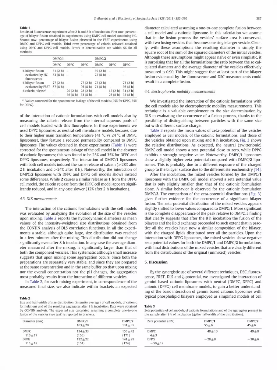
Table 1Results of fluorescence experiment after 2 h and 8 h of incubation. First row: percent-age of bilayer fusion obtained in experiments using DMPC cell model containing HC.Second row: percentage of bilayer fusion observed in the FRET experiments usingDMPC and DPPG cell models. Third row: percentage of calcein released obtainedusing DPPC and DPPG cell models. Errors in determination are within 5% for allmethods.
DMPC/1 DMPC/2
DMPC DPPC DPPG DMPC DPPC DPPG
% bilayer fusionevaluated by HCfluorescence
51 (2 h)83 (8 h)
–
–
–
–
39 (2 h)72 (8 h)
–
–
–
–
% bilayer fusionevaluated by FRET
77 (2 h)87 (8 h)
–
–
77 (2 h)95 (8 h)
72 (2 h)74 (8 h)
–
–
73 (2 h)95 (8 h)
% calcein releasea –
–
29 (2 h)36 (8 h)
28 (2 h)33 (8 h)
–
–
12 (2 h)25 (8 h)
31 (2 h)33 (8 h)
a Values corrected for the spontaneous leakage of the cell models (25% for DPPC, 35%for DPPG).
387S. Aleandri et al. / Biochimica et Biophysica Acta 1828 (2013) 382–390
of the interaction of cationic formulations with cell models also bymeasuring the calcein release from the internal aqueous pools ofcell models loaded with this fluorophore. In these experiments weused DPPC liposomes as neutral cell membrane models because, dueto their higher main transition temperature (41 °C vs 24 °C of DMPCliposomes), they feature a reduced permeability compared to DMPCliposomes. The values obtained in these experiments (Table 1) werecorrected for the spontaneous leakage of the cell model in the absenceof cationic liposomes; this was found to be 25% and 35% for DPPC andDPPG liposomes, respectively. The interaction of DMPC/1 liposomeswith both cell models induced the same release of calcein (>28% after2 h incubation and >34% after 8 h). Noteworthy, the interaction ofDMPC/2 liposomes with DPPC and DPPG cell models shows insteadsome differences. While 2 causes a similar release as 1 from the DPPGcell model, the calcein release from the DPPC cell model appears signif-icantly reduced, and in any case slower (12% after 2 h incubation).
4.3. DLS measurements
The interaction of the cationic formulations with the cell modelswas evaluated by analyzing the evolution of the size of the vesiclesupon mixing. Table 2 reports the hydrodynamic diameters as meanvalues of the intensity-weighted size distributions obtained fromthe CONTIN analysis of DLS correlation functions. In all the experi-ments a stable, although quite large, size distribution was reachedin a few minutes after the mixing. This distribution did not changesignificantly even after 8 h incubation. In any case the average diam-eter measured after the mixing, is significantly larger than that ofboth the component vesicles. This systematic although small increasesuggests that upon mixing some aggregation occurs. Since both thepreparations are separately very stable, and since they are preparedat the same concentration and in the same buffer, so that uponmixingnor the overall concentration nor the pH changes, the aggregationmost probably results from the interaction of different vesicles.
In Table 2, for each mixing experiment, in correspondence of themeasured final size, we also indicate within brackets an expected
Table 2Size and half-width of size distribution (intensity average) of cell models, of cationicformulations and of the resulting aggregates after 8 h incubation. Data were obtainedby CONTIN analysis. The expected size calculated assuming a complete one-to-onefusion of the vesicles (see text) is reported in brackets.
Diameter (nm) DMPC/1103±20
DMPC/2131±35
DMPC110±17
134±33(150)
155±42(171)
DPPG115±18
132±22(154)
141±29(174)
diameter calculated assuming a one-to-one complete fusion betweena cell model and a cationic liposome. In this calculation we assumethat in the fusion process the vesicles' surface area is conserved,with two fusing vesicles that becomes one single larger vesicle. Clear-ly, with these assumptions the resulting diameter is simply thesquare root of the sum of the squared diameters of the initial vesicles.Although these assumptions might appear naïve or even simplistic, itis surprising that for all the formulations the ratio between the so cal-culated diameter and the average diameter of the vesicles effectivelymeasured is 0.90. This might suggest that at least part of the bilayerfusion evidenced by the fluorescence and DSC measurements couldresult in a complete fusion.
4.4. Electrophoretic mobility measurements
We investigated the interaction of the cationic formulations withthe cell models also by electrophoretic mobility measurements. Thistechnique is a valuable complement to the size determination byDLS in evaluating the occurrence of a fusion process, thanks to thepossibility of distinguishing between particles with the same sizebut with different surface charge.
Table 3 reports the mean values of zeta-potential of the vesiclesemployed as cell models, of the cationic formulations, and those ofthe vesicles obtained upon mixing and 8 h incubation, Fig. 3 showsthe relative distributions. As expected, the neutral (zwitterionic)DMPC cell model shows a zeta potential close to zero, while DPPGexhibits a strongly negative value. Noteworthy, DMPC/1 liposomesshow a slightly higher zeta potential compared with DMPC/2 lipo-somes. This is probably due to a different exposure of the chargedgroup to the bilayer surface due to the different stereochemistry [14].
After the incubation, the mixed vesicles formed by the DMPC/1liposomes with the DMPC cell model showed a zeta potential valuethat is only slightly smaller than that of the cationic formulationalone. A similar behavior is observed for the cationic formulationDMPC/2. The comparison of the zeta-potential distributions (Fig. 3)gives further evidence for the occurrence of a significant bilayerfusion. The zeta-potential distribution of the mixed vesicles appearsslightly shifted to lower values compared to DMPC/1. More significantis the complete disappearance of the peak relative to DMPC, a findingthat clearly suggests that after the 8 h incubation the fusion of thebilayers and the lipid exchange proceeded to such extent that in prac-tice all the vesicles have now a similar composition of the bilayer,with the charged lipids distributed over all the particles. Upon theinteraction with DPPG liposomes, the mixed vesicles show negativezeta potential values for both the DMPC/1 and DMPC/2 formulations,with final distributions of the mixed vesicles that are clearly differentfrom the distributions of the original (unmixed) vesicles.
5. Discussion
By the synergistic use of several different techniques, DSC, fluores-cence, FRET, DLS and ζ-potential, we investigated the interaction ofgemini based cationic liposomes with neutral (DMPC, DPPC) andanionic (DPPG) cell membrane models, to gain a better understand-ing of the basic interaction of gemini based cationic liposomes withtypical phospholipid bilayers employed as simplified models of cell
Table 3Zeta potentials of cell models, of cationic formulations and of the aggregates present inthe sample after 8 h of incubation (±the half-width of the distribution).
Zeta potential (mV) DMPC/155±6
DMPC/245±6
DMPC4±3
48±10 49±8
DPPG−50±12
−28±8 −30±6

Fig. 3. Zeta potential distributions of the cell models (dashed line) and of the cationic formulations (dotted line) before the mixing, and of the single populations (continuous line)that appear shortly after mixing. Here the “mixed” distributions are those measured after 8 h incubation.
388 S. Aleandri et al. / Biochimica et Biophysica Acta 1828 (2013) 382–390
membranes. As we will show in details, this complex experimentalapproach allowed us to point out the key role of the stereochemistryof the gemini cationic component of the liposomes formulations insuch interaction. In fact, while our findings clearly suggest that inany case as a result of the gemini-liposome/cell-model interactionthere is a significant exchange of lipids between the bilayers, thereis also significant evidence that such exchange is quantitatively differ-ent for the two gemini stereoisomers.
The finding that gemini-based cationic liposomes show fusogenicproperties is interesting.
Even long-term contacts between protein-free liposomes mimick-ing the compositions of biological membranes usually do not result infusion. Actually, the propensity of lipid bilayer to hemifuse usuallydepends on the presence of “fusogenic” lipids, i.e. lipids that show apacking parameter [35]
Pp ¼ v=lA > 1
(here v is the volume of the hydrophobic tail, l its length and A is thearea of the cross section of the hydrophilic headgroup). A value ofPp>1, meaning that the hydrophobic part is somehow “larger” thanthe hydrophilic one, increases the propensity of these lipids to formstructures with a “negative” spontaneous curvature (the curvatureis considered negative when the surface bulges in the direction ofthe hydrophobic tails). The presence of such “negative curvature”lipids is considered a prerequisite for the formation of a “hemifusionstalk” where the proximal leaflets of two bilayer in close contactbegin to fuse (while the distal ones remain unfused) [16]. In fact, inthe stalk the curvature must be negative.
While in general charged liposomes are considered “non-fusogenic”due to the strong electrostatic repulsion between their head that favors
a positive curvature, there are several evidences that cationic gemini, asthe length of the spacer decreases and at relatively low degree ofprotonation tend to form inverted structures (Pp>1) [36,37]. In differ-ent conditions, they form wormlike micelles, ribbons, inverted hexago-nal phases [38] and more complex aggregates of elongated vesicles,where the clustering could be also due to the embedding of the twotails in bilayers of different vesicles [22]. This last tendency could alsobe reconnected to the fact that for an “activated collision” that resultin the formation of a stalk, a local “defect” in the bilayer organizationis needed, i.e. a lipid that protruding from the bilayer, increases theprobability of fusion in the collision [15]. Such “pointlike protrusions”favor the bilayer fusion [17] both by decreasing the distance to a nearbybilayer and by decreasing the hydrophobic energy of the monolayerrupture.
Due to the different requirements of the experimental techniques,DSC measurements were carried out on highly concentrated MLVs,while fluorescence and DLS measurements were performed on morediluted LUV samples. Nevertheless, data from the different experi-ments gave complementary information and are perfectly consistentin pointing out an extensive bilayer fusion and lipid exchange. Theextent of the fusion (without indications on the organization of thebilayer) between the cell models and the cationic formulations wasevaluated i) by analyzing the thermotropic behavior of the mixturesby DSC; ii) by following the variation of the surface potential of theneutral cell model containing HC; iii) by monitoring (by FRET exper-iments) the lateral diffusion of the fluorescent probes included in thecell models; iv) by measuring the leakage of calcein; and v) by mon-itoring the changes of the vesicle size (hydrodynamic radius by DLS)and ζ-potential (electrophoretic mobility). Since the fusion is usuallyaccompanied by leakage of the internal aqueous phase, the “calcein”experiment gives only an indirect estimation of the extent of fusion.

389S. Aleandri et al. / Biochimica et Biophysica Acta 1828 (2013) 382–390
In any case, the results obtained by all these different methodsand techniques pointed out consistently to a significant lipid-transfer between the gemini-based cationic liposomes and thecell models.
Particularly, DSC results show for both cationic formulations ahigh extent of lipid rearrangement upon interaction with both thecell models. The kinetics of lipid rearrangement is significantly fasterfor the liposomes formulated with the gemini 1 compared to 2,independently of the composition of the cell models and of theirsurface charge (neutral or anionic). However, the thermograms at8 h in the experiments with DMPC/1 liposomes show amore complexbehavior than that of DMPC/2 liposomes, in fact two new peaks,partially overlapping with that of the cell model, are observed. Inother words after a faster exchange, gemini 1 seems to experimentgreater difficulties than gemini 2 in diffusing and rearrange withinthe bilayer. This finding suggests a better miscibility of DMPC/2liposomes with the cell models.
This significantly different thermotropic behavior points out theimportant role of the stereochemistry of the gemini component inthe mode of lipid rearrangement upon the interaction of the cationicliposomes with phospholipid bilayers.
The DSC results are well consistent with the fluorescence experi-ments on the neutral cell model.
In fact, both the HC and FRET fluorescence experiments showa slower and minor extent of lipid rearrangement upon theinteraction of the neutral cell model containing the fluorescentprobes with DMPC/2 liposomes compared to DMPC/1 liposomes.Also the experiments of calcein release confirm a different kineticsof interaction.
On the other hand, in all the fluorescence experiments the differ-ences between the two gemini cationic formulations were negligiblewhen they interact with the anionic (DPPG) cell model. The reason ofsuch different behavior is unclear, but it is probably to be connectedwith the presence of the electrostatic interactions between geminiand DPPG. The presence of these interactions, being in general muchstronger and long ranged than sterical interactions, tends in fact toattenuate the differences between stereoisomers. The size of the aggre-gates formed by the interaction of the cell models with the DMPC/1liposomes did not show any dependence on the composition of thecell model. Conversely, the size of the aggregates formed by the interac-tion with the DMPC/2 liposomes depended on cell model, the largersize, and the larger polydispersity, being observed upon mixing withthe neutral cell model.
As expected on the basis of the surface potential values reportedpreviously [8], DMPC/2 liposomes feature lower zeta potential com-pared with DMPC/1 liposomes, probably because of a different expo-sure of the cationic head group of the gemini surfactant [18], and thisfeature could be, among others, responsible of the different kinetic,extent and mode of lipid rearrangement. However, the systemsformed by the two gemini upon the interaction with both the cellmodels feature the same zeta potential (≈50 mV and ≈−30 mVupon interaction with neutral and anionic cell model, respectively),thus suggesting that after fusion the mode of exposure of the cationichead groups is similar for the two gemini components.
Summarizing, the extent, the kinetic and the mode of interac-tion of gemini cationic liposomes with cell models were shownto depend on the cell model and on the stereochemistry of thegemini. In particular, DMPC/2 liposomes showed a significantlyslower lipid rearrangement, possibly due to a different exposureof the cationic gemini head group, though accompanied by ahigher lipid miscibility with both cell models when compared toDMPC/1 liposomes.
These evidences could explain the different biological behaviors ofthe cationic formulation i.e. different uptake and intracellular distri-butions of the delivered drug that are controlled by the interactionwith specific compartments of the cell membrane.
6. Conclusions
DSC, fluorescence, DLS and electrophoresis experiments were car-ried out to evaluate how and to what extent the stereochemistry ofthe two gemini surfactants included in liposome formulations affecttheir ability of interacting and fusing with neutral and anionic mem-brane models.
DMPC/2 liposomes, that feature the highest efficacy in drug deliv-ery [8], showed a lower zeta potential and, upon the interactionwith both the neutral and anionic cell model, a slower rate of lipidrearrangement and a higher miscibility compared to the correspond-ing formulation containing 1; the stereochemistry of the gemini, byaffecting the exposure to water of the cationic head group of the sur-factant, controls some physicochemical properties of liposomes (zetapotential, bilayer organization) important for their interaction withlipid bilayers.
Obviously, cell membranes are very complex lipid bilayers, theyare composed by hundreds of lipids that can interact differentlywith liposome bilayers and are organized in domains with specificcomposition (rafts); furthermore cell membrane contains many dif-ferent receptors, that could play a fundamental role in the interactionwith liposomes and in their internalization. On the other hand, themodels used are very simple and can mimic a region of cell mem-brane featuring a specific composition and/or surface charge.
The results of this investigation clearly show that the surfacecharge of liposomes (in this case controlled by the different stereo-chemistry of gemini component), cell membrane composition andlipid miscibility might control the uptake and the biodistribution ofthe drug delivered by liposomes.
References
[1] G. Poste, D. Papahadjopoulos, The influence of vesicle membrane properties onthe interaction of lipid vesicles with cultured cells, Ann. N. Y. Acad. Sci. 308(1978) 164–184.
[2] A. Chonn, P.R. Cullis, Recent advances in liposomal drug-delivery systems, Curr.Opin. Biotechnol. 6 (1995) 698–708.
[3] C. Bombelli, F. Faggioli, P. Luciani, G. Mancini, M.G. Sacco, Efficient transfection ofDNA by liposomes formulated with cationic gemini amphipiles, J. Med. Chem. 48(2005) 5378–5382.
[4] C. Bombelli, S. Borocci, M. Diociaiuti, F. Faggioli, L. Galantini, P. Luciani, G. Mancini,M.G. Sacco, Role of the spacer of cationic gemini amphiphiles in the condensationof DNA, Langmuir 21 (2005) 10271–10274.
[5] C. Bombelli, G. Caracciolo, P. Di Profio, M. Diociaiuti, P. Luciani, G. Mancini, C.Mazzuca, M. Marra, A. Molinari, D. Monti, L. Toccacieli, M. Venanzi, Inclusion ofa photosensitizer in liposomes formed by DMPC/gemini surfactant: correlationbetween physico-chemical and biological features of the complexes, J. Med.Chem. 48 (2005) 4882–4891.
[6] A. Molinari, M. Colone, A. Calcabrini, A. Stringaro, L. Toccacieli, G. Arancia, S.Mannino, A. Mangiola, G. Maira, C. Bombelli, G. Mancini, Cationic liposomes,loaded with m-THPC, in photodynamic therapy for malignant glioma, Toxicol.In Vitro 21 (2007) 230–234.
[7] A. Molinari, C. Bombelli, S. Mannino, A. Stringaro, L. Toccacieli, A. Calcabrini, M.Colone, A. Mangiola, G. Maira, P. Luciani, G. Mancini, G. Arancia, m-THPC-mediatedphotodynamic therapy of malignant gliomas: assessment of a new transfectionstrategy, Int. J. Cancer 121 (2007) 1149–1155.
[8] C. Bombelli, A. Stringaro, S. Borocci, G. Bozzuto, M. Colone, L. Giansanti, R.Sgambato, L. Toccaceli, G. Mancini, A. Molinari, Efficiency of liposomes in thedelivery of a photosensitizer controlled by the stereochemistry of a geminisurfactant component, Mol. Pharm. 7 (1) (2010) 130–137.
[9] C.R. Safinya, K. Ewert, A. Ahmad, H.M. Evans, U. Raviv, D.J. Needleman, A.J. Lin, N.L.Slack, C. George, C.E. Samuel, Cationic liposome-DNA complexes: from liquid crystalscience to gene delivery applications, Philos. Trans. R. Soc. A 364 (2006) 2573–2596.
[10] F. Bordi, C. Cametti, S. Sennato, M. Diociaiuti, Direct evidence of multicompartmentaggregates in polyelectrolyte-charged liposome complexes, Biophys. J. 91 (2006)1513–1520.
[11] S. Sennato, F. Bordi, C. Cametti, M. Diociaiuti, P. Malaspina, Charge patch attrac-tion and reentrant condensation in DNA-liposome complexes, Biochim. Biophys.Acta 1714 (2005) 11–24.
[12] A.J. Lin, N.L. Slack, A. Ahmad, C.X. George, C.E. Samuel, C.R. Safinya, Three-dimensional imaging of lipid gene-carriers: membrane charge density controlsuniversal transfection behavior in lamellar cationic liosome-DNA complexes,Biophys. J. 84 (2003) 3307–3316.
[13] B.R. Lentz, V. Malinin, M. Emdadul Haque, K. Evans, Protein machines and lipidassemblies: current views of cell membrane fusion, Curr. Opin. Struct. Biol. 10(2000) 607–615.

390 S. Aleandri et al. / Biochimica et Biophysica Acta 1828 (2013) 382–390
[14] W. Wickner, R. Schekman, Membrane fusion, Nat. Struct. Mol. Biol. 15 (2008)658–664.
[15] S. Lev, Non-vesicular lipid transport by lipid-transfer proteins and beyond, Nat.Rev. Mol. Cell Biol. 11 (2010) 739–750.
[16] L.V. Chernomordik, M.M. Kozlov, Mechanics of membrane fusion, Nat. Struct. Mol.Biol. 15 (2008) 675–683.
[17] L.V. Chernomordik, M.M. Kozlov, Protein–lipid interplay in fusion and fission ofbiological membranes, Annu. Rev. Biochem. 72 (2003) 175–207.
[18] C. Bello, C. Bombelli, S. Borocci, P. di Profio, G. Mancini, Role of the spacer stereo-chemistry on the aggregation properties of cationic gemini surfactants, Langmuir22 (2006) 9333–9338.
[19] D.K. Struck, D. Hoekstra, R.E. Pagano, Use of resonance energy transfer to monitormembrane fusion, Biochemistry 20 (1981) 4093–4099.
[20] N. Düzgünes, H. Faneca, M.C. Pedroso de Lima, Methods to monitor liposomefusion, permeability, and interaction with cells, in: Volkmar Weissig (Ed.),Liposomes: Methods and Protocols, Volume 2: Biological Membrane Models,Methods Mol. Biol., vol. 606, Springer, 2001, pp. 209–232.
[21] A.A. Yaroslavov, A.V. Sybachin, E. Kesselman, J. Schmidt, Y. Talmon, S.A.A. Rizvi,F.M. Menger, Liposome fusion rates depend upon the conformation of polycationcatalysts, JACS 133 (2011) 2881–2883.
[22] S. Aleandri, M.G. Bonicelli, F. Bordi, S. Casciardi, M. Diociaiuti, L. Giansanti, F.Leonelli, G. Mancini, G. Perrone, S. Sennato, How stereochemistry affects thephysicochemical features of gemini surfactant based cationic liposomes, SoftMatter 8 (2012) 5904–5915.
[23] R.L. Biltonen, D. Lichtenberg, The use of differential scanning calorimetry as a toolto characterize liposome preparations, Chem. Phys. Lipids 64 (1993) 129–142.
[24] T. Förster, Zwischenmolekulare Energiewanderung und Fluoreszenz, Ann. Phys.437 (1948) 55–75.
[25] J.R. Lakowicz, Principles of Fluorescence Spectroscopy, 3rd ed. Springer, 2006.[26] G. Van Meer, K. Simons, An efficient method for introducing defined lipids into
the plasma membrane of mammalian cells, J. Cell Biol. 97 (1983) 1365–1374.
[27] A. Memoli, L.G. Palermiti, V. Travagli, F. Alhaique, Effects of surfactants on thespectral behaviour of calcein (II): a method of evaluation, J. Pharm. Biomed.Anal. 19 (1999) 627–632.
[28] A. Andersson, J. Danielsson, A. Graslund, L. Maler, Kinetic models for peptide-induced leakage from vesicles and cells, Eur. Biophys. J. 36 (2007) 621–635.
[29] H.S. Dhadwal, R.R. Ansari, W.V. Meyer, A fiber-optic probe for particle sizing inconcentrated suspensions, Rev. Sci. Instrum. 62 (1991) 2963–2968.
[30] S. Provencher, Contin: a general purpose constrained regularization program forinverting noisy linear algebraic and integral equations, Comput. Phys. Commun.27 (1982) 229–242.
[31] S. Provencher, Constrained regularization method for inverting data representedby linear algebraic or integral equations, Comput. Phys. Commun. 27 (1982)213–227.
[32] W.W. Tscharnuter, Mobility measurements by phase analysis, Appl. Optics 40(2001) 3995–4003.
[33] M.T. Connah,M. Kaszuba, A.Morfesis, High resolution zeta potential measurements:analysis of multi-component mixtures, J. Dispersion Sci. Technol. 23 (2002)663–669.
[34] M. Minor, A.J. van der Linde, H.P. van Leeuwen, J. Lyklema, Dynamic aspects ofelectrophoresis and electroosmosis: a new fast method for measuring particlemobilities, J. Colloid Interface Sci. 189 (1997) 370–375.
[35] J.N. Israelachvili, Intermolecular and Surface Forces, 3rd edition Academic Press, 2011.[36] J.E. Klijn, M.C.A. Stuart, M. Scarzello, A. Wagenaar, J.B.F.N. Engberts, pH-Dependent
Phase Behaviour of Carbohydrate-Based Gemini Surfactants. Effect of the Lengthof the Hydrophobic Spacer, J. Phys. Chem B 110 (2007) 5204–5211.
[37] J.E. Klijn, M.C.A. Stuart, M. Scarzello, A. Wagenaar, J.B.F.N. Engberts, pH-dependentphase behavior of carbohydrate-based gemini surfactants. The effects of carbohy-drate stereochemistry, head group hydrophilicity, and nature of the spacer, J. Phys.Chem. B 111 (2007) 5204–5211.
[38] R. Oda, Ivan Huc, J.-C. Homo, B. Heinrich, M. Schmutz, S. Candau, Elongated aggre-gates formed by cationic gemini surfactants, Langmuir 15 (1999) 2384–2390.


