Embed Size (px)
Citation preview

Author Pro
of
Review
10.1517/17425247.2.2.xxx © 2005 Ashley Publications Ltd ISSN 1742-5247 1
Ashley Publicationswww.ashley-pub.com
Cationic liposomes for gene deliverySérgio Simões†, Ana Filipe, Henrique Faneca, Miguel Mano, Nuno Penacho, Nejat Düzgünes & Maria Pedroso de Lima†University of Coimbra, Laboratory of Pharmaceutical Technology, Faculty of Pharmacy, Coimbra, Portugal
Cationic liposome-DNA complexes (lipoplexes) constitute a potentially viablealternative to viral vectors for the delivery of therapeutic genes. This reviewwill focus on various parameters governing lipoplex biological activity, fromtheir mode of formation to in vivo behaviour. Particular emphasis is given tothe mechanism of interaction of lipoplexes with cells, in an attempt to dissectthe different barriers that need to be surpassed for efficient gene expressionto occur. Aspects related to new trends in the formulation of lipid-basedgene delivery systems aiming at overcoming some of their limitations will becovered. Finally, examples illustrating the potential of cationic liposomes inclinical applications will be provided.
Keywords: biological barriers, clinical trials, gene therapy, lipoplexes, nonviral vectors
Expert Opin. Drug Deliv. (2005) 2(2):xxx-xxx
1. Introduction
The efficient gene delivery into target cells is a critical issue for the success of genetherapy approaches. The drawbacks associated with the use of viral vectors,namely those related with safety problems, have prompted investigators to developalternative methods for gene delivery, cationic lipid-based systems (lipoplexes)being the most representative. However, some disadvantages including limitedefficiency of delivery and gene expression, toxicity at higher concentrations,potentially adverse interactions with negatively charged macromolecules in serumand on cell surfaces, and impaired ability to reach tissues beyond the vasculatureunless directly injected into the tissue, represent restrictions to their widesuccessful application.
Despite extensive research in the last decade, which led to the development of ele-gant strategies to enhance lipoplex biological activity, these systems are still far frombeing viable alternatives to the use of viral vectors in gene therapy. Basic knowledgeof the structure–activity relationships of lipoplexes and of the mechanisms involvedin the process of intracellular gene delivery is still scarce. It is believed that suchknowledge is crucial to further improve the biological performance of these systems;therefore, gaining insights into these mechanistic aspects should constitute one ofthe main goals in this field. Table 1 summarises the main factors and relatedproperties affecting the biological activity of the lipoplexes.
2 Factors governing the biological activity of lipoplexes: complex formation and related properties
It is well recognised that the mode of formation of the complexes strongly deter-mines the final physicochemical features of the lipoplexes and, consequently,modulates their biological activity. In addition to the morphology of the lipo-plexes, other properties, including size, charge density, colloidal stability and theability to protect DNA, are strongly dependent on the mode of lipoplex forma-tion. Therefore, understanding the parameters that modulate such properties is of
1. Introduction
2 Factors governing the biological
activity of lipoplexes: complex
formation and related
properties
3 Mechanisms of interaction of
cationic liposome–DNA
complexes with cells
4. New trends in lipid-based gene
delivery systems
5. Perspectives for clinical
application of cationic
liposomes as gene carriers
6. Expert opinion and conclusion

Author Pro
of
Cationic liposomes for gene delivery
2 Expert Opin. Drug Deliv. (2005) 2(2)
Table 1. Factors influencing the biological activity of lipoplexes.
Main stages and related properties Affecting factors
Mode of lipoplex formation- morphology and structure
Nature of cationic and helper lipid, stoichiometry of cationic lipid and DNA, nature of the medium (ionic strength, pH and temperature), DNA structure
Control of physicochemical properties- size- net charge- colloidal stability
Mode of lipoplex preparation (type of liposomes, DNA structure, order of addition and rate of mixing, lipid and DNA concentration), role of adjuvants (polycations, surfactants, cryoprotectants)
In vitro and in vivo performance- stability in the presence of serum- resistance to DNA nuclease degradation
Route of administration, nonspecific interaction with serum components, interaction with blood cells, net charge of the lipoplexes, nature of the colipid (cholesterol versus dioleoylphosphatidylethanolamine)
-pharmacokinetics/biodistribution Interaction with serum components, opsonisation, prolonged circulation time (inclusion of poly[ethyleneglycol])
-passive versus active targeting-surpass the endothelial barrier-transfection efficacy-cytotoxicity
Size, use of ligands and antibodiesUnknownLevel and duration of gene expressionType and concentration of lipid, type of cell
Lipoplex–cell interaction- mode of cellular internalisation
Size, liposome composition, net charge and topology of the complexes, presence of ligands
-escape from endosomes Nature of helper lipid, use of endosome disrupting agents
-DNA dissociation from the complex Net charge of the complex, nature and valency of the cationic lipid. Cellular factors unknown
-trafficking of DNA into the nucleus Degree of DNA condensation/compaction, protection from nucleases, size of the plasmid, targeting to the nucleus (nuclear localisation signal peptides). Cellular factors unknown
crucial importance for the successful application of lipo-plexes both in vitro and in vivo. Among such parameters, thenature of cationic and ‘helper’ lipids, the stoichiometry ofcationic lipid and DNA, the DNA structure, the mode oflipoplex preparation and the nature of the medium in whichthey are prepared are considered the most relevant.
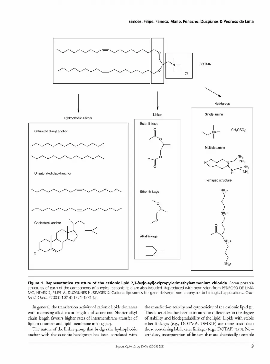
2.1 Nature of the cationic liposome componentsCationic liposomes are frequently composed of a cationiclipid and a neutrally charged lipid (colipid). Cationic lipidsinclude a group of amphiphiles that exhibit a positive charge,which triggers their interaction with negatively charged DNAleading to the formation of complexes containing condensedDNA. Since the first description by Felgner et al. [1] of thepotential of such types of lipids (2,3-bis[oleyl]oxipropyl-trimethylammonium chloride [DOTMA]) for transfection,an increasing number of new cationic lipids of different struc-tures have been synthesised and their respective transfectionactivities in a wide variety of cell types have been reported.Cationic lipids are frequently composed of a positivelycharged headgroup bridged by a linker group to a hydropho-bic lipid anchor, with a specific role being attributed to eachof these components. Figure 1 shows the structure ofDOTMA and illustrates some possible structures of each ofthe components of a typical cationic lipid [2].
The headgroup is composed of either single or multipleprotonatable amines. Multivalent headgroups (e.g., 2,3
dioleyloxy-N-[2[sperminecarboxaminino]ethyl]-N,N-dime-thyl-1-propanaminium trifluroacetate [DOSPA] and diocta-decyl amino glycyl spermine [DOGS]) [3] are more efficient incondensing DNA and more active than monovalent lipids(e.g., DOTMA, 1,2-dioleoyl-3-trimethylammonium pro-pane [DOTAP], 3 β[N-[N′,N′-dimethyl amino ethane]-car-bamoyl]cholesterol [DC-Chol] and 1,2-dimyristyloxypropyl-3-dimethyl-hydroxy ethyl ammonium bromide [DMRIE])[4], which may be related to the greater ability of the former tocondense and protect DNA. Nevertheless, progressivelyincreasing the number of positive charges may result in aninteraction with DNA that is too strong and would hamperits subsequent dissociation. In addition, multivalent cationiclipids are more prone to form micelles, which may lead to theformation of less stable and more toxic complexes. The orien-tation of the headgroup in relation to the backbone was alsoshown to be an important aspect affecting structure–activity.Linear structures (formed on coupling of a primary amine tothe lipid anchor) were shown to mediate lower levels of trans-fection as compared with T-shaped structures (in which asecondary amine is coupled to the lipid anchor) [5].
The hydrophobic moiety is composed of either a diacylbackbone or a cholesterol anchor. Although both groups havebeen extensively tested, cationic lipids containing cholesterolwere shown to be more active than those containing diacylchains, with this effect being more obvious for lipids with theT-shaped headgroups [5].
Author Pro
of
Simões, Filipe, Faneca, Mano, Penacho, Düzgünes & Pedroso de Lima
Expert Opin. Drug Deliv. (2005) 2(2) 3
In general, the transfection activity of cationic lipids decreaseswith increasing alkyl chain length and saturation. Shorter alkylchain length favours higher rates of intermembrane transfer oflipid monomers and lipid membrane mixing [6,7].
The nature of the linker group that bridges the hydrophobicanchor with the cationic headgroup has been correlated with
the transfection activity and cytotoxicity of the cationic lipid [5].This latter effect has been attributed to differences in the degreeof stability and biodegradability of the lipid. Lipids with stableether linkages (e.g., DOTMA, DMRIE) are more toxic thanthose containing labile ester linkages (e.g., DOTAP) [6,8,9]. Nev-ertheless, incorporation of linkers that are chemically unstable
O
O
N+
Cl-
Saturated diacyl anchor
Unsaturated diacyl anchor
Cholesterol anchor
X
Single amine
N+CH3OSO3
-
Multiple amine
N NH
NH
NH2
NH2
NH2
NH2
T-shaped structure
NH2+
NH3+
NH2+
NH3+
O
DOTMA
Hydrophobic anchor Linker
Headgroup
Ester linkage
O
O
O
O
Ether llinkage
O
O
Alkyl linkage
Figure 1. Representative structure of the cationic lipid 2,3-bis[oleyl]oxipropyl-trimethylammonium chloride. Some possiblestructures of each of the components of a typical cationic lipid are also included. Reproduced with permission from PEDROSO DE LIMAMC, NEVES S, FILIPE A, DUZGUNES N, SIMOES S: Cationic liposomes for gene delivery: from biophysics to biological applications. Curr.Med. Chem. (2003) 10(14):1221-1231 [2].

Author Pro
of
Cationic liposomes for gene delivery
4 Expert Opin. Drug Deliv. (2005) 2(2)
can limit their application, as the stability of lipoplexes on stor-age or in biological fluids can be drastically affected. Linkersshould thus be selected so that a balance between stability andbiodegradability in the cell can be achieved.
The importance of associating a colipid to improve theability of cationic liposomes to transfect cells has been dem-onstrated. In vitro studies show clearly that liposomes com-posed of an equimolar mixture ofdioleoylphosphatidylethanolamine (DOPE) and cationic lip-ids (e.g., DOTMA, DOTAP) can mediate higher levels oftransfection than those containing only the cationic lipid or adifferent helper lipid such as DOPC [10-12]. This fact has beenattributed to the ability of DOPE to undergo a transitionfrom a bilayer to an hexagonal configuration under acidic pH,which may facilitate fusion with or destabilisation of targetmembranes, in particular endosomal membranes [6,13,14]. Inaddition, the motional properties of DOPE, in contrast toDOPC, were correlated with the transfection potential ofDOPE-containing complexes [11]. More recently, it was sug-gested that DOPE can also play a role in facilitating the disas-sembling of the lipid-based DNA formulations after theirinternalisation and escape of DNA from endocytic vesicles[15,16]. This was based on the assumption that the aminegroup of polyethylene (PE) can interact with DNA phosphategroups, thus leading to weakening of the binding of cationiclipids to DNA [15]. Although the benefits of using DOPEhave been demonstrated empirically, recent work has shownthat the choice of the helper lipid can dictate the structureand activity of cationic liposome–DNA complexes. Choles-terol has also been employed as a colipid to prepare cationicliposomes, resulting in the formation of more stable com-plexes than those containing DOPE. In contrast to what hasbeen observed in in vitro studies, the inclusion of cholesterolin the bilayer of cationic liposomes resulted in very activecomplexes on in vivo administration [17-22]. Moreover, inclu-sion of cholesterol in the liposome composition enables theuse of increased concentrations of lipid and DNA withoutaffecting lipoplex stability. This, in turn, allows increaseddoses of DNA to be delivered and expressed. On the otherhand, the choice of DOPE as the helper lipid for cationicliposomes was described to result in a decrease of the levels oftransfection in vivo [22]. These findings suggest that the func-tion of the helper lipid in liposomes is different in vivo fromthat in vitro, also supporting the hypothesis that the in vivobehaviour of lipoplexes cannot necessarily be established fromin vitro data. Incorporation of poly(ethylene glycol) phos-pholipid conjugates (PEG–PE) into the liposomal membranehas been explored aiming at improving the colloidal stabilityof lipoplexes both in vitro and in vivo [19,20] and will bediscussed in more detail in section 2.3.
2.2 Stoichiometry of cationic lipid and DNASeveral studies have shown that highly positively chargedcomplexes, in which DNA is completely sequestered and con-densed, exhibit a homogeneous size distribution (mean
diameter of 100 – 450 nm). A similar size distribution is alsoobserved when complexes are prepared with an excess ofDNA over cationic lipids (i.e., negatively charged complexes),although in this case the presence of free DNA is generallyobserved [23-26]. On the other hand, complexes prepared froma lipid–DNA charge ratio of ∼ 1:1 exhibit a neutral zetapotential, suggesting that all the cationic lipid molecules areneutralised by DNA [12,26-28]. Such neutral complexes arecharacterised by a heterogenous size distribution (mean diam-eter of 350 – 1200 nm) and usually present a much lower col-loidal stability than those exhibiting an excess of net positiveor negative charge. This can be attributed to a lack of electro-static repulsive forces among the complexes that would pre-vent their aggregation [6,12,26,29,30]. It should be noted thatsome controversy has been reported regarding the (+/-) chargeratio at which neutral complexes are formed. In fact, the sur-face charge of the lipoplexes (usually assessed by zeta potentialmeasurements) does not always correspond to the theoreti-cally calculated charge ratio. Among other factors, differencesin the mode of lipoplex formation resulting from variations inthe experimental conditions used by different research groups,or differences in the cationic lipid concentration amongbatches, can be responsible for such discrepancy. The influ-ence of lipid–DNA stoichiometry on the physicochemicalproperties of the complexes becomes even more difficult toevaluate considering that, for a fixed lipid–DNA charge ratio,the increase in concentration of lipid and DNA results in asignificant change of their size and colloidal stability, whichcan be attributed to enhanced precipitation at higher concen-trations due to smaller interparticle separation [23]. It seems,therefore, that charge ratio alone is not sufficient to predictthe mode of formation and physicochemical features of thecomplexes and, consequently, their biological activity.
The ability to protect the carried DNA against nucleasedegradation is considered to be a crucial feature affecting thebiological activity of the complexes [31]. Besides size andcharge, the stoichiometry of cationic liposome–DNA com-plexes determines their resistance to the inhibitory effect ofthe nucleases. Negatively charged complexes (prepared withan excess of DNA over cationic lipid) cannot protect DNAefficiently, which can be explained by the susceptibility ofnoncomplexed DNA to nuclease degradation. In contrast,positively charged complexes (containing an excess of positiveover negative charges) are able to fully condense and coatDNA, thus acquiring a high degree of resistance to DNases.Curiously, complexes prepared so that a balance between pos-itive and negative charges is achieved (neutrally charged com-plexes) have been shown to be capable of protecting DNA.The degree of DNA protection obtained for different formu-lations can be assessed by evaluating the accessibility of ethid-ium bromide to the DNA associated with the complexes [32].A more biologically relevant assay to evaluate the protectionconferred by cationic liposomes to DNA degradation is basedon the assessment of DNase I resistance mediated by thecomplexes [32].
Author Pro
of
Simões, Filipe, Faneca, Mano, Penacho, Düzgünes & Pedroso de Lima
Expert Opin. Drug Deliv. (2005) 2(2) 5
2.3 In vivo lipoplex behaviourPositively charged complexes have been described as beingable to completely condense DNA and to mediate the highestlevels of transfection in vivo [12,21,26]. Favourable interactionswith and binding to the cell surface, as well as an efficient pro-tection of the foreign DNA against nucleases, can partiallyexplain these observations [1,4,23]. Surprisingly, it has been rec-ognised that within this type of complex, those exhibitinglarge sizes (> 200 nm) are more effective in mediating trans-fection than small complexes (50 – 100 nm) [23,33,34].Whether these findings are due to more efficient lipoplex–cellinteractions (presumably favoured by a more extensive deposi-tion of the large complexes at the cell surface), to the ability ofcertain size classes of the complexes to trigger cellular internal-isation events (such as phagocytosis), or to the fact that morecopies of the plasmids may be carried in the larger complexes,are still open questions. It was demonstrated recently that, forhighly positively charged complexes, free liposomes coexistwith the cationic liposome–DNA complexes and play animportant role in mediating transfection in vivo, namely onintravenous administration in mice [21]. This enhancing effectwas attributed to an increase of the retention time and effi-cient protection of DNA, and presumably to the ability of thefree liposomes in promoting intracellular gene delivery by thelipoplexes [21,35]. In addition, it was shown that lipoplexes pre-pared at high cationic lipid/DNA (+/-) charge ratios were alsoable to overcome the inhibitory effect of serum on lipofection[33,36]. This resistance to the inhibitory effect of serum ontransfection can also be achieved by prolonging the time ofcomplex formation [37]. Curiously, this time-dependent matu-ration was only observed for monovalent cationic lipids.Moreover, the process of maturation, which was acceleratedby high charge ratios, high concentration and high tempera-ture, resulted in the formation of homogeneous particles witha mean diameter of 170 – 400 nm.
Different approaches have been explored aiming at enhanc-ing the biological activity of lipoplexes and overcoming someof the biological barriers faced by the lipoplexes on theirin vivo administration. Whether coating of lipoplexes withfusogenic peptides or with negatively charged proteins, in thepresence or absence of polycations (including protamine sul-phate) would constitute a promising strategy to modulatetheir colloidal stability and transfection efficacy in the pres-ence of serum has been evaluated. These studies show a signif-icant enhancement of transfection and resistance to thepresence of serum when human serum albumin was associatedto lipoplexes, independently of the composition of the cati-onic liposomes used in their preparation [38,39]. Transferrin–lipoplexes were also found to mediate extensive transfection inthe presence of serum [12].
The use of cationic liposomes modified with the phosphol-ipid derivative of the polymer PEG (PEG–PE) may constitutea promising approach to the development of an efficient phar-maceutical carrier for systemic in vivo gene delivery. Thisstrategy was shown to stabilise cationic liposome–DNA
complexes for prolonged storage while maintaining their bio-logical activity [19], and to result in complexes that are highlyactive in vivo [20]. Meyer et al. [40] have reported that incorpo-ration of PEG–PE into cationic liposomes prevents lipoplexaggregation and increases their stability.
Alternative approaches include insertion of the polymerafter the formation of cationic liposome–DNA complexes [41]
and the use of PEG–lipid conjugates with cleavable bondsthat would allow the shedding of PEG molecules under acidicconditions such as those found in the endosomal lumen. Inthis context, it should be mentioned that a promising strategywas reported involving the efficient entrapment of plasmidDNA into cationic liposomes containing a PEG–ceramideconstruct by employing a detergent dialysis procedure [42-44].
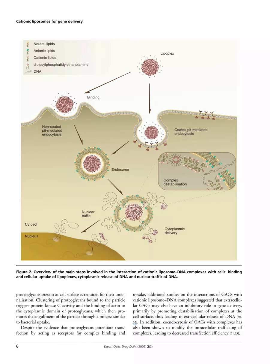
3 Mechanisms of interaction of cationic liposome–DNA complexes with cells
Despite the extensive use of cationic liposomes for gene deliv-ery both in vitro and in vivo, the mechanisms by which DNAis delivered into cells are not yet fully understood.
Studies aiming at clarifying these mechanisms demon-strated that the efficiency of gene delivery mediated by cati-onic liposomes is strongly dependent on the physicochemicalproperties of the lipoplexes, particularly size, morphology andsurface charge, which ultimately determine the processes ofbinding, cellular uptake and intracellular processing. Figure 2illustrates the key steps governing the process of intracellulargene delivery mediated by cationic liposome–DNAcomplexes, which are discussed in this section.
3.1 Cell association/binding of lipoplexesBased on the observation that lipoplexes exhibiting a net posi-tive charge frequently mediate high levels of transfection, a rolefor sulfated membrane-associated proteoglycans in the cellbinding of cationic liposome–DNA complexes has been sug-gested [45]. Sulfated proteoglycans are highly negativelycharged components of cell membranes, and consist of a groupof proteins covalently linked to one or more polysulfatedglicosaminoglycan (GAGs) polysaccharides [46].
Studies involving approaches that lead to the reduction ofcell-associated proteoglycans, or approaches using mutant cellsunable to synthesise proteoglycans, provided evidence that sur-face proteoglycans play a role in the binding of lipoplexes tocells, both in vitro [45] and in vivo [47]. In this context, it is inter-esting to note that, apart from being involved in a variety ofother cellular processes, proteoglycans play an important role inthe binding and entry of many viruses into cells [48], such asherpes simplex virus (HSV), human immunodeficiency virus-1and adeno-associated viruses, all of which have been success-fully used for gene therapy purposes.
Recent work by Kopatz et al. supports the involvement ofproteoglycans in the internalisation of cationic liposome–DNA complexes into cells [49]. According to the proposedmodel, the authors show that binding of cationic particles to
Author Pro
of
Cationic liposomes for gene delivery
6 Expert Opin. Drug Deliv. (2005) 2(2)
proteoglycans present at cell surface is required for their inter-nalisation. Clustering of proteoglycans bound to the particletriggers protein kinase C activity and the binding of actin tothe cytoplasmic domain of proteoglycans, which then pro-motes the engulfment of the particle through a process similarto bacterial uptake.
Despite the evidence that proteoglycans potentiate trans-fection by acting as receptors for complex binding and
uptake, additional studies on the interactions of GAGs withcationic liposome–DNA complexes suggested that extracellu-lar GAGs may also have an inhibitory role in gene delivery,primarily by promoting destabilisation of complexes at thecell surface, thus leading to extracellular release of DNA [50-
52]. In addition, coendocytosis of GAGs with complexes hasalso been shown to modify the intracellular trafficking ofcomplexes, leading to decreased transfection efficiency [51,53].
Neutral lipids
Anionic lipids
Cationic lipids
dioleoylphosphatidylethanolamine
DNA
Binding
Non-coatedpit-mediatedendocytosis
Cytosol
Nucleus
Nucleartraffic
Cytoplasmicdelivery
Complexdestabilisation
Endosome
Coated pit-mediatedendocytosis
Lipoplex
Figure 2. Overview of the main steps involved in the interaction of cationic liposome–DNA complexes with cells: bindingand cellular uptake of lipoplexes, cytoplasmic release of DNA and nuclear traffic of DNA.

Author Pro
of
Simões, Filipe, Faneca, Mano, Penacho, Düzgünes & Pedroso de Lima
Expert Opin. Drug Deliv. (2005) 2(2) 7
3.2 Cellular uptake of lipoplexesFollowing binding to the cell surface, endocytosis has beenrecognised as the major pathway of internalisation of cationicliposome–DNA complexes [54,55]. Although fusion of thesecomplexes with the plasma membrane was shown to occurconcomitantly with endocytosis, this process is not correlatedwith efficient intracellular gene delivery [26,56,57].
Despite the fact that endocytosis of lipoplexes has beenshown to be required for efficient transfection, the precisenature of the endocytic pathways involved in the internalisa-tion of lipoplexes has been poorly investigated. Endocytosismay occur through several distinct mechanisms, usuallydivided into two main categories: phagocytosis, a processrestricted to specialised mammalian cells; and pinocytosis,which occurs in all mammalian cells and encompasses mac-ropinocytosis, clathrin-mediated endocytosis, caveolae-medi-ated endocytosis, and clathrin- and caveolae-independentendocytosis (Figure 3) [58].
As indicated above, particle size has been shown to be acritical parameter governing cellular uptake and biologicalactivity of lipoplexes. Nonetheless, it has been difficult toevaluate the exact effect of lipoplex size on transfection, giventhe size heterogeneity often associated with the colloidal insta-bility of cationic liposome–DNA complexes (from 100 nm upto several micra).
Recently, Rejman et al. investigated the effect of particlesize on endocytosis, through systematic analysis of the mecha-nisms of internalisation and intracellular processing of fluo-rescent latex particles of defined sizes in nonphagocytic cells[59]. This very interesting analysis revealed that, in the absenceof any ligand, size of particles may dictate per se the pathwayof internalisation and the subsequent intracellular processing.Particles of small size (< 200 nm) were found to be internal-
ised through clathrin-mediated endocytosis and to reach thelysosomal compartment rapidly, whereas particles of largersize (> 200 nm but < 1 µm) were internalised preferentially bycaveolae-mediated endocytosis. The slow kinetics of caveolae-mediated internalisation may facilitate endosomal escape ofDNA (either free or still complexed with cationic lipids) intothe cytoplasm prior to reaching lysosomes, thus diminishinglysosomal degradation. These findings may help explain thehigher transfection efficiency frequently observed for largelipoplexes (> 200 nm) [60,61].
In addition to clathrin- and caveolae-mediated endocytosis,the contribution of other processes, such as macropinocytosis,phagocytosis, or other less-described internalisation pathways,to the internalisation of cationic liposome–DNA complexes(particularly those exhibiting large sizes) resulting in efficienttransfection should not be excluded, and requires furtherinvestigation.
Based on the evidence that endocytosis represents themajor pathway of lipoplex internalisation, attempts havebeen made to enhance cell internalisation by specificallytargeting cationic lipid-based systems to cells, through theassociation of protein or peptide ligands or antibodiesdirected toward receptors that mediate endocytosis, such aslectins and asialoglycoprotein, asialofetuin, integrin, folate,Her2/neu and low-density lipoprotein receptors [62]. In thisregard, several groups have demonstrated the fact that asso-ciation of transferrin to the lipoplexes enhanced transfec-tion in a large variety of cells, including dividing andnondividing cells [12,63]. Moreover, studies have indicatedthat triggering internalisation of the lipoplexes through anonspecific endocytic process (namely phagocytosis) can beachieved by associating certain proteins (e.g., albumin) tothe lipoplexes [39].
Phatocytosis(particle-dependent)
Pinocytosis
Clathrin-mediated
endocytosis(~ 120 nm)
Caveolin-mediated
endocytosis(~ 60 nm)
Clathrin- andcaveolin-independent
endocytosis(~ 90 nm)
Figure 3. Schematic representation of different endocytic pathways. Endocytosis may occur through several distinct mechanisms,which are usually divided into two main categories: phagocytosis, a process restricted to specialised mammalian cells; and pinocytosis,which includes macropinocytosis, clathrin-mediated endocytosis, caveolae-mediated endocytosis and clathrin- and caveolae-independentendocytosis. The endocytic pathways differ in vesicle size and structure, nature of the cargo, as well as on the mechanism of vesicleformation. Reproduced with permission from CONNER SD, SCHMID SL: Regulated portals of entry into the cell. Nature (2003)422(6927):37-44.

Author Pro
of
Cationic liposomes for gene delivery
8 Expert Opin. Drug Deliv. (2005) 2(2)
A better knowledge of the endocytic pathways involved inthe internalisation of different complexes will thus be of cru-cial importance for the rational improvement of new genedelivery systems, namely for approaches aiming at targetingintracellular pathways more favourable to transfection.
However, it should be noted that promotion of the extentof binding and internalisation of the lipoplexes does notnecessarily translate into a similar enhancement of transgeneexpression. Several studies from various laboratories have indi-cated that no correlation can be established between theextent of binding/cell association of the lipoplexes or theamount of DNA associated with the cell and the observed lev-els of transfection [57,62]. These observations reinforce thatsuccessful intracellular delivery of DNA is a complex multi-stage process that is also largely dependent on the capacity oflipoplexes to overcome the additional cellular barriers facedon cell entry, including the endosomal release and nucleartraffic of DNA [31,62,64].
3.3 Cytoplasmic delivery of DNAFollowing internalisation, the release of the complexes fromthe endocytic compartments into the cytoplasm is of crucialimportance to avoid DNA degradation at the lysosomallevel. However, the mechanism by which the lipoplexesinduce disruption of the endosomes in order to gain accessto the cytoplasm is also a question that still needs to be fullyresolved. According to a mechanistic model that has beenlargely accepted, the destabilisation of the endosomal mem-brane by the internalised complexes induces ‘flip-flop’ ofanionic lipids from the cytoplasmic leaflet to the lumenalleaflet of the endosomal membrane. The subsequent forma-tion of charge-neutral ion pairs between the cationic andanionic lipids is thought to result in the displacement of theDNA from the complexes, leading to the release of DNAinto the cytoplasm [65].
In this context, it should be noted that the presence ofDOPE in the liposome formulations plays an important rolein mediating destabilisation of the endosomal membrane, asthe acidification of the endosomal lumen activates thefusogenic properties of this lipid. It was demonstrated thatfollowing internalisation of the complexes via endocytosis,DOPE-containing cationic liposomes may promote fusionwith the endosomal membrane under acidic conditions, thusallowing the release of DNA into the cytoplasm [66]. Moreo-ver, as stated above, DOPE may be involved in helping theDNA dissociation from the lipoplexes due to the ability of itsamine group to compete with cationic lipid for DNA phos-phate groups, on lipoplex internalisation [15]. It is possible thatpore formation at the endosomal membrane may also beinvolved in the escape of the complexes or of free DNA intothe cytoplasm.
In an attempt to improve the endosomal release of DNA,pH-sensitive fusogenic peptides have been associated to lipo-plexes. This association is expected to result in a triggered desta-bilisation of the endosomal membrane on acidification of its
lumen, in a manner similar to that used by certain types ofenveloped viruses to infect their target cells. By following thisapproach, it has been demonstrated that the association ofeither the Glu-Ala-Leu-Ala fusogenic peptide or the fusion pep-tide derived from the influenza virus haemagglutinin to lipo-plexes results in a significant enhancement of transfection. Thiseffect is particularly relevant for professional phagocytic cells(human macrophages) [63]. Nonetheless, it remains to be clari-fied whether or not the benefits of such a strategy are counter-acted by potential immune responses elicited on in vivoapplication. Alternative approaches, which were also shown toenhance transfection by potentiating the endosomal escape ofDNA, involve the use of lysosomotropic agents such as chloro-quine, or of compounds that promote the osmotic swelling ofendosomes (e.g., sucrose or lipopolyamines) [62].
3.4 Nuclear entry of DNAOnce in the cytoplasm, DNA has to reach the nucleus andsurpass the nuclear membrane for transcription to occur. Aswith the other steps involved in the intracellular gene deliverymediated by lipoplexes, the knowledge of DNA traffickinginto the nucleus is still scarce.
In actively replicating cell lines, nuclear entry of DNA isthought to be extensively facilitated by the nuclear membranedisassembly that occurs during mitosis [67]. However, in theabsence of cell division, how DNA gains access to the cellnucleus remains to be clarified. Passive diffusion of plasmidDNA into the nucleus is unlikely to occur, as pores act as asize exclusion sieve that avoids the free exchange of large mac-romolecules, such as proteins > 60 kDa and plasmid DNAmolecules. Alternatively, DNA may be imported into thenucleus through an active process, most likely following itsnonspecific association with proteins containing nuclear local-isation signals (NLSs) or their receptors (karyopherin-α/β).Based on existing data, it cannot be excluded that traffickingof the complexes through the endosomal pathway may beinvolved in the import of DNA into the nucleus, through anunknown pathway.
A crucial question that is also tightly related to the nucleartraffic of DNA concerns the degree of condensation/compac-tion of the DNA during the process. Assuming that DNA islipid-free, a rapid movement into the nucleus appears to berequired in order to avoid its cytoplasmic degradation, as indi-cated by the finding that free DNA microinjected into thecytoplasm is degraded within a short time [68]. Therefore, itseems that partial coating of DNA with lipid would be advan-tageous at this stage, not only to ensure protection of DNAagainst cytoplasmic nucleases, but also to reduce the size ofthe plasmid. Moreover, it can be speculated that traces of cati-onic lipid still associated with DNA may play a role in thedestabilisation of the nuclear membrane, thus facilitatingDNA nuclear entry. In this context, studies by Zabner et al.have shown that microinjection of free plasmids into thenucleus of oocytes results in gene expression, whereas micro-injection of lipoplexes do not, suggesting that lipid coating of
Author Pro
of
Simões, Filipe, Faneca, Mano, Penacho, Düzgünes & Pedroso de Lima
Expert Opin. Drug Deliv. (2005) 2(2) 9
DNA inhibits transcription [69]. Nonetheless, it has beenrecently shown that microinjection of lipoplexes inside thenucleus of HeLa cells may also result in efficient transfection,clearly demonstrating that the dissociation of lipoplexes canalso occur inside the nucleus [70].
In view of the very limited number of plasmid copies thatare translocated into the nucleus when transfection ismediated by cationic liposomes, different strategies have beenattempted to promote the nuclear entry of DNA. The major-ity of these strategies are based on the association of NLS pep-tides to DNA, and have been inspired by the observation thatcertain proteins bearing NLSs (e.g., histones, transcriptionfactors, viral proteins etc.) have the ability to be actively trans-located into the nucleus by a receptor-mediated process. Pep-tides derived from the well characterised NLS present in theSV40 large T antigen have been the most frequently used toenhance transfection of nonviral gene delivery vectors. Differ-ent versions of applying this strategy have been reported,including the simple association of NLS peptides with lipo-plexes [71], their conjugation with polycations [72], or throughcoupling of NLS sequences to DNA, either by using a hairpinoligonucleotide enriched with amino groups [73] or a bifunc-tional peptide nucleic acid–NLS peptide [74]. Alternative strat-egies to promote nuclear delivery of DNA include itscondensation, prior to the addition of cationic liposomes,with cationic peptides, such as the adenoviral mu peptide[75,76]. The karyophilic cell penetrating peptide derived fromHIV-1 tat protein has also been shown to enhance transfec-tion mediated by cationic liposomes [77,78], suggesting that cellpenetrating peptides can also be of great value in theimprovement of existing nonviral gene delivery vectors.
4. New trends in lipid-based gene delivery systems
Several advantages, including lack of immunogenicity, safety,ability to package large molecules of DNA and ease of prepa-ration have been associated with cationic liposomes [79-81].Although their ability to mediate efficient transfection in tis-sue culture has been largely demonstrated, it has been recog-nised that their in vitro efficiency does not correlate with theirability to deliver DNA after in vivo administration [18,19,82-85].
Decreased transfection efficiency in vivo is due in part tothe interaction of the lipoplexes with blood components, suchas serum proteins, which inhibit transfection [60,86-89]. In vitrostudies indicated that the higher the serum (or plasma) con-tent, the larger is the inhibition observed [90-92]. Evidence ofthe effect of serum components on the physicochemical prop-erties of cationic liposome–nucleic acid complexes and oftheir interactions with cells has been reported. It was shownthat serum components can decrease nucleic acid delivery intocells and promote dissociation of the complexes [93], whichtogether with an increase of the size of lipoplexes in theplasma may explain their pattern of biodistribution, namelythe high levels of transgene expression frequently observed in
the lungs [94]. However, lung transfection mediated by cati-onic liposomes induces a strong cytokine response, which maybe undesirable for some applications [95]. Furthermore, intra-venously injected cationic liposome–DNA complexes unspe-cifically interact with blood cells such as macrophages,monocytes, neutrophils, platelets and erythrocytes, whichoften leads not only to disassembly and clearance of thecomplexes before they can reach target tissues [81,96,97], butalso to haemagglutination, [98] potent inflammatory reactionand elevated serum levels of liver enzymes [99].
To overcome some of the referred limitations, differentapproaches have been taken to modulate the properties oflipoplexes in vivo, namely through the encapsulation of pre-condensed DNA into neutral or negatively chargedliposomes.
Liposomes composed of neutral or zwitterionic lipidspresent longer circulation times and lower toxicities as com-pared with cationic liposome formulations due to the decreaseof nonspecific interactions with serum negatively charged pro-teins and blood cells. Neutral liposomes have already beensuccessfully applied as carriers for several drugs, improvingtheir biodistribution profile, while decreasing drug-associatedtoxicity [100-102]. Small neutral liposomes have been shown topassively accumulate in tumours and sites of inflammation,where the vasculature is malformed or permeabilised [103], andavoid rapid clearance in vivo. In addition, active targetingstrategies are more easily applied to neutral liposomes than tocationic liposomes given their increased circulation lifetimes[104] and the absence of nonspecific electrostatic interactions.
Anionic liposomes exhibiting pH-sensitivity have beenshown to mediate gene transfer, but, similarly to other non-cationic formulations, they present major disadvantages whencompared with cationic liposomes:
• low DNA entrapment efficiency, which can be attributedon one hand to their low internal aqueous volume as a con-sequence of their small size (required for systemic adminis-tration) and on the other hand to the large DNA molecularweight [95,104]
• lower extent of cellular internalisation• the fact that once in the cytoplasm, noncationic liposomes
are not so efficient in protecting DNA against nucleasesand in mediating its nuclear entry
Overall, this may justify the very low transfection efficiencyachieved when this type of liposomes are used for in vivo genedelivery. In addition, it was shown that the passive encapsulationmethod of DNA into anionic liposomes requires the use of ahigh concentration of lipids, which leads to the generation ofhigh amounts of empty liposomes. Procedures commonly usedto improve liposome encapsulation such as repeated freeze–thaw-ing cycles and sonication may also cause DNA damage.
Although several agents have been used to condenseDNA prior to its complexation with cationic liposomes,which led to promising results in terms of particle size,resistance to nuclease activity and transfection enhance-
Author Pro
of
Cationic liposomes for gene delivery
10 Expert Opin. Drug Deliv. (2005) 2(2)
ment, this section will focus mainly on the encapsulationof condensing agent–DNA complexes into neutral or nega-tively charged formulations. Among the agents used forDNA condensation prior to its encapsulation or complexa-tion with liposomes, polylysine and polyethylenimine(PEI) have been the most extensively used. Nevertheless,alternative methods for condensing DNA prior to itsencapsulation into liposomes have been reported.
Efficient encapsulation of DNA into small and neutralliposomes was achieved by the addition of ethanol and cal-cium chloride to an aqueous mixture of small unilamellarvesicles and plasmid DNA, leading to the formation of lipo-somes with average diameters of < 200 nm and trappingefficiencies of ≤ 80% [95].
Another approach consisting of condensing plasmid DNAwith spermine to reduce its size prior to encapsulation intoliposomes was found to yield in vitro transfection efficienciesof the same order of magnitude as lipoplexes, but with lowercytotoxicity [105].
A novel type of liposomal vector for gene therapy, desig-nated artificial virus-like particle, was proposed by Fahr et al.The lipid composition mimics that of retroviruses and thestrategy consists of condensing DNA with low molecularweight branched PEI. The resulting particles are able toencapsulate condensed DNA, provide for endosomolyticproperties, and exhibit small size (< 200 nm) and a negativesurface charge. These features resulted in the absence of toxic-ity and reduced interactions with the biological environment,thus conferring serum resistance. Equipment of these particleswith a cyclic Arg-Gly-Asp peptide ligand as a targeting devicerenders them selective for tumour endothelial and melanomacells expressing high levels of αvβ3-integrins, and allows for anefficient delivery of the enclosed genetic material. The specifi-city of the vector system for melanoma cells could be furtherimproved by using a melanocyte-specific tyrosinase promoterto drive transgene expression [106].
The work of Turner et al. emphasised that several aspectsshould be considered when designing strategies based on theliposome encapsulation of precondensed DNA. The authorsdemonstrated that the gene transfer activity of lipopolyplexesdepends not only on the nature of the anionic lipids, but alsoon the mole ratio cationic polymer–DNA–lipid. Further-more, it was shown that the observed transfection activity inJurkat cells was significantly higher for targeted lipopoly-plexes, through coupling of anti-CD3 antibody to the distalend of distearoylphosphatidylethanolamine–PEG incorpo-rated at the liposomal membrane, than for nontargetedformulations [107].
The inclusion of condensing agents bearing nuclear target-ing properties is also a promising strategy. Plasmid vectorswere complexed with the nuclear localising protein highmobility group-1, a nonhistone nuclear routing protein to tar-get DNA to myocyte nuclei, prior to the encapsulation inliposomes coated with noninfectious haemagglutinating virusof Japan (HVJ). HVJ–liposome-mediated transfer is efficient
for the transfection of both oligonucleotides and plasmidsinto cardiac myocytes in vitro without effect on cell morphol-ogy or viability at the concentrations used. This vector hasalso been shown to be efficient in vivo after intracoronaryinjection into the myocardium. Similar to adenovirus,HVJ-mediated transfection does not require cell replicationand can be used to transfect terminally differentiated cellssuch as cardiac myocytes [108].
Murphy et al. reported a study of a model system that dem-onstrated an alternative approach to the compaction of DNAby cationic amphiphiles, in such a way that small and stableparticles of condensed DNA are formed. The authors showthat mixtures of a cationic peptide (acetyl-Cys-Trp-(Lys)3-Pro-(Lys)2-amide)–PE conjugate and anionic detergent form solu-ble complexes with plasmid DNA. Under appropriate condi-tions, these complexes are stable in solution, neither aggregatenor precipitate and contain DNA that appears fully condensedand compacted to a small hydrodynamic diameter. The sameauthors report that this compacted DNA can be subsequentlycombined with noncationic lipid to form small, homogeneous,nuclease-resistant liposome complexes [109].
Other authors explored targeting of lipid-based particles(LPDII), prepared from pH-sensitive anionic liposomes com-posed of DOPE–cholesteryl hemisuccinate (CHEMS) andDNA–polylysine complexes, to specific cells through the cou-pling of folic acid to a PEG–lipid conjugate, which resulted inimproved transfection efficiency. Depending on the lipid–DNA ratio, either positive (high lipid–DNA ratios) or nega-tive (low lipid–DNA ratios) particles could be generated.However, cationic particles were highly active in transfectionbut were not tissue-specific. The efficient receptor-dependenttransfection activity of the anionic LPDII particles makesthem promising candidates for tissue-specific gene delivery[80]. In this regard, Reddy and Low demonstrated that thepH-dependent caged form of DOPE, C-DOPE (N-cit-raco-nyl-DOPE), when incorporated into pH-sensitive lipo-somes composed of cholesterol hemisuccinate and DOPE,significantly improved their transfection efficiency [110].
In contrast to LPDII formulations incorporating DOPE, thetransfection efficiency of LPDII vectors composed of DNA–PEIcomplexed with anionic liposomes (diolein–CHEMS) was sus-tained in media containing ≤ 50% foetal bovine serum [111]. Theauthors have also found that crosslinking of PEI–DNA poly-plexes with dithiobis(succicimidylpropionate) (DSP) or dimethyl3,3′-dithiobispropionimidate 2HCl (DTBP) at molar ratios> 10:1 (DSP or DTPB–PEI) stabilised these complexes againstpolyanion disruption, and that this stabilising effect was reversi-ble on reduction with 20 mM dithioerythritol (DTE). The com-bination of serum resistance conferred by the diolein/CHEMSliposomes and the increased stability owed to crosslinking maymake LPDII vectors more stable in the systemic circulation afterintravenous delivery [112].
As an alternative to liposome encapsulation, several studieshave reported the complexation of polyplexes (polymer–DNAcomplexes) to ensure precondensation of DNA, with
Author Pro
of
Simões, Filipe, Faneca, Mano, Penacho, Düzgünes & Pedroso de Lima
Expert Opin. Drug Deliv. (2005) 2(2) 11
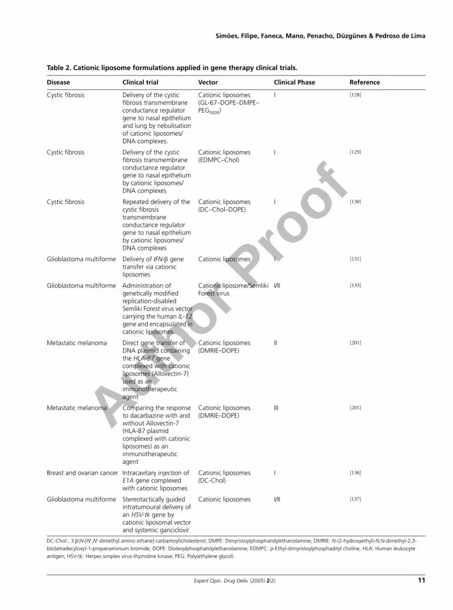
Table 2. Cationic liposome formulations applied in gene therapy clinical trials.
Disease Clinical trial Vector Clinical Phase Reference
Cystic fibrosis Delivery of the cystic fibrosis transmembrane conductance regulator gene to nasal epithelium and lung by nebulisation of cationic liposomes/DNA complexes.
Cationic liposomes(GL-67–DOPE–DMPE–PEG5000)
I [128]
Cystic fibrosis Delivery of the cystic fibrosis transmembrane conductance regulator gene to nasal epithelium by cationic liposomes/DNA complexes
Cationic liposomes(EDMPC–Chol)
I [129]
Cystic fibrosis Repeated delivery of the cystic fibrosis transmembrane conductance regulator gene to nasal epithelium by cationic liposomes/DNA complexes
Cationic liposomes(DC–Chol–DOPE)
I [130]
Glioblastoma multiforme Delivery of IFN-β gene transfer via cationic liposomes
Cationic liposomes I [131]
Glioblastoma multiforme Administration of genetically modified replication-disabled Semliki Forest virus vector carrying the human IL-12 gene and encapsulated in cationic liposomes
Cationic liposome/Semliki Forest virus
I/II [133]
Metastatic melanoma Direct gene transfer of DNA plasmid containing the HLA-B7 gene complexed with cationic liposomes (Allovectin-7) used as an immunotherapeutic agent
Cationic liposomes(DMRIE–DOPE)
II [201]
Metastatic melanoma Comparing the response to dacarbazine with and without Allovectin-7 (HLA-B7 plasmid complexed with cationic liposomes) as an immunotherapeutic agent
Cationic liposomes(DMRIE–DOPE)
III [201]
Breast and ovarian cancer Intracavitary injection of E1A gene complexed with cationic liposomes
Cationic liposomes(DC-Chol)
I [136]
Glioblastoma multiforme Stereotactically guided intratumoural delivery of an HSV-tk gene by cationic liposomal vector and systemic ganciclovir
Cationic liposomes I/II [137]
DC-Chol:; 3 β(N-[N′,N′-dimethyl amino ethane]-carbamoyl)cholesterol; DMPE: Dimyristoylphosphatidylethanolamine; DMRIE: N-(2-hydroxyethyl)-N,N-dimethyl-2,3-bis(tetradecyloxy)-1-propanaminium bromide; DOPE: Dioleoylphosphatidylethanolamine; EDMPC: p-Ethyl-dimyristoylphosphadityl choline; HLA: Human leukocyte antigen; HSV-tk: Herpes simplex virus-thymidine kinase; PEG: Poly(ethylene glycol).
Author Pro
of
Cationic liposomes for gene delivery
12 Expert Opin. Drug Deliv. (2005) 2(2)
preformed negatively charged liposomes, namely pH-sensitiveliposomes [80,106,107,110-115].
In comparison with conventional liposome formulations,the reported approach was shown to result in efficient con-densation and protection of plasmid DNA. Furthermore, tar-geting of the formulations to a specific cell (through couplingof a PEG–lipid conjugate to a ligand) led to improvedtransfection efficiency.
Moreover, further improvements can be achieved by usingdifferent strategies, namely by incorporation of a nuclear tar-geting sequence into the plasmid DNA aiming at promotingits nuclear entry [114], or by the use of condensing agents con-taining nuclear localisation signals [116]. Through structuralanalysis of ternary complexes composed of DNA, cationicDNA binding protein and anionic liposomes, Hagstrom et al.reported that DNA was located on the outside of the lipo-somes, providing evidence that plasmid DNA encapsulation isnot essential for transfection competency in vitro [116].
Another alternative approach to avoid liposomeencapsulation of polyplexes was reported by Lee et al., whichconsisted of generating water soluble lipopolymers (WSLP) onconjugation of a lipid (cholesterol) to a cationic polymer(PEI 1800 Da) for complexation with DNA. It was demon-strated that WSLP–DNA complexes undergo internalisationthrough the cellular cholesterol pathway, leading to hightransfection efficiency of smooth muscle cells without causingcytotoxicity. The injection of WSLP–pDNA complexes intorabbit myocardium showed that WSLP mediated highertransfection efficiency than PEI (1800 Da) and led to longergene expression than naked pDNA. [117].
5. Perspectives for clinical application of cationic liposomes as gene carriers
The first gene therapy clinical trial was approved in 1989 andconsisted of using tumour infiltrating lymphocytes, containinga marker gene (the gene coding for resistance to neomycin)delivered by a retroviral vector [118]. This represented the start-ing point for a period of great enthusiasm for clinical genetherapy. However, the great expectations associated with theapplication of this therapeutic approach have been hamperedby the unsatisfactory results obtained [119]. Moreover, genetherapy faced a serious drawback in the late 1990s, when apatient, enrolled in Phase I clinical trials involving intravascu-lar gene transfer mediated by an adenovirus type 5-based vec-tor that contained the human ornithine transcarbamylasecDNA, experienced lethal complications [120,121]. This caseraised serious safety and toxicity concerns regarding the use ofviral vectors in clinical trials [119]. Nevertheless, sometime laterCavaszana-Calvo et al. published the first clinical success ofgene therapy [122]. In a Phase I clinical trial, children sufferingfrom severe combined immunodeficiency-X1 were submittedto an ex vivo gene therapy protocol involving autologous infu-sion of haematopoietic stem cells transduced with a Moloneyretrovirus-derived vector containing the gene encoding the γ-c
cytokine receptor subunit of IL-2, -4, -7, -9 and -15 receptors[122,123]. Unfortunately, the enthusiasm generated by theapparent cure of 9 out of the 10 infants turned to alarm whennearly 3 years after treatment T-cell leukaemia emerged in 2 ofthe children. This was attributed to retroviral vector integra-tion in proximity of the LMO2 proto-oncogene promoter,leading to aberrant transcription and expression of LMO2[124]. All these safety problems prompted the scientists todevelop alternatives to viral vectors. Cationic liposomes havebeen one of the most used nonviral vectors in worldwidehuman clinical trials of gene therapy. Table 2 summarises themain gene therapy clinical trials using cationic liposomes. Sofar, the clinical application of cationic liposomes has beenessentially focused on cancer and monogenic diseases, cysticfibrosis (CF) being, among the latter, the most frequentlyaddressed disease. CF is caused by mutation of the CF trans-membrane conductance regulator (CFTR) gene. The normalCFTR protein is localised at the apical membrane of epithelialcells and plays a vital role in regulating transepithelial iontransport and water balance. In CF, the airway secretions dehy-drate, mucociliary clearance, is impaired and opportunisticbacterial infections take hold. This causes inflammation andprogressive lung damage, which is usually fatal by early adult-hood [10]. Studies performed in transgenic CF mutant miceshowed that the electrophysiological defect in the animal’s tra-chea can be partially or wholly corrected by instillation or aero-solisation of human CFTR cDNA complexed with cationicliposomes [125,126]. However, in most of the performed humanclinical trials, gene transfer efficiency was low, and most likelyinsufficient to achieve clinical benefit. This low gene transferefficiency is probably due to the extracellular barriers such asmucus, the gycocalyx, tight junctions and mucociliary clear-ance, which limits the biological access of gene transfer agents[127]. Alton et al. showed that the administration, by nebulisa-tion of patients’ lungs, of complexes prepared from a plasmidcontaining the CFTR cDNA (pCF-1–CFTR) and cationicliposomes composed of GL-67–DOPE–dimyristoylphosphati-dylethanolamine–PEG5000 (molar ratio 1:2:0.05), resulted in asignificant degree of correction of the chloride abnormality inall the eight patients involved in the clinical trial [128].
However, the authors detected no alterations in the sodiumtransport abnormality and observed influenza-like symptomsin seven out of the eight treated patients enrolled in the clini-cal trial [128]. Using complexes prepared from p-ethyl-dimyris-toylphosphadityl choline (EDMPC)–Chol cationic liposomesand a plasmid expressing hCFTR, Noone et al. concludedthat the cationic liposome/DNA complexes are safe but werenot capable of mediating consistent evidence of gene transferto the nasal epithelium [129]. Nevertheless, Hyde et al. wereable to demonstrate that liposome–DNA complexes (DC-Chol–DOPE–CFTR cDNA) could be successfully readminis-tered to the nose of CF patients. The authors showed thatrepeated administration of these complexes was safe, with noactivation of the immune system, and resulted in efficientgene transfer. Changes were detected in CFTR-dependent
Author Pro
of
Simões, Filipe, Faneca, Mano, Penacho, Düzgünes & Pedroso de Lima
Expert Opin. Drug Deliv. (2005) 2(2) 13
cellular function, following each of the three doses (adminis-tered 4 weeks apart) in 6 out of 10 treated patients [130]. Allthese studies show that in order to improve the clinical prog-nosis of CF patients, current cationic lipid-based formulationswill need to be optimised, aiming at increasing the level andpersistence of transgene expression.
Most of the approved gene therapy clinical trials addresscancer, in which protocols involving the use of cationic lipo-somes are included. These protocols include the transfer ofvarious types of genes for the application of differentantitumour strategies, such as immunopotentiation, oncogeneinactivation and ‘suicide’ gene therapy. Recently, Yoshida et al.published a clinical study involving five patients with malig-nant glioma (glioblastoma multiforme or anaplastic astrocy-toma), in which the safety and effectiveness of IFN-β genetransfer via cationic liposomes were evaluated. Transgeneexpression and antitumour activity were detected in four outof five treated patients. Two patients showed a partial response(> 50% tumour reduction) and two others had stable disease10 weeks after beginning therapy [131], thus suggesting the fea-sibility and safety of IFN-β gene therapy mediated by cationicliposomes. These clinical trials were based on a preclinicalstudy on the antitumour effect and mechanism of action ofcationic liposome-mediated murine IFN-β gene therapy inmouse B16F1 melanoma. The authors observed that intratu-mour administration of cationic liposome–DNA complexesresulted in a 5.5-fold reduction in the mean volume of subcu-taneous melanoma and eradication of the tumour in 18% ofthe treated mice, by directly inducing cell death and stimulat-ing natural killer cells [132]. In addition, Ren et al. tested anovel therapy strategy in adult patients with recurrent gliob-lastoma multiforme, which was aimed at evaluating biologicalsafety, maximum tolerated dose and antitumour efficacy of agenetically modified replication-disabled Semliki Forest virusvector carrying the human IL-12 gene and encapsulated incationic liposomes. Preclinical work with these systems inbreast and prostate cancer animal models demonstrated theirbiosafety and antitumour efficacy [133]. Several clinical trials inPhase II and one in Phase III have been performed using Allo-vectin-7® formulation (Vical Inc, San Diego, CA, USA),which consists of a DNA plasmid containing the human leu-kocyte antigen-B7 gene complexed with DMRIE–DOPE cat-ionic liposomes [201].
The HER-2/neu oncogene encodes an epidermal growthfactor receptor-related transmembrane protein and is over-expressed in many types of human cancers. Therefore, thisoncogene represents an excellent molecular target for anti-tumour strategies, namely in HER-2/neu-overexpressinghuman cancers, such as human breast and ovarian cancers[134]. Preclinical studies have demonstrated that the adenovi-rus type 5 E1A gene is associated with antitumour activitiesby transcriptional repression of HER-2/neu and induction ofapoptosis [135]. In this context, Hortobagyi et al. evaluated thefeasibility of intracavitary injection of E1A gene complexedwith DC–Chol cationic liposomes in patients with breast and
ovarian cancers in a Phase I clinical trial. The authorsobserved E1A gene expression that was accompanied byHER-2/neu downregulation, increased apoptosis and reducedproliferation [136]. In a prospective Phase I/II clinical study,Voges et al. treated eight patients suffering from recurrentglioblastoma multiforme with stereotactically guided intratu-mour convection-enhanced delivery of an HSV-1 thymidinekinase gene-bearing cationic liposomal vector and systemicganciclovir. The treatment was well tolerated without majorside effects in two out of eight patients. The authors observeda > 50% reduction of tumour volume and focal treatmenteffects in six out of eight patients [137].
The main objective in gene therapy is to achieve successfulin vivo gene transfer into target tissues. The choice of the genedelivery system varies according to the application. For exam-ple, prolonged and sustained expression is needed for treatingmonogenic hereditary diseases, whereas short duration of geneexpression may be sufficient for most cancer treatments [138].
6. Expert opinion and conclusion
Due to their safety and versatility, cationic liposomes haveemerged as promising alternatives to viral vectors for thedevelopment of gene therapy approaches. Numerous in vivoapplications have been reported in the literature, focusingnot only on aspects related to their pharmacokinetics andbiodistribution, but also on their toxicity and immuno-genicity. The demonstrated efficacy of such systems inmediating delivery of polynucleotides, including therapeu-tic genes and antisense oligonucleotides, have made thempromising candidates for the treatment of several diseases,which has been reflected in their extensive and increasingapplication in a large number of clinical trials. However, itis generally recognised that the efficacy of lipid-based genedelivery systems is still far from that observed for viral vec-tors, especially when high levels and long-term transgeneexpression are required.
Aiming at circumventing these limitations, much work hasbeen devoted to the synthesis of new cationic lipids and thedesign of new plasmid constructs with more efficient promot-ers/enhancers. However, the progress achieved is still far frombeing satisfactory. This has prompted investigators to focustheir research activities on not only the nature of the lipoplexcomponents, but also on the variables affecting their forma-tion, mode of interaction with cells and in vivo behaviour(e.g., colloidal stability). The aim is to generate complexes ofsmall size with a narrow distribution, while presenting a neu-tral or negatively charged surface (to prevent nonspecificinteractions with blood components) that ensure completeprotection of DNA, exhibit specific targeting and have theability to promote efficient intracellular delivery of carriedmaterial and to facilitate its translocation into the nucleus,thus leading to high and sustained levels of transgene expres-sion without causing cytotoxicity. In this regard, it will berather laborious and difficult to design a nonviral vector capa-
Author Pro
of
Cationic liposomes for gene delivery
14 Expert Opin. Drug Deliv. (2005) 2(2)
ble of fulfilling the conflicting requirements imposed by eachof the different stages involved in the gene delivery process.Nevertheless, it is also clear that governing formulation varia-bles may allow the tailoring of lipoplexes to particular trans-fection applications. Therefore, the biophysical properties ofthe complexes such as size, charge, stability and the extent ofinteraction with cells should be controlled in such a way thattheir application to different protocols (in vitro, ex vivo andin vivo) and routes of administration can be optimised.
Recently, different formulation strategies have been attemptedto confer viral attributes to lipoplexes, namely through the asso-
ciation of certain proteins or peptides. Whether these improve-ments result in a system that, while exhibiting satisfactory abilityto mediate in vivo transfection, would lead to such a complexitythat could endanger its versatilily and large scale production orcould limit extended/repeated in vivo use due to immunogenic-ity, are important questions that remain to be addressed.
Thus, before embarking on extensive and expensive animalexperiments and clinical trials, it is advisable to pursue funda-mental research focused on the mechanisms by which lipoplexesare formed and deliver their DNA, as well as on methods bywhich the different biological barriers they face can be overcome.
BibliographyPapers of special note have been highlighted as either of interest (•) or of considerable interest (••) to readers.
1. FELGNER PL, GADEK TR, HOLM M et al.: Lipofection: a highly efficient, lipid-mediated DNA-transfection procedure. Proc. Natl. Acad. Sci. USA (1987) 84(21):7413-7417.
2. PEDROSO DE LIMA MC, NEVES S, FILIPE A, DUZGUNES N, SIMOES S: Cationic liposomes for gene delivery: from biophysics to biological applications. Curr. Med. Chem. (2003) 10(14):1221-1231.
3. BEHR JP, DEMENEIX B, LOEFFLER JP, PEREZ-MUTUL J: Efficient gene transfer into mammalian primary endocrine cells with lipopolyamine-coated DNA. Proc. Natl. Acad. Sci. USA (1989) 86(18):6982-6986.
4. FERRARI ME, NGUYEN CM, ZELPHATI O, TSAI Y, FELGNER PL: Analytical methods for the characterization of cationic lipid-nucleic acid complexes. Hum. Gene Ther. (1998) 9(3):341-351.
5. MARSHALL J, YEW NS, EASTMAN SJ et al.: Nonviral vectors for gene therapy, In: Nonviral vectors for gene delivery. L Huang, M Hung, E Wagner (Eds). (1999) Academic Press. 39-68.
6. FELGNER JH, KUMAR R, SRIDHAR CN et al.: Enhanced gene delivery and mechanism studies with a novel series of cationic lipid formulations. J. Biol. Chem. (1994) 269(4):2550-2561.
7. LEE ER, MARSHALL J, SIEGEL CS et al.: Detailed analysis of structures and formulations of cationic lipids for efficient gene transfer to the lung. Hum. Gene Ther. (1996) 7(14):1701-1717.
8. FELGNER PL, TSAI YJ, SUKHU L et al.: Improved cationic lipid formulations for
in vivo gene therapy. Ann. NY Acad. Sci. (1995) 772:126-139.
9. SCHEULE RK, ST GEORGE JA, BAGLEY RG et al.: Basis of pulmonary toxicity associated with cationic lipid-mediated gene transfer to the mammalian lung. Hum. Gene Ther. (1997) 8(6):689-707.
10. HUI SW, LANGNER M, ZHAO YL, ROSS P, HURLEY E, CHAN K: The role of helper lipids in cationic liposome-mediated gene transfer. Biophys. J. (1996) 71(2):590-599.
11. MOK KW, CULLIS PR: Structural and fusogenic properties of cationic liposomes in the presence of plasmid DNA. Biophys. J. (1997) 73(5):2534-2545.
12. SIMOES S, SLEPUSHKIN V, GASPAR R, DE LIMA MC, DUZGUNES N: Gene delivery by negatively charged ternary complexes of DNA, cationic liposomes and transferrin or fusigenic peptides. Gene Ther. (1998) 5(7):955-964.
13. KOLTOVER I, SALDITT T, RADLER JO, SAFINYA CR: An inverted hexagonal phase of cationic liposome-DNA complexes related to DNA release and delivery. Science (1998) 281(5373):78-81.
14. ZUIDAM NJ, BARENHOLZ Y: Electrostatic and structural properties of complexes involving plasmid DNA and cationic lipids commonly used for gene delivery. Biochim. Biophys. Acta (1998) 1368(1):115-128.
15. HARVIE P, WONG FM, BALLY MB: Characterization of lipid DNA interactions. I. Destabilization of bound lipids and DNA dissociation. Biophys. J. (1998) 75(2):1040-1051.
16. SIMOES S, SLEPUSHKIN V, PIRES P et al.: Mechanisms of gene transfer mediated by lipoplexes associated with
targeting ligands or pH-sensitive peptides. Gene Ther. (1999) 6(11):1798-1807.
17. LIU Y, MOUNKES LC, LIGGITT HD et al.: Factors influencing the efficiency of cationic liposome-mediated intravenous gene delivery. Nat. Biotechnol. (1997) 15(2):167-173.
18. WANG J, GUO X, XU Y, BARRON L, SZOKA FC, JR.: Synthesis and characterization of long chain alkyl acyl carnitine esters. Potentially biodegradable cationic lipids for use in gene delivery. J. Med. Chem. (1998) 41(13):2207-2215.
19. HONG K, ZHENG W, BAKER A, PAPAHADJOPOULOS D: Stabilization of cationic liposome-plasmid DNA complexes by polyamines and poly(ethylene glycol)-phospholipid conjugates for efficient in vivo gene delivery. FEBS Lett. (1997) 400(2):233-237.
20. STERNBERG B, HONG K, ZHENG W, PAPAHADJOPOULOS D: Ultrastructural characterization of cationic liposome-DNA complexes showing enhanced stability in serum and high transfection activity in vivo. Biochim. Biophys. Acta (1998) 1375(1-2):23-35.
21. SMITH JG, WEDEKING T, VERNACHIO JH, WAY H, NIVEN RW: Characterization and in vivo testing of a heterogeneous cationic lipid-DNA formulation. Pharm. Res. (1998) 15(9):1356-1363.
22. SONG YK, LIU F, CHU S, LIU D: Characterization of cationic liposome-mediated gene transfer in vivo by intravenous administration. Hum. Gene Ther. (1997) 8(13):1585-1594.
23. TOMLINSON E, ROLLAND AP: Controllable gene therapy: pharmaceutics of non-viral gene delivery systems. J. Control. Rel. (1996) 39:357-372.

Author Pro
of
Simões, Filipe, Faneca, Mano, Penacho, Düzgünes & Pedroso de Lima
Expert Opin. Drug Deliv. (2005) 2(2) 15
24. RADLER JO, KOLTOVER I, SALDITT T, SAFINYA CR: Structure of DNA-cationic liposome complexes: DNA intercalation in multilamellar membranes in distinct interhelical packing regimes. Science (1997) 275(5301):810-814.
25. EASTMAN SJ, SIEGEL C, TOUSIGNANT J et al.: Biophysical characterization of cationic lipid: DNA complexes. Biochim. Biophys. Acta (1997) 1325(1):41-62.
26. PIRES P, SIMOES S, NIR S, GASPAR R, DUZGUNES N, PEDROSO DE LIMA MC: Interaction of cationic liposomes and their DNA complexes with monocytic leukemia cells. Biochim. Biophys. Acta (1999) 1418(1):71-84.
27. LEDLEY FD: Pharmaceutical approach to somatic gene therapy. Pharm. Res. (1996) 13(11):1595-1614.
28. KOE GS, WAY HL, QUETINGCO GM et al.: The effect of mixing on the formation of DNA/liposome complexes. Pharm. Res. (1997) 14:S57.
29. MAHATO RI, ROLLAND A, TOMLINSON E: Cationic lipid-based gene delivery systems: pharmaceutical perspectives. Pharm. Res. (1997) 14(7):853-859.
30. ZELPHATI O, NGUYEN C, FERRARI M, FELGNER J, TSAI Y, FELGNER PL: Stable and monodisperse lipoplex formulations for gene delivery. Gene Ther. (1998) 5(9):1272-1282.
31. PEDROSO DE LIMA MC, SIMOES S, PIRES P, FANECA H, DUZGUNES N: Cationic lipid-DNA complexes in gene delivery: from biophysics to biological applications. Adv. Drug Deliv. Rev. (2001) 47(2-3):277-294.
32. PEDROSO DE LIMA MC, FANECA H, MANO M, PENACHO N, DUZGUNES N, SIMOES S: Biophysical characterization of cationic liposome-DNA complexes and their interaction with cells. Methods Enzymol. (2003) 373:298-312.
33. TEMPLETON NS, LASIC DD, FREDERIK PM, STREY HH, ROBERTS DD, PAVLAKIS GN: Improved DNA: liposome complexes for increased systemic delivery and gene expression. Nat. Biotechnol. (1997) 15(7):647-652.
34. DESHPANDE D, BLEZINGER P, PILLAI R, DUGUID J, FREIMARK B, ROLLAND A: Target specific optimization of cationic lipid-based systems for
pulmonary gene therapy. Pharm. Res. (1998) 15(9):1340-1347.
35. SONG Y, KLIU D: Free liposomes enhance the transfection activity of DNA/lipid complexes in vivo by intravenous administration. Biochim. Biophys. Acta (1998) 1372(1):141-150.
36. YANG JP, HUANG L: Overcoming the inhibitory effect of serum on lipofection by increasing the charge ratio of cationic liposome to DNA. Gene Ther. (1997) 4(9):950-960.
37. YANG JP, HUANG L: Time-dependent maturation of cationic liposome-DNA complex for serum resistance. Gene Ther. (1998) 5(3):380-387.
38. FANECA H, SIMOES S, PEDROSO DE LIMA MC: Association of albumin or protamine to lipoplexes: enhancement of transfection and resistance to serum. J. Gene Med. (2004) 6(6):681-692.
39. SIMOES S, SLEPUSHKIN V, PIRES P, GASPAR R, PEDROSO DE LIMA MC, DUZGUNES N: Human serum albumin enhances DNA transfection by lipoplexes and confers resistance to inhibition by serum. Biochim. Biophys. Acta (2000) 1463(2):459-469.
40. MEYER O, KIRPOTIN D, HONG K et al.: Cationic liposomes coated with polyethylene glycol as carriers for oligonucleotides. J. Biol. Chem. (1998) 273(25):15621-15627.
41. USTER PS, ALLEN TM, DANIEL BE, MENDEZ CJ, NEWMAN MS, ZHU GZ: Insertion of poly(ethylene glycol) derivatized phospholipid into pre-formed liposomes results in prolonged in vivo circulation time. FEBS Lett. (1996) 386(2-3):243-246.
42. MAURER N, MORI A, PALMER L et al.: Lipid-based systems for the intracellular delivery of genetic drugs. Mol. Membr. Biol. (1999) 16(1):129-140.
43. ZHANG YP, SEKIROV L, SARAVOLAC EG et al.: Stabilized plasmid-lipid particles for regional gene therapy: formulation and transfection properties. Gene Ther. (1999) 6(8):1438-1447.
44. WHEELER JJ, PALMER L, OSSANLOU M et al.: Stabilized plasmid-lipid particles: construction and characterization. Gene Ther. (1999) 6(2):271-281.
45. MISLICK KA, BALDESCHWIELER JD: Evidence for the role of proteoglycans in cation-mediated gene transfer. Proc. Natl.
Acad. Sci. USA (1996) 93(22):12349-12354.
• This paper constitutes one of the first reports suggesting the involvement of proteoglycans on transfection mediated by polycation complexes.
46. KJELLEN LLINDAHL U: Proteoglycans: structures and interactions. Ann. Rev. Biochem. (1991) 60:443-475.
47. MOUNKES LC, ZHONG W, CIPRES-PALACIN G, HEATH TD, DEBS RJ: Proteoglycans mediate cationic liposome-DNA complex-based gene delivery in vitro and in vivo. J. Biol. Chem. (1998) 273(40):26164-26170.
48. SAWITZKY D: Protein-glycosaminoglycan interactions: infectiological aspects. Med. Microbiol. Immunol. Berl (1996) 184(4):155-161.
49. KOPATZ I, REMY JS, BEHR JP: A model for non-viral gene delivery: through syndecan adhesion molecules and powered by actin. J. Gene Med. (2004) 6(7):769-776.
50. RUPONEN M, YLA-HERTTUALA S, URTTI A: Interactions of polymeric and liposomal gene delivery systems with extracellular glycosaminoglycans: physicochemical and transfection studies. Biochim. Biophys. Acta (1999) 1415(2):331-341.
51. RUPONEN M, HONKAKOSKI P, TAMMI M, URTTI A: Cell-surface glycosaminoglycans inhibit cation-mediated gene transfer. J. Gene Med. (2004) 6(4):405-414.
52. WIETHOFF CM, SMITH JG, KOE GS, MIDDAUGH CR: The potential role of proteoglycans in cationic lipid-mediated gene delivery. Studies of the interaction of cationic lipid-DNA complexes with model glycosaminoglycans. J. Biol. Chem. (2001) 276(35):32806-32813.
53. RUPONEN M, RONKKO S, HONKAKOSKI P, PELKONEN J, TAMMI M, URTTI A: Extracellular glycosaminoglycans modify cellular trafficking of lipoplexes and polyplexes. J. Biol. Chem. (2001) 276(36):33875-33880.
54. WROBEL I, COLLINS D: Fusion of cationic liposomes with mammalian cells occurs after endocytosis. Biochim. Biophys. Acta (1995) 1235(2):296-304.
55. FRIEND DS, PAPAHADJOPOULOS D, DEBS RJ: Endocytosis and intracellular processing accompanying transfection

Author Pro
of
Cationic liposomes for gene delivery
16 Expert Opin. Drug Deliv. (2005) 2(2)
mediated by cationic liposomes. Biochim. Biophys. Acta (1996) 1278(1):41-50.
56. STEGMANN T, LEGENDRE JY: Gene transfer mediated by cationic lipids: lack of a correlation between lipid mixing and transfection. Biochim. Biophys. Acta (1997) 1325(1):71-79.
57. DA CRUZ MT, SIMOES S, PIRES PP, NIR S, DE LIMA MC: Kinetic analysis of the initial steps involved in lipoplex-cell interactions: effect of various factors that influence transfection activity. Biochim. Biophys. Acta (2001) 1510(1-2):136-151.
58. CONNER SD, SCHMID SL: Regulated portals of entry into the cell. Nature (2003) 422(6927):37-44.
•• This paper is a comprehensive review of the different endocytic internalisation pathways.
59. REJMAN J, OBERLE V, ZUHORN IS, HOEKSTRA D: Size-dependent internalization of particles via the pathways of clathrin- and caveolae-mediated endocytosis. Biochem. J. (2004)377(Pt 1):159-169.
•• This paper represents a very important contribution to the understanding of the effect of particle size on transfection.
60. ROSS PC, HUI SW: Lipoplex size is a major determinant of in vitro lipofection efficiency. Gene Ther. (1999) 6(4):651-659.
61. XU Y, HUI SW, FREDERIK P, SZOKA FC, JR.: Physicochemical characterization and purification of cationic lipoplexes. Biophys. J. (1999) 77(1):341-353.
62. BALLY MB, HARVIE P, WONG FM, KONG S, WASAN EK, REIMER DL: Biological barriers to cellular delivery of lipid-based DNA carriers. Adv. Drug Deliv. Rev. (1999) 38(3):291-315.
63. SIMOES S, SLEPUSHKIN V, PRETZER E et al.: Transfection of human macrophages by lipoplexes via the combined use of transferrin and pH-sensitive peptides. J. Leukoc. Biol. (1999) 65(2):270-279.
64. ZUHORN IS, HOEKSTRA D: On the mechanism of cationic amphiphile-mediated transfection. To fuse or not to fuse: is that the question? J. Membr. Biol. (2002) 189(3):167-179.
•• This paper constitutes an excellent review on the current knowledge of the mechanisms involved in gene delivery mediated by lipoplexes.
65. XU Y, SZOKA FC, JR.: Mechanism of DNA release from cationic liposome/DNA
complexes used in cell transfection. Biochemistry (1996) 35(18):5616-5623.
• In this paper the authors propose a model that is still widely accepted regarding the dissociation of lipoplexes and cytoplasmic delivery of DNA.
66. NOGUCHI A, FURUNO T, KAWAURA C, NAKANISHI M: Membrane fusion plays an important role in gene transfection mediated by cationic liposomes. FEBS Lett. (1998) 433(1-2):169-173.
67. BRUNNER S, SAUER T, CAROTTA S, COTTEN M, SALTIK M, WAGNER E: Cell cycle dependence of gene transfer by lipoplex, polyplex and recombinant adenovirus. Gene Ther. (2000) 7(5):401-407.
68. PAGE RL, BUTLER SP, SUBRAMANIAN A, GWAZDAUSKAS FC, JOHNSON JL, VELANDER WH: Transgenesis in mice by cytoplasmic injection of polylysine/DNA mixtures. Transgenic Res. (1995) 4(6):353-360.
69. ZABNER J, FASBENDER AJ, MONINGER T, POELLINGER KA, WELSH MJ: Cellular and molecular barriers to gene transfer by a cationic lipid. J. Biol. Chem. (1995) 270(32):18997-19007.
70. CORNELIS S, VANDENBRANDEN M, RUYSSCHAERT JM, ELOUAHABI A: Role of intracellular cationic liposome-DNA complex dissociation in transfection mediated by cationic lipids. DNA Cell Biol. (2002) 21(2):91-97.
• This paper addresses an interesting study reporting efficient dissociation of lipoplexes inside the nucleus.
71. ARONSOHN AI, HUGHES JA: Nuclear localization signal peptides enhance cationic liposome-mediated gene therapy. J. Drug Target. (1998) 5(3):163-169.
72. CHAN CK, JANS DA: Enhancement of polylysine-mediated transferrinfection by nuclear localization sequences: polylysine does not function as a nuclear localization sequence. Hum. Gene Ther. (1999) 10(10):1695-1702.
73. ZANTA MA, BELGUISE-VALLADIER P, BEHR JP: Gene delivery: a single nuclear localization signal peptide is sufficient to carry DNA to the cell nucleus. Proc. Natl. Acad. Sci. USA (1999) 96(1):91-96.
74. BRANDEN LJ, MOHAMED AJ, SMITH CI: A peptide nucleic acid-nuclear localization signal fusion that mediates
nuclear transport of DNA. Nat. Biotechnol. (1999) 17(8):784-787.
75. MURRAY KD, ETHERIDGE CJ, SHAH SI et al.: Enhanced cationic liposome-mediated transfection using the DNA-binding peptide mu (mu) from the adenovirus core. Gene Ther. (2001) 8:453-460.
76. TAGAWA T, MANVELL M, BROWN N et al.: Characterisation of LMD virus-like nanoparticles self-assembled from cationic liposomes, adenovirus core peptide mu and plasmid DNA. Gene Ther. (2002) 9:564-576.
77. RUDOLPH C, PLANK C, LAUSIER J, SCHILLINGER U, MULLER RH, ROSENECKER J: Oligomers of the arginine-rich motif of the HIV-1 TAT protein are capable of transferring plasmid DNA into cells. J. Biol. Chem. (2003) 278:11411-11418.
78. HYNDMAN L, LEMOINE JL, HUANG L, PORTEOUS DJ, BOYD AC, NAN X: HIV-1 Tat protein transduction domain peptide facilitates gene transfer in combination with cationic liposomes. J. Control. Release. (2004) 99:435-444.
79. LASIC D, TEMPLETON N: Liposomes in Gene therapy. Adv. Drug Deliv. Rev. (1996) 20:221-226.
80. LEE RJ, HUANG L: Folate-targeted, anionic liposome-entrapped polylysine-condensed DNA for tumor cell-specific gene transfer. J. Biol. Chem. (1996) 271(14):8481-8487.
81. SINGHAL A, HUANG L: Gene transfer in mammalian cells using liposomes as carriers, In: Gene therapeutics: methods and applications of direct gene transfer. W JA, (Ed.) Birkhauser, Boston, MA, USA (1994) 118- 142.
82. SOLODIN I, BROWN CS, BRUNO MS et al.: A novel series of amphiphilic imidazolinium compounds for in vitro and in vivo gene delivery. Biochemistry (1995) 34(41):13537-1344.
83. GORMAN CM, AIKAWA M, FOX B et al.: Efficient in vivo delivery of DNA to pulmonary cells using the novel lipid EDMPC. Gene Ther. (1997) 4(9):983-992.
84. CROOK K, STEVENSON BJ, DUBOUCHET MPORTEOUS DJ: Inclusion of cholesterol in DOTAP transfection complexes increases the delivery of DNA to cells in vitro in the presence of serum. Gene Ther. (1998) 5(1):137-143.

Author Pro
of
Simões, Filipe, Faneca, Mano, Penacho, Düzgünes & Pedroso de Lima
Expert Opin. Drug Deliv. (2005) 2(2) 17
85. EGILMEZ NK, IWANUMA YBANKERT RB: Evaluation and optimization of different cationic liposome formulations for in vivo gene transfer. Biochem. Biophys. Res. Commun. (1996) 221(1):169-173.
86. ESCRIOU V, CIOLINA C, LACROIX F, BYK G, SCHERMAN D, WILS P: Cationic lipid-mediated gene transfer: effect of serum on cellular uptake and intracellular fate of lipopolyamine/DNA complexes. Biochim. Biophys. Acta (1998) 1368(2):276-288.
87. PLANK C, MECHTLER K, SZOKA FC Jr, WAGNER E: Activation of the complement system by synthetic DNA complexes: a potential barrier for intravenous gene delivery. Hum. Gene Ther. (1996) 7(12):1437-1446.
88. VITIELLO L, BOCKHOLD K, JOSHI PBWORTON RG: Transfection of cultured myoblasts in high serum concentration with DODAC:DOPE liposomes. Gene Ther. (1998) 5(10):1306-1313.
89. BOUSSIF O, LEZOUALC’H F, ZANTA MA et al.: A versatile vector for gene and oligonucleotide transfer into cells in culture and in vivo: polyethylenimine. Proc. Natl. Acad. Sci. USA (1995) 92(16):7297-7301.
90. VITIELLO L, CHONN A, WASSERMAN JD, DUFF C, WORTON RG: Condensation of plasmid DNA with polylysine improves liposome-mediated gene transfer into established and primary muscle cells. Gene Ther. (1996) 3(5):396-404.
91. ZHOU XH, KLIBANOV AL, HUANG L: Lipophilic polylysines mediate efficient DNA transfection in mammalian cells. Biochim. Biophys. Acta (1991) 1065(1):8-14.
92. LEWIS JG, LIN KY, KOTHAVALE A et al.: A serum-resistant cytofectin for cellular delivery of antisense oligodeoxynucleotides and plasmid DNA. Proc. Natl. Acad. Sci. USA (1996) 93(8):3176-3181.
93. ZELPHATI O, UYECHI LS, BARRON LG, SZOKA FC Jr: Effect of serum components on the physico-chemical properties of cationic lipid/oligonucleotide complexes and on their interactions with cells. Biochim. Biophys. Acta (1998) 1390(2):119-133.
94. ELIYAHU H, SERVEL N, DOMB AJ, BARENHOLZ Y: Lipoplex-induced hemagglutination: potential involvement in
intravenous gene delivery. Gene Ther. (2002) 9(13):850-858.
95. BAILEY AL, SULLIVAN SM: Efficient encapsulation of DNA plasmids in small neutral liposomes induced by ethanol and calcium. Biochim. Biophys. Acta (2000) 1468(1-2):239-252.
96. REMY JS, KICHLER A, MORDVINOV V, SCHUBER F, BEHR JP: Targeted gene transfer into hepatoma cells with lipopolyamine-condensed DNA particles presenting galactose ligands: a stage toward artificial viruses. Proc. Natl. Acad. Sci. USA (1995) 92(5):1744-1748.
97. HUG PS, LEIGHT RG: Liposomes for the transformation of eukaryotic cells. Biochim. Biophys. Acta (1991) 1097(1):1-17.
98. OGRIS M, BRUNNER S, SCHULLER S, KIRCHEIS R, WAGNER E: PEGylated DNA/transferrin-PEI complexes: reduced interaction with blood components, extended circulation in blood and potential for systemic gene delivery. Gene Ther. (1999) 6(4):595-605.
99. LI S, WU SP, WHITMORE M et al.: Effect of immune response on gene transfer to the lung via systemic administration of cationic lipidic vectors. Am. J. Physiol. (1999) 276(5 Pt 1):L796-804.
100. GULATI M, BAJAD S, SINGH S, FERDOUS AJ, SINGH M: Development of liposomal amphotericin B formulation. J. Microencapsul. (1998) 15(2):137-151.
101. TARDI PG, BOMAN NL, CULLIS PR: Liposomal doxorubicin. J. Drug Target. (1996) 4(3):129-140.
102. GABIZON A: Tailoring liposomes for cancer drug delivery: from the bench to the clinic. Ann. Biol. Clin. (1993) 51(9):811-813.
103. OSTRO MJ, CULLIS PR: Use of liposomes as injectable-drug delivery systems. Am. J. Hosp. Pharm. (1989) 46(8):1576-1587.
104. WANG CY, HUANG L: pH-sensitive immunoliposomes mediate target-cell-specific delivery and controlled expression of a foreign gene in mouse. Proc. Natl. Acad. Sci. USA (1987) 84(22):7851-7855.
105. SHANGGUAN T, CABRAL-LILLY D, PURANDARE U et al.: A novel N-acyl phosphatidylethanolamine-containing delivery vehicle for spermine-condensed plasmid DNA. Gene Ther. (2000)7(9):769-783.
106. FAHR A, MULLER K, NAHDE T, MULLER R, BRUSSELBACH S: A new colloidal lipidic system for gene therapy. J. Liposome Res. (2002) 12(1-2):37-44.
•• This paper reports the use of a cyclic Arg-Gly-Asp- containing peptide as a target device for lipid-based vectors. This was shown to increase their ability to interact with the endothelial cells and to transfect cells in a ligand-dependent manner. Specificity was further improved by combining Arg-Gly-Asp-mediated transductional targeting with transcriptional targeting.
107. TURNER C, WEIR N, CATTERALL C, BAKER TS, JONES MN: The transfection of Jurkat T-leukemic cells by use of pH-sensitive immunoliposomes. J. Liposome Res. (2002) 12(1-2):45-50.
• The authors have shown the importance of the pH-sensitivity of targeted liposomes to increase their transfection efficiency.
108. ELLISON KE, BISHOPRIC NH, WEBSTER KA et al.: Fusigenic liposome-mediated DNA transfer into cardiac myocytes. J. Mol. Cell. Cardiol. (1996) 28(7):1385-1399.
109. MURPHY EA, WARING AJ, HAYNES SM, LONGMUIR KJ: Compaction of DNA in an anionic micelle environment followed by assembly into phosphatidylcholine liposomes. Nucleic Acids Res. (2000) 28(15):2986-2992.
110. REDDY JA, LOW PS: Enhanced folate receptor mediated gene therapy using a novel pH-sensitive lipid formulation. J. Control. Release. (2000) 64(1-3):27-37.
111. GUO W, GOSSELIN MA, LEE RJ: Characterization of a novel diolein-based LPDII vector for gene delivery. J. Control. Release. (2002) 83(1):121-132.
112. GOSSELIN MA, GUO W, LEE RJ: Incorporation of reversibly cross-linked polyplexes into LPDII vectors for gene delivery. Bioconjug. Chem. (2002) 13(5):1044-1053.
113. SHI G, GUO W, STEPHENSON SM, LEE RJ: Efficient intracellular drug and gene delivery using folate receptor-targeted pH-sensitive liposomes composed of cationic/anionic lipid combinations. J. Control. Release. (2002) 80(1-3):309-319.
114. REDDY JA, DEAN D, KENNEDY MD, LOW PS: Optimization of folate-conjugated liposomal vectors for folate receptor-mediated gene therapy. J. Pharm. Sci. (1999) 88(11):1112-1118.
Author Pro
of
Cationic liposomes for gene delivery
18 Expert Opin. Drug Deliv. (2005) 2(2)
•• This paper reports a novel plasmid containing a segment of Simian virus 40 DNA, which has been employed to facilitate its transport into the nucleus when deliverd by pH-sensitive targeted liposomes.
115. MAGUIRE LA, ZHANG H, SHAMLOU PA: Preparation of small unilamellar vesicles (SUV) and biophysical characterization of their complexes with poly-l-lysine-condensed plasmid DNA. Biotechnol. Appl. Biochem. (2003) 37(Pt 1):73-81.
116. HAGSTROM JE, SEBESTYEN MG, BUDKER V et al.: Complexes of non-cationic liposomes and histone H1 mediate efficient transfection of DNA without encapsulation. Biochim. Biophys. Acta (1996) 1284(1):47-55.
117. LEE M, RENTZ J, HAN SO, BULL DA, KIM SW: Water-soluble lipopolymer as an efficient carrier for gene delivery to myocardium. Gene Ther. (2003) 10(7):585-593.
118. KOHN DB, HERSHFIELD MS, CARBONARO D et al.: T lymphocytes with a normal ADA gene accumulate after transplantation of transduced autologous umbilical cord blood CD34+ cells in ADA-deficient SCID neonates. Nat. Med. (1998) 4(7):775-780.
119. BAUM BJ, GOLDSMITH CM, KOK MR et al.: Advances in vector-mediated gene transfer. Immunol. Lett. (2003) 90(2-3):145-149.
120. RAPER SE, WILSON JM, YUDKOFF M et al.: Developing adenoviral-mediated in vivo gene therapy for ornithine transcarbamylase deficiency. J. Inherit. Metab. Dis. (1998) 21(Suppl. 1):119-137.
121. RAPER SE, YUDKOFF M, CHIRMULE N et al.: A pilot study of in vivo liver-directed gene transfer with an adenoviral vector in partial ornithine transcarbamylase deficiency. Hum. Gene Ther. (2002) 13(1):163-175.
• This paper emphasises the safety risks associated with the repeated administrations of adenoviral vectors in gene therapy.
122. CAVAZZANA-CALVO M, HACEIN-BEY S, DE SAINT BASILE G et al.: Gene therapy of human severe combined immunodeficiency (SCID)-X1 disease. Science (2000) 288(5466):669-672.
•• This paper represents a hallmark in gene therapy, as it reports its first clinical success, which consisted of treating
children suffering from severe combined immunodeficiency-X1.
123. HACEIN-BEY-ABINA S, LE DEIST F, CARLIER F et al.: Sustained correction of X-linked severe combined immunodeficiency by ex vivo gene therapy. N. Engl. J. Med. (2002) 346(16):1185-1193.
124. HACEIN-BEY-ABINA S, VON KALLE C, SCHMIDT M et al.: LMO2-associated clonal T cell proliferation in two patients after gene therapy for SCID-X1. Science (2003) 302(5644):415-419.
• This paper reports a serious drawback in gene therapy associated to the use of viral vectors. Two patients developed leukaemia 3 years after being enrolled in gene therapy clinical trials, this adverse effect being attributed to retroviral vector genome integration.
125. HYDE SC, GILL DR, HIGGINS CF et al.: Correction of the ion transport defect in cystic fibrosis transgenic mice by gene therapy. Nature (1993) 362(6417):250-255.
126. MCLACHLAN G, HO LP, DAVIDSON-SMITH H et al.: Laboratory and clinical studies in support of cystic fibrosis gene therapy using pCMV-CFTR-DOTAP. Gene Ther. (1996) 3(12):1113-1123.
127. GRIESENBACH U, FERRARI S, GEDDES DM, ALTON EW: Gene therapy progress and prospects: cystic fibrosis. Gene Ther. (2002) 9(20):1344-1350.
128. ALTON EW, STERN M, FARLEY R et al.: Cationic lipid-mediated CFTR gene transfer to the lungs and nose of patients with cystic fibrosis: a double-blind placebo-controlled trial. Lancet (1999) 353(9157):947-954.
129. NOONE PG, HOHNEKER KW, ZHOU Z et al.: Safety and biological efficacy of a lipid-CFTR complex for gene transfer in the nasal epithelium of adult patients with cystic fibrosis. Mol. Ther. (2000) 1(1):105-114.
130. HYDE SC, SOUTHERN KW, GILEADI U et al.: Repeat administration of DNA/liposomes to the nasal epithelium of patients with cystic fibrosis. Gene Ther. (2000) 7(13):1156-1165.
•• The authors demonstrated for the first time that liposome–DNA complexes could be successfully re-administered to the nose of CF patients.
131. YOSHIDA J, MIZUNO M, FUJII M et al.: Human gene therapy for malignant gliomas (glioblastoma multiforme and anaplastic
astrocytoma) by in vivo transduction with human interferon beta gene using cationic liposomes. Hum. Gene Ther. (2004) 15(1):77-86.
•• The authors observed transgene expression and a significant antitumour activity in patients with malignant glioma following transfer of IFN-β gene via cationic liposomes.
132. RYUKE Y, MIZUNO M, NATSUME A et al.: Growth inhibition of subcutaneous mouse melanoma and induction of natural killer cells by liposome-mediated interferon-beta gene therapy. Melanoma Res. (2003) 13(4):349-356.
133. REN H, BOULIKAS T, LUNDSTROM K, SOLING A, WARNKE PC, RAINOV NG: Immunogene therapy of recurrent glioblastoma multiforme with a liposomally encapsulated replication-incompetent Semliki forest virus vector carrying the human interleukin-12 gene-a Phase I/II clinical protocol. J. Neurooncol. (2003) 64(1-2):147-154.
134. XING X, YUJIAO CHANG J, HUNG M: Preclinical and clinical study of HER-2/neu-targeting cancer gene therapy. Adv. Drug. Deliv. Rev. (1998) 30(1-3):219-227.
135. YAN DH, CHANG LS, HUNG MC: Repressed expression of the HER-2/c-erbB-2 proto-oncogene by the adenovirus E1a gene products. Oncogene (1991) 6(2):343-345.
136. HORTOBAGYI GN, UENO NT, XIA W et al.: Cationic liposome-mediated E1A gene transfer to human breast and ovarian cancer cells and its biologic effects: a Phase I clinical trial. J. Clin. Oncol. (2001) 19(14):3422-3433.
137. VOGES J, RESZKA R, GOSSMANN A et al.: Imaging-guided convection-enhanced delivery and gene therapy of glioblastoma. Ann. Neurol. (2003) 54(4):479-487.
138. EL-ANEED A: An overview of current delivery systems in cancer gene therapy. J. Control. Release. (2004) 94(1):1-14.
Websites
201. http://www.wiley.co.uk/genmed/clinical/The journal of gene medicine website for Gene Therapy Clinical Trials Worldwide (2004).
AffiliationSérgio Simões1†, Ana Filipe3, Henrique Faneca3, Miguel Mano3, Nuno Penacho3, Nejat Düzgünes4 & Maria Pedroso de Lima2,3

Author Pro
of
Simões, Filipe, Faneca, Mano, Penacho, Düzgünes & Pedroso de Lima
Expert Opin. Drug Deliv. (2005) 2(2) 19
†Author for correspondence1University of Coimbra, Laboratory of Pharmaceutical Technology, Faculty of Pharmacy, Coimbra, PortugalTel: +1 351 239 859927;
Fax: +1 351 239 827126;E-mail: [email protected] of Coimbra, Department of Biochemistry, Coimbra, Portugal3University of Coimbra, Center for
Neurosciences and Cell Biology, Coimbra, Portugal4University of the Pacific, Department of Microbiology, School of Dentistry, San Francisco, CA 94115, USA