Embed Size (px)
Citation preview

Folding and Oxidation of the Antibody Domain CH3
Michael J.W. Thies1, Fabio Talamo2,3, Marcus Mayer1, Stefan Bell1
Margherita Ruoppolo4, Gennaro Marino2,3 and Johannes Buchner1*
1Institut fur OrganischeChemie und BiochemieTechnische UniversitatMunchen, Lichtenbergstrasse 485747 Garching, Germany
2Dipartimento di ChimicaOrganica e BiochimicaUniversita di Napoli“Federico II”, Napoli, 80134Italy
3Centro Internationaledi Servisi di Spettrometriedi Massa, CNR-Universitadi Napoli, “Federico II”Napoli, 80131 Italy
4Dipartimento di Biochimica eBiotecnologie MedicheUniversita degli Studi diNapoli, “Federico II”Napoli, 80131 Italy
The non-covalent homodimer formed by the C-terminal domains of theIgG1 heavy chains (CH3) is the simplest naturally occurring model systemfor studying immunoglobulin folding and assembly. In the native state,the intrachain disulfide bridge, which connects a three-stranded and afour-stranded b-sheet is buried in the hydrophobic core of the protein.Here, we show that the disulfide bridge is not required for folding andassociation, since the reduced CH3 domain folds to a dimer with definedsecondary and tertiary structure. However, the thermodynamic stabilityof the reduced CH3 dimer is much lower than that of the oxidized state.
This allows the formation of disulfide bonds either concomitant withfolding (starting from the reduced, denatured state) or after folding (start-ing from the reduced dimer). The analysis of the two processes revealedthat, under all conditions investigated, one of the cysteine residues, Cys86, reacts preferentially with oxidized glutathione to a mixed disulfidethat subsequently interacts with the less-reactive second thiol group ofthe intra-molecular disulfide bond. For folded CH3, the second step in theoxidation process is slow. In contrast, starting from the unfolded andreduced protein, the oxidation reaction is faster. However, the overallfolding reaction of CH3 during oxidative folding is a slow process.Especially, dimerization is slow, compared to the association startingfrom the denatured oxidized state. This deceleration may be due to mis-folded conformations trapped by the disulfide bridge.
q 2002 Elsevier Science Ltd. All rights reserved
Keywords: immunoglobulin; cysteine residues; oxidation; protein stability;conformation*Corresponding author
Introduction
Antibodies exhibit a characteristic domain struc-ture called the immunoglobulin fold. The b-barrelis formed of two sheets connected by a disulfidebridge.1 This covalent linkage, which is highlyconserved within the immunoglobulin (Ig) fold,2 is
part of the hydrophobic core of the protein. Invivo, folding and disulfide bond formation isinitiated already during translation and translo-cation into the endoplasmic reticulum (ER).3 Theseprocesses are completed after translocation in thelumen of the ER with the assistance of proteindisulfide isomerase and molecular chaperones.4 – 6
The intramolecular disulfide bridge influences thethermodynamic stability of antibodies and theirfragments significantly.7 – 11 Detailed mechanisticstudies on protein folding and oxidation werecarried out on antibody fragments and single anti-body domains. These investigations revealed thatthe reduction of the intradomain disulfide bridgeof the constant immunoglobulin domain CL doesnot affect the three-dimensional structure of theprotein.7 However, the stability is decreased signifi-cantly compared to the native oxidized state.Beside thermodynamic effects, the presence ofdisulfide bonds can influence the kinetic of proteinfolding.12,13 In the case of the antibody CL domainin the reduced state, folding is decelerated
0022-2836/02/$ - see front matter q 2002 Elsevier Science Ltd. All rights reserved
Present addresses: M. J. W. Thies, November AG,Gesellschaft fur Molekulare Medizin, Ulrich-Schalk-Strasse 3, 91056 Erlangen, Germany; F. Talamo,Fondazione Centro San Raffaele, DIBIT-hSR, Milano,20132 Italy.
E-mail address of the corresponding author:[email protected]
Abbreviations used: CAM, carboxyamidomethylgroup; DTNB, dithionitrobenzoate; ESI-MS, electrospraymass spectrometry; GdmCl, guanidinium chloride; GSH,reduced glutathione; GSSG, oxidized glutathione; HPLC-SEC, high-pressure liquid chromatography-size-exclusion chromatography; IAA, iodacetamide; MALDI-MS, matrix assisted laser desorption/ionization massspectrometry; TFA, trifluoracetic acid.
doi:10.1016/S0022-2836(02)00375-3 available online at http://www.idealibrary.com onBw
J. Mol. Biol. (2002) 319, 1267–1277

100-fold compared to the folding of the oxidizeddomain.14 Starting from the reduced, denaturedstate of CL, concomitant folding and oxidationleads to a folding intermediate that lacks theinternal disulfide bridge.14 Native protein isobtained after the subsequent oxidation. Althoughnearly 100% of reduced and denatured CL can betransformed into the native state, the renaturationof larger proteins, such as the immunoglobulinFab fragment, is accompanied by unproductiveside-reactions.15 It has been shown that, underrefolding conditions that allow the reshuffling ofincorrect disulfide bonds, about 40% of the Fabmolecules reach their functional state. The kineticsof functional reactivation are very slow, with ahalf-time of 15 hours at 10 8C, whereas therenaturation of denatured Fab fragment with intactdisulfide bonds is a fast process.16,17 The presenceof the intradomain disulfide bonds is a prerequisitefor the formation of functional antibody molecules,since renaturation under reducing conditions didnot result in the formation of active protein.15
A simple model system for folding and oxi-dation of immunoglobulin domains that includesassociation is the C-terminal domain of the anti-body heavy chain. Previously, it had been shownthat the CH3 dimer can be isolated by limitedproteolysis or recombinant expression inEscherichia coli.18,19 Refolding of the oxidized dimeris completely reversible and involves a step that islimited by the isomerization of a proline–peptidebond (34F– 35P).18,19 Due to this slow isomerizationreaction, a native-like folding intermediateaccumulates.19 Transforming the respectiveproline–peptide bond from the trans to the nativecis configuration is the prerequisite to form nativeCH3.19 In these studies, folding was followed withthe internal disulfide bond intact ignoring thefolding conditions that are present in vivo.
Here, we set out to elucidate the folding andoxidation process of reduced CH3.
Results
Reduced CH3 is a dimer
To investigate the influence of the immuno-globulin intradomain disulfide bond on the confor-mation of the antibody CH3 domain, we preparedthe reduced form of the protein (see Materials andMethods). The spectroscopic analysis revealed thatreduced CH3 had an intact secondary and tertiarystructure (Figure 1). Compared to the nativeoxidized domain, the far-UV CD spectrum ofreduced CH3 showed a significant increase inthe intensity of the signal at 220 nm (from 22500to 27000 deg cm2 dmol21) (Figure 1(a)). The mini-mum was shifted from 220 nm to 217 nm. Further-more, a local maximum at 235 nm present in thespectrum of the oxidized form disappeared. In thenear-UV CD spectrum (Figure 1(b)), two localminima, at 285 nm and 295 nm, were detected in
the reduced and in the oxidized state. The mainminimum was slightly red-shifted from 274 nm to279 nm in the reduced state. The overall intensitywas decreased for the reduced protein. The maxi-mum of fluorescence emission was at 345 nm or342 nm for the oxidized and the reduced state ofCH3, respectively (Figure 1(c)), suggesting that thetryptophan residues are in a similar environment.In the oxidized state of CH3, the fluorescence ofone tryptophan residue is quenched by thedisulfide bond.19 Accordingly, a fourfold higherfluorescence signal was observed here for the
Figure 1. Spectroscopic properties of reduced CH3.(a) Far-UV CD spectra of native (- - -), reduced (—) andunfolded CH3 (· · ·). The protein concentration was200 mg/ml (8.2 mM dimer) in 0.1 M Tris–HCl, pH 8(oxidized state) and 0.1 M Tris–HCl, pH 7 (reducedstate), respectively. Unfolded CH3 was generated byincubating the protein in 3 M GdmCl, 0.1 M Tris–HCl,pH 8. (b) Near-UV CD spectra of CH3: native state (- - -),reduced state (—) and unfolded state (· · ·). (c) Fluor-escence spectra of native CH3 (- - -), reduced CH3 (—)and the unfolded antibody domain (· · ·). The CH3 con-centration was 10 mg/ml (0.4 mM dimer). All measure-ments were carried out at 20 8C.
1268 Reoxidation of CH3
reduced protein. This allowed us to monitordisulfide bond formation of CH3 by fluorescencespectroscopy (see below).
Taken together, these results show that thereduction of the internal disulfide bridgeinfluences the overall structure of the CH3 domainonly slightly. Compared to the oxidized form, thespectra of the reduced protein show a change insecondary structure, whereas the tertiary structureseems to remain unaffected.
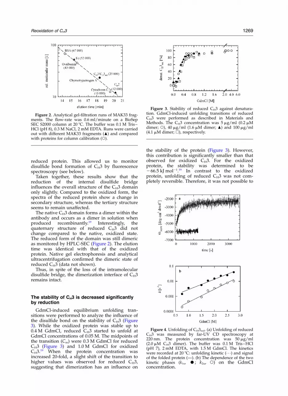
The native CH3 domain forms a dimer within theantibody and occurs as a dimer in solution whenproduced recombinantly.19 Interestingly, thequaternary structure of reduced CH3 did notchange compared to the native, oxidized state.The reduced form of the domain was still dimericas monitored by HPLC-SEC (Figure 2). The elutiontime was identical with that of the oxidizedprotein. Native gel electrophoresis and analyticalultracentrifugation confirmed the dimeric state ofreduced CH3 (data not shown).
Thus, in spite of the loss of the intramoleculardisulfide bridge, the dimerization interface of CH3remains intact.
The stability of CH3 is decreased significantlyby reduction
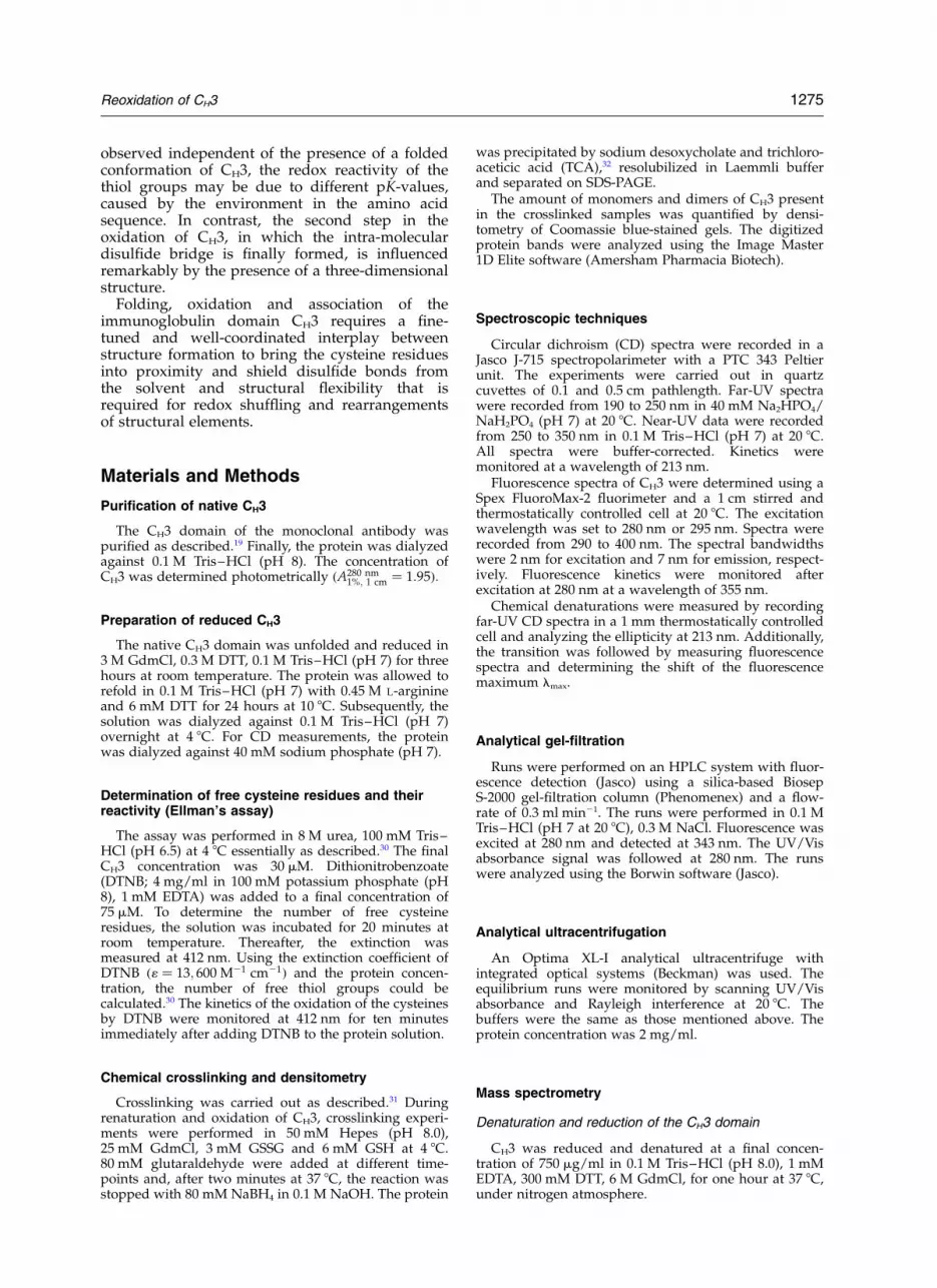
GdmCl-induced equilibrium unfolding tran-sitions were performed to analyze the influence ofthe disulfide bond on the stability of CH3 (Figure3). While the oxidized protein was stable up to0.4 M GdmCl, reduced CH3 started to unfold atGdmCl concentrations of 0.05 M. The midpoints ofthe transition (Cm) were 0.3 M GdmCl for reducedCH3 (Figure 3) and 1.0 M GdmCl for oxidizedCH3.19 When the protein concentration wasincreased 20-fold, a slight shift of the transition tohigher values was observed for reduced CH3,suggesting that dimerization has an influence on
the stability of the protein (Figure 3). However,this contribution is significantly smaller than thatobserved for oxidized CH3. For the oxidizedprotein, the stability was determined to be266.5 kJ mol21.19 In contrast to the oxidizedprotein, unfolding of reduced CH3 was not com-pletely reversible. Therefore, it was not possible to
Figure 2. Analytical gel-filtration runs of MAK33 frag-ments. The flow-rate was 0.6 ml/minute on a BioSepSEC S2000 column at 20 8C. The buffer was 0.1 M Tris–HCl (pH 8), 0.3 M NaCl, 2 mM EDTA. Runs were carriedout with different MAK33 fragments (O) and comparedwith proteins for column calibration (W).
Figure 3. Stability of reduced CH3 against denatura-tion. GdmCl-induced unfolding transitions of reducedCH3 were performed as described in Materials andMethods. The CH3 concentration was 5 mg/ml (0.2 mMdimer; W), 40 mg/ml (1.6 mM dimer; O) and 100 mg/ml(4.1 mM dimer; A), respectively.
Figure 4. Unfolding of CH3red. (a) Unfolding of reducedCH3 was measured by far-UV CD spectroscopy at220 nm. The protein concentration was 50 mg/ml(2.0 mM CH3 dimer). The buffer was 0.1 M Tris–HCl(pH 7), 2 mM EDTA, with 1.5 M GdmCl. The kineticswere recorded at 20 8C: unfolding kinetic (· · ·) and signalof the folded protein (—). (b) The dependence of the twokinetic phases (k1u, †; k2u, W) on the GdmClconcentration.
Reoxidation of CH3 1269

calculate the thermodynamic parameters of thetransition. As an estimate, the reduced dimer hasan eightfold lower stability than the oxidizeddimer.
Besides the low thermodynamic stability,reduced CH3 exhibited a kinetic instability againstchemical denaturants. As shown in Figure 4(a),reduced CH3 unfolded in a fast reaction withinone hour, with a half-time of 900 seconds. Themuch more stable oxidized protein had a half-time of 2800 seconds.19 Interestingly, the analysisof the unfolding kinetic of reduced CH3 revealedtwo rate constants (k1u ¼ 5:6 £ 1023 s21 (64% of theamplitude) and k2u ¼ 8:9 £ 1024 s21 (36% ofthe amplitude); u stands for unfolding), whereasthe unfolding process of the oxidized domain
could be described by one rate constantðku ¼ 2:5 £ 1024 s21Þ.19 Both rate constants ofreduced CH3 were influenced by the concentrationof the denaturant, the slow phase being affectedmore strongly than the fast unfolding phase(Figure 4(b); Table 2).
Oxidation of CH3 during folding
To analyze how folding and disulfide bond for-mation are coupled, folding of CH3 was monitoredby fluorescence spectroscopy under oxidizing con-ditions in the presence of a redox system consistingof reduced and oxidized glutathione (Figure 5(a)).At pH 8, the kinetic trace could be described ade-quately only by a three-exponential function withrate constants kfo1 ¼ 2:3 £ 1023 s21; kfo2 ¼3:1 £ 1024 s21 and kfo3 ¼ 5:6 £ 1025 s21 (fo standsfor folding and oxidation). The overall process isvery slow, reaching completion after 24 hours.
Whereas the fluorescence kinetics reflect bothoxidation and the concomitant changes in theprotein structure, oxidation was monitored directlyby mass spectrometry (MS) to gain insight into themechanism of disulfide formation of CH3. Reducedand denatured CH3 was diluted in refolding bufferwith the redox system mentioned above, aliquotswere withdrawn at different times and the cysteineresidues were alkylated with iodacetamide (IAA).The carboxy-amido-methylation reaction increasedthe molecular mass of oxidation intermediates by57 Da for each free SH group, thus allowing theseparation of intermediates containing differentnumbers of disulfide bonds. Using electroionisa-tion spray (ESI)-MS, alkylated intermediates canbe quantified accurately by measuring the totalion current produced by each species (Figure5(b)).20 The measured molecular masses, theexpected mass values, and the identification of thedetected intermediates are summarized in Table 1.At the beginning of the oxidation of CH3, the fullyreduced species (2H) disappeared fast ðk ¼2:8 s21Þ: After ten minutes, the decrease of 2H con-tinued very slowly (Table 2). Fully reduced proteincould be detected up to 120 minutes after initiatingrefolding. Intermediates with one reduced cysteineresidue and one mixed disulfide with glutathione(1G1H) appeared rapidly, with the same rate con-stant as that determined for the disappearance of2H at early stages of refolding and predominatedduring the first 25 minutes. Later, the fullyoxidized species (1S) became most abundant. The
Figure 5. Folding and oxidation of CH3. (a) The fluor-escence kinetics were measured at a wavelength of355 nm (excitation 280 nm). The protein concentrationwas 7.5 mg/ml (0.3 mM dimer) in 0.1 M Tris–HCl (pH8), 3 mM GSSG, 6 mM GSH, 4 8C. (b) Refolding and oxi-dation of CH3 analyzed by ESI-MS after derivatizationof the free cysteine residues with IAA: (W) The 2Hspecies, (K) 1G1H, (P) 2G and (B) 1S. The buffer con-ditions and protein concentrations were the same as forthe fluorescence measurements.
Table 1. ESI-MS analysis of the intermediates formed during refolding of CH3
Measured mass (Da) Identificationa Expected mass (Da) Possible isomers
12,363.57 ^ 0.65 2CAM (2H) 12,363.93 112,612.51 ^ 0.34 1G 1CAM (1G1H) 12,612.03 212,247.25 ^ 0.46 1S (1S) 12,247.71 112,860.53 ^ 0.72 2G (2G) 12,860.10 1
a CAM, carboxyamidomethyl groups; G, mixed disulfide with glutathione; S, intramolecular disulfide; H, free cysteine.
1270 Reoxidation of CH3

1S species was formed with a rate constant of1.3 £ 1022 s21 and reached a level of 100% after 180minutes. Interestingly, in addition to these species,a non-productive 2G intermediate was formed.This mixed disulfide derivative accumulated at aconcentration of about 10% in the first 30 minutesand then disappeared.
Dimerization of CH3 during folding andoxidation: association of the monomers isvery slow
We were interested in determining at whichstage of this reaction dimerization of CH3 takesplace. Therefore, we analyzed the time-course ofassociation by chemical crosslinking and sub-sequent SDS-PAGE. The time-course of dimeriza-tion was quantified by densitometry of themonomer and dimer bands (Figure 6(a)).
Figure 6(b) shows that dimerization of CH3under redox conditions occurred with a half-timeof 200 minutes. For the oxidized CH3 domain, thehalf-time for dimerization was determined to beten minutes.19 Thus, association of CH3 is deceler-ated significantly compared to dimerization of theoxidized domain.19 The amount of dimer reached100% after 24 hours. Interestingly, the dimerizationprocess was remarkably slower than the oxidation
of CH3 (see Figure 5(b)), but correlates well withthe overall kinetics of the folding and oxidationprocess monitored by fluorescence spectroscopy(Figure 5(a)).
Oxidation after refolding: the compactstructure of reduced folded CH3 preventsfast oxidation
To determine to what extent disulfide bond for-mation depends on the steric environment of therespective cysteine residues and on protein struc-ture formation in general, we next analyzed theoxidation process starting from the structuredprotein. To this end, the protein was first refoldedunder reducing conditions. GSSG was then addedto start the oxidation reaction. This process wasfollowed by monitoring the decrease in fluor-escence intensity (Figure 7(a)). The fluorescencekinetics consisted of three phases with the corre-sponding rate constants koaf1 ¼ 1:8 £ 1023 s21;koaf2 ¼ 3:6 £ 1024 s21 and koaf3 ¼ 4:8 £ 1025 s21 (oafstands for oxidation after folding). This reactionwas completed after 16 hours (Table 2).
ESI-MS measurements revealed the relativeintensities of the intermediates formed during theoxidation of folded CH3 (Figure 7(b)). The 2Hspecies decreased rapidly during the first 15
Table 2. Comparison of kinetic constants of reduced and oxidized CH3 shown as half-times
Unfolding Oxidation during folding Folding Oxidation after refolding
Dimerizationt1/2 (min-
utes)
CH3red t1u1=2 ¼ 120 s;
t2u1=2 ¼ 780 s
tfo11=2 ¼ 300 s; tfo2
1=2 ¼ 2240 s;tfo31=2 ¼ 12; 380 s
– toaf11=2 ¼ 390 s; toaf2
1=2 ¼ 1930 s;toaf31=2 ¼ 14; 440 s
200
CH3ox
(19)t1=2 ¼ 2800 s – t1
1=2 ¼ 65 s; t21=2 ¼ 1330 s;
t31=2 ¼ 4330 s
– 10
CH3red, CH3 reduced form; CH3ox, CH3 oxidized form.
Figure 6. Dimerization of CH3during folding and oxidation.Crosslinking experiments werecarried out in 50 mM Hepes (pH8.0), 25 mM GdmCl, 3 mM GSSGand 6 mM GSH at 4 8C. The proteinconcentration was 7.5 mg/ml(0.3 mM CH3 dimer). During refold-ing, samples of 5 mg of proteinwere taken, crosslinked with glutar-aldehyde for two minutes andprecipitated. (a) Coomassie-stainedSDS/polyacrylamide gel. Lanes1–19 represent the time-course ofdimerization: lane 1, 0.5 minute;lane 2, one minute; lane 3, twominutes; lane 4, five minutes; lane5, ten minutes; lane 6, 15 minutes;lane 7, 20 minutes; lane 8, 30
minutes; lane 9, 45 minutes; lane 10, one hour; lane 11, two hours; lane 12, three hours; lane 13, four hours; lane 14,five hours; lane 15, six hours; lane 16, ten hours; lane 17, 11 hours; lane 18, 22.5 hours; lane 19, 23 hours. (b) Thetime-course of the formation of the CH3 dimer determined by densitometry.
Reoxidation of CH3 1271

minutes together with the accumulation of the1G1H (45%) species, which represents the mixeddisulfide with glutathione. The kinetic of theformation and disappearance of 1G1H is com-parable to that of 1G1H during oxidative foldingof the denatured and reduced protein (Figure 5(b)).After 15 minutes, however, the remaining 2Hspecies disappeared very slowly over 24 hours.The fully oxidized species, 1S, appeared after 15minutes and became the most abundant after 45minutes. The relative amount reached 100% after24 hours. It should be noted that the non-productive intermediate 2G, containing two mixeddisulfides with glutathione, accumulated at a levelhigher than 15% from 15 minutes onwards.
Taken together, these experiments show thatoxidation of folded CH3 is a very slow process.The rapid formation and accumulation of the1G1H intermediate is not correlated with the fastformation of the fully oxidized species. Thisindicates that there is an intrinsic difficulty informing the disulfide in the structured CH3domain, possibly due to the environment of therespective cysteine residues.
To further analyze the influence of the proteinstructure on the oxidation reaction, we oxidizedCH3 in a partially folded state in the presence oflimited concentrations of denaturant (1 MGdmCl). Aliquots were collected at different timesduring refolding and intermediates were analyzedby ESI-MS after alkylation, as described (Figure 8).In this experiment, a steady state was reachedafter 30 minutes. It is important to underline thefact that the concentration of species 2H and1G1H was approximately equal from 30 minutesonwards, while species 1S was present throughoutat a very low level. The kinetic analysis showedfurther that the non-productive intermediate 2Gaccumulated at a concentration higher than 15%.
As a control, the reduced and denatured CH3domain was diluted into a buffer containing 6 MGdmCl. The kinetic analysis of species formed inthe presence of the redox system showed clearlythat 2H is the only stable species present insolution (data not shown).
Taken together, these results suggest that, start-ing from the structured domain, oxidation of CH3is hampered by the steric constraints. On the otherhand, formation of the structure of CH3 is necess-ary for protecting this covalent bond againstreduction.
Assignment of disulfide bonds within thepopulation of 1G1H intermediates
From the analysis described above, the questionarose whether in the 1G1H species one of the twocysteine residues is preferentially labelled orwhether the 1G1H intermediate represents amixture of the two isomers differing in the cysteineresidue involved in the formation of the mixeddisulfide with glutathione. In order to address thispoint, the sequence position of the modifiedcysteine residue was assigned using a massmapping strategy.21 Aliquots of the folding andoxidation reaction were withdrawn after one, two,
Figure 7. Oxidation after refolding. The protein wasallowed to refold for three hours under reducing con-ditions. Then, the formation of the disulfide bond wasinduced by addition of GSSG. The protein concentrationwas 7.5 mg/ml (0.3 mM dimer) in 0.1 M Tris–HCl (pH8), 3 mM GSSG, 6 mM GSH, 4 8C. (a) Fluorescence kineticmeasured at a wavelength of 355 nm (excitation 280 nm).(b) ESI-MS analysis of the CH3 species after derivatiza-tion of the free cysteine residues: (W) 2H species, (K)1G1H, (P) 2G and (B) 1S.
Figure 8. Refolding and oxidation of CH3 under desta-bilizing conditions. The reaction was performed in 1 MGdmCl, 0.1 M Tris–HCl (pH 8), 3 mM GSSG and 6 mMGSH at 4 8C. The protein concentration was 7.5 mg/ml(0.3 mM dimer). The reaction solution was analyzed byESI-MS after derivatization of the free cysteine residueswith IAA. (W) The 2H species, (K) 1G1H, (P) 2G and(B) 1S.
1272 Reoxidation of CH3

15 or 30 minutes, alkylated as described and lyo-philized. The samples were then hydrolyzed withcyanogen bromide and the mixture of peptideswas analyzed by matrix-assisted laser desorption/ionization(MALDI)-MS. Figure 9 shows theMALDI-MS analysis of the peptide mixturederived from the hydrolysis of the sample with-drawn after one minute when the species 1G1Hreached almost 40% (Figure 5(b)). The signals atm/z 5793.6 and 5545.2 represent peptide 60–108linked to a molecule of glutathione or containing acarboxyamidomethylated cysteine residue, respect-ively. In addition, the spectrum shows the presenceof a signal at m/z 1123.5 corresponding to peptide20–29 containing a carboxyamidomethylatedcysteine residue. The two signals at m/z 3583.8and 2061.8, respectively, were assigned to peptidesalong the CH3 sequence as reported in Figure 9.This analysis shows clearly that only cysteine 86was involved in the formation of the mixed disul-fide with glutathione. No signal corresponding topeptide 20–29 linked to a molecule of glutathionewas ever observed. The analysis of the peptidemixture derived from the hydrolysis of the samplewithdrawn after two minutes revealed the pre-sence of the same mass signals, confirming thatspecies 1G1H is homogeneous and containscysteine 86 involved in a mixed disulfide withglutathione.
The same analysis was performed on aliquotswithdrawn after 15 and after 30 minutes of therefolding in the presence of 1 M GdmCl when the1G1H species reached a concentration of about45% (see Figure 8). The MALDI spectra of thesesamples showed the presence of the same signalsdescribed above, thus strengthening the conclusion
that cysteine 86 is more reactive than cysteine 28 informing the mixed disulfide with glutathione.
To investigate whether the reactivity of thecysteine residues is influenced by the conformationof CH3 or the sequential context, we analyzed thereaction of the reduced and denatured CH3 domainwith dithionitrobenzoate (DTNB). The kinetics ofoxidation could be fitted by a two-exponential fitresulting in the rate constants of k1 ¼ 2:8 s21 andk2 ¼ 0:6 s21 (data not shown). This indicates thatone cysteine residue reacts with DTNB slightlyfaster than the other. Because of the absence ofany protein structure, this behavior must be dueto the amino acid sequence flanking cysteineresidues 28 and 86, respectively.
Discussion
Proteins that traverse the secretory pathwaystypically contain one or more disulfide bonds thatstabilize the folded conformation by lowering theentropy of the unfolded state.22 Systematic experi-mental studies revealed a significant impact ofdisulfide bonds on the thermodynamics andkinetics of protein folding.13,23,24
In immunoglobulin domains, the disulfidebridge connecting the sheets of the b-sandwichstructure is well conserved.10,25 – 27 It has beenshown experimentally that it is an exceptionallyimportant factor in stabilizing the structure of theconstant part of the immunoglobulin light chain,of two different variable domains of the lightchain and of an scFv fragment.7 – 11 Here, we haveevaluated the impact of the intramoleculardisulfide bond on the structure and stability of the
Figure 9. Disulfide bond assignment within the population of 1G1H intermediates. MALDI-MS analysis of thepeptide mixture derived from the hydrolysis of the sample withdrawn one minute after starting folding and oxidationof CH3. The amount of 1G1H in the solution was about 40%.
Reoxidation of CH3 1273

antibody CH3 dimer. Reduced CH3 shows a muchlower stability than the oxidized state. Comparedto the contribution of the disulfide bond to thestability of the CL domain7,28 the 4D5-SSþ scFvfragment11 and of different mutants of the REIv-kvariable domain,9 the influence of the disulfidebridge on the stability of CH3 is remarkably greater.In contrast to the oxidized domain, unfolding ofCH3 occurs in two phases: a fast phase, character-ized by a large change in CD-signal intensity,which seems to be due to the destruction of thedomain interface, and a slow phase with a smallincrease in signal intensity, as would be expectedfor the unfolding of an isolated domain. The result-ing rate constant corresponds to that of the unfold-ing phase of oxidized CH3. A similar behavior wasobserved in the case of the wild-type and theVL
þVH2 variant of the scFv fragment 4D5.29
Interestingly, reduced CH3 shows a slightlyaltered secondary structure, but the tertiarystructure, as judged by fluorescence and CD spec-troscopy, seems to remain unchanged compared tothe native oxidized state. The increased signal inthe far-UV CD spectrum may indicate a more com-pact secondary structure due to the removal of thedisulfide spacer between the two b-sheets of CH3.These results are consistent with that obtained forthe CL domain after reduction.7 However, the CL
domain is monomeric, whereas reduced CH3 is adimer. The domain interface of oxidized CH3 buriesa significant part of the surface upon association.The conformation of this surface seems to be con-served in reduced CH3, since the protein retains itsdimeric state. A similar behavior was observed forthe reduced 4D5-SS scFv fragment.11 Here, how-ever, the two domains are linked artificially by apeptide sequence that favors association byincreasing the local concentration of the twodomains. Our results show that in reduced CH3non-covalent interactions are sufficient to maintainthe b-sheet structure and promote stable andspecific dimerization.
The necessity of a fine-tuned coordinationbetween structure formation and oxidation in theformation of native protein is clearly illustrated bythe refolding/oxidation experiments of CH3. Here,redox reactions are decelerated by the burial ofthe reactive groups in stable tertiary structure. Thevelocity of disulfide bond formation in folded,reduced CH3 illustrates this effect. We observed aneightfold deceleration of the reaction compared tothe oxidation concomitant with refolding ofdenatured CH3. The rate of disulfide bond for-mation depends also on the proximity in space ofthe respective cysteine residues, defined here asthe probability of their sulfur atoms to come withinthe distance required for thiol/disulfide exchange.Consequently, the formation of the internaldisulfide bridge of CH3 is influenced significantlyby the concentration of denaturant in the solution.When oxidation and folding take place simul-taneously, disulfide bond formation occurs withinthree hours via mixed disulfide intermediates,
complexes of one or two glutathione moleculeswith the free thiol groups of the CH3 domain.These derivatives accumulate at the beginning ofthe reaction and disappear subsequently becauseof the formation of oxidized CH3. In contrast, fold-ing monitored by fluorescence is completed onlyafter 24 hours. These kinetics may be due to mis-folded states of CH3 that are caught in their confor-mation by the disulfide bridge. Subsequently,rearrangements in these conformational statesmay occur only slowly. This is in agreement withprevious observations that fast disulfide bond for-mation can decelerate folding by stabilizingentropically misfolded conformations.22 Similar tothe overall kinetics of folding and oxidation deter-mined by fluorescence spectroscopy, the dimeriza-tion process of CH3 starting from the unfolded andreduced protein is very slow. In contrast tooxidized CH3, where monomers dimerize in asingle-exponential reaction,19 dimerization ofreduced CH3 seems to involve two differentreactions. Fast association seems to involve CH3molecules that are properly folded and oxidized.This species reaches the dimeric state within thetime range necessary for the oxidation of CH3. Theslow association reaction may represent incom-pletely or misfolded states that have to undergostructural changes before association can takeplace. The changes in fluorescence intensityobserved during the later stages of folding couldbe due to rearrangements of the contact surfacesduring dimerization. Our results suggest that fold-ing and oxidation of the CH3 domain are tightlycoupled. Folding is required to bring the cysteineresidues within bonding distance; on the otherhand, structure formation negatively influencesredox shuffling.
In agreement with this interpretation, increasingthe amount of denaturant in the solution slowsCH3 oxidation. Under unfolding conditions, theformation of disulfide bonds no longer takesplace. Remarkably, the mixed disulfide derivativeof CH3 with glutathione (1G1H) is formed withthe same velocity under all conditions investigated.Within the first 15 minutes, the amount of 1G1Hreaches 40 to 45%. In a partially folded state, oxi-dation of CH3 arrests after 30 minutes. The fullyoxidized species is present at a very low level,whereas reduced and partially oxidized CH3molecules predominate. Under unfolding con-ditions, the distance between the cysteine residuesand the high conformational flexibility of the poly-peptide chain completely prevent the formation ofthe internal disulfide bridge of CH3. The fullyreduced species is the only one observed in sol-ution since, in the absence of structure, disulfidebonds are accessible for reducing compounds.
Under all conditions investigated, oxidation ofCH3 occurs via a homogeneous 1G1H species inwhich exclusively cysteine 86 forms the mixeddisulfide with glutathione. Cysteine 28 is muchless reactive and oxidized in a second step formingthe redox species 2G. Since this behavior is
1274 Reoxidation of CH3
observed independent of the presence of a foldedconformation of CH3, the redox reactivity of thethiol groups may be due to different pK-values,caused by the environment in the amino acidsequence. In contrast, the second step in theoxidation of CH3, in which the intra-moleculardisulfide bridge is finally formed, is influencedremarkably by the presence of a three-dimensionalstructure.
Folding, oxidation and association of theimmunoglobulin domain CH3 requires a fine-tuned and well-coordinated interplay betweenstructure formation to bring the cysteine residuesinto proximity and shield disulfide bonds fromthe solvent and structural flexibility that isrequired for redox shuffling and rearrangementsof structural elements.
Materials and Methods
Purification of native CH3
The CH3 domain of the monoclonal antibody waspurified as described.19 Finally, the protein was dialyzedagainst 0.1 M Tris–HCl (pH 8). The concentration ofCH3 was determined photometrically ðA280 nm
1%; 1 cm ¼ 1:95Þ:
Preparation of reduced CH3
The native CH3 domain was unfolded and reduced in3 M GdmCl, 0.3 M DTT, 0.1 M Tris–HCl (pH 7) for threehours at room temperature. The protein was allowed torefold in 0.1 M Tris–HCl (pH 7) with 0.45 M L-arginineand 6 mM DTT for 24 hours at 10 8C. Subsequently, thesolution was dialyzed against 0.1 M Tris–HCl (pH 7)overnight at 4 8C. For CD measurements, the proteinwas dialyzed against 40 mM sodium phosphate (pH 7).
Determination of free cysteine residues and theirreactivity (Ellman’s assay)
The assay was performed in 8 M urea, 100 mM Tris–HCl (pH 6.5) at 4 8C essentially as described.30 The finalCH3 concentration was 30 mM. Dithionitrobenzoate(DTNB; 4 mg/ml in 100 mM potassium phosphate (pH8), 1 mM EDTA) was added to a final concentration of75 mM. To determine the number of free cysteineresidues, the solution was incubated for 20 minutes atroom temperature. Thereafter, the extinction wasmeasured at 412 nm. Using the extinction coefficient ofDTNB ð1 ¼ 13; 600 M21 cm21Þ and the protein concen-tration, the number of free thiol groups could becalculated.30 The kinetics of the oxidation of the cysteinesby DTNB were monitored at 412 nm for ten minutesimmediately after adding DTNB to the protein solution.
Chemical crosslinking and densitometry
Crosslinking was carried out as described.31 Duringrenaturation and oxidation of CH3, crosslinking experi-ments were performed in 50 mM Hepes (pH 8.0),25 mM GdmCl, 3 mM GSSG and 6 mM GSH at 4 8C.80 mM glutaraldehyde were added at different time-points and, after two minutes at 37 8C, the reaction wasstopped with 80 mM NaBH4 in 0.1 M NaOH. The protein
was precipitated by sodium desoxycholate and trichloro-aceticic acid (TCA),32 resolubilized in Laemmli bufferand separated on SDS-PAGE.
The amount of monomers and dimers of CH3 presentin the crosslinked samples was quantified by densi-tometry of Coomassie blue-stained gels. The digitizedprotein bands were analyzed using the Image Master1D Elite software (Amersham Pharmacia Biotech).
Spectroscopic techniques
Circular dichroism (CD) spectra were recorded in aJasco J-715 spectropolarimeter with a PTC 343 Peltierunit. The experiments were carried out in quartzcuvettes of 0.1 and 0.5 cm pathlength. Far-UV spectrawere recorded from 190 to 250 nm in 40 mM Na2HPO4/NaH2PO4 (pH 7) at 20 8C. Near-UV data were recordedfrom 250 to 350 nm in 0.1 M Tris–HCl (pH 7) at 20 8C.All spectra were buffer-corrected. Kinetics weremonitored at a wavelength of 213 nm.
Fluorescence spectra of CH3 were determined using aSpex FluoroMax-2 fluorimeter and a 1 cm stirred andthermostatically controlled cell at 20 8C. The excitationwavelength was set to 280 nm or 295 nm. Spectra wererecorded from 290 to 400 nm. The spectral bandwidthswere 2 nm for excitation and 7 nm for emission, respect-ively. Fluorescence kinetics were monitored afterexcitation at 280 nm at a wavelength of 355 nm.
Chemical denaturations were measured by recordingfar-UV CD spectra in a 1 mm thermostatically controlledcell and analyzing the ellipticity at 213 nm. Additionally,the transition was followed by measuring fluorescencespectra and determining the shift of the fluorescencemaximum lmax.
Analytical gel-filtration
Runs were performed on an HPLC system with fluor-escence detection (Jasco) using a silica-based BiosepS-2000 gel-filtration column (Phenomenex) and a flow-rate of 0.3 ml min21. The runs were performed in 0.1 MTris–HCl (pH 7 at 20 8C), 0.3 M NaCl. Fluorescence wasexcited at 280 nm and detected at 343 nm. The UV/Visabsorbance signal was followed at 280 nm. The runswere analyzed using the Borwin software (Jasco).
Analytical ultracentrifugation
An Optima XL-I analytical ultracentrifuge withintegrated optical systems (Beckman) was used. Theequilibrium runs were monitored by scanning UV/Visabsorbance and Rayleigh interference at 20 8C. Thebuffers were the same as those mentioned above. Theprotein concentration was 2 mg/ml.
Mass spectrometry
Denaturation and reduction of the CH3 domain
CH3 was reduced and denatured at a final concen-tration of 750 mg/ml in 0.1 M Tris–HCl (pH 8.0), 1 mMEDTA, 300 mM DTT, 6 M GdmCl, for one hour at 37 8C,under nitrogen atmosphere.
Reoxidation of CH3 1275

Oxidation after refolding of CH3
The renaturation was started by diluting 100-fold thereduced and denatured protein in 0.1 M Tris–HCl (pH8.0), 1 mM EDTA containing DTT at a final concentrationof 3 mM. The process was carried out at 4 8C at a finalprotein concentration of 7.5 mg/ml. After three hours ofrefolding, 6 mM GSSG was added and the oxidationcarried out at 4 8C.
Refolding and oxidation of CH3
Oxidative refolding was started by diluting 100-foldthe reduced and denatured protein in 0.1 M Tris–HCl(pH 8.0), 1 mM EDTA, containing 6 mM GSSG. Theprocess was carried out at 4 8C at a final protein concen-tration of 7.5 mg/ml.
Formation of the disulfide bond of the CH3 domain in thepresence of GdmCl
The process was started by diluting 100-fold thereduced and denatured protein in 0.1 M Tris–HCl (pH8.0), 1 mM EDTA, containing 6 mM GSSG and 1 MGdmCl. Alternatively, the refolding buffer contained 36or 60 mM GSSG. The reaction was carried out at 4 8C ata final protein concentration of 7.5 mg/ml.
Alkylation of the intermediates during refolding
The refolding of the CH3 domain was monitored on atime-course basis by sampling aliquots of the refoldingmixtures at appropriate intervals. The protein sampleswere alkylated with IAA as described.33,34 IAA wasfreshly dissolved to a concentration of 2.2 M in 1 MTris–HCl (pH 6.5), 10 mM EDTA, at 65 8C and cooled toroom temperature before use. During preparation ofreagents, the solutions were protected from light to mini-mize photolytic production of iodine, which is a verypotent oxidizing agent for thiol groups. The refoldingaliquots (2 ml) were added to 1.1 ml of IAA solution ata final IAA concentration of 0.8 M. Alkylation was per-formed for 15 seconds in the dark at room temperature.After 15 seconds, 0.9 ml of 0.45% (v/v) trifluoraceticacid (TFA) was added and the aliquots were quicklyvortex mixed and desalted by HPLC using a Vydacreversed-phase C4 column (0.46 cm £ 25 cm). Theelution system consisted of solvent A (0.1% TFA inwater) and solvent B (0.07% TFA in 95% acetonitrile/5%water). Refolding intermediates were desalted with alinear gradient of solvent B from 20% to 95% in fiveminutes at a flow-rate of 1 ml/minute. Eluted proteinswere monitored at 220 nm, collected manually andanalyzed directly by mass spectrometry.
Peptide mapping
Refolding intermediates were alkylated as describedand lyophilized. The carboxyamidomethylated proteinswere hydrolyzed with cyanogen bromide in 200 ml of70% TFA using a tenfold molar excess of reagent overmethionine residues for 18 hours at room temperaturein the dark. The cyanogen bromide reaction was stoppedby adding ten volumes of cold water and lyophilizingthe sample.
Mass spectrometric analysis
ESI-MS analyses were carried out using a BIO-Q triplequadrupole mass spectrometer equipped with an electro-spray ion source (Micromass). Formic acid was added tothe eluted protein samples to a final concentration of 5%(v/v). Samples were introduced into the ion source byinfusion at a flow-rate of 5 ml/minute. Spectra wererecorded by scanning the quadrupole at ten seconds/scan. Data were acquired and elaborated by theMassLynx software. Mass scale calibration was per-formed by means of multiply charged ions from aseparate injection of horse heart myoglobin (averagemolecular mass 16,951.5 Da). Each set of refolding datawas obtained as the means of three independent foldingexperiments. The differences between refolding experi-ments performed completely independent of each otherwere about 5%. The disulfide intermediates that formedcould be identified by ESI-MS. Each trapped inter-mediate is characterized by the presence of an intra-molecular disulfide bond (indicated as 1S), mixeddisulfides with the exogenous glutathione (nG) and car-boxyamidomethyl groups (nCAM). The number ofCAM groups corresponds to the number of free thiolgroups present in the refolding intermediates and istherefore indicated as nH. It is important to emphasizethat in the species 1G1H, the position of the effectivecysteine residue involved in the mixed disulfide is notidentified (Table 1).
Peptide mixtures were analyzed by MALDI-time-of-flight (TOF)-MS. The MALDI-MS analyses were carriedout using a Voyager DE mass spectrometer (PerSeptiveBiosystems, Boston, MA, USA). The mass range was cali-brated using bovine insulin (average molecular mass5734.6 Da) and a matrix peak (379.1 Da) as internal stan-dards. Samples were dissolved in 0.2% (v/v) TFA at10 pmol/ml. An aliquot (1 ml) was applied to a sampleslide together with 1 ml of a solution of a-cyano-4-hydroxycinnamic acid (10 mg/ml) in acetonitrile/0.1%TFA (1:2, v:v). The matrix and the sample wereallowed to air-dry before spectra were collected. Massspectra were generated from the sum of 50 laser shots.
Acknowledgments
We thank Hauke Lilie for stimulating discussions andcritically reading the manuscript. The work was sup-ported by funds from the European Union and theFonds der Chemischen Industrie.
References
1. Deisenhofer, J. (1981). Crystallographic refinementand atomic models of a human Fc fragment and itscomplex with fragment B of protein A fromStaphylococcus aureus at 2.9 and 2.8 A resolution.Biochemistry, 20, 2361–2370.
2. Williams, A. F. & Barclay, A. N. (1988). The immuno-globulin superfamily—domains for cell surfacerecognition. Annu. Rev. Immunol. 6, 381–405.
3. Bergman, L. W. & Kuehl, W. M. (1979). Formation ofan intrachain disulfide bond on nascent immuno-globulin light chains. J. Biol. Chem. 254, 5690–5694.
4. Bardwell, J. C. A. (1994). Building bridges:disulphide bond formation in the cell. Mol. Microbiol.14, 199–205.
1276 Reoxidation of CH3

5. Freedman, R. B., Brookway, D. E. & Lambert, N.(1984). Protein disulphide-isomerase and the for-mation of native disulphide bonds. Biochem. Soc.Trans. 12, 929–932.
6. Melnick, J., Aviel, S. & Argon, Y. (1992). The endo-plasmic reticulum stress protein GRP94, in additionto BiP, associates with unassembled immunoglobulinchains. J. Biol. Chem. 267, 21303–21306.
7. Goto, Y. & Hamaguchi, Y. (1979). The role of theintrachain disulfide bond in the conformation andstability of the constant fragment of the immuno-globulin light chain. J. Biochem. 86, 1433–1441.
8. Rudikoff, S. & Pumphrey, J. G. (1986). Functionalantibody lacking a variable-region disulfide bridge.Proc. Natl Acad. Sci. USA, 83, 7875–7878.
9. Frisch, C., Kolmar, H., Schmidt, A., Kleemann, G.,Reinhardt, A., Pohl, E. et al. (1996). Contribution ofthe intramolecular disulfide bridge to the foldingstability of REIv, the variable domain of a humanimmunoglobulin kappa light chain. Fold. Des. 1,431–440.
10. Proba, K., Honegger, A. & Pluckthun, A. (1997). Anatural antibody missing a cysteine in VH: conse-quences for thermodynamic stability and folding.J. Mol. Biol. 265, 161–172.
11. Worn, A. & Pluckthun, A. (1998). An intrinsicallystable antibody scFv fragment can tolerate the lossof both disulfide bonds and fold correctly. FEBSLetters, 427, 357–361.
12. Creighton, T. E. (1997). Protein folding coupled todisulfide bond formation. Biol. Chem. 378, 731–744.
13. Abkevich, V. I. & Shakhnovich, E. I. (2000). What candisulfide bonds tell us about protein energetics, func-tion and folding: simultations and bioinformaticsanalysis. J. Mol. Biol. 300, 975–985.
14. Goto, Y. & Hamaguchi, Y. (1982). Unfolding andrefolding of the constant fragment of the immuno-globulin light chain. Kinetic role of the intrachaindisulfide bond. J. Mol. Biol. 156, 911–926.
15. Buchner, J. & Rudolph, R. (1991). Renaturation,purification and characterization of recombinantFab-fragments produced in Escherichia coli. Bio/Technology, 9, 157–162.
16. Schmidt, M. & Buchner, J. (1992). Interaction of GroEwith an all-beta-protein. J. Biol. Chem. 267,16829–16833.
17. Lilie, H., McLaughlin, S., Freedman, R. & Buchner, J.(1994). Influence of protein disulfide isomerase(PDI) on antibody folding in vitro. J. Biol. Chem. 269,14290–14296.
18. Isenman, D. E., Lancet, D. & Pecht, I. (1979). Foldingpathways of immunoglobulin domains. The foldingkinetics of the Cg3 domain of human IgG1.Biochemistry, 18, 3327–3336.
19. Thies, M. J. W., Mayer, J., Augustine, J. G., Frederick,C. A., Lilie, H. & Buchner, J. (1999). Folding andassociation of the antibody domain CH3: prolyl
isomerization preceeds dimerization. J. Mol. Biol.293, 67–79.
20. Ruoppolo, M., Freedman, R. B., Pucci, P. & Marino,G. (1996). Glutathione-dependent pathways ofrefolding of RNase T1 by oxidation and disulfide iso-merization: catalysis by protein disulfide isomerase.Biochemistry, 35, 13636–13646.
21. Pucci, P. & Morris, H. R. (1985). A new method forrapid assignment of SS bridges in proteins. Biochem.Biophys. Res. Commun. 126, 1122–1128.
22. Anfinsen, C. & Scheraga, H. (1975). Principles thatgovern the folding of protein chains. Advan. ProteinChem. 29, 205–299.
23. Clarke, J. & Fersht, A. (1993). Engineered disulfidebonds as probes of the folding pathway of bar-nase—increasing the stability of protein against therate of denaturation. Biochemistry, 32, 4322–4329.
24. Yokata, A., Izutani, K., Takai, M., Kubo, Y., Noda, Y.,Koumoto, Y. et al. (2000). The transition state in thefolding–unfolding reaction of four species of three-disulfide variant of hen lysozyme: the role of eachdisulfide bridge. J. Mol. Biol. 295, 1275–1288.
25. Alzari, P. M., Lascombe, M. B. & Poljak, R. J. (1988).Three-dimensional structure of antibodies. Annu.Rev. Immunol. 6, 555–580.
26. Davies, D. R., Padlan, E. A. & Sherriff, S. (1990). Anti-body–antigen complexes. Annu. Rev. Biochem. 59,439–473.
27. Padlan, E. A. (1996). X-crystallography of antibodies.Advan. Protein Chem. 49, 57–133.
28. Goto, Y., Tsunenaga, M., Kawata, Y. & Hamaguchi,K. (1987). Conformation of the constant fragment ofthe immunoglobulin light chain: effect of cleavageof the polypeptide chain and the disulfide bond.J. Biochem. 101, 319–329.
29. Ramm, K., Gehrig, P. & Pluckthun, A. (1999).Removal of the conserved disulfide bridges fromthe scFv fragment of an antibody: effects on foldingkinetics and aggregation. J. Mol. Biol. 290, 535–546.
30. Sedlak, J. & Lindsay, R. H. (1968). Estimation of total,protein-bound, and nonprotein sulfhydryl groups intissue with Ellman’s reagent. Anal. Biochem. 25,192–205.
31. Jaenicke, R. & Rudolph, R. (1989). Folding proteins.In Protein Structure: A Practical Approach (Creighton,T. E., ed.), pp. 191–223, IRL Press, Oxford.
32. Bensadoun, A. & Weinstein, W. (1976). Assay of pro-teins in the presence of interfering materials. Anal.Biochem. 70, 241–250.
33. Gray, W. R. (1993). Disulfide structures of highlybridged peptides: a new strategy for analysis. ProteinSci. 2, 1732–1748.
34. Torella, C., Ruoppolo, M., Marino, G. & Pucci, P.(1994). Analysis of RNase A refolding intermediatesby electrospray/mass spectrometry. FEBS Letters,352, 301–306.
Edited by R. Huber
(Received 2 January 2002; received in revised form 3 April 2002; accepted 10 April 2002)
Reoxidation of CH3 1277


![Calorimetric and Raman Study of the Phase Transition in [N(CH3)4]2ZnBr4](https://img.dokumen.tips/doc/110x75/6314d4c285333559270ce0b4/calorimetric-and-raman-study-of-the-phase-transition-in-nch342znbr4.jpg)


