Embed Size (px)
Citation preview
Floral Anatomy of the Santalaceaeand Some Related Forms
FRANK H. SMITH,ASSISTANT PROFESSOR OF BOTANY
ELIZABETH C. SMITH
OREGON STATE COLLEGECORVALLIS, OREGON. Printed at
The College Press. 1943.
OREGON STATE MONOGRAPHSStudies in Botany
Number 5, September 1942Published by Oregon State College
Oregon State System of Higher EducationCorvallis, Oregon
TABLE OF CONTENTS
Page
Explanation of plates 5
Preface 7
Acknowledgments 9
Introduction 11
Santalaceae 12
Darbya 13
Colpoon 16
Santalum 19
Osyris 22
Acanthosyris 24
Eucarya 28
Pyrularia 30
Comandra 31
Geocaulon 34
Nanodea 35
Buckleya 38
Exocarpus 40
Anthobolus 41
Henslowia 43
Jodina 45
Myoschilos 45
Choretrum 47
Leptomeria 49
Thesium 51
Thesidium 52
Omphacomeria 52
Osyridocarpus 54
Quinchamalium 55
Arjona 57
The staminate flower 58
Calyptosepalum 60
Discussion 60
TABLE OF CONTENTSContinued
Page
Olacaceae 67
Ochanostachys 67
Heisteria 68
Anacolosa 70
Strombosia 73
°lax 74
Aptandra 76
Schoepfia 76
Discussion 79
Grubbiaceae 80
Grubbia 80
Myzodendraceae 82
Myzodendron 82
Loranthaceae 83
Phoradendron 83
Arceuthobium 85
Relationships within the Santalales 86
Summary 88
Literature cited 93
EXPLANATION OF PLATES
The semidiagrammatic illustrations were drawn at various magnifications with theaid of a camera lucida. The magnification used for each figure was determined by thesize of the section and the detail to be shown. In general, illustrations of sections ofthe pedicel and basal portions of a flower are reproduced at greater magnifications thanthose of the upper portions. Sections of small flowers are reproduced at greater magni-fications than those of large ones.
The labels used are listed below together with their meanings.B bract or bract traceB-Se trace composed of fused bract and sepal tracesC commissural trace located between sepal positionsD diskDC dorsal carpellary traceP petal traceP-St trace composed of fused petal and stamen tracesRB recurrent bundles of the receptacleRT residual vascular tissueS xylem-like sclerenchymaSa. seta or trace to seta of MyzodendronSe sepal trace or tracesSe-St trace composed of fused sepal and stamen tracesSt stamen traceVC ventral carpellary trace or tracesX traces that arise from the recurrent bundles and move
inward to positions between the dorsal carpellary traces

PREFACE
The nature of the inferior ovary has been under discussion for manyyears. There are two theories regarding the morphology of the inferiorovary that have been widely accepted by various botanists. According to thefirst, the appendicular theory, an inferior ovary results from extreme adna-tion with the sepals, petals, and stamens fused with each other and with theovary wall. Thus the outer tissues of such an inferior ovary consist of thefused bases of the floral appendages. According to the second, the receptacu-lar theory, an inferior ovary results from invagination of the floral axis sothat the ovary itself is embedded in tissues of the receptacle. Thus the outertissues of such an inferior ovary are axial or receptacular in nature. A thirdpossibility that has been suggested is that an inferior ovary may consist ofreceptacular tissues in the lower portion and floral tissues above. This situa-tion occurs in the floral cup of several genera of the Rosaceae (Bonne, 1925,
1928 and Jackson, 1934). These are forms with perigynous flowers, how-ever, and the ovary is not inferior. The structure of the apple has also beeninterpreted in this manner but incorrectly so, as was shown by Mac Daniels(1940). The sepals, petals, and stamens of a flower with an inferior ovaryappear to be borne on or above the ovary. This would be an actual fact in areceptacular inferior ovary but only apparent for the appendicular type, sincein the latter the floral parts originate below the ovary, as in an hypogynousflower, but are so fused with the ovary in their lower portions as to lose theiridentity.
While the appendicular theory is probably older than the receptaculartheory, the latter has been, until recent years, more widely accepted. Thiswas due primarily to the work of Payer (1857) and other developmentalmorphologists. In general, taxonomists still describe the inferior ovary interms of the receptacular theory. The work of Eames (1931) and his col-
laborators has shown rather conclusively that the inferior ovary in the greatmajority of plant families is the result of adnation and hence is appendicularin nature. Thus the appendicular theory has come to be accepted by mostplant anatomists of recent times.
The authors recently described the floral anatomy of Darbya in whichthe inferior ovary is interpreted as receptacular. It was specifically stated,however, that this interpretation applies only to the Santalaceae. Acceptanceof the receptacular theory for this family does not deny the existence of theappendicular inferior ovary in other families. In fact, the evidence used tosupport the receptacular nature of the inferior ovary in the Santalaceae mayalso be considered as confirming the appendicular nature of the inferior ovaryin other forms. This is so because the anatomy of the receptacular inferior
7

ovary is so characteristic and entirely distinct from that of the appendiculartype. The floral anatomy of the inferior ovary as it occurs in the highermonocots and in most of the higher dicots, can be adequately interpreted onlyin terms of cohesion and adnation, while the anatomy of the inferior ovaryin the Santalaceae seems impossible of explanation except in terms of invagi-nation of the floral axis. There has been a tendency on the part of somebotanists to assume that these two theories are mutually exclusive, and thatto accept one is to reject the other. The inferior ovary has undoubtedlyappeared independently in different plant lines and there is no reason whyall should have the same structure. For example, in the Olacaceae, afew species of which are described in this paper, it seems likely that two andperhaps all of the three possible types of inferior ovary occur in this onefamily.
Assuming that the receptacular nature of the inferior ovary in Darbyahas been established, the primary purpose of this publication is to describethe many modifications in structure of the receptacular inferior ovary as itoccurs in the Santalaceae. Some additional species in other families of theSantalales were also studied in an attempt to trace the origin of the receptac-ular inferior ovary in the Santalaceae and to obtain some indication as torelationships within the order.
8
ACKNOWLEDGMENTS
The authors are grateful to Professor A. J. Eames, who has been gen-erous with his time and has offered many helpful suggestions and criticismsduring the progress of the work and in preparation of the manuscript. Thesenior author is also indebted to the Department of Botany, Cornell Univer-sity, where laboratory facilities and privileges were granted him as a ResidentDoctor. Acknowledgments are also due the herbaria of Cornell University,of the University of California, and the Gray Herbarium for furnishingmuch of the material for study. Material of Comandra Richardsiana waskindly supplied by Dr. N. C. Fassett of the University of Wisconsin.
This work was supported in part by a grant from the General ResearchCouncil, Oregon State College.
Department of BotanyOregon State CollegeSeptember 24, 1942
9
FRANK H. SMITHELIZABETH C. SMITH

Floral Anatomy of the Santalaceaeand Some Related Forms
INTRODUCTION
The Santalales comprise a heterogeneous group that has received muchattention from the taxonomist but little from the floral anatomist. Relation-ships are somewhat obscure and the position of some groups is much indoubt. The taxonomic literature, almost without exception, describes theinferior ovary of the Santalaceae, Grubbiaceae, Myzodendraceae and Loran-thaceae as embedded in tissues of the receptacle. This interpretation seemsto be based for the most part on popular adherence to the receptacular theoryof the nature of the inferior ovary rather than on anatomical evidence. Thenature of the inferior ovary in the Santalales has been variously interpretedby the comparatively few floral anatomists who have studied species belong-ing to this order. Van Tieghem (1869b), from a study of Thesium andOsyris, and Dowding (1931), from a study of Comandra and Arceuthobium,concluded that the ovary is appendicular. But Schaeppi and Steindl (1937),from their work on Osyris, and Smith and Smith (1942), in their study ofDarbya, considered the ovary to be receptacular though they did not agreeentirely in their interpretations. Rao (1942) made rather minor examina-tions of floral anatomy in connection with a study of the embryo sacs ofThesium, Santalum, Osyris and Scleropyron. While no definite statementwas made, his descriptions imply acceptance of the appendicular theory. Healso stated that from the flower primordia there first develops the perianthfollowed successively by the androecium, gynoecium, and central placenta.
Investigators have disagreed in their interpretation of the stalk bearingthe ovules in various members of the Santalales. This structure has beeninterpreted as consisting of carpel tissue only (Van Tieghem, 1869b andSmith and Smith, 1942), of axial tissue only (Johnson, 1889 and Rao, 1942),of axial and carpel tissue (Schaeppi and Steindl, 1937) and, on the basis ofcarpel polymorphism, of a whorl of reduced fertile carpels (Dowding, 1931,and Saunders, 1940).
The floral anatomy of the Santalales, especially of the Santalaceae, showsextreme variation between genera. This is probably to be expected sinceparasitic forms tend to become more highly diversified than nonparasiticforms. An attempt is made here to explain these variations in terms of theanatomical structure of Darbya, which is considered to represent a some-what primitive condition of floral anatomy in the Santalaceae. In this family
11
12 OREGON STATE COLLEGESTUDIES IN BOTANY
there are all gradations between perfect flowers and staminate and pistillateflowers with no trace of organs of the opposite type. In general only perfector pistillate flowers are considered here since the structure of the inferiorovary is of primary interest.
For most species only herbarium material was studied. This method ofinvestigation has decided disadvantages since it is impossible in most casesto study flowers of different ages. Also there were available only one or afew flowers of some species. While there may be some errors regardingminor details in floral anatomy, the authors believe that the descriptions ofthe major features are essentially correct. It was also necessary to usesomewhat atypical flowers of some species, as for example a 5-merousflower instead of a more typical 4-merous one. The technique for handlingherbarium material has been previously described (Smith and Smith, 1942).
To avoid confusion the nomenclature used in Engler and Pranti's"Natiirliche Pflanzenfamilien" (1935) has been followed. Taxonomic de-scriptions are, for the most part, taken from the same source.
SANTALACEAE
The flowers of various species in the Santalaceae may be perfect or im-perfect; and polygamous, monoecious and dioecious forms occur. The peri-anth is simple, consisting of four or five, seldom three, sepals. The stamensare as many as the sepals and are opposite to and usually adherent withthem. The ovary is inferior, partly inferior or (in the Anthoboleae) essen-tially superior, with two to five carpels. The ovary contains a single locule orthree to five locules at the base, merging into a single locule above. Theovules, which hang from the tip of a central placental stalk, number one tothree, rarely four to five. The Santalaceae occur as trees, shrubs, or herbsand probably all are parasitic or semiparasitic. Usually there are smallbracts below the flower which, in some species, have flowers in their axiles.These bracts may be large enough to enclose or partly enclose the flowers.The receptacle surrounds the ovary and is joined with it. In general theovary occupies only the upper portion of the receptacle. The lower part isusually fleshy. There is a gradual transition between the stem and the fleshy,receptacular portion of the ovary. A disk is usually present but it receivesno vascular supply. The lobes of the disk alternate with the sepals.
Pilger, in "Natiirlichen Pflanzenfamilien" (1935), divided the familyinto the following three tribes, the last two of which are rather difficult toseparate :
Tribe 1. Anthoboleae. Perianth hypogynous. Ovary superior, 1-10-cubed with a sessile ovule. Only the base of the ovary is embedded in the

FLORAL ANATOMY OF THE SANTALACEAE 13
receptacle. As the fruit develops the receptacle becomes fleshy and enlarged.Tribe 2. Osyrideae. Perianth more or less epigynous. The receptacle
either does not extend above the ovary or it extends only a short distanceabove it as a bell-shaped structure, which is more or less covered by thedisk.
Tribe 3. Thesieae. Perianth epigynous. Receptacle extends more orless above the ovary, usually in the form of a tube, and is not covered by thedisk.
The genera to be discussed here are listed by Pilger in the followingorder:
Anthoboleae OsyrideaeContinuedAnthobolus ComandraExocarpus Nanodea
Osyrideae MyoschilosChoretrum AcanthosyrisLeptomeria SantalumHenslowia Col goon
Iodina EucaryaBuckleya ThesieaeOmphacomeria OsyridocarpusDarbya ThesidiumPyrularia ThesiumOsyris ArjonaGeocaulon Quinchamalium
This list includes all of the genera in the family with the exception offive belonging to the Osyrideae. The order of discussion followed in thispaper is based on similarities in floral anatomy and is somewhat differentfrom that followed by Pilger.
Darbya umbellulata A. Gray
Since Darbya apparently represents a somewhat primitive condition inthe Santalaceae, the floral anatomy of the pistillate flower is summarizedbriefly here even though it has previously been described (Smith and Smith,1942).
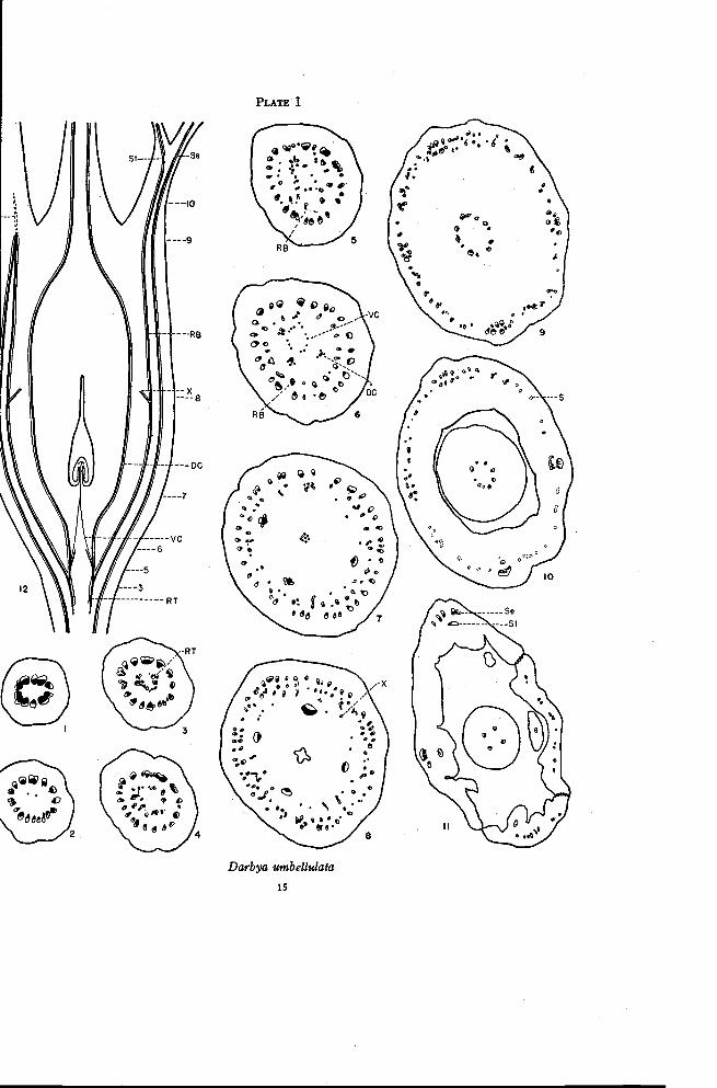
The ovary is completely inferior with a single locule and a central placen-tal stalk bearing two or three ovules at the tip. The disk is thin, has four orfive lobes alternating with the sepals and is borne on the floral tube. The vas-cular tissue in the pedicel forms an almost continuous cylinder (Plate I, Fig-ure 1). At the base of the ovary this cylinder begins to separate radially into
14 OREGON STATE COLLEGE-STUDIES IN BOTANY
numerous strands (Figure 2). These strands swing outward and pass up theouter portion of the ovary to approximately the level at which the floral tubeis freed from the ovary. The number of traces in this ring of bundles con-tinually increases by radial separation throughout this distance (Figures 2to 8). A second ring of bundles is present inside the first for the full lengthof the ovary. These inner traces are inverted with the xylem located outsidethe phloem (Figures 4 to 8). At approximately the level at which the floraltube is freed from the ovary, the outer ring of normally oriented traces andthe inner ring of inverted traces, except those in the positions of the sepals,come together in a series of anastomoses (Figure 9, and left side of Figure12). This region is the only one in which there are strong connections be-tween the two rings of bundles. Continuing upward from these connectionsthere are a few short strands of xylem-like sclerenchyma (S, Figures 10and 12). The traces in the two rings have been interpreted as axial or recep-tacular traces. The traces of the inner ring are considered to be invertedbecause of invagination of the tip of the floral axis. From this interpretationthe outer ring of bundles may be referred to as ascending receptacular tracesand the inner ring as recurrent receptacular traces.
Directly opposite the sepals the ascending and recurrent bundles do notconnect at the top of the ovary as do the bundles between the sepal positions.Instead they swing closer together to form a large compound amphicribralstrand with the individual traces rather loosely arranged (Figure 9). Theinner traces opposite each sepal give rise to a single inverted trace (Figure10) that then swings inward to enter the base of a stamen (Figure 11). Theexact point of origin of the stamen trace is difficult to determine (Smith andSmith, 1942). The outer traces continue as large bundles for a short distanceafter the divergence of the stamen traces. At approximately the level atwhich the sepal lobes are differentiated, each trace divides radially to formthe vascular bundles of the sepal (Figure 11).
Traces to the carpels originate near the ends of the recurrent traces. Incross sections of the base of the ovary the recurrent traces appear as xylemonly, or as xylem and phloem in the inverted position (Figure 3). Thesetraces soon form a complete ring, which gives off branches to the inside(Figures 4 and 5). Three or four branches are more strongly developedthan the others. These are normal collateral bundles and they swing out-ward slightly at higher levels while the traces between them move inward(Figure 6). These inner traces, the ventral carpellary traces, move inwardand form a compact strand of phloem, which enters the placental stalk (Fig-ures 7 and 12). The larger traces, which are the dorsal carpellary traces,continue to swing outward for awhile and then pass upward through the
°O fiLO 11".
a ,. . e.O 1,. I. .a . 0 6
O Go6 c. o)t. .=
9 9 10%, et i 4 .116
Olt,6 66 d ee
Darbya umbellulata
15
16 OREGON STATE COLLEGESTUDIES IN BOTANY
ovary.At approximately the level of the top of the locule branch traces(X, Figures 8 and 12) move inward from the ring of inverted traces andtake up positions between the dorsal traces. Thus there is formed at thislevel a third ring of bundles with the xylem and phloem always normallyoriented. This third or innermost ring may be referred to as the dorsal-Xring of bundles. As has been mentioned previously (Smith and Smith,1942), the nature of the X-traces is in doubt. The dorsal-X traces swinginward at the top of the ovary and form anastomoses with each other justbefore the style is freed (Figure 9). A varying number of traces continueinto the base of the style (Figure 10) but soon they all disappear except thedorsal carpellary traces (Figure 11) each of which extends to the base of astigma lobe.
It is apparent that the portion of the recurrent bundles below the levelof origin of the carpellary traces constitutes vascular tissue of the receptaclethat is not used up in the formation of traces to the floral organs. This tissueis designated as residual. Since this is directed downward as a result of in-vagination, it also follows that the placental stalk must be composed entirelyof carpellary tissues.
There are certain cross sections that are considered critical in the inter-pretation of floral anatomy in other members of the Santalaceae. Figure 3illustrates a section at or below the base of the ovary, showing the residualtissue located apparently in the pith of the pedicel. Figure 5 shows theorigin of the carpellary traces from the recurrent traces. Figure 7, takenjust below the locule, shows the ascending receptacular traces, the invertedrecurrent traces, the dorsal carpellary traces and, in the center, the strandsrepresenting the ventral carpellary traces that enter the placental stalk.Figure 8, taken through the upper portion of the locule, shows the ascendingand recurrent traces and a third ring of normally oriented bundles, consistingof the dorsal carpellary traces plus the X-traces. Figure 9, taken near thetop of the ovary, shows the sepal-stamen complexes and, between these, theanastomosing of the ascending and recurrent traces. It is in this region thatthe receptacular traces turn downward as a result of invagination. The ex-tent of the xylem-like sclerenchyma (Figure 12) may indicate that the recep-tacle actually extends somewhat above this level.
Colpoon compressum Berg.
The flowers of Colpoon are perfect or pistillate with well developedstaminodia. Although they are usually 4-merous, in the material availablefor this study they are 5-merous. There are three to five ovules suspendedfrom a thick placental stalk. The lobes of the disk alternate with the sepals.

FLORAL ANATOMY OF THE SANTALACEAE 17
The floral tube is either very short or completely lacking. The four largetraces in the pedicel above the bracts separate radially as the pedicel graduallyenlarges so that numerous strands are present at the level at which the re-sidual tissue first appears in the pith region (Plate II, Figure 1). Immediatelyabove this level is a more or less saucer-shaped plate of vascular tissue in thecenter of the pedicel (Figure 2). There may be an occasional weak con-nection between the inner traces and the ascending receptacular bundles.From the central plate of tissue there are gradually differentiated a weak am-phicribral bundle, the ventral carpellary trace, in the center; three strongercollateral bundles, the dorsal carpellary traces, toward the outside; and rela-tively few inverted recurrent traces between the dorsal carpellary traces andthe ascending receptacular bundles. Thus in Figure 3, taken just below thelocules, the traces in order from the center toward the outside are the fusedventral traces ; the three dorsal carpellary traces ; the recurrent traces, whichare inverted or represented only by phloem; and the ascending traces.
Colpoon, unlike Darbya, has separate locules throughout the length ofthe ovules (Figure 4). These locules merge into one approximately at thelevel of attachment of the ovules (Figure 5). As might be expected, thedorsal carpellary traces are located directly opposite the locules. Throughoutthis distance the number of traces in the ring of recurrent bundles is in-creased partly by the inward swing of branch traces from the outer ring andtheir rotation to the inverted position, but principally by the appearance ofnew traces. In successive sections of the ovary from the base upward, thesenew traces appear first as phloem only but gradually develop xylem in the in-verted position (Figures 4 and 5). The difficulties in determining the extentand origin of the stamen traces are the same here as in Darbya. At the base ofthe locules (Figure 4) the ring of recurrent traces is rather sparse. In thepositions of the sepals (Figure 7), however, there is always one strong in-verted trace. This may originate either from the plate of tissue in the pedicelas phloem only with the subsequent formation of xylem, as a branch fromtraces on either side of the sepal trace, or rarely from the sepal trace itself.This single trace inside of each sepal trace eventually leads to a stamen.
Near the top of the locule the X-traces arise from the recurrent bundlesand move in to form with the dorsal carpellary traces a third ring of bundles(Figure 5). Thus at this level, as in Darbya, there are three concentricrings of bundles, the ascending, the recurrent, and the dorsal-X rings. Atthe extreme top of the locule the dorsal-X ring of bundles moves in and,after anastomoses are formed (Figure 6), gradually disappears. None ofthese traces enter the base of the style (Figure 7). At the level of the topof the locule, which is just below the level at which the sepal lobes are freed,
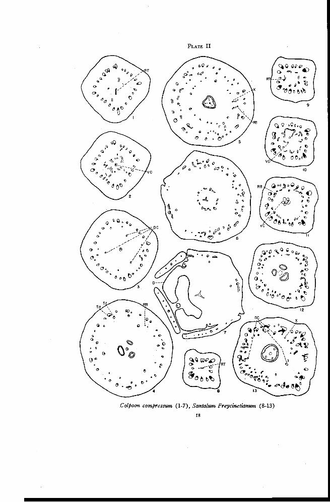
PLATE II
Colpoon compressum (1-7), Santa lum Freycinetianum (8-13)
18
FLORAL ANATOMY OF THE SANTALACEAE 19
the ascending and recurrent traces come together in anastomoses (Figure 6).All except those in the positions of the sepals then disappear. The invertedtrace opposite each sepal loses its xylem and continues into the stamen as aphloem trace (Figure 7). The sepal trace is much more compact than inDarbya and tends to give off lateral traces and to retain a strong dorsal traceinstead of separating radially into more or less equal strands as in Darbya.The sepals, stamens, and lobes of the disk are freed almost simultaneously.
According to Saunders (1933, 1940) the sepals in the flowers of Colpoonhave commissural marginal veins. No evidence was found to support thisstatement, though commissural marginal veins do occur in other genera of thefamily.
Santalum Freycinetianum Gaudich.
The flowers of Santalum are perfect and 4- or 5-merous. The ovary isessentially only partly inferior as the floral tube is freed just above thelevel of ovule attachment. There is but one locule, though there may beslight depressions in its base in which the ovules rest. The placental stalk isextended for some distance beyond the ovules. There is a large floral tubewith a well developed disk the lobes of which are freed simultaneously withthe sepals and stamens. The style is elongate with two to four short stigmalobes.
Since Santalum Freycinetianum, S. cuneatum (Hillebr.) Rock., S. hal-eakalae Hillebr., S. paniculatum H. and A. var angustifolium Sk. and S.pyrularium A. Gray are essentially similar in their floral anatomy, onlyS. Freycinetianum is described in detail.
There are four large traces in the base of the pedicel. These dividerapidly so that numerous traces are present in the region in which residualtissue first appears (Plate II, Figure 8). The residual tissue consists atfirst of xylem only. Later phloem appears and the residual tissue becomesarranged in a definite ring of inverted traces (Figure 9). The traces soonbecome connected by anastomoses and from these phloem is given off to theinside (Figure 10). The phloem strands come together to form a largestrand, the ventral carpellary strand, which remains in the center (Figure 11)and leads to the placental column. The ascending and recurrent traces moveoutward. Occasionally a small trace connects the two rings of bundles. Nodorsal carpellary traces are evident.
The single locule may have partitions at its extreme base. Thus crosssections through this region show apparently three locules, each with a singleovule (Figure 12). The other species studied do not show this condition asfrequently as does S. Freycinetianum. The number of traces in the ovarywall is increased in the outer ring by branching and in the inner ring prin-
20 OREGON STATE COLLEGESTUDIES IN BOTANY
cipally by the appearance of new traces. At approximately the level of at-tachment of the ovules to the placental column there are numerous connec-tions between the ascending and recurrent traces (Figure 13). The X-tracesoriginate at the same level and move inward toward the locule. In Darbyathe X-traces originate some distance below the level at which the ascendingand recurrent traces connect with each other. Three or four of the X-tracesin Santalum, those in the positions of dorsal carpellary traces, are morestrongly developed than the others. The X-traces move into the long taperedupper portion of the ovary where their number is gradually reduced to threeor four, which are in the positions of dorsals (Plate III, Figures 1 to 3). Theabsence of dorsal carpellary traces below the level at which the X-tracesmove in may be interpreted in two ways. It may be due either to theirfusion with the recurrent traces through this distance or to their completedisappearance as a result of a gradual reduction in size. In other genera thedorsal carpellary traces may be represented only by weak phloem traces(e. g. Choretrum) and in still others the dorsals may appear independentlyin the ovary wall just below the X-traces (e. g. Arjona). Indications arethen that the dorsal carpellary traces may actually disappear in the regionbelow the X-traces. They are retained above because the X-traces normallyreinforce the dorsals.
As was previously mentioned, the floral tube in Santalum is freed onlya short distance above the level of attachment of the ovules. This is justabove the level at which the X-traces move in. In addition to the sepal-stamen complexes, numerous small traces enter the floral tube. There aresome anastomoses between these small traces and many of the traces areinverted. Thus even though most of the ascending and recurrent traces areconnected just below the level at which the floral tube is freed, it seemslikely that much of the floral tube itself is receptacular. Usually one in-verted trace persists inside each sepal trace. In addition the small tracesflanking each sepal trace swing around to the inverted position (Figure 2).Thus there are formed at this level loosely arranged amphicribral strands,each representing a sepal-stamen complex. The small inverted traces thatoriginate near each sepal trace may then become incorporated with either thesepal or stamen traces. Eventually there are differentiated from each sepal-stamen complex a single large sepal trace and a single stamen trace, which isusually amphicribral (Figure 3). As the stamen trace moves in, it usuallyleaves behind a small amount of xylem, which persists for some distancejust inside the sepal trace.
Meanwhile some of the small traces between the sepal positions disap-pear and some additional traces appear that are inverted. These additional
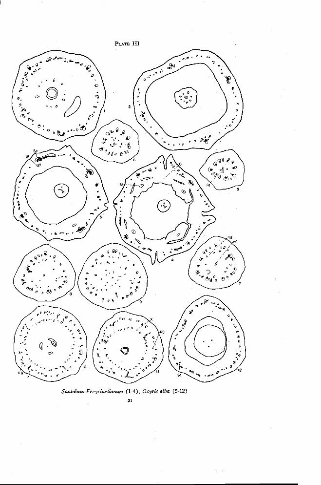
PLATE III
Santalum Freycinetianum (1-4), Osyris alba (5-12)
21
22 OREGON STATE COLLEGE-STUDIES IN BOTANY
inverted traces arise from branches of the sepal traces and are directeddownward. The sepal traces give off large branches at almost right anglesjust before the calyx lobes are freed (Figure 3). From these, small branchtraces extend both upward and downward, the latter being inverted. Noinverted traces appear above this level (Figure 4). The stamens, calyx lobes,and disk are freed almost simultaneously.
Santalum differs essentially from Darbya in only two details. Thedorsal carpellary traces present in Darbya throughout the length of the ovaryare absent in Santalum from the base of the ovary to the level at which theX-traces appear. In Darbya the sepal traces gradually separate radially tosupply the sepals while in Santalum the sepal traces give off branches atright angles and from these branches traces extend both upward and down-ward, the latter being inverted. The distribution of the inverted traces inSantalum seems to indicate that here the floral tube is probably receptacularup to approximately the level of branching of the sepal traces.
It is difficult to reconcile the above description with that given by Raofor S. album (1942). He described four large strands, which enter theperianth segments after giving off branches to the stamens, and smallerstrands between the large ones which supply the carpels. Either two dis-tinct rings of branches do not occur in S. album or they were overlooked.Rao did describe two rings of traces in Scleropyron but he considered themas outer and inner series of the carpellary strands supplying different layersof the fruit wall. Rao considered that the placental column in S. album, aswell as in other forms he studied, does not receive any vascular trace fromthe main floral strands.
Osyris alba L.
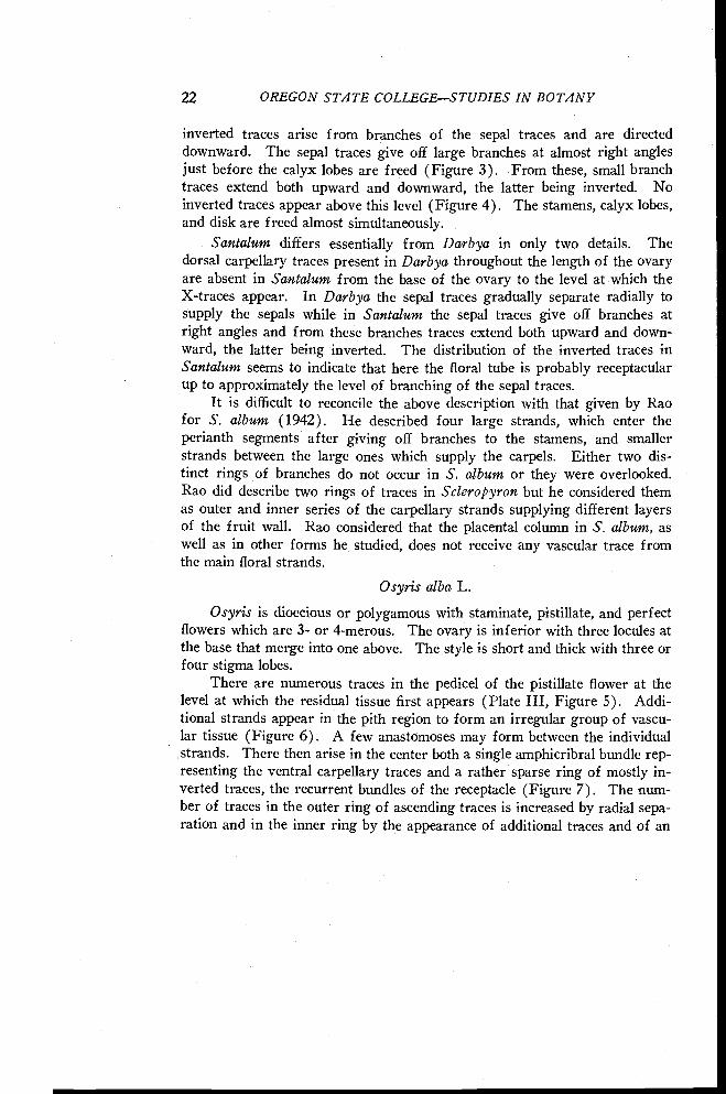
Osyris is dioecious or polygamous with staminate, pistillate, and perfectflowers which are 3- or 4-merous. The ovary is inferior with three locules atthe base that merge into one above. The style is short and thick with three orfour stigma lobes.
There are numerous traces in the pedicel of the pistillate flower at thelevel at which the residual tissue first appears (Plate III, Figure 5). Addi-tional strands appear in the pith region to form an irregular group of vascu-lar tissue (Figure 6). A few anastomoses may form between the individualstrands. There then arise in the center both a single amphicribral bundle rep-resenting the ventral carpellary traces and a rather sparse ring of mostly in-verted traces, the recurrent bundles of the receptacle (Figure 7). The num-ber of traces in the outer ring of ascending traces is increased by radial sepa-ration and in the inner ring by the appearance of additional traces and of an
FLORAL ANATOMY OF THE SANTALACEAE 23
occasional branch from the outer traces. The recurrent traces here are veryweak, consisting of phloem only or of phloem with a small amount of xylem.Over a distance of about twenty sections, three stronger traces move inwardin the positions of dorsal carpellary traces (Figures 7, 8, and 9). Theseoriginate from or frequently near the ascending traces. In this same region,numerous very weak phloem traces move inward from the recurrent tracestoward the ventral carpellary strand. These seem to be in the nature offlesh bundles since most of them soon disappear and only a very fewactually connect with the ventral strand. Figure 9 is taken from just belowthe locule.
The dorsal carpellary traces soon branch many times. Numerousbranches move inward from the inverted traces and, to a limited extent,from the ascending traces and assume positions between the dorsals. Thesetraces are apparently the X-traces that appear here below the locule insteadof above it as in Darbya. At the base of the locule (Figure 10), appearfirst a ring of ascending receptacular traces toward the outside, then a veryfew inverted recurrent traces inside, and finally a more pronounced ring ofnormally oriented bundles, the dorsal-X ring, toward the center. At firstglance the two rings of traces in Figure 7 appear to be comparable to those inSantalum (Plate II, Figure 12) and to those in Darbya (Plate I, Figure 7)at the same level. In both Santalum and Darbya, however, the inner ringconsists of inverted traces while in Osyris it consists of normally orientedtraces. The origin of the X-traces below the locule and the small number ofinverted traces present in Osyris account for this difference.
Near the top of the locule (Plate III, Figure 11) additional strongtraces in the positions of the dorsal carpellary traces move inward from theouter ring. The entire inner ring of traces then moves inward to enter thebase of the style and the floral tube is freed from the ovary. Anastomosesare formed between the dorsal-X traces and only the traces in the positionsof dorsal carpellaries persist in the style (Figure 12).
There are few inverted traces above the level at which the X-tracesappear. In the floral tube numerous anastomoses appear between traces(Figure 12). It is probably at this same level that the ascending receptaculartraces become recurrent. An occasional stamen trace may be identified. Un-fortunately the material available was very poorly preserved for structureabove this level but the vascular structure seems to be similar to that inDarbya.
Osyris differs from Darbya in the branching of the dorsals and especiallyin the origin of the X-traces below the level of the base of the locule insteadof above the locule. The ring of X-traces is apparently well developed at
24 OREGON STATE COLLEGESTUDIES IN BOTANY
the expense of the recurrent traces, which are sparsely scattered throughoutthe length of the ovary.
The above description of Osyris agrees in general with that given byVan Tieghem (1869b) and by Schaeppi and Steindl (1937). Van Tieghemconsidered the outer ring of traces as belonging to the calyx and the innerring as consisting primarily of the three dorsal carpellary traces. He re-garded the inferior ovary here as appendicular. He recognized that theinner traces, particularly those in the ventral strand, originate from vasculartissue that appears in the pith region and is thus "vascularly independent" ofthe main vascular supply. While Schaeppi and Steindl considered the outertraces as belonging to the receptacle, they did not describe invagination andthey considered the X-traces as traces of the ovary because they do not appearin the staminate flower. The connections between the outer and inner ringsof traces were interpreted as indicating that, the ca.rpellary bundles are ratherweakly connected below to the main vascular supply.
Acanthosyris falcata Griseb.
The flowers of Acanthosyris are 4- or 5-merous and perfect. The ovaryhas one locule and a much twisted placental stalk bearing three ovules at itstip. The style is somewhat long and the stigma is indistinctly 5-lobed. Thefloral anatomy of this species is the most complex and difficult of explanationof the entire family.
There are four large traces in the pedicel just above the much reducedbracts (Plate IV, Figure 1). Even at this level there is a tendency towardthe amphicribral condition with phloem or a small inverted trace located justinside each of the main bundles. The pedicel enlarges rapidly as the maintraces begin to separate in radial planes. The small inner traces branch andtend to merge with the main traces. At the same time a somewhat scatteredring of traces arises independently in the cortical region of the pedicel (Fig-ure 2). These traces may appear as xylem, as phloem or as complete tracesmore or less inverted. The pedicel then decreases in diameter rather rapidlyand the innermost strands merge completely with the main traces (Figure 3).The traces in the cortical region branch. Some of them disappear and mostof the rest of them lose their xylem. Five traces tend to remain morestrongly developed than the others. These are in the positions of the sepals(Figure 10). The outermost traces, which usually consist of phloem only,continue to branch and occasionally form strands connecting with each otherand also with the main traces. The traces in the cortical region then becomeso rearranged that the five somewhat larger inverted traces in the sepal posi-tions are slightly inside the others (Figure 4). At the same level vascular
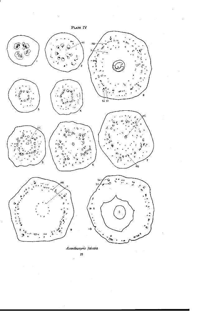
PLATE IV
0 .,",::0 °0 0,,,,,
7. --4..,,--0 ;' R9 po /
0p 0 r 0 0
o °oc ° o G t
II,...D .Q %
t I 0F.
0 .V
6, ?Po
00. c.<1, zo :%. 00 6
.5
Acanthosyris falcata
25
26 OREGON STATE COLLEGESTUDIES IN BOTANY
tissue arises independently in the pith region. The number of strands in thepith region is increased rapidly by the appearance of new traces and by theinward movement of an occasional branch from the main traces.
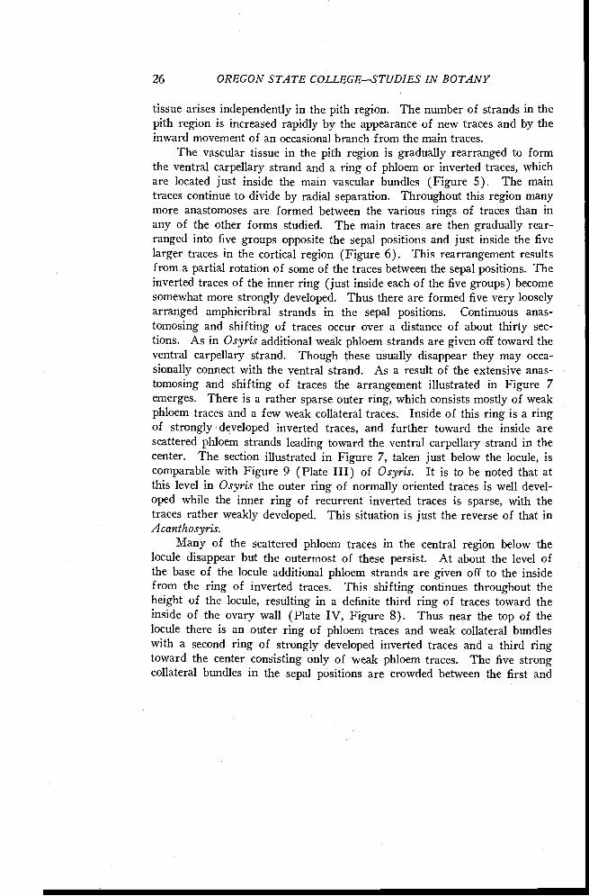
The vascular tissue in the pith region is gradually rearranged to formthe ventral carpellary strand and a ring of phloem or inverted traces, whichare located just inside the main vascular bundles (Figure 5). The maintraces continue to divide by radial separation. Throughout this region manymore anastomoses are formed between the various rings of traces than inany of the other forms studied. The main traces are then gradually rear-ranged into five groups opposite the sepal positions and just inside the fivelarger traces in the cortical region (Figure 6). This rearrangement resultsfrom a partial rotation of some of the traces between the sepal positions. Theinverted traces of the inner ring ( just inside each of the five groups) becomesomewhat more strongly developed. Thus there are formed five very looselyarranged amphicribral strands in the sepal positions. Continuous anas-tomosing and shifting of traces occur over a distance of about thirty sec-tions. As in Osyris additional weak phloem strands are given off toward theventral carpellary strand. Though these usually disappear they may occa-sionally connect with the ventral strand. As a result of the extensive anas-tomosing and shifting of traces the arrangement illustrated in Figure 7emerges. There is a rather sparse outer ring, which consists mostly of weakphloem traces and a few weak collateral traces. Inside of this ring is a ringof strongly developed inverted traces, and further toward the inside arescattered phloem strands leading toward the ventral carpellary strand in thecenter. The section illustrated in Figure 7, taken just below the locule, iscomparable with Figure 9 (Plate III) of Osyris. It is to be noted that atthis level in Osyris the outer ring of normally oriented traces is well devel-oped while the inner ring of recurrent inverted traces is sparse, with thetraces rather weakly developed. This situation is just the reverse of that inAcanthosyris.
Many of the scattered phloem traces in the central region below thelocule disappear but the outermost of these persist. At about the level ofthe base of the locule additional phloem strands are given off to the insidefrom the ring of inverted traces. This shifting continues throughout theheight of the locule, resulting in a definite third ring of traces toward theinside of the ovary wall (Plate IV, Figure 8). Thus near the top of thelocule there is an outer ring of phloem traces and weak collateral bundleswith a second ring of strongly developed inverted traces and a third ringtoward the center consisting only of weak phloem traces. The five strongcollateral bundles in the sepal positions are crowded between the first and
FLORAL ANATOMY OF THE SANTALACEAE 27
second rings. The five small inverted traces of the outer ring, one just out-side each of the strong collateral bundles, are the five inverted traces shownin Figure 4. These are continuous through the flower to the top of theovary.
The inner ring of phloem traces moves in to enter the base of the style(Figure 9) and can therefore be regarded as the dorsal-X ring. All of thesetraces disappear, however, before the style is freed. At the same time thereare an increasing number of connections between the inverted traces and theouter traces, the connections usually being through xylem only. There arethus formed in the outer ring normally oriented collateral bundles thatbecome increasingly pronounced. By the time the calyx tube is freed, all thexylem of the inner traces has been "transferred" to the outer traces (rightside of Figure 10). The large trace in each sepal position moves out andcombines with the small trace just outside it to form the sepal trace. Theinverted trace just inside each of these becomes the stamen trace. The bun-dles of the outer and inner rings, except for the sepal and stamen bundles,come together in the base of the floral tube, form anastomoses and graduallydisappear just before the sepal traces branch to supply the sepals. Thesepals, stamens and disk are freed almost simultaneously.
A comparison of Figure 8 with Figure 5 (Plate II) of Colpoon or withFigure 8 (Plate I) of Darbya helps to explain what may have happened inAcanthosyris to give this anomalous anatomical structure. In both Colpoonand Darbya there are three rings of well developed traces at the level indi-cated, the ascending traces, the recurrent traces, and the dorsal-X traces. Atthis level in Acanthosyris, both the ascending and the X-traces are repre-sented only by weak phloem traces while the inverted recurrent traces arestrongly developed. Much anastomosing and shifting of traces also occurthroughout the lower portion of the ovary, always so that the more stronglydeveloped traces are located deep inside the ovary wall. These facts consid-ered together make the following interpretation seem the most likely. InAcanthosyris, as in Osyris, the X-traces apparently originate below thelocule, in the former perhaps even lower than in the latter. Thus the "nodes"or levels of origin of the X-traces are very close to the ventral carpellarytraces. In following the strongly developed traces from the base to the topof the ovary, their location gradually shifts to the outside. At the base ofthe ovary the most strongly developed traces are those normally orientedtraces that lie deep in the tissues of the ovary inside approximately two ringsof weaker traces (Plate IV, Figures 4 and 5). Above this the shift is out-ward to mostly inverted traces with a definite ring of weaker traces bothinside and outside (Figure 8), and at the top of the ovary to the outermost
28 OREGON STATE COLLEGE-STUDIES IN BOTANY
ring of normally oriented traces (Figure 9 and right side of Figure 10). Inboth Darbya and Colpoon the main path of water conduction from the pedi-cel, as indicated by the distribution of xylem, is up through the ascendingtraces and down through the recurrent traces to the inner parts of the flower.For some unknown reason the main path of conduction, as indicated byxylem development, seems to have shifted in Acanthosyris so that it cutsacross to the innermost traces at the base of the ovary, moves out to the in-verted traces, and finally out to the outermost ring of ascending traces. Whilethis interpretation may seem illogical from a physiological point of view,the confusion caused by the crowding of the "nodes" where the ventrals andX-traces originate may make this shift possible. This interpretation is sup-ported also by the fact that in the basal portion of the ovary, where thegreatest change must occur, there are many more anastomoses and connec-tions between rings of traces than in any other form studied.
Eucarya acuminata (R. Br.) Sprague and Summerhayes
The flowers of Eucarya are perfect and 4-merous. The ovary is partlyor completely inferior with a single locule and a placental stalk bearing two orthree ovules. The stalk extends up into the short style much as in Santalum.There are as many stigma lobes as there are ovules.
Eucarya presents a much simpler and more direct modification of the re-ceptacular inferior ovary than Acanthosyris. There are four large traces inthe pedicel that divide radially to form numerous strands at the level atwhich the residual tissue first appears (Plate V, Figure 1). A short distanceabove this level a loose plate of vascular tissue appears in the pith region(Figure 2). The plate consists chiefly of phloem. Xylem, when it is pres-ent, appears in the traces adjacent to the main vascular bundles and in theinverted position. A strand of phloem continues upward from the plate toenter the placental stalk (Figure 3). The remaining traces in the pith regionmove outward and upward. Some of them connect to the main traces, othersdisappear and a few persist just inside the main bundles. Three of thesepersistent strands are dorsal carpellary traces. They move in near thelocule, one opposite each ovule, and persist as weak phloem traces (Figure4). In addition to these a single weak strand (Figure 3), which later be-comes a stamen trace, appears just inside each of the four larger traces of theouter ring of bundles. One instance was found in which a single stranddivided to form both a dorsal and a stamen trace. There are very few tracespresent at the level of the base of the locule to represent recurrent traces. Afew additional traces appear at higher levels and an occasional trace swings
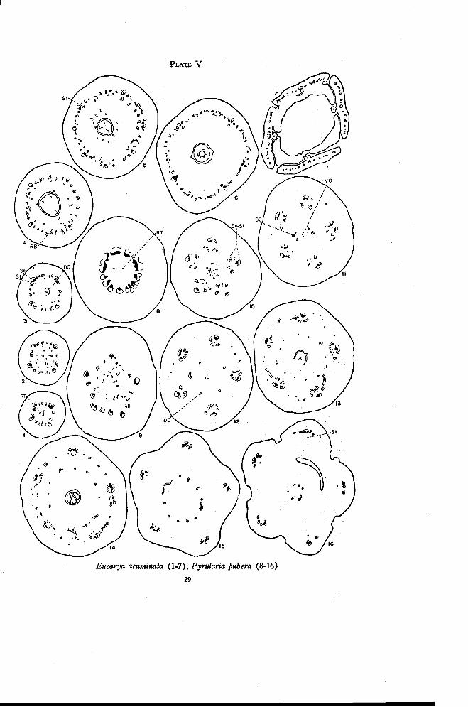
PLATE V
Eucarya acuminata (1-7), Pyrularia pubera (8-16)29
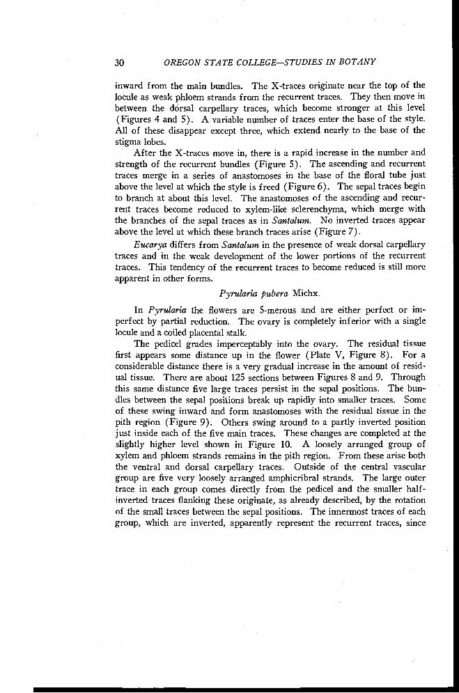
30 OREGON STATE COLLEGESTUDIES IN BOTANY
inward from the main bundles. The X-traces originate near the top of thelocule as weak phloem strands from the recurrent traces. They then move inbetween the dorsal carpellary traces, which become stronger at this level(Figures 4 and 5). A variable number of traces enter the base of the style.All of these disappear except three, which extend nearly to the base of thestigma lobes.
After the X-traces move in, there is a rapid increase in the number andstrength of the recurrent bundles (Figure 5). The ascending and recurrenttraces merge in a series of anastomoses in the base of the floral tube justabove the level at which the style is freed (Figure 6). The sepal traces beginto branch at about this level. The anastomoses of the ascending and recur-rent traces become reduced to xylem-like sclerenchyma, which merge withthe branches of the sepal traces as in Santalum. No inverted traces appearabove the level at which these branch traces arise (Figure 7).
Eucarya differs from Santalum in the presence of weak dorsal carpellarytraces and in the weak development of the lower portions of the recurrenttraces. This tendency of the recurrent traces to become reduced is still moreapparent in other forms.
Pyrularia pubera Michx.
In Pyrularia the flowers are 5-merous and are either perfect or im-perfect by partial reduction. The ovary is completely inferior with a singlelocule and a coiled placental stalk.
The pedicel grades imperceptably into the ovary. The residual tissuefirst appears some distance up in the flower (Plate V, Figure 8). For aconsiderable distance there is a very gradual increase in the amount of resid-ual tissue. There are about 125 sections between Figures 8 and 9. Throughthis same distance five large traces persist in the sepal positions. The bun-dles between the sepal positions break up rapidly into smaller traces. Someof these swing inward and form anastomoses with the residual tissue in thepith region (Figure 9). Others swing around to a partly inverted positionjust inside each of the five main traces. These changes are completed at theslightly higher level shown in Figure 10. A loosely arranged group ofxylem and phloem strands remains in the pith region. From these arise boththe ventral and dorsal carpellary traces. Outside of the central vasculargroup are five very loosely arranged amphicribral strands. The large outertrace in each group comes directly from the pedicel and the smaller half-inverted traces flanking these originate, as already described, by the rotationof the small traces between the sepal positions. The innermost traces of eachgroup, which are inverted, apparently represent the recurrent traces, since
FLORAL ANATOMY OF THE SANTALACEAE 31
most of them originate from the scattered strands shown in the pith regionin Figure 9. Thus the loosely arranged amphicribral strands originate herein approximately the same manner as in Darbya and Santalum. In Pyrularia,however, they originate low in the ovary and all traces between them dis-appear. While there is some branching from these groups throughout thelength of the ovary, the branches seem to be mostly in the nature of fleshbundles.
From the central group of vascular strands shown in Figure 10, fivetraces move outward (Figure 11). Two of these become much more stronglydeveloped than the others (Figure 12). These five traces are the dorsalcarpellary traces. The ventral traces, represented by a variable number ofxylem and phloem strands, remain in the center and continue up into theplacental column. The dorsal carpellary traces branch to form a ring ofmore than five traces at the level of the base of the locule (Figure 13). Theamphicribral strands give off a few traces to the inside at various levelsthroughout the height of the locule and the traces of the inner ring becomegradually stronger. These branch traces probably represent the X-tracesthat arise at various levels instead of simultaneously at one level as in formspreviously described. Toward the top of the locule (Figure 14) an innerring of well developed bundles is formed which moves inward (Figure 15)toward the base of the style. This is the dorsal-X ring of traces. A variablenumber of traces from the ring enter the style (Figure 16). Only two per-sist and these are in the positions of the two stronger dorsal carpellary tracesshown in Figure 12.
The flesh bundles between the main traces become stronger toward thetop of the locule (Figures 13 and 14) and then gradually disappear. The am-phicribral strands become more compactly arranged (Figure 15). Most ofthe inner traces of each group disappear by fusing with adjacent traces, butat least one weak phloem trace persists as a stamen trace. The main sepaltrace then divides radially, as in Darbya, as the floral tube is freed (Figure16). The sepals, stamens, and disk are freed almost simultaneously.
Comandra pallida A. DC.
The flowers of Comandra are perfect and 4- or 5-merous. The ovary iscompletely inferior with a single locule and a much twisted placental stalkbearing two or three ovules at the top. Comandra pallida, C. umbellata (L.)Nutt., C. Richardsiana Fernald, and C. californica Eastw. were studied, butonly C. pallida is described in detail.
There is a continuous vascular cylinder in the pedicel above the bracts.The residual tissue appears as scattered xylem strands in the pith region at
32 OREGON STATE COLLEGESTUDIES IN BOTANY
approximately the level at which the sepal-stamen traces begin to move out(Plate VI, Figure 1). These strands become connected to the outer tracesby vascular tissue that moves in from between the main lobes of the stele(Figure 2). This situation is very similar to that in Pyrularia (Plate V,Figure 9) in which the residual tissue is connected to the smaller tracesbetween the sepal positions. The ventral carpellary traces are represented bya single amphicribral strand (Plate VI, Figures 2 and 3) formed by thegrouping of the xylem traces in the center in a single strand and the inwardmovement of phloem through gaps in the stele. Occasional small traces movelaterally from the lobes of the stele and usually become inverted.
The main traces become partly or completely amphicribral as they moveout to enter the ovary wall (Figure 3). The method of formation is similarto that in Pyrularia (Plate V, Figures 9 and 10), the xylem strands at themargins tending to swing around toward the inside and phloem moving inbehind them. The resultant amphicribral bundles are more compact through-out than in Pyrularia. As these large traces move out, three, or more rarelyfour, normally oriented traces are left behind as the dorsal carpellary traces.These originate either from the margins of the main traces before they closeto become amphicribral (Plate VI, Figure 3) or from the connecting tissuebetween the ventral strand and the traces between the lobes of the stele. Thedorsals may at first be partly or completely inverted since they frequentlyoriginate from the margins of the main traces after these have swung partlyaround. The dorsals immediately become rearranged, however, and are nor-mally oriented at the level of the base of the locule (Figure 4).
Additional small traces are given off from the main traces throughoutthe length of the ovary. These are usually inverted and frequently directeddownward. Toward the top of the locule, weak traces move in between thedorsal carpellary traces (Figure 5). These are the X-traces and they orig-inate mostly from the small traces between the sepal positions. Their modeof origin offers additional evidence that the small inverted traces between thesepal positions represent largely the recurrent traces. Just before the styleis freed, the traces of the dorsal-X ring move in abruptly (Figure 6) andform numerous anastomoses below the base of the style. Only three tracesfrom this ring, those in the positions of the dorsals, persist as weak tracesand extend for a short distance into the base of the style.
Near the top of the locule the main traces, which are combined sepal-stamen traces, become distinctly amphicribral (Figure 5). The small bun-dles remaining between the main traces after the X-traces are given off formanastomoses with each other (Figure 6) and rapidly disappear as the floraltube is freed from the ovary. At the level at which the stamen traces sepa-
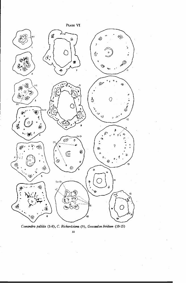
PLATE VI
Comandra pallida (1-8), C. Richardsiana (9), Geocaulon lividum (10-15)
33
34 OREGON STATE COLLEGE-STUDIES IN BOTANY
rate from the sepal traces (Figure 7) all of the smaller traces have disap-peared. The sepal traces immediately branch repeatedly to supply the sepals(Figures 7 and 8). As in Santalum there may be a few inverted tracesdirected downward from the first branches of the sepal traces. The stamensare usually freed before the disk.
In C. Richardsiana the residual tissue extends over a much greater dis-tance than in C. pallida. The main traces, however, become arranged asamphicribral bundles within a shorter distance. There are fewer invertedbranch traces given off laterally from the main bundles but these are muchlarger (Figure 9) than in C. pallida. The dorsal carpellary traces are fartherfrom the locule and more inverted traces are directed downward from thebranches of the sepal traces. Comandra californica is very similar to C. Rich-ardsiana. Comandra umbellata differs from C. pallida principally in that thedorsal carpellary traces are smaller and farther from the locule, the maintraces are not so distinctly amphicribral, and more inverted traces are di-rected downward from the branches of the sepal traces.
Dowding (1931) described but did not illustrate the floral anatomy ofC. pallida and C. umbellata. His description, however, differs greatly fromthat given here. He interpreted the anatomy of the flower in terms of theappendicular theory and the theory of carpel polymorphism. According tohis description the perianth tube is traversed by five main vascular bundlesthat run to the sepals and superimposed stamens. Though this much is inagreement, no support was found for his statement that five strands branchoff from the main vascular bundles to supply the carpels and that five alternatestrands supply the placental column. Neither could evidence be found thatthe gynoecium is composed of two alternate whorls of carpels differentiatedinto an outer sterile and an inner fertile whorl.
Geocaulon lividum (Richardson) Fernald
In Geocaulon the central flowers in each cyme are usually pistillate andthe outer flowers staminate. All are 5-merous. The ovary is inferior witha single locule and a placental stalk that is less twisted than that in Comandra.Although Geocaulon was formerly considered to be Comandra, its separationfrom the latter genus is in accord with differences in floral anatomy, althoughclose affinity is also indicated.
There is a continuous vascular cylinder in the pedicel as in Comandra.The residual tissue appears in much the same manner but extends for only avery short distance. Anastomoses are formed between the residual tissueand the margins of the main traces as the latter move out to enter the ovarywall (Plate VI, Figure 10). A single amphicribral strand continues upward
FLORAL ANATOMY OF THE SANTALACEAE 35
into the placental column. Three traces arise from the anastomoses or fromthe margins of the main traces. These probably represent the dorsal carpel-lary traces. Their subsequent behavior, however, is quite different fromthat of the dorsal carpellary traces in Comandra. They move out into theovary wall as far as the five principal traces and begin to branch immediately.Thus at the level of the base of the locule (Figure 11) there are three groupsof small traces derived from the three dorsals. These are not inverted as aremost of the small traces at the corresponding level in Comandra (Figure 4).A few additional traces, however, are inverted or amphicribral. These arisefrom the main traces, as in Comandra. The margins of the main tracesswing inward gradually to produce the amphicribral condition. There is onlya small amount of xylem in each trace, as one might expect considering thefleshy nature of the fruit.
Numerous traces are given off from the main traces beginning at ap-proximately the base of the locule. These are usually amphicribral or in-verted. Since the normally oriented traces derived from the dorsals grad-ually disappear, all of the small bundles present near the top of the locule(Figure 12) are branches from the principal traces. The X-traces thenmove in toward the base of the style (Figure 13). These originate mostlyfrom the small traces just as in Comandra but occasionally also from thelarge traces. There are no indications of dorsal carpellary traces at this leveland all traces in this ring disappear before the style is freed.
The small traces left between the sepal positions after the X-traces aregiven off disappear in the base of the floral tube. A few short, invertedtraces, directed downward, are given off at this level (Figure 14). Thestamen traces, leading to staminodia, then separate tangentially from thesepal-stamen bundles. The sepal traces branch immediately afterward(Figure 15). The staminodia are freed usually slightly before the disk andsepals.
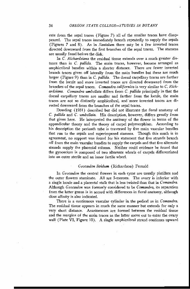
Nanodea muscosa Gaertn. f.
The flowers of Nanodea are 4-merous and either perfect, or pistillatewith aborted stamens. The ovary is inferior with a single locule and a shortcentral placenta bearing at its tip two ovules, only one of which matures.The style is short and has a stigma that is indistinctly two-lobed. The onlyavailable material of this species was of young fruits and these were inrather poor condition.
In the pedicel above the bracts the vascular cylinder is continuous andencloses a very small pith (Plate VII, Figure 1). The cylinder then sep-arates radially (Figure 2) into traces that gradually move out into theenlarged base of the flower. Smaller traces continue to separate from the
36 OREGON STATE COLLEGE STUDIES IN BOTANY
margins of the larger ones. The first of these smaller traces remain nearthe center of the ovary (Figure 3) while those produced later swing aroundto become partly or completely inverted, thus forming a loosely arrangedamphicribral strand in each sepal position (Figures 4 and 5). At this leveltwo of the traces either do, not become amphicribral, or accompanied each bya single small inverted trace become partly so (Figure 5). One of thetraces arises directly from the stele of the pedicel, the other from the marginof one of the larger traces. These two traces are apparently the dorsal car-pellary traces since they continue directly into the style. The innermosttraces become connected by anastomoses (Figure 5) and move in (Figure 6)to enter the placental stalk.
While at first glance the floral structure of Nanodea may appear quitedifferent from that of Comandra and Geocaulon, a careful comparison re-veals many similarities. The principal difference lies in the development ofresidual tissue. Fruits of Nanodea that were studied showed no indicationof residual tissue. Residual tissue is well developed in Comandra but onlyslightly developed in Geocaulon. In Comandra as the residual tissue and theventral strand form anastomoses with the margins of the sepal-stamen traces,the latter move out to enter the base of the ovary (Plate VI, Figure 2). Thesame is true but less obvious in Geocaulon (Plate VI, Figure 10). InNanodea, however, the first traces given off from the margins of the sepal-stamen bundles form anastomoses, which give rise to the ventral carpellarystrand (Plate VII, Figures 3 to 6). Thus it appears that in Nanodea boththe residual tissue and the ventral strand below these anastomoses have dis-appeared, thus establishing a more direct vascular connection between theaxis and the placental stalk. Of course it should be kept in mind that theanatomy of the young fruits of Nanodea is compared with that of the flowersof Comandra and Geocaulon.
The behavior of the dorsal carpellary traces in Nanodea differs onlyslightly from that in Comandra. The two traces move out into the ovarywall almost as far as the sepal-stamen bundles. They are appreciably closer,however, to the stony endocarp layer (indicated by a dotted line) just abovethe base of the locule (Figure 7). The placental trace here is at one side ofthe seed. No small branch traces are given off from the main bundles andno X-traces appear at the top of the ovary. Instead, the dorsals move inwardthrough the sclerenchyma of the endocarp (Figure 8) and assume positionson opposite sides of the locule (Figure 9) near the top of the ovary. Herethey are lost in the sclerenchyma tissue and apparently do not enter the baseof the style. The sepal-stamen traces tend to become collateral as the in-verted traces swing back and around and fuse with the main trace of each
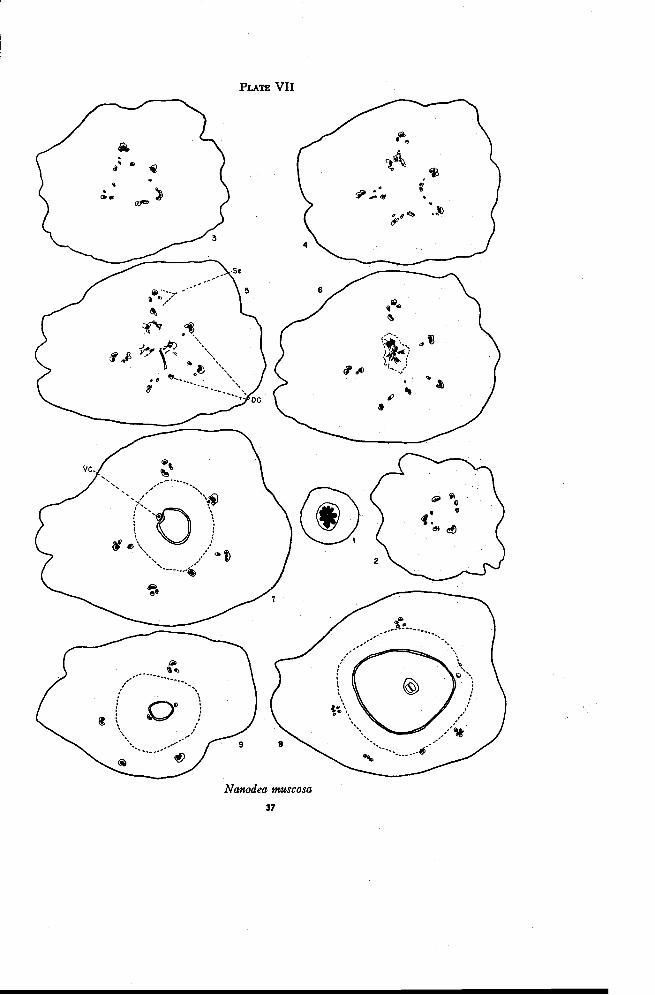
PLATE VII
Nanodea muscosa
37
38 OREGON STATE COLLEGESTUDIES IN BOTANY
group. The material available apparently consisted of fruit from pistillaterather than from perfect flowers, since it showed only occasional traces lead-ing to staminodia.
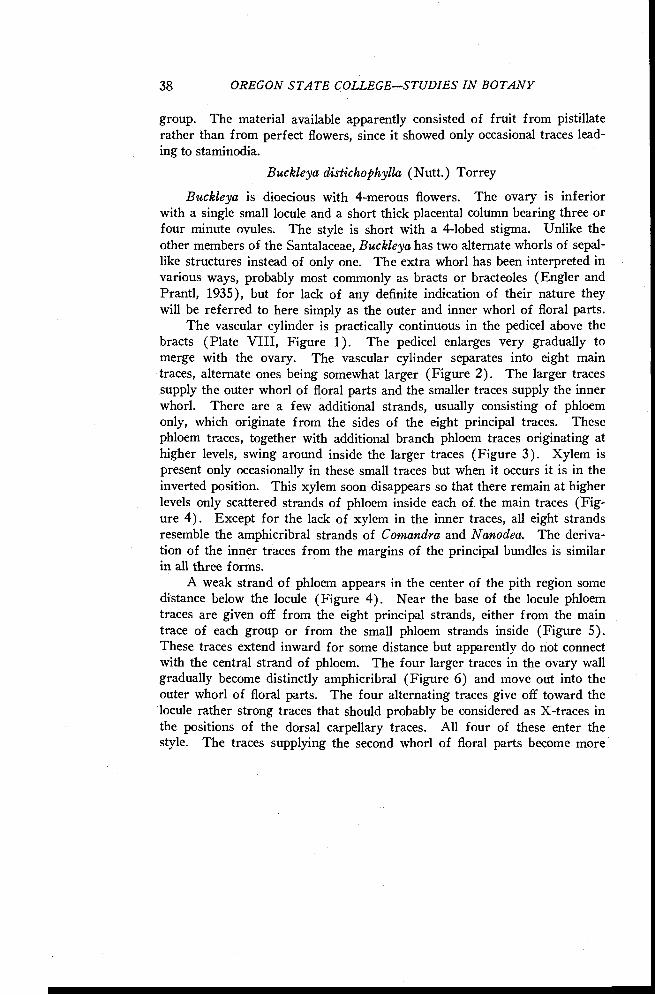
Buckleya distichophylla (Nutt.) Torrey
Buckleya is dioecious with 4-merous flowers. The ovary is inferiorwith a single small locule and a short thick placental column bearing three orfour minute ovules. The style is short with a 4-lobed stigma. Unlike theother members of the Santalaceae, Buckleya has two alternate whorls of sepal-like structures instead of only one. The extra whorl has been interpreted invarious ways, probably most commonly as bracts or bracteoles (Engler andPrantl, 1935), but for lack of any definite indication of their nature theywill be referred to here simply as the outer and inner whorl of floral parts.
The vascular cylinder is practically continuous in the pedicel above thebracts (Plate VIII, Figure 1). The pedicel enlarges very gradually tomerge with the ovary. The vascular cylinder separates into eight maintraces, alternate ones being somewhat larger (Figure 2). The larger tracessupply the outer whorl of floral parts and the smaller traces supply the innerwhorl. There are a few additional strands, usually consisting of phloemonly, which originate from the sides of the eight principal traces. Thesephloem traces, together with additional branch phloem traces originating athigher levels, swing around inside the larger traces (Figure 3). Xylem ispresent only occasionally in these small traces but when it occurs it is in theinverted position. This xylem soon disappears so that there remain at higherlevels only scattered strands of phloem inside each of the main traces (Fig-ure 4). Except for the lack of xylem in the inner traces, all eight strandsresemble the amphicribral strands of Comandra and Nanodea. The deriva-tion of the inner traces from the margins of the principal bundles is similarin all three forms.
A weak strand of phloem appears in the center of the pith region somedistance below the locule (Figure 4). Near the base of the locule phloemtraces are given off from the eight principal strands, either from the maintrace of each group or from the small phloem strands inside (Figure 5).These traces extend inward for some distance but apparently do not connectwith the central strand of phloem. The four larger traces in the ovary wallgradually become distinctly amphicribral (Figure 6) and move out into theouter whorl of floral parts. The four alternating traces give off toward thelocule rather strong traces that should probably be considered as X-traces inthe positions of the dorsal carpellary traces. All four of these enter thestyle. The traces supplying the second whorl of floral parts become more
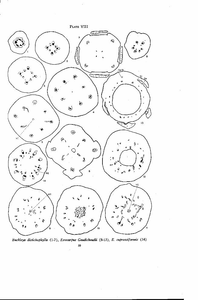
PLATE VIII
Buckleya distichophylla (1-7), Exocarpus Gaudichaudii (8-13), E. cupressiformis (14)
39
40 OREGON STATE COLLEGESTUDIES IN BOTANY
nearly collateral after the departure of the X-traces (Figure 7). The secondwhorl of floral parts is then freed and the ovary tapers rapidly to formthe style.
All eight of the principal strands thus behave alike except at the top ofthe ovary. Since the branch traces leading inward at the top of the ovaryarise only from the traces supplying the second whorl of floral parts it mightbe assumed that the second whorl is composed of sepals and the first whorlof bracts. The staminate flower is of no assistance in identifying the extrawhorl in the pistillate flower. The staminate flower is supplied by fourtraces in the pedicel, each of which moves out into the base of a sepal,where it divides to form the sepal and stamen traces.
Exocarpus Gaudichaudii A. DC.
The staminate flowers of Exocarpus have reduced pistils. The pistil-late flowers have rather weakly developed staminodia. The ovary in thepistillate flower is either apparently superior or partly surrounded by thereceptacle, and has a single locule with one more or less sessile ovule. As thefruit develops, the axis below the ovary becomes enlarged and fleshy. Onlywell developed fruits were available for study. Exocarpus Gaudichaudii,E. cupressiformis Labill. and E. aphyllus R. Br. were all studied but onlythe first will be described in detail. Since the seed was poorly preserved, itis omitted from drawings of sections made through the locule. So also is theextent of the sclerenchymatous endocarp.
There are four or five traces in the lower portion of the pedicel. Someof these divide radially at the base of the enlarged portion of the , axis.Small phloem strands appear independently just inside some of the maintraces (Plate VIII, Figure 8). Occasionally a few of the main traces tendto become amphicribral but this condition is only temporary. If the maintraces are followed over a considerable distance some will be found thatdivide once, some twice, and some not at all (Figure 9). At the same timethe number of phloem traces inside the main traces is increased by the ap-pearance of new strands and by the formation of a few weak branch tracesthat swing inward from the larger bundles. These inner traces may developa small amount of xylem in any position. In E. cupressiformis the innertraces are well developed and definitely inverted (Figure 14) and thus maybe considered as recurrent traces, or as residual traces below the level atwhich the ventral carpellary strand originates.
Anastomoses appear in the pith region in E. Gaudichaudii (Figure 9)that connect with the recurrent traces to form a large flat plate of vasculartissue in the center (Figure 10). The ventral carpellary strand continues
FLORAL ANATOMY OF THE SANTALACEAE 41
upward from this plate toward the placental knob and the remaining vasculartissue moves outward to connect with some of the outer traces (Figure 11).A comparison with Figure 13 shows that these connections are mostly withtraces between the sepal positions. Figure 11 illustrates a section taken justbelow the locule. The region above this is characterized by much branchingand anastomosing of traces (Figure 12). In this same region the ascendingand recurrent traces come together, the dorsal-X ring of traces is given offand the sepal-stamen traces are differentiated, all of which probably con-tribute to the confused branching and anastomosing. While the sepal-stamentraces are extremely small (Figure 13), the traces that continue in the ovarywall are well developed. There are no inverted traces above the level atwhich the small sepals are freed. Each staminodium receives a weak tracefrom one of the sepal-stamen bundles. The traces in the ovary wall grad-ually disappear and only a few enter the base of the style.
It appears from the above description that the enlarged receptacle hereis definitely invaginated. Also, since the ventral carpellary strand originatesapproximately halfway down in the enlarged axis, the ovary is not as com-pletely superior as it appears to be.
One characteristic of this genus particularly difficult to explain is thepresence in E. cupressiformis of small xylem strands, or complete vascularbundles, within the phloem of the larger bundles (Figure 14). Thesestrands swing out into the phloem at the base of the enlarged portion of theaxis, continue to a level just below that at which the sepals are freed, andthen swing back to the xylem. The only condition approaching this appearsin Acanthosyris where several bundles are arranged close together in thesame radius in each sepal position (Plate IV, Figure 8). The two outertraces of each group merge at a higher level to form a single trace to eachsepal.
Anthobolus leptomerioides F. Miill.
Anthobolus is dioecious and has flowers that are 3- or 4-merous. Thereare no evidences of staminodia in the pistillate flowers. The ovary is appar-ently superior on a fleshy axis just as in Exocarpus. There is a single smalllocule with one sessile ovule.
The stele in the pedicel may be divided into a variable number ofstrands. Extending outward from the margins of these are short tracesconsisting of xylem only and ending in xylem-like sclerenchyma (Plate IX,Figure 1). The main traces divide radially in the lower portion of the en-larged axis and produce additional xylem-like sclerenchyma from the xylem(Figure 2). Residual tissue appears here in the center of the pith region asone or more xylem strands. At higher levels some anastomoses are formed
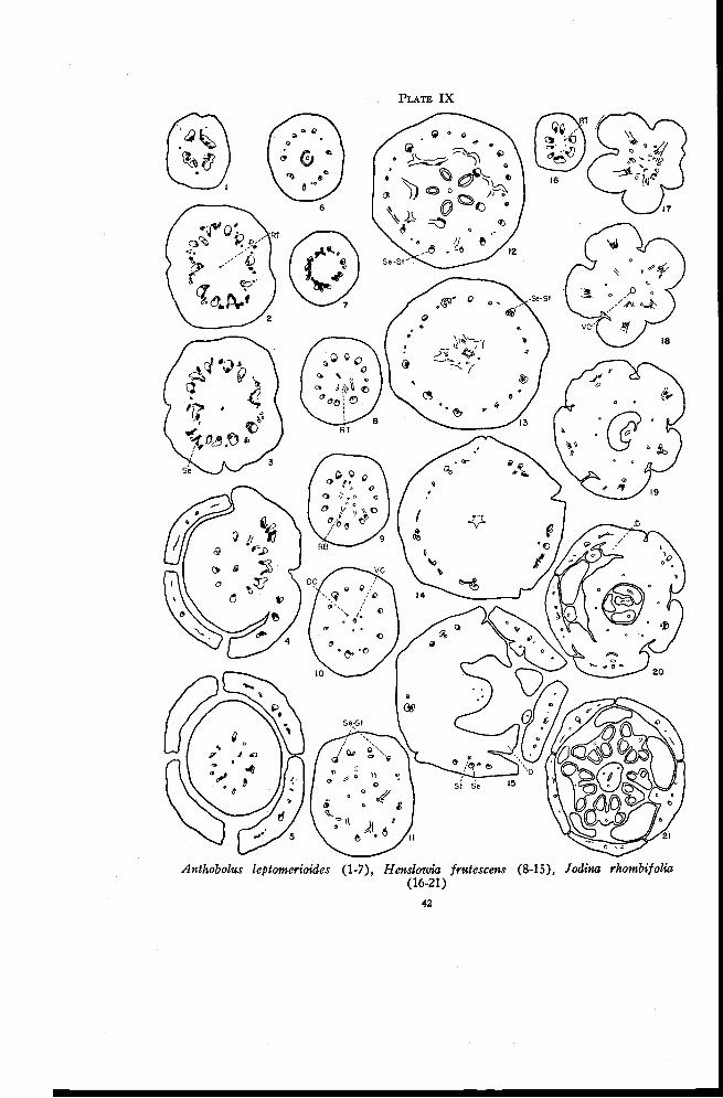
PLATE IX
Anthobolus leptomerioides (1-7), Henslowia frutescens (8-15), Jodina rhombifolia(16-21)
42
FLORAL ANATOMY OF THE SANTALACEAE 43
between the main traces, and the sepal traces begin to move out (Figures 3and 4). Either one large trace or three smaller traces enter each sepal.There are no indications of stamen traces or of staminodia. Following thedeparture of the sepal traces the remaining traces form a ring of simple col-lateral bundles. At some distance below the locule a few weak xylem tracesmay extend inward toward the ventral carpellary strand (Figure 5) but theyapparently do not become connected with it. The locule is high in the ovary(Figure 6). Just above the locule the traces in the ovary wall form anas-tomoses with each other (Figure 7) and then all traces rapidly disappear.
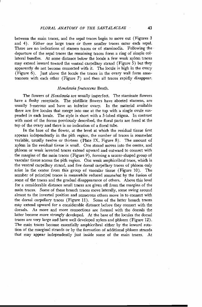
Henslowia frutescens Benth.
The flowers of Henslowia are usually imperfect. The staminate flowershave a fleshy receptacle. The pistillate flowers have aborted stamens, areusually 5-merous and have an inferior ovary. In the material availablethere are five locules that merge into one at the top with a single ovule sus-pended in each locule. The style is short with a 5-lobed stigma. In contrastwith most of the forms previously described, the floral parts are freed at thetop of the ovary and there is no indication of a floral tube.
In the base of the flower, at the level at which the residual tissue firstappears independently in the pith region, the number of traces is somewhatvariable, usually twelve or thirteen (Plate IX, Figure 8). The amount ofxylem in the residual tissue is small. One strand moves into the center, andphloem or weak inverted traces extend upward and outward to connect withthe margins of the main traces (Figure 9), forming a saucer-shaped group ofvascular tissue across the pith region. One weak amphicribral trace, which isthe ventral carpellary strand, and five dorsal carpellary traces of phloem onlyarise in the center from this group of vascular tissue (Figure 10). Thenumber of principal traces is meanwhile reduced somewhat by the fusion ofsome of the traces and the gradual disappearance of others. Above this levelfor a considerable distance small traces are given off from the margins of themain traces. Some of these branch traces move laterally, some swing aroundalmost to the inverted position and numerous others move in to connect withthe dorsal carpellary traces (Figure 11). Some of the latter branch tracesmay extend upward for a considerable distance before they connect with thedorsals. As more and more connections are formed with the dorsals thelatter become more strongly developed. At the base of the locules the dorsaltraces are very large and have well developed xylem and phloem (Figure 12).The main traces become essentially amphicribral either by the inward rota-tion of the marginal strands or by the formation of additional phloem strandsthat may appear independently just inside some of the main traces. At
44 OREGON STATE COLLEGE-STUDIES IN BOTANY
higher levels the locules gradually merge into one. The sepal-stamen tracescan be identified as definite amphicribral traces (Figure 13) located oppositethe carpels. The last traces to move in toward the dorsals arise between thesepal positions and apparently receive the small strands located inside thesetraces. Thus only the sepal-stamen traces remain amphicribral while thetraces between them become once more collateral. The inner traces thenform anastomoses and move in toward the base of the style. Just beforethe style is freed all but five of these traces disappear. These five weaktraces in the positions of the dorsals (Figure 14) enter the base of the style(Figure 15) and continue upward almost to the 5-lobed stigma.
Meanwhile the outer traces located between the sepal positions formanastomoses, some of which are inverted (Figure 13 and left side of Figure14) and gradually disappear. Each main amphicribral strand then divides tof orm a stamen and a sepal trace (Figure 14). At higher levels the stamentraces move in to enter the rudimentary stamens (Figure 15) and each of thesepal traces forms two branch traces each of which soon divides again. Thesepals, stamens, and disk are freed almost simultaneously.
The receptacular nature of the inferior ovary of Henslowia is not asapparent as that of Darbya and closely related forms. A comparison withsome of the species already described reveals that despite the presence of alarger number of carpels the vascular anatomy here represents an advancedsystem derived from the Darbya type. The distinct inner ring of invertedrecurrent traces has almost disappeared even though the more primitivenumber of carpels has been retained. The following characteristics are evi-dences of an advanced vascular system : First, the appearance of residualtissue in the pith region before any connections are formed with the maintraces (Figure 8) ; second, the origin of branch traces from the margins ofthe main traces and their inward rotation to form amphicribral sepal-stamentraces (Figures 11, 12, and 13), a behavior comparable to that in Comandraand closely related forms ; third, the anastomosing of the small traces, some ofwhich are inverted, between the sepal positions and their subsequent disap-pearance (Figures 13 and 14), a behavior characteristic of most of the formsalready described; and, fourth, the nature of the stamen traces, which are atfirst inverted, later amphicribral, a behavior likewise characteristic of theforms previously described. Thus it seems that the ascending and recurrenttraces in Henslowia have become reduced to form more nearly a single ringof collateral bundles. The reduction is comparable to that in the series fromColpoon to Pyrularia and it apparently occurred early in the phylogenetic de-velopment of this and the following genera of the Santalaceae. It becomeseven more complete in the forms yet to be described.
FLORAL ANATOMY OF THE SANTALACEAE 45
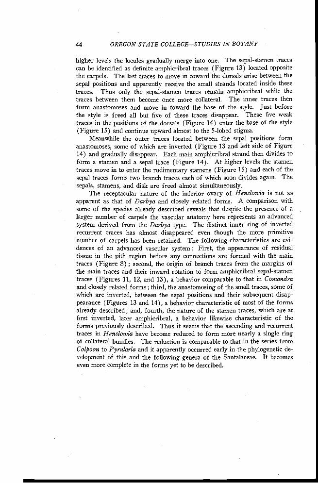
Jodina rhombifolia Hook. and Am.
The flowers of Jodina are perfect and 5-merous. The ovary is onlypartly inferior in the young flower but it becomes completely inferior in theolder flower. There is a single locule with a twisted placental stalk, whichbears three ovules at its tip. The style is thick with a stigma that is indis-tinctly 3-lobed. The fruit is drupe-like with a fleshy exocarp divided intofive easily detachable segments.
A variable number of traces occur in the pedicel. Residual tissue ap-pears below the ovary as scattered protoxylem strands in the pith region(Plate IX, Figure 16). At the base of the flower, five of the main strandsbegin to move out into the segments of the ovary (Figure 17). Anas-tomoses are formed that connect the remaining traces with each other andalso with the residual tissue. Just above this level the five large strands,the sepal-stamen traces, move out rather abruptly (Figure 18). From theanastomoses of the traces between the sepal positions there persists a ring ofphloem traces that continues upward into the ovary wall (Figure 19). Asingle strand of phloem from the traces in the pith region continues upwardto enter the placental column.
The sepal-stamen bundles gradually become amphicribral by the rotationof the marginal strands to a partly or completely inverted position. At ap-proximately the level of the base of the locule (Figure 19) the stamen tracesseparate from the sepal-stamen bundles. The sepal traces then branch im-mediately to supply the sepals (Figure 20). The sepals, stamens, and diskare freed at a level that is a little more than halfway up the ovary. The ringof phloem traces between the outer traces and the locule (Figure 19) con-tinues in the ovary, but in the base of the style the number of traces in thering is reduced to three (Figure 21) which are in the positions of dorsalcarpellary traces. This ring of traces must be considered as the dorsal-Xring of traces that originates here below the locule.
Myoschilos oblongus Ruiz and Pay.
The flowers of Myoschilos are perfect and 5-merous. The ovary isinferior with a single locule and a short thick placental column bearing at itstip three to five ovules. The style is short with a 2- to 5-lobed stigma.
The number of traces in the pedicel is variable, usually five to seven(Plate X, Figure 1). Some of these traces become reduced in size, just as inHenslowia, while others branch. The residual tissue appears as scatteredxylem strands in the pith region (Figure 2). Five strong bundles, the sepal-stamen bundles, move outward, leaving near the residual tissue a few addi-
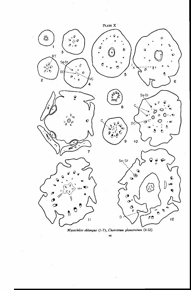
PLATE X
Myoschilos oblongus (1-7), Choretrum glomeratum (8-12)
46
FLORAL ANATOMY OF THE SANTALACEAE 47
tional xylem strands that originate from the margins of the traces. This be-havior is similar to that at a comparable level in Jodina (Plate IX, Figure 17).The traces between the sepal positions become reduced to phloem strands(Plate X, Figures 3 and 4). The residual tissue together with the tracesfrom the margins of the principal traces forms a compact group of strands inthe center (Figure 3). These strands form a few anastomoses and then sep-arate to form a ventral carpellary strand and three dorsal carpellary traces(Figure 4).
Xylem appears in the small phloem strands in the outer ring of traces.Additional small traces may be formed from branches from these strandsand from the sepal-stamen bundles. Thus at the level of the base of thelocule (Figure 5) numerous small traces are interspersed between the fivesepal-stamen bundles and the three dorsal carpellary traces lie just insidethem. Near the top of the locule the small traces between the sepal-stamenbundles form a few anastomoses and then they all move in between thedorsals (Figure 6) to form the dorsal-X ring of traces. Anastomoses areformed in this ring below the base of the style (Figure 7). Only threetraces in the ring, those in the positions of the dorsals, enter the base of thestyle and these soon disappear.
The sepal-stamen traces begin to move out at the level of the top ofthe locule (Figure 6). They never become amphicribral as they do in formspreviously described. The stamen traces separate tangentially from thesepal-stamen bundles (Figure 7) and the sepal traces branch immediately tosupply the sepals. Occasionally there may be a small branch directed down-ward in the base of the sepals. Sepals and stamens are freed before the disk.
Choretrum glomeratum R. Br.
Choretrum, Leptomeria, Thesium, Thesidium and Omphacomeria aregrouped together in a single series because in all these forms there are twiceas many traces in the pedicel as there are floral parts. Alternate traces be-come the sepal-stamen traces and each of the remaining traces branches tosupply a lateral trace to the sepals on either side of it. These lateral traceshave been termed commissural marginal traces by Saunders (1933, 1940)who considered that their presence indicates that each sepal actually consists ofone sepal plus half of the neighboring petal on either side. Thus sepals withcommissural marginal veins show petaloid coloring while those with only asingle trace, and therefore individual sepals, are green. There seem to bemany exceptions, however, to this generalization in the Santalaceae.
The flowers of Choretrum are perfect and 5-merous. The ovary isinferior with five locules at the base, which merge into one above. There is
48 OREGON STATE COLLEGESTUDIES IN BOTANY
a thick central placental column bearing five ovules. The style is short andthick with a 5-lobed stigma. Choretrum glomeratum, C. Candollei F. Mull.and C. spicatum F. Mii ll. were studied but only C. glomeratum is describedin detail.
There are ten main traces in the pedicel (Plate X, Figure 8) and inaddition an occasional phloem strand. As these traces move out to enter thebase of the ovary most of them give off weak phloem traces to the inside(Figure 9). At the same time phloem appears independently in the pithregion and connects upward and outward to these inner (phloem) traces. InC. Candollei the traces connecting with the phloem traces occasionally showxylem in the inverted position. As the principal traces continue to moveoutward, the phloem traces are left behind. From these and from the plateof phloem tissue in the center, the following traces arise : the ventral car-pellary strand, which later enters the placental column (Figure 10) ; the dor-sal carpellary traces of phloem only, one opposite each locule; and a ring ofphloem bundles, probably representing the recurrent traces, located betweenthe dorsals and the main traces. In both C. spicatum and C. Candollei thisring occasionally shows xylem in the inverted position. Dorsal carpellarytraces are lacking in both these forms.
At or slightly below the base of the locules alternate strands in the outerring of bundles divide so that fifteen large traces are present (Figure 10).Each of the five sepals receives three of these traces (Figure 11), a sepal-stamen trace that comes directly from the pedicel and two lateral traces, eachresulting from the division of the alternate trace ; i.e., the commissural traceon either side of the sepal-stamen bundle. At this level the phloem in theplacental column separates into five strands supplying the ovules. The lo-cules merge into one just below the level of ovule attachment. The dorsalcarpellary traces and the recurrent traces move in toward the locule. It isprobably more correct to consider the latter traces as X-traces, and that therecurrent traces are used up in the production of the X-traces. Weak anas-tomoses are formed between the dorsal-X traces. All these traces soon dis-appear except for five phloem strands in the positions of the dorsals, whichenter the base of the style (Figure 12) and extend about half way to the5-lobed stigma. A short floral tube is freed from the style. The sepal-stamen traces soon become amphicribral and the stamen traces then separateas phloem only or as weak inverted traces. The lateral sepal traces divide toform most of the traces in the sepals. The filaments are short and almostcompletely fused with the sepals.
FLORAL ANATOMY OF THE SANTALACEAE 49
Leptomeria pauciflora R. Br.
The flowers of Leptomeria are perfect and 5-merous. As in Choretrumthe ovary is inferior with five locules at the base that merge into one above.The style is small with a 5-lobed stigma. Leptomeria pauciflora, L. acidaR. Br. and L. Cunninghamii Miq. were studied but only L. pauciflora is de-scribed in detail.
As in Choretrum there are ten traces in the pedicel (Plate XI, Figure 1).Some distance below the ovary a single xylem strand representing the re-sidual tissue appears in the center of the pith region. A short distanceabove, phloem appears around the xylem strand to form an amphicribralbundle (Figure 2). At the same level phloem strands move outward towardthe main traces. These phloem strands either connect with the phloem ofthe principal traces or they take up positions just inside the main traceswhere they continue for a short distance and then disappear. The amphi-cribral trace in the center continues upward into the placental column.
Since the commissural traces that form the lateral traces of adjacentsepals begin to branch at a higher level than in Choretrum, only the tenprincipal traces are present at the base of the locules (Figure 3). A singlephloem strand appears just inside each of the sepal-stamen traces, which arelocated opposite the locules. These phloem strands either appear inde-pendently in these positions or originate as branches from the phloem of themain traces. They probably represent only the recurrent traces, althoughthere is the possibility that the recurrent traces and the dorsal carpellarytraces are fused. At the level of ovule attachment weak traces are given offfrom these phloem strands and move in toward the locule (Figure 4), leavingone or two strands of phloem inside each dorsal sepal bundle. Xylem in thenormal position appears temporarily in the inner traces (Figure 5) beforethey enter the base of the style. Xylem may also appear in the invertedposition in the small traces inside the dorsal sepal bundles.
The traces alternating with the sepal-stamen strands branch at the levelof the top of the locule (Figures 4 and 5) increasing the number of principaltraces to fifteen. The phloem trace inside each of the larger bundles nowswings inward to enter the stamen (Figure 6). The lateral sepal traces dis-appear at the base of the sepals in a group of xylem-like sclerenchyma. Thedorsal sepal traces may or may not branch in the sepals.
Leptomeria Cunninghamii and L. acida are essentially the same as L.pauciflora except that the residual tissue does not extend as far down in thepedicel.
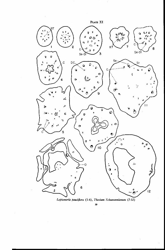
PLATE XI
Leptomeria pauciflora (1-6), Thesium Schumannianum (7-12)
so
FLORAL ANATOMY OF THE SANTALACEAE 51
Thesium Schumannianum Schlecht.
The flowers of Thesium are perfect and 4- or 5-merous. The ovary isinferior with a single locule and a slender and somewhat twisted placentalcolumn bearing two or three ovules. The style is short with a stigma that isindistinctly 3-lobed.
In the 5-merous flower there are ten traces in the pedicel at the levelat which the residual tissue first appears (Plate XI, Figure 7). The residualtissue consists of a few scattered strands of phloem and only an occasionalxylem cell in the inverted position. The residual tissue appears in the pithregion either independently or as branches from some of the main traces. Afew anastomoses are formed between the residual traces (Figure 8) and theythen move in to form a single phloem strand that enters the placental column(Figure 9). At the same time the principal traces begin to move outwardinto the ovary wall, leaving behind three dorsal carpellary traces (Figures8 and 9). The dorsal traces may originate either independently or asbranches from the sepal-stamen or the commissural traces, usually the latter.Occasional branch traces are given off laterally from the main traces.
The dorsal carpellary traces become stronger near the top of the locule.The traces between the sepal-stamen bundles branch one or more times(Figure 10) and from these branches weak traces arise that move in betweenthe dorsal carpellary traces (Figure 11). The traces of the dorsal-X ringthen move in toward the base of the style but disappear completely beforethe style is freed. The sepal-stamen bundles usually become amphicribral.Sometimes, however, a single inverted trace is formed inside by the inwardrotation of a strand from the margin (Figure 11). Each of the tracesbetween the sepal positions then swings over near the adjacent sepal-stamenstrand (left side of Figure 11) where it persists for some time as an invertedtrace. It is difficult to explain why these traces are inverted when those inChoretrum and Leptomeria, which apparently have the same origin, arenormally oriented. Just before the floral tube is freed these inverted tracesconnect with the sepal traces (right side of Figure 12). Above this levelthere arise several small, normally oriented traces that continue into thesepal (left side of Figure 12). The inverted stamen traces become amphi-cribral but may be reduced to phloem only as they enter the stamens.
This description agrees in general with that of T. linophyllum given byVan Tieghem (1869b) though it differs in that here the carpellary traces arenot completely independent of the main traces.
52 OREGON STATE COLLEGESTUDIES IN BOTANY
Thesidium fragile Sonder.
The flowers of Thesidium are imperfect and 4-merous. The pistillateflower has an inferior ovary with a single locule and an erect or twistedplacental column bearing two or three ovules. The style is short with a stigmathat is indistinctly 2- or 3-lobed. There are no stamen traces or staminodiain the pistillate flower.
Just above the level at which the traces to the bracts are given off, thethree or four strands present in the pedicel divide radially to form eighttraces that enter the base of the flower (Plate XII, Figure 1). One or moreof these traces may consist of phloem only. The xylem eventually disap-pears from all but the four dorsal sepal traces. The behavior of the corn-missural traces is quite variable. The commissural trace shown at 7 o'clockin Figure 2 behaves as a dorsal carpellary trace and enters the base of thestyle. The one at 9 o'clock divides to form two traces, probably the marginaltraces of adjacent sepals, which disappear before the sepals are freed. Theone at 12 o'clock divides to form the two marginal traces and another dorsalcarpellary trace. The one at 4 o'clock divides only once to form a dorsal car-pellary trace and a phloem trace, which soon disappears. The commissuraltraces and the dorsal carpellary traces associated with them are very muchreduced in this species. The ventral carpellary strand appears independentlyin the pith region (Figure 2) and continues into the placental stalk. Thereare no connections between this strand and any of the other traces.
At the top of the locule (Figure 3) the dorsal traces move inward and
a few weak traces are given off from the sepal bundles. These branchtraces either connect with the dorsals or move laterally and soon disappear.The dorsal carpellary traces continue into the base of the style (Figure 4)where they also disappear. The sepal traces may or may not branch as thesepals are freed.
Omphacomeria acerba (R. Br.) A. DC.
Omphacomeria is monoecious or polygamous with 4-merous flowers.The ovary is practically inferior with a single locule. The style is short andthick with a 2-lobed stigma.
Since the flowers are 4- instead of 5-merous the pedicel contains eighttraces (Plate XII, Figure 5) instead of ten as in Choretrum, Leptomeriaand Thesium. The residual tissue appears in the pith region and connectsimmediately with the main traces (Figure 6). A ring of phloem tracesarises from these anastomoses (Figure 7). The number of traces in thering gradually is reduced to two (Figures 8 and 9), which enter the base ofthe style. Probably the number of carpels has likewise been reduced to two.
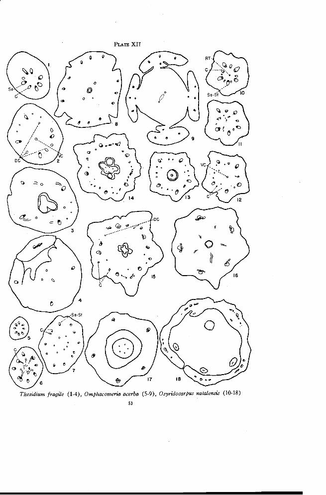
PLATE XII
Thesidium fragile (1-4), Omphacomeria acerba (5-9), Osyridocarpus natalensis (10-18)
53
54 OREGON STATE COLLEGESTUDIES IN BOTANY
There is no indication of a ventral carpellary strand. Alternate traces in thering of principal bundles branch low in the ovary (Figure 6), forming thetwelve traces that are present in the ovary wall (Figure 7). As in Choretrumthese continue directly into the sepals (Figures 8 and 9). The stamentraces arise from the sepal-stamen bundles as inverted or amphicribraltraces.
Osyridocarpus natalensis A. DC.
The flowers of Osyridocarpus are perfect and 5-merous. The ovary isinferior with a single locule and a slender twisted placental column bearingtwo or three ovules. The style is long with a stigma that is capitate or in-distinctly 2- or 3-lobed.
At the level at which the residual tissue first appears, there are tentraces in the pedicel (Plate XII, Figure 10). The five traces in the sepalpositions are considerably larger than the others. Occasionally an additionalsmall trace may appear. The residual tissue consists at first of a fewstrands of xylem, which soon disappear and are replaced by phloem (Figure11). A few weak phloem traces connect to the traces, especially the smallertraces, of the outer ring at this level. The single strand of phloem tissue inthe center continues upward into the placental column (Figures 12 and 13).At the level at which the phloem traces in the pith region connect to theouter traces the bundles between the sepal positions begin to branch (Figure11). Some of these branch traces connect with the residual tissue as justdescribed. Other traces continue upward in the outer part of the ovary(Figures 11 to 13).
There are five very weak traces representing the dorsal carpellary traces.These are not continuous throughout the ovary but may appear and disappearat various levels. Just above the point of ovule attachment (Figure 15) allfive traces are present. A comparison of Figures 14 and 15 shows that thesetraces are opposite the points of ovule attachment. Near the top of theovary the dorsals move inward (Figure 16) and enter the base of the style(Figure 17). The dorsal carpellary traces are alternate with the sepals in-stead of opposite them as in Choretrum, Leptomeria and Henslowia.
Near the top of the ovary the smaller traces in the outer ring formanastomoses with each other (Figure 14) and occasionally with one or moreof the dorsal carpellary traces. Above this level two traces continue upwardfrom each group of anastomoses between the sepal-stamen bundles (Figure15). These two traces are derived indirectly from the single trace in theregion between the sepals just as in Choretrum and Leptomeria though herethe situation is complicated by anastomoses near the top of the ovary. Each
FLORAL ANATOMY OF THE SANTALACEAE 55
of these traces then moves to the nearest sepal-stamen trace and fuses withit completely (Figure 16). The floral tube is freed just above this level.The sepal-stamen bundles then become amphicribral by the rotation of themarginal traces to the inverted position (Figure 17). The stamen tracesseparate a short distance below the level at which the stamens are freed whilethe sepal traces branch laterally to supply the sepals (Figure 18).
From the behavior of the commissural traces here it seems possible thatOsyridocarpus may form a connecting link between Choretrum, Leptomeriaand Thesium on the one hand and Arjona and Quinchamalium on the other.
Quinchamalium majus Brongn.
The flowers of Quinchamalium are perfect and 5-merous. The floweris surrounded at the base by a tubular structure formed by the union offloral bracts. The floral tube is very much elongate, small and cylindricalwith the stamens adnate to the base of the sepals. The ovary is inferior withthree locules below, which merge into one above the level of ovule attach-ment. There is a short placental column bearing three ovules at the tip. Thestyle is filiform and extends approximately to the middle of the floral tube.The stigma is capitate.
The residual tissue appears in the pith region before the tubular bractis entirely freed (Plate XIII, Figure 1). One trace to the bract moves outwell below the others and the bract is freed on that side first. As the re-maining three traces of the bract move out, anastomoses (consisting mostlyof phloem) are formed between most of the traces present at this level(Figure 2). The residual tissue connects upward and outward to these anas-tomoses. As the bract is freed, the phloem between the main traces dis-appears (Figure 3). The ventral carpellary strand, consisting of phloemand scattered xylem cells, continues upward into the placental column. Thethree locules appear immediately above this level (Figure 4). There is nochange in vascular structure from here up to the level of ovule attachment.At this level (Figure 5) numerous anastomoses are formed between thesepal-stamen traces. Branch traces move in toward the locule in the posi-tions of dorsal carpellary traces and then continue upward to enter the baseof the style (Figure 6). Weaker traces move inward at the same time asthose in the positions of the dorsal carpellary traces but extend for only ashort distance. The anastomoses then disappear and the floral tube andcircular disk are freed in close succession.
Approximately in the middle of the floral tube single traces appear inde-pendently between the sepal-stamen bundles (Figure 7). These gradually
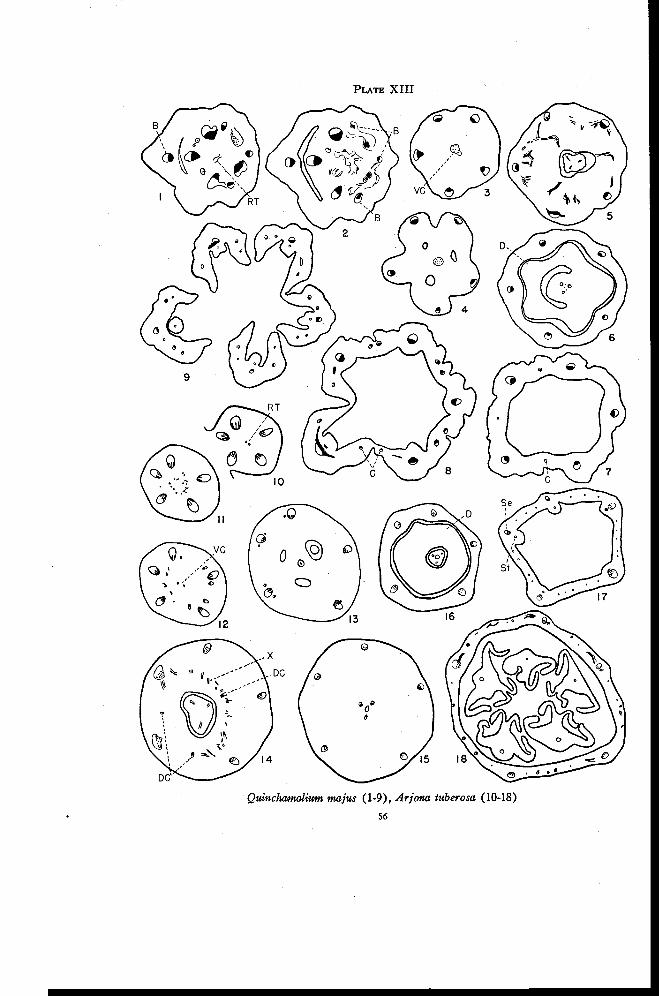
PLATE XIII
Quinchantatium majus (1-9), Arjona tuberosa (10-18)
56
FLORAL ANATOMY OF THE SANTALACEAE 57
enlarge as they extend toward the top of the tube. Just before the calyxlobes are formed, each of these traces divides into two branches that enteradjacent sepals (Figure 8). Thus the lateral traces of adjacent sepals hereare derived from a single commissural trace as in Choretrum and Leptomeria.Here, however, the commissural traces appear independently about halfwayup the floral tube. Thus they have lost direct connection with the main vas-cular system. The sepal-stamen bundles do not become amphicribral. Asthe stamen traces are given off from the sepal-stamen bundles, the lateraltraces connect with the dorsal sepal traces. Both the lateral and dorsal sepaltraces branch to supply the sepals (Figure 9).
Arjona tuberosa Cay.
The flowers of Arjona are perfect and are usually 5-merous. Theovary is inferior with three locules below, which merge into one above and ashort thick placenta bearing three ovules. The style is filiform with a stigmathat is capitate or indistinctly 3-lobed. Since Arjona pusilla Hook. f. andA. longifolia Phil. differ only slightly from A. tuberosa, only the last men-tioned species is described in detail.
Above the level at which the bracts are given off there are five tracesin the pedicel. The residual tissue appears in the pith region just as the lastbract is freed (Plate XIII, Figure 10). In the center appear additionalstrands that lead upward and outward toward the sepal-stamen bundles(Figure 11). At least one xylem strand remains in the center, developsphloem to form an amphicribral strand and enters the placental column(Figures 12 and 13). The traces that move outward assume positions oneither side of the sepal-stamen bundles (Figure 12), and then continue up-ward beside them for varying distances before fusion occurs. In A. longi-folia, however, these traces are immediately incorporated into the sepal-stamen bundles.
Near the top of the locule weak traces appear independently in theovary in the positions of dorsal carpellary traces (left side of Figure 14).Immediately above, anastomoses are given off from the main traces andmove inward with the dorsals (right side of Figure 14) to form the dorsal-Xring of traces. Before the style is freed, all traces in this ring disappearexcept the three dorsal traces (Figure 15), which enter the style (Figure 16)and extend to the lobes of the stigma. In A. pusilla the dorsal carpellarytraces do not appear below the level at which anastomoses are formed.
Here, as in Quinchamalium, there is a ring-shaped disk. The floraltube contains only the five principal traces for approximately two-thirds ofits length. The stamen traces originate as separate phloem traces inside each
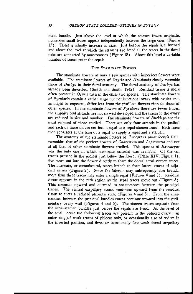
58 OREGON STATE COLLEGESTUDIES IN BOTANY
main bundle. Just above the level at which the stamen traces originate,numerous small traces appear independently between the large ones (Figure17). These gradually increase in size. Just before the sepals are formedand above the level at which the stamens are freed all the traces in the floraltube are connected by anastomoses (Figure 18). Above this level a variablenumber of traces enter the sepals.
THE STAMINATE FLOWER
The staminate flowers of only a few species with imperfect flowers wereavailable. The staminate flowers of Osyris and Henslowia closely resemblethose of Darbya in their floral anatomy. The floral anatomy of Darbya hasalready been described (Smith and Smith, 1942). Residual tissue is moreoften present in Osyris than in the other two species. The staminate flowersof Pyrularia contain a rather large but nonfunctional ovary with ovules and,as might be expected, differ less from the pistillate flowers than do those ofother species. In the staminate flowers of Pyrularia there are fewer traces,the amphicribral strands are not so well developed and the traces in the ovaryare reduced in size and number. The staminate flowers of Buckleya are themost reduced of those studied. There are only four strands in the pediceland each of these moves out into a sepal as a sepal-stamen trace. Each tracethen separates at the base of a sepal to supply a sepal and a stamen.
The anatomy of the staminate flowers of Exocarpus sandwicensis Baill.resembles that of the perfect flowers of Choretrum and Leptomeria and notat all that of other staminate flowers studied. This species of Exocarpuswas the only one in which staminate material was available. Of the tentraces present in the pedicel just below the flower (Plate XIV, Figure 1),five move out into the flower directly to form the dorsal sepal-stamen traces.The alternate, or commissural, traces branch to form lateral traces of adja-cent sepals (Figure 2). Since the laterals may subsequently also branch,more than three traces may enter a single sepal (Figures 4 and 5). Residualtissue appears in the pith region as the sepal traces move out (Figure 3).This connects upward and outward to anastomoses between the principaltraces. The ventral carpellary strand continues upward from the residualtissue to enter a reduced placental stalk (Figures 4 and 5). From the anas-tomoses between the principal bundles traces continue upward into the rudi-mentary ovary wall (Figures 4 and 5). The stamen traces separate fromthe sepal-stamen bundles just before the sepals are freed. At the level ofthe small locule the following traces are present in the reduced ovary: anouter ring of weak traces of phloem only, or occasionally also of xylem inthe inverted position, and three or occasionally five weak dorsal carpellary
PLATE XIV
o
0e
00
0 O O 0
0 0 0 0 0 00O 0 0 0
Exocarpus sandwicensis (1-6), hypothetical primitive santalaceous species (7)
59
60 OREGON STATE COLLEGESTUDIES IN BOTANY
traces inside (Figure 5). The only essential difference between Figure 5and Figure 10 (Plate X) of Choretrum is in the number of locules. Thereare five locules in Choretrum but only one in Exocarpus. The sepals,stamens, and disk are freed somewhat irregularly (Plate XIV, Figure 6).All traces in the ovary disappear except the dorsal carpellary traces, whichenter the base of the style. Figure 6 closely resembles Figure 12 (Plate X)of Choretrum and Figure 6 (Plate XI) of Leptomeria.
Calyptosepalum sumatranum S. Moore
It has been suggested that Calyptosepalum may possibly belong to theSantalaceae but its position here is doubtful. Only male flowers are known.There are four sepals arranged in pairs with the outer pair larger and en-closing the inner pair. The stamens are opposite the sepals. Traces aregiven off first to the two outer sepals and then to the two inner sepals. Theremaining traces form a complete ring of bundles in the center. Four ofthe bundles move out to form stamen traces opposite the sepals. Threeothers form weak dorsal carpellary traces that enter a rudimentary ovarywith no locule. In addition to the structure described thus far there are sev-eral cortical traces, some of which are inverted, which arise independently inthe lower portion of the flower and connect with the main traces in the upperportion. On the basis of floral anatomy there seems to be no reason forplacing Calyptosepalum in the Santalaceae. Though it seems to bear someslight resemblance to the Olacaceae, its relation to this family seems almostas doubtful.
DISCUSSION
The inferior ovary of the Santalaceae is extremely varied in its anatomi-cal structure. Every species studied shows the characteristics that indicate areceptacular inferior ovary in either the primitive or some derived condition.The three most important characteristics of a receptacular inferior ovaryare : first, the presence of recurrent inverted traces ; second, the presence ofresidual tissue that is directed downward into the pith region; and third, theorigin of the carpellary bundles from the recurrent inverted traces. In thespecies that are more primitive in the family, these characteristics are moreevident than in the advanced species. Since they can be recognized to someextent in all species studied, the inferior ovary throughout the family may beconsidered as receptacular. In all species the placental column is interpretedas consisting entirely of carpellary tissue, since the residual tissue of thefloral axis is directed downward. The number of carpels varies from five totwo, with three the most common number. Saunders (1940) concluded, on
FLORAL ANATOMY OF THE SANTALACEAE 61
the basis of carpel polymorphism, that the ovary in Colpoon consists of alter-nate whorls of sterile and fertile carpels (G 5 + 5 or G 4 + 4). She desig-nated the ovary of Osyris as G 4 + 4 and of Thesium as G 3 + 3. No evi-dence was found to support this interpretation of the ovary of the Santa-laceae.
Van Tieghem (1869b) considered that the carpels in the flowers ofThesium and Osyris have a vascular system independent of that of thesepals. Schaeppi and Steindl (1937) and Rao (1942) agreed with this in-terpretation. The carpellary traces show definite connections with the mainvascular supply in the more primitive species, such as in Darbya. Thesetraces arise from the recurrent inverted traces in the base of the flower.Since, in the more advanced forms, the recurrent traces become more andmore reduced, it is to be expected that the carpellary traces may show atendency to lose their connections with the main vascular supply or that newconnections may be established. That the latter occurs more frequentlyhas been shown in many of the species described. Connections always appearexcept in species where the recurrent traces have practically disappeared bygradual reduction in size. In only a few species, such as in Thesidium andOmphacomeria, is the ventral carpellary strand completely free from con-nections to the other traces.
As might be expected, because of the parasitic nature of the family, alarge number of variations in the anatomy of the inferior ovary occur in theSantalaceae. Variations in one characteristic may occur quite independentlyof variations in other characteristics in the same species. The ovary of Col-poon, for instance, has retained three locules and three well developed dorsalcarpellary traces, but the recurrent traces are less clearly defined than inDarbya, in which the ovary has been reduced to one locule. Santalum hasa single locule in the ovary and rather strong recurrent traces but shows noevidence of dorsal carpellary traces below the level at which the X-tracesappear. Thus the presence or absence of dorsal carpellary traces in differentgenera is not dependent on the advanced or primitive condition of the restof the flower.
There are indications that in the Santalaceae the dorsal carpellary tracesin some species actually disappear. That is to say, they fail to differentiaterather than that they are not evident because of fusion with other traces. InSantalum, for example, the recurrent traces are variable in size but the tracesopposite the points of ovule attachment are not more strongly developed asmight be expected if the dorsals were fused with them. Also, if dorsalswere present there should be in the inner ring some indication of collateraltraces in the positions of dorsal carpellary traces. Instead, all of the traces
62 OREGON STATE COLLEGESTUDIES IN BOTANY
in this ring are inverted. The dorsal traces are evident above the level atwhich the X-traces appear, however, because in most instances the X-tracesseem to reinforce or strengthen the dorsals. Because of this, the dorsals arenot so likely to disappear above these rather strong connections as they arebelow. In various forms, such as in Osyris and Arjona, the dorsals arestrongly developed above the level of the X-traces and weakly developed orlacking below. No dorsal traces are evident at the level of the locules inArjona. Before the X-traces move in, however, the dorsal traces appearindependently in the proper positions and are strongly developed above theanastomoses with the X-traces. Also, in Osyridocarpus the dorsals mayappear and disappear at irregular intervals in the same flower.
On the other hand, the recurrent traces may be lacking as distinct traceseither because of failure to differentiate or because they are incorporatedwith the ascending traces. In genera closely related to Colpoon, Darbya andSantalum, it is evident that the distinct rings of ascending and recurrenttraces have been gradually replaced by large amphicribral strands. Many ofthe traces located between the sepal positions form anastomoses low in theovary and disappear. Other traces swing inward and rotate to the invertedposition to form the inner portions of the amphicribral strands. The amphi-cribral strands undoubtedly contain more than just the dorsal sepal andstamen traces. In the genera related to Henslowia and Choretrum, on theother hand, the recurrent traces are early reduced in size until, within theseries, they gradually disappear. It is interesting to note in Thesidium andOmphacomeria, which are advanced forms in this series, the complete inde-pendence of the ventral carpellary strand from the main vascular supply.The loss of the recurrent traces in these forms apparently resulted in theremoval of any connection between the ventral strand and the main traces.
On the basis of the floral anatomy of the genera studied it appears likelythat the primitive santalaceous flower was probably 5-merous with a partly orcompletely inferior ovary that had five more or less separate locules and awell developed vascular structure of the type described for Darbya. A crosssection of this hypothetical flower, taken at the level of the locules, wouldappear as in Figure 7, Plate XIV.
From this primitive type two main lines seem to have been developedwithin the family (Plate XV). In one line there was an early reduction inthe number of carpels and locules but a retention of the well developed ringsof ascending and recurrent traces. Colpoon, Darbya and Santalum representthe more primitive members of this group. Eucarya shows definite indica-tions of relationship to Darbya and Santalum but the recurrent traces showa considerable reduction in both size and number. Characteristic of the
FLORAL ANATOMY OF THE SANTALACEAE 63
anatomy of the remaining members of this group is the formation of themore or less loosely arranged amphicribral strands representing sepal-stamenbundles. In Colpoon (Plate II, Figure 6) and Darbya (Plate I, Figure 9)these amphicribral strands are formed near the top of the ovary or in thebase of the floral tube by the persistence of a varying number of bothascending and recurrent traces in the sepal positions. The traces betweenthe sepal positions form anastomoses and disappear. In Santalum theloosely arranged amphicribral strands originate in the base of the floral tubepartly by the persistence of ascending and recurrent traces and partly by therotation to the inverted position of the small traces on either side of thesepal traces (Plate III, Figures 1 and 2). In Pyrularia both of these ten-dencies are expressed in the base of the ovary. The traces between thesepal positions form anastomoses and disappear and small traces on bothsides of the sepal traces swing inward and rotate to the inverted position(Plate V, Figures 9 and 10). Thus a large, loosely arranged amphicribralstrand appears in each sepal position throughout the length of the ovary.The traces or their anastomoses between the sepal positions show a strongertendency to form connections with the residual tissue or the ventral car-pellary strand than do the traces in the sepal positions. This is a possibleindication that the recurrent traces between the sepal positions are the tracesthat originally gave rise to the ventral carpellary traces.
In Comandra and Geocaulon more compactly arranged amphicribralstrands are formed in the ovary wall. There is a continuous stele in thepedicel in these genera, which separates to form five large strands in eachsepal position. Few traces appear between the sepal positions. The largestrands become amphicribral as they move out from the stele because themarginal portions swing inward and rotate to the inverted position (PlateVI, Figures 2, 3, and 10). The origin of the amphicribral strands inNanodea is similar, though in this genus the traces become amphicribralmuch more gradually. The number of carpels here is reduced to two.Nanodea probably is the most advanced form in this series.
Osyris and Acanthosyris appear to represent a side branch from themain line of development in this group. They should probably be consideredas derivatives from the line at the level of Darbya or Santalum. In Osyris theX-traces appear low in the ovary, are well developed and have establishedconnections with the ascending traces. The recurrent traces are much re-duced in size and number above the level of origin of the X-traces. Thusthe main path of conduction to the carpels, as indicated by the development
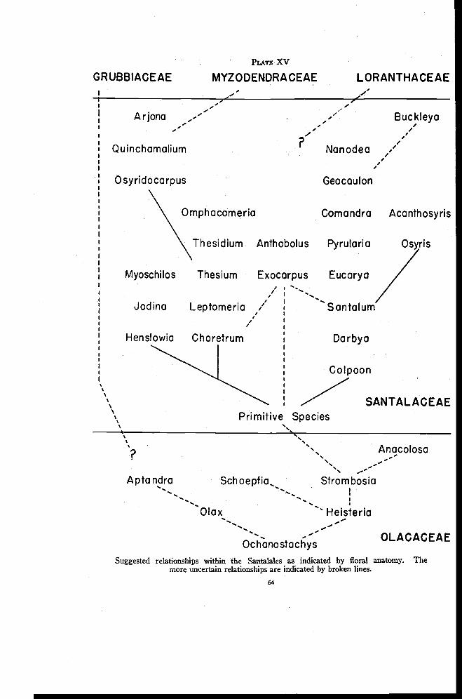
PLAIT XV
GRUBBIACEAE MYZODENDRACEAE LORANTHACEAE
A rjona
Quinchamalium
0 syridocarpus\Omphacomeria Coma ndra Acanthosyris
Thesidium Anthobolus Pyrularia Osyris
Myoschilos Thesium Exocarpus Eucarya
Nanodea
Geocaulon
Buckleya
Jodina Leptomeria
Hen stowia Choretrum
Santalum
Darbya
Colpoon
Primitive SpeciesSANTALACEAE
Anacolosa. --
Apta ndra Schoepfia, Strombosia-. .
I. . . .
Olax Heisteria
OchanostachysSuggested relationships within the Santalales as indicated by floral anatomy. The
more uncertain relationships are indicated by broken lines.
OLACACEAE
64
FLORAL ANATOMY OF THE SANTALACEAE 65
of xylem, has been shifted inward low in the flower. The X-traces appar-ently originate at a lower level in Acanthosyris than in Osyris so that thecarpellary traces and X-traces originate at approximately the same level.This results in a much confused system of vascular tissue in the base of theflower. The development of the xylem at successive levels seems to indicatethat the path of conduction is shifted to the innermost traces low in the ovaryand passes outward first to the recurrent traces and finally to the ascendingtraces. This may explain why the more strongly developed traces are atfirst normally oriented in the inner layer of the ovary wall, then inverted inan intermediate position and finally normally oriented in the outer layer ofthe ovary wall.
Although Buckleya resembles Comandra and Nanodea in the nature ofthe main traces, there seems to be insufficient evidence definitely to associateit with these forms. That Buckleya is considerably advanced is indicated bythe structure of both the staminate and pistillate flowers. There is no definiteindication as to the nature of the extra whorl of floral parts in the pistillateflowers of this genus.
The second line of development in the Santalaceae with regard to floralanatomy seems to have been an early reduction of the recurrent and car-pellary traces with the retention of the five carpels of the primitive santala-ceous flower. There then followed a further reduction in the inner tracesand in the number of carpels. The inferior ovary in this group thus differsmarkedly from that in the first group in that inverted recurrent traces arerepresented by much reduced strands or are entirely lacking. The receptacu-lar nature of the inferior ovary in this group is indicated by the presence inthe pith region of residual tissue that connects upward and outward to themain traces, by the presence of occasional inverted traces (e.g., in Hens-lowia), and usually by the rotation to the inverted position of the marginalstrands of the sepal-stamen bundles to form amphicribral bundles in thesame manner as in Santalum, Pyrularia and other members of the firstgroup.
Within this second group there seem to be two subdivisions. In one,all traces between the sepal-stamen traces disappear near the top of theovary just as in the first line of development described. Each sepal thenreceives only a single strand, the sepal-stamen trace, which first divides tan-gentially to form the stamen trace and then radially to supply the sepal. Thethree genera in this subdivision are Henslowia, Jodina and Myoschilos. Theyare characterized by a reduction from five carpels to three and also by a re-duction in the number of traces representing the recurrent bundles. In thesecond subdivision of this group, each sepal receives three traces, a dorsal
66 OREGON STATE COLLEGESTUDIES IN BOTANY
sepal-stamen trace and two lateral sepal traces. The pedicel contains twiceas many traces as there are floral parts in each whorl. Alternate traces thenbecome dorsal sepal-stamen traces and the remaining traces, which are com-missural, branch to produce lateral traces of adjacent sepals. The genera inthis subdivision are Choretrum, Leptomeria, Thesium, Thesidium and Om-phacomeria. Within this subdivision there is a reduction in the number ofcarpels from five to two and a marked reduction in the number of recurrentand carpellary traces.
Osyridocarpus, Quinchamalium and Arjona seem to have been derivedfrom this group, which has commissural marginal traces. In Osyridocarpusthere are twice as many traces in the pedicel as there are floral parts in eachwhorl. The commissural traces, which alternate with the sepal-stamenbundles, do not continue directly through to the sepals but instead form aseries of anastomoses. At higher levels in the ovary, however, the anas-tomoses form the lateral traces so that each sepal receives one dorsal andtwo lateral traces just as in Choretrum and related genera. Each lateral sepaltrace fuses for a time with its adjacent dorsal sepal-stamen bundle but sepa-rates in the base of the sepal. In Quinchamalium each sepal receives threetraces, but the lateral traces are derived from traces that appear independentlyhalfway up the floral tube. It can probably be assumed that here the com-missural traces have lost their connections below with the main vascularsupply. Arjona resembles Quinchamalium in practically all respects exceptin the absence of lateral sepal traces, and hence is included with it.
Anthobolus and Exocarpus have been considered previously as ratherprimitive members of the Santalaceae because of their superior or nearlysuperior ovaries. As has already been pointed out in describing the struc-ture of these forms, the ovary is not as nearly superior as it appears sincethe ventral carpellary strands arise deep in the fleshy receptacle that bearsthe ovary. The lack of a clearly defined ring of inverted recurrent traces,except for a short distance in Exocarpus cupressiformis, the single loculeand the lack of dorsal carpellary traces all indicate a rather advanced condi-tion and are suggestive of Santalum, which at times has a practically superiorovary. On the other hand, the structure of the staminate flowers of Exocar-pus resembles that of the perfect flowers of Choretrum and Leptomeria.There is also the possibility that Anthobolus and Exocarpus were deriveddirectly from the primitive santalaceous species and may thus be related tothe two main series only in having a common origin. While the origin ofAnthobolus and Exocarpus may be in doubt, it seems entirely unlikely thatthese genera represent primitive forms in the Santalaceae.
FLORAL ANATOMY OF THE SANTALACEAE 67
The relationships indicated by floral anatomy in the Santalaceae aregiven in Plate XV. This chart should not be interpreted as phylogenetic inthe usual sense of the term. The arrangement of genera is based almostentirely on floral anatomy of perfect or pistillate flowers. Taxonomicfeatures generally have not been considered. The primary function of thechart is to show the relation of variations in structure of the receptacularinferior ovary. Since floral anatomy is very important in determiningphylogeny, however, it is to be expected that the arrangement of the generagiven approaches rather closely a phylogenetic arrangement.
The division of the Santalaceae into three tribes is supported by thefloral anatomy of the genera studied. The Anthoboleae are distinctly sep-arated from the Osyrideae and the Thesieae. It is difficult to separate thetwo latter tribes on purely taxonomic considerations since this involves theuse of characteristics more or less present in both tribes. The results ob-tained in this study indicate that Choretrum, Leptomeria, Omphacomeria,Henslowia, Jodina and Myoschilos are more closely related to members ofthe Thesieae than they are to those of the Osyrideae. Relationship is par-ticularly evident between Choretrum and Leptomeria on the one hand andThesium and Thesidium on the other. Numerous changes in the relativepositions of the genera within the Thesieae and Osyrideae are suggested bytheir floral anatomy. Probably the most important of these is the placing ofColpoon and Santalum in the position of more primitive forms and ofBuckleya in an advanced position.
OLACACEAE
Olacaceae is considered by most taxonomists as the most primitivefamily of the Santalales. Sleumer, in "Natiirlichen Pflanzenfamilien" (1935)lists twenty-three genera that are arranged in three subfamilies and furthersubdivided into six tribes. Since the authors are interested in the Olacaceaeprimarily because of their possible relationship to the Santalaceae as indicatedby floral anatomy, taxonomic groupings within the family are not discussed.It may be mentioned, however, that the seven species studied represent allbut one of the six tribes.
Most of the flowers in this family are perfect with both sepals andpetals present. The ovary is superior or inferior with two to five locules be-low, which merge into one above. There are two to five ovules suspendedfrom the central placental column.
Ochanostachys amentacea Mast.In Ochanostachys the calyx is small and cup-shaped. The twelve to
fifteen stamens are so arranged that two are opposite each of the four or five
68 OREGON STATE COLLEGESTUDIES IN BOTANY
petals and one is in each position alternating with the petals. The ovary issuperior with three locules below, which merge into one above the level ofovule attachment. The style is short and thick with a 3-lobed stigma.
In the pedicel there are usually eight or nine traces (Plate XVI, Fig-ure 1), which divide radially to form numerous traces in the base of theflower (Figure 2). Four large traces, and occasionally additional smallerones, move out to supply the calyx tube (Figure 3). Each large trace imme-diately branches to form three traces, which then divide to form numeroustraces in the calyx tube (Figure 4). The calyx tube is freed immediatelyabove the level at which these traces originate. Four petal traces then moveout followed by the twelve stamen traces. The section illustrated in Figure 4is cut at a slight angle across the flower with the lower side of the figure at alower level. In the small amount of material available, no evidence was ob-tained to indicate that the stamens are borne in more than one whorl. Asthe stamen traces move out, three weak traces move in. The latter un-doubtedly represent ventral carpellary traces. They soon disappear, how-ever, and do not enter the placental column. The remaining traces enter theovary wall. The corolla tube with its adnate stamens is freed just below thelevel of the base of the locules (Figure 5). The traces remaining after thedeparture of the stamen and ventral carpellary traces divide to form a ringof numerous small bundles that continue upward in the ovary wall. Thestamens, arranged so that usually two are opposite each petal and four arealternate with the petals, are then freed from the corolla tube (Figure 6).Vascular tissue appears in the partitions near the top of the locules and thenconnects to the ovules. The strands continue upward in the ovary wall and,as the locules merge, become connected above the top of the locule. Extend-ing upward from these connections are three inverted traces that alternatewith the locules (Figure 7). These are the ventral carpellary traces that bya somewhat devious route are continued above the central placenta to thebase of the style. Meanwhile the traces in the outer ovary wall move in andform anastomoses with each other near the level of ovule attachment (Figure6). Some of the traces disappear and at the base of the style the remainderform anastomoses with the inverted traces (Figure 7). The three invertedtraces then disappear and three traces that are normally oriented and in thepositions of dorsal carpellary traces continue up the style almost to the 3-lobed stigma.
Heisteria nitida Engl.
The calyx of Heisteria is small in the flower but it enlarges considerablyas the fruit matures. There are usually five petals and ten stamens. Five ofthe stamens are epipetalous and shorter than the five that alternate with the
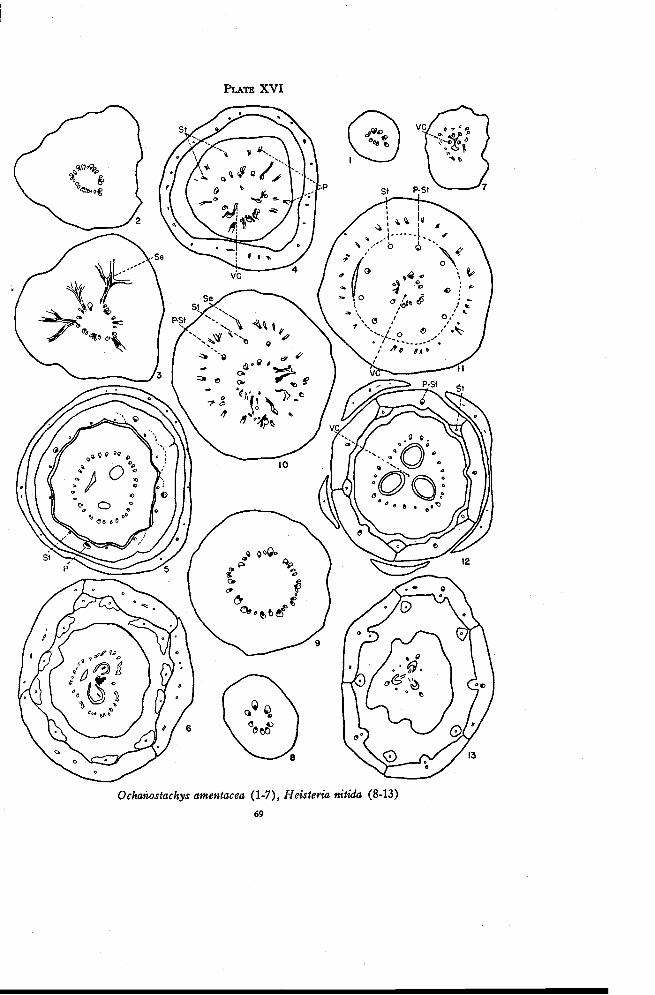
PLATE XVI
Ochanostachys amentacea (1-7), Heisteria nitida (8-13)
69
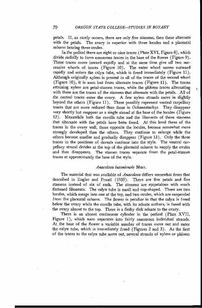
70 OREGON STATE COLLEGE-STUDIES IN BOTANY
petals. If, as rarely occurs, there are only five stamens, then these alternatewith the petals. The ovary is superior with three locules and a placentalcolumn bearing three ovules.
In the pedicel there are eight or nine traces (Plate XVI, Figure 8), whichdivide radially to form numerous traces in the base of the flower (Figure 9).These traces move inward rapidly and at the same time give off two suc-cessive whorls of traces (Figure 10). The outer whorl moves outwardrapidly and enters the calyx tube, which is freed immediately (Figure 11).Although originally xylem is present in all of the traces of the second whorl(Figure 10), it is soon lost from alternate traces (Figure 11). The tracesretaining xylem are petal-stamen traces, while the phloem traces alternatingwith these are the traces of the stamens that alternate with the petals. All ofthe central traces enter the ovary. A few xylem strands move in slightlybeyond the others (Figure 11). These possibly represent ventral carpellarytraces that are more reduced than those in Ochanostachys. They disappearvery shortly but reappear as a single strand at the base of the locules (Figure12). Meanwhile both the corolla tube and the filaments of those stamensthat alternate with the petals have been freed. At this level three of thetraces in the ovary wall, those opposite the locules, become somewhat morestrongly developed than the others. They continue to enlarge while theothers become smaller and gradually disappear (Figure 13). Only the threetraces in the positions of dorsals continue into the style. The ventral car-pellary strand divides at the top of the placental column to supply the ovulesand then disappears. The stamen traces separate from the petal-stamentraces at approximately the base of the style.
Anacolosa luzoniensis Merr.
The material that was available of Anacolosa differs somewhat from thatdescribed in Engler and Prantl (1935). There are five petals and fivestamens instead of six of each. The stamens are epipetalous with muchflattened filaments. The calyx tube is small and cup-shaped. There are twolocules, which merge into one at the top, and two ovules, which are suspendedfrom the placental column. The flower is peculiar in that the calyx is freedbelow the ovary while the corolla tube, with its adnate anthers, is fused withthe ovary almost to the top. There is a fleshy disk adnate to the ovary.
There is an almost continuous cylinder in the pedicel (Plate XVII,Figure 1), which soon separates into fairly numerous individual strands.At the base of the flower a variable number of traces move out and enterthe calyx tube, which is immediately f reed (Figures 2 and 3). As the firstof the traces to the calyx tube move out, several strands of xylem or phloem
FLORAL ANATOMY OF THE SANTALACEAE 71
or both extend into the pith region (Figure 2) and almost disappear.The two locules appear immediately above (Figure 3). Two pairs of phloemstrands are arranged in a single row along the partition or placental column.These phloem strands either originate independently near the main traces orthey originate from the inwardly directed strands shown in Figure 2. Thetraces of each of these pairs fuse near the top of the placental column (Figure5) and both of the traces thus formed supply both ovules.
At the level of the base of the locules the petal traces begin to moveout and the traces on either side of each petal trace begin to swing aroundto the inverted position. These traces are easily distinguishable at approxi-mately the middle of the ovary (Figure 4). At this level small traces aregiven off to the inside, move in near the locules (Figure 5) and form anas-tomoses with each other above the level of ovule attachment (Figure 6).These traces originate in the same manner and follow the same paths as theX-traces in the Santalaceae. The number of traces in this inner ring is thengradually reduced to four, which enter the style (Figure 8). Perhaps this isan indication that there are four carpels but only two locules in this species.After these traces are given off to the inside, the outer traces between thepetal positions soon lose their xylem (Figure 5). In each petal position atthis level there is a loose amphicribral strand of the type seen in the Santa-laceae. The outermost trace from each strand moves out to enter the corollatube (Figure 6), which is freed immediately. The remaining traces of eachgroup, as well as the phloem strands that have persisted between the petalpositions, enter the fleshy disk, which is adnate to the ovary. The invertedand partly inverted traces branch laterally in the disk. Both these and thephloem traces between the petal positions then gradually disappear. Thestamens are freed opposite each petal (Figure 7) but no stamen traces aredistinguishable at any level in the flower.
The nature of the inferior ovary here is somewhat in doubt. Since theexact origin of the ventral carpellary traces is difficult to determine, this typeof evidence cannot be considered. The loosely arranged amphicribralstrands and the behavior of the stylar traces, however, would seem to indicatea receptacular inferior ovary as in the Santalaceae. If the inferior ovaryhere is receptacular, then it seems likely that the so-called calyx is actually awhorl of bracts. The gradual disappearance of the traces between the am-phicribral strands is also typical of the Santalaceae. In Anacolosa, however,these traces disappear in the disk after the corolla tube has been freedinstead of at the base of the calyx tube as in the Santalaceae. Before theirdisappearance in the disk the inner traces of each amphicribral group formanastomoses. It seems most likely that the inferior ovary here is receptacular
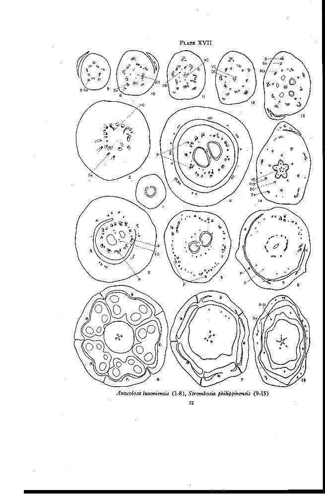
PLATE XVII
Anacolosa luzoniensis (1-8), Strombosia philippinensis (9-15)
72
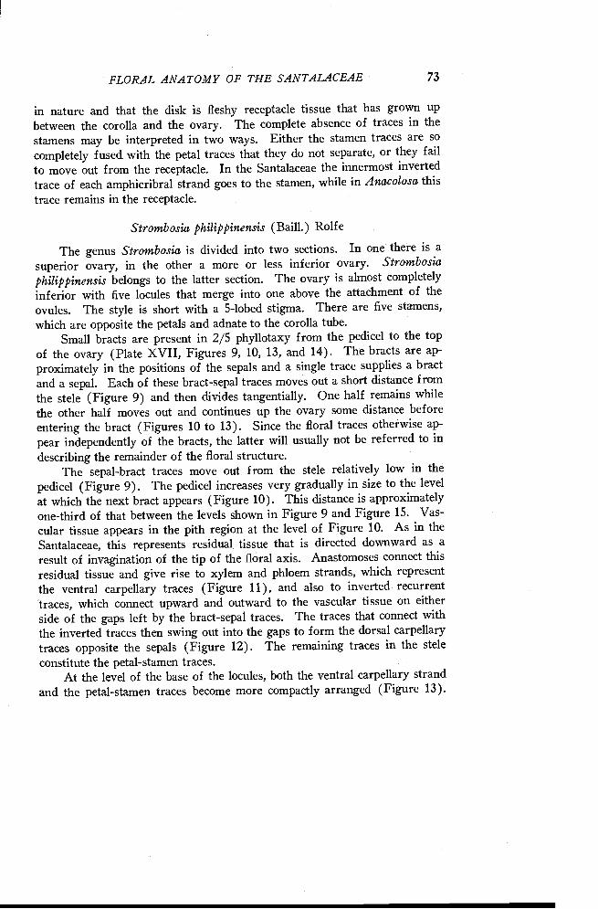
FLORAL ANATOMY OF THE SANTALACEAE 73
in nature and that the disk is fleshy receptacle tissue that has grown upbetween the corolla and the ovary. The complete absence of traces in thestamens may be interpreted in two ways. Either the stamen traces are socompletely fused with the petal traces that they do not separate, or they fail
to move out from the receptacle. In the Santalaceae the innermost invertedtrace of each amphicribral strand goes to the stamen, while in Anacolosa this
trace remains in the receptacle.
Strombosia philippinensis (Baill.) Rolfe
The genus Strombosia is divided into two sections. In one there is asuperior ovary, in the other a more or less inferior ovary. Strombosiaphilippinensis belongs to the latter section. The ovary is almost completelyinferior with five locules that merge into one above the attachment of theovules. The style is short with a 5-lobed stigma. There are five stamens,which are opposite the petals and adnate to the corolla tube.
Small bracts are present in 2/5 phyllotaxy from the pedicel to the topof the ovary (Plate XVII, Figures 9, 10, 13, and 14). The bracts are ap-proximately in the positions of the sepals and a single trace supplies a bractand a sepal. Each of these bract-sepal traces moves out a short distance fromthe stele (Figure 9) and then divides tangentially. One half remains whilethe other half moves out and continues up the ovary some distance beforeentering the bract (Figures 10 to 13). Since the floral traces otherwise ap-pear independently of the bracts, the latter will usually not be referred to indescribing the remainder of the floral structure.
The sepal-bract traces move out from the stele relatively low in thepedicel (Figure 9). The pedicel increases very gradually in size to the level
at which the next bract appears (Figure 10). This distance is approximatelyone-third of that between the levels shown in Figure 9 and Figure 15. Vas-cular tissue appears in the pith region at the level of Figure 10. As in theSantalaceae, this represents residual tissue that is directed downward as aresult of invagination of the tip of the floral axis. Anastomoses connect thisresidual tissue and give rise to xylem and phloem strands, which representthe ventral carpellary traces (Figure 11), and also to inverted recurrenttraces, which connect upward and outward to the vascular tissue on eitherside of the gaps left by the bract-sepal traces. The traces that connect withthe inverted traces then swing out into the gaps to form the dorsal carpellarytraces opposite the sepals (Figure 12). The remaining traces in the steleconstitute the petal-stamen traces.
At the level of the base of the locules, both the ventral carpellary strandand the petal-stamen traces become more compactly arranged (Figure 13).
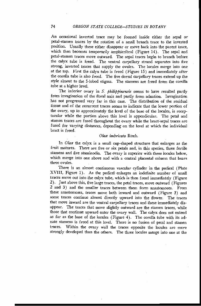
74 OREGON STATE COLLEGESTUDIES IN BOTANY
An occasional inverted trace may be formed inside either the sepal orpetal-stamen traces by the rotation of a small branch trace to the invertedposition. Usually these either disappear or move back into the parent trace,which then becomes temporarily amphicribral (Figure 14). The sepal andpetal-stamen traces move outward. The sepal traces begin to branch beforethe calyx tube is freed. The ventral carpellary strand separates into fivestrong, inverted traces that supply the ovules. The locules merge into oneat the top. First the calyx tube is freed (Figure 15) and immediately afterthe corolla tube is also freed. The five dorsal carpellary traces extend up thestyle almost to the 5-lobed stigma. The stamens are freed from the corollatube at a higher level.
The inferior ovary in S. philippinensis seems to have resulted partlyfrom invagination of the floral axis and partly from adnation. Invaginationhas not progressed very far in this case. The distribution of the residualtissue and of the recurrent traces seems to indicate that the lower portion ofthe ovary, up to approximately the level of the base of the locules, is recep-tacular while the portion above this level is appendicular. The petal andstamen traces are fused throughout the ovary while the bract-sepal traces arefused for varying distances, depending on the level at which the individualbract is freed.
()lax imbricate Roxb.
In Olax the calyx is a small cup-shaped structure that enlarges as thefruit matures. There are five or six petals and, in this species, three fertilestamens and five staminodia. The ovary is superior with three locules below,which merge into one above and with a central placental column that bearsthree ovules.
There is an almost continuous vascular cylinder in the pedicel (PlateXVIII, Figure 1). As the pedicel enlarges an indefinite number of smalltraces move out into the calyx tube, which is then freed immediately (Figure2). Just above this, five large traces, the petal traces, move outward (Figures2 and 3) and the smaller traces between them form anastomoses. Fromthese anastomoses, traces move both inward and outward (Figure 3) andsome traces continue almost directly upward into the flower. The tracesthat move inward are the ventral carpellary traces and these immediately dis-appear. The traces that move slightly outward are the stamen traces, whilethose that continue upward enter the ovary wall. The calyx does not extendas far as the base of the locules (Figure 4). The corolla tube with its ad-nate stamens is freed at this level. There is no fusion of petal and stamentraces. Within the ovary wall the traces opposite the locules are morestrongly developed than the others. The three locules merge into one at the
PLATE XVIII
Olax imbricata (1-6), Aptandra Spruceana (7-17)75
76 OREGON STATE COLLEGESTUDIES IN BOTANY
level of ovule attachment (Figure 5). Anastomoses are formed between thetraces in the ovary wall and then all but those in the positions of dorsal car-pellary traces disappear. The dorsals continue in the style (Figure 6) almostto the 3-lobed stigma. The stamens and staminodia are freed at approxi-mately the same level as the corolla lobes.
Aptandra Spruceana Miers
The flowers of Aptandra are 4-merous. The stamens are united in atube and are located opposite the petals. The stamen tube is separated fromthe corolla tube by thick, scale-like glands that are adnate to the stamen tubeat its base. The ovary is superior with two locules below, which merge intoone above and with a central placental column that bears two ovules. Thestyle is slender with a thickened club-shaped stigma.
In the upper portion of the pedicel there is an almost continuous cylinder(Plate XVIII, Figure 7) which soon separates into eight strands (Figure 8).The smaller traces then move out and each branches immediately into threetraces (Figure 9). The lateral traces disappear as the calyx tube is freed(Figure 10). At this same level the four traces remaining in the center giveoff branches from both sides and the strong petal traces move outward(Figure 11) into the corolla tube, which is then freed immediately. Fourstrong traces remain in the petal positions and alternate with four phloemtraces. The latter divide once or twice and four traces then move inward(Figure 12). The inner traces merge into a mass of phloem-like tissue inthe center, the ventral carpellary strand (Figure 13), which then disappearsbelow the level of the locules. The four strongly developed traces oppositethe petal traces are the stamen traces. The phloem traces between these arereduced to four (Figure 14), which enter the ovary wall. The stamen tracesmove out slightly and almost at the same time the mass of tissue inside thecorolla tube is differentiated into a fleshy disk that is lobed at the top, astamen tube, and the 2-loculed ovary (Figure 15). It seems very probablethat the glandular disk between the stamen tube and the corolla tube repre-sents reduced stamens. The two locules merge into one near the top of theovary (Figure 16). The four traces in the ovary wall enter the style (Figure17) and extend almost to the stigma. In this species, as in Anacolosa, thereare possibly four carpels but only two locules. The stamens are united bytheir anthers as well as by their filaments.
Schoepfia californica Brandeg.
According to the accepted description of the flowers of Schoepfia, thecalyx, while not distinguishable is, if present, perhaps united with the re-
FLORAL ANATOMY OF THE SANTALACEAE 77
ceptacle. Subtending bracts are united to form a cup-shaped involucre below
the ovary. Five petals are united into a corolla tube bearing five stamensopposite the petals. The ovary in this species is inferior with three loculesbelow, which merge into one above the level of attachment of the ovules.
The three ovules are borne on the tip of the central placental column.Throughout the pedicel, gradual changes occur in the number and ar-
rangement of traces. In the material studied there are eight traces low inthe pedicel (Plate XIX, Figure 1 ). One of these divides and the ninetraces thus formed become arranged on opposite sides, leaving two gap-likebands of parenchyma in the stele (Figure 2). Some of the traces then swing
over into the gaps (Figure 3). In the upper portion of the pedicel some ofthe traces fuse, reducing the number to seven (Figure 4). The number oftraces is increased again by radial separation and a variable number, usuallythree or four, move out from the stele (Figure 5). These traces then branchmany times and enlarge considerably to form an outer ring of well developedtraces (Figure 6). The involucre, which is freed immediately above, re-ceives all of these traces (Figure 7). Meanwhile the traces in the centralregion divide radially to form additional traces, some of which consist of
phloem only. This radial separation of traces continues throughout thelength of the ovary. Some distance below the locules phloem traces aregiven off to the inside (Figure 8). These originate mostly from the smallertraces, especially from those consisting of phloem only. They move inwardto form an almost continuous ring of phloem tissue, which later forms sev-eral large strands of phloem in the base of the placental column (Figure 9).These strands, representing the ventral carpellary traces, later merge into asingle large phloem strand.
Just at the level of the base of the locules, three traces in the positionsof dorsal carpellary traces move inward from the traces in the ovary wall
(Figure 9). These traces appear to arise at slightly different levels in theflower. The petal-stamen traces gradually enlarge to form quite well de-veloped traces near the top of the ovary. They move out slightly while thetraces between them form numerous anastomoses and move inward (Figure10). As the corolla tube is freed (Figure 11) the traces in the ovary wallbegin to disappear. Anastomoses continue to be formed between thesetraces but apparently none are formed with the dorsal carpellary traces.Only a few of these traces persist at the level of ovule attachment (Figure12) and soon all disappear except the three dorsals, which continue in thestyle almost to the 3-lobed stigma. The stamens are freed a little more thanhalfway up the corolla tube.
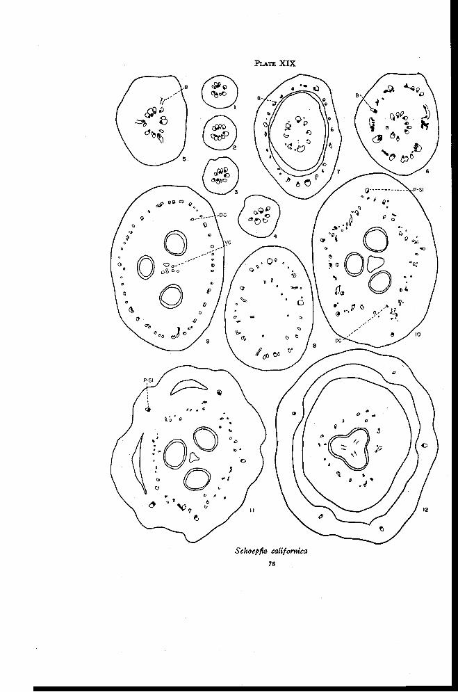
PLATE XIX
Schoepfia californica
78
FLORAL ANATOMY OF THE SANTALACEAE 79
DISCUSSION
The presence in this family of two and possibly all three of the possibletypes of inferior ovary is of especial interest. The three possible types, ashave been previously discussed, are the appendicular, the receptacular, and acombination in which the inferior ovary is receptacular below and appendicu-lar above. The inferior ovary of Schoepfia californica appears clearly tobe appendicular. The floral traces originate in the manner of a typical ap-pendicular ovary and there are no inverted or amphicribral traces to indicatethat invagination has occurred. The inferior ovary of Strombosia philip-pinensis appears just as clearly to be receptacular below the base of the loculesand appendicular above. The receptacular nature of the base of the ovary is
indicated by the presence of downwardly directed residual tissue and of in-verted recurrent traces that connect the residual tissue with bundles of thestele at a level near the base of the locules. The vascular anatomy of theflower above this level is that of an appendicular inferior ovary. The floralparts are fused but the traces to the various parts, except the petal-stamentraces, are separate. The inferior ovary of this species is complicated by theadherence of bracts, fused to both the receptacular and appendicular portionsof the ovary.
If the authors' interpretation of the anatomy of Anacolosa luzoniensisis correct, then the receptacular inferior ovary also is present in this family.It seems difficult to explain the amphicribral strands and the traces to the diskon any other basis. On the other hand, if the cup-shaped structure beneaththe flower is the calyx, as it has been considered, it must be concluded thatelongation and invagination of the axis have occurred above the level atwhich the calyx is freed. On the basis of information available at present,this does not seem plausible. Anacolosa resembles Schoepfia in that in boththere is present the cup-shaped structure at the base of the ovary and also asingle whorl of floral organs borne at the top of the ovary. In both formsthe whorl at the top of the ovary is interpreted as corolla, while the cup-shaped structure in Anacolosa is interpreted as calyx and that in Schoepfiaas involucre. The interpretation of the inferior ovary in Anacolosa wouldbe simplified if it could be shown that the cup-shaped structure is an in-volucre instead of a calyx. Much additional work should be done on thefloral anatomy of this family and also on the relation of bracts and floralparts.
The relatively few species of Olacaceae studied offer insufficient evidenceto justify any conclusions regarding relationships within the family. Thereare some definite similarities, however, between some genera that have here-tofore not been considered as closely related. Ochanostachys and Olax show
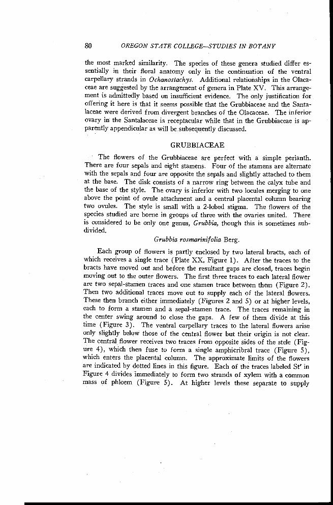
80 OREGON STATE COLLEGESTUDIES IN BOTANY
the most marked similarity. The species of these genera studied differ es-sentially in their floral anatomy only in the continuation of the ventralcarpellary strands in Ochanostachys. Additional relationships in the Olaca-ceae are suggested by the arrangement of genera in Plate XV. This arrange-ment is admittedly based on insufficient evidence. The only justification foroffering it here is that it seems possible that the Grubbiaceae and the Santa-laceae were derived from divergent branches of the Olacaceae. The inferiorovary in the Santalaceae is receptacular while that in the Grubbiaceae is ap-parently appendicular as will be subsequently discussed.
GRUBBIACEAE
The flowers of the Grubbiaceae are perfect with a simple perianth.There are four sepals and eight stamens. Four of the stamens are alternatewith the sepals and four are opposite the sepals and slightly attached to themat the base. The disk consists of a narrow ring between the calyx tube andthe base of the style. The ovary is inferior with two locules merging to oneabove the point of ovule attachment and a central placental column bearingtwo ovules. The style is small with a 2-lobed stigma. The flowers of thespecies studied are borne in groups of three with the ovaries united. Thereis considered to be only one genus, Grubbia, though this is sometimes sub-divided.
Grubbia rosmarinifolia Berg.
Each group of flowers is partly enclosed by two lateral bracts, each ofwhich receives a single trace (Plate XX, Figure 1). After the traces to thebracts have moved out and before the resultant gaps are closed, traces beginmoving out to the outer flowers. The first three traces to each lateral flowerare two sepal-stamen traces and one stamen trace between them (Figure 2).Then two additional traces move out to supply each of the lateral flowers.These then branch either immediately (Figures 2 and 5) or at higher levels,each to form a stamen and a sepal-stamen trace. The traces remaining inthe center swing around to close the gaps. A few of them divide at thistime (Figure 3). The ventral carpellary traces to the lateral flowers ariseonly slightly below those of the central flower but their origin is not clear.The central flower receives two traces from opposite sides of the stele (Fig-ure 4), which then fuse to form a single amphicribral trace (Figure 5),which enters the placental column. The approximate limits of the flowersare indicated by dotted lines in this figure. Each of the traces labeled St' inFigure 4 divides immediately to form two strands of xylem with a commonmass of phloem (Figure 5). At higher levels these separate to supply
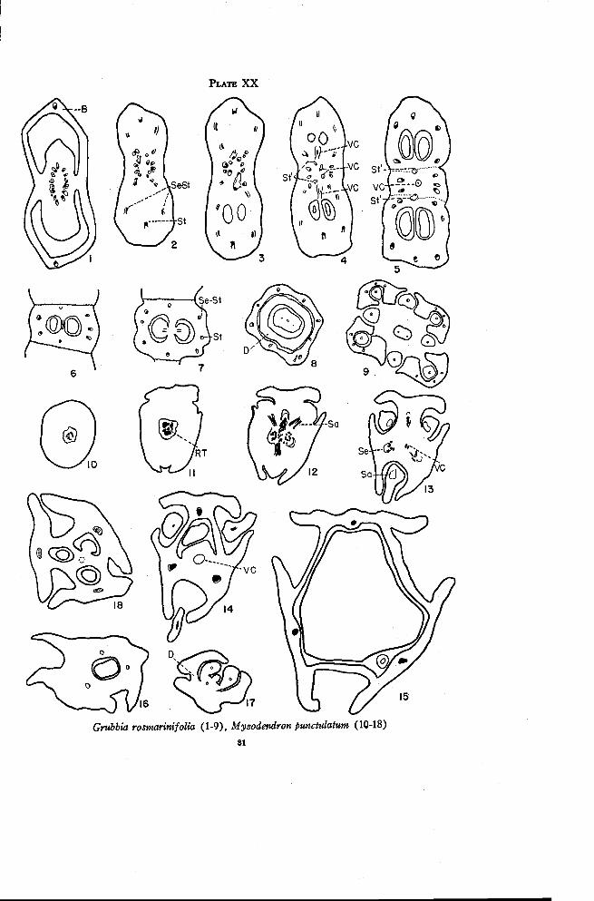
PLATE XX
Grubbia rosmarinifolia (1-9), Myzodendron punctulatum (10-18)
81
82 OREGON STATE COLLEGESTUDIES IN BOTANY
stamens in separate flowers. In this region the vascular supplies to thethree flowers may be easily identified. The upper flower has five large traces,the two lower of which each subsequently divide to form a stamen anda sepal-stamen trace. Half of the upper St' trace, moreover, supplies astamen in this flower. The central flower also has five large traces of whichthe upper left one later divides. This flower receives a half of each ofthe St' traces. The lower flower has seven traces and receives half of thelower St' trace. Thus each flower is eventually supplied by eight traces inthe ovary wall, four sepal-stamen traces and, alternating with these, fourstamen traces (Figure 7). The ventral carpellary strand is flattened in thepartition between the locules (Figure 6) and is used up in supplying theovules (Figure 7). Above the point of attachment of the ovules the loculesmerge into a single cavity extending for some distance up the style. Thecalyx tube, to which all the filaments are fused, is freed almost simultaneouslywith the disk (Figure 8). The stamens that alternate with the sepals arefreed from the tube first, followed by the stamens opposite the sepals. Ineach sepal the sepal trace branches to form three (Figure 9). At the baseof the style two traces appear in the positions of dorsal carpellary traces.There is no evident connection between these and any of the other floraltraces.
The inferior ovary seems to be entirely appendicular in nature sincethere are indications neither of residual tissue directed downward in the pithregion nor of inverted recurrent traces.
MYZODENDRACEAE
The species in the Myzodendraceae are dioecious with usually 3-merousflowers. The pistillate flower has an inferior ovary. In the young pistillateflower there are three locules below and three ovules suspended from acentral placental column. Only one ovule develops and this enlarges to fillthe entire cavity. The placental stalk is crowded to one side of the locule.In the ovary wall there are three longitudinal furrows, each of which containsa seta, which becomes long in the fruit. These setae have been considered ashomologous with stamens. Myzodendron is the only genus in this family.
Myzodendron punctulatum Banks and Sol.
The pedicel of Myzodendron has a very slender stele with a small pith(Plate XX, Figure 10). While interpretation is difficult because of thesmall stele and irregular distribution of the xylem, it seems likely that xylemappears independently in the pith region (Figure 11) at the base of the
FLORAL ANATOMY OF THE SANTALACEAE 83
ovary. Traces are given off first to the setae (Figure 12) and then justabove this to the ovary wall in the sepal positions (Figure 13). Since thetraces to the setae are given off first, it seems rather improbable that thesestructures are staminodia. After the departure of the traces to the ovarywall there is a small amount of xylem left in the pith region. This graduallydisappears and is replaced by phloem-like tissue (Figure 14), which con-tinues into the placental column. In an older flower, such as the one de-scribed here, a single seed is present and the placental stalk is crowded overto one side of the locule (Figure 15). In the younger flower, however, oneovule is suspended in each of three locules (Figure 18). The locules mergeinto one above the level of ovule attachment. The three traces in the ovarywall do not enter the sepals. This was recognized by Johnson (1889). In-stead, the traces swing inward at the top of the ovary as the sepals are freed(Figure 16) and enter the style, which almost immediately branches intothree stigmas (Figure 17). A small ring-shaped disk is present between thesepals and the style.
The extreme reduction in Myzodendron makes it difficult of interpreta-tion. Unfortunately only limited material was available. The probable oc-currence of residual tissue in the pith region below the ventral carpellarystrand seems to indicate invagination of the axis. Hence the inferior ovaryis probably receptacular.
LORANTHACEAE
In the species of the Loranthaceae that were studied, the pistillateflowers are characterized by an inferior ovary with a single locule. There islittle or no differentiation between the ovule and the placenta. There aretwo or three sepals and sometimes a circular disk. There are no indicationsof either stamens or staminodia.
Phoradendron villosum Nutt.
The material studied of Phoradendron consisted of young fruits con-taining well developed seed. There are ten to twelve traces in the pedicel(Plate XXI, Figure 1). At the base of the ovary three large sepal tracesmove outward at slightly different levels (Figure 2). The remaining traces,some of which divide radially, become rearranged into a somewhat irregularring of bundles just below the level of the base of the locule (Figure 3).There are no traces connecting these strands with the placental knob. As thisring of bundles moves into the ovary wall there is some increase in number oftraces by radial separation and the bundles become smaller in size (Figure
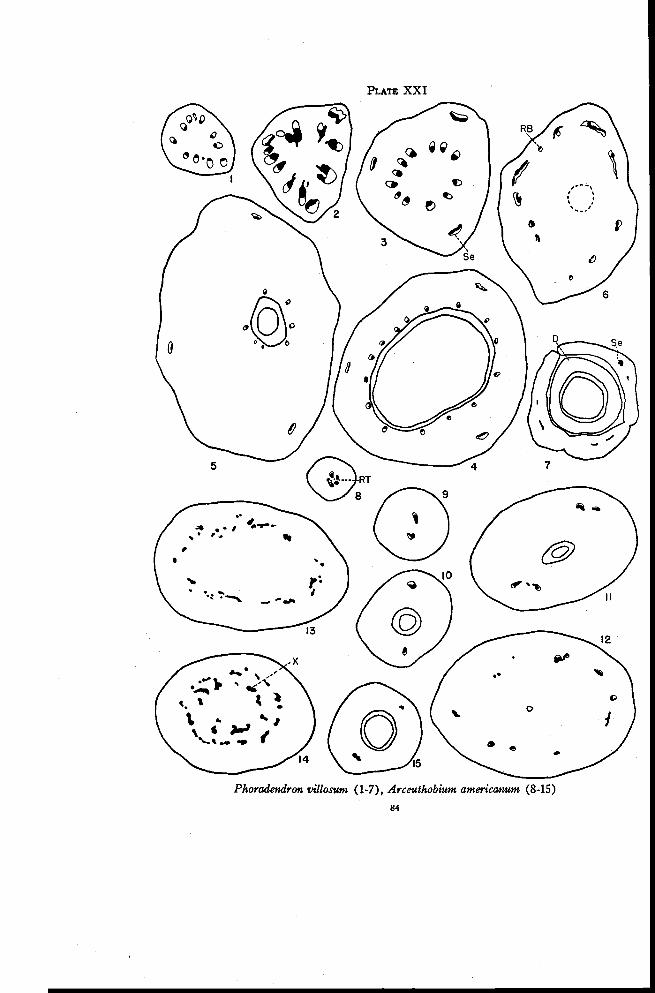
PLATE XXI
Phoradendron villosion (1-7), Arceuthobium americanum (8-15)
84
FLORAL ANATOMY OF THE SANTALACEAE 85
4). As the locule closes, these traces move inward toward the base of thestyle and their number is gradually reduced by the disappearance of traces(Figure 5). All of these traces disappear above the locule. Meanwhile atapproximately the level of the top of the locule, additional traces appear inde-
pendently in the outer portion of the ovary. Most of these traces are in-verted and directed downward (Figure 6). They connect to the sepal tracesat higher levels and then disappear rather rapidly. Few traces persist toenter the sepals (Figure 7). The sepals, disk, and style are freed almostsimultaneously.
Arceuthobium americanum Nutt.
In the flowers of Arceuthobium there are four or five traces in the pedi-cel just below the ovary. There are apparently a few strands of residual tis-
sue in the pith region (Plate XXI, Figure 8). The vascular tissue graduallyseparates into two traces (Figure 9), which enter the ovary wall on opposite
sides of the locule (Figure 10). There is no trace leading to the placentalknob. Near the top of the locule each of the traces branches once (Figure11). At slightly higher levels additional traces appear independently in the
ovary wall (Figure 12). Most of these traces consist only of xylem andgradually the phloem disappears from all traces. Toward the top of theovary the traces are all connected by a series of anastomoses (Figure 13),which are formed over a considerable distance. Gradually more and moreof the anastomoses lead inward until finally two distinct rings of xylemtraces can be recognized (Figure 14). The inner ring, possibly representingthe X-traces, is directed toward the base of the style but all of the tracesdisappear before the style is freed. The traces of the outer ring disappearmore gradually and usually only two persist at the level at which the shortcalyx tube is freed (Figure 15).
There is no indication in the floral anatomy of the number of carpelsin either Arceuthobium or Phoradendron. Van Tieghem (1869a) describedtwo carpels in Viscum album. Dowding (1931), on the basis of morphologi-cal development, concluded that the ovary of Arceuthobium americanum con-sists of an outer whorl of four sterile and an inner whorl of four fertilecarpels. These are much reduced forms of the Loranthaceae. A study ofmore primitive members of the family is desirable.
The appearance of traces directed downward in the ovary wall, especiallyof the inverted ones of Phoradendron, suggests that the inferior ovary isprobably receptacular. The presence of X-traces in Arceuthobium and thecomplete absence of any indication of ventral carpellary strands in bothgenera are also suggestive of a receptacular inferior ovary. In the Santa-
86 OREGON STATE COLLEGESTUDIES IN BOTANY
laceae there is in general a gradual reduction in the connections of the ventralcarpellary strand to the main traces. In some forms (e.g., Anthobolus, Buck-leya, Thesidium) the ventral strands are entirely independent of the mainvascular supply. In Viscum also the carpels are independent of the mainvascular supply (Van Tieghem, 1869a).
RELATIONSHIPS WITHIN THE SANTALALES
The constitution of the Santalales and the relationships of the familiesincluded have been discussed by taxonomists for many years. The variousgroupings and arrangements suggested were discussed recently by Harms in"Natiirlichen Pflanzenfamilien" (1935) and are not considered here in detail.In addition to the families considered here there are two others, the Opilia-ceae and the Octoknemaceae, which are usually considered to belong to theSantalales. Both are closely related to the Olacaceae.
The Olacaceae together with the Opiliaceae were placed by Hutchinson(1926) in a separate order, the Olacales. In the Santalales he placed firstthe Loranthaceae, followed by the Santalaceae, the Grubbiaceae and theMyzodendraceae. On the basis of progressively increasing parasitism andof reduction in complexity of the ovule, Schellenberg (1932) considered theorder of families in the Santalales to be the Olacaceae, the Grubbiaceae, theSantalaceae, the Myzodendraceae and the Loranthaceae. He considered theGrubbiaceae and the Myzodendraceae, however, as possibly convergentforms not directly related to the Santalales. The same arrangement is usedin "Natiirlichen Pflanzenfamilien" (1935). Rend le (1938) regarded theMyzodendraceae as a much reduced ally of the Santalaceae.
The Olacaceae is clearly the least advanced of the families of the San-talales described here. The number of floral whorls, the number of stamens,the more complete separation of the locules and the lack of fusion of petaland stamen traces in 0 chanostachys and Olax are all indications of the moreprimitive nature of this family. Also, as indicated by Schellenberg (1932),the presence both of nonparasitic forms in this family and of species inwhich the ovules have two integuments covering the nucellus indicates thatthe Olacaceae should be placed lowest in the series.
It seems likely that divergent lines in the Olacaceae may have given riseto the Grubbiaceae and the Santalaceae. Within the Olacaceae both adna-tion and invagination have occurred to form two or three types of inferiorovary. The inferior ovary of Grubbia appears to be appendicular and thuswould be derived from forms in which invagination of the floral axis has notoccurred. The inferior ovary of Grubbia resembles very closely the superiorovary of Aptandra except that in the former the number of dorsal carpellary
P
FLORAL ANATOMY OF THE SANTALACEAE 87
traces has been reduced to two and these appear only in the style. Schoepfia
has an inferior ovary that is clearly appendicular and in this respect resem-bles Grubbia. There is not enough evidence available to suggest a directconnection between the Grubbiaceae and any given genus in the Olacaceae.
The Santalaceae was probably derived from the Olacaceae. This isthe opinion of most taxonomists. There are similarities throughout bothfamilies in the structure of the ovary and of the ovules, especially with
regard to placentation. A particularly striking similarity is the invaginationof the floral axis, which is characteristic of the Santalaceae and which occursin Strombosia and probably in Anacolosa. The receptacular inferior ovaryresulting from invagination probably occurs rather infrequently in dicots.
This together with similarities in ovary, ovule, placentation and parasitismseems to indicate that the primitive santalaceous flower may have been de-rived from the Olacaceae, possibly from the tribe Anacoloseae. It may beespecially significant that Strombosia, which shows definite indications ofinvagination, has five carpels, five locules each with a suspended ovule, and
strong dorsal carpellary traces, all of which are characteristic of the hypo-thetical primitive santalaceous flower. Since the inferior ovary of Strom-bosia is receptacular below and appendicular above, while the inferior ovaryin the Santalaceae is receptacular throughout, the question arises as to the
method of origin of the latter type. Before studying the floral anatomy ofStrombosia, the authors had suggested (1942) that the receptacular inferiorovary in the Santalaceae might have originated by invagination of the floralaxis to form a cup-shaped receptacle that subsequently became fused with
the ovary. The structure of Strombosia suggests the possibility that thereceptacular inferior ovary in the Santalaceae may have been derived froman inferior ovary which was receptacular below and appendicular above by aprogressive increase in the amount of invagination. Conclusions regardingthese two possibilities do not seem to be justified on the basis of evidence
available at present. Additional studies in the Olacaceae are certainly desir-able in determining the probable method of origin of the inferior ovary in
the Santalaceae.The Myzodendraceae was considered both by Skottsberg in "Natfirlichen
Pflanzenfamilien" (1935) and by Rendle (1938) as a reduced ally of theSantalaceae. While evidence obtained from the floral anatomy of Myzoden-dron punctulatum is not conclusive, it strongly suggests that the inferiorovary in Myzodendron is receptacular in nature. This would seem to indi-cate its relationship to the Santalaceae. Skottsberg, on the basis of the struc-ture of the ovary and of the circular disk, suggested a relationship to Arjonaand to Quinchamalium, Myzodendron is a very much reduced form and its
88 OREGON STATE COLLEGESTUDIES IN BOTANY
origin is obscure. The only conclusion justifiable at present seems to bethat there seem to be more reasons for relating it to Arjona and to Quin-chamalium than to any other genera in the Santalaceae.
The Loranthaceae has been considered both as more primitive and asmore advanced than the Santalaceae. The two species of this family thatwere studied are too advanced to indicate relationships. Studies are neededof the more primitive members of the family. The only conclusion possiblehere is that in Phoradendron and Arceuthobium the inferior ovary is prob-ably receptacular. The advanced condition of the ovary in these forms, asevidenced by reduction and loss of traces, seems to indicate that the Loran-thaceae is more advanced than the Santalaceae and was probably derivedfrom it. According to Van Tieghem (1869a) the sepals of Viscum receiveone dorsal trace and two lateral traces originating from commissural strands,as in Thesium and closely related forms. This fact alone, however, ishardly enough to connect the Loranthaceae with this particular series in theSantalaceae.
The Santalales, as treated here, has been considered as a member of theCelastrales (Pool, 1941), as a derivative of the Celastrales (Hutchinson,1926), and as an ally of the Proteales (Rendle, 1938). No evidence basedon floral anatomy is available from either the Celastrales or the Proteales toindicate the origin of the Santalales. It may be stated, however, that theSantalales apparently does not belong in the main line of development towardthe Umbellales as in the Bessyian system. The inferior ovary in the Umbelli-f erae is appendicular ( Jackson, 1933). It seems improbable that a receptacu-lar inferior ovary could give rise to the type of ovary that has been inter-preted as appendicular. At present it seems most likely that the Santalalesconstitutes a separate line of development and probably has not given riseto other orders. Whether this line originates from the Celastrales, theProteales, or from some other order remains to be determined.
SUMMARY
Descriptions are given of the floral anatomy of the Santalaceae based ona study of twenty-four genera and forty species of the family. The inferioror partly inferior ovary of all species studied is considered to be receptacular.The ovary is embedded in tissues of the receptacle as a result of invaginationof the floral axis. The receptacular inferior ovary probably occurs infre-quently in the Angiosperms. Thus far the Santalales is the only orderin which floral anatomy indicates the occurrence of such a type. The fol-
FLORAL ANATOMY OF THE SANTALACEAE 89
lowing characteristics may be considered as typical of the receptacularinferior ovary in the Santalaceae :
1. Inverted recurrent traces. These are characteristic of the moreprimitive receptacular inferior ovary and, when present, are a definiteand easily recognizable indication of invagination.
2. Amphicribral condition of the main traces. It can be shown thatthe large, loosely arranged amphicribral strands in some species of theSantalaceae are the result of phylogenetic fusion of ascending and re-current receptacular traces in the positions of the sepals. The tracesbetween the sepal positions disappear near the top of the ovary in moreprimitive forms and low in the ovary in more advanced forms.
3. Downwardly directed residual tissue in the pith region of thepedicel. In the more primitive forms the carpellary traces originate from
the inverted recurrent traces. The recurrent traces continue downwardbeyond the level of origin of the carpellary traces. It is because ofinvagination then that the residual tissue is directed downward insteadof upward as in the appendicular type of inferior ovary.
4. Origin of the carpellary traces from a group of vascular tissue inthe pith region, which connects upward and outward to the main vascu-lar supply. In the more primitive forms the carpellary traces originatefrom the inverted recurrent traces low in the ovary, while in the moreadvanced forms the ascending and recurrent traces tend to become fusedand the residual tissue tends to persist in the pith region. As a result ofthese two tendencies a saucer- or cone-shaped group of tissue, with thetip of the cone directed downward, persists in the pedicel and connectsupward and outward to the main traces. In an appendicular type ofinferior ovary the vascular tissue giving rise to the carpellary traces is inthe form of a cone with the tip directed upward.
5. Lack of dorsal carpellary traces. This characteristic is of some-what dubious value. It seems likely that in the Santalaceae dorsal car-pellary traces may be lacking in some species because they have lost con-nections with the main vascular supply and have gradually disappearedthrough continued reduction in size. This should not be confused withthe nonappearance of dorsal traces because of fusion with other traces.
6. Complete independence of the dorsal or ventral carpellary traces,or both, from the main vascular supply. This characteristic is evenmore dubious than the preceding, but it does appear in certain speciesof the Santalaceae. In many species there are only weak connectionsbetween the main traces and the carpellary traces, especially the ventral
carpellary traces. In the more advanced species these connections dis-appear completely.
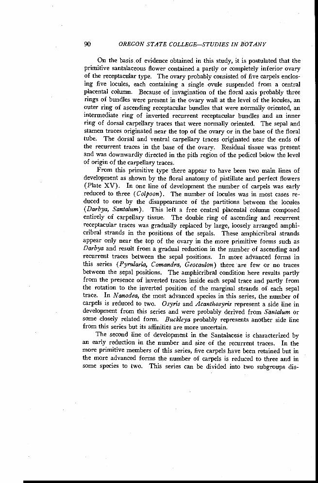
90 OREGON STATE COLLEGESTUDIES IN BOTANY
On the basis of evidence obtained in this study, it is postulated that theprimitive santalaceous flower contained a partly or completely inferior ovaryof the receptacular type. The ovary probably consisted of five carpels enclos-ing five locules, each containing a single ovule suspended from a centralplacental column. Because of invagination of the floral axis probably threerings of bundles were present in the ovary wall at the level of the locules, anouter ring of ascending receptacular bundles that were normally oriented, anintermediate ring of inverted recurrent receptacular bundles and an innerring of dorsal carpellary traces that were normally oriented. The sepal andstamen traces originated near the top of the ovary or in the base of the floraltube. The dorsal and ventral carpellary traces originated near the ends ofthe recurrent traces in the base of the ovary. Residual tissue was presentand was downwardly directed in the pith region of the pedicel below the levelof origin of the carpellary traces.
From this primitive type there appear to have been two main lines ofdevelopment as shown by the floral anatomy of pistillate and perfect flowers(Plate XV). In one line of development the number of carpels was earlyreduced to three (Colpoon). The number of locules was in most cases re-duced to one by the disappearance of the partitions between the locules(Darbya, Santalum). This left a free central placental column composedentirely of carpellary tissue. The double ring of ascending and recurrentreceptacular traces was gradually replaced by large, loosely arranged amphi-cribral strands in the positions of the sepals. These amphicribral strandsappear only near the top of the ovary in the more primitive forms such asDarbya and result from a gradual reduction in the number of ascending andrecurrent traces between the sepal positions. In more advanced forms inthis series (Pyrularia, Comandra, Geocaulon) there are few or no tracesbetween the sepal positions. The amphicribral condition here results partlyfrom the presence of inverted traces inside each sepal trace and partly fromthe rotation to the inverted position of the marginal strands of each sepaltrace. In Nanodea, the most advanced species in this series, the number ofcarpels is reduced to two. Osyris and Acanthosyris represent a side line indevelopment from this series and were probably derived from Santalum orsome closely related form. Buckleya probably represents another side linefrom this series but its affinities are more uncertain.
The second line of development in the Santalaceae is characterized byan early reduction in the number and size of the recurrent traces. In themore primitive members of this series, five carpels have been retained but inthe more advanced forms the number of carpels is reduced to three and insome species to two. This series can be divided into two subgroups dis-
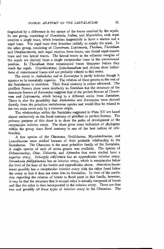
FLORAL ANATOMY OF THE SANTALACEAE 91
tinguished by a difference in the nature of the traces received by the sepals.
In one group, consisting of Henslowia, Jodina, and Myoschilos, each sepal
receives a single trace, which branches tangentially to form a stamen and asepal trace. The sepal trace then branches radially to supply the sepal. Inthe other group, consisting of Choretrum, Leptomeria, Thesium, Thesidiumand Omphacomeria, each sepal receives three traces, one dorsal sepal-stamentrace and two lateral traces. The lateral traces in the adjacent margins ofthe sepals are derived from a single receptacular trace in the commissural
position. In Thesidium these commissural traces disappear before theyreach the sepals. Osyridocarpus, Quinchamalium and Arjona show indica-tions of commissural traces and are probably related to this series.
The ovary in Anthobolus and in Exocarpus is partly inferior though itappears to be essentially superior. The relation of these genera to the rest ofthe Santalaceae is uncertain. Their floral anatomy is rather advanced. Thepistillate flowers show some similarity to Santalum but the structure of thestaminate flowers of Exocarpus suggests that of the perfect flowers of Choret-rum and Leptomeria, which belong to a different series from Santalum.There is also the possibility that Anthobolus and Exocarpus were deriveddirectly from the primitive santalaceous species and would thus be related tothe two main series only by a common origin.
The relationships within the Santalales suggested in Plate XV are basedalmost exclusively on the floral anatomy of pistillate or perfect flowers. Theprimary purpose of this chart is to show the paths of development of the
receptacular inferior ovary. The chart gives some indication of phylogenywithin the group since floral anatomy is one of the best indices of rela-
tionship.A few species of the Olacaceae, Grubbiaceae, Myzodendraceae, and
Loranthaceae were studied because of their probable relationship to the
Santalaceae. The Olacaceae is the most primitive family of the Santalales.A single species of each of seven genera was available. The species ofOchanostachys, Olax, Heisteria, and Aptandra that were studied have asuperior ovary. Schoepfia californica has an appendicular inferior ovary.Strombosia philippinensis has an inferior ovary, which is receptacular belowthe level of the base of the locules and appendicular above. Anacolosa luzon-iensis possibly has a receptacular inferior ovary with the calyx freed belowthe ovary so that it does not enter into its formation. In view of the confu-sion regarding the relation of bracts to floral parts in this family, however,it may be that the structure that is termed calyx is actually composed of bractsand that the calyx is then incorporated in the inferior ovary. There are thustwo and possibly all three types of inferior ovary in the Olacaceae. The
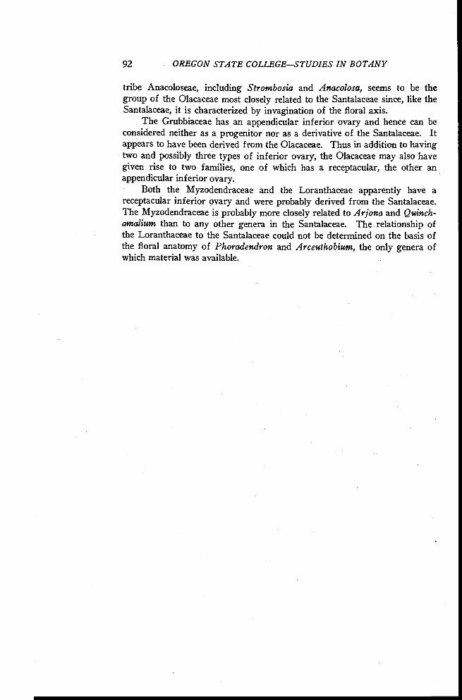
92 OREGON STATE COLLEGESTUDIES IN BOTANY
tribe Anacoloseae, including Strombosia and Anacolosa, seems to be thegroup of the Olacaceae most closely related to the Santalaceae since, like theSantalaceae, it is characterized by invagination of the floral axis.
The Grubbiaceae has an appendicular inferior ovary and hence can beconsidered neither as a progenitor nor as a derivative of the Santalaceae. Itappears to have been derived from the Olacaceae. Thus in addition to havingtwo and possibly three types of inferior ovary, the Olacaceae may also havegiven rise to two families, one of which has a receptacular, the other anappendicular inferior ovary.
Both the Myzodendraceae and the Loranthaceae apparently have areceptacular inferior ovary and were probably derived from the Santalaceae.The Myzodendraceae is probably more closely related to Arjona and Quinch-amalium than to any other genera in the Santalaceae. The relationship ofthe Loranthaceae to the Santalaceae could not be determined on the basis ofthe floral anatomy of Phoradendron and Arceuthobium, the only genera ofwhich material was available.
FLORAL ANATOMY OF THE SANTALACEAE 93
LITERATURE CITED
BONNE, GABRIEL. 1925. Sur les faisceaux de rebroussement dans la coupe florale decertaines Rosacees. Comptes Rendus Acad. Sci. 181 :189-191.
1928. Recherches sur le pedicelle et la fleur des Rosacees. Paris.DOWDING, E. 1931. Floral morphology of Arceuthobiuen americanum. Bot. Gaz.
91: 42-54.EAMES, A. J. 1931. The vascular anatomy of the flower with refutation of the theory
of carpel polymorphism. Amer. Jour. Bot. 18: 147-188.ENGLER, A., and K. PRANTL. 1935. Die natiirlichen Pflanzenfamilien. Band 16b.HUTCHINSON, J. 1926. The families of flowering plants. I. Dicotyledons. Macmillan,
London.JACKSON, GEMMA. 1933. A study of the carpophore of the Umbellifeme. Amer. Jour.
Bot. 20 : 121-144.1934. The morphology of the flowers of Rosa and certain closely re-
lated genera. Amer. Jour. Bot. 21: 453-466.JOHNSON, T. 1889. The nursing of the embryo and some other points in Myzodendron
punctulatum. Banks and Sol. Annals Bot. 3: 179-206.MACDANIELS, L. H. 1940. The morphology of the apple and other pome fruits. Cornell
Univ. Agric. Exper. Sta. Memoir 230.PAYER, J. 1857. Traite d'organogenie comparee de la fleur. Paris.Poor., R. J. 1941. Flowers and flowering plants. McGraw Hill, New York.RAO, L. N. 1942. Studies in the Santalaceae. Annals Bot. N. S. 6: 151-175.RENDLE, A. B. 1938. The classification of flowering plants. II. Dicotyledons. Cam-
bridge Univ. Press. London.SAUNDERS, EDITH R. 1933. The cause of petaloid colouring in apetalous flowers. Jour,
Linn. Soc. London. 49 : 199-218.1940. Floral Morphology. II. Heller, Cambridge.
SCHAEPPI, H., and F. STEINDL. 1937. Bliitenmorphologische und embryologische Unter-suchungen an Osyris alba L. Ber. Schweiz. Bot. Ges. 47: 369-392.
SCHELLENBERG, G. 1932. tTher Systembildung und iiber die Reihe der Santalales. Ber.Deutsch. Bot. Ges. 50a: 136-145. Festschrift.
SMITH, F. H., and E. C. SMITH. 1942. Anatomy of the inferior ovary of Darbya.Amer. Jour. Bot. 29 : 464-471.
VAN TIEGHEM, M. Ph. 1869a. Anatomie des fleurs et du fruit du Gui. Ann. Sci. Nat.V Serie Bot. 12 : 101-124.
1869b. Anatomie de la fleur des Santalacees. Ibid. 12: 340-346.


