Embed Size (px)
Citation preview

MORPHOLOGY, HISTOLOGY, AND FINE STRUCTURE
Fine Structure of the Compound Eyes of Callitettix versicolor(Insecta: Hemiptera)
LEI-PO JIA1,2AND AI-PING LIANG1,3
Ann. Entomol. Soc. Am. 1–9 (2015); DOI: 10.1093/aesa/sav007
ABSTRACT The external morphology and internal anatomy of the compound eyes of the adults of therice spittlebug Callitettix versicolor (F., 1794) (Insecta: Hemiptera: Cercopidae) are described forthe first time with light, scanning, and transmission electron microscopy observations. The eyes ofC. versicolor are of the apposition type and each eye consists of�2,042 ommatidia. Each ommatidium iscomposed of a plano-convex corneal lens, a eucone-type crystalline cone, eight retinular cells, two pri-mary pigment cells, and an undetermined number of secondary pigment cells (probably six). The rhab-domeres of the eight retinular cells form a centrally fused rhabdom surrounded by palisades. The distalend of the rhabdom is funnel shaped around the proximal region of the crystalline cone. The microvilli ofthe rhabdom are arranged in orthogonal directions suggesting polarization sensitivity. With a smallF-number of 1.48 and a large interommatidial angle Du of 7.7�, the eye possesses high light sensitivitybut low spatial resolution. With a large ommatidial acceptance angle Dq of 11.6�, the eye can merelyform a burred image of its surroundings, but the ratio Dq/Du of 1.5 indicates that the eye can provide arelatively good contrast.
KEY WORDS Hemiptera, Callitettix versicolor, compound eye, rhabdom, microvilli
Compound eyes play important roles in insect’s forag-ing, homing, prey detection, and other activities. Thebasic structural and functional units of the compoundeyes are ommatidia. Each ommatidium generally con-sists of a dioptric apparatus, a few primary and second-ary pigment cells, and retinular cells (Paulus 1979).The morphological characteristics of a compound eye,such as the organization of the retinular cells and themicrovillus orientations, can provide valuable clues onthe eye’s capability and limitation (Meyer-Rochow andMonalisa 2009).
In Hemiptera, previous studies on the compoundeyes have focused mainly on the heteropteran species(Schwind 1983, Settembrini 1984, Fischer et al. 2000,Reisenman et al. 2002). However, the fine structure ofthe eyes of the species in other hemipteran groups, i.e.,the Sternorrhyncha and the Auchenorrhyncha, is notwell documented. To our knowledge, Drosicha steb-bingi (Sternorrhyncha: Coccoidea) and Philaenus spu-marius (Auchenorrhyncha: Cercopoidea) are the onlytwo species in these groups that have had their com-pound eyes studied (Ramachandran 1963, Keskinenand Meyer-Rochow 2004).
The Cercopidae (Hemiptera: Auchenorrhyncha: Cer-copoidea) is a superfamily of Auchenorrhyncha and the
largest xylemophagous group of insects with �1,500described species (Liang and Webb 2002). This groupof insects is usually called spittlebugs because their lar-vae live in the spit-like bubbles of foam they secrete,while their adults inhabit in a terrestrial setting. Pre-vious work on the structure of the compound eyes ofthe spittlebug species is limited to only one study ofthe meadow spittlebug Philaenus spumarius (L.)(Keskinen and Meyer-Rochow 2004).
Here, we are dealing with the compound eyes of an-other spittlebug Callitettix versicolor (F., 1794) (Hemi-ptera: Cercopidae) which, compared with P. spumarius,belongs to a different genus. Another difference be-tween the two species is that P. spumarius has a world-wide distribution (Keskinen and Meyer-Rochow 2004),but C. versicolor is only found in southern China, In-dia, Myanmar, Thailand, Vietnam, and Malaysia (Met-calf 1962). The adults of C. versicolor mainly feed onxylem fluid of rice and maize and are considered oneof the most notorious economic pests in these areas(Chen and Liang 2012). Field observations reveal thatthe adults usually stay at the lower (abaxial) surfaces ofleaves to avoid high-intensity sunlight for most of theday, and feed themselves at dawn (7 to 8 am) and dusk(5 to 7 pm; Lei et al. 1992). Mating activities occuraround 5 pm (Lei et al. 1992) or between 4 and 6 pm(Cai and Xu 2001).
Successful rearing of this insect species under labo-ratory conditions (Chen and Liang 2012) makes it pos-sible for advanced behavioral and ecologicalresearches. Given that visual capacity and limitationhave great influence on the insect’s mating and feeding
1 Key Laboratory of Zoological Systematics and Evolution, Instituteof Zoology, Chinese Academy of Sciences, 1 Beichen West Road,Chaoyang District, Beijing 100101, P.R. China.
2 University of Chinese Academy of Sciences, Chinese Academy ofSciences, 19A Yuquanlu, Beijing 100049, P.R. China.
3 Corresponding author, e-mail: [email protected].
VC The Authors 2015. Published by Oxford University Press on behalf of Entomological Society of America.All rights reserved. For Permissions, please email: [email protected]

behavior, a thoroughly anatomical investigation on themajor visual organ, i.e., the compound eyes of theadults of this species before any further studies seemsnecessary.
The objectives of the present study are to provide adetailed description of the external morphology and in-ternal anatomy of the compound eyes in the adult of C.versicolor and give a brief discussion on the potentialfunctions and limitations of the compound eyes of thispest species.
Materials and Methods
Insect Specimens. The adult specimens of C. ver-sicolor (�10 mm in body length) used in this studywere the third or fourth generation of a laboratory col-ony. The adults of this spittlebug were reared on riceseedlings and maintained in the controlled laboratorycondition at 27�C, a photoperiod of 14:10 (L: D) h, and70% relative humidity (Chen and Liang 2012).
Scanning Electron Microscopy (SEM). Afterdecapitation, the heads of three adults were dehydratedin an alcohol series, critical point-dried with liquid car-bon dioxide (Leica EM CPD300, Leica Microsystems,Wetzlar, Germany), and sputter-coated with gold at40 mA current for 100 s (Leica EM SCD050, LeicaMicrosystems, Wetzlar, Germany). The specimens werefinally observed under a FEI Quanta 450 scanningelectron muscope (FEI, Hillsboro, OR), operated at avoltage of 30 kV.
Transmission Electron Microscopy (TEM). Thespecimens were decapitated and the heads were imme-diately fixed in 2.5% glutaraldehyde buffered in 0.1 Mcacodylate buffer (pH 7.4) during the day in the light-adapted state for at least 24 h. The specimens werepostfixed in 1% OsO4, also buffered in 0.1 M cacody-late buffer (pH 7.4) for 2 h at room temperature. Thesamples were then rinsed three times in the same buf-fer and dehydrated in a graded series of ethanol. Thespecimens were passed through different acetone/Epon mixtures (3:1, 1:1, 1:3, pure Epon), embedded inpure Epon (Serva, Heidelberg, Germany) and hard-ened at a temperature of 60�C for 3 d. Semithin sec-tions for light muscopy (LM; from cornea to basementmembrane) were cut on an ultramutome (Leica EMUC6þFC6, Wetzlar, Germany) with a glass knife andstained with a 0.5% aqueous solution of toluidine blue.Ultrathin sections (� 65 nm) were cut on an ultramu-tome (Leica EM UC6þFC6, Wetzlar, Germany) with adiamond knife (Diatome, Bienne, Switzerland) andpicked up on uncoated 200-mesh copper grids. Theultrathin sections were then stained with 2% aqueousuranyl acetate for 15 min and Reynolds’ lead citrate for5 min and observed under a Spirit transmission elec-tron muscope (FEI Tecnai Spirit, Hillsboro, USA) or aJEOL-1400 transmission electron muscope (JEOL,Tokyo, Japan), both operated at 100 kV.
Light Microscopy (LM). The semithin sections ofthe eyes cut with glass knifes (see section TEM) wereplaced on glass slides and mounted with rhamsan gum.After the gum dried out, the samples were then
observed with a regular optical objective (10�,NA¼ 0.3) and an oil immersion objective (100�,NA¼ 1.25) under a Nikon eclipse Ni-E muscope sys-tem (Nikon, Tokyo, Japan).
Statistics. All measurements were performed indigital photographs and analyzed using the softwareImage pro plus 6.0. SEM photographs were used todetermine the dorso–ventral and anterior–posteriorlengths of the eye, total number of the ommatidia andthe facet diameters. TEM photographs of cross-sec-tions of the eyes were used to determine the rhabdomdiameters, the diameters of pigment granules of pri-mary pigment cell and secondary pigment cell, as wellas retinular cells. LM mugraphs of longitudinal sectionsof the ommatidia were used to determine the ommati-dial lengths and the radii of the curvature of the facet.The curvature radii of corneal facet lens, as show inFig. 3A, were calculated based on the coordinate dataof three homologous points on their surface (Tanakaet al. 2009).
Results
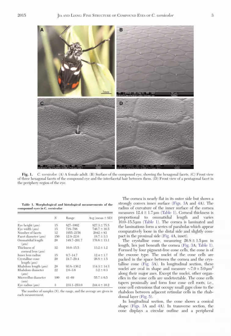
External Morphological Features of theEyes. The adult of C. versicolor possesses a pair ofbrown hemispherical compound eyes on both sides ofthe head (Fig. 1A). Each eye measures927.3 6 75.5mm in its dorso–ventral direction (eyeheight) and 746.7 6 16.5mm in the anterior–posteriordirection (eye width; Fig. 1A; Table 1). Each eyeconsists of 2,042 6 83 facets (Table 1). Most of the fac-ets are typical hexagonal in shape (Fig. 1C), but thoselocated close to the periphery region of the eye arepentagonal (Fig. 1D). The center-to-center distancesbetween neighboring facets measure 18.76 3.3mm(Table 1). Very sparse interfacetal hairs are presentbetween facets and randomly distributed across thewhole compound eye (Fig. 1B). The interfacetal hairis needle-shaped and situated between three neighbor-ing facets (Fig. 1C). Each interfacetal hair measures2.0–3.5mm in length and 0.7–1.2mm in diameterat its base. The surface of the facets is covered witharrays of corneal nipples, which are randomlyarranged and no orderly nipple pattern is observed(Fig. 1C and D).
Internal Anatomical Features of theEyes. General Organization. Each ommatidium iscomposed of two distinct structures: the dioptric appa-ratus, consisting of corneal lens and crystalline cone,and the photoreceptive layer, made up of retinular cellsand their rhabdomeres (Fig. 2). A “clear-zone” has notbeen found throughout the eye and the crystalline coneis in physical contact with the rhabdom and the com-pound eye of the adult species of C. versicolor, thus,conforms to the apposition compound eye type. Thelength of an ommatidium near the center of the eye is�200mm. Ommatidial lengths decrease to �150mmtowards the dorsal margin of the eye (Table 1).
Dioptric Apparatus. The dioptric apparatus consistsof a plano–convex corneal lens (cornea) and a crystal-line cone (Fig. 3A).
2 ANNALS OF THE ENTOMOLOGICAL SOCIETY OF AMERICA
The cornea is nearly flat in its outer side but shows astrongly convex inner surface (Figs. 3A and 4A). Theradius of curvature of the inner surface of the corneameasures 12.4 6 1.7mm (Table 1). Corneal thickness isproportional to ommatidial length and varies10.0–15.5mm (Table 1). The cornea is laminated andthe laminations form a series of parabolas which appearcomparatively loose in the distal side and slightly com-pact in the proximal side (Fig. 4A, inset).
The crystalline cone, measuring 26.8 6 1.5mm inlength, lies just beneath the cornea (Fig. 3A; Table 1).Formed by four pigment-free cone cells, the cone is ofthe eucone type. The nuclei of the cone cells arepacked in the space between the cornea and the crys-talline cone (Fig. 3A). In longitudinal section, thesenuclei are oval in shape and measure �7.0� 5.0mm2
along their major axes. Except the nuclei, other organ-elles in the cone cells are undetectable. The cone cellstapers proximally and form four cone cell roots, i.e.,cone cell extensions that occupy small gaps close to therhabdom between adjacent retinular cells in the rhab-domal layer (Fig. 5).
In longitudinal section, the cone shows a conicalshape (Figs. 3A and 4A). In transverse section, thecone displays a circular outline and a peripheral
Fig. 1. C. versicolor. (A) A female adult. (B) Surface of the compound eye, showing the hexagonal facets. (C) Front viewof three hexagonal facets of the compound eye and the interfacetal hair between them. (D) Front view of a pentagonal facet inthe periphery region of the eye.
Table 1. Morphological and histological measurements of thecompound eyes in C. versicolor
N Range Avg (mean 6 SD)
Eye height (lm) 15 827–1002 927.3 6 75.5Eye width (lm) 15 718–798 746.7 6 16.5Number of facets 12 1955–2156 2042 6 83Facet diameter (lm) 150 12.9–22.6 18.7 6 3.3Ommatidial length
(lm)20 149.7–201.7 176.6 6 13.1
Thickness ofcorneal lens (lm)
32 10.0–15.5 13.2 6 1.2
Inner lens radius 15 9.7–14.7 12.4 6 1.7Crystalline cone
length (lm)20 24.7–29.4 26.8 6 1.5
Rhabdom length (lm) 20 93.8–130.2 114.3 6 14.3Rhabdom diameter
(lm)22 2.6–3.6 3.2 6 0.3
Microvillus diameter(nm)
100 41–69 55.7 6 6.5
Eye radius (lm) 3 233.1–253.0 244.4 6 10.2
The number of samples (N), the range, and the average are given ineach measurement.
2015 JIA AND LIANG: FINE STRUCTURE OF COMPOUND EYES OF C. versicolor 3

concentric layer of high-electron density (Figs. 3B and4B), and each cone cell contributes one quadrant ofthe cone and consequently a cross-like contact face canbe seen (Figs. 3B and 4B).
The cone is tapered proximally and penetrates intothe rhabdom layer at least 10mm (Fig. 4C). In thecone/rhabdom overlapping area, the cone is “intruded”by four rhabdomeres in transverse section (Fig. 4D).As the cone tapers, the sizes of the four rhabdomeresincrease (Fig. 4E).
Primary and Secondary Pigment Cells. There aretwo primary pigment cells enveloping the crystallinecone from the distal end close to the cornea to its prox-imal tip where it meets the rhabdom. The nuclei of theprimary pigment cells, situated at the proximal side ofthe crystalline cone, are radially elongated and measure�7.2� 2.8mm2 along their major axes (Figs. 3B and4B). Primary pigment cells contain numerous electron-dense screening pigment granules measuring0.54–1.12mm in diameter.
An undetermined number (probably six) of secon-dary pigment cells surround the primary pigmentcells in each ommatidium. Unlike primary pigmentcells, the secondary pigment cells reach from the cor-nea down to the basement membrane and fill the spacebetween adjacent ommatidia. The spherical nuclei ofthe secondary pigment cells, measuring �2.5mm indiameter, are present around the central cone (Figs. 3Band 4B). The pigment granules in secondary pigmentcells are as large as those found in primary pigmentcells.
Retinular Cells and Rhabdom. Each ommatidiumcontains eight retinular cells. They lie just beneath thecone cells and their rhabdomeres form a centrallyfused rhabdom.
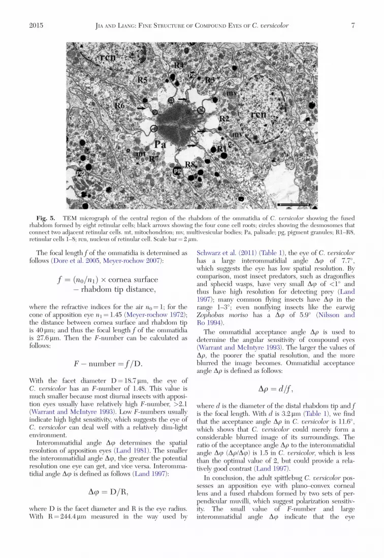
The oval nuclei of the retinular cells measure�4.7� 3.4mm2 along their major axes (Fig. 5). Thepositions of these nuclei of retinular cells extend fromthe distal to the middle parts of the rhabdomal layer.Except the prominent nuclei, the cytoplasm of theretinular cells also contains a large number of mito-chondria, many multivesicular bodies, some roughendoplasmic reticulum, and some randomly scatteredscreening pigment granules (Fig. 5). These pigmentgranules are 0.35–0.71mm in diameter and are conspic-uously smaller than those of the primary and secondarypigment cells. Adjacent retinular cells are connectedby desmosomes (Fig. 5) near the edge of the rhabdom.Four cone cell roots are also found near the desmo-somes (Fig. 5). According to the positions of the fourcone cell roots, the retinular cell which has conecell root on both sides is numbered R1, and theremaining retinular cells are numbered R2–R8counterclockwise.
All retinular cells contribute muvilli to the rhabdom,which has an average length of 114.36 14.3mm (Table1). Rhabdom diameter is more or less constant andmeasures 3.2 6 0.3mm (Table 1). In longitudinal sec-tion, the rhabdom is not twisted and surrounded by cis-ternae of the smooth endoplasmic reticulum, known aspalisades (Fig. 6A). The pigment-free palisades meas-ure �0.25–0.6mm in width around the rhabdom.
The distal-most region of the rhabdom is stillopen and funnel shaped, allowing the extension ofcone to penetrate into the rhabdomeres (Fig. 4C). Inlongitudinal section, the distal part of the rhabdomextends into a funnel shape around the proximal regionof the crystalline cone (Fig. 4C). In transverse section,the rhabdomeres of four retinular cells appear andexpand to the center of the cone (Figs. 3C, 4D,and 4E).
At the central region of the rhabdom, the coneextension disappears and the rhabdom shows a squar-ish cross-section profile (Fig. 6B). The muvilli arearranged in orthogonal directions (Figs. 5 and 6B).The muvilli of rhabdomeres of R2, R3, R4, and R7 areall aligned in the same direction, but at right angles tothose of R1, R5, R6, and R8 (Figs. 5 and 6B). Eachmuvillus measures 55.7 6 6.5 nm in diameter (Table 1).
At the proximal end of the ommatidium, the retinu-lar cells form eight axons as a bundle penetrating thebasement membrane (Fig. 6C). Each axon contains
Fig. 2. Ommatidium of C. versicolor. (A) Lightmicrograph of one ommatidium in longitudinal view. (B)Semischematic drawing of one ommatidium in longitudinalview. bm, basement membrane; cc, crystalline cone; co,cornea; ppc, primary pigment cell; rcn, nucleus of retinularcell; rh, rhabdom; spc, secondary pigment cell.
4 ANNALS OF THE ENTOMOLOGICAL SOCIETY OF AMERICA

numerous neurofilaments, some mitochondria, andvery scarce pigment granules.
Discussion
Anatomical Aspects. The compound eye of adultC. versicolor belongs to the apposition type of eyes.Apposition eyes are generally equipped in diurnallyrather than nocturnally active insects because of therelatively low light sensitivity (Warrant and McIntyre1993). However, with proper structural modifications,apposition eyes in some nocturnal insects like the heli-ctid bee Megalopta genalis (Greiner et al. 2004) orunderwater insects like the diving beetle Agabus japo-nicus (Jia and Liang 2014), can be effectively adaptedto dim-light environment.
Anatomically, the eye of C. versicolor is similar tothat of its close relative, the meadow spittlebugP. spumarius (Keskinen and Meyer-Rochow 2004). Anoticeable difference between the two species is thatC. versicolor possesses a nearly flat outer corneal sur-face but strongly curved inner surface, i.e., plano–convex cornea, while P. spumarius possesses a biconvexcornea (Keskinen and Meyer-Rochow 2004). Plano–convex corneal design generally presents in the com-pound eyes of many aquatic insects such as theNotonecta (Schwind 1980), and crustacean such as
the crab Ozius truncatus (Shaw and Stowe 1982). Theadvantage of this corneal design for aquatic animals isthat the optical power changes slightly when thespecies emerge from the water into the air (Schwind1980, Sanders and Halford 1995). Some nocturnal beesand wasps also feature flattened outer and stronglyconvex inner curvatures, but their diurnal relatives donot have the same characteristics (Greiner et al. 2004,Greiner 2006b). Thus, the plano–convex cornealdesign may improve light sensitivity and representan optical adaptation for nocturnal vision (Greiner2006a).
The rhabdom in C. versicolor is fused and extendsdistally into a funnel shape embracing the proximal endof the crystalline cone. According to Fisher et al.(2000), all heteropteran species possess open rhabdom,while other hemipteran groups, i.e., the Sternorrhynchaand the Auchenorrhyncha, possess fused rhabdom.This study clearly endorses the conclusion of Fisheret al. (2000). Although the optical function of funnel-shaped rhabdomal design is unknown, it occurs inmany insects such as the locust Locusta migratoria(Wilson et al. 1978), the stick insect Carausius morosus(Meyer-Rochow and Keskinen 2003), the cricket Gryl-lus bimaculatus (Sakura et al. 2003), and larvae of someMecoptera species (Suzuki and Nagashima 1989,Melzer et al. 1994).
Fig. 3. LM micrographs of different levels of the compound eye in C. versicolor. (A) Longitudinal section of the corneallens and the crystalline cone. (B) Transverse section of the distal region of the compound eye showing the tetrad of the coneand the nuclei of the secondary pigment cells (red arrows) and the primary pigment cells (black arrows). (C) Transverse sectionof the distalmost region of rhabdom showing the rhabdomeres that are still apart from each other (red arrowheads). (D)Transverse section of the rhabdom. cc, crystalline cone; ccn, nucleus of cone cell; co, cornea; rh, rhabdom.
2015 JIA AND LIANG: FINE STRUCTURE OF COMPOUND EYES OF C. versicolor 5

The muvilli of the rhabdom are arranged in two setsorienting at right angles to each other, which suggestspolarization sensitivity. In many insects, a group ofommatidia suited at the dorsal rim area of compoundeyes are specialized in detecting polarized light. Assummarized by Labhart and Meyer (1999), the rhab-dom in these dorsal rim area ommatida has commonanatomical characteristics: 1) strict alignment of themuvilli and 2) orthogonal muvillus arrangement. In C.versicolor, the muvilli are extremely aligned along therhabdomere and arranged in orthogonal directions(Fig. 6C), and thus meet the requirements list above.The rhabdom in C. versicolor is also nontwisted
along its optical axe (Fig. 6A), which is of importancein discussing polarization sensitivity because twistedrhabdom can greatly decrease polarization sensitivity(Wehner et al. 1975). All these morphologicalfeatures suggest that C. versicolor could discriminatelinearly polarized light, yet behavioral and electrophy-siological studies are needed to determine if it isthe case.
Optical Aspects. To estimate the optical propertyand limitation of the eyes of C. versicolor, the followingparameters were calculated: the F-number, the inter-ommatidial angle Du and the ommatidial acceptanceangle Dq.
Fig. 4. TEM micrographs of the crystalline cone of the ommatidia of C. versicolor. (A) Longitudinal section (somewhatoblique) of the laminated cornea and the crystalline cone; inset showing the cornea. (B) Transverse section of the crystallinecone, showing the cross-like contact face confined in a circular outline of the crystalline cone. (C) Longitudinal section of thecone/rhabdom overlapping area, showing the proximal end of the cone and the funnel-shaped rhabdom. (D) and (E)Transverse sections of the cone/rhabdom overlapping area showing the cone surrounded by four rhabdomeres. cc, crystallinecone; ccn, nucleus of the cone cell; co, cornea; pg, pigment granule; ppcn, nucleus of the primary pigment cell; rh, rhabdom;spc, secondary pigment cell; spcn, nucleus of the spc. Scale bars¼ 5 lm in A, 2 lm in inset of A, 2 lm in B, and 1 lm in C–E.
6 ANNALS OF THE ENTOMOLOGICAL SOCIETY OF AMERICA
The focal length f of the ommatidia is determined asfollows (Dore et al. 2005, Meyer-rochow 2007):
f ¼ ðn0=n1Þ � cornea surface� rhabdom tip distance;
where the refractive indices for the air n0¼ 1; for thecone of apposition eye n1¼ 1.45 (Meyer-rochow 1972);the distance between cornea surface and rhabdom tipis 40mm; and thus the focal length f of the ommatidiais 27.6mm. Then the F-number can be calculated asfollows:
F� number ¼ f=D:
With the facet diameter D¼ 18.7mm, the eye ofC. versicolor has an F-number of 1.48. This value ismuch smaller because most diurnal insects with apposi-tion eyes usually have relatively high F-number, >2.1(Warrant and McIntyre 1993). Low F-numbers usuallyindicate high light sensitivity, which suggests the eye ofC. versicolor can deal well with a relatively dim-lightenvironment.
Interommatidial angle Du determines the spatialresolution of apposition eyes (Land 1981). The smallerthe interommatidial angle Du, the greater the potentialresolution one eye can get, and vice versa. Interomma-tidial angle Du is defined as follows (Land 1997):
Du ¼ D=R;
where D is the facet diameter and R is the eye radius.With R¼ 244.4mm measured in the way used by
Schwarz et al. (2011) (Table 1), the eye of C. versicolorhas a large interommatidial angle Du of 7.7�,which suggests the eye has low spatial resolution. Bycomparison, most insect predators, such as dragonfliesand sphecid wasps, have very small Du of <1� andthus have high resolution for detecting prey (Land1997); many common flying insects have Du in therange 1–3�; even nonflying insects like the earwigZophobas moriso has a Du of 5.9� (Nilsson andRo 1994).
The ommatidial acceptance angle Dq is used todetermine the angular sensitivity of compound eyes(Warrant and McIntyre 1993). The larger the values ofDq, the poorer the spatial resolution, and the moreblurred the image becomes. Ommatidial acceptanceangle Dq is defined as follows:
Dq ¼ d=f ;
where d is the diameter of the distal rhabdom tip and fis the focal length. With d is 3.2mm (Table 1), we findthat the acceptance angle Dq in C. versicolor is 11.6�,which shows that C. versicolor could merely form aconsiderable blurred image of its surroundings. Theratio of the acceptance angle Dq to the interommatidialangle Du (Dq/Du) is 1.5 in C. versicolor, which is lessthan the optimal value of 2, but could provide a rela-tively good contrast (Land 1997).
In conclusion, the adult spittlebug C. versicolor pos-sesses an apposition eye with plano–convex corneallens and a fused rhabdom formed by two sets of per-pendicular muvilli, which suggest polarization sensitiv-ity. The small value of F-number and largeinterommatidial angle Du indicate that the eye
Fig. 5. TEM micrograph of the central region of the rhabdom of the ommatidia of C. versicolor showing the fusedrhabdom formed by eight retinular cells; black arrows showing the four cone cell roots; circles showing the desmosomes thatconnect two adjacent retinular cells. mt, mitochondrion; mv, multivesicular bodies; Pa, palisade; pg, pigment granules; R1–R8,retinular cells 1–8; rcn, nucleus of retinular cell. Scale bar¼ 2 lm.
2015 JIA AND LIANG: FINE STRUCTURE OF COMPOUND EYES OF C. versicolor 7

possesses high light sensitivity and low spatial resolu-tion. The eye also has a comparatively large ommatidialacceptance angle Dq that may result in a considerableblurred image.
Acknowledgements
We thank two anonymous reviewers for their constructiveand thorough comments which helped to improve the manu-script. We are also grateful to Zhang Kui-Yan (Institute ofZoology, CAS), Liang Jing-Nan (Institute of Microbiology,CAS), and Sun-Lei (National Laboratory of Biomacromole-cules, Institute of Biophysics, CAS) for their help with theassistance of the SEM and TEM preparation. This work wassupported by the following sources: the National BasicResearch Program of China (973 Program; grant2011CB302102), the National Natural Science Foundation ofChina (grants 31172128 and 31372249), and the National Sci-ence Fund for Fostering Talents in Basic Research (Specialsubjects in animal taxonomy, NSFC-J1210002), all awardedto A.P.L.
References Cited
Cai, L., and L. J. Xu. 2001. Biological characters of Callitettixversicolor and Abidama liuensis. J. Anhui Agric. Sci. 29:185–186.
Chen, X., and A. P. Liang. 2012. Laboratory rearing of Callitet-tix versicolor (Hemiptera: Cicadomorpha: Cercopidae), withdescriptions of the immature stages. Ann. Entomol. Soc. Am.105: 664–670.
Dore, B., H. Schiff, and M. Boido. 2005. Photomechanical ad-aptation in the eyes of Squilla mantis (Crustacea, Stomato-poda). Ital. J. Zool. 72: 189–199.
Fischer, C., M. Mahner, and E. Wachmann. 2000. Therhabdom structure in the ommatidia of the Heteroptera(Insecta), and its phylogenetic significance. Zoomorphology120: 1–13.
Greiner, B. 2006a. Adaptations for nocturnal vision in insect ap-position eyes. Int. Rev. Cytol. 250: 1–46.
Greiner, B. 2006b. Visual adaptations in the night-active waspApoica pallens. J. Comp. Neurol. 495: 255–262.
Greiner, B., W. A. Ribi, and E. J. Warrant. 2004. Retinal andoptical adaptations for nocturnal vision in the halictid beeMegalopta genalis. Cell Tissue Res. 316: 377–390.
Jia, L. P., and A. P. Liang. 2014. An apposition-like compoundeye with a layered rhabdom in the small diving beetleAgabus japonicus (Coleoptera, Dytiscidae). J. Morphol. 275:1273–1283.
Keskinen, E., and V. B. Meyer-Rochow. 2004. Post-embryonic photoreceptor development and dark/lightadaptation in the spittle bug Philaenus spumarius (L.)(Homo-ptera, Cercopidae). Arthropod Struct. Dev. 33: 405–417.
Labhart, T., and E. P. Meyer. 1999. Detectors for polarizedskylight in insects: a survey of ommatidial specializations in
Fig. 6. TEM micrographs of the rhabdom of the ommatidia of C. versicolor. (A) Longitudinal section of the central regionof the rhabdom showing that the rhabdom is not twisted and is surrounded by palisades. (B) Transverse section of the centralregion of the rhabdom showing the rectangle outline of the rhabdom and the orthogonal arrangement of the microvilli. (C)Transverse section of eight axons showing many mitochondria (black arrows). pa, palisade; R1–R8, retinular cells 1–8. Scalebars¼ 1 lm.
8 ANNALS OF THE ENTOMOLOGICAL SOCIETY OF AMERICA

the dorsal rim area of the compound eye. Microsc. Res. Tech.47: 368–379.
Land, M. F. 1981. Optics and vision in invertebrates. pp.471–592. In H. Autrum, (Ed), Hand-book of sensory Physiol-ogy. Springer, Berlin.
Land, M. F. 1997. Visual acuity in insects. Annu. Rev. Entomol.42: 147–177.
Lei, T. S., J. G. Lu, D. F. Zhou, and X. C. Liu. 1992. Study onthe biological characters and control of Callitettix versicolor.Entomol. Knowl. 29: 334–336.
Liang, A. P., and M. D. Webb. 2002. New taxa and revisionarynotes in Rhinaulacini spittlebugs from southern Asia (Homo-ptera: Cercopidae). J. Nat. Hist. 36: 729–756.
Melzer, R. R., H. F. Paulus, and N. P. Kristensen. 1994. Thelarval eye of nannochoristid scorpionflies (Insecta, Mecop-tera). Acta Zool. 75: 201–208.
Metcalf, Z. P. 1962. General catalogue of the Homoptera.Fasc. VII Cercopoidea. Part 3. Aphro-phoridae. Part 4. Clas-topteridae. Raleigh North Carolina State College, Paperno. 1324.
Meyer-Rochow, V. B. 1972. The eyes of Creophilus erythroce-phalus F. and Sartallus signatus sharp (Staphylinidae:Coleoptera). Z. Zellforsch.133(1): 59–86.
Meyer-rochow, V. B. 2007. Structure and putative function ofdark- and light-adapted as well as UV-exposed eyes of thefood store pest Psyllipsocus ramburi Selys-longchamps(Insecta: Psocoptera: Psyllipsocidae) J. Insect Physiol. 53(2):157–169.
Meyer-Rochow, V. B., and E. Keskinen. 2003. Post-embry-onic photoreceptor development and dark/light adaptation inthe stick insect Carausius morosus (Phasmida, Phasmatidae).Appl. Entomol. Zool. 38: 281–291.
Meyer-Rochow, V. B., and M. Monalisa. 2009. A six-rhabdomere, open rhabdom arrangement in the eye of thechrysanthemum beetle Phytoecia rufiventris: some ecophysi-ological predictions based on eye anatomy. Biocell 33:115–120.
Nilsson, D. E., and A. I. Ro. 1994. Did neural pooling for nightvision lead to the evolution of neural superposition eyes? J.Comp. Physiol. A 175: 289–302.
Paulus, H. F. 1979. Eye structure and the monophyly ofthe arthropoda, pp 299–384. In A. P. Gupta, (Ed),Arthropod Phylogeny. Van Nostrand Reinhold Co., NewYork, NY.
Ramachandran, S. 1963. The structure and development ofthe compound eye in the male of Drosicha stebbingii
(stebbing) (Homoptera: Coccoidea, Margarodidae). Proc. R.Entomol. Soc. Lond. A. 38: 23–31.
Reisenman, C., T. Insausti, and C. Lazzari. 2002. Light-induced and circadian changes in the compound eye of thehaematophagous bug Triatoma infestans (Hemiptera: Redu-viidae). J. Exp. Biol. 205: 201–210.
Sakura, M., K. Takasuga, M. Watanabe, and E. Eguchi.2003. Diurnal and circadian rhythm in compound eye ofcricket (Gryllus bimaculatus): changes in structure and pho-ton capture efficiency. Zool. Sci. 20: 833–840.
Sanders, J. S., and C. E. Halford. 1995. Design and analysis ofapposition compound eye optical sensors. Opt. Eng. 34:222–235.
Schwarz, S., A. Narendra, and J. Zeil. 2011. The properties ofthe visual system in the Australian desert ant Melophorusbagoti. Arthropod. Struct. Dev. 40: 128–134.
Schwind, R. 1980. Geometrical optics of the Notonecta eye: ad-aptations to optical environment and way of life. J. Comp.Physiol. 140: 59–68.
Schwind, R. 1983. Zonation of the optical environment and zo-nation in the rhabdom structure within the eye of the back-swimmer, Notonecta glauca. Cell Tissue Res. 232: 53–63.
Settembrini, B. P. 1984. The compound eyes of Triatoma infes-tans and Rhodnius prolixus (Hemiptera: Reduviidae). J. Med.Entomol. 21: 477–479.
Shaw, R., and S. Stowe. 1982. Neurobiology: Structure andFunction. In H. L. Atwood, (Ed), The biology of crustacea,Chapter 7. Academic Press, New York, NY.
Suzuki, N., and T. Nagashima. 1989. Ultrastructure of the lar-val eye of the hanging fly, Bittacus leavipes Navas (Mecoptera,Bittacidae). Proc. Arthropod. Embryol. Soc. Jpn. 24: 27–29.
Tanaka, G., A. R. Parker, D. J. Siveter, H. Maeda, and M.Furutani. 2009. An exceptionally well-preserved Eocenedolichopodid fly eye: function and evolutionary significance.P. Roy. Soc. B -Biol. Sci. 276: 1015–1019.
Warrant, E. J., and P. D. McIntyre. 1993. Arthropod eye de-sign and the physical limits to spatial resolving power. Prog.Neurobol. 40: 413–461.
Wehner, R., G. D. Bernard, and E. Geiger. 1975. Twistedand non-twisted rhabdoms and their significance for polariza-tion detection in the bee. J. comp. Physiol. 104: 225–245.
Wilson, M., P. Garrard, and S. McGinness. 1978. The unitstructure of the locust compound eye. Cell Tissue Res. 195:205–226.
Received 9 May 2014; accepted 29 January 2015.
2015 JIA AND LIANG: FINE STRUCTURE OF COMPOUND EYES OF C. versicolor 9


