Embed Size (px)
Citation preview
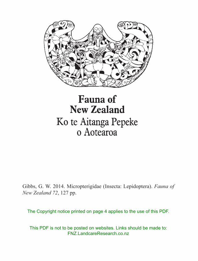
Gibbs, G. W. 2014. Micropterigidae (Insecta: Lepidoptera). Fauna of New Zealand 72, 127 pp.
The Copyright notice printed on page 4 applies to the use of this PDF.
This PDF is not to be posted on websites. Links should be made to: FNZ.LandcareResearch.co.nz
EDITORIAL BOARD
Dr R. M. Emberson, c/- Department of Ecology, P.O. Box 84, Lincoln University, New Zealand
Dr M. J. Fletcher, NSW Agricultural Scientific Collections Unit, Forest Road, Orange, NSW 2800, Australia
Prof. G. Giribet, Curator of Invertebrates, Museum of Comparative Zoology, Harvard University, 26 Oxford Street, Cambridge, MA 02138, U.S.A.
Dr R. J. B. Hoare, Landcare Research, Private Bag 92170, Auckland, New Zealand
Dr M.-C. Larivière, Landcare Research, Private Bag 92170, Auckland, New Zealand
Mr R. L. Palma, Natural Environment Department, Museum of New Zealand Te Papa Tongarewa, P.O. Box 467, Wellington, New Zealand
Dr C. J. Vink, Canterbury Museum, Rolleston Ave, Christchurch, New Zealand
SERIES EDITOR
Dr Z.-Q. Zhang, Landcare Research, Private Bag 92170, Auckland, New Zealand
Associate EditorsDr T. R. Buckley, Dr R. J. B. Hoare, Dr M.-C. Larivière, Dr R. A. B. Leschen,
Dr D. F. Ward, Dr Z. Q. Zhao, Landcare Research, Private Bag 92170, Auckland, New Zealand
Honorary EditorDr T. K. Crosby, Landcare Research, Private Bag 92170, Auckland, New
Zealand
Fauna of New ZealandKo te Aitanga Pepeke o Aotearoa
Number / Nama 72
Micropterigidae(Insecta: Lepidoptera)
George W. GibbsSchool of Biological Sciences, Victoria University, P.O. Box 600,
Wellington 6140, New [email protected]
Lincoln, New Zealand2014
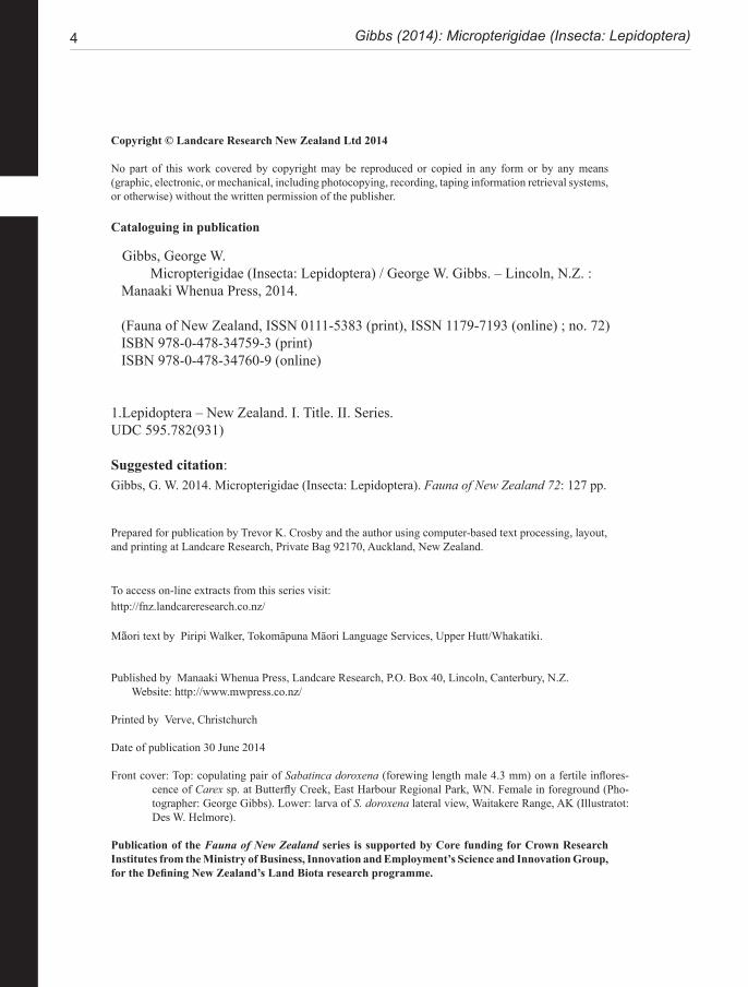
4 Gibbs (2014): Micropterigidae (Insecta: Lepidoptera)
Copyright © Landcare Research New Zealand Ltd 2014
No part of this work covered by copyright may be reproduced or copied in any form or by any means (graphic, electronic, or mechanical, including photocopying, recording, taping information retrieval systems, or otherwise) without the written permission of the publisher.
Cataloguing in publication
Gibbs, George W. Micropterigidae (Insecta: Lepidoptera) / George W. Gibbs. – Lincoln, N.Z. :Manaaki Whenua Press, 2014.
(Fauna of New Zealand, ISSN 0111-5383 (print), ISSN 1179-7193 (online) ; no. 72)ISBN 978-0-478-34759-3 (print)ISBN 978-0-478-34760-9 (online)
1.Lepidoptera – New Zealand. I. Title. II. Series.UDC 595.782(931)
Suggested citation:Gibbs, G. W. 2014. Micropterigidae (Insecta: Lepidoptera). Fauna of New Zealand 72: 127 pp.
Prepared for publication by Trevor K. Crosby and the author using computer-based text processing, layout, and printing at Landcare Research, Private Bag 92170, Auckland, New Zealand.
To access on-line extracts from this series visit:http://fnz.landcareresearch.co.nz/
Māori text by Piripi Walker, Tokomāpuna Māori Language Services, Upper Hutt/Whakatiki.
Published by Manaaki Whenua Press, Landcare Research, P.O. Box 40, Lincoln, Canterbury, N.Z.Website: http://www.mwpress.co.nz/
Printed by Verve, Christchurch
Date of publication 30 June 2014
Front cover: Top: copulating pair of Sabatinca doroxena (forewing length male 4.3 mm) on a fertile inflores-cence of Carex sp. at Butterfly Creek, East Harbour Regional Park, WN. Female in foreground (Pho-tographer: George Gibbs). Lower: larva of S. doroxena lateral view, Waitakere Range, AK (Illustratot: Des W. Helmore).
Publication of the Fauna of New Zealand series is supported by Core funding for Crown Research Institutes from the Ministry of Business, Innovation and Employment’s Science and Innovation Group, for the Defining New Zealand’s Land Biota research programme.
Fauna of New Zealand 72 5
POPULAR SUMMARY HE WHAKARĀPOPOTOTANGA
Class InsectaOrder LepidopteraFamily Micropterigidae
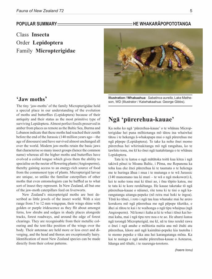
Illustration / Whakaahua: Sabatinca aurella, Lake Mathe-son, WD (Illustrator / Kaiwhakaahua: George Gibbs).‘Jaw moths’
Ngā ‘pūrerehua-kauae’Ka noho ko ngā ‘pūrerehua-kauae’ o te whānau Microp-terigidae hei puna mōhiotanga mō tātou ina whaiwhai tātou i te hekenga ā-whakapapa mai o ngā pūrerehua me ngā pēpepe (Lepidoptera). Te take ka noho ēnei momo pūrerehua hei whirinakitanga mō ngā rangahau, ko te tawhito tonu, me kī ko ēnei ngā tuatahitanga o te whānau Lepidoptera.
Tata ki te katoa o ngā mātātoka toitū kua kitea i ngā takiwā pēnei te Moana Baltic, i Pēma, me Repanona ka tohu kua eke ēnei pūrerehua ki te taumata o te hekenga me te huringa āhua i mua i te mutunga o te wā Jurassic (140 manomano tau ki muri – te wā o ngā mokoweri) ā, kei te noho tonu mai ki tēnei ao, i ōna tōpito katoa, me te tata ki te kore rerekētanga. He kauae taketake tō ngā pūrerehua-kauae o nāianei, rite tonu ki te tini o ngā ka-rangatanga aitanga-pepeke (nō reira mai hoki te ingoa). Tēnā ko tēnei, i roto i ngā tau kua whanake mai he arero korukoru mō ngā pūrerehua me ngā pēpepe tiketike, e āhei ai rātou te kai i te waihonga o ngā tipu whaipua (ngā Angiosperm). Nā konei i kaha ai ki te whai i tēnei kai ho-mai kaha, mai i ngā tipu rere noa o te ao. He ahurei katoa ngā torongū Micropterigid, me kī, nā te tino rerekē rawa o ēnei i ngā anuhe e mōhiotia nuitia ana mō ētahi atu pūrerehua, kāore anō ngā kaimātai-pepeke kia tautohu i te momo pepeke e tika ana hei karangatanga mōna. Ka kai te nuinga o ngā anuhe pūrerehua-kauae o Aotearoa, hāunga anō tētahi, i te raurenga-torotoro.
The tiny ‘jaw-moths’ of the family Micropterigidae hold a special place in our understanding of the evolution of moths and butterflies (Lepidoptera) because of their antiquity and their status as the most primitive type of surviving Lepidoptera. Almost perfect fossils preserved in amber from places as remote as the Baltic Sea, Burma and Lebanon indicate that these moths had reached their zenith before the end of the Jurassic (140 million years ago—the age of dinosaurs) and have survived almost unchanged all over the world. Modern jaw-moths retain the basic jaws that characterise so many insect groups (hence the common name) whereas all the higher moths and butterflies have evolved a coiled tongue which gives them the ability to specialise on the nectar of flowering plants (Angiosperms), thereby gaining access to an energy-rich source of food from the commonest type of plants. Micropterigid larvae are unique, so unlike the familiar caterpillars of other moths that even entomologists can be baffled as to what sort of insect they represent. In New Zealand, all but one of the jaw-moth caterpillars feed on liverworts.
New Zealand’s micropterigid moths are best de-scribed as little jewels of the insect world. With a size range from 5 to 12 mm wingspan, their wings shine with golden or purple iridescence as they flit about amongst ferns, low shrubs and sedges in shady places alongside tracks, forest roadways, and around the edge of forest clearings. They are recognisable from their metallic col-ouring and the tent-like position of the wings over the body. Their antennae are held more or less erect and di-verging, and the head and thorax are exceptionally hairy. Identification of most New Zealand species can be made directly from their colour patterns.
(haere tonu)
6 Gibbs (2014): Micropterigidae (Insecta: Lepidoptera)
Geo
rge
Gib
bs
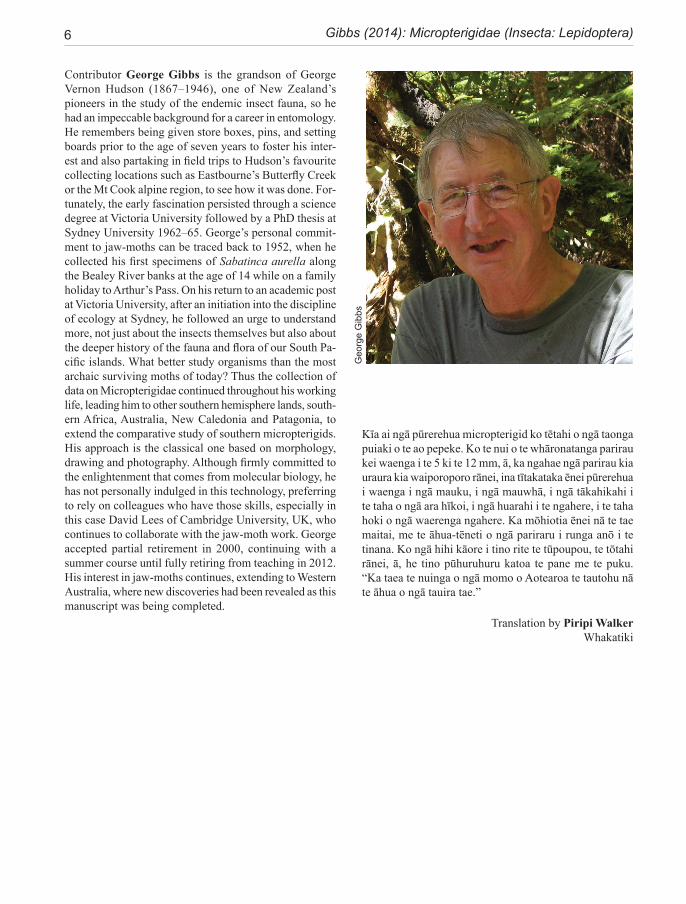
Contributor George Gibbs is the grandson of George Vernon Hudson (1867–1946), one of New Zealand’s pioneers in the study of the endemic insect fauna, so he had an impeccable background for a career in entomology. He remembers being given store boxes, pins, and setting boards prior to the age of seven years to foster his inter-est and also partaking in field trips to Hudson’s favourite collecting locations such as Eastbourne’s Butterfly Creek or the Mt Cook alpine region, to see how it was done. For-tunately, the early fascination persisted through a science degree at Victoria University followed by a PhD thesis at Sydney University 1962–65. George’s personal commit-ment to jaw-moths can be traced back to 1952, when he collected his first specimens of Sabatinca aurella along the Bealey River banks at the age of 14 while on a family holiday to Arthur’s Pass. On his return to an academic post at Victoria University, after an initiation into the discipline of ecology at Sydney, he followed an urge to understand more, not just about the insects themselves but also about the deeper history of the fauna and flora of our South Pa-cific islands. What better study organisms than the most archaic surviving moths of today? Thus the collection of data on Micropterigidae continued throughout his working life, leading him to other southern hemisphere lands, south-ern Africa, Australia, New Caledonia and Patagonia, to extend the comparative study of southern micropterigids. His approach is the classical one based on morphology, drawing and photography. Although firmly committed to the enlightenment that comes from molecular biology, he has not personally indulged in this technology, preferring to rely on colleagues who have those skills, especially in this case David Lees of Cambridge University, UK, who continues to collaborate with the jaw-moth work. George accepted partial retirement in 2000, continuing with a summer course until fully retiring from teaching in 2012. His interest in jaw-moths continues, extending to Western Australia, where new discoveries had been revealed as this manuscript was being completed.
Translation by Piripi WalkerWhakatiki
Kīa ai ngā pūrerehua micropterigid ko tētahi o ngā taonga puiaki o te ao pepeke. Ko te nui o te whāronatanga parirau kei waenga i te 5 ki te 12 mm, ā, ka ngahae ngā parirau kia uraura kia waiporoporo rānei, ina tītakataka ēnei pūrerehua i waenga i ngā mauku, i ngā mauwhā, i ngā tākahikahi i te taha o ngā ara hīkoi, i ngā huarahi i te ngahere, i te taha hoki o ngā waerenga ngahere. Ka mōhiotia ēnei nā te tae maitai, me te āhua-tēneti o ngā pariraru i runga anō i te tinana. Ko ngā hihi kāore i tino rite te tūpoupou, te tōtahi rānei, ā, he tino pūhuruhuru katoa te pane me te puku. “Ka taea te nuinga o ngā momo o Aotearoa te tautohu nā te āhua o ngā tauira tae.”
Fauna of New Zealand 72 7
ABSTRACTNew Zealand’s fauna of archaic Lepidoptera, the Micropterigidae, is revised, with the addition of four new species: Sabatinca pluvialis, S. weheka, S. bimacula, S aurantissima. The synonomy of Palaeomicra Meyrick, 1886 and Micropardalis Meyrick, 1912 with Sabatinca Walker, 1863, proposed by Kristensen and Nielsen, 1979, is supported here. Three new synonomies are established: Sabatinca pas-salota Meyrick is synonymised as a junior synonym of S. chrysargyra Meyrick; S. barbarica Philpott is synonymised as a junior synonym of S. caustica Meyrick; and S. aurantiaca Philpott is synonymised as a junior synonym of S. aemula Philpott. The outcome of long-standing confusion between incongruella Walker and chalcophanes Meyrick, initiated by Meyrick in 1912, is discussed because it influenced a series of publications by Tillyard and Philpott between 1919 and 1927. Adults and larvae are described and illustrated; adults in colour from life as well as museum specimens, larval examples in colour from life to show the variety of pigmentation patterns. Larvae have been matched to adult species by barcoding (8 cases), rearing (4 cases), unambiguous habitat association (6 cases); but despite these efforts three species remain where the larval form is unknown. Within Sabatinca, three monophyletic species-groups are recognised, established by DNA phylogenetic analysis and supported by morphological characters; within one of which two further informal sub-groupings are adopted based on character traits of adults, larvae and DNA (although the latter without strong support).
Current understanding of life cycles, foodplants, and general ecology are reviewed. All known Sabatinca species are confirmed as hepatic feeders, those in New Zealand utilising only the foliose types of liverwort. The precise diet of Zealandopterix larvae remains undetermined. Phenology patterns are dis-cussed—typical life cycles being annual, with larval growth throughout winter and a relatively short pupal stadium prior to the spring/summer flight season. Members of the calliarcha-group of species appear to incorporate a diapause, resulting in a two-year cycle and more erratic seasonal emergence of adults.
The broader systematic position of New Zealand Micropterigidae is de-scribed based on current molecular understanding that the world fauna is sub-divided into five strongly-supported clades—two northern and three southern hemisphere, with two occurring in New Zealand and New Caledonia: Sabat-inca clade and ‘Australian’ clade. The historical biogeography and phylogeny of SW Pacific Micropterigidae is compared with the tectonic interpretation for this region, within both a deeper Zealandian perspective and from the perspec-tive of sister species divergence patterns in the genus Sabatinca, finding that in all except one case, the New Zealand speciation events occurred prior to the development of modern geological landforms. With the incorporation of DNA phylogenetics into a predominantly morphological analysis of diversity, it has been possible to evaluate the strength of phylogenetic signal in the basic mor-phological structures of alpha-taxonomy. Certain features of the male phallus, gonopore, bulbous ejaculatorius, and female signa and spermatheca are evalu-ated in relation to their phylogenetic signal with gradings from ‘phylogeneti-cally meaningful’ to ‘of taxonomic value only’.
Species of New Zealand Micropterigidae are widely distributed from North Cape to Stewart Island with a maximum concentration in NW Nelson region. No single species occurs throughout, the most widespread (S. chalcophanes) ex-tends from Auckland to Fox Glacier, the most restricted (S. pluvialis) at present known only from Secretary Island in Fiordland. Of the 19 New Zealand species, four are endemic to North Island, 11 to South Island, and four occur in both
8 Gibbs (2014): Micropterigidae (Insecta: Lepidoptera)
islands. They are not known from Three Kings Islands, Chatham Islands, Lord Howe Island, or Subantarctic Islands.
The maculation and colours of New Zealand Micropterigidae are com-prehensively described. In contrast to larvae, which are cryptically coloured, the brilliant iridescent colours of adult Sabatinca appear, to our eyes, to defy crypsis, yet in the dappled light of their complex habitats, these small moths are by no means easy to see. Two species (S. calliarcha, S. doroxena) exhibit an eye-catching pattern in which the upper part of the forewing of a resting moth (tornus) features a black patch containing several brilliant white spots. It is suggested that this theme, especially since it re-occurs in seven New Caledo-nian Sabatinca species and a number of other similar-sized moths that rest with their wings tent-like (e.g. certain Glyphipterix species), is likely to have survival value by mimicking the facial view of a jumping spider (Salticidae), one of their key predators. Unfortunately the hypothesis remains to be tested.
Gibbs, G. W. 2014. Micropterigidae (Insecta: Lepidoptera). Fauna of New Zea-land 72, 127 pp.urn:lsid:zoobank.org:pub:D6BC8C34-6D93-4EC7-BCB3-5670B2CFE744Doi: 10.7931/J2/FNZ.72
Received: 19 May 2014; accepted by Robert Hoare: 19 June 2014; published 30 June 2014
CHECKLIST OF NEW ZEALAND TAXAGenus Sabatinca Walker 1863 .................................. 30 Palaeomicra Meyrick, 1886 Micropardalis Meyrick, 1912
incongruella-group ................................................. 31incongruella Walker 1863 ......................................... 32 munda Felder & Rogenhofer, 1875 eodora Meyrick 1918demissa Philpott, 1923 ............................................... 34
calliarcha-group ..................................................... 35calliarcha Meyrick, 1912 .......................................... 37pluvialis new species ................................................. 39lucilia Clarke, 1920 ................................................... 40heighwayi Philpott, 1927 ........................................... 41weheka new species ................................................... 43
chrysargyra-group .................................................. 44 aurella-subgroup .................................................... 48chrysargyra (Meyrick, 1885) ..................................... 48 passalota (Meyrick, 1923) new synonomy
aemula Philpott, 1924 ............................................... 49 aurantiaca Philpott, 1924 new synonomy aenea Hudson, 1923 .................................................. 51ianthina Philpott, 1921 .............................................. 52bimacula new species ................................................ 53aurella Hudson, 1918 ................................................ 54doroxena (Meyrick,1888) .......................................... 56 chalcophanes-subgroup .......................................... 58chalcophanes (Meyrick, 1885) .................................. 58caustica Meyrick, 1912 ............................................. 59 barbarica Philpott, 1918 new synonomy aurantissima new species .......................................... 61quadrijuga Meyrick, 1912 ......................................... 63
Genus Zealandopterix Gibbs, 2010 ........................... 64zonodoxa (Meyrick, 1888) ......................................... 66 rosicoma (Meyrick, 1914)
Fauna of New Zealand 72 9
CONTENTSAbstract ........................................................................ 7Checklist of taxa .......................................................... 8Acknowledgments .........................................................9Introduction ................................................................ 10Background to the Study of New Zealand Micro-
pterigidae ............................................................. 10The diversity of micropterigids in New Zealand ....... 11Methods and conventions .......................................... 12Morphology of relevance to systematics ................... 14Life History and Biology ........................................... 18Phylogeny .................................................................. 22Congruence between morphology and phylogeny ..... 25Biogeography ............................................................. 26Conservation .............................................................. 39Key to adults of New Zealand genera ........................ 30SYSTEMATICS ......................................................... 30Genus Sabatinca Walker ............................................ 30 incongruella-group ................................................. 31 calliarcha-group ..................................................... 35 chrysargyra-group .................................................. 44Genus Zealandopterix Gibbs ..................................... 64References .................................................................. 68Appendix 1 Localities of specimens .......................... 72Illustrations ................................................................ 75Distribution maps ..................................................... 113Taxonomic index ...................................................... 120
ACKNOWLEDGMENTSA lot of the stimulus for taxonomic research depends on serendipity and the meeting of like interests and passions. Although not directly responsible for my interest in these archaic little lepidopteran jewels, I owe a particular sense of gratitude to John Dugdale, now retired from the New Zealand Arthropod Collection (NZAC), for his many years of encouragement and the sharing of ideas and ultimately his anatomical sketches made from specimens that came his way during an outstanding professional life in entomology. I am equally grateful to the inspiration of Niels Kristensen, Copenhagen Zoological Museum, whose warm hospitality and reciprocal visits have taught me so much about the morphological interpretation of primitive moths and how to be a good scientist. The geographical coverage of a taxonomic revision depends greatly on as-tute field collectors, never more so than when the quarries are no larger than 5mm. Thus my special indebtedness
and thanks go to a team of expert field fossickers of tiny moths: John Dugdale, Brian Patrick, Robert Hoare, John Grehan, Robin Craw, Ian Henderson, and Tom Davies, who have found obscure jaw-moths and their larvae in obscure places, thus greatly extending the nation-wide coverage of this revision.
Taxonomy would be futile without the support of Museum collections and curators. For willingly assisting with this study my thanks go to John Early (AMNZ), Ri-cardo Palma, (MONZ), Cor Vink (CMNZ), John Marris and Carol Muir (LUNZ) for responding to enquiries and loans of specimens when requested. Also to the staff at NZAC, especially Robert Hoare who has answered my countless questions, Leonie Clunie who has spent hours searching the spirit collections for obscure micropterigid larval specimens and Brenda May for finding them while pursuing her weevils. Inevitably, much of the specimen collection and life history work took place on the conser-vation estate. I am grateful to the Department of Conser-vation staff for their authority to access this estate and especially to those like Dave Crouchley (Te Anau) who went ‘the extra mile’ to help me get to Secretary Island and Lisa Mills (Fox Glacier) who monitored malaise traps to help determine the flight season of Sabatinca we-heka while I was resident in Wellington. I am indebted to Rodney Lewington and David Glenny for their liver-wort identifications based on nibbled leaf samples from captive larvae. The SEM images have been meticulously prepared by Karen Reader and David Flynn at Victoria University, to whom I am extremely grateful; also to Bir-git Rhode (NZAC) who produced the automontage im-ages of museum specimens. The phylogenetic analysis, based on barcode sequencing of New Zealand specimens by the Canadian Barcode of Life Data Systems (BOLD) was carried out by David Lees, Zoology Department, Cambridge University, UK, to whom I am totally in-debted for our cooperative project on the phylogeny of the Micropterigidae of the World (MICOW). Financial support for some of the author’s field work has been contributed by National Geographic Society and Victo-ria University. I am grateful to Don Davis, Smithsonian Institution, Washington DC, and Robert Hoare (NZAC) for critically reading the manuscript and making valuable suggestions and NZAC generously gave me permission to use the painstaking habitus drawings by Des Helmore of Sabatinca larvae. Finally, I must stress that without the encouragement and years of loving support from my wife Keena, in particular her tolerance of the endless entomo-logical excuses for holidays off the beaten track, this project could never have been completed. Fortunately, we both share the spiritual uplift that comes from being intimately involved with the natural world.
10 Gibbs (2014): Micropterigidae (Insecta: Lepidoptera)
INTRODUCTIONThe family Micropterigidae, comprising more than 140 species world-wide (Zeller-Lukashort et al. 2007), is regarded as the most basal of living lepidopterans (Kris-tensen, 1984, 1998). These small moths, with wingspans between 5 and 12 mm, occur worldwide on all continents and several continental islands. The fossil record supports the contention of antiquity with exquisite examples from Lebanese amber, dated about 136 Ma (Azar et al. 2002), which closely resemble living taxa in both size and external morphological features. Additional amber micropterigid fossils are from Myanmar dated 93–100 Ma (Declòs et al. 2007) and Baltic amber dated 37 Ma (Pervosky et al. 2007), implying that these moths have survived world-wide without significant phenotypic change since the end of the Jurassic. One of the Baltic amber examples, Baltimartyria Skalski, has provisionally been interpreted as a member of the ‘southern sabatincoid’ clade (Mey 2011). It is noteworthy that their lifestyles are frequently independent of flowering plants (Angiospermae), on which the higher Lepidoptera are so dependent, with larvae that are liverwort-feeders and mandibulate adults which can feed on a wide range of spores from ferns, lycopods, and possibly bryophytes. Many micropterigids visit the flowers of angiosperms (Fig. 49), especially grasses and sedges (Fig. 53), to feed on pollen, and the larvae of some Euro-pean species are known to browse angiosperm seedlings. In New Zealand, the moths are characteristically golden or metallic and are most often seen flitting in the semi-shade of forest or shrubland understorey, close to the ground.
To set this SW Pacific regional fauna in context, it is necessary to consider the overall pattern of diver-sity in the family Micropterigidae as revealed by recent molecular studies (Kobayashi et al. 2001, Gibbs et al. 2004, Lees 2010, Gibbs & Lees 2014) and from previ-ous morphological understanding. A primary dichotomy was initially recognised by Kristensen & Nielsen (1979), who delineated a Micropterix-group in the northern hemisphere and a Sabatinca-group which included the remainder of known taxa in both northern and south-ern hemispheres. Since then many new taxa have been discovered, especially around the southern hemisphere, leading to reassessment of this dichotomy. Although the post-1979 literature has not shaken the validity of the ‘Micropterix-group’, the integrity of the ‘Sabatinca-group’ has been challenged (Gibbs 1983, Kristensen & Nielsen 1982, 1983, Kaltenbach & Speidel 1982, Minet 1985). However, apart from the recognition of a distinct ‘Australian-group’ (Gibbs 1983), and a discussion of the validity of the Sabatinca-group in the Pacific region (Mi-net 1985), no further consideration of SW Pacific microp-
terigid lineages was published until Gibbs (2010) revised the Australian fauna. The molecular studies, initiated by Yukimasa Kobayashi (Kobayashi et al. 2000) based on the 16S rRNA gene and now extended to include 18S and COI, (Kobayashi, pers. comm.), Gibbs & Lees 2014), provide evidence for five lineages of world genera, each with a predominantly discrete geographical distribution, two in the northern hemisphere, three southern: 1) the original Micropterix-group in Europe and Asia; 2) the ‘Australian group’, dominated by Tasmantrix Gibbs in eastern Australia and including Aureopterix Gibbs from New Caledonia and Queensland and Zealandopterix Gibbs from New Zealand, as well as a newly-discovered West Australian taxon; 3) an E Asian/North American lineage centred between Japan and Taiwan (Neomicrop-terix Issiki, Kurokopteryx Hashimoto, Palaeomicroides Issiki, Paramartyria Issiki, Issikiomartyria Hashimoto); Vietnam (Vietomartyria Hashimoto & Mey), and North America (Epimartyria Walsingham); 4) the Sabatinca lineage largely confined to New Zealand and New Cal-edonia but with an undescribed outlier in SW Australia; and 5) a widely distributed, but more weakly supported, southern hemisphere group extending from South Africa (Agrionympha Meyrick, +1 new genus), and Madagas-car (2 new genera), to Australia (Austromartyria Gibbs), Chile (Hypomartyria Kristensen & Nielsen), Ecuador, and Costa Rica (at least 2 new genera), which are referred to loosely as the ‘southern sabatincoid’ taxa (Gibbs et al. 2004, Gibbs & Kristensen 2011, Gibbs & Lees 2014). This group also includes Squamicornia Kristensen & Nielsen, from Ecuador.
As discussed above, two of the lineages occur in New Zealand; the majority in the genus Sabatinca, now de-fined to include 70 species known to the author, which are largely confined to the 90% submerged continental plate of Zealandia (sensu Mortimer 2008), and distrib-uted between the emergent islands of New Caledonia and New Zealand. The majority of the undescribed taxa in this lineage (50+) are from New Caledonia. The Austral-ian-group lineage (of 11 species with two undescribed taxa from Western Australia) is represented in New Zea-land by a single northern North Island species (Zealand-opterix zonodoxa).
BACKGROUND TO THE STUDY OF NEW ZEALAND MICROPTERIGIDAE
Francis Walker’s description of genus Sabatinca (Walker 1863) was based on Sabatinca incongruella (Fig. 3) which presumably was collected from the Nelson area (where it occurs today), although cited as: ‘Auckland, New Zealand, from Mr Oxley’s collection.’ T. R. Oxley, a professional
Fauna of New Zealand 72 11
photographer and the first resident lepidopteran collector based in New Zealand, lived in Nelson, but his specimens forwarded to Walker were mislabelled in BMNH as ‘Auck-land’ (Dugdale 1988). Edward Meyrick, a classics master at Cathedral Grammar School, Christchurch, England, vis-ited New Zealand between 1879 and 1886, making a large collection of New Zealand Lepidoptera, which included two micropterigid species: one from Hawke’s Bay, and another from Lake Wakatipu. He presented a paper at the 1st October 1885 meeting of the Philosophical Institute of Canterbury, in which he mentioned these as Palaeomicra n.g. chalcophanes (Fig. 18) and P. chrysargyra (Fig. 9) respectively. They were formally described the following year (1886) but it was not until 1912, back in England, that he realised Sabatinca had precedence over his genus Palaeomicra. Unfortunately, without critically examining the specimens, he wrongly assumed that Walker’s incon-gruella and his chalcophanes were the same insect, thereby establishing a source of confusion that persisted for nearly 40 years, influencing a number of important contributions by Tillyard (1919, 1922), Philpott (1923b, 1924c, 1927a, 1927c), and Hudson (1928), in which morphological de-scriptions of S. chalcophanes were incorrectly attributed to S. incongruella. Enlightenment finally became official after 93 years when Kristensen & Nielsen (1979: 140) examined the specimens in BMNH and reviewed the status of world genera.
Edward Meyrick established the genus Microparda-lis (1912a), on the basis of wing venation, for the spe-cies he had described as Palaeomicra doroxena Meyrick, 1888 (Fig. 17). However, Alfred Philpott (1923–1927), who added six species to the fauna and contributed seven publications on micropterigid morphological topics, ig-nored Micropardalis and assigned all New Zealand spe-cies to Sabatinca Walker. Kristensen & Nielsen (1979: 140), in a generic catalogue of the family Micropterigi-dae followed this view, listing Palaeomicra Meyrick, 1886 and Micropardais Meyrick, 1912a, as synonyms of Sabatinca Walker, 1863. Kristensen (1984a: 169) later stated that the ‘very heterogeneous assemblage’ of New Zealand Micropterigidae ‘seem to be at most subgeneri-cally distinct’, provided the ‘Australian species group’ of taxa (which included the New Zealand ‘Sabatinca’ zono-doxa (Fig. 23)) was separately recognised.
Meyrick’s intensive collecting and cataloguing of the New Zealand lepidopteran fauna became the foun-dation for G. V. Hudson’s contribution that was to fol-low in his fully illustrated volumes on moths in 1928 and 1939. Hudson had described one species (aurella (Fig. 16)) himself, later added another (aenea (Fig. 12)) and included the then current knowledge of the micropterigid fauna in both volumes, amounting to 19 species. John
Dugdale compiled a catalogue of New Zealand Lepidop-tera (1988) which forms the basis of our understanding today. He retained Micropardalis for doroxena and au-rella but included all other species (including zonodoxa) within Sabatinca. Joel Minet (1985), on the other hand, when describing two new species of Sabatinca Walker from New Caledonia, discussed the overall status of the ‘Sabatinca-group’, reaching the conclusion that both Micropardalis and Palaeomicra should be elevated to the status of genus, in conjunction with a more focussed view of Sabatinca to include the New Caledonian taxa. The approach adopted here, although based on much the same reasoning, retains the overall scope of the genus Sabatinca for the dominant micropterigid genus of con-tinental Zealandia and defines four species-groups to recognise the sub-clades revealed by molecular phyloge-netic analysis (Gibbs & Lees 2014). Support for these subclades is not sufficient to warrant taxonomic status and, moreover, the splitting of the traditional Sabatinca generic epithet has the potential to jeopardize the useful-ness of the Linnean binomen.
On a broader scale, phylogenetic analysis of the world-wide family Micropterigidae has progressed in re-cent years to the point where subdivision into five mono-phyletic lineages can be proposed with some confidence (Kobayashi et al. 2000; Gibbs et al. 2004; Gibbs 2010; Gibbs & Lees 2014). Although these lineages may ulti-mately be defined at the subfamily level, this revision of the New Zealand fauna does not take that step but uses the opportunity to highlight the fundamental morpholog-ical and biological distinctions between two of them: the Sabatinca lineage, and the ‘Australian-group’ lineage.
DIVERSITY OF MICROPTERIGIDAE IN NEW ZEALAND
The two well-defined clades (lineages) that occur in New Zealand, are defined as:
1. The ‘Australian clade’, distributed across the SW Pacific in Australia, New Caledonia, and New Zealand. This clade, currently containing eleven species in five genera (Gibbs 2010) and two undescribed species from Western Australia, is represented in New Zealand by the monotypic genus Zealandopterix Gibbs. Molecular phylogeny tends to assign this clade to the basal posi-tion in the family Micropterigidae but not with a high degree of confidence. It is characterised by the follow-ing synapomorphies: hindwing lacking all evidence for the presence of an R vein (Fig. 29); antennal scape only moderately swollen and barrel-shaped, either without or with just the barest hint of an indentation at its mid-length (Fig. 41); antennal flagellomeres with rugose mi-
12 Gibbs (2014): Micropterigidae (Insecta: Lepidoptera)
cro-sculpturing and the sensory branches of the antennal ascoids arranged in a linear configuration, each ascoid arising comb-like from a circumferential groove around the flagellomere (Faucheux 2004) (Fig. 45). The larvae are unpigmented, roughly circular in cross-section with longitudinal furrows and 8 pairs of short abdominal pro-legs (Fig. 82). They live in soil or rotten wood.
2. The ‘Sabatinca clade,’ previously regarded as con-fined to the Zealandia continental block (i.e., New Zea-land and New Caledonia) (Gibbs & Lees 2014), has be-come ‘Australasian’ with the surprise discovery in 2007 of an isolated disjunct species in SW Australia (Gibbs, in prep). The lineage is species-rich, containing 18 New Zealand species but reaching its zenith in New Caledo-nia where more than three times that number is known (although only three species are described) (Gibbs & Lees 2014). It is characterised by: hindwing with R vein distinct in basal half (Fig. 25) or reduced to a ‘vestigial spur’ (Fig. 26, 27); antennal scape greatly swollen with a strong indentation on its mesal surface (Fig. 38–40); antennal flagellomeres with dense papillate micro-sculp-turing and the sensory branches of the antennal ascoids arising from a circular or ovoid base, spreading like the spokes of a wheel (Fig. 42–44) (as in all micropter-igid taxa (Faucheux 1997) apart from those in Austral-ian clade above). The larvae are cryptically pigmented, hunchbacked, hexagonal in cross-section (Fig. 58, 59), lack abdominal prolegs and live amongst damp terrestrial periphyton, where they feed on liverworts (Fig. 249).
The lineage is presented here as three species groups (sub-clades), the incongruella-group shared with New Caledonia, and two groups endemic to New Zealand—a calliarcha-group and a chrysargyra-group; the latter subdivided further into an aurella-subgroup and a chal-cophanes-subgroup.
METHODS AND CONVENTIONSCOLLECTION
1. Adult moths. There is no substitute for sweep-net col-lection of adults from amongst vegetation in damp semi-shady places. The area must be suitable for the growth of foliose liverwort species (with the exception of Zealand-opterix) and should preferably also have a source of adult food such as fern or lycopod spores or sedge pollen. Adult activity occurs in dappled sunlight, shade, or even during light drizzle and rain, but not at dawn or dusk. Occasion-ally, when sufficiently abundant during a period of peak emergence, these small metallic moths may be observed flitting around over a concentrated area or assembling (e.g., Fig. 53) so that they can readily be collected directly into
tubes. Although this behaviour strongly suggests a role for pheromone communication, tests conducted so far have proved negative (Kozlov & Zvereva 1999). Their flights are short, often akin to a wing-powered hop, although sometimes cover considerable distances when they follow a direct flight path; they are not circling and evasive like some small moths. When disturbed, the moths tend to drop to the ground and slither down into the dead litter. Several New Zealand species (Z. zonodoxa, S. lucilia, S. incon-gruella, S. chrysargyra, S. chalcophanes, S. doroxena, S. ianthina) have been collected at UV light but, apart from the first two species, the other occurrences should probably be regarded as exceptional and not taken as a collection guide. Malaise traps set in appropriate locations (especially over small seepages with obvious hepatics) can be an ef-ficient way of collecting micropterigids.2. Larvae. Another approach is to utilise their potential larval habitat—the dense carpet of bryophytes (mosses and liverworts) that smothers suitable substrates in moist forest environments. This complex community (referred to loosely here as ‘periphyton’) can be collected from the ground, tree trunks or fallen branches, rock piles etc. To extract the larvae, samples of fresh periphyton are best placed in a Berlese-type of funnel with a low heat/light source overhead to drive them out over a period of several days. The use of a layer of moist plaster of Paris in the collection vessel enables living larvae to be obtained for photography or rearing purposes and, from examination of the source liverworts, it may be possible to determine host plant specificity. Emergence cages can also provide a very useful means of collecting adult moths from these samples when taken prior to their normal emergence time and kept fresh by regular misting.
REARINGThis approach is not recommended as a generalised ‘col-lection technique’ for obtaining adults, but is indispensible for resolving foodplant identity or in one case was the key to solving the identity of a mystery larva. The small cryptic larvae, annual or biennial life cycle, and slow develop-ment rate in an environment where virtually 100% RH is necessary to keep the host plants fresh and the larvae active, combine to make rearing a challenging occupation, enough to test any patience.
SPECIMEN PREPARATIONSpecimens for pinning and spreading are best anaesthetised for a few seconds with ethyl acetate until they fall, then pinned while still relaxed with a micro-pin that has been wetted with a strong solution of nicotine. This technique allows spreading and taping of the wings before muscle
Fauna of New Zealand 72 13
contraction sets in, thus minimising loss of wing scales or other damage, while the insecticide kills the specimen. For other purposes such as morphological study or for DNA extraction, the specimens are best put directly into 95% ethanol.
Cuticular preparations used for all figures in this paper were prepared in the usual way beginning with maceration in 10% KOH, but drawn (with camera lu-cida) from glycerine mounts in which the normal 3-di-mensional proportions are retained i.e., not excessively flattened as in slide mounts with unsupported coverslips. In this case the coverslips were supported on soft wax pads which could be depressed in stages to aid the orien-tation of the specimen before drawing. Glycerine-filled microvials containing genitalia have been lodged with the museum specimens.
PHOTOGRAPHYSmall moths like these, with an iridescent sheen, can lose their natural ‘living’ characteristics very rapidly once dead and pinned in an insect collection. A principle adopted for this study has been to regard good colour images of living individuals as indispensible for compiling accurate written descriptions. For this publication the images were obtained with a Pentax 100D digital reflex camera, us-ing a 100mm Macro lens on a 100 mm extension tube. Lighting is vital and has been developed by using two Sunpak B3000 flash units placed 20 cm apart on a sheet aluminium base to which the camera is attached. The flash units are triggered by a remote slave unit mounted on one of the flashes and activated by the built-in camera flash. To avoid losing valuable specimens, the insect is held inside a loose circular tent, suspended from a 600 mm diameter collapsible thin steel spring (modified from a mosquito-net support ring) and hanging down 700 mm onto a bench-top, where a fresh leaf provides a movable substrate to orientate the moth. The camera/flash complex is supported on a folded hand towel on the bench-top so it can be rapidly moved to capture various view-angles required to reveal the reflective colours.
IDENTIFICATIONIn most cases intact specimens of New Zealand microp-terigids are readily identified from their wing colouration patterns, or antennal features, hence the emphasis on colour images from living insects. Some, however, (most notably aemula and chrysargyra) are confusing or vari-able and require dissection of genitalia for confirmation. Discrete allopatric distribution data may resolve some identity challenges but it is also wise to confirm these determinations by dissection. Keys are provided for both approaches.
With larvae, which are likely to be collected at almost any time of year, some are distinctively pigmented in their final two instars or possess a characteristic setal morphology, but many require more detailed examination of chaetotaxy, or in some cases DNA barcoding to be sure.
CONVENTIONSSpecies concept. The species concept adopted here is a combination of the Phylogenetic Species Concept (PSC) and the Morphological Species Concept (MSC) in which the ultimate judge of morphological variability is derived from the phylogenetic barcode analysis. In general terms the ‘reality’ of the New Zealand taxa reviewed here is not an issue. No species complexes or recent evolutionary radiations have been identified. The most challenging case of species determination encountered in this study was discrimination between S. aemula and S. chrysargyra in the field when plotting their respective distributions. Although wing maculation proved unreliable, genitalic dissection was unambiguous. Other cases involved allopatric popu-lations between which morphological differences could be detected, e.g., the disjunct distribution of S. calliarcha between the Coromandel Range, North Island and Nelson region, South Island. Similarly, with S. chrysargyra from low-mid altitudes and putative S. passalota at high alti-tudes in the Lake Wakatipu region, where morphological differences had been used for species diagnosis (Meyrick 1923). In these examples molecular phylogenetic analysis was accepted as the arbitrator.Repositories. Institutional abbreviations for repository of specimens are as follows:AMNZ: Auckland Museum, Auckland, New ZealandANIC: Australian National Insect Collection, Canberra,
AustraliaBMNH: Natural History Museum, London, EnglandBPNZ: Brian Patrick private collection, Birdling’s Flat,
New ZealandCMNZ: Canterbury Museum, Christchurch, New ZealandGGNZ: author’s private collection, Eastbourne, New
ZealandLUNZ: Entomology Resarch Museum, Lincoln Univer-
sity, New ZealandMONZ: Museum of New Zealand, Wellington, New
ZealandNHNZ: Neville Hudson private collection, Auckland,
New ZealandNZAC: New Zealand Arthropod Collection, Auckland,
New ZealandOMNZ: Otago Museum, Dunedin, New ZealandZMUC: Zoological Museum, Copenhagen, Denmark
14 Gibbs (2014): Micropterigidae (Insecta: Lepidoptera)
Labels. Data for primary types are based largely on Dug-dale (1988) or from re-examination of specimens, with the proviso that types held overseas have not been examined. Types held in New Zealand have been checked but not dissected. No ambiguities arose when the specimens were examined.
The two-letter codes for collecting localities in New Zealand follow Crosby et al. (1998).
MORPHOLOGY OF RELEVANCE TO SYSTEMATICS
The morphological terminology used here broadly fol-lows Kristensen (2003) whose contributions on basal homoneurous moths have established the understanding on which this investigation is based. Details of the terms used are outlined below.
ADULT MORPHOLOGYHead. (Fig. 30–33) Variation in head capsule shape and dominance of the compound eyes in Lepidoptera is expressed in terms of the interocular index (Davis 1975) (vertical height of eye/distance between eyes). Larger eyes (index >0.8) have been interpreted as adaptations for nocturnal activity (Kristensen 2003). Within the New Zealand Micropterigidae, the vertex region maybe high and domed as in some Sabatinca species (e.g., Fig. 31), or relatively low as in Zealandopterix (Fig. 33). This is reflected in the supraocular index (Kristensen & Neilsen 1979) (height above eyes/overall height vertex to gena). A high index (>0.4) is possibly related to the attachment area needed on the supraocular part of the cranium for musculature which operates in the pharate adult of dec-tious pupae with hypertrophied mandibles (Kristensen 2003). All micropterigids have pupal mandibles but it is not known whether their relative sizes are correlated with this index. Ocelli are present in all known Micropterigidae except Aureopterix (Australia, New Caledonia) but their position with respect to the compound eyes can vary (see contrast Fig. 31 vs Fig. 33).
The sabatincoid lineage is characterised by the exceptionally hairy head and thorax, a feature of all micropterigids but reaching its zenith in the New Zealand and New Caledonian faunas (Fig. 4, 50). On the head these long piliform scales arise from five clearly defined and slightly domed areas of the head capsule—frontal, supra-antennal, sub-ocellar, post-ocellar, and on the vertex. The area between ocellus and compound eye remains as naked, microtrichiated cuticle, hence non-reflective (clearly visible in Fig. 50 above the compound eye). Similar tufts of scales also arise from the scape
and pedicel of the antennae (see Fig. 38, 50) and, characteristically in Sabatinca species, the tuft arising from the pedicel extends along the antenna for several flagellomeres. Characteristic also are the tufts of long piliform scales arising from the tegulae and mesoscutum of the thorax (Fig, 54). Their colour varies and may be diagnostic.
The antennae provide many taxonomic characters but, unfortunately, can be easily damaged. Their colour and black banding patterns provide an aid to identifica-tion. Length may be important, but more so is the num-ber of flagellomeres which in New Zealand species range from 21 to 44, normally fewer in females than males; and their shape which varies from elongate filiform (4× long-er than wide, Fig. 42) to submoniliform (1.2× longer than wide, Fig. 45). Moniliform flagellomeres (like a shuttle-cock) do not occur in any New Zealand micropterigids. The form of the ascoid sensilla organs on micropterigid antennae is a very important character for discriminating between lineages within the family, as mentioned above. The scape and pedicel are enlarged in all micropterigids. In the New Zealand Sabatinca taxa the barrel-shaped scape can reach up to 4× the diameter of the first flagel-lomere (Fig. 38–40) with its dorsal surface indented at mid-length. The pedicel is likewise enlarged to almost the same diameter but is shorter, approximately spheri-cal. Both segments carry dense tufts of extremely long piliform scales. Note that individual variation in num-bers of antennal flagellomeres and the precise position of black bands can occur, so that some allowance needs to be made when interpreting the values given in the text.
The maxillary palps are long (1.3–2.0× head width), 5-segmented, elbowed at joints 1 and 3, with 4th segment the longest at 2.3–2.9× 1st segment and sculptured with very fine transverse striae (Fig. 34–37). The terminal (5th) segment is shortest in Zealandopterix (0.6× 1st segment), and in chrysargyra-group species (0.5–0.9× 1st seg-ment), but longer in calliarcha-group (0.8–1.1). These palps are involved with the manipulation of spores and pollen grains during feeding (Fig. 48). The labial palps are very short, usually 2-segmented in Micropterigidae (Kristensen, 1998); but 3-segmented in all New Zealand species except S.demissa and the chrysargyra-group of species. Von Rath’s organ is situated on the distal end of the terminal segment.Thorax and Wings. The nature of the dorsal scale vestiture of the thorax, especially on the the tegulae and mesothorax, provides a useful character for discriminating between the two genera of New Zealand Micropterigidae. This vesiture comprises both flat lamellar scales and dense erect tufts of long piliform scales. In the sabatincoid
Fauna of New Zealand 72 15
species the tufts arise from two discrete areas—the tegulae and the middle of the mesoscutum (Fig. 54). The broad surface of the remainder of the mesoscutum is clothed with reflective lamellar scales, as is the whole area, including tegulae, in Zealandopterix (Fig. 55). The metanotum is essentially naked but for a few long, very thin hairs. Lamellar scales provide colour to the leg segments, usually forming contrasting colour bands around the segments, especially on the tarsus.
Wing maculation in the Sabatinca lineage is exuber-ant in comparison to Northern Hemisphere and Austral-ian micropterigids, where the groundplan appears to be essentially dark grey, brown or black pigmentation with a metallic sheen, often broken by a number of transverse whitish bands or blotches. On Zealandia, however, this pattern is rare, (found only in Zealandopterix and S. ian-thina), most taxa exhibiting an array of colour and pat-tern that could rival butterflies. Although providing an intriguing challenge to explain in terms of adaptive radia-tion, the feature is of enormous benefit to the taxonomist for species identification.
Wing venation of Micropterigidae provides no apo-morphies at family level (Kristensen, 1984a) and can be equally uninformative for genus-level phylogenetic signals. Despite this, there are variations that correlate with certain phylogenetic relationships or with wing shape and size (Fig. 25–29). In the forewing, all known Southern Hemisphere genera share the forked condition of both Sc and R, except for the eastern Australian Aus-tromartyria, an undescribed western Australian taxon, and the Ecuadorian Squamicornia, where R remains un-forked, as in Northern Hemisphere genera. Variations in the apical area of the wing commonly involve the branch-ing pattern of RS3 + RS4 veins (the situation pertaining to most New Zealand species). The common RS3+RS4 stalk, present in the majority of Sabatinca species, can become progressively shorter to the point where in three species the two veins arise separately from the hyaline area surrounding the inter-RS and RS-M crossvein (Fig. 27) (hence described as ‘sessile’). This latter condition is associated with a broadening of the apical area (and was the basis for establishment of Micropardalis by Meyrick 1912a).
In the hindwing, attention has focussed on the condition of the R vein in Sabatinca species (Tillyard 1919, Philpott 1923, Kristensen & Nielsen 1982, Huang et al. 2010, Gibbs & Kristensen 2011). Four basic patterns of hindwing R vein topology are evident in southern micropterigids— 1: R is wholly separate from Sc in the basal region and remains separate throughout (Austromartyria, Hypomartyria but no New Zealand taxa); or 2: R is separate from Sc in the basal region,
from its stem at about one quarter, but coalesces with Sc2 over the distal third (as in Fig. 25—the calliarcha-group species). A variation of this pattern occurs in S. chrysargyra and S. aemula where what appears to be the basal stem of R arises from the middle of the wing, immediately before the RS fork, resulting in a very short R vein approaching the appearance of an oblique cross-vein (as in Fig. 28); or 3: R is represented by only a short ‘vestigial spur’ beyond the distal fork of Sc2 (as in Fig. 26, 27) (a condition found in all Sabatinca species examined so far, except calliarcha-group species and the two chrysargyra-group species mentioned above and except species 10 and 18 from New Caledonia); or finally 4: no trace of a separate R vein remains (as in Fig. 29—Zealandopterix, Agrionympha, Aureopterix, Nannopterix, Tasmantrix). The four descriptive categories given above make no attempt to interpret the evolutionary significance of these configurations. That would require analysis beyond the limits of this revision and dependent on a more complete review of presently undescribed southern hemisphere micropterigid taxa. The issue is further complicated by whether the groundplan hindwing Sc vein is interpreted as being forked (as assumed here) or unforked (Huang et al. 2010). It should be noted that in some specimens, especially of S. doroxena and S. aurella, and also Agrionympha species discussed by Gibbs & Kristensen (2011), a doubling of the entire Sc vein stem is clearly evident prior to the fork, thus supporting the contention that this vein is forked in the ancestral state. Although CuA is strongly represented in the hindwing, CuP is indistinct and normally connected to A1 vein by an oblique cross-vein in the incongruella- and calliarcha-group species.Male abdomen and genitalia. The 5th abdominal sternite typically bears a pair of glandular orifices in both sexes, raised on a short peduncle, each bearing a radial array of long piliform setae. These glands are absent in S. weheka. Although all are of the same general form in New Zealand species, there is considerable variability in size and the pattern of setal insertion (e.g., Fig. 46, 47). Their function is widely considered to be pheromone production, more specifically the production of sex pheromones in females (Djernæs 2011), although the only investigative study done in New Zealand (on S. chalcophanes and S. demissa) could find no evidence of sex pheromones (Kozlov & Zvereva 1999). On the evidence of repetitive observations of ag-gregations of both sexes at certain optimal feeding sites (e.g., dehiscing fern sporangia, angiosperm flower clusters; see Fig. 53.) the foremost function of the pheromone is more likely to be one of aggregation, with a secondary role as a sex attractant.
16 Gibbs (2014): Micropterigidae (Insecta: Lepidoptera)
In the adults of all Micropterigidae the 8th abdominal spiracle is non-functional. Segment 8 retains the tergite (T8), but in all New Zealand taxa the sternite (S8) remnant has been lost. A pair of thin anterior extensions from the lower part of the anteromedian sulcus, which can be seen in a number of genera, e.g., some Tasmantrix species and many Sabatinca, had previously been interpreted as a remnant of the 8th sternite (Gibbs 2010), but the newly discovered micropterigid fauna of Madagascar has shed doubt on this view. Examples are now known where both the independent S8 remnant and the anteromedian sulcus extension coexist on the same specimen. Moreover, the origin of segment 9 muscles on the anterior extension indicate it is part of segment 9 (N.P. Kristensen, pers. comm.). In the present account, the extension is referred to as an anterior flange of the sulcus. A truly independent S8 sclerite is found in Micropterix, Agrionympha, Aureopterix and two Tasmantrix species, but not in Sabatinca or Zealandopterix.
Segment 9 dominates this region, providing struc-tural support for the post-genital components. Its single large sclerite, the vinculum, is wrapped around the seg-ment (e.g., Fig. 106), massive ventrally and extending anteriorly to telescope into S7 mid-ventrally; but is atten-uated in various ways in its dorsal part. The ground-plan form was probably a complete ring sclerite (Kristensen 1984c), a situation found in three New Zealand spe-cies (of Sabatinca), in which a broad melanised ‘bridge’ across the mid-dorsal line closes the ring and supports the terminal diaphragm. In all other New Zealand spe-cies and in all New Caledonian Sabatinca species, the segment 9 ring is either incomplete dorsally, or is con-nected dorsally by only a narrow vestige of the bridge. The posterior margin of the vinculum is clearly defined (and more or less vertical) in all taxa discussed here, in contrast to Micropterix and an undescribed genus from Madagascar (Davis et al, in prep.) where vinculum and T10 are synscleritous. The vinculum of the Sabatinca clade is characterised by strong oblique melanisation along the anteromarginal sulcus. Variations within Sa-batinca involve reduction and loss of the dorsal bridge. The entire dorsal part of the vinculum is lost in Zealan-dopterix and the anteromedian sulcus neither thickened nor melanised. The relative size of sclerite 9 is expressed here in terms of the ratio of its mid-ventral length to the same measurement of S6, a sclerite which is not involved in genital modification.
The post-genital complex comprises tergum 10 (T10) and the gonopods, or valvae, which articulate with the posterior margin of segment 9, enclosing what is best described as a genital atrium, where the anus and phal-lus project from the terminal diaphragm. Tergum 10 is a
discrete uni- or bi-lobed component forming the roof of the atrium, sclerotised and bearing numerous setae and scales dorsally but often naked ventrally above the anal cone. T10 can be greatly attenuated in some species of the aurella-subgroup. The anal cone is supported later-ally by a pair of ill-defined sclerites, variously referred to as anal cone sclerites (Minet 1985), anal plates (Philpott 1923c), or venter 10 sclerotisation (Kristensen 1984b). The anal cone sclerites usually bear a number of short macrosetae. These sclerites may be attached to the ven-tral part of T10 or be independent. Because of the diffi-culty of giving definitive descriptions of them from con-ventional preparations, they are not considered as useful taxonomic features here, apart from noting the number of macrosetae.
The densely micro-scaled phallocrypt, a flexible sleeve-like collar of the phallus, emerges into the genital atrium through the terminal diaphragm below the anal cone and between the bases of the valvae. Whether the apex of the phallus is clearly visible in genitalic prepara-tions will depend partly on the position of the phallus within the phallocrypt at the time of death—an element of chance—and partly on the optical density of the phal-locrypt micro-scales, which varies between species. The latter issue can be overcome by further clearing, but is most effectively tackled by removal of the phallus and then careful teasing away of the phallocrypt sleeve to re-veal the distal phallus morphology. This aspect of male genitalic preparation is stressed because the distal phal-lus provides a wealth of useful taxonomic detail as well as a strong phylogenetic signal. Recent interpretation of phallic morphology in Lepidoptera (Kristensen 2003) has replaced the term aedeagus with ‘distal phallus’ to describe the nature of the phallus in Micropterigidae. Thus, the entire phallus is best described in terms of a phallobase—the anterior portion—and a posterior distal phallus, the junction being the point at which the phal-locrypt arises. The gonopore of micropterigids is made conspicuous by a series of melanised radial folds of cuti-cle (more aptly described as ‘gonopore teeth’) surround-ing the aperture. The shape of the gonopore is extremely variable and thus useful for species or species-group di-agnosis but it should be noted that the aperture is capable of changing shape in relation to the reproductive activity of the moth at the time of fixation. For example, in a New Caledonian species with a typical heart-shaped gonopore in which the dorsal lip is deeply invaginated, one speci-men examined showed the invagination everted so that the gonopore had become almost circular. A pair of lat-eral ear-like ‘lappets’, associated with the anterolateral margins of the gonopore in many Sabatinca species, can be useful for taxonomic distinctions. This type of lappet
Fauna of New Zealand 72 17
is absent in the calliarcha-group of species, their place being taken by a variety of longitudinal keel-like flanges. Reference is sometimes made to a ‘ventral branch or ‘ventral bulb’, which, as the name suggests, projects ventrally beyond the gonopore. It is regarded as an ‘un-derlying synapomorphy’ of Micropterigidae (Kristensen 1984c) and takes a variety of forms in the New Zealand taxa, but never approaches the elongate projection seen in northern hemisphere Micropterix or Epimartyria (Kristensen 1984b). The ventral bulb is developed into a distinctive keel-like structure in the calliarcha-group species. In New Zealand micropterigid taxa, the overall phallus length relative to the length of segment 6 ster-nite, can vary from a modest 1.9× (in Zealandopterix) to 10.0× (in S. quadrijuga). The extra length arises from the phallobase component of the phallus extending ante-riorly through the abdomen, and becoming looped back on itself once or even twice (see Fig. 196). Another fea-ture of the phallobase in the Sabatinca lineage is enlarge-ment of the bulbus ejaculatorius, providing attachment for a mass of muscle fibres that constitute the ejaculatory pumping device (Kristensen 1984b); the anterior diam-eter may expand to 9× that of the narrowest region (e.g., Fig. 156). The anterior aperture is markedly oblique in the majority of sabatincoid taxa but only marginally so in the chrysargyra-group.
The lateral valvae of micropterigids are relatively simple lobes, lacking additional arms and processes, but can terminate in a bilobed apex in some species. Spe-cialised tufts of thickened setae often occur on the inner surface, usually inclined toward the base of the valve, so as to perform a grasping function during copulation, and hence described here as ‘retro-setae’. A small, dis-crete, mesally-projecting spine may be present near the apex of the valve, defined here as a ‘valve tooth’ (e.g., Fig. 163), bluntly or acutely pointed, robust, and devoid of setal bases—clearly serving a grasping function. The valvae are connected across the mid-ventral area by the arms of the median plate, a thin, horizontal flange-like apodeme in the mid-line which projects forward inside the vinculum, readily visible in profile (lateral) view but, unless well stained, often difficult to see in ventral view.Female genitalia. The micropterigid segment 8 is nor-mally unmodified in female micropterigids apart from the loss of its functional spiracle, but in the Sabatinca lineage segment 8 can become conspicuously modified and spe-cialised. In chrysargyra-group species this specialisation is minimal and involves a modification to sternite 8 (S8) in five species, where an internal thickened, stain-absorbant transverse rib runs across the mid-ventral line and, in its extreme form, can reach to the lateral margins of the
sternite (Fig. 228). A trace of this rib is present along the anterior margin of S8 in the remaining two species of this group. The presence of this rib is marked externally in unstained specimens by a band of de-melanised cuticle. Within the incongruella-group the two New Zealand spe-cies are unmodified, whereas New Caledonian species in this group can be highly specialised in the pleural region of this segment (Gibbs & Lees 2014). The calliarcha-group exhibits a wide variety of modifications to segment 8 which, in its extreme, may result in a complex sclerotised pleural pocket (heighwayi, weheka) (Fig. 215), and/or the presence of a thickened melanised strip along the anterior margin, associated with a patch of dense, often specialised, microtrichia (all species in this group).
The female segment 9 exhibits contrasting states in the two micropterigid lineages in New Zealand. A broad ring sclerite occurs in the Sabatinca-clade, occasionally eroded along the mid-dorsal line, but in Zealandopterix there is no sclerite, the entire segment is soft and mem-braneous, highly extensile, with only a single row of se-tae around the circumference in the position equivalent to the posterior edge of the sclerite (Fig. 208). Some dis-tinctive specialisations have developed on the 9th sclerite in three species of the calliarcha-group and in two of the chalcophanes-subgroup species.
Segment 10 is retracted into segment 9 in repose and capable of varying degrees of extension for oviposition. The densely setose terminal lobes (anal papillae) are supported by a pair of lateral sclerites: in incongruella-group and calliarcha-group species these are elongate, distinctively U-shaped melanised sclerites (Fig. 210); but in chrysargyra-group and Zealandopterix the termi-nal sclerites are compact ovoid to squarish, typical of mi-cropterigids in general (Fig. 228). Each sclerite consists of an unspecialised proximal zone, melanised, bearing short setae and microtrichia, but usually delimited from a densely setose terminal portion bearing longer, thicker, posteriorly directed setae.
Internally, as in all Micropterigidae, the wall of the genital chamber forms a characteristic cup-like structure, referred to as the genital papilla, at the point where the spermathecal duct enters. Its walls are laminated, folded and stain darkly with chlorazol black, rendering it the most visible feature of female reproductive system prep-arations. However, some caution is needed when making comparisons because the papilla is labile and can become everted (presumably during insertion of the spermato-phore) and highly distorted, so is not of constant mor-phology. The orientation of the papilla cup varies among species groups and certain Sabatinca species (e.g., luci-lia, heighwayi, weheka) incorporate an irregular lobed sclerite into the base of the cup.
18 Gibbs (2014): Micropterigidae (Insecta: Lepidoptera)
Although delicate and the most difficult region of the genitalia to prepare and examine successfully, the spermatheca morphology (Fig. 223) offers consistent characters with strong phylogenetic signal. This elon-gate, sacular organ is made up of three regions—a short proximal thick-walled, heavily staining ductus, which passes through the lumen of the papilla cup; a domi-nant, elongate, thin-walled sac, utriculus, often subdi-vided into several distinct zones; and a shorter narrow distal lagena, which is relatively uniform throughout the family. The utricular sac can be variously modified. A thick-walled ‘valve-like’ structure can occur near the proximal end where the internal duct is constricted. In certain species (mainly calliarcha-group), a radial pat-tern of micro-bristles is visible lining the constriction, suggestive of a filtering function.There may also be an expanded pouch or, exceptionally, an appendix-like sac in this area. Variations in the form of the main utricu-lus sac provide diagnostic characters for the subclades of the Sabatinca lineage. For instance, the presence of a distinctive spherical multiloculate organ at its distal ex-tremity, resembling a bunch of grapes, is diagnostic for incongruella- and calliarcha-group species and applies equally to all New Caledonian species so far examined in this genus. In contrast, the chrysargyra-group species lack this feature where the distal section of the utriculus is represented by a long, narrow, convoluted duct. Other minor distinctions are presented in the taxonomy section and serve to emphasise that what might appear to be ran-dom variations can often have considerable phylogenetic significance. The utriculus is a simple, undifferentiated tubular organ in Zealandopterix.
The corpus bursae is especially large and bulbous in the Sabatinca clade, lacking signa in all chrysargyra-group species, but armed with four large tri-radiate signa in the other New Zealand species of Sabatinca. The cor-pus is quite small and without signa in Zealandopterix. Remains of a discrete spermatophore body have been found within the corpus of S. incongruella and S. dem-issa, and Epimartyria auricrinella (North America)—all species with large triradiate signa .
LIFE HISTORY AND BIOLOGYEGG
Large for a moth of this size, with the implication that fecundity must be relatively low in this family. This is confirmed by a study of the ovarian morphology in a Japanese species of Neomicropterix (Kobayashi 1994), which indicates that 30–40 mature eggs are present at eclosion. The eggs are spherical or slightly ovoid in shape and characterised by the development of large numbers of
gelatinous processes which come to smother the surface a few hours after oviposition (Fig. 63). They result from an exudate from the oocyte and are not secreted by the follicle (Chauvin & Chauvin 1980).
LARVALarval morphotypes. Two distinctive types of micropter-igid larva occur in the New Zealand fauna and indeed in the world fauna at large. In New Zealand, all but one species conform to the ‘sabatincoid’ morphotype (Gibbs & Lees 2014). These look nothing like the typical caterpillars of other lepidopterans. Instead, they are hunch-backed and slug-like, more-or less hexagonal in cross-section (Fig. 59) and lack abdominal prolegs. In common with the alterna-tive morphotype, the prognathous head capsule is capable of being retracted entirely within the prothorax. They are free-living, feeding on foliose liverworts. Their setae are distinctive and well-developed, especially the dorsal series, and the cuticle is pigmented in various shades of green, brown and black, rendering them highly cryptic. This type, with 8 pairs of abdominal spiracles, is found around the southern hemisphere and also in Asia and North America and has recently been reviewed by Hashimoto (2006). The alternative ‘micropteroid’ morphotype, modelled on the European Micropterix-clade and described most recently by Klausnitzer et al. (2002), and Hasenfuss & Kristensen (2003) are subterranean and unpigmented with short clubbed setae. Their trunk is round or oval in cross-section, with 7 pairs of functional abdominal spiracles, and small abdominal prolegs on segments A1-8. They have been extracted from soil, grass roots and rotten logs and are deemed to be fungal or detrital feeders, although some are capable of eating seedling angiosperms (Carter & Dugdale 1982). In the southern hemisphere, this type of larva occurs only in the Australian group species, which includes Zealandopterix zonodoxa.
The trunk cuticle of micropterigid larvae is uniquely specialised (Kristensen 1998) with liquid-filled cham-bers (each corresponding to one epidermal cell) in a honeycomb pattern, the exo- and endocuticle separated by fluid-filled spaces. This structure suggests it might have a role in the semi-aquatic lifestyles of these larvae where all conditions from total immersion to the threat of desiccation are likely. The threat of submergence in wa-ter is further ameloriated by the development of a com-plex micro-sculptured plastron surface (Davis & Landry 2012). The larval cuticle is overlain with a sticky pellicle to which foreign bodies often adhere.
Larval chaetotaxy. Before discussing larval tax-onomy, it should be stressed that establishing a direct link between a larval type and a corresponding species of adult can present a challenge. To my knowledge, al-though larvae of virtually all New Zealand species have
Fauna of New Zealand 72 19
been collected, only four species have been successfully reared to adult from larvae (unpublished data) in order to establish their identity. The result is that the majority of larval determinations made for this study have been confirmed with the aid of molecular barcoding.
Chaetotaxy, a system for describing the numbers and position of setae on the head and trunk of larvae, has be-come an integral part of lepidopteran taxonomy (Hasen-fuss & Kristensen 2003). It is included here because micropterigid larvae are likely to be encountered, some-times quite commonly, in samples of litter or periphy-ton and are seldom recognised as lepidopteran, let alone identified to species. However, it is now known that trunk chaetotaxy can be used to discriminate between species clades, while pigmentation can often define species. Al-though all setae are shown on the setal maps presented here (Fig. 88–95), only the larger macrosetae are includ-ed in species descriptions. The benchmark publication by Hinton (1946), which established a nomenclature for homologous setae in lepidopteran larvae, unfortunately excluded the Micropterigidae, since he believed them worthy of their own order, Zeugloptera. Once Micropter-igidae had been restored to the Lepidoptera (Kristensen 1971) there was a strong incentive to establish a system of setal nomenclature that reflected their lepidopteran heritage. Davis (1987), accepting that Micropterigidae represent the most archaeic lineage of Lepidoptera, made the first attempt to assign Hinton-based nomenclature to larval chaetotaxy. His study was based on the North American Epimartyria. An extensive review of Japanese micropterigid larvae by Hashimoto 2001, 2006, has mod-ified Davis’ scheme further. I have adopted Hashimoto’s nomenclature here, but with reconsideration of the dor-sum of the prothorax as discussed below.
The prothorax carries by far the most setae and some of these are the most difficult to locate. Three pairs of minute setae that occur on the ventral region, around the cuticular invagination for the retractable head capsule, have often escaped the attention of previous workers and may require special preparation to reveal them. This is best done by dissecting off the whole ventral area of cuticle from segment A1 to the head capsule, extending laterally to the level of the spiracle, and clearing in KOH. Alternatively, especially with small forms, it is preferable to treat the whole larva, complete with head capsule, in KOH until cleared of internal tissues, stain in chlorazol black and mount in glycerol using a coverglass supported on wax pads so the larva remains inflated. This way it is a simple matter to roll the specimen in order to examine all surfaces and also find landmark internal epidermal or-gans such as tracheal trunks.
In New Zealand larvae, the frontal row of macro-setae on the prothorax has important taxonomic signifi-
cance. These setae vary from a maximum of seven pairs to minimum of five but agreement on their respective homologies has never been achieved. I regard the two setae closest to the mid-dorsal line as D1 and D2, which is in agreement with the conclusions of Hashimoto, 2006. Seta D1 is well back from the anterior cuticular ridge (which marks the head invagination fold). Seta D2 is the most median of the frontal row which in total comprises 4–6 near equal-sized pairs of setae that project forward over the head (if extended) or over the recess into which the head is retracted. In sabatincoid larvae these anterior prothoracic setae are situated on raised bases along the antero-lateral cuticular ridges. From a world-wide in-vestigation of larval diversity (unpublished data) I have come to interpret this row of setae that extend laterally from D2, as follows: the next in line are XD1 and XD2. This terminology reflects that they are unique to the dorsum of the prothorax, not represented on T2 or T3. Hinton (1946) established the XD notation on this basis. Hashimoto (2001, 2006) has disregarded the XD setae, naming them instead as part of the L group, but I find that both are universally present on all micropterigid larvae. Beyond XD2, on the more lateral part of the segment, are a maximum of three setae (in a triangle), minimum one. Based on the New Zealand Sabatinca larvae these three setae appear almost identical in configuration (and therefore homologous?) to those normally interpreted as L1, L2, and L3 on the meso- and metathorax. Moreover, in this latter situation, losses of L group setae are com-monplace in different micropterigid lineages (see Table 1 below) and I would argue that this principle also ap-plies to the prothoracic L setae. Thus, I am suggesting that all variability in the number of dorsal prothoracic macro-setae is taking place within the L group. In New Zealand taxa, the prothoracic L group can retain all three setae (incongruella and calliarcha-group species), or be reduced to two (Zealandopterix and the chrysargyra-subgroup species) or be further reduced to a single L seta (chalcophanes-subgroup species). This separation of chrysargyra-group species into two subgroups on the basis of a larval setal character is confirmed by the CO1 sequences employed for bar-coding and in the male phal-lus morphology.
New Zealand larvae of the Sabatinca-clade can be distinguished from those in New Caledonia by the pres-ence of a D2 seta on the mesothorax which is absent in all known New Caledonian taxa. Note that the SD setal group of higher Lepidoptera is not found on micropterig-id larvae. The L group is consistently present on all seg-ments but where only a single seta occurs it is not always clear which it represents. Hinton (1946) regarded the longest as L1. If we adopt this principle then, based on the setation of calliarcha-group larvae (Fig. 89), where
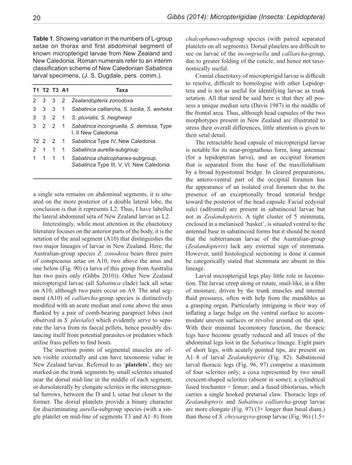
20 Gibbs (2014): Micropterigidae (Insecta: Lepidoptera)
a single seta remains on abdominal segments, it is situ-ated on the more posterior of a double lateral lobe, the conclusion is that it represents L2. Thus, I have labelled the lateral abdominal seta of New Zealand larvae as L2.
Interestingly, while most attention in the chaetotaxy literature focuses on the anterior parts of the body, it is the setation of the anal segment (A10) that distinguishes the two major lineages of larvae in New Zealand. Here, the Australian-group species Z. zonodoxa bears three pairs of conspicuous setae on A10, two above the anus and one below (Fig. 90) (a larva of this group from Australia has two pairs only (Gibbs 2010)). Other New Zealand micropterigid larvae (all Sabatinca clade) lack all setae on A10, although two pairs occur on A9. The anal seg-ment (A10) of calliarcha-group species is distinctively modified with an acute median anal cone above the anus flanked by a pair of comb-bearing paraproct lobes (not observed in S. pluvialis) which evidently serve to sepa-rate the larva from its faecal pellets, hence possibly dis-tancing itself from potential parasites or predators which utilise frass pellets to find hosts.
The insertion points of segmental muscles are of-ten visible externally and can have taxonomic value in New Zealand larvae. Referred to as ‘platelets’, they are marked on the trunk segments by small sclerites situated near the dorsal mid-line in the middle of each segment, or dorsolaterally by elongate sclerites in the intersegmen-tal furrows, between the D and L setae but closer to the former. The dorsal platelets provide a binary character for discriminating aurella-subgroup species (with a sin-gle platelet on mid-line of segments T3 and A1–8) from
chalcophanes-subgroup species (with paired separated platelets on all segments). Dorsal platelets are difficult to see on larvae of the incongruella and calliarcha-group, due to greater folding of the cuticle, and hence not taxo-nomically useful.
Cranial chaetotaxy of micropterigid larvae is difficult to resolve, difficult to homologise with other Lepidop-tera and is not as useful for identifying larvae as trunk setation. All that need be said here is that they all pos-sess a unique median seta (Davis 1987) in the middle of the frontal area. Thus, although head capsules of the two morphotypes present in New Zealand are illustrated to stress their overall differences, little attention is given to their setal detail.
The retractable head capsule of micropterigid larvae is notable for its near-prognathous form, long antennae (for a lepidopteran larva), and an occipital foramen that is separated from the base of the maxillolabium by a broad hypostomal bridge. In cleared preparations, the antero-ventral part of the occipital foramen has the appearance of an isolated oval foramen due to the presence of an exceptionally broad tentorial bridge toward the posterior of the head capsule. Facial ecdysial sulci (adfrontal) are present in sabatincoid larvae but not in Zealandopterix. A tight cluster of 5 stemmata, enclosed in a melanised ‘basket’, is situated ventral to the antennal base in sabatincoid forms but it should be noted that the subterranean larvae of the Australian-group (Zealandopterix) lack any external sign of stemmata. However, until histological sectioning is done it cannot be categorically stated that stemmata are absent in this lineage.
Larval micropterigid legs play little role in locomo-tion. The larvae creep along or rotate, snail-like, in a film of moisture, driven by the trunk muscles and internal fluid pressures, often with help from the mandibles as a grasping organ. Particularly intriguing is their way of inflating a large bulge on the ventral surface to accom-modate uneven surfaces or revolve around on the spot. With their minimal locomotory function, the thoracic legs have become greatly reduced and all traces of the abdominal legs lost in the Sabatinca lineage. Eight pairs of short legs, with acutely pointed tips, are present on A1–8 of larval Zealandopterix (Fig. 82). Sabatincoid larval thoracic legs (Fig. 96, 97) comprise a maximum of four sclerites only; a coxa represented by two small crescent-shaped sclerites (absent in some); a cylindrical fused trochanter + femur; and a fused tibiotarsus, which carries a single hooked pretarsal claw. Thoracic legs of Zealandopterix and Sabatinca calliarcha-group larvae are more elongate (Fig. 97) (3× longer than basal diam.) than those of S. chrysargyra-group larvae (Fig. 96) (1.5×
Table 1. Showing variation in the numbers of L-group setae on thorax and first abdominal segment of known micropterigid larvae from New Zealand and New Caledonia. Roman numerals refer to an interim classification scheme of New Caledonian Sabatinca larval specimens, (J. S. Dugdale, pers. comm.).
T1 T2 T3 A1 Taxa
2 3 3 2 Zealandopterix zonodoxa3 3 3 1 Sabatinca calliarcha, S. lucilia, S. weheka 3 3 2 1 S. pluvialis, S. heighwayi3 2 2 1 Sabatinca incongruella, S. demissa, Type
I, II New Caledonia?2 2 2 1 Sabatinca Type IV, New Caledonia2 1 1 1 Sabatinca aurella-subgroup1 1 1 1 Sabatinca chalcophanes-subgroup,
Sabatinca Type III, V, VI, New Caledonia
Fauna of New Zealand 72 21
longer than basal diam.). The Sabatinca larval leg pos-sesses a curious inflatable bladder-like sac which arises from a mesal desclerotised region of the femur.
Marked differences exist between the setal types on the trunk of different micropterigid larvae and can assist with taxonomic resolution. In New Zealand, only the calliarcha-group of Sabatinca species has ‘conventional’ acutely-pointed macro-setae (Fig. 86). In all other spe-cies, the trunk macrosetae are thickened in some way and distinctively ridged (Fig. 83–87), giving the appearance of a compound structure. The relative lengths of these setae has taxonomic significance and is thus cited here in terms of an overall length measurement and the ra-tio of setal length/length of a mid-abdominal segment, as measured along the dorsal mid-line. The cranial setae are very small, as are those of the more ventral parts of the trunk where they are camouflaged amongst the coarse honeycomb texture of the cuticle. In some cases a small raised papilla is present but lacks a seta (e.g., the SV group of chrysargyra-group larvae). These minor setal features are shown on the chaetotaxy maps but not dis-cussed in the descriptions.
PUPAPupae are known for Sabatinca lucilia, S. heighwayi, S. weheka, S. pluvialis, S. chrysargyra and S. chalcophanes, but not for Zealandopterix. They are formed within a thin-walled oval silk cocoon, concealed amongst the foliage of the liverworts on which the larvae have been feeding (Fig. 61). Leaves are drawn together and fragments of leaves and other particles may be incorporated to render the structure highly cryptic. These New Zealand examples conform closely to the published descriptions of Microp-terigid pupae (Tillyard, 1922; Lorenz, 1961; Yasuda, 1962; Hashimoto, 2006) differing mainly in the numbers and relative sizes of certain setae.
The pupa is of the type referred to as decticous ex-arate (Fig. 62, 102–105). Decticous denotes the presence of functional mandibles: a pair of sharp sclerotised man-dibles, operated by the mandibles of the pharate adult and used for opening the cocoon prior to emergence of the adult moth. The mandibles of micropterigid pupae are conspicuous as the only melanised part of the pupa (Fig. 105), but are not markedly asymmetrical or hypertro-phied as in Agathiphaga, Heterobathmia, Eriocrania, or Acanthopteroctetes. Their sharply pointed apices overlap slightly in the mid-line and there is a small supernumery tooth about half way along the inner margin of the right mandible. The distal mandible, beyond the supernumery tooth, is heavily sclerotised and finely serrate along its inner margin. Sheaths for the long maxillary palps are
free, elbowed around the mandibles with the distal joints passing across the ventral surface of the eyes.
The clypeus and labrum are clearly demarcated in sabatincoid pupae and Asian/North American microp-terigids but not in Micropterix (Hasenfuss & Kristensen, 2003). The antennae arise above the eye-piece and ex-tend dorsally around the eye and along the forewing to terminate on the ventral side midway between the end of leg 2 and leg 3. The leg sheaths overlap each other along the mid-ventral region between the wings, with leg 3 bent into an ‘S’-shape leaving the tarsus projecting clear of the abdomen as a free and mobile appendage, with the first tarsal joint bent into a right-angle.
The term exarate refers to the moveable appendages of these pupae when compared with the obtect ‘soldered’ pupae of higher Lepidoptera. There is a considerable de-gree of mobility possible resulting from the combined action of leg 3 and wriggling of the abdominal segments. The right-angled tibia and tarsus of leg 3 projects freely from the ventral surface of the pupa. The wing sheaths are readily detached from the abdomen during preserva-tion but to what extent this can occur in living pupae is unknown. Sheaths of palps and legs become detached when the pupa is active on emergence and are thus sepa-rated on the exuviae.
Relatively long robust macro-setae occur on the head, and thorax (Fig. 105). In S. heighwayi, which can be regarded as a groundplan Sabatinca form, these setae are distributed as follows: a trio of long setae on the low-er facial region between the eyes, in the positions of the single M and pair of C1 setae of the larval stage. Directly dorsal to these and forming an overall cluster of five setae on the fronto-clypeal area, is another pair, equivalent to the AF setae of the larva. These five setae seem to be uni-versally present in all micropterigid pupae that have been studied, as are a pair of long setae between the antennal bases on the dorsum of the head. More laterally, on the occipital region of the head, is a group of long setae close together, three in the case of S. heighwayi. Much smaller macrosetae occur on the clypeus and labrum. In addition to the long seta dorsally on the clypeus (as above) is a pair of two very small setae towards each lateral angle. Six equal small setae occur in two rows on the labrum, as in the larva, a dorsal row of two with four along the ventral margin.
On the thorax, the dome of the mesoscutum carries a pair of long setae on each side and in a similar posi-tion on the metascutum is a pair of equally long setae. The abdominal segments of all micropterigid pupae bear a pair of very short setae subdorsally on each segment, the inner one being about twice the length of the outer in S. heighwayi. These setae are absent on A8, replaced by
22 Gibbs (2014): Micropterigidae (Insecta: Lepidoptera)
a pair of small sclerotised tooth-like spines which point posteriorly. The terminal piece of the abdomen, repre-senting segments 9 and 10 and enclosing the external genitalia, is devoid of setae.
Any thorough comparative treatment between New Zealand taxa is premature since very few specimens exist and only few species are represented. However, it can be noted that quite major species differences in the dimen-sions of the head setae and in the number of long setae in the thoracic clusters can occur. Thus, while in calliarcha-group species and S. chrysargyra, the six labral setae on each side are all relatively short and equal, those of S. chalcophanes are represented by four short medial setae (one in dorsal pair, three in ventral row) and two very long setae at the outer end of each row, equivalent to those of the clypeus. On the thorax, S heighwayi with two equally long setae on the metascutum differs from S. weheka where the same group consists of a single much shorter seta. On the abdomen the pair of spines on A8 of S. heighwayi and S. weheka is represented by only a single spine in S. chalcophanes and there are no spines on S. chrysargyra pupae.
Northern hemisphere pupae are described for Mi-cropterix calthella (Lorenz 1961) and Neomicropterix nipponensis (Yasuda 1962) and N. matsumurana (Hashi-moto 2006). Hashimoto (2006) records that the number of setae is frequently variable on both sides of the body, also noted in New Zealand pupae. While it is not prac-tical to make detailed comparison with Micropterix on the basis of the Lorenz drawings, other than to note that the long head setae concur with New Zealand species, a single seta is figured for the proscutum and mesoscu-tum, and the abdominal pairs of setae are shown as in New Zealand taxa apart from an absence of spines on A8. In both M. calthella and M. aruncella the long head and thoracic setae are furcate (Carter & Dugdale 1982). Pupal chaetotaxy of Neomicropterix and Sabatinca are in general agreement for the head and thorax, but dif-fer on the abdomen, where the Japanese species exhibit a greater complement of setae than New Zealand species, with pairs of microsetae in both dorsal and lateral posi-tions (Hashimoto 2006).
PHENOLOGYTable 2 shows the seasonal distribution of collection records for New Zealand Micropterigidae. The reliability of any interpretations concerning the relationship between seasonal flight records and the life cycle of a species will depend on the geographic range of the sampling and the number of samples. Thus, the following generalised com-ments must be regarded as provisional, especially for the rarer species.
Based on the occurrence of adult moths and larvae of each species throughout the year, it appears that in gener-al, New Zealand micropterigids have an annual life cycle with spring to early summer flight periods. Peak months for adults are November and December but the first spe-cies to appear in early spring is S. aurantissima in which almost the entire known flight period takes place during September, followed by S. bimacula and S. quadrijuga in October. These three species are all restricted to South Island. By March, only S. chalcophanes and Z. zonodoxa are still on the wing.
When we look in more detail we find two distinct types of seasonal strategy, distinguished by the immature stage in which they overwinter—the majority, as above, spend autumn and winter as active larvae. From what is known, it appears that all incongruella- and chrysargyra-group species belong to this group, which reaches larval maturity sometime during winter or spring, and spends one or two months in the pupal stage before emerging for their flight period. There is no evidence of pupal dor-mancy (diapause) in this group. Small, early instar larvae can be collected in the field during late summer months after their flight season, and large larvae in late winter or early spring. There is a possibility that the common spe-cies with 5 or 6-month flight records (e.g., chalcophanes and aurella respectively), might be double-brooded over the summer, at least in some favourable locations, but this has yet to be confirmed.
In contrast to the above predominant type, adults of calliarcha-group species, are characterised by the spas-modic appearance of individuals over many months. These species can occasionally be found in large num-bers at a very restricted locality, at the peak of their flight season, but otherwise their appearance is erratic and unpredictable. There is evidence that at least some of them undergo an extended pupal period, implying a pupal diapause. This pattern is clearly demonstrated by the South Island sister species S. heighwayi and S. we-heka where mature larvae have consistently occurred at the end of December, reaching the pupal stage during January. Instead of emerging after the normal pupal de-velopment period (i.e., in autumn), they remain until the following spring, 9-10 months later. Not all larvae follow this pattern. For instance, seemingly mature larvae of S. heighwayi have been maintained under ambient condi-tions in captivity throughout winter. Any conclusions are tentative since these alternative strategies have only been recorded from a few captive individuals and field sam-pling is far from sufficient to substantiate these seasonal hypotheses.
It is tempting to speculate that the early New Zealand lepidopterists, who made such a significant contribution
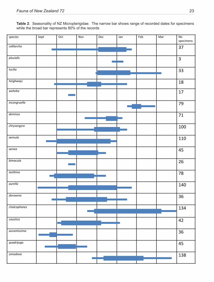
Fauna of New Zealand 72 23
Table 2. Seasonality of NZ Micropterigidae. The narrow bar shows range of recorded dates for specimens while the broad bar represents 80% of the records
species Sept Oct Nov Dec Jan Feb Mar No specimens
calliarcha 37
pluvialis 3
lucilia 33
heighwayi 18
weheka 17
incongruella 79
demissa 71
chrysargyra 100
aemula 110
aenea 45
bimacula 26
ianthina 78
aurella 140
doroxena 36
chalcophanes 134
caustica 42
aurantissima 36
quadrijuga 45
zonodoxa 138
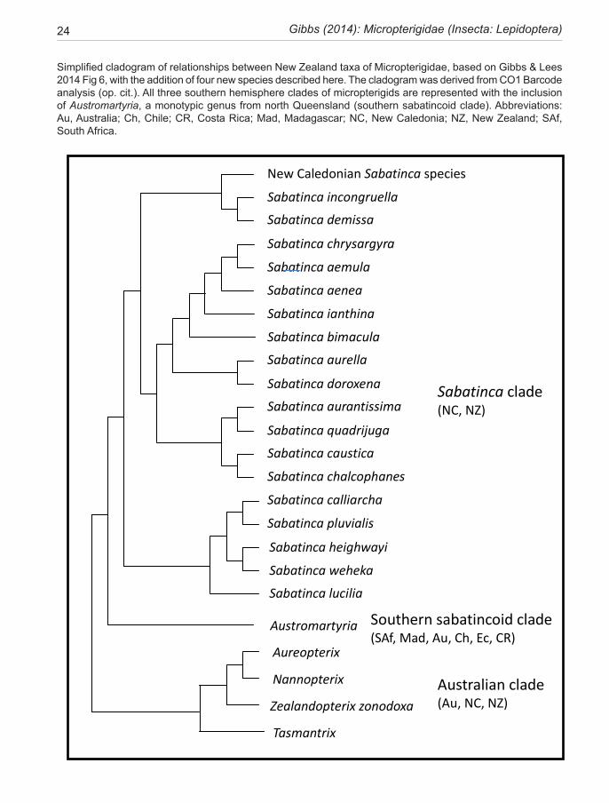
24 Gibbs (2014): Micropterigidae (Insecta: Lepidoptera)
Simplified cladogram of relationships between New Zealand taxa of Micropterigidae, based on Gibbs & Lees 2014 Fig 6, with the addition of four new species described here. The cladogram was derived from CO1 Barcode analysis (op. cit.). All three southern hemisphere clades of micropterigids are represented with the inclusion of Austromartyria, a monotypic genus from north Queensland (southern sabatincoid clade). Abbreviations: Au, Australia; Ch, Chile; CR, Costa Rica; Mad, Madagascar; NC, New Caledonia; NZ, New Zealand; SAf, South Africa.
Tasmantrix
Sabatinca incongruella
Sabatinca demissa
New Caledonian Sabatinca species
Sabatinca chrysargyra
Sabatinca aemula
Sabatinca aenea
Sabatinca ianthina
Sabatinca bimacula
Sabatinca aurella
Sabatinca doroxena
Sabatinca aurantissima
Sabatinca quadrijuga
Sabatinca caustica
Sabatinca chalcophanes
Sabatinca calliarcha
Sabatinca pluvialis
Sabatinca lucilia
Sabatinca heighwayi
Sabatinca weheka
Zealandopterix zonodoxa
Southern sabatincoid clade (SAf, Mad, Au, Ch, Ec, CR)
Aureopterix
Nannopterix
Sabatinca clade (NC, NZ)
Australian clade (Au, NC, NZ)
Austromartyria
Fauna of New Zealand 72 25
towards our knowledge of Micropterigidae between 1900 and 1930, failed to locate three of the new species described here simply because they were not in the field sufficiently early in the season (aurantissima (Sept), bimacula (Oct), weheka (Oct)) to encounter them.
PHYLOGENYA molecular phylogenetic analysis of the family Microp-terigidae was initiated by Y. Kobayashi and H. Suzuki at Tokyo Metropolitan University in 1997. Their analysis included five New Zealand taxa (four of Sabatinca + Zealandopterix) and used the ribosomal mitochondrial 16S gene. This ongoing project gradually incorporated samples from all continents (except India, where micropterigids were not known at the time (Lees et al. 2010.)), until it could be regarded as a worldwide phylogeny. At the time of writing, this molecular analysis has been extended to include nuclear rRNA genes 18S and 28S, but remains unpublished. A separate programme, with the goal of barcoding Micropterigidae of the World (MICOW) has been established in Barcode of Life Data Systems (BOLD) (Ratasingham & Hebert 2007). It is more comprehensive, with the New Zealand component incorporating multiple examplars of all the New Zealand taxa in a survey cover-ing about 170 taxa world-wide. Analysis of this database forms the basis of the following discussion (see Gibbs & Lees 2014 for methodology and accessibility of data; and simplified cladogram p. 24).
The most significant finding on a world scale, is the retrieval of five major clades in the family Micropterigi-dae, two northern hemisphere, three southern. The five clades are unambiguously defined in all analyses and ap-pear monophyletic, but their inter-relationship topology cannot be supported with a high degree confidence. This topology, and even evidence of a primary southern-north-ern hemisphere dichotomy within the family, remains to be tested with additional molecular markers. Reassur-ingly, a number of morphological traits can be correlated with the five world clades (Hashimoto 2006). Here, the phylogenetic relationships and biogeographic interpreta-tion of New Zealand species are discussed, based on the molecular analysis presented in Gibbs & Lees (2014).
As well as its contribution to taxonomic species grouping, bar-code analysis has identified larval morphs, confirmed the three synonomies proposed in this revision from morphological data, and also confirmed the identity of pairs of sister species (discussed within the relevant species descriptions and in relation to biogeography). The identification of three subclades within Sabatinca on the main islands of New Zealand has important im-plications for biogeographic inference, particularly with regard to the occurrence of Sabatinca in New Caledonia.
CONGRUENCE BETWEEN MORPHOLOGY AND PHYLOGENY
This study has revealed a degree of heterogeniety within New Zealand Sabatinca that warrants subdivision into species groups, definable from either morphological or molecular characters. The use of these two independent character sets for analysing species diversity enables an evaluation of the strength of phylogenetic signals con-tained in the basic morphological structures that we use for alpha-taxonomy. Some selected genital characters are reviewed below with a view to noting features that carry a reliable phylogenetic signal as opposed to those which have taxonomic value only. ‘Reliable’ is defined as a phylo-genetic signal that is supported by both the morphological and molecular character sets.
This review has confirmed an extant New Zealand fauna of 18 species in the genus Sabatinca. There is no evidence (with the possible exception of the aemula/chrysargyra species pair (see above)) of sibling spe-cies complexes that might illustrate a burst of recent di-vergence (i.e., evolved within the last 5 Ma). It is thus not surprising to find that the New Zealand species are stable, well-defined entities, easily recognisable from a glance at a colour image, and moreover, that their genital morphology, especially the male, provides unambiguous confirmation of identity. Interestingly, the situation is en-tirely different in New Caledonia where a number of spe-cies complexes have been revealed within the Sabatinca fauna.Male genitalia: Phylogenetic analysis indicates that development of a massive vinculum (segment 9 sclerite), which seems so characteristic of members of the genus Sabatinca, is clearly a widespread groundplan feature. An elongated vinculum can appear in four of the five world lineages of Micropterigidae, although often in only one or two species. It is absent from the remarkably consistent Northern Hemisphere Micropterix clade. The robust vinculum is best interpreted as a structure of major functional significance but with little phylogenetic signal. By way of contrast, the micropterigid phallus is found to reveal more phylogenetic information than any other part of the genital complex. Some traits are significant at the lineage level, others at genus or species level. Those with lineage significance are described below:
Relative length of the phallobase: In three of the five lineages of Micropterigidae, the phallobase is con-siderably longer than the distal phallus (e.g., in the Zea-landian Sabatinca lineage, the ratio of phallobase: overall phallus is in the range 0.6–0.9. However, in the Austral-ian lineage (e.g., Tasmantrix) this relationship is reversed (ratio 0.3–0.4) and in the Asian-North American lineage (e.g., Neomicropterix) it can be close to equal (0.5–0.6).
26 Gibbs (2014): Micropterigidae (Insecta: Lepidoptera)
Bulbus ejaculatorius aperture: The anterior termi-nation of the cuticular phallobase tube is clearly defined in genitalic preparations and seems to be relatively con-sistent in the limited number of comparative specimens available for this study. The plane of the terminal phal-lobase aperture is essentially at right angles to the longi-tudinal axis in the Micropterix lineage of Europe. In the Asian lineage the phallobase tube is bent at its anterior end to bring the aperture into a longitudinal ventral orien-tation, parallel with the axis of the phallus. The anterior aperture is oblique in the remaining three lineages.
Gonopore details: In the hypothetical groundplan micropterigid, the gonopore is probably an open, more-or-less circular aperture which can be either terminal or toward the dorsal side of the phallus. In a few cases, no-tably the genus Tasmantrix and its allies in the Australian lineage, there is a dorsal component which overhangs the gonopore like the upper part of a vertebrate jaw. A series of close-set, sclerotised radial folds which surround the margin of the gonopore are a probable micropterigid au-tapomorphy (Kristensen 1984b), but in the two northern hemisphere lineages there is an additional blind ventral branch of the distal phallus extending beyond the gonop-ore. This ventral branch seems universally present in Mi-cropterix species but has only been reported from Epima-rtyria in the Asian-North American lineage (Hashimoto 2006). In each of the three southern hemisphere lineages there are numerous examples of an expanded ‘ventral bulb’ developed below the gonopore but no discrete ventral branch as in Micropterix and Epimartyria. The morphological and phylogenetic significance of these features have not yet been investigated. It goes without saying that the remarkable degree of gonopore diversity can be valuable for species-level determinations.
Characters of the phallus are illustrated and cited ex-tensively in this revision for all taxonomic levels.Female genitalia: The presence of large tri-radiate signa in the corpus bursae of certain micropterigids invites taxonomic analysis but appears to offer little of phyloge-netic value. Four signa occur consistently in members of the calliarcha group and the New Zealand incongruella group of the Sabatinca lineage. Four remarkably similar signa also occur in certain taxa of the Asian/N American lineage. An intriguing correlation between the existence of signa and the presence of a sclerotised ‘pleural pocket’ modification of segment 8 (see discussion p.14) is found only in the Sabatinca lineage and is extensively devel-oped in many New Caledonian species (where females in 27 out of 41 species examined had this feature in conjunction with the development of signa). It has been suggested that large signa are likely to be associated with the breaking up of spermatophores and indeed dissections
of Sabatinca specimens have sometimes found fragments of spent spermatophores remaining within the corpus in these cases (Gibbs & Lees 2014). A dissected female specimen of Epimartyria (North America) also contained spermatophore fragments in conjunction with large trident signa (unpublished observation).
In contrast to the signa discussed above, an exami-nation of spermathecal morphology will repay the time needed to make suitable preparations of these delicate structures:
Spermatheca: Although relatively consistent throughout many lepidopteran groups, the degree of spermathecal differentiation in the New Zealand Microp-terigidae is significant and offers valuable phylogenetic insight. While ductus and lagena remain relatively con-stant throughout the family, the utriculus varies from the most basic simple form in Zealandopterix (Fig. 209—a single elongate thin-walled chamber), to the complex utricular differentiation of certain Sabatinca species, where up to four distinct regions can be distinguished. Of particular interest is the strong correlation with the phy-logenetic relationships established by barcoding analysis. For instance, the utricular modifications shown in Fig. 229–235 and Fig. 238–241, spanning the aurella-sub-group and chalcophanes-subgroup respectively, clearly indicate patterns that support subdivision into two basic forms within the chrysargyra-group clade. Quite dif-ferent modifications characterise the incongruella- and calliarcha-group clades. In particular, the presence of a discrete spherical loculate organ at the junction between utriculus and lagena (Fig. 221–226). This spherical organ is a feature of the New Caledonian Sabatinca fauna as well as these two New Zealand clades.
BIOGEOGRAPHYBiogeographic links between Australia, New Caledonia, and New Zealand have become a topic for debate, par-ticularly in regard to the role of vicariance or dispersal to account for the trans-oceanic relationships we find today. The topic is further complicated by the question of how much land existed and where it was situated on the crustal block of Zealandia as it separated from eastern Gondwana and was steadily inundated by the sea throughout the Ce-nozoic, leading ultimately to the modern isolated islands of New Caledonia and New Zealand. These small archaic moths and their moist breeding habitats clearly survived throughout the Cretaceous and Cenozoic periods. The Micropterigidae are thus almost ‘ideal’ candidates for historical biogeographic interpretation. First, their deep fossil record (amber fossils dated from 136, 97, and 37 Ma. see Introduction) and the molecular phylogenetic analysis (Gibbs & Lees 2014) support the notion of an antiquity
Fauna of New Zealand 72 27
which extends back to the break-up of the Laurasian and Gondwanan tectonic plates. Second, they survive today on every habitable temperate continent, in both northern and southern hemispheres, and offer suitably speciose groups for analysis. Third, their antiquity, together with the present world distribution pattern of each of the five major micropterigid lineages on separate continents or con-tinental fragments, implies that trans-oceanic dispersal has not contributed to, nor disrupted, their current distribution pattern. Within the New Zealand region, these moths are known from the three main islands and a number of the larger offshore islands that were connected to the mainland during glacial periods of lowered sea levels (e.g., Poor Knights, Little Barrier, Great Barrier, Kapiti) but not from the Kermadecs, Three Kings, Chatham Islands, or any of the Subantarctic Islands.
In an earlier, more intuitive, review of the family in relation to tectonic events in this region, Gibbs (1983) used a morphological/panbiogeographic model in an attempt to explain diversity across the region. The con-clusion was that in the SW Pacific region—eastern Aus-tralia, New Caledonia, and New Zealand—two distinct ‘species-groups’ overlap, occupying a complex biogeo-graphic region surrounding the Tasman and Coral Seas. Since that time, the world molecular phylogeny, dis-cussed above (Kobayashi et al. 2000, Gibbs 2004), and the COI bar-code analysis (Gibbs & Lees 2014), have resolved three well-supported micropterigid clades in the SW Pacific: an Australian clade that dominates in Aus-tralia with outliers in New Caledonia and North Island; a Sabatinca clade that dominates in New Caledonia and New Zealand but has a surprise outlier in SW Australia; and a southern sabatincoid clade with but a single repre-sentative in northern Queensland.
In terms of SW Pacific biogeography, the most sig-nificant conclusion from the Gibbs & Lees 2014 phylo-genetic analysis is that it provides the first evidence that the New Caledonian Sabatinca fauna is likely to have been derived from New Zealand. Of the three known Sa-batinca subclades, two are confined to the islands of New Zealand today and one, the incongruella-clade, contains two sister clades, one in New Zealand, the other with at least 50 species in New Caledonia. Rather than assign-ing their biogeographic history to the modern islands, it would be more honest to say these moths have evolved on the continental block of Zealandia (sensu Mortimer 2008), whatever topography that might imply, but sur-vive today only on the isolated emergent islands of New Caledonia and New Zealand.
Tentative dates, assigned by the application of a relaxed molecular clock calibrated from three microp-terigid fossils (Baltimartyria proavitella Rebel—Eo-cene-Oligocene; Parasabatinca aftimacri Whalley—
Aptian-Neocomian; and AMNH Bu 701 from Burmese amber illustrated by Grimaldi & Engel 2002.) and four trichopteran fossils, indicate that the divergence of the Sabatinca lineage might have been initiated at about the time when tectonic events were beginning to separate Zealandia from Australia/Antarctica (i.e., ca 84 million years ago (Ma), whereas the New Caledonian radiation of incongruella-group species began about 52 Ma (Gibbs & Lees 2014). This dating hypothesis, and the survival of the Sabatinca clade on the crustal block of Zealandia, prior to the uplift of the modern islands of New Caledo-nia (emergent ca 37 Ma (Schellart et al. 2006)) and New Zealand (which commenced uplift toward its present profile ca 23 Ma (Campbell & Hutching 2007)), imply the continuous existence of moist forested land (islands?) in the region. Recent geological studies of the Tasman Sea basin are beginning to offer plausible scenarios for such former land; e.g., Schellart et al. (2009), who have revealed a large slab of subducted continental crust be-tween New Caledonia and New Zealand which is thought to have finally sunk below sea-level at about this time.
The sole member of the ‘Australian-group’ clade in New Zealand, Zealandopterix, diverged from the Aus-tralian fauna about 83 Ma, according to the phylogenetic hypothesis of Gibbs and Lees (2014). These phyloge-netic studies are thus generally compatible with the 1983 hypothesis, namely that Trans-Tasman relationships in the SW Pacific Micropterigidae (Sabatinca and Zealand-opterix) represent vicariant processes that were probably established around the time of the opening of the Tasman Sea and confirming that the faunas of New Caledonia and New Zealand are more closely associated with each other than either is to the Australian fauna (Gibbs & Lees 2014).
Within the New Zealand fauna of 18 Sabatinca spe-cies the molecular analysis has retrieved a total of seven pairs of sister species, three of which [incongruella/dem-issa; aurella/doroxena; calliarcha/pluvalis] are likely to have been in existence prior to the uplift of modern New Zealand and are thus difficult to explain through refer-ence to modern geomorphology or geological phenom-ena. The remaining four pairs, all South Island examples and all allopatric pairs today, can be examined further to look for possible geologically-driven patterns.
The Alpine Fault is by far the most significant South Island tectonic feature, visible from space. This massive strike/slip fault, which has resulted in a total of over 470 km of lateral displacement, can be traced back to its in-ception 45 Ma but did not become vigorous until about 26 Ma and has been especially active over the past 5 Ma, moving at a rate of 20–30 mm per year horizontally and uplifting the Southern Alps (Campbell & Hutching 2007). Its lateral displacement, as a possible cause of
28 Gibbs (2014): Micropterigidae (Insecta: Lepidoptera)
South Island biogeographic disjunctions, has been de-bated by Heads (1998), Wallis & Trewick (2001), Heads & Craw (2004), Haase et al. (2007) and its potential role in the divergence of a pair of sibling micropterigid spe-cies (chrysargyra and aemula) has been noted by Gibbs (2006). Extensive recent West Coast collections of these two species have attempted to define the geographic gap between them more precisely and determine whether sympatric populations exist. To date none have been found. Instead, aemula, with a north-western distribution from the Nelson region south to Mt Hercules, appears to be separated by a mere 35 km from chrysargyra which is found from Franz Josef valley southwards. Both share the same flight season and are indistinguishable in the field, suggesting that further sampling might be needed to resolve the nature of the gap between these two spe-cies. The barcode analysis implies that this example is the only instance of speciation in the micropterigid fauna that falls within the Miocene-Holocene era (Gibbs & Lees 2014). Whether geographic fault movement was the driver for divergence, or whether more recent factors such as glacial extirpation (Wallis & Trewick 2001), have served to keep them apart remains a matter for specula-tion in this case.
The other three examples of sister species evidently relate to earlier events (dated around 25 Ma), but each shows divergence between a species, now centred in the northwest of the South Island, and a sister towards the south of the island. In all cases the molecular diagno-ses as sister pairs is correlated with morphological simi-larities. With the case of heighwayi (north) and weheka (south) in the calliarcha-group, the divergence is along the west coast; whereas the other two include a north-western member (chalcophanes or aurantissima) which has diverged from a south-eastern member (caustica or quadrijuga respectively). The northern members are sympatric over a wide area of north-western South Island while Sabatinca caustica and quadrijuga are sympat-ric throughout most of their ranges in the south-eastern South Island, the former extending onto Stewart Island. The South Island has undergone extensive geomorpho-logical changes since the estimated timing of species divergence—so much so that it is not realistic to offer as-sociated geological explanations. Moreover, the species pairs are widely separated which, in itself, indicates the speciation events themselves were likely to have taken place on a very different landscape from today. At 25 Ma it was close to the point of maximum marine inundation, implying that perhaps isolation on different islands may have initiated the process and been a driving force for all three examples of diversification. A similar explanation may apply to the example of wide geographical disjunc-tion in Sabatinca calliarcha discussed on p. 27, which is
distributed over both North and South Islands but with a gap between the Coromandel Peninsula and NW Nelson.
Attention was drawn to the trio of species doroxena, aurella, and ianthina in previous discussions of New Zea-land micropterigid biogeography (Gibbs 1983, 1989). In contrast to the above species pairs with wide allopatry, these three are broadly sympatric over the North Island with two (aurella, ianthina) also extending about half way along the western South Island. Both morphological and molecular interpretations imply a closely related trio, but phylogenetic analysis suggests they represent steps along a ladder leading to the remainder of the aurella-subgroup species. The relaxed molecular clock dating used in this study (Gibbs & Lees 2014) indicates that spe-cies divergence between doroxena and aurella occurred late Eocene to early Miocene, i.e., between about 37–18 Ma i.e., at a time of increasing marine inundation when Zealandia is considered to have been an extensive low-lying archipelago, without significant regional variation and possibly lacking montane regions. The advent of a new active plate boundary initiated the uplift of modern New Zealand (Campbell & Hutching 2007). A possible scenario might suggest that divergence from an early chrysargyra-group ancestor was initiated on a northern sector of the Zealandian archipelago and over the course of island submergence and possible recombination, out-lying populations were isolated, became distinct, and when amalgamated maintained their identity and original distributions until more active uplift of New Zealand in the early Miocene led to the dispersal of two taxa with very similar ecological requirements (aurella, ianthina), southward along the western South Island.
It should be noted that none of the examples of sister species divergence given above, apart from the case of aemula/chrysargyra, conforms to the prevailing moun-tain-building or glaciation hypotheses offered by many recent phylogeographic studies of New Zealand biota e.g., review by Wallis & Trewick (2009).
Intensive micropterigid collecting along the mid-West Coast South Island region (associated with the discovery of S. weheka and the mapping of the aemula-chrysargyra interface), has revealed an approximately coincident southern biogeographic limit for four wide-spread species of these moths, three of which (ianthina, aurella and chalcophanes) extend well into the North Island. The boundary falls within the well-known ‘beech gap’, a zone often attributed to the impact of glacial ex-tirpation during the last ice advance (e.g., Wardle 1988; Wallis & Trewick 2009). Whether this coincident bound-ary of micropterigids reflects a deeper historical pattern, the impact of Pleistocene ice, or a limit imposed by the present climate is not clear.
Fauna of New Zealand 72 29
CONSERVATIONThreats to the survival of micropterigids are unknown, apart from the loss of habitat, which can be a serious issue in New Zealand. Although most species have been found in reasonable numbers, it does not necessarily follow that they pose no conservation problems. Three species (S. pluvialis, S. weheka, and S. bimacula) are known from only one or two localities but these locations are embedded in National Parks and so are as secure as we can make them
today. Each of the other 16 species occurs at some or all of their range within protected areas. In short, although six species (incongruella, calliarcha, aenea, chalcophanes, caustica, and quadrijuga) were originally found in loca-tions that are unknown or unprotected, all are now known from many sites, the majority of which have some form of protection, and hence are not especially vulnerable. There are no documented or suspected examples of declining populations.
30 Gibbs (2014): Micropterigidae (Insecta: Lepidoptera)
KEY TO GENERA AND SPECIES-GROUPS OF GENUS SABATINCA
This key employs both superficial, externally-visible fea-tures, as well as morphological characters to provide for identification regardless of the condition of the specimen. However, it needs to be appreciated that the external scale characters will apply only to specimens in good condition, old or damaged individuals may have to be dissected to confirm their generic status.1 Tegulae and thoracic dorsum devoid of long hair-scales,
clothed with flat lamellar scales (Fig. 55); male ter-gum 10 dominated by a median cluster of about 6 posteriorly-directed setae, slightly longer than length of tergum (Fig. 202); female segment 9 totally un-sclerotised (Fig. 208); antennal ascoid branches aris-ing from a linear groove around the flagellomere (Fig. 45) [fwl 2.3-3.3 mm, iridescent purple with white head, legs and wing maculation] ................... ... ...................................…(p. 64)… Zealandopterix
—Tegulae and thoracic dorsum with tufts of long hair-scales similar to those on head (Fig. 54); male tergum 10 lacking a median cluster of long setae; female seg-ment 9 sclerite forming a complete ring (Fig. 227); antennal ascoid branches arising radially from a basal disc (Fig. 43) ............…(p. 30)… 2 (Sabatinca)
2 Apical half of antenna black, grading to ochreous at base (exceptions chalcophanes with multiple black bands, quadrijuga black throughout); labial palps 2-segmented (Fig. 32); male tergum 10 a median lobe (Fig. 149); female bursa lacking signa, spherical loculate organ of spermathecal utriculus absent ...... ............ ...…(p. 44)… Sabatinca chrysargyra-group
—Apical half of antenna predominantly ochreous with discrete black bands, either on each flagellomere, or incorporating several flagellomeres, or at tip only; la-bial palps 3-segmented (Fig. 30) (exception demissa 2-segmented); male tergum 10 bilobed; female bursa with conspicuous signa, a spherical, loculate organ present at distal extremity of utriculus (Fig. 223) ... 3
3 Antenna with discrete black apex, the remainder ochre-ous; forewing length greater than 5.1 mm; hindwing R1 vein entire over basal sector from fork with R to coalescence with Sc at about 2/3 (Fig. 25); male tergum 10 wide, with broad ‘U’shaped emargination (Fig. 119) ....…(p. 35)… Sabatinca calliarcha-group
—Antenna with two discrete black bands in apical half, the remainder ochreous; forewing length less than 4.9 mm; hindwing R1 reduced to a trace only proxi-mal to the point of coalescence with Sc at about 2/3 (Fig. 26); Male tergum 10 squarish-trapezoidal with a small shallow emargination on posterior edge (Fig. 107, 113) .....(p. 31)... Sabatinca incongruella- group
SYSTEMATICS
Genus Sabatinca WalkerSabatinca Walker, 1863: 511 (type species Sabatinca incongru-
ella Walker by original monotypy)Palaeomicra Meyrick, 1885. synonymised by Meyrick 1912:
124Micropardalis Meyrick, 1912. synonymised by Kristensen and
Nielsen 1979: 140
Note on synonomies.Meyrick’s genus Palaeomicra, although syn-
onymised by him in 1912, has surfaced on a number of occasions since then, mainly due to the fact that the chrysargyra-group clade can be strongly supported on the basis of morphological apomorphies of both adults and larvae (see p. 48). However, since its molecular sup-port as a stand-alone genus is ambiguous, the decision here is to regard this clade as a subgroup of Sabatinca.
The genus Micropardalis, established by Meyrick for doroxena on the basis that veins RS3 and RS4 of the forewing are separate (meaning not stalked as in Sabatin-ca), was extended to include aurella by Dugdale (1988). Both species share this character and the unique gross morphology of the male genitalia (which was not cited by Meyrick). The discovery of S. bimacula and a survey of wing venation across all Sabatinca species show that although the majority retain a RS3+RS4 stalk, the stalk varies in length, becoming progressively shorter in aenea and then ianthina before disappearing altogether in bi-macula, aurella and doroxena. This series represents the more derived end of the chrysargyra-group clade in the COI phylogeny on p. 24, indicating no more than a trend in the evolution of apical wing veins along the clade. The loss of this stalk also occurs in a number of New Caledo-nian Sabatinca taxa, as well as in Agrionympha (Gibbs & Kristensen 2011) and cannot be regarded as grounds for generic distinction.Diagnosis. Antennal ascoids with branches arising around a near-circular base; antennal flagellomeres with a densely papillate cuticular microsculpture; head capsule, tegulae, mesoscutellum with tufts of very long piliform scales; forewing veins RS3 and 4 usually stalked, RS4 reaching margin posterior to apex; hindwing vein R either discrete in its basal section only or represented by a vestigial ‘spur’ remnant on Sc2, never reaching to margin uncoalesced; male sternite 8 absent; male genital segment 9 (vinculum) with anteromarginal sulcus heavily melanised; tergum 10 discrete from vinculum, undivided.
Note: The range of variation among New Caledonian species of Sabatinca is considerably greater than in New Zealand and not necessarily accounted for in the descrip-tions that follow.
Fauna of New Zealand 72 31
Adult head. Head capsule usually without an epicra-nial sulcus. Interocular index 0.6–0.8; supraocular index 0.3–0.4. Head capsule with tufts of exceptionally long piliform scales on frons and vertex. Maxillary palps long, 5-segmented, with 4th segment the longest, usually slightly curved and bearing fine transverse striae along most of its length; 5th segment long, 0.8–1.1× 1st segment. Labial palps 2 or 3-segmented. Antennae less than half the forewing length, usually 0.3 of forewing in female and 0.4 in male; with 21–35 filiform, or submoniliform flagellomeres; scape and pedicel greatly swollen, scape 2.0–4.1× diameter of first flagellomere, with a median indentation on mesal surface; pedicel almost spherical; both scape and pedicel with dense tufts of extremely long piliform setae.Thorax. Mesoscutum clad with closely fitting scales but tegulae and mesoscutellum bearing dense tufts of long erect piliform scales (Fig. 54). Forewing venation (Fig. 25, 26) with both Sc and R deeply forked. Hindwing vein Sc forked; vein R not forked, coalesced with Sc2 over its distal third in all taxa; the basal section distinct in calliarcha-group, reduced in incongruella, demissa and chrysargyra-group species to a short vestigial spur at the point of coalescence (originally referred to as the ‘recurrent vein’ by Philpott 1923b); hindwing with 2–4 frenular bristles.Abdomen. Deeper than wide. T1 entirely membraneous in central area, lacking a transverse sclerite. T2 with vari-able melanisation, ranging from entire to eroded by an emargination on dorsal mid-line. S5 gland orifice typically ‘sabatincoid’ in both sexes, meaning raised on a cylindrical protuberance and bearing 5–17 long piliform setae (Fig. 46, 47). S5 glands absent in S. weheka. S8 remnants absent.Male genitalia. Sclerite 9 relatively massive, 1.5–3.2× medioventral length of S6, heavily melanised along ant-eromarginal sulcus; anteror extremities of this thickened margin often with a lightly melanised flange-like extension present; development of the dorsal region of S9 variable, ranging from the entire dorsal half being non-existent, through the typical Sabatinca form with tapering dorsal arms that terminate short of the mid-dorsal line, to those with a broad melanised dorsal ‘bridge’ which closes a complete ring sclerite. Valves relatively simple, single or double-lobed, sometimes incurved at apex with a small valve tooth; bearing numerous retro-setae on inner sur-faces but lacking any highly specialised setae. Median plate a fan-shaped horizontal flange. Tergum 10 discrete from T9, uni- or bi-lobed; lacking specialised setae. Anal cone sclerite large, conspicuous, bearing a patch of scat-tered setae proximally. Phallus of variable length, from short and straight to longer than abdomen and looped back on itself anteriorly; phallobase with trumpet-like bulbus ejaculatorius anteriorly, expanded to1.8–8.0 of its minimum diameter. Gonopore orientation dorsal or
postero-dorsal, usually heart-shaped but can be apically attenuated; lappets usually present; ventral bulb present or absent, sometimes with melanised ventral keel flanges. Phallocrypt with micro-scales.Female genitalia. 8th segment unmodified or with a spe-cialised pleural sclerotisation linking T8 with S8. Segment 9 sclerite a complete ring; but with small emargination anterodorsally and in some, irregular demelanisation along mid-dorsal line. Segment 10 lateral sclerites (papillae) either moderately elongate and U-shaped, or short and squarish. Spermathecal papilla rarely with an irregular ventral sclerite; utriculus subdivided into at least two distinct regions. Bursa copulatrix with or without signa, where present usually four of tri-radiate form.Larva. Larvae typical of the liverwort-feeding ‘sabat-incoid’ morphotype, i.e., cryptically pigmented, hunch-backed in profile, roughly hexagonal in cross-section and lacking abdominal prolegs (e.g., Fig. 74). Chaetotaxy of T1 and A1–7 required for diagnosis: T1 with 3 L setae; A1–7, with a single D macroseta (D1) (Fig. 89).
The incongruella-groupDiagnosis. Antennae ochreous with two black bands. Forewing length less than 4.9 mm; hindwing R1 reduced to a vestigial trace, proximal to the point of coalescence with Sc at about 2/3; male tergum 10 roughly trapezoidal with a small median emargination; female segment 9 a simple ring, emarginated on anterior mid-dorsal line. Larva with three L-group setae on T1 and two on T3; a single D-group seta on A8.Adult head. (Fig. 30) Head capsule usually without an epicranial sulcus. Head capsule with tufts of exception-ally long piliform scales on frons and vertex. Maxillary palps (Fig. 34) long, 5-segmented, 5th segment relatively long, 0.8–1.1× 1st segment. Labial palps 3-segmented, (2-segmented in S. demissa). Antennae less than half the forewing length, usually 0.3 of forewing in female and 0.4 in male; with 21–35 submoniliform flagellomeres; tufts of extremely long piliform setae on pedicel reaching to 6th to 9th flagellomere.Thorax. Mesoscutum clad with closely fitting lamellar scales but tegulae and mesoscutellum bearing dense tufts of long erect piliform scales. Wing venation typical for Sabatinca (Fig. 26). Hindwing with 2–4 frenular bristles.Abdomen. Elongate, ratio 2.5–3.0. T1 entirely membrane-ous in central area, lacking a transverse sclerite. T2 with variable melanisation, ranging from entire to emarginated on dorsal mid-line. Segment 5 gland small, (0.03–0.04 mm diameter) with about 9 piliform setae.Male genitalia. Genitalia never inclined upwards. Sclerite 9 massive, 2.6–3.4× S6, heavily melanised along
32 Gibbs (2014): Micropterigidae (Insecta: Lepidoptera)
anterolateral sulcus; dorsal arms broad, coalescing to form a wide melanised dorsal ‘bridge’ of about 0.2 of S9 length, bearing a double row of piliform scales towards the posterior margin; anterior flanges of the anteromarginal sulcus barely discernable. Valves 0.5× length of S9, convex outwards; broadly divided into two lobes, dorsal lobe clasper-like, curving mesally at apex, with a blunt tooth; ventral lobe which follows curvature of posterior margin of sclerite 9, creating a horizontal shelf at base of genital atrium; sparse retro-setae on inner surfaces. Median plate horizontally flat, about twice as long as wide, smooth-edged. Tergum 10 inclined slightly ventrad in profile; 0.3–0.4× S9; trapezoidal, with minimal emargination—either broad and shallow, or a ‘V’-shaped cleft; bearing only sparse setae. Anal cone sclerite squarish, conspicuous, bearing 6-12 scattered macrosetae proximally. Phallus straight (in NZ species) relatively short, 1.2–1.6× length of sclerite 9; phallobase 0.6–0.7 of total phallus length, bulbus ejaculatorius expanded slightly anteriorly to 1.7–2.4× its minimum diameter, with a markedly oblique aperture. Gonopore orientation dorsal to postero-dorsal, heart-shaped but can be apically attenuated; lappets present; ventral bulb present or absent. Phallocrypt with small comb-like micro-scales.Female genitalia. T8 and S8 sclerites either totally un-modified; or with a minor modification in the pleural region involving encroachment of both T8 and S8 sclerites into the pleural membrane. Segment 9 forming a simple com-plete ring, emarginated anteriorly. Segment 10 sclerites 2.3–2.4× longer than high, with U-shaped lateral support sclerites. Genital papilla often difficult to interpret if dis-torted; ventral sclerites absent; corpus bursae moderate to large, oval or elongate, with 4 tri-radiate signa which are either very small and ‘vestigial’ at 0.15 mm (demissa), or large 0.4 mm (incongruella) densely adorned with stiff bristles. Spermathecal utriculus in two sections - a basal thin-walled sac, capable of great expansion and a compact globular terminal portion comprised of multiple locules, resembling a bunch of grapes; lagena arising from distal extremity of the loculate utriculus. Remains of a discrete spermatophore can occur within the corpus of both these species—a smooth-walled bulbous capsule with long narrow neck region.Larva. The larvae of both New Zealand species are known. They are typical of the liverwort-feeding ‘sabatincoid’ type of larval Micropterigidae, with elongate thoracic legs for a micropterigid, about 3× basal diameter of femur, with 5 setae on the femur and 3 on tibiotarsus; coxal sclerotisa-tion minimal or absent. A basal ‘papilla’ is present on the mesal side of each thoracic leg. Abdominal legs absent, but extensible dome-like extrusions of the cuticle aid in
locomotion. The macrosetae are thickened and ridged in both incongruella and demissa, thus resembling chrys-argyra-group species, but distinguishable from those by their chaetotaxy (Fig. 94) in which L-group compliment on thoracic segments—3L on T1, 2 L on T2 and T3 is unique in New Zealand.Status and Conservation. The species are confined to restricted regions, one North Island, one South Island but occur in protected indigenous forest areas and are unlikely to become seriously threatened.Remarks. Although grouped together, these two species are not similar in general facies. They are united by their banded antennae, the possession of a broad dorsal ‘bridge’ across the 9th segment in the male (incidentally, a feature which does not occur in the known New Caledonian species of this clade), and the compact form of the male tergum 10.
The bulk of diversity (in excess of 50 species) in this subclade of Sabatinca occurs in New Caledonia where, overall, they exhibit quite an astounding range of vari-ation which includes maculation patterns, antennal fea-tures, adult morphology and larval characters including chaetotaxy. By comparison, the New Zealand species are extremely conservative and represent the phylogenetic stem of the New Caledonian radiation (Gibbs & Lees 2014).
KEY TO ADULTS OF NEW ZEALAND SPECIES OF INCONGRUELLA-GROUP
Antennae with 24 flagellomeres in male (21 female); forewings with several pale yellow or pinkish fas-ciae, narrowly bordered with black scales (Fig. 3); T10 with a shallow emargination (Fig. 113) ............ ..........................................…(p. 32)… incongruella
—Antennae with 35–38 flagellomeres in male (22 fe-male); forewings bronzy, essentially un-marked, but surface slightly undulating (Fig. 2); T10 with V-shaped emargination (Fig. 107) .…(p. 34)… demissa
Sabatinca incongruella Walker, 1863 Fig. 3, 26, 30, 34, 38, 46, 52, 60, 74, 86, 112–117, 211, 222, 252.Sabatinca incongruella, Walker, 1863: 511Sabatinca munda, Felder & Rogenhofer, 1875: pl clx fig 38.
Synonymised by Dugdale 1988: 52 Sabatinca eodora, Meyrick, 1918: 134.Sabatinca eodora, in Hudson 1928: 370, pl XLVI fig. 10. Syn-
onymised by Kristensen & Nielsen 1979: 140.
Fauna of New Zealand 72 33
Diagnosis. (Fig. 3, 252) Antennae of male with 24 flag-ellomeres, female 21. Forewing orange with indistinct pinkish fasciae; a pale yellow rectangular fascia at mid length on costa, edged proximally with black scales. Male tergum 10 with very shallow median emargination. Female spermatheca with an extremely wide ductus, almost as wide as long.Head and thorax. Eyes pale grey. interocular index 0.6; supraocular index 0.4. Maxillary palp (Fig. 34) 1.8× head width; ratio 1:0.9:1.8:2.5:1. Labial palp 3-segmented. An-tennae 0.4 of wing length in male, 0.3 female; scape 3.8× diameter of 1st flagellomere; male with 24 flagellomeres, female 21; submoniliform, 7th is 1.3× longer than wide; pedicel piliform scales reaching to 6th flagellomere. Long piliform scales of head, scape, pedicel and tegulae ochre-ous; antennal flagellomeres ochreous with two distinct black bands, the first at 13th–15th, the second at 21st–23rd; apical flagellomere ochreous; thoracic mesoscutum bronzy-brown scaled; an erect tuft of ochreous piliform scales on mesoscutellum. Femora pale creamy-yellow, tibiae and tarsi with dark brown bands on each segment.Wings. (Fig. 26) Forewing length of male 4.3 mm (3.8–4.7); female 4.9 mm (4.8–5.0); width ratio 3.1. Ground colour orange, with pale yellow and salmon-pink macula-tion; two large and three small yellow fasciae along costa; first extends from base to one third a roughly triangular patch of pale yellow scales tending white toward middle of wing where it forms a pale basal streak; second at half length to middle of wing, edged black on basal side; three or four small yellow spots edged black on basal side, along costa to apex; salmon-pink scales form three rather indistinct parallel transverse fascia from one third to three quarters; finally a scattered tracery of black scales, not of descriptive value. Fringes largely creamy-yellow but with several black sections.Hindwing with 2 frenular bristles; R1 vein present as recurrent trace only. Grey-scaled with purple reflections. Fringes silvery grey.Male abdomen and genitalia. (Fig. 112–117) Abdomen deeper than wide. T1 lacking a transverse sclerite; T2 deeply emarginated in mid-line. Segment 5 gland excep-tionally small, 0.026 mm. diameter, with about 9 piliform setae. T8 and S7 unmodified. Sclerite 9 massive, 3.0× S6, dorsal arms forming a broad (0.16 of ventral length of seg 9 sclerite) sclerotised bridge across the dorsum; long ante-rolateral sulcus strongly thickened by a densely melanised band, concave; a short anterior flange present, with about 10 scale bases; posterior margin almost straight. Valves small, 0.4× length sclerite 9, dorsal lobe terminating in a small inward-directed blunt tooth; ventral region form-ing a shelf; sparse slightly curved retro-setae on concave
inner surface; longer posterior-directed setae on outer surface. Median plate horizontal, 0.2 of sclerite 9, oblong, approx 2× longer than wide. Tergum 10 short, 0.3 length of sclerite 9, ventrally inclined; tapering in dorsal view to a wide apex with very shallow median emargination of 0.1; ventral surface with a few scattered setae. Anal cone prominent with large squarish sclerites laterally, each bearing 10–12 macrosetae and numerous microtrichia. Phallus straight, 1.2× sclerite 9 length; phallobase 0.7 of total phallus length, extending anteriorly to A6, where expanded 2.4× its minimum diameter; gonopore dorsal with posterior inclination; heart-shaped with dorsal lip depressed ventrally to about half depth of phallus; a pair of broad lappets at anterior angle of gonopore, lightly melanised but not projecting; ventral bulb absent; phal-locrypt micro-scales comb-tipped.Female abdomen and genitalia. (Fig. 211, 222) T1 lack-ing a transverse sclerite. T8 and S8 wholly separated by pleural membrane but almost meeting at their posterior angles where both sclerites are slightly extended and more heavily melanised; dorsal margin of S8 thickened, lacking microtrichia, and forming a shallow depression, but otherwise with a normal array of macrosetae. Seg-ment 9 sclerite forming a complete ring with a very small emargination anteriorly. Segment 10 lateral sclerites 2.4× longer than high, U-shaped. Spermatheca unique in the fauna for the dominance of its extraordinarily broad ductus and reduction of the normal cup-shaped genital papilla. The papilla region lacks sclerites but retains a diffuse, laminated structure, heavily stain-absorbing, occupying the entrance to corpus bursae and giving rise to the very wide ductus, its width approximately equal to its length; an almost ‘vestigial’ circular cup-like ‘papilla’ marks the posterior end of the laminated area on the dorsal side, possibly with a narrow duct connecting to the broad ductus and passing through the cup as in other species. Careful sectioning would be required to further elucidate the modifications of this species. From the distal end of the ductus, an even wider funnel-like junction connects to the utriculus sac, more typical, but also exceptionally wide and short, lacking transverse striations and internal spinulation; a typically spherical distal loculate organ present, from which the lagena arises, the latter of ap-proximately equal length to saccular utriculus. Corpus bursae very large, extending to anterior of segment A4, its anterior terminal wall markedly granulated, also with several longitudinal rows of micro-granules along the bursa walls; 4 large tri-radiate bristled signa; each signum with a thin oval base plate from which the three rigid sclerotised tynes arise in trident form, connected basally, the base with sharp recurved spine at each end, projecting
34 Gibbs (2014): Micropterigidae (Insecta: Lepidoptera)
through the corpus wall; tynes tapered, not widely separate at their apices, bearing a number of thick bristles toward the tips, inner margins serrate rather than bristled; median tyne slightly shorter than others, maximum tyne length 0.4 mm. Remains of a spermatophore can occur in the corpus of this species.Mature larva. Distinguishable by its predominantly black pigmentation with a reddish-brown dorsal tinge (Fig. 60, 74). Dorsum reddish, with irregular black pigmentation; muscle attachments not evident. Lateral flanks of trunk very darkly pigmented, almost black, the only excep-tion being an oval window around the bases of D1 seta between T3–A1 and to lesser extent around D1 setae on A5–A7; lateral integument with a characteristic pattern of three vertical ridges on each segment connected by many irregular ladder-like transverse bars. Macrosetae elongate spindle-shaped (Fig. 86), melanised, comprised of two or three longitudinal rods, apically tapered but not acutely pointed; D1 seta of A1 0.27 mm or 1.0× dorsal length of segments.Flight period. For five weeks between mid-January and the end of February, with 95% in the last week of January and first week of February.Type data. Lectotype: Male, designated by Kristensen and Nielsen (1979, p.140), BMNH. Published as: ‘Auck-land, New Zealand. From Mr Oxley’s collection’. Presum-ably collected from the Nelson area (where it occurs today) by T. R. Oxley, 1860.Material examined. Lectotype not seen. A total of 88 specimens; 46 males 29 females, 13 lacking an abdomen. Localities listed in Appendix 1.Distribution. Northern South Island, south to Reefton and east to Picton;
— / NN, SD, BR / —Remarks. This species has a long history of misidentifica-tion and confusion with S. chalcophanes, the outcomes of which are discussed on p. 11. Prior to that confusion, there was more. New Zealand’s first resident insect collector, T.R. Oxley, had, in fact, collected at least three specimens of this micropterigid, two of which went to Walker to provide the incongruella description, while another was forwarded to Dr Felder as part of the ‘Novara’ Expedition material. This latter was illustrated as Fig. 38 on pl. clx in the ‘Lepidoptera’ section of the ‘Novara’ results (Felder & Rogenhofer 1875), as Oecophora munda, thus generating the synonymy pointed out by Dugdale (1988).
Modification of the pleural region in the female sug-gests an incipient development of the pleural speciali-sations found in females of the calliarcha-group and a common feature of members of the New Caledonian fauna, where it has been interpreted as a mechanical aid
to clasping by the male valvae during copulation (Gibbs & Lees 2014).
Larvae of S. incongruella exhibit the same chaeto-taxy as S. demissa, but with entirely different pigmenta-tion and geographic distribution. In the field, adults are often associated with the large fern Pneumatopteris, on spores of which they are almost certain to feed.
Sabatinca demissa Philpott, 1923Fig. 2, 72, 73, 86, 94, 106–111, 210, 217, 221, 253, 254.Sabatinca demissa Philpott, 1923a: 154; Sabatinca demissa in Hudson, 1928: 368, pl XLIX fig. 15.
Diagnosis. (Fig. 2, 253, 254) Antennae of male with 35–38 flagellomeres, female 22. A small pale bronzy-brown spe-cies; speckled with darker scales but lacking definitive fasciae; strongly reflective due to slight undulation of wing surface; male tergum 10 rounded in dorsal view with a narrow median cleft to about half its length; female signa reduced to an ovoid disc with long terminal hairs totalling about 0.15 mm in length.Head and thorax. Interocular index 0.7; supraocular index 0.4. Maxillary palps short, 1.5× head width; ratio 1:0.7:1.4:1.7:0.8. Labial palps 2-segmented. Male anten-nae 0.7 of forewing length, with 36 (34–38) flagellomeres; female 0.4, with 22 flagellomeres; submoniliform, length/width of 7th is 1.3; basically ochreous with two distinct black bands—in male, first 19th-22nd, second 28th-31st; in female first at 9th-11th, second at 15th-18th; apical flagel-lomere ochreous, tapered. There is considerable variation in numbers of flagellomeres and the exact position of bands, some individuals having a small third black band in the basal ochreous section. Long piliform scales of head and thorax brownish-ochreous. Mesoscutum with brownish-yellow lamellar scales. Legs with pale ochreous scales, black banded on tibiae and tarsal segments.Wings. Forewing length of male 3.1 mm (2.9–3.3); female 3.2 mm (3.0–3.3); width ratio 3.3. Ground colour yellow-brown with strong golden iridescence, spangled with black scales but not forming discrete maculation apart from up to six small black spots along costa from one third to apex and a distinct spot in middle of wing just beyond half; some veins towards apex may be marked with a single line of black scales. The forewing of this species is not totally flat, tending to undulate slightly along its length, thereby creating a visually banded effect of light and dark reflec-tive transverse bars. Fringes ochreous with a prominent black section at tornus and other clusters of black scales particularly near apex.
Hindwing with 2 frenular bristles; R1 vein present as recurrent trace only. Grey-scaled with brassy reflections. Fringes silvery grey.
Fauna of New Zealand 72 35
Male abdomen and genitalia. (Fig. 106–111) T1 lacking transverse sclerite, T2 emarginated in mid-line. Segment 5 gland relatively large, 0.04 mm diameter, with 9 piliform setae. Sclerite 9 massive, 2.6–3.1× S6; dorsal arms form a broad (0.2 of ventral length of 9) sclerotised bridge across the dorsum; long anterolateral sulcus strongly thickened by a densely melanised band, slightly concave; a very short anterior flange present, lacking scale bases; posterior margin sinuous. Valves small, 0.4–0.5× sclerite 9, tapering slightly, length/width ratio 1: 0.7, with dorsal lobe bear-ing a small inward-directed, blunt-tipped tooth; ventral region shorter, forming a horizontal shelf; sparse slightly curved retro-setae on concave inner surface; outer surface with some long setae. Median plate tear-drop shaped, approximately 2.4× longer than wide, horizontal; 0.3 of sclerite 9. Tergum 10 0.3× length of sclerite 9, inclined slightly toward venter with narrow V-emargination of 0.5, with narrow blunt apices; ventral surface with marginal recurved setae. Anal cone with large squarish sclerites laterally, each bearing about 7 macrosetae. Phallus straight, 1.6× sclerite 9 length; phallobase extending to A5–6 where expanded 1.7× its minimum diameter; distal phallus 0.4× total phallus length, gonopore dorsal; heart-shaped but at-tenuated, radial sclerites reduced in size at posterior apex; a pair of very small sclerotised lappets, barely projecting laterally; ventral bulb present, rounded, unmelanised, extending apically beyond gonopore; phallocrypt micro-scales comb-tipped.Female abdomen and genitalia. (Fig. 210, 217, 221) T8, S8 unmodified, separated by pleural membrane. Segment 9 a broad ring sclerite, longer than high, with a small emar-gination anterodorsally; some irregular de-melanisation along mid-dorsal line, otherwise without modification. Segment 10 sclerites 2.4× longer than high, “U”-shaped. Genital papilla roughly cuboidal with a wide shallow cup, open dorsally, lacking sclerites. Spermatheca with short ductus, slightly elbowed with a patch of acutely pointed spines on the elbow, thick-walled with a valve-like constriction at junction with utriculus where a small appendix-like pouch arises; saccular section of utriculus elongate, relatively narrow, uniform, 2.5× length of lagena and reaching to the position of signa in corpus; a small globular loculate section, midway in width between utriculus and lagena. Corpus bursae long and narrow, reaching into A4; with 4 very reduced signa (Fig. 217) at about mid-length, each about 0.15 mm, consisting of a thin oval base-plate from which a reduced vestige of the typical tri-radiate Sabatinca signa arises, the arms simply a thin triangular extension of the base, sometimes with only a single arm, but always with a few long thickened bristles and a double recurved hook which projects through the corpus wall; corpus wall between papilla and signa set
with many transverse comb-like rows of micro-granules. Remains of a spermatophore can occur in this species.Mature larva. A pale green larva, distinguished by its small size (2.5 mm) and patch of dark greenish-black pig-ment on lateral pleura of posterior half of trunk, extending along spiracles A4-7 (Fig. 72, 73). Dorsum green, dorsal muscle attachments paired, adjacent, contained in oval depression; lateral flanks of T1 and T2 brownish; the ir-regular dark lateral pigment patch reaching the dorsal setal row only at D1 seta on segment A3. Macrosetae thickened and ridged (Fig. 86), very short and slightly clavate, 0.1 mm or 0.5× the dorsal length of segments.Flight period. For two months from mid-November to early January, with 84% between 11-31 December.Type material. Holotype: Male. Te Wairoa waterfall, nr Lake Tarawera, Nov 1923, A.J. Tillyard. NZAC.Material examined. Holotype, together with 57 males, 14 females. Localities listed in Appendix 1.Distribution. Northern half of North Island, north of the ‘Taupo-line’,
ND, AK, CL, WO, HB, TO / — / —Remarks. This smallest species of Sabatinca exhibits some aberrant features, e.g., 2-segmented labial palps, which is unique among all known species of Sabatinca, (including New Caledonia) and also the unusually long antennae of the male and unique shape of the male tergum 10 sclerite. Bar-coding indicates its nearest living relative is S. incongruella from the northern South Island.
This species is occasionally found assembling in large numbers on the fertile cones of various Lycopodium species (L.volubile, L. scariosum), where it is presum-ably feeding on ripe spores.
The calliarcha-groupDiagnosis. Forewing length greater than 4.9 mm (to maxi-mum of 6.4 mm); hindwing R vein (Fig. 25) fully present in basal section from fork to point of coalescence with Sc at about 2/3; median plate fan-shaped, thinning anteriorly to a transparent laciniate margin, phallus with gonopore on extended neck; specialised keel-like adornments dorsally, ventrally or both; female segments 8 and 9 modified and specialised to some degree, but no uniform pattern in the group. Larva (Fig. 89) with 3 L-group setae on T1 and 2 D-group setae on A8.Adult head. (Fig. 31) Head capsule usually without an epicranial sulcus. Interocular index 0.6-0.8; supraocular index 0.3–0.4. Head capsule with tufts of exceptionally long piliform scales on frons and vertex. Maxillary palps (Fig. 35) long, 5-segmented, with 4th segment the longest,
36 Gibbs (2014): Micropterigidae (Insecta: Lepidoptera)
usually slightly curved and bearing fine transverse striae along most of its length; 5th segment long, 0.8–1.1× 1st segment. Labial palps 3-jointed. Antennae less than half the forewing length, usually 0.3 of forewing in female and 0.4 in male; with 22–29 filiform flagellomeres; scape and pedicel (Fig. 39) greatly swollen, scape 2.0–4.1× diameter of first flagellomere, with median indentation on mesal surface; pedicel almost spherical; both scape and pedicel with dense tufts of extremely long piliform setae (those on pedicel reaching 7th to 9th flagellomere).Thorax. Mesoscutum clad with closely fitting lamellar scales but tegulae and mesoscutellum bearing dense tufts of long erect piliform scales. Hindwing venation (Fig. 25) with a complete R1 vein in basal section, coalesced with Sc distally beyond the fork; with 2–4 frenular bristles.Abdomen. Moderately deep; length/depth ratio 1.8–2.6. T1 entirely membraneous in central area, lacking a trans-verse sclerite. T2 with variable melanisation, ranging from entire to those emarginated on dorsal mid-line. Segment 5 gland of moderate size (0.03–0.06 mm diameter) with about 9–16 piliform setae.Male genitalia. (Fig. 118–147) Tergum 10 never inclined upwards. Sclerite 6 relatively massive, 1.9–3.1× S9, with broad band of dark melanisation along anteromarginal sulcus, a lightly sclerotised anterior flange present; dorsal arms well-developed, leaving a mid-dorsal gap in all ex-cept S. weheka, where a narrow bridge is present. Valves 0.3× length of S9, convex outwards; broadly divided (apart from calliarcha) into a dorsal clasper-like lobe, curving mesally at apex, with or without a small spine, and a ventral lobe, which follows curvature of posterior margin of sclerite 9, creating a horizontal shelf at base of genital atrium; retro-setae sparse on inner surfaces. Median plate broadly fan-shaped, horizontal, thin, taper-ing to an even thinner laciniate anterior margin. Tergum 10 about 0.5× S9; with a wide ‘U-shaped’ emargination, apices rounded; setae sparse or absent on ventral surface. Anal cone sclerite squarish, prominent, bearing 3-18 scat-tered setae. Phallus more or less straight, relatively short, 1.6–2.1× length of S9; phallobase 0.6–0.7× total phallus length, bulbus ejaculatorius expanded slightly to 1.7–2.4 of its minimum diameter with a strongly oblique terminal aperture. Gonopore region variable, with discrete neck region of smaller diameter than gonopore, orientation dorsal or postero-dorsal, usually heart-shaped but can be apically attenuated; typical lappets absent, their place taken by a pair of elongate lateral flanges which may pro-ject posteriorly beyond the gonopore; ventral bulb present, dominated by a melanised ventral keel which may or may not be divided. Phallocrypt invested in small micro-scales, with comb-like or rounded tips.
Female genitalia. (Fig. 212–216; 218–226) T8 sclerites variable, but all species with some form of modifica-tion, usually in the pleural region. Segment 9 forming a complete ring, or with a narrow gap dorsally, simple, or in some highly specialised and modified. Segment 10 sclerites considerably longer than high, with U-shaped lateral support sclerites. Genital papilla with an irregular ventral sclerite in two species; utriculus subdivided into sections—a proximal thin-walled region linked to the duc-tus, a thick-walled constriction, an elongate sac lined with spinules, and a distal globular loculate section at junction with lagena. Corpus bursae moderate to large, with 4 large tri-radiate signa adorned with copious bristles.Larva. Larvae of all species are known. They are typical of the liverwort-feeding ‘sabatincoid’ type of larval Mi-cropterigidae. The thoracic legs (Fig. 97) are elongate for a micropterigid, about 3× basal diameter of femur, with 5 setae on the femur and 3 on tibiotarsus; coxal sclerotisation minimal or absent. A basal ‘sac-like papilla’ is present on the mesal side of each thoracic leg. Abdominal legs absent, but extensible dome-like extrusions of the cuticle aid in locomotion. Unlike the other New Zealand micropterigid larvae, their dorsal macrosetae are normal simple, tapering bristles, with acutely pointed tips. The chaetotaxy is diag-nostic (Fig. 89, 92, 93), with 3 L-group setae on T1 and two D-group setae on A8. In life, larvae of this species-group feature a distinctive compact orange-coloured internal organ visible through the integument in the middle of segment A4 (Fig. 56–58), possibly the developing gonad.Status and Conservation. These, the largest of the New Zealand micropterigids, are elusive species, seldom encountered in any numbers. Indeed it has often proved almost impossible to revisit a known locality in the hope of finding more specimens. One is known from only its type locality where three males and a few larvae have been found. No doubt additional localities will be discovered in the future, probably further species.Remarks. As it stands, this clade embraces five endemic species, three of which have been recognised previously. Together they represent a coherent group from a molecular point of view but with quite a remarkable range of mor-phological variation (see Gibbs & Lees 2014) in terms of maculation patterns, antennal features, adult morphology and larval characters including chaetotaxy.
Each of the calliarcha-group species is extremely distinctive for its wing maculation apart from the two South Island sister species, S. heighwayi and S. weheka. The flight season of these species (Table 2), so far as is known, can be very brief (e.g., for S. weheka) or quite extended (as in case of S. calliarcha), from mid-October to late January or February. Occasional individuals may
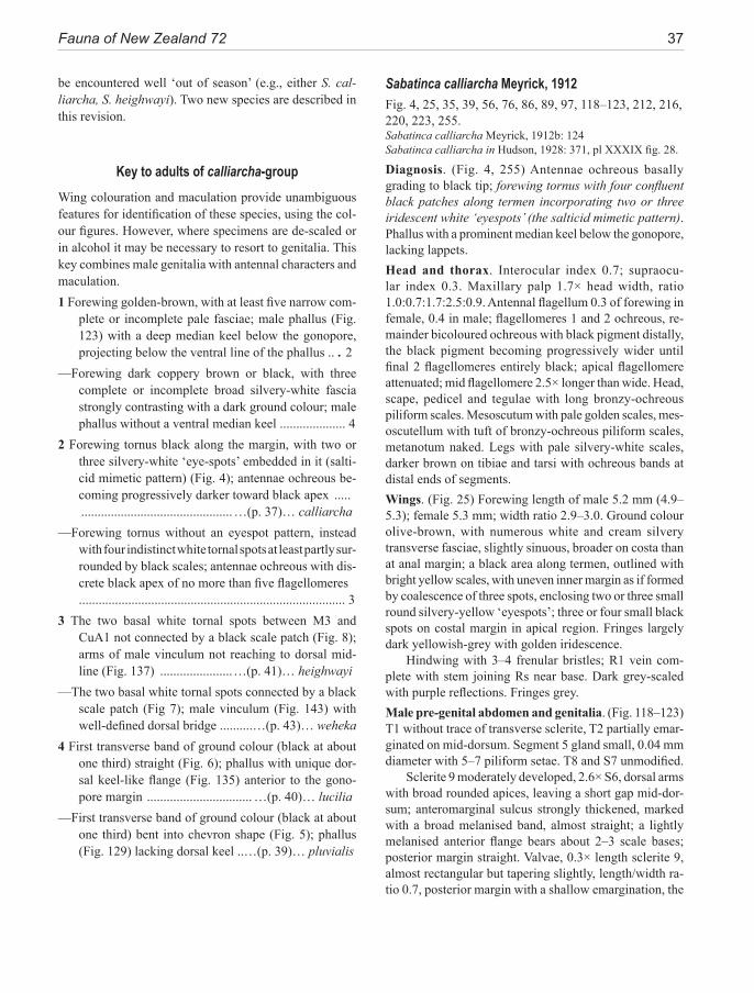
Fauna of New Zealand 72 37
be encountered well ‘out of season’ (e.g., either S. cal-liarcha, S. heighwayi). Two new species are described in this revision.
Key to adults of calliarcha-group Wing colouration and maculation provide unambiguous features for identification of these species, using the col-our figures. However, where specimens are de-scaled or in alcohol it may be necessary to resort to genitalia. This key combines male genitalia with antennal characters and maculation.1 Forewing golden-brown, with at least five narrow com-
plete or incomplete pale fasciae; male phallus (Fig. 123) with a deep median keel below the gonopore, projecting below the ventral line of the phallus .. . 2
—Forewing dark coppery brown or black, with three complete or incomplete broad silvery-white fascia strongly contrasting with a dark ground colour; male phallus without a ventral median keel .................... 4
2 Forewing tornus black along the margin, with two or three silvery-white ‘eye-spots’ embedded in it (salti-cid mimetic pattern) (Fig. 4); antennae ochreous be-coming progressively darker toward black apex ..... ..............................................…(p. 37)… calliarcha
—Forewing tornus without an eyespot pattern, instead with four indistinct white tornal spots at least partly sur-rounded by black scales; antennae ochreous with dis-crete black apex of no more than five flagellomeres ................................................................................. 3
3 The two basal white tornal spots between M3 and CuA1 not connected by a black scale patch (Fig. 8); arms of male vinculum not reaching to dorsal mid-line (Fig. 137) ......................…(p. 41)… heighwayi
—The two basal white tornal spots connected by a black scale patch (Fig 7); male vinculum (Fig. 143) with well-defined dorsal bridge ..........…(p. 43)… weheka
4 First transverse band of ground colour (black at about one third) straight (Fig. 6); phallus with unique dor-sal keel-like flange (Fig. 135) anterior to the gono-pore margin ................................ …(p. 40)… lucilia
—First transverse band of ground colour (black at about one third) bent into chevron shape (Fig. 5); phallus (Fig. 129) lacking dorsal keel ..…(p. 39)… pluvialis
Sabatinca calliarcha Meyrick, 1912Fig. 4, 25, 35, 39, 56, 76, 86, 89, 97, 118–123, 212, 216, 220, 223, 255.Sabatinca calliarcha Meyrick, 1912b: 124Sabatinca calliarcha in Hudson, 1928: 371, pl XXXIX fig. 28.
Diagnosis. (Fig. 4, 255) Antennae ochreous basally grading to black tip; forewing tornus with four confluent black patches along termen incorporating two or three iridescent white ‘eyespots’ (the salticid mimetic pattern). Phallus with a prominent median keel below the gonopore, lacking lappets.Head and thorax. Interocular index 0.7; supraocu-lar index 0.3. Maxillary palp 1.7× head width, ratio 1.0:0.7:1.7:2.5:0.9. Antennal flagellum 0.3 of forewing in female, 0.4 in male; flagellomeres 1 and 2 ochreous, re-mainder bicoloured ochreous with black pigment distally, the black pigment becoming progressively wider until final 2 flagellomeres entirely black; apical flagellomere attenuated; mid flagellomere 2.5× longer than wide. Head, scape, pedicel and tegulae with long bronzy-ochreous piliform scales. Mesoscutum with pale golden scales, mes-oscutellum with tuft of bronzy-ochreous piliform scales, metanotum naked. Legs with pale silvery-white scales, darker brown on tibiae and tarsi with ochreous bands at distal ends of segments.Wings. (Fig. 25) Forewing length of male 5.2 mm (4.9–5.3); female 5.3 mm; width ratio 2.9–3.0. Ground colour olive-brown, with numerous white and cream silvery transverse fasciae, slightly sinuous, broader on costa than at anal margin; a black area along termen, outlined with bright yellow scales, with uneven inner margin as if formed by coalescence of three spots, enclosing two or three small round silvery-yellow ‘eyespots’; three or four small black spots on costal margin in apical region. Fringes largely dark yellowish-grey with golden iridescence.
Hindwing with 3–4 frenular bristles; R1 vein com-plete with stem joining Rs near base. Dark grey-scaled with purple reflections. Fringes grey.Male pre-genital abdomen and genitalia. (Fig. 118–123) T1 without trace of transverse sclerite, T2 partially emar-ginated on mid-dorsum. Segment 5 gland small, 0.04 mm diameter with 5–7 piliform setae. T8 and S7 unmodified.
Sclerite 9 moderately developed, 2.6× S6, dorsal arms with broad rounded apices, leaving a short gap mid-dor-sum; anteromarginal sulcus strongly thickened, marked with a broad melanised band, almost straight; a lightly melanised anterior flange bears about 2–3 scale bases; posterior margin straight. Valvae, 0.3× length sclerite 9, almost rectangular but tapering slightly, length/width ra-tio 0.7, posterior margin with a shallow emargination, the
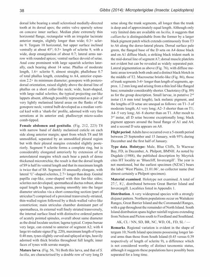
38 Gibbs (2014): Micropterigidae (Insecta: Lepidoptera)
dorsal lobe bearing a small sclerotised medially-directed tooth at its dorsal apex; the entire valve sparsely setose on concave inner surface. Median plate extremely thin horizontal flange, rectangular with an irregular laciniate anterior margin, slightly longer than wide; 0.3× scler-ite 9. Tergum 10 horizontal, but upper surface inclined ventrally at about 45°; 0.5× length of sclerite 9, with a wide, deep emargination of about 0.7; lateral arms nar-row with rounded apices; ventral surface devoid of setae. Anal cone prominent with large squarish sclerites later-ally, each bearing about 14 setae. Phallus of moderate length, 1.6× sclerite 9, almost straight; phallobase 0.7 of total phallus length, extending to A4, anterior expan-sion 2.2× its minimum diameter; gonopore with postero-dorsal orientation, raised slightly above the dorsal line of phallus on a short collar-like neck; wide, heart-shaped, with large radial sclerites; the typical projecting ear-like lappets absent, although possibly represented as a pair of very lightly melanised lateral areas on the flanks of the gonopore neck; ventral bulb developed as a median verti-cal keel with a ‘shark-fin’ profile, adorned with very fine serrations at its anterior end; phallocrypt micro-scales comb-tipped.Female abdomen and genitalia. (Fig. 212, 223) T8 with narrow band of darkly melanised cuticle on each side along anterior margin; apart from which T8 and S8 unmodified, separated by an unmodified pleural region but with their pleural margins extended slightly poste-riorly. Segment 9 sclerite forms a complete ring, but is considerably elongated anteriorly by extension of its anterolateral margins which each bear a patch of dense thickened microtrichia; the result is that the dorsal length of S9 is half its ventral length and that its maximum length is twice that of S8. Segment 10 unusually elongate, with lateral ‘U’-shaped sclerites, 2.7× longer than deep. Genital papilla cup-like, cone-shaped with thin fan-like sides; sclerites not developed; spermathecal ductus robust, about equal length to lagena, passing smoothly into the larger diameter utriculus via a short connecting section (part of utriculus?) comprised of a proximal transversely-striated, thin-walled region followed by a thick-walled valve-like constriction; main utriculus chamber dominant part of spermatheca, its external wall finely striated transversely, the internal surface lined with distinctive ordered pattern of acutely pointed spinules, overall about same diameter as the distal loculate section; lagena typical. Corpus bursae very large, can extend to anterior of segment A2; with 4 large tri-radiate signa (Fig. 220), maximum length of tynes 0.48 mm, tynes slightly curved and splayed at tips, heavily adorned with thick bristles throughout full length; inner faces of tynes with serrate margin.Mature larva. (Fig. 56, 76) Both this larva, and that of S. lucilia, are characterised by a double row of very long D
setae along the trunk segments, all longer than the trunk is deep and of approximately equal length. Although only very limited data are available on lucilia, it suggests that calliarcha is distinguishable from the former by a larger black pigment patch which extends continuously from T3 to A6 along the dorso-lateral pleura. Dorsal surface pale green, the flanged base of the D seta on A4 dense black and on A1 diffuse black; a striking black trident mark in the mid-dorsal line of segment A7; dorsal muscle platelets not evident but can be revealed as widely separated pair. Lateral pigmentation as above (Fig. 76), with diffuse me-lanic areas towards both ends and a distinct black blotch in the middle of T2. Macrosetae bristle-like (Fig. 86), those of trunk segments 3-4× longer than length of segments, ap-prox. 1.2 mm long and arising from a thin leaf-like flanged base; remainder considerably shorter. Chaetotaxy (Fig. 89) as for the group description. Small larvae, in putative 2nd instar (1.6 mm total length), lack melanic pigment, and the lengths of D setae are unequal as follows: on T1–3 of moderate length; A1 very long; A2–3 shorter than on T1; A4–5 very long; A6–8 shorter than on T1. After moult to 3rd instar, all D setae become exceptionally long; black pigment appears around the basal flange of A1 and A4; and a second D seta appears on A8.Flight period. Adults have occurred over a 5-month period between 25 September and 15 January, with 95% during December and the first half of January.Type data. Holotype: Male, Blue Cliffs, Te Waewae Bay, FD, in December, A. Philpott, BMNH. As noted by Dugdale (1988), the published description by Meyrick cites HT locality as ‘Bluecliff, Invercargill’. The year is not mentioned, but the earliest specimen (NZAC) bears the label ‘West Plains, 21.01.06’, no collector name (but almost certainly a Philpott specimen).Material examined. Holotype not examined. A total of 27♂, 8♀, distributed between Great Barrier Island and Invercargill. Localities listed in Appendix 1.Distribution. A very widespread species with a strongly disjunct pattern. Northern populations occur on Waitakere Ranges, Great Barrier Island and the Coromandel Ranges, with a gap throughout the remainder of North Island. South Island distribution spans higher rainfall regions extending from Nelson and Picton south to Fiordland and Southland.
AK, CL / NN, SD, BR, NC, WD, OL, FD, SL / —Remarks. Regional variation is evident in the shape of tergum 10; North Island specimens possessing longer lat-eral arms than those from South Island (0.45 versus 0.39 respectively of length of sclerite 9), a difference which is not considered worthy of distinct taxonomic status, although it suggests these populations have possibly been separated for a long time.
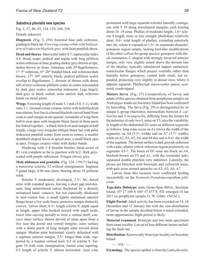
Fauna of New Zealand 72 39
Sabatinca pluvialis new speciesFig. 5, 57, 86, 93, 124–129, 244, 256.Female unknownDiagnosis. (Fig. 5, 256) Antennal base pale ochreous, grading to black tip. Forewing creamy-white with bold pat-tern of iridescent blackish-grey, with faint purplish sheen.Head and thorax. Interocular index 0.7; supraocular index 0.4. Head, scape, pedicel and tegula with long piliform scales ochreous at base grading darker grey-brown at tips; darker brown on frons. Antennae with 29 flagellomeres; 1st–5th ochreous, 6th–26th banded black and ochreous/dark brown, 27th–29th entirely black; pedicel piliform scales overlap to flagellomere 7. dorsum of thorax with dense tuft of erect ochreous piliform scales in centre surrounded by dark grey scales, somewhat iridescent. Legs largely dark-grey or black scaled, some narrow dark ochreous bands on tarsal joints.Wings. Forewing length of male 5.1 mm (5.0–5.1), width ratio 3.1. Ground colour creamy-white with bold blackish maculation; first fascia a broad blackish curved band from costa to anal margin at one quarter; remainder of wing from half to near apex with irregular black fascia in three parts but linked together—a black blotch on anal margin at half length, a large very irregular oblique black bar with paler iridescent purplish centre from costa to tornus, a smaller dumbbell-shaped fascia in apical area; a small black spot at apex. Fringes creamy-white with darker bands.
Hindwing with 3–4 frenular bristles; basal sector of R1 vein complete as far as junction with Sc2. Dark grey-scaled with purple reflections. Fringes silvery grey.Male abdomen and genitalia. (Fig. 124–129) T1 lacking a transverse sclerite; T2 without emargination. Segment 5 gland large, 0.06 mm diam, bearing about 16 piliform setae.
Sclerite 9 moderately developed, 2.5× S6, dorsal arms with rounded apices, leaving a short gap mid-dor-sum; long anterolateral sulcus thickened by a densely melanised band, concave, but not especially thickened in mid-ventral line, a small lightly melanised anterior flange bears a few scale bases; posterior margin distinctly convex. Valves short, 0.3× length sclerite 9, depth equal to length, upper lobe hooked inward with small tooth, lower lobe curving mesally to form a ventral shelf; con-cave inner surface almost devoid of setae apart from a few near the dorsal and ventral margins; outer surface with a dense patch of long straight setae toward distal margin. Median plate horizontal, clearly delimited with a sagittate anterior margin, 2.5× longer than wide, sup-ported by a median vertical keel; 0.2 of sclerite 9. Ter-gum 10 with wide emargination, lateral arms tapering, 0.5 length of sclerite 9, almost horizontal. Anal cone
prominent with large squarish sclerites laterally, contigu-ous with T 10 along dorsolateral margins, each bearing about 16–18 setae. Phallus of moderate length, 1.6× scle-rite 9 length, more or less straight; phallobase relatively short, 0.6× total length of phallus, extending anteriorly into A6, where it expands to 1.9× its minimum diameter; gonopore region simple, lacking keel-like modifications of the other calliarcha-group species; gonopore with dor-sal orientation, C-shaped with strongly recurved anterior margin, only very slightly raised above the dorsum line of the phallus; typically sabatincoid lappets melanised, semicircular flanges which project ventrally rather than laterally below gonopore; ventral bulb small, not ex-panded, projecting very slightly in dorsal view, where it appears squarish. Phallocrypt micro-scales sparse, scat-tered, comb-tipped.Mature larva. (Fig. 57) Conspecificity of larvae and adults of this species obtained from periphyton samples on Nothofagus trunks on Secretary Island has been confirmed by barcoding. The larva (Fig. 29) is distinguished by its unique L-group chaetotaxy intermediate between S. cal-liarcha and S incongruella, differing from the former by the presence of only two L setae on T3; also the variability in length of the abdominal D1 setae on different segments as follows: long setae occur on A1 (twice the width of the segment), on A4 (5.5× width) and on A7 (3.7× width); while on A2, A3, A5, A6, and A8 this seta is 0.6× the width of the segment. The dorsal surface is dark greyish-ochreous with a pale, almost yellow-ochreous region posteriorly on segments A5-7. The bases of D1 setae are black on A4, blackish-brown on T3 and A1, with the remainder pale; separated double platelets very indistinct. Laterally, the pleura are blotched with brownish and yellowish areas, with pale areas around spiracles on A2, A3, A6, A7.
Larvae from this location were confirmed feeding successfully on the liverwort Pseudomarsupidium pilif-erum.Type data. Holotype: male, Grono Spur, 860 m., Secretary Island, 45º 17.108´S 166º 57.677´E, FD, emerged 18 Jan 2011 ex periphyton sample, G. W. Gibbs. (NZAC).Flight Period. Adult activity has been recorded on 14–18 December and 12 January but with the size-distribution of larvae in the sample decribed below a much extended, more opportunistic flight period is likely.Material examined. Holotype and two male specimens from same locality. Larvae of four different instars includ-ing the final one.Distribution. Known only from type locality on Secretary Island.
— / FD / —Etymology. The species epithet is from the Latin pluvialis,
40 Gibbs (2014): Micropterigidae (Insecta: Lepidoptera)
meaning ‘of the rain’; it refers to the prodigious rainfall of western Fiordland where an annual precipitation of 5–7 m is a normal event; in this case allowing liverworts to flourish well above ground level on tree trunks where the larvae of this species occur.Remarks. This species was discovered by John Grehan in January 1982 on the Grono Spur ridge track above Gut Hut, Secretary Island. The male moth occurred on a rocky outcrop surrounded by stunted silver beech forest near the treeline at 780 m. At about the same time a distinctive larva was extracted by C. F. Butcher from a bryophyte sample obtained from the same track, clinging to Nothofagus trunks at 860 m in this high rainfall environment, and putatively assigned to this species. In December 2011 another bryophyte sample from the same site yielded further larvae and two male adults.
Sabatinca lucilia Clarke, 1920Fig. 6, 130–135, 213, 224, 257.Sabatinca lucilia, Clarke, 1920: 35.Sabatinca lucilia in Hudson 1928: 371, pl. XLIX fig. 9.
Diagnosis. (Fig. 6, 257) Antennae pale ochreous with black tips. Forewings with bold pattern of coppery-brown, black and creamy-white fascia, dominated by a broad transverse whitish band with a sinuous distal margin, edged with black, at mid-length. Male distal phallus with erect mid-dorsal toothed keel.Head and thorax. Interocular index 0.8; supraocular index 0.4 Maxillary palp ratio 1.0:0.9:2.1:2.5:1.0; 1.7× head width. Labial palps 3-segmented. Long piliform scales of head, scape, pedicel and thorax ochreous tinged with pink; those of pedicel reaching to 9th flagellomere. Antennal flagellum 0.3 of forewing length in female, 0.4 in male; with 27–28 flagellomeres in male, 23–24 in female; all brownish-ochreous except 2 or 3 at tip which entirely black; terminal flagellomere attenuated. Mesoscutum scales coppery-brown. Legs ochreous towards base but dark-brown scaled on tibiae and tarsi, with paler bands at distal extremities of joints.Wings. Forewing length of male 5.1 mm (4.6–5.4); female 5.1 (4.9–5.4); width ratio 2.9. Ground colour rich reflec-tive coppery orange-brown, grading to black around the fasciae; a broad transverse creamy-white fascia at mid-length, heavily edged with black scales; a second slightly narrower transverse creamy-white fascia at three quarters, interrupted in middle; apical area creamy-white. Fringes largely pale ochreous, but black arising from dark regions of maculation.
Hindwing with 3 frenular bristles; R1 vein complete to junction with Sc2, stem joining Rs basally.
Male abdomen and genitalia. T1 lacking a transverse sclerite, T2 fully melanised. Segment 5 gland small, 0.04 mm diameter with 8 piliform setae. T8 and S7 unmodified.
Sclerite 9 moderately developed, 2.4× S6, dorsal arms with broad rounded apices, leaving a moderately wide gap mid-dorsum; anteromargin sulcus thickened and densely melanised, slightly concave, with a small lightly melanised anterior flange present bearing a few scale bases; posterior margin slightly convex; outer sur-face densely adorned with scale bases. Valves small, 0.3× length sclerite 9, tapering, length/width ratio equal, upper lobe incurved but without a tooth on lower lobe, forming a shelf ventrally; convex outer surface densely covered with long posterior-directed setae, concave inner surface with sparse curved retro-setae. Median plate a thin hori-zontal flange, 2× longer than wide with an extremely thin laciniate anterior margin, 0.3 of sclerite 9. Tergum 10 with wide, deep emargination of 0.7, lateral arms narrow with apices very slightly inturned, 0.6 length of sclerite 9; ventral surface devoid of setae. Anal cone prominent with large squarish sclerites laterally, each bearing 16–17 setae. Phallus (Fig. 133–135) of moderate length, 1.9× sclerite 9, more or less straight; phallobase 0.8 of total phallus length, extending forward into A3 where expand-ed to 1.9× its minimum diameter; gonopore region with one median and two pairs of ornate keel-like flanges—an erect mid-dorsal flange immediately anterior to gonopore, which bears a large blunt posteriorly-directed ‘tooth’; a pair of dorso-lateral melanised flanges project laterally, their margins with 4 or 5-toothed serrations, running an-teriorly to the level of the dorsal flange and possibly rep-resenting lappets; and finally a pair of smoothly-rounded ventro-lateral flanges on the base of the neck region. The gonopore neck region raises it above the dorsal line of the phallus; heart-shaped, but compressed laterally (i.e., with concave lateral margins), and with a strongly in-folded anterior margin. Phallocrypt micro-scales comb-tipped.Female abdomen and genitalia. Fig. 213, 224) Abdo-men elongate, length/depth ratio 2.3; T1 without sclerites in dorsum but lightly sclerotised laterally, T2 normal; S2 with a small rectangular sclerite in mid-line. Anterolateral margins of T8 expanded and telescoped into T7, with a thickened darkly melanised margin: S8 unmodified but re-duced in width to 0.8 of S7; pleural area narrow but without modifications. Segment 9 a complete ring, extended into two anterolateral lobes where telescoped into segment 8, with thickened and melanised margin similar to that of T8; mid-ventral length 1.8× that of S8. The cuticle of both T8 and segment 9 anterolateral extended areas is adorned with radial patterns of dense modified microtrichia. Segment 10 elongate; sclerites U-shaped with anterior arms splayed
Fauna of New Zealand 72 41
slightly at anterior end, about 3× longer than high. Genital papilla dominated by a cylindrical, coiled, tapering cup supported posteriorly by the dorsal arms of two lateral, lightly sclerotized flanges ventral to the cup; spermathecal ductus robust but of variable length depending on degree of extension (compressed about equal length to lagena), passing smoothly into the larger diameter utriculus via a short connecting section (part of utriculus?) comprised of a proximal transversely-striated, thin-walled region followed by a thick-walled valve-like constriction; main utriculus chamber slightly swollen, its external wall finely striated transversely, the internal surface lined with dense array of ridges and scutes, overall about same diameter as its distal bulbous loculate section; lagena typical. Corpus bursae large, reaching to posterior margin of A4; with 4 large tri-radiate signa, maximum length of tynes ca 0.5 mm, each almost straight, not splayed at tips, densely adorned with thick bristles throughout full length; basal connecting bar free of bristles and similar in form to Fig. 220 (calliarcha).Larva. The larva is of the same type as S. calliarcha (i.e., with identical chaetotaxy and the same long D1 setae). Specimens found at Subritsky’s bush, close to Auckland (Kuschel, March 1975), and at Waipoua Forest (Craw 1985) are assumed to represent 4th and 3rd instars of S. lucilia respectively. The Waipoua specimen is almost without pigmentation but the Auckland specimen suggests that the species can be distinguished by the absence of dark lateral pleural pigmentation on segments A2-3, i.e., there is a gap in the dark lateral patch. As far as can be judged from these specimens, the pigmentation is prob-ably similar to S. calliarcha but the presence of a trident patch on dorsum of A7-8 cannot be confirmed or denied. The D1 setae of abdomen are about 4× dorsal length of segments, and arise from posterior angle of large fan-like dorsal flanges as in calliarcha.Flight period. This species has occurred over five months between the end of November and early March, with 80% during the four weeks between the end of November and mid December.Type data. Lectotype: Male. Kauri Gully, Northcote, Auckland, 10/01/19, labelled ‘Sabatinca lucilia Clarke Type T. LII 1919’; with the AMNZ label #21790. Clarke (1920:35) noted localities as: ‘Waitomo Caves Hotel, Dec, 1915, at hotel lights’ and ‘Kauri Gully, second week of January, 1919’ but did not designate a Type. Later, Clarke (1934:16) stated: ‘The type I netted, in 1915, at the Waitomo Caves.’ Since the Waitomo specimen has been lost, there is no ambiguity with Kauri Gully specimens, and the Type label is in Clarke’s hand, the specimen is here designated as a lectotype. AMNZ.
Material examined: Lectotype and 33 non-type speci-mens. Localities listed in Appendix 1.Distribution. Northern half of North Island, north of the ‘Taupo line’;
ND, AK, CL, WO, BP, TO / — / — Found on moist ferny banks in a wide variety of for-
est types from kauri and kanuka to Nothofagus.Remarks. Irregular in its occurrence, this species is no-table for being collected in quite large numbers at certain localities, but not reappearing on a regular seasonal basis. It has been taken at light on a number of occasions.
Sabatinca heighwayi Philpott, 1927Fig. 8, 31, 50, 59, 61, 62, 86, 136–141, 214, 219, 225, 250, 258.Sabatinca heighwayi Philpott, 1927: 90Sabatinca heighwayi in Hudson 1939: 471, pl. LXI fig. 32
Diagnosis. (Fig. 8, 258) Large size (fwl >6 mm); pale reddish-brown fw with brassy reflections and a number of narrow transverse whitish fasciae, incomplete in terminal area. Combination of bulbous distal phallus with absence of a dorsal ‘bridge’ on vinculum.Head and thorax. Interocular index 0.6; supraocular index 0.4. Maxillary palps 5-segmented; long, 2.0× head width with ratios 1.0:0.7:1.8:2.5:0.8. Labial palps 3-segmented. Antennae 0.3 of wing length in both sexes; scape 3.5× diameter of 1st flagellomere; male with 24 flagellomeres, female 22; filiform, 7th is 1.8× longer than wide; terminal flagellomere attenuated. Pedicel piliform scales reach-ing to at least 9th flagellomere. Long piliform scales of head, scape, pedicel and tegulae pale yellowish; antennal flagellomeres all ochreous, apart from black tip of 4-5 segments. Mesoscutum clad with brassy scales. Femora creamy-white, tibiae and tarsi dark brown-scaled with some paler bands.Wings. Forewing length of male 6.1 mm (5.9–6.3); female 6.2 mm (5.9–6.4); width ratio 3.0. Ground colour reflective golden-brown; basal third with scattered small patches of white scales; remainder with four more or less distinct parallel whitish transverse fasciae arising from costa; first fascia at one third, a whitish bar obliquely toward apex, edged with dark brown scales, either terminating about middle of wing or extending to anal margin; second fascia incomplete but arises similarly from about half length from costa; third fascia indistinct, at about two thirds; fourth almost straight across wing but broken in middle; apex with about five marginal white patches edged with brown. Fringes ochreous or pale brown, ochreous bands extending from white patches.
42 Gibbs (2014): Micropterigidae (Insecta: Lepidoptera)
Hindwing with 4 frenular bristles; vein R1 complete, its stem joining RS basally. Dark-grey with purple reflec-tions, fringes silvery-grey.Male abdomen & genitalia. (Fig. 136–141) T1 lacking a transverse sclerite, T2 fully melanised, without emargina-tion. Segment 5 gland small, 0.04 mm dia with 5 piliform setae. T8 and S7 unmodified.
Sclerite 9 moderately developed, 2.3× S6, dorsal arms with very broad rounded apices, leaving a narrow gap mid-dorsally; anteromarginal sulcus strongly thick-ened and darkly melanised, slightly concave, appearing especially heavily thickened in ventral mid-line; a large lightly melanised anterior flange present, bearing a few microsetae. Valves short, 0.3× length sclerite 9, nearly twice as deep as long, upper lobe hooked inward with a sharp apex but not developed as a mucron, lower lobe curving mesally to form a ventral shelf; convex outer surface densely adorned with long straight posteriorly-directed piliform setae, concave inner surface with short curved retro-setae, sparse. Median plate horizontal, very large and fan-shaped, almost as wide as long, anterior margin extremely thin and irregular with a delicate la-ciniate margin; 0.3 of sclerite 9. Tergum 10 with wide emargination of 0.9, lateral arms relatively narrow with rounded apices, 0.5 length of sclerite 9; ventral surface devoid of setae. Anal cone with large squarish scler-ites laterally, each bearing about 8 setae. Phallus (Fig. 139–141 of moderate length, 1.8–1.9× sclerite 9, almost straight; phallobase 0.7× total phallus length, extending forward to posterior margin of A3, where expands 2.2× minimum diameter; distal phallus complexly modified, swollen and bulbous in relation to phallobase, somewhat flattened anteriorly; gonopore postero-dorsally extended on a narrower tubular neck, the aperture crescentic due to an overarching anterior margin; ventral bulb devel-oped as a deep median ventral keel projecting posteriorly slightly beyond the gonopore where it is divided; a pair of elongated lateral flanges run along the sides, project-ing beyond the gonopore as slightly upturned lateral horns, smooth, without serrations; these lateral flanges, which are perhaps derived from the groundplan sabatin-coid lappets, extend anteriorly to terminate on the widest part of the distal phallus bulb in a small spine. A curved transverse ‘shoehorn-shaped’ diaphragm joins the lat-eral flanges of each side at their mid-point (narrowest) to the rim of the gonopore (appearing as an H-shaped frame when viewed ventrally). Anterior phallocrypt mi-cro-scales sparse, simple acute teeth, although typical comb scales numerous towards posterior.Female abdomen and genitalia. (Fig. 214, 225) Segment 5 gland with only 1-3 piliform setae. Segment 8 massive, heavily melanised, forming a complete ring which abruptly reduces the diameter of abdomen; pleural area specialised,
with anterior emargination and a lozenge-shaped pleural pocket on its sclerotised posterior margin—the pocket in the form of an oval depression with smooth lining on its concave surface. Segment 9 not extendable, essentially a ring sclerite but with melanisation reduced along the dorsal mid-line, leaving only a very narrow sclerite postero-dorsally with a cluster of setae on its dorsal mid-line; a distinctive pair of lateral setose process, project-ing towards posterior from ventral region. Segment 10 sclerites moderately long, about 1.5× longer than deep. Genital papilla region at entrance to corpus bursae with a massive ‘winged’ U-shaped sclerite at base of bursa copulatrix, its extremities extending posteriorly as flanges to support the sides of the papilla cup; spermatheca with long thick-walled ductus, of equal length to lagena; passing smoothly into the larger diameter utriculus via a short con-necting section (part of utriculus?) comprised of a proxi-mal transversely-striated, thin-walled, slightly expanded region followed by a thick-walled valve-like constriction; main utriculus chamber slightly wider, parallel-sided, its external wall finely striated transversely, the internal surface lined with dense array of acute spinules; loculate distal bulb of utriculus separated from main chamber by a constricted neck section which extends into the main sac a considerable distance. Corpus bursae large, reaching to anterior margin segment A4, with 4 large tri-radiate bris-tled signa (Fig. 219), longest tynes ca. 0.6 mm.Larva. Larvae of the pair of sister species heighwayi and weheka (Fig. 58, 75) do not have the long D setae of the above calliarcha-group species, instead, their trunk D setae are 0.27 mm or 0.7× segment length. Mature larvae have been recovered from the foliose liverwort, Plagiochila circumdentata, that grows on rock surfaces in humid ravines, and reared to adult, thus confirming their identity and the fact that this species normally overwinters as a diapause pupa. Their pigmentation and overall habitus are indistinguishable from S. weheka apart from the absence of the small L3 seta on T3 of S. heighwayi. Dorsal surface yellowish-green with a pattern of of yellowish chevron marks, strongly lobed in a regular segmental pattern which includes the leaf-like basal lobes of the D setae; on some specimens dark pigment areas are conspicuous on the dorsum of T1 and T2 and the basal flanges of the D setae on A4 and A7; platelets inconspicuous; lateral pleura relatively uniform yellowish-green with a series of small oblique bicoloured (white above and black beneath) patches dorsad of the spiracles between A1–A7, larger and extending to the lateral fold on A1 and A4. Only confirmed host plant Plagiochila circumdentata.Flight period. For five months between the end of Septem-ber and mid-January, with 83% between mid-November and early December.
Fauna of New Zealand 72 43
Type data. Holotype: Female, Lesley Valley, Mt Arthur Tableland, 17 Nov, 1915, W. Heighway. Sabatinca heigh-wayi Philpott 1927: 90. NZAC.Material examined. Holotype and 11 males, 12 females. Localities listed in Appendix 1.Distribution. North-western South Island, south to Lewis Pass.
/ NN, BR, SD / — It occurs in dense moist forest (usually of southern
beech) from sea-level to 720 m.Remarks. One of the largest jaw-moth species (only matched by S. weheka and some congeners in New Cal-edonia) but very elusive, normally encountered only as isolated individuals from spring through until late summer. Winter passed in pupal stage.
Sabatinca weheka new speciesFig. 7, 58, 75, 92, 142–147, 215, 218, 226, 244, 245, 246, 251, 259.Diagnosis. (Fig. 7, 259) Large size (fwl ca 6 mm); fw with a number of narrow transverse whitish fasciae, dominated by a broad white fascia at 1/3 with kink toward anal margin, heavily shaded with dark brown scales on proximal side. A pair of unique posterior horns on ventral lobe of valvae.Head and thorax. Interocular index male 0.7; supraocu-lar index 0.4. Maxillary palps 5-segmented; long, 1.8× head width with ratios 1.0:0.6:1.5:2.1:0.8. Labial palps 3-segmented. Antennae 0.4 of wing length in male, 0.3 female; scape 4.0× diameter of 1st flagellomere; male with 28 flagellomeres, female 26; filiform, 7th is 1.7× longer than wide; terminal flagellomere attenuated; all ochreous apart from 2–5 black flagellomeres at tip, but terminal joint dark ochreous. Pedicel piliform scales reaching to 9th flagellomere. Long piliform scales of head, scape, pedicel and tegulae pale yellowish. Mesoscutum clad with brassy lamellar scales. Femora creamy-white, tibiae and tarsi dark brown-scaled with some paler bands.Wings. Forewing length of male 6.0 mm (5.8–6.2); female 5.9 mm (5.8–6.2); width ratio 2.9. Ground colour reflective golden-brown; basal third with scattered small patches of white scales; remainder with three more or less distinct parallel white transverse fasciae arising from costa; first fascia at one third, complete, a broad shining white bar almost straight in costal half but kinked towards anal mar-gin, strongly margined with dark brown scales on proximal (but not on distal) side; second fascia white at about ½, incomplete, narrow and irregular; third fascia at 2/3, of two separate white spots from costa, edged with dark brown scales; apical area margined by white spots - three small
white spots along costa (including at apex) and four larger marginal white spots along termen, heavily edged with black, often conjoined. Fringes brownish-ochreous with pale ochreous bands extending from white spots. Hindwing with 4 frenular bristles; vein R1 complete, its stem joining Rs basally. Pale grey basally, darkening towards apex, with purple reflections, fringes silvery-grey apart from a band of ochreous scales at 2/3 along anal margin.Male abdomen and genitalia. (Fig. 142–147) T1 lacking a transverse sclerite, T2 fully melanised, without emar-gination. Segment 5 gland absent. T8 and S7 unmodified.
Sclerite 9 moderately developed, 2.7× S6, dorsum connected by a moderate melanised bridge bearing a sin-gle sinuous row of setae; anteromarginal sulcus thickened and darkly melanised, concave, heavily thickened in ven-tral mid-line; a narrow tapering anterior flange present, bearing a few microsetae. Valves short, 0.5× length scle-rite 9, upper extremity not recurved, adorned terminally with a roughened cuticular texture along the posterior margin, lower extremity with projecting posterior horn, acutely pointed and unique to this species (Fig. 144); convex outer surface densely clothed with long straight posteriorly-directed piliform setae, concave inner surface with short curved retro-setae, sparse. Median plate hori-zontal, wider than long, fan-shaped, anterior margin thin and irregular with a delicate laciniate margin; 0.3 length of sclerite 9. Tergum 10 robust, thickened in mid-line, a moderate emargination of 0.7, lateral arms relatively broad with blunt apices, 0.5 length of sclerite 9; a short transverse row of about 5 setae across the posterior mar-gin in the mid-line are the longest setae on the tergum; ventral surface devoid of setae. Anal cone sclerites prom-inent, each bearing about 11 stiff setae and clothed with dense microtrichia. Phallus (Fig. 145–147) of moderate length, 1.8× sclerite 9, almost straight; phallobase 0.7× total phallus length, extending forward into A4, where it expands 2.2× minimum diameter; distal phallus com-plex, modified similar to S. heighwayi; swollen to 3× diameter of phallobase, bulbous anteriorly, with the go-nopore extended posteriorly on a long neck, its aperture crescentic due to indented upper lip; ventral bulb shorter than gonopore, developed as a keel with a melanised me-dian supporting ridge which becomes divided anteriorly; a pair of flattened paddle-like lateral appendages on either side of the gonopore (Fig. 144), adorned on their outer side with some acute scale-like spines and termi-nating posteriorly with an inwardly hooked apex, pos-sibly representing a modification of lappets and certainly the equivalent of the lateral appendages of S. heighwayi; a curved interior diaphragm connects and supports the paddles anteriorly, ventral to the neck of the gonopore. Anterior phallocrypt micro-scales sparse, simple blunt or acute teeth.
44 Gibbs (2014): Micropterigidae (Insecta: Lepidoptera)
Female abdomen and genitalia. (Fig. 215, 218, 226) Segment 5 gland absent. Segment 8 massive with T8 and S8 joined by a sclerotised pleural pocket posteriorly, heavily melanised, with an anterior emargination in pleural region, forming a complete ring which abruptly reduces the diameter of abdomen; densely clothed with comb-edged microtrichia along anterior marginal areas of both T8 and S8; a pair of lateral semicircular lobes extend from the pos-terior margin of T8, overhanging the telescoped segment 9; pleural pocket vertically oval, heavily sclerotised along its anterior margin and lined with small scale-like pustules on the antero-dorsal concave surface. Segment 9 telescoped into segment 8, although slightly extendable; largely mem-braneous, with some vestigial melanisation, indicated as a diffuse dumb-bell shaped band across the ventral region; posterior region of segment with numerous macrosetae, the posteriormost forming a neat regularly-spaced row around the segment. Segment 10 ‘U’ sclerites moderately long, about 1.6× longer than deep, anterior ends splayed apart. Genital papilla with a massive, dense, ‘W-shaped’ sclerite at entrance to corpus bursae; spermatheca ductus wall spirally thickened; connecting section to utriculus expanded as pouch, thin-walled with transverse striations, followed by a short thick-walled ‘valve-like’ section; main utriculus sac the longest section, widest in middle, lined with dense needle-like spinules, loculate distal bulb separated from main chamber by a constricted neck which extends into the utriculus; lagena typical. Corpus bursae large, to middle of segment A5, with 4 large tri-radiate bristled signa, longest tynes ca. 0.4 mm.Larva. (Fig. 58, 75) It was the discovery of larvae that first revealed the existence of this species. This pale yellowish-green larva is superficially indistinguishable from its sister, S. heighwayi, differing only by the presence of the small L3 seta on segment T3. Dorsum yellowish-green with a pattern of yellowish chevron marks, conspicuously lobed in a regular segmental pattern which incorporates the leaf-like basal flanges of the D setae; platelets inconspicuous; lateral pleura the same uniform colour but with a series of oblique bicoloured patches (white above, black below), dorsad of the spiracles on segments A1–A7, the patches larger on segments A1 and A4, extending to the lateral fold; a more diffuse oblique patch of darker pigment on the upper pleura of A7 from the base of the D seta toward the spiracle. The foliose liverwort, Plagiochila deltoidea, which tends to grow above ground level on tree trunks or dead fallen branches in high rainfall forest, is the only known host plant.Flight period. Observed only from mid-October to the end of the month.Type data. Holotype: Male, Lake Matheson, 120 m, 43º
26.707´S, 169º 58.063´E, WD, 18 Oct. 2011, G.W. Gibbs. Emerged from a sample of periphyton predominately Plagiochila deltoidea. Paratypes: 12 males and 4 females from above locality, emerged as above 13–26 Oct, 2011; and caught at type locality 28 Oct, 2011, GW Gibbs.Material examined. Type material only.Distribution. Known as adults from only the type locality at Lake Matheson, WD but a small larva from Grono Spur, Secretary Island (FD) has confirmed another locality by barcode determination.
— / WD, FD / —Etymology. The local Maori name weheka (or wehenga in standard Maori) means literally ‘the parting of the ways’ and is derived for this species epithet from the name of the Department of Conservation Area Office at Fox Glacier. The word relates to the two rivers, Fox R. and Cook R., which separate adjacent to Lake Matheson. In a broader sense it could be taken to refer to the putative ancestral West Coast South Island Sabatinca species which has been separated into two widely allopatric sister populations, one northern (heighwayi) and one southern (weheka) at some time in its history.Remarks: Although this species closely resembles its sister species, S. heighwayi, in many features, its morphol-ogy is quite distinctive, representing a deep divergence confirmed by barcoding. This southern moth has a slightly darker appearance with more strongly marked maculation emphasised by the dark brown pigmentation in the ground colour. To date, known only from under forest canopy in wet high-rainfall forest near sea-level at Lake Matheson and at 860 m on Secretary Island, Doubtful Sound. Like its sister species, it overwinters as a pupa but this one has adopted a slightly different seasonal strategy by emerging very early in the season (end October) with mature larvae pupating in January.
The chrysargyra-groupDiagnosis. Labial palps 2-segmented; male tergum 10 a simple median lobe (e.g., Fig. 158); valves of male genita-lia equal to or longer than segment 9, bulbus ejaculatorius with a transverse terminal aperture; female segment 10 sclerite squarish, length:height ratio less than 1; female bursa lacking signa; spermatheca a long narrow duct.
Larva with a single L-setae on T2 and T3.Adult head. Head capsule with or without an epicra-nial sulcus. Interocular index 0.6–0.8; supraocular index
Fauna of New Zealand 72 45
0.3–0.4. Head capsule with long tufts of piliform scales. Maxillary palps long, 5-segmented; 4th segment longest, usually slightly curved, bearing fine transverse striae along most of its length; 5th segment short, 0.5–0.9× 1st segment. Labial palps 2-segmented. Antennae moderately long (0.5–0.6 of forewing length ♂), with 24–42 filiform or submoniliform flagellomeres; scape and pedicel greatly swollen, scape 2.6–4.0× diameter of first flagellomere; pedicel almost spherical; scape with median indentation on mesal surface; both scape and pedicel with dense tufts of long piliform scales (those on pedicel extending over first 2–4 flagellomeres).Thorax. Mesoscutum clad with closely fitting lamellar scales, but tegulae, posterior end of mesoscutum and mesoscutellum with tufts of long erect piliform scales. Protibia with well-developed epiphysis.Wings. (Fig. 27, 28) Wing venation as for incongruella-group except that in aurella, doroxena and bimacula the forewing veins RS3 and RS4 do not share a common stem (Fig. 27). Where the common stem is present, it can vary between very short (as in ianthina at 0.1× the distance between end of the ‘cell’ and the wing apex) and long (0.5× this distance in aemula and caustica). Long-stemmed apical veins appear to be associated with attenuation of the wing apex, whereas loss of the stem correlates with broadening of the apical area. In the hindwing, R is al-ways coalescent with Sc over its distal section beyond the fork; but basally can exist either as a separate vein from Sc, or represented only by a recurrent stub at the fork of Sc+ R, or totally absent (coalesced with Sc throughout?). Chrysargyra-group species exhibit all three conditions. Hindwing with 2–4 frenular bristles.Pre-genital abdomen. Abdomen variable in cross-section but usually more or less circular. T1 membraneous apart from lateral margins (4 spp.) with variable presence of a narrow transverse melanised sclerite across the central region (3 spp.) which can be reduced to small lateral remnants (3 spp.). T2 sclerite membraneous anteriorly in mid-line, but may be completely divided into two lateral plates (1 sp.). Segment 5 glands present in both sexes, sabatincoid in form, i.e., with a raised, more-or less cylin-drical prominence, diameter 0.03–0.07 mm, bearing 5–16 long piliform scales. In male, T7 unmodified (although reduced in aurantissima) but T8 reduced in most species; no trace of S8.Male genitalia In repose, the genitalia are usually tilted upward, very conspicuously in some. Sclerite 9 highly variable in size, ranging from 1.4–2.7× longer than S6, with or without a dorsal ‘bridge’; dorsal arms absent in five species; anterior flange of anteromarginal sulcus absent. Tergum 10 a median lobe, forming the roof of the
genital atrium, shape ranging from short and spatulate to extremely attenuated, sometimes (3 spp.) with a very small median indentation or division of the apex. Anal cone with a pair of lateral sclerites, not clearly delimited and usually free in the membrane except in three species where they articulate with the lateral extremities of the tergum. Valves simple, lacking branches or processes of any kind, but with divided apex in three species; mesal surface bearing a dense patch of thick retro-setae toward distal extremity. Median plate a prominent horizontal flange, squarish, ante-rior margin smooth, not laciniate. Phallus with long to very long phallobase (phallobase: distal phallus ratio 0.6–0.9), often looped within the abdomen, with a wide cone-shaped bulbus ejaculatorius 2–8× diameter of narrowest region, the aperture transverse, not oblique; gonopore with dorsal or terminal orientation, basically heart-shaped but often with a greatly attenuated ventral lip, sometimes with an over-arching dorsal lip, the gonopore bordered by sclero-tised radial folds and supported by a sclerotised ‘frame’ which bears small lappets at its dorso-lateral corners and in some (5 spp.) an additional pair of ventral sclerotised spines below gonopore. Phallocrypt adorned with dense micro-scales of various form, darkly pigmented in some.Female genitalia. T8 and S8 always separated by a mem-braneous pleural region, S8 often (5 spp.) with a thickened transverse rib across midventral-line (stains heavily with chlorazol black) and extending towards the lateral margin. Segment 9 forming a complete ring, but usually reduced dorsally as a result of an anterior emargination; normally unmodified, but in two species with a transverse rib similar to that described above for S8. Anterior border of segments 8 and 9 sclerites microtrichiated but remainder smooth. Segment 10 not greatly extendable (except 3 spp.), lat-eral sclerites usually equidimensional or slightly higher than long, weakly melanised anteriorly, never U-shaped. Genital papilla orientation variable, never with a melanised sclerite. Spermatheca in three sections—short, relatively narrow, basal duct emerging from the papilla, with thick walls; a much longer thin-walled utriculus, variously spe-cialised into one, two or three distinct zones, one of which may be narrow and convoluted; a short loculate lagena. Corpus bursae of moderate to large size, lacking signa but sometimes with minor modifications in neck region.Larva. To date, mature larvae of nine chrysargyra-group taxa have been confidently associated with their respective adults, thus enabling a reasonably comprehensive descrip-tion of their features. Two widespread taxa, aemula and ianthina, have eluded capture and/or barcode detection. Larvae of this group are typical of the liverwort-feeding sa-batincoid type of larval Micropterigidae. The thoracic legs (Fig. 96) are extremely short (about 1.5× the basal diameter
46 Gibbs (2014): Micropterigidae (Insecta: Lepidoptera)
of femur), considerably shorter than calliarcha-group or incongruella-group larvae, with 4 bulbous micro-setae on femur and 3 on tibiotarsus, the most distal of which is longer and acutely pointed. Abdominal prolegs absent, re-placed by small paired elevations on the abdominal venter that have been described as cone sensilla, and interpreted as neither forerunners nor vestiges of abdominal prolegs in other Lepidoptera (Dupont, 2012). A basal inflatable ‘papilla’ of unknown function is present on the mesal side of each thoracic femur.
Larval taxonomy in this group is based on distinctive pigmentation patterns, chaetotaxy and dorsal platelets. The chrysargyra-group larvae can be distinguished from other Sabatinca larvae by the presence of only a single L group seta (Fig. 91) on thoracic segments T2 and T3 (other Sabatinca with 2 or 3). Based on the number of prothoracic L setae, larvae can be readily assigned to ei-ther the aurella-subgroup (Fig. 95, with 2 L setae and sin-gle dorsal platelets) or the chalcophanes-subgroup (Fig. 91, with 1 L seta and double dorsal platelets). The dor-sal macrosetae are thickened in all known species (Fig. 84, 85), giving the appearance of compound setae due to the ridged nature of the shaft. Their morphology is often valuable for discriminating species.Taxonomic history. Despite their diminutive size, short flying seasons and secretive behaviour, an early flurry of lepidopterological interest in the late 19th C and early 20th C led to the description of 12 New Zealand species in this group prior to 1924 (9 regarded valid in this revision). Their primitive status was known and clearly the race was on to find them. However, eighty years then elapsed with-out any further species being added, until recently, when collections from less accessible areas or at ‘unseasonal’ times outside the ‘accepted’ summer insect season have revealed two new western South Island species described in this revision.
Meyrick (1886) initially established the genus Pala-eomicra for chrysargyra, chalcophanes, and later (1888) doroxena, before realising the priority of Sabatinca Walker, which led him to establish the genus Micropar-dalis for doroxena (Meyrick, 1912:132:7). During the 1980’s the present author’s research on systematics and biogeography of SW Pacific Micropterigidae led him, without formal taxonomic revision, to re-employ Mey-rick’s Palaeomicra for species in the chrysargyra-group, in order to distinguish them from what he regarded as the strict Sabatinca species (Gibbs, 1983, 1989, 2006). Now, with molecular analysis and improved morphologi-cal understanding (Gibbs & Lees 2014), it is clear there are three major clades of Sabatinca in New Zealand and New Caledonia and, without more extensive molecular evidence, we are unable to give them equal status at this time. We believe this diversity is best expressed in terms
of species groups within the widely-known genus Sabat-inca.Distribution. The chrysargyra-group species are distrib-uted throughout New Zealand, but no single species is nationwide. Absent from New Caledonia.Remarks. Two monophyletic sub-groups of the chrysar-gyra-group can be recognised, distinguished by features of both larvae and adults and supported by COI bar-code analysis:
aurella-subgroup—male genitalia lacking a pair of prominent ventral spines below the gonopore; lar-vae with 5 macrosetae on the dorso-lateral ridge of the prothorax (Fig. 95), projecting above the head recess on each side (i.e., D2, XD1, XD2, L1, L2), dorsal platelets single. Included species: aurella, doroxena, ianthina, bi-macula, aenea, aemula, chrysargyra.
chalcophanes-subgroup—male genitalia with a pair of prominent ventral spines below the gonopore; larvae with 4 macrosetae on the dorso-lateral ridge of the pro-thorax (Fig. 91), projecting above the head recess on each side (i.e., with only 1 L seta), dorsal platelets dou-ble, widely separated. Included species: chalcophanes, caustica, quadrijuga, aurantissima.
These are the most frequently encountered microp-terigids in New Zealand fauna. Adults tend to appear ear-ly in the season, between mid-September and early Janu-ary, depending on species and location. Five species are on the wing in September. The sister species S. aemula and S.chrysargyra are confusingly similar, necessitating dissection to confirm their identity.
Key to adults of S. chrysargyra-groupTwo keys are provided. The first is superficial, based on colour pattern features to enable identification of species rapidly, in conjunction with the colour photos. The sec-ond provides a ‘phylogenetic’ key using male genitalic characters to associate species according to their putative relationships.
A. Key based on forewing colours1 Forewing ground colour dark brown-black with strong
purple or bronzy iridescence, fasciae white .......... 2—Forewing ground colour golden, iridescent, with vari-
ous oblique fasciae of lighter or darker tones......... 42 Fasciae of many small white patches, most notable be-
ing 4 costal spots beyond 1/3rd; antennae entirely black ....................................…(p. 63)… quadrijuga
—Fasciae dominated by a strong white transverse band at mid-length; antennae black apically grading to ochreous base ......................................................... 3
Fauna of New Zealand 72 47
3 Basal half of forewing with broad longitudinal white fascia continuous with the strong transverse bar at mid-length; apical half of wing with scattered ochre-ous scales and some minor white patches ............... ............................................... …(p. 53)… bimacula
—Basal half of forewing unmarked, a strong narrow transverse fascia at mid-length; apical half of wing with a few extremely small white marginal spots .......................................................…(p. 52)… ianthina
4 Forewing almost unmarked apart from a small black spot in centre; some faint transverse or oblique bands evident in certain lights ......................................... 5
—Forewing conspicuously marked with strong trans-verse or oblique fasciae of various colours ............ 7
5 Faint fasciae pale brown, oblique and longitudinal .... ............................ …(p. 59)… caustica (pale forms)
—Faint fasciae white, forming complete or incomplete transverse bands at 1/2 and 2/3rd, but only seen from certain angles ......................................................... 6
6 (This couplet unreliable—require dissection to con-firm) Termen and tornus of forewing usually lacking white and black spots ............................................... ...................…(p. 48)… chrysargyra (southern S.I.)
—Termen and tornus of forewing with white spots, each usually associated with some black scales ................................(p. 49)… aemula (northern S.I.)
7 Major fasciae black, consisting of 2 parallel bars from costa to middle of wing; variable minor black fasciae in apical half; also an indistinct white fascia across apical area and some white and black-scaled spots around termen .............................…(p. 51)… aenea
—Major fasciae otherwise (brownish or purple-brown) ..................................................................................8
8 All fasciae oblique, never perpendicular to costal mar-gin, indistinct against ground colour .................... 9
—Third, fourth and fifth fasciae roughly perpendicular to costal margin, very strongly marked in contrasting colour to ground colour ........................................ 10
9 Forewing with complex overall maculation of pale brown fasciae; 4 small black spots along costa be-tween 1/3 and apex; antennae ochreous with a num-ber of black bands .......... …(p. 58)… chalcophanes
—Forewing with complex overall maculation of pale and dark brown fasciae; costa lacking black spots; antennae black apically grading to ochreous base ...........................…(p. 59)… caustica (dark variations)
10 Fascia along termen black, in two patches, containing a total of 5 silvery ‘eye spots’ . ..(p. 56)… doroxena
—Fascia present along termen but never black with eye-spots ...................................................................... 11
11 Three distal forewing fasciae iridescent golden, ap-pearing as if raised bands above the wing surface … ............................................. (p. 61)… aurantissima
—Three distal forewing fasciae pale iridescent silvery-blue .............................................…(p. 54)… aurella
B. Key based on male genitalia1 Segment 9 sclerite forming a complete ring ............ 2—Segment 9 sclerite incomplete dorsally ................... 72 Dorsal connecting ‘bridge’ of 9th sclerite broad, i.e., not
less than 0.1× total length of sclerite 9 ..................... ............................................... …(p. 53)… bimacula
—Dorsal connecting ‘bridge’ very narrow and ribbon-like ......................................................................... 3
3 Valve apex divided, resulting in two bluntly rounded apical lobes ........................................................... 4
—Valve apex undivided ............................................... 64 Tergum 10 elongate triangular, but not more than 1.5×
length of sclerite 9 ..................…(p. 52)… ianthina—Tergum 10 extremely attenuated, more than 2× length
of sclerite 9 ............................................................. 55 Tergum 10 with a prominent ventral keel in basal half
...................................................…(p. 54)… aurella—Tergum 10 lacking a ventral keel …(p. 56)… doroxena6 Valve apex very broadly rounded ...…(p. 51)… aenea—Valve apex clavate …(p. 49)… aemula7 Tergum 10 constricted at 1/3rd, with expanded apical
portion which is very finely cleft in the mid-line .... ......................................... …(p. 61)… aurantissima
—Tergum 10 broadly triangular or subtriangular ......... 88 Distal phallus lacking ventral spines at gonopore, can
be withdrawn into phallocrypt ................................. ...........................................…(p. 48)… chrysargyra
—Distal phallus with a pair of prominent ventral spines below gonopore which prevent withdrawal of phal-lus into phallocrypt .................................................. 9
9 Phallobase without a constriction; valves elongate with slightly clubbed apex bearing a dense patch of short thick inward-directed setae .…(p. 63)… quadrijuga
—Phallobase with a constriction immediately posterior to the bulbus ejaculatorius; valves broadly ovate, lacking specialised thickened setae on inner face 10
10 Valves broaden distally; phallus terminates anteriorly at segment A4 ................ …(p. 58)… chalcophanes
—Valves more-or less parallel-sided but maximum width at mid-length; phallus terminates anteriorly at seg-ment A6 ....................................…(p. 59)… caustica
48 Gibbs (2014): Micropterigidae (Insecta: Lepidoptera)
The aurella subgroupA monophyletic group of seven species; distal phallus lacking paired ventral spines or lobes below the gonopore; utriculus duct long, narrow, and convoluted; two L-group setae present on the larval prothorax.
Sabatinca chrysargyra (Meyrick, 1885) Fig. 9, 10, 36, 40, 48, 51, 53, 66, 84, 105, 148–152, 229, 243, 245, 246, 260.Palaeomicra chrysargyra Meyrick, 1885: 182Sabatinca passalota (Meyrick, 1923): 169 new synonymySabatinca chrysargyra in Hudson, 1928: 369. Pl XXXIX fig.17.
Diagnosis. (Fig. 9, 10, 260) Indistinguishable from S. aemula in the field. A pale shining golden-yellow species with two indistinct oblique, interrupted white transverse fascia (only visible at certain angles) and some variable small black spots in centre. Male tergum 10 (Fig. 149) subtriangular with truncate, slightly concave apex; neck of female corpus bursae with a row of tooth-like internal spines along its dorsal surface.Head and thorax. Epicranial sulcus detectable in female, not in male. Interocular index 0.6, supraocular index 0.3. Maxillary palps (Fig. 36) 1.6× head width. Antennal scape (Fig. 40) 3.4× diameter of 1st flagellomere; male flagellum 0.6 of forewing length with 37 flagellomeres, female 0.5 of forewing with 29, short filiform, 7th is 1.9× longer than wide. Long piliform scales of head, scape and pedicel pale ochreous; flagellum base ochreous to 8th, grading into black 9th to 11th, remainder wholly black. Legs ochreous with black bands on tarsi.Wings. Forewing length of male 4.4 mm (3.6–5.1), female 4.4 mm (3.8–4.8); width ratio 2.8–2.9. Maculation not readily evident, since it depends very much on the angle of view (compare Fig. 48, 51, 53); ground colour pale shining ochreous with orange iridescence; two irregular white fasciae across wing, the first just before mid-length, the second at two thirds, both often interrupted in middle; termen with 4–7 small white spots along margin; 2–4 small marginal black spots situated between white spots and 1–3 small black spots in line along the middle of the wing. Fringes uniform pale ochreous.
Hindwing with 2–3 frenular bristles; R1 vein com-plete with a short stem to Rs in some individuals, pre-sent as a recurrent trace only in others. Grey-scaled with brassy reflections.Male abdomen and genitalia. T1 with only the smallest trace of transverse sclerite present on lateral margin; T2 completely divided by broad membraneous area in mid-line. Segment 5 gland larger than aemula, 0.07–0.08 mm diameter at base, bearing 7-9 piliform setae. S7
unmodified, but T8 reduced in length and unmelanised in mid-line.
Genitalia (Fig. 148–152) less than 0.2 of abdomen length, inclined upward about 20° in repose. Sclerite 9 1.7× length S6; dorsal arms reduced to rounded projec-tions at the antero-dorsal angle of S9 with the barest trace of a tapering arm extending into the dorsal membrane; anteromarginal sulcus slightly thickened but not darkly melanised, almost straight; posterolateral margin slightly sinuous. Valve length 1.3× S9, length/width ratio 3.2:1, dorsal margin straight, ventral margin very slightly con-cave, with rounded clavate apex bearing a dense oval patch of short thickened retro-setae on mesal surface, the shortest with simple blunt tips but some longer with flat-tened 3–5 pronged tips; both surfaces of valves densely setose, outer fine, straight, inner slightly more robust, re-curved. Median plate a horizontal flange, 0.5 of sclerite 9, rectangular with convex anterior margin. Tergum 10 a simple subtriangular plate; 1.1× sclerite 9; apex trun-cate (Fig. 149); lacking specialised ventral setae but with scattered short setae and a few fine setae with recurved tips near the anterolateral margins. Anal cone sclerites distinct, but difficult to view in their entirety, subtriangu-lar, with about 25–30 setae. Phallus long, 4.9× sclerite 9 length, phallobase 0.9× total phallus length, reaching into A3, where bulbus ejaculatorius is expanded 8× minimum diameter, recurved at anterior end; gonopore terminal, strongly crescentic with the dorsal lip completely over-hanging the ventral lip like a gaping shark’s mouth; radial folds distinctive, sharp recurved shark-like teeth on dor-sal lip, more typical and inturned on ventral lip; a lightly sclerotised frame around the gonopore carries a pair of small lappets dorsally; ventral bulb absent; phallocrypt with very large, dense, lightly melanised micro-scales, most acute-tipped on ventral side but multi-tuberculate (3–5-toothed) dorsally.Female abdomen and genitalia. (Fig. 229) Segment 8 unmodified. Segment 9 sclerite a ring, about same width as T8, but with an anterior ‘V-shaped’ membraneous emargination on dorsal mid-line. Segment 10 sclerites slightly higher then long. Genital papilla dorsally orien-tated; spermatheca with long narrow convoluted utriculus, about 3–4× length of lagena, its proximal end thin-walled, striated, shorter than lagena. Neck of corpus bursae with a thickened strip of cuticle dorsally, bearing a ribbon of opposed internal tooth-like acute spines (un-melanised).Mature larva. (Fig. 66) A rather bland larva, lacking any distinctive features but with identity confirmed by barcoding. Best distinguished by the prominence of its darkly pigmented median dorsal platelets, coupled with its southern South Island distribution. The only other
Fauna of New Zealand 72 49
known New Zealand micropterigid larva with similar platelets is the North Island S. doroxena. Dorsum with extensive weak grey-brown pigmentation throughout, platelets dark brown, circular; lateral pleura diffusely pigmented grey-brown on all segments, more or less continuous at level of spiracles, completely dominating toward the ends on segments T1–2 as well as A7–9, but with the greenish ground colour showing through strongly on segments A2-4; raised bases of D setae whitish on T3, A2, A3, A5, A6, those of T2, A1, A4, A7 darker brownish. Macrosetae typical for the group, 0.27 mm long, 0.6 of dorsal length of segments. Chaetotaxy as for the aurella-subgroup.Flight period. Adults have occurred over a 4-month period between early October and mid-January, with 93% during December and the first two weeks of January. Strangely, there are no records of this species for November.Type data. Palaeomicra chrysargyra: Lectotype: Male, ‘Lake Wakatipu New Zealand 15/12/82’ E. Meyrick. ‘Sabatinca chrysargyra Meyr. (1886) 4/12 E. Meyrick det. In Meyrick Coll’ [selected by K.R Tuck] designated by Dugdale (1988:52) [Note: locality given as ‘Lake Wakatipu (1,000ft)’] BMNH (not examined).
Sabatinca passalota: Holotype: Male, ‘Lake Wakati-pu, G.V. Hudson’. Cited by Dugdale (1988:53). [Note: GV Hudson’s register records 4 specimens from Ben Lo-mond, Lake Wakatipu, 20/1/14, at 2000ft with one sent to Meyrick. A further 5 specimens were recorded from Bold Peak (Meyrick’s type locality for passalota), Lake Wakatipu, 1/1/21, at 3,200ft but none were sent to Mey-rick.] BMNH (not examined).Material examined. A total of 100 specimens from lo-calities listed in Appendix 1; with particular emphasis on localities along the West Coast to determine the northern-most limit of S. chrysargyra and to elucidate the altitudinal range in relation to the status of passalota.Distribution. South western South Island (excluding Stewart Island), northern limit at Franz Josef Glacier.
— / WD, MK, OL, FD, SL / —Typically a species of more exposed habitats, al-
though often on margins of forest or within forest along-side small streams and seepages.Remarks. Easily confused in the field with S. aemula—see notes for that species on sister-species relationship and distribution. The similarity is with the paler varieties of aemula. Colour-forms of the latter, in which the forewing apical area is strongly pigmented with black spots, have not been found in chrysargyra. S. chrysargyra has an unusually broad altitudinal range from sea-level on the West Coast to high subalpine fellfield up to 1,230 m. At Franz Josef, its
northernmost recorded locality, this species has occurred at both 200 m in the valley and 1200 m on Alex Knob.Note on chrysargyra/passalota synonymy. Meyrick (1886, 1923) described both these species from Lake Wakatipu—chrysargyra at lake level (340 m) and pas-salota from Bold Peak (920 m). By way of distinction he notes that ‘forewings more long and pointed’ in passalota than chrysargyra (Meyrick, 1923: 169). Extensive further collections emphasise the widespread distribution of this common species in the south-western South Island, with populations extending from forest margins near sea-level to above tree-line in snow-tussock habitats, e.g., at Harris Saddle (1100 m), Alex Knob (1200 m) or Chancellor Hut (1220 m). Modern specimens from as near as practicable to Meyrick’s type localities have been checked for wing measurements, genitalia and barcoding. Measurements of length to width ratios of both forewing and hindwing indicate that Meyrick was indeed correct in his observation that high altitude specimens have smaller, narrower wings than lowland specimens. However, there is no significant difference in male genitalia. Bar-coding determinations from CO1 gene confirms this decision but suggests the phylogeography of S. chrysargyra could be a rewarding study in the light of climate fluctuations and mountain-building in the Pliocene-Pleistocene.
Sabatinca aemula Philpott, 1924Fig. 11, 28, 42, 153–156, 230, 242, 261, 262.Sabatinca aemula Philpott 1924: 668Sabatinca aurantiaca Philpott, 1924: 669 new synonymySabatinca aemula in Hudson, 1928: 369, pl LI fig. 26.
Diagnosis. (Fig. 11, 261, 262) Indistinguishable from S. chrysargyra in the field. A pale shining golden-yellow species with two indistinct white transverse fasciae (only visible at certain angles) and some variable small black spots in centre and often around the apical margins. Tegumen 10 (Fig. 154) subtriangular with broad rounded apex, neck of female bursa lacking spines along its dorsal internal surface.Head and thorax. Epicranial sulcus present. Interocular index 0.7, supraocular index 0.4. Maxillary palps 1.7× head width. Antennal scape 3.5× diameter of 1st flagel-lomere; male with about 40 flagellomeres, female 35, short, almost submoniliform, (Fig. 42) 7th is 1.7× longer than wide. Head, scape and pedicel piliform scales long, pale ochreous, sometimes with reddish tinge; genal region and mouthparts pale ochreous. Flagellum ochreous to 7th, grading darker to 12th, remainder fully black. Tegulae with long ochreous piliform scales. Legs largely ochreous but tarsi banded with black.
50 Gibbs (2014): Micropterigidae (Insecta: Lepidoptera)
Wings. Forewing length of male 5.1 mm (4.5–5.4); female 5.5 mm (4.9–5.8); width ratio 2.8–2.9. Maculation not readily evident, since it depends on the angle of view; ground colour pale shining ochreous, varying from cream coloured to orange-brown; two irregular white fasciae across wing, the first just before mid-length, the second at two thirds, both often interrupted in middle; tip region with 5–7 small white marginal spots; 2–8 small black spots be-tween the marginal white spots and from 1-3 in line along the middle of the wing. Fringes uniform pale ochreous.
Hindwing with 4 frenular bristles; R1 vein with complete stem (Fig. 28), but short and curving onto Rs at about mid-length. Grey-scaled with purple reflections.Male abdomen and genitalia. Abdominal T1 lacking a full transverse sclerite but with lateral remnants clearly present, T2 slightly emarginated in mid-line anteriorly but continuous posteriorly. Segment 5 gland smaller than chrysargyra, 0.05–0.06 mm dia. bearing 6–14 piliform setae. T8 reduced by anterior emargination to about half length of T7; sternites unmodified.
Genitalia (Fig. 153–156) tilted upwards in repose. Sclerite 9, 2.0× length of S6; dorsal arms reduced to rounded projections at the antero-dorsal angle of S9 with a very lightly melanised tapering arm extending into the dorsal membrane; anteromarginal sulcus slightly thick-ened but not darkly melanised, almost straight; postero-lateral margin with an angular projection at its widest point. Valves not especially long (1.1 of sclerite 9) but relatively narrow, length/width ratio 4.0:1; with a strong-ly recurved clavate apex, dorsal margin slightly sinuous, ventral margin with a prominence at one quarter bearing a small dense patch of thickened setae on inner side, the margin concave over remainder of its length; mesal setae diverse - a dense crescentic patch of thickened melanised retro-setae around apical club, the longer ones flattened with 3–5 pronged comb-like tips; outer and inner sur-faces of valvae quite densely setose, the outer longer and finer than inner. Median plate fan-shaped, 0.3× length sclerite 9, a thin flange slightly arched dorsally, wider than long with rounded anterior margin; a patch of minute pustules on either side of the median plate base. Tergum 10 a simple subtriangular plate, 0.8× length of sclerite 9; apex convex, broadly rounded (Fig. 154); without specialised setae. Anal cone with rectangular lat-eral sclerites, bearing about 20–25 setae. Phallus more or less straight but often recurved anteriorly, 3.5× sclerite 9, reaching to A4; phallobase 0.9× total phallus length, expansion of bulbus ejaculatorius 9× minimum diameter; gonopore terminal, full width of distal phallus, strongly crescentic with the dorsal lip overhanging the ventral lip like a gaping shark’s mouth, radial folds forming sharp
projecting shark-like recurved teeth on dorsal and ventral lip, but more inturned on ventral lip; a lightly melanised frame around the gonopore with a pair of small lappets dorsally; ventral bulb absent; phallocrypt with very large, dense, lightly melanised micro-scales, most acute-tipped on ventral side but with multi-pronged comb-like tips dorsally.Female abdomen & genitalia. (Fig. 230) S8 with prominent transverse band of demelanised thickening at mid length. Segment 9 ring sclerite, slightly longer than S8 mid-ventrally but reduced to about half length by an anterior ‘V-shaped’ membraneous emargination on dorsal mid-line. Segment 10 sclerites slightly higher than long. Spermatheca with extremely long narrow, convoluted utriculus, approximately 6× length of lagena, its short proximal end differentiated with fine transverse striations. Neck of corpus bursae with a slightly thickened ribbon of cuticle along its dorsal surface but lacking the acute spines present in chrysargyra.Larva. In spite of intensive searches for larvae of S. ae-mula at sites where adults are known to be common, none have been confirmed for this species. To date, all putative aemula larvae, when barcoded or reared to adult, have turned out to be S. aurella, a species likely to be present at aemula habitats.Flight Period. Adults have occurred over four months of the year between mid September and the end of December with the peak in November.Type data. Sabatinca aemula: Holotype: Male, ‘Cobb Valley, 9/12/22’ A. Philpott. Designated by A. Philpott (1924b) NZAC. (Examined but not dissected).
Sabatinca aurantiaca: Holotype: Male, ‘Dun Mt, 22/11/22’ A. Philpott. Designated by A. Philpott (1924b) NZAC. (Examined but not dissected).Material examined. Holotypes of aemula and aurantiaca and a total of 110 specimens, including 81 males and 29 females from the localities listed in Appendix 1.Distribution. In well-lit seepages from the Cobb Valley south to Mt Hercules along west coast and from Dun Mountain, Nelson south to Boyle River and Lewis Pass along the main divide.
— / NN, MB, BR, WD / —Remarks. The sister taxa S. aemula and S. chrysargyra represent the most recent speciation event within the New Zealand Micropterigidae according to phylogenetic analy-sis and molecular dating, with a hypothetical divergence date of about 6 MY. Their apparently allopatric geographic distribution is of particular interest, with S. aemula occur-ring in the northwest of South Island, largely to the west of the Alpine Fault and S. chrysargyra in the southwest,
Fauna of New Zealand 72 51
largely to the east of the Fault. The two species have yet to be found sympatrically at any one locality. On the West Coast their distributions are separated somewhere between Franz Josef valley and Mt Hercules, a distance of about 37 km. Both share the same flight season and both can occur anywhere between sea-level and about 1100 m. A biogeographic scenario involving lateral displacement along the Alpine Fault is discussed on p. 27.Note on aemula/aurantiaca synonymy. Philpott de-scribed both species in the same paper (1924b), claiming first (p. 668) that ‘this (aemula) and the following spe-cies (S. aurantiaca) are superficially very similar to S. chrysargyra (Meyr.) The present form is a rather larger insect and has less whitish suffusion. Good structural dif-ferences are to be found in the genitalia’. And again (p. 669) ‘The species (aurantiaca) may be separated from both chrysargyra and aemula by the dark apical strigae and the general darker colouring; the genitalia of the male show sufficiently definite differentiating characters.’
Whilst confirming the distinction from chrysargyra, and its size difference, more extensive collecting has revealed continuous intergrading variation between the aemula colour-form and the aurantiaca colour-form, leaving no justification for retaining the latter. Moreover, both colour-forms frequently occur in the same popula-tion. The only defining character cited by Philpott (shape of tergum 10 apex - Philpott, Fig. 5c, 5e. 1924b) is not supported by more extensive sampling. Bar-coding of five specimens from widely spaced localities and incor-porating both aemula and aurantiaca maculation-types confirms the synonymy.
Sabatinca aenea Hudson, 1923Fig. 12, 47, 67, 157–161, 231, 263.Sabatinca aenea Hudson, 1923: 181Sabatinca aenea in Hudson, 1928: 370, pl XLVII fig. 12.
Diagnosis. (Fig. 12, 263) A pale shining ochreous species with two very prominent, slightly oblique black fasciae on costal margin of forewing and several minor indistinct narrow white fasciae.Head and thorax. Epicranial sulcus present. Interocular index 0.7, supraocular index 0.4. Maxillary palps 1.6× head width. Antennal scape 4× diameter of 1st flagellomere; antennal flagellum 0.6 of forewing length in male, 0.5 in female; male with 35 flagellomeres, female 28, filiform, 7th is 2.5× longer than wide. Head, scape, pedicel and thorax dorsum with long pale brownish-ochreous piliform scales; mesoscutum scales pale silvery-ochreous; antennal flagellum with 6 basal flagellomeres ochreous, about 6 with ochreous and black bands, then grading progressively darker to black for remaining 23. Legs glistening creamy-white scaled, tarsi grading to black.
Wings. Forewing length of male 4.5 mm (3.0–4.9); female 4.7 mm (4.5–4.9); width ratio 3.1. Ground colour pale golden-ochreous with two strong black oblique fasciae on costa at about mid-length, extending half-way across the wing; wing base reddish-brown, extending along anal margin to one third; a number of fine white fasciae as incomplete transverse bands before and beyond black bars with one continuous across wing at three quarters; apex with five white spots and some black scales. Fringes creamy-white with some indistinct darker bands.
Hindwing with 4 frenular bristles; Grey-scaled with pale ochreous-grey fringes.Male abdomen and genitalia. Tergum 1 lacking a transverse sclerite. T2 with deep anterior emargination. Segment 5 gland (Fig. 47) large, cylindrical, 0.08 mm diameter, with 6–8 setae arising toward posterior margin. Posterior sternites and tergites normal, apart from a slight reduction of T8.
Genitalia (Fig. 157–161) tilted upward only slightly in repose. Segment 9 sclerite 1.4× S6; attenuated dorsally to form an extremely narrow connecting ‘bridge’ across dorsum (Fig. 158); anteromarginal sulcus and posterior margins thickened but not melanised. Valve length/width ratio 2.3:1, oval, widest at mid-length, apex broadly rounded; two patches of short (longest only 0.1 of valve) slightly thickened retro-setae on concave mesal surface, longer ones with flattened comb-tips - one around apex, the other along postero-ventral side distal to the ventral angle; remainder of mesal surface very sparsely setose. Median plate 0.5 of sclerite 9 length, slightly longer than wide with smooth slightly convex anterior margin. Ter-gum 10 an elongate, somewhat triangular median sclerite with slightly concave lateral margins and a rounded apex, 1.5× sclerite 9; length/width ratio 2.1:1; more or less straight in lateral profile, lacking specialised setae. Anal cone sclerites not contiguous with T10, somewhat rectan-gular with about 14–16 short setae in tight cluster. Phal-lus extending to segments A1 or A2 depending on degree of looping which varies from a slightly recurved proxi-mal apex to a complete loop, 6× medioventral length of sclerite 9; phallobase 0.9 of phallus, bulbus ejaculatorius expansion 7 x; gonopore postero-dorsal, heart-shaped to crescentic with ventral lip attenuated to an acute apex be-yond the gonopore; dorsal lip radial folds large, dentate, recurved; small blunt lappets at dorsolateral angles of gonopore; ventral lip radial folds typical, tightly packed; phallocrypt densely clothed with fine overlapping, acutely pointed, melanised micro-scales with one to four tynes, which obliterate underlying details.Female abdomen and genitalia. S8 with transverse unmelanised thickening at mid-length, heavily stained by chlorazol black. Segment 9 ring sclerite about same
52 Gibbs (2014): Micropterigidae (Insecta: Lepidoptera)
length as S8 mid-ventrally, but reduced by half dorsally by an anterior ‘V-shaped’ membraneous emargination on mid-line. Segment 10 sclerites slightly higher than long. Genital papilla vertically orientated; spermathecal utriculus (Fig. 231) with two zones, a short simple striated section proximally, a long narrower convoluted section approx. 5× diameter of papilla; lagena equal to diameter of papilla. Corpus bursae with a ring of small pleats around the neck region, otherwise unadorned.Larva. (Fig. 67) A darkly pigmented larva, generally very similar to S. doroxena with dark grey-green pigmentation and blackish dorsal and lateral platelets; relieved by only the D1 setal bases of segments T2&3, A2&3, A5&6, A8 and the L seta base on segment A8, which are pale ochre-ous. The lateral flanks are unevenly pigmented; most continuous toward dorsum, mottled over lower region with ochreous patches in segmental sulci and a row of small indistinct spots at about half height. Abdominal platelets single; lateral platelets particularly conspicuous on segments A1–2 where highlighted against an ochre-ous backround. Dorsal setae relatively short, slightly thickened, about 0.6× length of segments. Macrosetae slightly shorter than either chrysargyra or aurella, about 0.22 mm long, 0.6× length of average dorsal segment. Host plant unknown.Flight period. Adults have been found over a three month period between early October and mid-December with peak emergence during November.Type data. Holotype: Male, ‘Governor’s Bay, Banks Peninsula’ S. Lindsay. [specimen 1069a in GV Hudson’s register]. Collected in October by Mr S. Lindsay. NMNZMaterial examined. A total of 45 specimens including 37 males and 8 females from the localities listed in Ap-pendix 1.Distribution. Restricted to eastern central South Island, from Puhipuhi Valley, KA, south to Peel Forest, SC.
/ KA, NC, MC, SCRemarks. The phylogenetic analysis cited here places this species between aemula and ianthina in the aurella subclade, where the chrysargyra/aemula sister pair lie at one end and aurella/doroxena pair at the other. The gonopore morphology of the phallus is congruent with its phylogenetic position between aemula and ianthina.
Sabatinca ianthina Philpott, 1921Fig. 15, 44, 167–171, 233, 264, 265.Sabatinca ianthina Philpott, 1921: 342Sabatinca ianthina in Hudson, 1928: 368, pl XLIX fig. 24.
Diagnosis. (Fig. 15, 264, 265) An iridescent purple species with narrow white bar across forewing at mid-length; a
few very small white spots around termen.Head and thorax. Epicranial sulcus absent. Interocular index 0.6, supraocular index 0.4. Maxillary palps 1.5× head width. Antennal flagellum 0.5 of wing length; scape 3.7× diameter of 1st flagellomere; male with 24 flagellomeres, female 24, filiform, 7th is 3.2× longer than wide. The as-coid sensory arms are unusually short and erect (Fig. 44). Long piliform scales of head, scape and pedicel reddish-ochreous; antennal flagellomeres banded ochreous and black to 7th, remainder entirely black. Mesoscutum lamellar scales dark brown with bronzy iridescence; piliform scales brownish-ochreous. Legs dark, largely blackish with white bands on tibiae and 1st tarsal joint.Wings. Forewing of male 3.9 mm (3.6–4.1); female 4.4 mm (4.0–4.6); broad-winged, length:width ratio of 2.7. Maculation distinctive, ground colour iridescent deep purple with simple white markings; base of wing shining white; a single narrow white transverse band at mid-length; up to 6 small white spots on margin at apex, 3 on costa and 3 on termen. Fringes uniform grey.
Hindwing with 3 frenular bristles; R1 vein present as recurrent trace only. Wing and fringes dark grey-scaled with purple reflections.Male abdomen and genitalia. T1 lacking a transverse sclerite in dorsum, T2 emarginated in mid-line. Segment 5 gland 0.04 mm diameter, with 10-16 piliform setae. T8 reduced.
Genitalia (Fig. 167–171) inclined upward about 40° in repose. Sclerite 9 well developed, 2.0–2.2× S6; lack-ing dorsal arms but with a substantial melanised ‘bridge’ across the dorsum; anteromarginal sulcus thickened but unmelanised, almost straight; posterior margin strongly convex. Valve length/width ratio 0.57; slightly broader proximally than distally, with narrowly cleft apex to about 0.15 of valve length, dorsal and ventral apical lobes both rounded, ventral slightly shorter; outer surface finely se-tose; inner surface entirely clothed with setae—the ma-jority short, thickened, with three zones of robust longer retro-setae—one on each apical lobe and one forming an oval patch along the ventral margin at its deepest point, where longest setae, about one third length of valve, are situated; the longest setae of each zone with squared tips. Median plate 0.4× length of sclerite 9, a circular, hori-zontal flange. Tergum 10 elongate triangular in dorsal view, with a blunt apex, sides slightly concave; dorsum more or less straight in profile, bulbous anteriorly with slightly concave ventral margin; length 1.1× sclerite 9; length/width ratio 1.8:1; bearing scattered setae dorsally, most short but an area of extremely long fine setae (0.8 of overall tergum length) on mid-line at about one quarter; in dorsal view, a semi-circular median posterior region appears demelanised but the effect is largely due to a
Fauna of New Zealand 72 53
more strongly melanised ventral surface; almost devoid of setae ventrally but with a comb-like row of 5-8 short thickened setae on either side at about one third. Anal cone with a pair of weakly melanised semicircular scle-rites, each bearing a single seta. Phallus almost straight, with a terminal loop in segment 4; 3.2–3.3× length of sclerite 9; phallobase 0.8× total length of phallus, reach-ing into A4, where bulbus ejaculatorius expands 5.3× minimum diameter; gonopore dorsal, elongate heart-shaped, dorsal lip one third of ventral lip; small rounded lappets at anterior margin; ventral bulb rounded below gonopore with an unmelanised keel-like thickening ven-trally; phallocrypt micro-scales sparse, like fish-scales with finely serrated margins.Female abdomen and genitalia. Segment 8 sclerites unmodified. Segment 9 longer than S8, with slightly concave profile ventrally; segment 10 highly extensible with long intersegmental membrane, melanised sclerites slightly higher than long. Spermathecal utriculus (Fig. 233) thin-walled, in two zones which grade imperceptibly from one to the other, proximal section of wider diameter than distal with fine transverse striations toward genital papilla, length about 5× diameter of papilla, gradually narrowing to extremely long distal section, sinuous and slightly convoluted, about 10× diameter of papilla, lagena about 2× papilla diameter. Corpus bursae unadorned.Larva. Larvae identifiable to this species have not yet been recovered.Flight period. This species has occurred over a period of 4 months from the last week of September until mid-December with a broad peak of emergence through Oc-tober and November.Type data. Holotype: Male, ‘Dun Mountain, 15/11/20,’ A. Philpott. Designated by A. Philpott, one of a ‘fair number … at about 2000ft’. NZACMaterial examined. Holotype and 78 specimens from localities listed in Appendix1.Distribution. North and South Islands from 38°S to 43°S.
BP, HB, TO, TK, WN / NN, BR, WD / —Occurs over a wide altitude range from close to sea-
level to above tree-line at 1400 m.Remarks. The only sabatincoid species to exhibit the ‘ground plan’ iridescent purple maculation with a trans-verse white mid fascia so characteristic of members of the Australian micropterigid lineage. It thus superficially resembles Zealandopterix zonodoxa (which belongs to that lineage) but geographic overlap between them is minimal (both are recorded as sympatric from only White Pine Bush, Hawke’s Bay).
Sabatinca bimacula new speciesFig. 13, 14, 54, 68, 81, 84, 162–166, 227, 232, 244, 266, 267.Diagnosis. (Fig. 13, 14, 266, 267) A sexually dimorphic, iridescent purple species, with silvery-white fasciae domi-nated by a basal ‘L’-shaped marking along anal margin and across full width of forewing at about mid-length.Head and thorax. Epicranial sulcus present. Interocular index 0.7, supraocular index 0.4. Maxillary palps 1.7× head width. Antennal scape 3.5× diameter of 1st flagellomere; male with 26 flagellomeres, female 24, filiform, 7th is 4× longer than wide. Head, scape, pedicel and tegulae piliform scales creamy-white (Fig.55); antennal flagellum with 1st flagellomere ochreous, 2nd-6th banded with black becoming progressively darker, 7th to tip entirely black. Legs dark, femora mixed black and ochreous scales, tibiae and tarsi largely black. Thorax dorsum dark brown scaled in centre, silvery-white bands either side.Wings. Forewing length of male 4.6 mm (4.5–4.8); female 4.5 mm (4.4–4.7); width ratio 2.9. Ground colour dark brownish-black with strong purple/golden reflections, fasciae silvery white with a uniquely sexually dimorphic maculation. Male (Fig. 13)—two major white fasciae, the first broadly ‘L’-shaped from base of wing, one arm running parallel with anal margin but separated from it by a narrow brownish-black band, the other arm extending to costa just before mid-length; the second white fascia (present only in the male) smaller, a short transverse bar from costa to middle of wing at about three quarters; a series of four small white patches along apical margin, three on costa and one on termen at end of fringes; a few scattered gold scales in apical area. Female (Fig. 14)—the basal white ‘L’-shaped fascia far more extensive, occupy-ing almost the entire basal half of wing, apart from a brown patch along the costa; whole apical area brown interrupted only by traces of white marginal spots and a scattering of gold scales in centre. Fringes alternating patches of pale ochreous and black scales, male with one additional black fringe band.
Hindwing with 3 frenular bristles; recurrent R1 vein absent. Dark grey-scaled with brassy purple reflections.Male abdomen and genitalia. T1 with transverse scler-ite. Segment 5 gland 0.04–05 mm diameter, with 12–14 piliform setae. T8, S6 and S7 unmodified.
Genitalia (Fig. 162–166) slightly tilted upwards in repose. Segment 9 sclerite 1.6× S6; forming a complete ring with a substantial melanised ‘bridge’ across the dorsum, devoid of scales and setae; anteromedial sulcus thickened but not especially melanised, almost straight. Valves broad, length/width ratio 1.7:1, oval with strongly convex ventral margin, apex attenuated on dorsal margin
54 Gibbs (2014): Micropterigidae (Insecta: Lepidoptera)
to form an inward-turning apical tooth; distal two-thirds of concave inner surface densely clothed with a variety of thickened retro-setae, the longest being about one third length of valve—marginal setae short, central setae long, most terminating in thickened, slightly toothed apex. Me-dian plate rectangular with semi-circular anterior margin, 0.3× length of sclerite 9. Tergum 10 approximately tri-angular, its sides concave, tapering to an abrupt, trun-cate apex, but with a small rectangular extension in the mid-line, length/width ratio 1.68:1; setae absent on ven-tral surface. Anal cone lateral sclerites small, isolated, somewhat triangular, bearing about 3 setae. Phallus (Fig. 166) short for genus, 1.8 x sclerite 9; reaching only to posterior margin of A6; phallobase 0.6× total phallus length, with bulbus ejaculatorius enlarged 2.6 x, more or less straight; gonopore terminal, crescentic across full width of aedeagus, with large slightly raised dorsal lip; a pair of very small lappets anterior to gonopore; ventrally a slightly curved strongly melanised keel sclerite domi-nates the distinct ventral bulb; phallocrypt clothed with comb-like micro-scales, multi-pointed dorsally, simple, acute ventrally.Female abdomen and genitalia. (Fig. 227, 232) T8 longer than S8 (1.4×); S8 unmodified, apart from a very small thickened transverse rib on ventral mid-line along anterior margin. Segment 9 ring sclerite about same length as S8, but somewhat reduced dorsally by an anterior emargination on dorsal mid-line. Segment 10 sclerites longer than wide (ratio 1.2). Spermatheca with short duct, barely reaching lip of papilla; utriculus in two sections—a long narrow thin-walled proximal region with fine transverse stria-tions (5× diameter of papilla), a short convoluted section about equal to papilla. Corpus bursae to posterior margin of A6, unadorned.Larva. (Fig. 68) A mature larva, confirmed by barcod-ing for this species, has been recovered from tree-trunk periphyton collected on Grono Spur, Secretary Island at 860 m. and forms the basis of this description. A number of similar larvae have also occurred in extractions from the type locality in Percy Valley. Distinguished by unique ‘hour-glass’ pigmentation of the dorsum (Fig. 81) and its very short setae (Fig. 84). The dorsal surface remains almost unpigmented apart from small brownish patches towards the centre-line and strongly melanic pigmented intersegmental regions on 3rd and 6th intersegmental grooves; dorsal platelets pale and inconspicuous; lateral flanks pigmented towards the spiracles, with strongly pigmented star-shaped blotch centred on D1 seta of A4 and another smaller discrete intersegmental blotch between T3 and A1 at level of D1 seta; macrosetae very short, 0.15 mm, D1 is 0.3 of dorsal length of segments,
thickened, tapering distally, with longitudinal ridges and truncate tip. Chaetotaxy as for the species group. Larvae from Secretary Island, Doubtful Sound have been found to feed on the liverwort Bazzania involuta.Flight period. Short, recorded only from mid to end OctoberType data. Holotype: male, Percy Valley, 45º 36.425´S, 167º 20.555´E, 17 Oct 2008, G.W. Gibbs, 330 m., FD (NZAC). Paratypes: 6 males 9 females same locality on 11 Oct 2001, at 380 m; 4 males 6 females from the roadside access to Percy Valley on 17 Oct 2008, at 330 m. NZAC, NMNZ.Material examined. Type material only.Distribution. Adult moths are known from only two locations in FD, 700 m apart. However, a mature larva from Grono Spur, 860 m, Secretary Island (FD) has been determined as this species by barcoding. It is highly prob-able that this species will be found to occur more widely throughout Fiordland.
— / FD / —Etymology. Species epithet based on the dimorphic macu-lation in this species. The only other example known in the Micropterigidae is Micropterix aruncella in Europe, where the male displays silver cross-lines which are absent in the female.Remarks. Extremely distinctive, and notable among micropterigids for its degree of sexual dimorphism in wing maculation. The restricted distribution of the two populations in the Percy Valley suggests this species may be very local in its occurrence, perhaps explaining its apparent rarity. The male genitalia resemble S. aenea in many respects, especially tergum 10, but the phallus is very much shorter than in other members of this species group.
Sabatinca aurella Hudson, 1918 revised combinationFig. 16, 65, 77, 84, 95, 172–176, 228, 234, 242, 245, 246, 268, 269.Sabatinca aurella Hudson 1918: 62Micropardalis aurella (Meyrick), 1923: 169Micropardalis aurella in Dugdale, 1988: 52Sabatinca aurella in Hudson, 1928: 367, pl XLVI fig. 20.
Diagnosis. (Fig. 16, 268, 269) A pale shining golden species with five transverse forewing fasciae, basal two magenta, apical three with pale silvery-blue iridescence. Male genitalia very prominent, nearly half abdominal length, projecting dorsally, tergum 10 extremely attenuated with a semicircular ventral keel-like flange (Fig. 172).Head and thorax. Epicranial sulcus absent. Interocular
Fauna of New Zealand 72 55
index 0.7, supraocular index 0.4. Maxillary palps 1.7× head width. Antennal scape 3.5× diameter of 1st flagel-lomere; male with 24 flagellomeres, female 22; filiform, 7th is 3.1× longer than wide. Long piliform scales of head, scape, pedicel and tegulae pale ochreous, darker toward tips; antenna with 1st or 1st and 2nd flagellomeres ochreous, 2nd– 6th with black bands, progressively darker, remainder entirely black; 0.5 of forewing length in male, 0.4 in fe-male. Femora creamy-white, tibiae and tarsi largely black with white band on 2nd tarsal segment.Wings. Forewing length of male 4.6 mm (4.2–5.1); female 4.8 mm (4.7–5.2); width ratio 2.8-2.9. Ground colour iridescent pale golden yellow, with five major transverse fasciae; basal fascia broad, magenta; second fascia also magenta, slightly oblique outwards from costa across full width, with reflective white spot in anal sector of the band; remaining three fasciae narrower, with strong silvery-blue iridescence, outlined magenta scales; the first of these is just beyond mid length across full width; the second from costa to nearly half; the third full width or interrupted near termen; several dark magenta spots along the termen with silvery-blue iridescent centres. Fringes pale ochreous with indistinct black patch at tornus.
Hindwing with 3 frenular bristles; stem of Sc vein doubled throughout; R1 vein present as recurrent trace but absent in some. Dark grey-scaled with purple reflec-tions.Male abdomen and genitalia. T1 without any trace of a transverse sclerite; T2 deeply emarginated in anterior mid-dorsal line. Segment 5 gland 0.05 mm diameter, with 12–15 piliform scales. T8 reduced in size and bearing a patch of dense hair-scales on each lateral margin; S7 nar-rowed ventrally.
Male genital segments (Fig. 172–175) of aurella are similar to doroxena; and both share a greatly attenuated T10 and elongate terminally bilobed valvae, the genitalia are very large and strongly upturned in repose. Segment 9 sclerite moderately developed, 1.9× S6; lacking dor-sal arms but with a narrow transverse ‘bridge’, devoid of scales, across the dorsum; anteromarginal sulcus thick-ened but not darkly melanised, straight. Valves elongate, 1.8× of S9; apical fourth narrowly cleft to form dorsal and ventral lobes, lower lobe slightly longer with clavate apex, upper lobe acutely pointed terminating in an in-turned tooth; inner surface with three areas of thickened retro-setae—a sparse row along dorsal margin from apex to about mid-length, increasing in length proximally; an oval patch of moderately long setae (0.25× valve length) at mid-length on ventral margin; and a patch of short se-tae in the apex of ventral lobe. Median plate 0.3× length of sclerite 9; a horizontal flange, widest at half length,
wider than long with convex anterior margin. Tergum 10 greatly attenuated to an elongate, slightly up-turned narrow apex, bluntly rounded; length/width ratio 2:1; mid-dorsal area only weakly melanised proximally; lat-eral margins fringed with a dense array of setae, inclined posteriorly over most of the length but reversed along apical region; dorsal surface with some very long, fine setae proximally, 0.7 of overall tergum length; ventral surface lacking specialised setae but with a thin, lightly melanised semi-circular keel flange in proximal third, bearing about 6–7 short setae around its anterior margin. Anal cone with a pair of roughly rectangular lateral scle-rites, each bearing about 8 small setae. Phallus straight posteriorly, 2.5× sclerite 9, reaching to A5; but kinked at the anterior bulbus ejaculatorius end where it expands 3 ×; gonopore dorsal, dorsal lip about half length of ventral lip which tapers to an acute apex; ‘teeth’ well-developed anteriorly, diminishing towards apex; lappets present, minimal; ventral bulb tapering to an acute, slightly up-turned apex beyond gonopore; phallocrypt clothed with small micro-scales with acutely pointed comb-like mar-gins.Female abdomen and genitalia. (Fig. 228, 234) S8 with a transverse band of demelanised cuticle toward anterior margin; representing a transverse rib of thickened cuticle, strongly stain-absorbing. Segment 9 ring sclerite, slightly longer than T8, not conspicuously emarginated on dorsal mid-line. Segment 10 highly extensible, long interseg-mental membrane between 9 and 10; sclerites slightly longer than high. Genital papilla with 3–4 small digital processes on posterior side of rim (Fig. 234). Spermathe-cal utriculus 3× longer than diameter of papilla, in three sections—proximal section slightly longer than distal, thin-walled with transverse striations; passing abrubtly into narrower, convoluted distal section which terminates in a bulbous section, elongate and comprised of a series of chambers, the terminal one largest, about 3× diameter of lagena. Lagena length about 2× diameter of papilla.Mature larva. (Fig. 65, 77, 95) A commonly collected larva of somewhat variable appearance. Usually rather pale yellow-green, often suffused with greyish pigmenta-tion on pleura, dorsum and prothorax but distinguished by the presence of an oblique, but discontinuous, band of black pigment from the base of the D seta on A4 ventrally to cover the spiracle on A1 and extend slightly beyond it. Dorsum generally pale yellowish-green with some weakly developed areas of darker pigment forming wide ‘inverted-V’ shaped marks, more general blackish pig-ment on segments A7 and A8; lateral pleura with variable suffused pigment but paler along the dorsal ridges either side of the blackish D setal base on A4 (as is true of most
56 Gibbs (2014): Micropterigidae (Insecta: Lepidoptera)
chrysargyra-group larvae); dorsal platelets evident but not strongly pigmented. Macrosetae moderately long, D1 is 0.28 mm, 0.7–0.8 of length of average dorsal segment; parallel-sided, with longitudinal ridges and a rounded tip (Fig. 84 aur). Host plants include Heteroscyphus normalis, and possibly Plagiochila intertexta.Flight period. Early September– end of January, de-pending on locality and altitude, with peak from middle November to end December.Type data. Lectotype: Female. ‘Tararua Mts in Nov about -08 (ex coll. Sunley)’. Designated by Dugdale (1988:52). [Specimen 804b in GV Hudson’s register] Collected at about 900m (‘about 3000ft’ Hudson, 1918) by R.M. Sunley. NMNZMaterial examined. 140 specimens from the localities listed in Appendix 1.Distribution. From Coromandel peninsula south to Fox Glacier, broadly overlapping both S. doroxena and S. ianthina in the North Island.
CL, BP, HB, TK, TO, RI, WN / NN, SD, MB, KA, BR, NC, WD / —Remarks. From barcoding, this species appears most closely related to S. doroxena. It should rank as one of New Zealand’s most frequently encountered jaw-moths, found in both North Island and South Island. Where its distribution overlaps S. doroxena, it occurs at higher alti-tudes, either within forest or in more exposed subalpine grasslands/shrublands. It can often be sympatric with S. ianthina. Its higher altitude bias might well account for it not being reported as often as S. chalcophanes, which has a similar distribution.
Sabatinca doroxena (Meyrick, 1888) revised combination
Fig. 1, 17, 27, 49, 64, 78, 79, 84, 177–181, 235, 248, 270, 271.Palaeomicra doroxena Meyrick, 1888: 92.Micropardalis doroxena Meyrick, 1912: 132: 7Sabatinca dorozena in Hudson, 1928: 367, pl XXXIX, fig. 27.Micropardalis doroxena in Dugdale 1988: 52Sabatinca doroxena (Kristensen & Nielsen 1979: 140)
Diagnosis. (Fig. 17, 270, 271) A pale shining creamy-yel-low species with five rather indistinct transverse forewing fasciae and two black areas on the termen which contain two or three shining silvery ‘eyespots’. Male genitalia very prominent, projecting upward, tergum 10 extremely attenuated as in aurella, but lacking a keel flange.Head and thorax. Interocular index 0.8, supraocular index 0.3. Maxillary palps 1.5× head width. Antennal
scape 3.3× diameter of 1st flagellomere; antennal flagel-lum 0.5 of forewing length in male, 0.4 in female; male with 27–28 flagellomeres, female 26, filiform, 7th is 3.5× longer than wide. Long piliform scales of head, scape and pedicel pale ochreous; antennal flagellum with 1 basal flagellomere ochreous, about 12 with ochreous and black bands, tending progressively darker, remainder entirely black. Legs pale ochreous with black bands on all joints except pro- and meso-femur.Wings. (venation Fig. 27) Forewing length of male 4.3 mm (3.9–4.7); female 4.7 mm (4.4–5.1); width ratio 2.7. Ground colour pale shining creamy-yellow, becoming more strongly yellow around the distal black patches; muted maculation of 5 pale orange-brown fascia, 2-5 only margined with brown—central area iridescent silvery-white, 2 large black contrasting terminal patches contain-ing small iridescent ‘eye-spots’; base of wing shining white with narrow brown streak along anal margin; first brown fascia ‘L’-shaped, across wing from costa to middle then to anal margin at half length; second fascia oblique from one third on costa to merge with second fascia at anal margin; third fascia a transverse bar at mid-length; fourth from costa to half; fifth a spot on costa near apex, sometimes connected to the apical black patch with 2 silvery eyespots, a second black patch along termen to tornus, containing 3 eyespots. Fringes uniform pale ochreous, black at base.
Hindwing with 3 frenular bristles; Sc vein sometimes doubled; R1 vein present as recurrent trace only. Grey-scaled with purple reflections, fringes silvery-grey.Male abdomen and genitalia. T1 with transverse sclerite present on dorsum, in two parts with a gap in mid-line; T2 emarginated anteriorly on mid-line. Segment 5 gland 0.04 mm diameter, with 10–15 piliform scales. S7 reduced along ventral mid-line, T8 reduced in area, with a postero-lateral oval patch of dense setae.
Genitalia (Fig. 177–181) conspicuous (valves nearly one third abdomen length), sharply upturned about 60° in repose. Sclerite 9 1.9× length of S6, lacking dorsal arms but with a very narrow, parallel-sided, transverse dorsal ‘bridge’ between its lateral margins; anteromarginal sulcus thickened but not melanised, very slightly concave. Valve length/width ratio 2.9:1, slightly bent, parallel-sided, apical fifth narrowly cleft into dorsal and vental lobes, both with bluntly rounded apices; inner surface bearing thickened, square-tipped retro-setae in three areas—a double row along the dorsal margin from apical lobe to about mid-length, a dense oval patch on apex of ventral lobe, and another broad band along the ventral margin from about one quarter to beyond mid-length, longest setae about one quarter length of valve. Median plate 0.5 of sclerite 9, a horizontal flange,
Fauna of New Zealand 72 57
roughly rectangular, longer than wide, with convex margins. Tergum 10 (Fig. 177) greatly attenuated with slightly expanded apex; gently sinuate in lateral view, convex upward proximally, then concave with upturned apex; length/width ratio 2.6:1; mid-dorsal area very weakly melanised along anterior marginal region but with strongly melanised lateral margins; lateral margins fringed with long hair-scales; dorsal surface with some extremely long fine setae over proximal area; ventral surface concave, without keel flange, almost devoid of setae except for about six short setae at one third. Anal cone with a pair of small melanised sclerites, elongate rectangular, bearing about 5–7 short setae. Phallus essentially straight but kinked at anterior end, 2.1× sclerite 9 length; phallobase 0.7× total length of phallus, reaching into A6 where bulbus ejaculatorius expands 3.0× minimum diameter; gonopore dorsal, elongate triangular with attenuated lower lip to an acute apex, upper lip short, 0.3 of overall gonopore aperture, with rounded apex; gonopore ‘teeth’ well developed anteriorly but diminishing toward apex; lateral margins of gonopore produced into minor lappets on each anterolateral corner; ventral bulb present, with broadly rounded apex; phallocrypt clothed in overlapping micro-scales with finely serrate margins.Female abdomen and genitalia. S8 with a transverse band of demelanised thickened cuticle toward anterior side, across the mid-line; heavily stain-absorbing. Segment 9 sclerite a broad ring, slightly longer than T8, but with an anterior ‘V-shaped’ membraneous emargination on dorsal mid-line. Segment 10 highly extensible, long intersegmen-tal membrane between 9 and 10; sclerites slightly longer than high. Posterior side of genital papilla rim raised with several small digital process. Spermathecal utriculus (Fig. 235) uniquely complex with a notable appendix-like pouch in its proximal section and a prominent terminal bulb in distal section; thickened ductus swollen at junction with utriculus, proximal utriculus shorter than distal utriculus, largely comprised of a swollen, finely striated blind ‘ap-pendix’ pouch, distal section arising as lateral branch from about half way along proximal section, narrow, slightly convoluted, leading into swollen terminal organ with about 5 chambers in line, the final one essentially spherical and about 3× diameter of lagena.Mature larva. (Fig. 64) Distinguished, along with its North Island distribution, by the dark pigmentation of the dorsal muscle platelets. The dorsal surface is patterned with greenish black pigmentation along its entire length, over a pale greenish yellow ground colour, D setal bases usually blackish on A1, A4, A7–8; predominant pigmen-tation on lateral flanks dark olive-green, contrasting with
the pale greenish-cream dorsal setal bases (apart from seg-ments cited above). Although this band of colour contrast is typical of a number of New Zealand micropterigid larvae, in this case the boundary between pale dorsum and darker lower flank forms a saw-tooth pattern due to the convex/concave profile of each segment; a series of small round pale-green spots occurs at level of spiracles. Macrosetae slightly longer than most in this group, ridged, with a rounded apex; D setae 0.33 mm, 1.1× dorsal length of abdominal segments. Host plant Heteroscyphus normalis.Flight period. From the end of September to the middle of January with a peak period from mid October until mid December.Type data. Holotype: Male, ‘Waitakere Range, Auck-land’, no date, E. Meyrick. Palaeomicra doroxena (Mey-rick) 1888: 92. BMNH (not examined).Material examined. 36 specimens from localities listed in Appendix 1.Distribution. Collected throughout the North Island from Te Paki to the Cook Strait coast, but never from the South Island.
ND, AK, CL, WO, BP, GB, TO, HB, TK, WN / — / —
Remarks. The distinctive black ‘eye-spot’ patches along the termen, which occur in this species, are also known in New Zealand from S. calliarcha and from seven New Caledonian Sabatinca species, including S. kristenseni (Gibbs & Lees, 2014). A tentative interpretation of this independently evolved pattern is that it crudely mimics the frontal head image of a salticid jumping spider with its row of eyes set against a contrasting darker background. The argument is that these spiders (frequently found coexist-ing with micropterigids), as potential predators, might be deceived by this image for long enough to delay or cancel their prey-catching behaviour (see Rota & Wagner, 2006) and thus permit the moth to survive. Unfortunately, tests of this intriguing hypothesis have not yet been carried out.
S. doroxena appears in the barcode analysis as the sister-species of aurella. It occurs only in North Island, but is found throughout the range from Te Paki in the far north to the Rimutaka Range, Wellington. It occurs from deep forest to open shrubland sites, often associated with the blossoms of flowering plants, e.g., of Cordyline spe-cies or exotic yellow-flowered buttercups of the genus Ranunculus (Fig. 49).
58 Gibbs (2014): Micropterigidae (Insecta: Lepidoptera)
The chalcophanes subgroup The four remaining species of the chrysargyra-group are retrieved in barcode analyses as a monophyletic clade of two species pairs (Gibbs & Lees, 2014, Fig. 6)—chal-cophanes/caustica and quadrijuga/aurantissima. They are characterised by the male distal phallus bearing a distinctive pair of ventral spines below the gonopore; the female segment 9 ring sclerite notable for the presence of a thickened transverse rib across its ventral mid-line; and the spermathecal utriculus elongate but not with the long narrow convoluted section so characteristic of the aurella-subgroup. Larvae of chalcophanus-subgroup species have paired, separated muscle platelets on dorsum and a single prothoracic seta in the L-group.
Males of the chalcophanes/caustica pair feature broad simple lobe-like tergum 10 sclerite and valves, whereas in the aurantissima/quadrijuga pair the tergum is elongate with a broad anterior base and the valves are long, narrow and upwardly bent. However, it is a feature of the distal phallus which emphasises the distinction be-tween these two pairs, namely that the ventral gonopore spines of the former pair are massive, to the extent of preventing the total withdrawal of the phallus into the phallocrypt. Distinctions are also evident in the female spermathecal morphology.
Sabatinca chalcophanes (Meyrick, 1885) Fig. 18, 32, 70, 80, 85, 87, 91, 96, 98, 99, 100, 102–104, 182–186, 237, 240, 242, 245, 248, 249, 272.Palaeomicra chalcophanes Meyrick, 1885: 182Sabatinca incongruella Walker, (synonymised by Meyrick,
1912a: 124.) Sabatinca incongruella in Hudson, 1928: 370, pl XXXIX, fig.
19.Sabatinca chalcophanes (Kristensen & Nielsen 1979: 140)
Diagnosis. (Fig. 18, 272) Antennae with multiple black bands; a pale shining golden-yellow species with an in-distinct pattern of linear and oblique pale bronzy-brown fasciae; three black spots on costa near apex.Head and thorax. (Fig. 32) Epicranial sulcus present. Interocular index 0.8, supraocular index 0.3. Maxillary palps 1.6× head width, 5th segment short, 0.5× 1st segment. Antennal scape 3.1× diameter of 1st flagellomere; male with 37 flagellomeres, female with 24, submoniliform, 7th is 1.4× longer than wide. Long piliform scales of head, scape and pedicel pale ochreous; pedicel piliform scales reaching to flagellomere 5; antennal flagellum pale ochre-ous at base to 5th flagellomere, between 6th–28th (6th–14th in female) an inconsistent mix of ochreous and black-banded, usually grouped into about 4 black bands, tip 9 or 10 fully black, preceded by 1–6 ochreous flagellomeres. Legs pale
ochreous with black bands on tibia and all tarsal segments.Wings. Forewing length of male, 5.3 mm (4.9–5.8), female 5.1 (4.8–5.4); width ratio 3.1–3.2. Ground colour pale shin-ing creamy-yellow; maculation a complex pattern of pale orange-brown fasciae with some mainly marginal black spots; costal and anal margins bronzy-black edged to about one quarter, fading to brownish towards centre of wing; an indistinct basal brownish streak lying along cubital fold, oblique to the anal margin, reaching about half; an ‘X’-shaped fascia, centred in the middle of the wing, extending from costa to anal margin, each arm ending in a marginal black spot; a patch of darkly pigmented scales near centre of X; apical area with irregular pale brown fasciae forming an oblique bar parallel to termen and another line around apical margin; total of 3–5 black spots on costal margin towards apex. Fringes pale creamy-yellow with black areas coinciding with margin wing spots.
Hindwing with 3 frenular bristles: R1 present as re-current trace only. Grey-scaled with brassy or purple iri-descence, fringes grey.Male abdomen and genitalia. T1 entirely membrane-ous; T2 largely membraneous in mid-line, but melanised across posterior; T8, S6 and S7 unmodified. Segment 5 gland protuberance small, 0.03 mm diameter, with 5-9 piliform setae.
Genitalia (Fig. 182–186) not inclined upward in re-pose. Sclerite 9 moderately developed, 2.6× S6, dorsal arms well developed with acute apices, leaving a short gap in dorsal mid-line; long anterolateral sulcus thick-ened and melanised, slightly concave. Valves simple, 1.3× sclerite 9, about twice as long as wide (2.0:1); dor-sal margin very slightly concave, ventral margin strongly convex, apex broadly rounded; mesal surface with 3–4 rows of dense, slightly curved, retro-setae around distal apex, becoming more sparse towards middle of valve and only a few scattered smaller setae basally, all mesal se-tae robust and most moderately long but more than 30 of outer rows of setae thickened; a distinct prominence situ-ated on the mesal surface of thickened proximal hinge margin, bearing 8–10 small setae. Median plate horizon-tal flange, 0.3 of sclerite 9, semicircular, wider than long. Tergum 10 roughly triangular, bluntly truncated, usually with a small mid-dorsal apical indentation, 0.6 of scler-ite 9; apex upturned in profile; ventral surface with two patches of about 12 long slightly curved thickened setae on lateral margins near apex. Anal cone sclerites elon-gate, lying along the ventrolateral margins of the cone, attached proximally to the lateral corners of tergum 10; with about 10 short setae proximally, remainder densely microtrichiated. Phallus (Fig. 186–186) long, 3.4× scle-rite 9, phallobase 0.8× total phallus length, reaching to
Fauna of New Zealand 72 59
A4; conspicuous ‘S’-bend in A7 where it expands to a small bulb at mid-length, distal half darkly melanised, with constriction leading into a wide unmelanised anteri-or cone-like section, expanded 6.7× from minimum phal-lobase diameter; gonopore dorsal with a wide, semicir-cular opening, small blunt lappets projecting from lateral extremities; ventral bulb present, deep, with pair of mela-nised ventral spines with broad spatulate tips, extending below ventral bulb and remaining outside the phallocrypt in repose. Phallocrypt with fine acute micro-scales.Female abdomen and genitalia. (Fig. 237) Segment 8 lacking any specialisation but S8 a distinctly narrower sclerite than T8 (0.6× length of T8). Segment 9 sclerite a complete ring, about same length as T8, with an elongate internal rib across ventral mid-line close to its anterior margin, strongly stain-absorbing. Segment 10 sclerites about 2× higher than long, rectangular to reniform. Genital papilla elongated and cylindrical (Fig. 239) anterodorsally orientated; spermatheca moderately long; ductus with curious conical ‘appendix’ on one side at junction with utriculus; utriculus in two unequal sections, the proximal section about 3× longer than distal, folded back on itself the two sections connected by a very short narrow con-voluted section, both sections of uniform width with very fine transverse striations but differing in wall character-istics—the distal section appearing more optically dense. Neck of corpus bursae unadorned.Mature larva. (Fig. 70) The most frequently collected larval micropterigid and one that has been reared to adult. A greenish-brown, often strongly patterned larva distin-guished by the strongly contrasting longitudinal lateral line separating the yellow-green dorsum from the dark grey-green pigmented lower abdominal pleura, extend-ing from A1 to the posterior end; but interrupted by the blackish pigmented base of the A4 dorsal seta. Dorsum irregularly patterned with grey-green pigment, mainly in form of wide “V”-shaped markings in each segment; more darkly pigmented on prothorax and mesothorax; the raised D1 setal bases pale yellow-green except for those on A1 and A4 which are dark blackish-green; dorsal muscle platelets paired, widely separated. Macrosetae thickened, with 4 longitudinal ridges, broadest at mid-length, tapering slightly to an abrupt tip with 4 acute projecting points; D1 setae on abdomen 0.27 mm long, about 0.6–0.8× dorsal segment length. Hostplants: a variety of foliose liverworts including Hymenophyton flabellatum.Flight Period. This species has been observed over a pe-riod of six months, November to April. It tends to have a peak emergence time in each locality but overall this seems highly variable with occasional specimens appearing ‘out of season’. It is the longest season for any New Zealand
species, possibly implying two broods in some regions.Type data. Lectotype: Male. ‘Makatoku, New Zealand 8/2/83’, designated by Dugdale (1988: 52). ‘Sabatinca incongruella Walker 2/14 E. Meyrick det in Meyrick Coll.’, ‘Lectotype Palaeomicra chalcophanes Meyrick teste K.R. Tuck 1978.’ BMNH. [Meyrick’s misinterpretation of Walker’s incongruella and the outcome is discussed on p. 11.] (LT not examined).
Type locality note: the place name cited by Meyrick does not exist in the New Zealand geographic gazette. It is likely that he mis-spelt Makotuku, a small township near Norsewood on a headwater tributary of the Manawatu River. Meyrick’s diary shows he visited this location on 8th and 9th March 1883 a location that was likely to have been surrounded by native forest in 1883 and would thus have supported populations of S. chalcophanes. The LT label quoted in Dugdale (1988: 52) should be amended to March, in line with Meyrick’s (1885: 182) original de-scription.Material examined. 134 specimens from the localities listed in Appendix 1.Distribution. In North Island the species has occurred throughout apart from Northland; in the South it is absent in the east, south of Queen Charlotte Sound.
AK, WO, BP, TK, TO, GB, HB, RI, Wi, WN, WA / NN, BR, SD, MB, WD / —Remarks. This sister species of S. caustica is the most widely distributed of all New Zealand micropterigids, ranging from the Auckland region in the north to Fox Glacier in the south but absent on the eastern side of the South Island. It occurs commonly in moist situations in lowland rainforest and flies later in the season than many of the other chrysargyra-group species. Due to its abundance, S. chalcophanes has been chosen for investigations into the possible role of aggregation pheromones in archaic moths (Kozlov & Zvereva, 1999). Behavioural trials and tests of active compounds on antennal responses found no evidence of pheromones in this species, which is perhaps not surprising since it is not known for its aggregations. The authors concluded that visual clues are probably used to meet sexual partners.
Sabatinca caustica Meyrick, 1912Fig. 19, 20, 71, 85, 187–191, 241, 247, 273, 274.Sabatinca caustica Meyrick, 1912b: 124Sabatinca barbarica Philpott, 1918: 132 new synonymySabatinca caustica in Hudson, 1928: 369, pl XXXIX, fig. 18.
Diagnosis. A small narrow-winged species (Fig. 19, 20, 273, 274) with concave termen on forewing; basically pale shining golden-yellow, but varying from almost unmarked
60 Gibbs (2014): Micropterigidae (Insecta: Lepidoptera)
specimens to those with a strong pattern of linear and oblique dark brown fasciae; antennae gradually darkening to black tip, never with black bands.Head and thorax. Interocular index 0.8, supraocular index 0.3. Maxillary palps 1.7× head width; 5th segment very short. Antennal scape 2.7× diameter of 1st flagellomere; male with about 39 flagellomeres, female 28, submonili-form, 7th is 1.2× longer than wide. Long piliform scales of head, scape, pedicel and tegulae pale ochreous, tinged reddish-brown in darker specimens; piliform scales of pedicel reach to 2nd flagellomere; antennal flagellum with first 12 flagellomeres pale ochreous, grading into black from 13th to 29th, tip black. Legs ochreous with narrow black band distally on tibiae and on each tarsal joint.Wings. Forewing length of male 4.2 mm (3.9–4.9), female 4.1 (3.8–4.5); narrow-winged, length:width ratio of 3.0-3.2 with slightly concave termen. Ground colour pale shining ochreous-yellow but maculation extremely variable: at ‘unmarked’ extreme the wing is almost unicolourous with a patch of about 6 grey scales forming a spot in the centre; at heavily marked extreme, basal third with broad coppery brown bands along costal and anal margins, joined by a less distinct oblique fascia; distal two-thirds with three strong dark brown bands, two forming a right-angled ‘V’-shaped maculation from costa at about mid-length, the third form-ing another band parallel to the termen, expanding near costa to reach apex. Colour intensity of the majority of specimens lie somewhere between these extremes (e.g., Fig. 19, 20) but the above maculation invariably present.
Hindwing with 2 frenular bristles; R1 present as re-current trace only. Grey-scaled with brassy iridescence.Male abdomen and genitalia. T1 lacking transverse sclerite; T2 with broad membraneous area in mid-line but a melanised bar posteriorly. T8, S6 and S7 unmodi-fied. Segment 5 gland small, 0.03 mm diameter, with 9 piliform setae.
Genitalia (Fig. 187–191) not upwardly inclined in re-pose. Sclerite 9 moderate, 1.5× S6, with dorsal arms well developed, apex acute, with small gap in mid-dorsal line; anteromarginal sulcus thickened and melanised, slightly concave. Valves simple rounded lobes, 1.3× sclerite 9, al-most twice as long as wide (1.9:1); dorsal margin slightly concave, ventral margin convex; mesal surface with 2–3 rows of dense, slightly curved, retro-setae around distal apex, becoming more sparse towards middle of valve and only a few scattered smaller setae basally, all mesal setae robust but 20-30 of outer rows shorter and distinctly thickened; a very small mound situated on the mesal surface of thickened proximal hinge margin, bears 4-8 small setae. Median plate a horizontal flange, square with rounded sides, 0.4 of sclerite 9. Tergum 10 a simple
triangular hood-like sclerite with broadly rounded apex, apex indented in some specimens, length/width ratio 0.7; ventral surface devoid of specialised setae, largely mem-braneous with only the apical region sclerotised. Anal cone sclerites narrow, lying along ventral sides of anal cone and attached proximally to antero-lateral corners of tergum 10, with about 3 small setae near proximal end. Phallus relatively short, 2.8× sclerite 9; slightly sinuous, extending to A4 or A5; phallobase 0.7 of total phallus, with wide, cone-like proximal end, expanded 6.2× mini-mum diameter of phallobase, followed by a constriction and a swollen bulb at about one third; gonopore dorsal, heart-shaped to triangular, full width of aedeagus, sclero-tised frame bearing a pair of small blunt lappets at lateral corners of gonopore and (Fig. 189) a pair of very promi-nent long ventral melanised spines, slightly divergent with spatulate tips, which extend to about 2.5× depth of aedeagus. These spines prevent the aedeagus from be-ing fully retracted in repose. The distal end of ejacula-tory duct is clearly evident within the phallus, its walls thickened with a patch of scale-like pouches, capable of eversion through the gonopore. Phallocrypt clothed with very minute, acutely pointed micro-scales.Female abdomen and genitalia. T1 with transverse sclerite, dumbbell-shaped but not reaching to margin. S8 unmodified. Segment 9 ring sclerite slightly longer than S8; with demelanised diamond-shaped areas dorsally; a very small transverse thickened rib across the mid-ventral line towards anterior margin. Segment 10 sclerites higher than long, ratio 0.6. Genital papilla (Fig. 241) elongated, with a curious anterior extension of the rim which ap-pears to be connected with the neck of the bursa; ductus relatively long, its junction with utriculus with just a trace of an ‘appendix’; utriculus in two approximately equal short sections, separated by a narrower, kinked, junction; proximal part of utriculus shorter than distal, similar length to ductus, thick-walled with fine transverse striations; distal portion with more substantial wall and a septate appearance especially proximally; lagena relatively short.Mature larva. (Fig. 71) This was the first sabatincoid larva to be discovered and described (as S. barbarica by Tillyard, 1922). It resembles the larva of S. chalcophanes in general colouration but the dorsal setae are thicker and lack the acute terminal points of chalcophanes. Dorsum pale yellow-green with some irregular darker pigmenta-tion on most abdominal segments, especially between A3 and A7; prothorax and mesothorax with darker, brownish pigmentation. Lateral pleura greenish-brown with an ex-tensive darkly pigmented area from A1 to posterior end, a strong longitudinal lateral line separating the yellow-green dorsum from the dark grey-green pigmented lower
Fauna of New Zealand 72 61
abdominal pleura between A1 and A8, interrupted by the dark pigment at A4 dorsal seta base; meso-and metathorax pale yellow-green. Macrosetae thickened, short, 0.25 mm, about 0.5× length of segments, with 4 lateral ridges and an abruptly truncated tip, with rounded knobs at the end of the ridges.Flight period. According to specimen records, the species has been found over a three month period between early October and mid-December. However, Philpott (1918) refers to the timing of his eight specimens of barbarica as December and January but specimen labels on these specimens show only six records from November. Recent collections have added specimen data from October and December, but virtually nothing from November, thus raising the possibility that we could be dealing with two species here. Since no modern specimens taken in October have been available for barcoding, the possibility of two discrete entities must still remain open.Type data. Sabatinca caustica. Lectotype: Male, des-ignated by J.S. Dugdale (1988:52). ‘Seaward Moss, In-vercargill, New Zealand. 23.10.10’ A. Philpott. Labelled ‘Invercargill New Zealand AP 23.10.10’ ‘Sabatinca caus-tica Meyr 3/4 E. Meyrick det. In Meyrick Coll’ [selected by K.R. Tuck] BMNH. (Not examined).
Sabatinca barbarica. Holotype: Male, designated by A. Philpott., ‘Tisbury, Invercargill, 21.12.16, A. Phil-pott’. NZAC. (Examined but not dissected).Material examined. BMNH specimens not examined. A total of 87 specimens including 81 males and 6 females—a significant preponderance of males. Localities listed in Appendix 1.Distribution. Restricted to SL and SI and including ‘a few’ recorded from the Takitimu Range by C.E. Clarke (1933).
— / SL, SI / —Remarks. The sister species relationship to chalcophanes is evident in both larvae and adults. Their geographic distributions are widely allopatric, with caustica restricted to the SE corner of South Island, including Stewart Island and chalcophanes a more northern and western species, reaching south to the west coast glaciers.Note on synonymy caustica/barbarica. Forewing maculation of caustica ranges from almost unmarked pale shining ochreous to quite strongly marked forms with dark brown fasciae, thus exhibiting greater variation than found in other New Zealand micropterigid species (apart from the sexual dimorphism of S. bimacula). With such a contrast between pale and heavily marked specimens, one might anticipate that the extremes could be regarded as distinct species when only a few specimens are available. Meyrick (1912) remarked that the ‘amount of variation is remarkable’ in the four original caustica specimens from
Seaward Moss, Invercargill. However, Philpott’s (1918) description of barbarica, from eight specimens taken at Seaward Bush, Invercargill suggests that his species ‘is larger and more vividly marked’ than caustica, but does not comment on variation. Later, however, when discussing genitalic morphology of Sabatinca species (Philpott, 1924c: 360), he notes that he cannot detect any differences between caustica and barbarica, and although he hinted that possible differences in wing markings exist, he preferred ‘to allow each form to retain specific rank’. With further collecting from a wide range of locations, the conclusion from morphological and molecular evidence is that the separation of these two taxa is not justified. Bar-coding of recent specimens has confirmed the lack of divergence between the populations sampled.
Sabatinca aurantissima new speciesFig. 21, 69, 85, 192-196, 236, 238, 242, 275.Diagnosis. A large golden iridescent species (Fig. 21, 275) with five transverse fasciae on forewing; the basal two magenta, the distal three highly reflective, appearing as if convex above the wing surface. Male genitalia with tergum 10 narrowed to a waist proximally, expanding to a rounded apex with a small mid-line cleft (Fig. 193). Likely confusion with S. aurella but a larger species.Head and thorax. Epicranial sulcus absent. Interocular index 0.8, supraocular index 0.3. Maxillary palps 1.6× head width. Antennal scape 3.5× diameter of 1st flagel-lomere; male with 29 flagellomeres, female 27, filiform, 7th is 4× longer than wide. Long piliform setae on scape and pedicel ochreous-brown, darker toward tips; reaching to 4th flagellomere; antennal flagellum with ochreous and black banded flagellomeres to 10th, remainder to tip fully black. Legs dark ochreous, largely black on tibiae and tarsi.Wings. Forewing length of male 5.5 mm (5.3–5.8); female 6.1 mm (5.9–6.3); width ratio 2.9. Ground colour iridescent golden yellow, with five major transverse fasciae; a basal fascia dark brownish-magenta, wider along anal margin than costa, second fascia also magenta oblique outwards from costa to about two thirds; remaining three fasciae of uniquely iridescent scales with magenta pigment but so highly reflective that it is difficult to determine their true colour; the first of these is at mid length across full width; the second from costa to half; the third full width, often interrupted, more or less parallel with termen; several narrow bands of these scales along the termen. Fringes ochreous with black patches at tornus, midway along termen and at apex.
Hindwing with 3–4 frenular bristles; R1 vein repre-sented as recurrent stub. Dark grey-scaled, with purple reflections.

62 Gibbs (2014): Micropterigidae (Insecta: Lepidoptera)
Male abdomen and genitalia. Abdomen elongate, ratio 3.1. Tergite 1 unmelanised but with a narrow transverse sclerite towards posterior margin; T2 with small emargina-tion on anterior mid-line and cut away laterally dorsal to the spiracle; T7 and T8 slightly reduced in size, with a shallow transverse groove near anterior margin. Segment 5 gland 0.04 mm diameter, with 5-6 piliform setae. Sternite 7 significantly reduced along mid-ventral line (to half of maximum length).
Genitalia (Fig. 192–196) distinctly tilted upward about 30° in repose. Segment 9 sclerite large, 2.1× S6; dorsal arms acutely pointed, almost meeting in mid-dorsal line; long anterolateral sulcus thickened and heavily melanised, smoothly concave. Valvae elongate, upturned, splayed apart and very narrow; 1.5× sclerite 9, length/width ratio 8:1; curving gently dorsally, with a rounded apex; medial surface bearing a diverse array of setal types: the slightly expanded apical zone with a dense toothbrush-like patch of short thick rigid spine-like setae; middle third with long fine setae, some anteriorly inclined, some posteriorly, inner section almost devoid of setae; a small mound on the medial surface near the base carries about 7–8 short setae. Median plate 0.5 of sclerite 9, horizontal, square with rounded corners. Ter-gum 10 moderately long, clavate-shaped, length/width ratio 1.7:1; narrowed at one third with broader apex (in dorsal view fish-shaped), apex with minute cleft in mid-line; on dorsum two pear-shaped pits subapically one either side of median groove and some scattered long very fine setae, most notably a pair immediately poste-rior to the pits and an extremely long median seta, nearly equal to the entire tergum, in the mid-line close to the anterior margin; whole tergum upturned; lacking a ven-tral keel, but with two dense lateral patches of very long, stiff, slightly curved retro-setae at about mid-length, the longest nearly half length of tergum, these setae with spe-cialised extremely fine brush broders apically. Anal cone with lateral sclerites, melanised but somewhat diffuse, bearing an oblique row of 11–18 short setae. Phallus (Fig. 194–196) very long, 5× sclerite 9; phallobase 0.8× total phallus length, reaching into A4, with two loops—anteriorly in A4-5 and posteriorly in A7, the anterior end expanding 2.7× minimum diameter; melanised only over the straight posterior section beyond posterior loop; en-tire gonopore region distinctively swollen and bulbous, the gonopore terminal, a transverse aperture with over-hanging dorsal lip; a pair of strongly developed lappets project laterally to more than twice width of phallus (Fig. 195), melanised, their ventral surface with scale-like tex-ture; ventral bulb region bearing a pair of discrete poste-rior-directed, flattened blunt spines adjacent to mid-line. A curious modification of the ejaculatory duct occurs in
this species within the distal phallus, whereby the duct is convoluted and folded upon itself a number of times, suggesting a highly extendable form that is capable of considerable extrusion. Phallocrypt adorned with an as-sortment of comb-edged micro-scales.Female abdomen and genitalia. T1 largely membraneous with three melanised transverse bands; the anterior margin, especially its anterolateral angles, a narrow iosolated band at 2/3, the posterior margin. T2 fully melanised. Segment 5 gland 0.06 mm diameter with 9 piliform setae. Genitalia (Fig. 236, 238) with segment 8 unmodified. Segment 9 a ring sclerite about same length as S8, but slightly emar-ginated anteriorly on dorsal mid-line; with a transverse band of thickened heavily staining cuticle ventrally near anterior margin. Segment 10 sclerites slightly longer than high (1.2 x). Genital papilla simple, cylindrical in form. Spermatheca with a straight, relatively narrow ductus, slightly expanded on ventral side at junction with utriculus; utriculus a little more than twice length of ductus, thick-walled, finely crimped suggesting it is highly distendable or possibly septate within; with a valve-like constriction at 1/3, recurved a short distance near distal end; lagena about half length of utriculus. Neck of corpus bursae unmodified, corpus extending to segment 7, signa absent.Larva. (Fig. 69) Mature larvae from the lower Mokihinui Gorge (NN), confirmed by barcoding. Distinguished by their unique pinkish-brown colouration and large size at maturity, 8 mm total length. Dorsum predominantly rich greyish-brown with basal papillae of D1 setae on T3, A2, A3, A5 and A7 pale creamy-brown; lateral pleura uni-formly pigmented apart from some diffuse paler ‘scribbles’ above spiracles, a pinkish tinged area around the D1 setae from A3–A5. Macrosetae clavate and flattened, scale-like, widest at about two thirds whence tapering to a broad curved apex which lacks projecting ridges, the longitudinal ridges conspicuous, slightly serrate toward apex; 0.45 mm long, about 0.5× longitudinal distance between segments. Host plant unknown.Type data. Holotype: Male, Pororari R. track, 60 m, 24 Sep 2011, 42° 06.729’, 171° 20.894’, BR. GW Gibbs. NZAC. Paratypes: 35 specimens from the localities cited in Appendix 1.Material examined. A total of 28 male, 8 female speci-mens from the localities listed in Appendix 1.Distribution. Restricted to NW Nelson-Buller from Oparara Basin in the north to Point Elizabeth in the south. All locations are in the coastal ranges, within 12 km of the coast and with an altitude range of 25–320 m. above sea-level.
— / NN, BR / —
Fauna of New Zealand 72 63
Etymology. The golden colouration of New Zealand micropterigids has been encapsulated in the names of three species to date. This one exceeds the splendour of its predecessors so hence the superlative form.Remarks. Bar-coding indicates that this species is sister to quadrijuga from Otago-Southland. Although their maculation is in no way similar, the relationship is indeed reflected in the gross morphology of male and female genitalia, especially the valves and specialised form of the gonopore region. In the field this species completely overlaps the distribution of aurella and is easily confused with that species from which it is best distinguished by its larger size and the strikingly reflective nature of the three outer wing fasciae. It is the earliest species to emerge with its peak season confined almost entirely to September. Mature larvae occur in late autumn (May) but have never been encountered in winter, indicating pupation is probably initiated during May.
Sabatinca quadrijuga Meyrick, 1912 Fig. 22, 43, 63, 197-201, 239, 276, 277.Sabatinca quadrijuga Meyrick, 1912: 126Sabatinca quadrijuga in Hudson, 1928: 369, pl XXXIX, fig. 25.
Diagnosis A dark purplish-brown species (Fig. 22, 276, 277) with small patches of white scales, most notably five roughly triangular patches along the full length of costa; fringes strongly barred dark brown and ochreous. Male genitalia with unique parallel-sided tergum 10 with bluntly pointed apex (Fig. 198).Head and thorax. Epicranial sulcus present. Interocular index 0.7, supraocular index 0.4. Maxillary palps 1.5× head width. Antennal scape 2.6× diameter of 1st flagel-lomere; antennal flagellum 0.5 of forewing length in both sexes; male with 31 flagellomeres, female 28, filiform, 7th is 2.6× longer than wide, entirely black. Long piliform scales of head, scape and pedicel and thorax dorsum dark rusty-brown. Legs largely black, with creamy-white spurs and bands on tibiae and tarsal joints.Wings. Forewing of male 5.0 mm (4.7–5.4); female 4.9 mm (4.5–5.2); width ratio 3.1. Maculation unique; ground colour brownish-black with dull purple reflections; spangled with white scales in middle but coalescing into 5 distinct white patches along the costa. Base of wing brownish-ochreous at anal angle; first white patch at one quarter on costa extends obliquely and irregularly to anal margin at mid-length; remaining white costal patches roughly triangular, from one third, evenly spaced to apex; smaller additional marginal patches of white scales along termen from apex to about two thirds, each with a tuft of dull white fringe scales beyond it. Fringes otherwise black.
Hindwing with 3 frenular bristles; R1 vein present as recurrent trace only.Male abdomen and genitalia. T1 with narrow full-width sclerite across dorsum. T2 with small anterior emar-gination. T8 and S6 and S7 unmodified. Segment 5 gland protuberance 0.05 mm diameter with 7–12 piliform setae.
Genitalia (Fig. 197–201) upturned about 30° in re-pose. Sclerite 9 moderately developed, 1.8× S6; dorsal arms reaching dorsum but with gap in mid-line, apices acutely pointed; anteromarginal sulcus melanised. Valves narrow (length/width ratio 4.2), upwardly curved with slight clavate apex, 1.2× sternite 9; inner surface with an oval apical patch of closely set short thick blunt-tipped setae directed mesally, remainder with longer retrosetae, quite sparse except for a patch at mid-length near ventral margin. Median plate a squarish horizontal flange, 0.3 of sclerite 9, slightly fan-shaped, wider anteriorly than pos-teriorly. Tergum 10 a simple median plate, 0.7× sclerite 9, but subdivided into a rounded proximal area and a squar-ish distal region with parallel sides and a bluntly pointed apex; apex upturned in profile; ventral surface with two areas of long, almost straight setae, posterolaterally. Anal cone prominent, sclerites large, articulated with lateral corners of tergum 10, bearing about 20 short setae. Phal-lus very long, 9.0× length of sclerite 9; phallobase 0.9× total phallus length, reaching into segment 4 with a loop in segment 7, where it expands to 3.0× minimum diam-eter; gonopore dorsal with a wide semicircular aperture; the conspicuous, lightly melanised frame bears a dorsal pair of blunt lappets and also a pair of flattened blunt ven-tral spines; phallocrypt with dense mix of comb-tipped and acute micro-scales.Female abdomen and genitalia. Almost identical to au-rantissima. T8 unmodified. Segment 9 ring sclerite longer than T8 (1.3 x), anterior ventral margin of segment 9 ring slightly thickened, staining heavily with chlorazol black; dorsal mid-line slightly emarginated on anterior side. Seg-ment 10 not greatly extendable, its sclerites vertically ori-entated, ratio 1:0.6. Genital papilla and spermatheca (Fig. 239) with same morphology as aurantissima, including the ventral projection at junction of ductus and utriculus and the valve toward base of utriculus; the utriculus long and narrow, twice-folded, thick-walled with fine transverse striations throughout. Neck of corpus bursae unspecialised, corpus extending to anterior margin segment 7.Larva. The only larval specimens available are penulti-mate instar (reared from egg) and do not provide adequate data on pigmentation, apart from the fact that they are darkly pigmented. Chaetotaxy places them in the same group as S. chalcophanes, i.e., with 4 setae along dorso-lateral ridge of T1 (both L2 and L3 absent); D1 setae of
64 Gibbs (2014): Micropterigidae (Insecta: Lepidoptera)
abdomen short, conforming to the pattern in this species-group, about 0.6× length of segment; dorsal muscle at-tachments double.Flight Period. September to November, but virtually all specimens have been taken during October.Type data. Holotype. Male. ‘Invercargill, A. Philpott’ A unique male [courtesy G.V. Hudson] BMNH. (Not examined)Material examined. A total of 45 specimens from the localities listed in Appendix 1. Where sex ratios recorded, a very strong preponderance of males.Distribution. Only known in the south-eastern part of South Island from DN and SL.
— / DN, SL / —Remarks. This distinctive, but far from eye-catching species, is restricted to the SE South Island, almost fully overlapping the distribution of S. caustica. In life it has the deceptive appearance of a small black caddis, especially noticeable with older worn specimens which lack the white scales. Bar-coding and male genitalic structure indicates its nearest relative is S. aurantissima.
Genus Zealandopterix GibbsZealandopterix Gibbs, 2010: 34 (type species Zealandopterix
zonodoxa by original monotypy)
Diagnosis. Labial palps 3-segmented; thoracic tegulae and dorsum of thorax with closely fitting lamellar scales (Fig. 55); hindwing without a separate R1 vein (Fig. 29); male tergum 10 semicircular in dorsal view with a tuft of long setae projecting posteriorly from the mid-line. Female segment 9 entirely membraneous, its sclerite represented only by a row of setae; segment 10 slightly longer than wide; female bursa lacking signa.Head and thorax. Interocular index of male 1.0, female 0.7. Ocelli present; situated on dorsal margin of compound eye (Fig. 33). Antennal scape about twice the length and width of the first flagellomere, swollen, but not indented at mid-length; ratios of scape:pedicel:first flagellomere are length = 2:1.1:1 and width = 2.3:1.8:1; scape and pedicel with tufts of long piliform scales. Flagellum of male with 42 (40–44) flagellomeres, female 30 (28-31); female an-tenna shorter than male. Flagellomeres filiform throughout, proximal 4-10 clothed with lamellar scales; remainder with whorls of long narrow fluted scales that reach toward the following flagellomere, obscuring the underlying features. Ascoid sensilla (Fig. 45) linear, comb-like, (an autapomor-phy of the ‘Australian clade’) one opposing pair on each flagellomere around the circumference, each with a row of
10–12 curved sensory branches, arising from a transverse groove; surface of flagellomeres with a reticulate pattern of fine sculptural ridges. Mandibles functional; maxillary palps 5-segmented, ratio 1:1.1:1.4:2.3:0.6; palp segment 4 almost straight, with fine transverse striations (Fig. 37). Labial palps short, 3-segmented.
Thoracic tegulae, mesoscutum and mesoscutellum with flat, closely-fitting lamellar scales (Fig. 55). Fore-tibia with well-developed epiphysis. Forwing with both Sc and R1 forked , Rs3 and Rs4 stalked, Rs4 posterior to apex. Hindwing with R1 not present as an independent vein, the Sc+R1 vein forked, without a trace of the recur-rent vein stub (Fig. 29).Pre-genital abdomen. T1 lacking any trace of a sclerite. External orifice of segment 5 gland typically sabatincoid in both sexes with a raised protuberance, 0.02 mm diameter, bearing about 5–8 long piliform scales.Male genitalia. (Fig. 202–207) The male genital segments differ in almost every respect from the genus Sabatinca. Segment 8 sternite absent. The segment 9 sclerite (vin-culum) is relatively small, scoop-like around the ventral half of the segment, with a bulbous posterior region and a narrow ‘vinculum tongue’ extending forward; but lack-ing dorsal arms which are so characteristic of Sabatinca; antero-marginal sulcus neither thickened nor melanised. Valvae simple, trapezoidal: median plate a short thickened ventral peg with a pair of long lateral ‘wings’ spread-ing across more than half the segment width. Tergum 10 unique in the Australian clade; a small semicircular, dome-like, sclerite, isolated from the dorsal extremities of segment 9 sclerite. Anal tube well developed with a diffuse ventral melanised region but lacking macrosetae. Phallus relatively short, 1.5× length of vinculum; phal-lobase slightly shorter than distal phallus (0.8 x); gonopore region shares features of both the Australian clade and Sabatinca—a wide, crescentic dorsal aperture as in some Sabatinca; but the proximal (anterior) margin overhung by a dorsal hood raised above the dorsal median line of the phallus so that the radial folds in this part are under the hood, somewhat reminiscent of other Australian clade species; ventral bulb well developed; phallocrypt clothed with extremely small scales.Female genitalia. (Fig. 208, 209) Morphology generally in accord with that described for the ‘Australian clade’ of the family (Gibbs, 2010). Thus, segment 8 is unmodified; segment 9 elongate, capable of considerable extension beyond segment 8. In Zealandopterix the segment 9 ring-sclerite is absent, a situation unknown in any other Micropterigidae to date; its position being represented by a single row of setae around the distal extremity of the segment. Segment 10 lateral sclerites slightly elongate,
Fauna of New Zealand 72 65
emarginated proximally. Spermatheca simple; utriculus unspecialised and undifferentiated—a single thin-walled section; lagena loculae compressed; genital papilla project-ing above the dorsal wall of genital chamber with ventral shelf supporting the entrance to the corpus bursae. Corpus bursae small, without spines or signa.Larva. (Fig. 82) Larvae attributable to Z. zonodoxa have been collected from Waitangi State Forest, and Ngaiotonga Saddle, Northland, New Zealand (W. Kuschel, 3 Nov 1981) and from Fairy Falls track (J.S. Dugdale, 14 Aug 1982) and Whatipu Road (B. May, 3 Sep 1978), Waitakere Range, Auckland. These larvae closely resemble the Aus-tralian larvae described for Tasmantrix (Gibbs, 2010); i.e., they are more or less circular in cross-sectional shape at mid-length, with eight pairs of abdominal prolegs; an un-pigmented integument with a finely honeycombed texture. The macrosetae (Fig. 83) are short and strongly clubbed (clavate). Spiracles indistinct, a prothoracic spiracle on posterior margin of T1 at the level of L3 seta (on T2); and seven abdominal spiracles closely postero-dorsal to seta L2 on A1–7.
As in all Micropterigidae, the head is prognathous and capable of being completely withdrawn into the prothorax, but unlike the sabatincoid-type of larva, the attachment of the cervical integument is around the pos-terior margin and not half-way along the head capsule. The head capsule is largely unmelanised, semi-transpar-ent, revealing the mandibular musculature; an anterior melanised oral frame is the only pigmented region of the larva. The 3-segmented antennae are longer than half the overall head length. Stemmata absent, as is any indica-tion of an adfrontal sulcus.
The trunk is delimited into dorsal, lateral and sub-ventral regions by shallow longitudinal depressions; ad-ditionally, two longitudinal series of segmental depres-sions, marking muscle attachment points (platelets), lie along trunk segments; the first along mid-dorsal line toward posterior margin of each segment - a narrow rod-like plate; the second placed mid-segment between D2 and L1 setae - small more or less circular plates. Thoracic legs relatively long, unpigmented; coxa semicircular, fe-mur a tapering cone with three long basal macro-setae; tibiotarsus greatly elongated with two small setae at mid-length; a short recurved tarsal claw with one small seta at its base. Abdominal segments 1-8 each with a pair of well-developed prolegs consisting of a short tapering cone region, invested with honeycombed integument, terminating in a sharply pointed apical region about 1/3 length of the cone.
Chaetotaxy. (Fig. 90) Macro-setae relatively short, thickened, most with a flattened, clubbed apex but others
rod-like and bluntly pointed; setae in SV group largely rod-like but those associated with proleg bases on abdo-men relatively acute. Prothorax the longest of all seg-ments, almost as long as it is high, with 9 pairs of setae; four pairs on the dorsum: D1 and D2 thick, clubbed; two XD setae, XD1 adjacent to anterior margin of head fold is thin, acute, XD2, directly below D2 thick and clubbed; two L-group setae longitudinally aligned with those of trunk, L1 in middle of segment, rod-like, blunt; L2 small, ovoid, closer to T2 margin; two SV setae in longitudinal line above T1 leg base, SV1 above coxa, SV2 anterior, more acute. A single mini-seta MV1 ventrally, on mar-gin of head fold; V1 seta absent. Meso- and meta-thorax with 8 pairs setae: on dorsal region, the two D setae dif-ferentiated with D1 above thick, clubbed, D2 vertically below, rod-like blunt and slightly bent; 3 L setae in trian-gle on lateral region, L2 most posterior, L1 and L2 thick, clubbed, L3 small rod-like; 2 SV setae on subventral fold in longitudinal line, SV2 near anterior margin, thin acute, SV1 clubbed, V1 absent.
Abdominal segments 1–7 with 6 pairs of setae; D1 and D2 as for T2–3; two L setae, L1 slightly anterior to L2, clubbed, L2 small, more rod-like; two SV setae; SV1 rod-like in middle of segment, SV2 acutely pointed, ante-rolateral to the basal cone of each proleg. The same setae occur on A8, but D and L setal pairs are oriented longitu-dinally instead of vertically, with both L setae large and clubbed. Three pairs of longer, thickened macro-setae occur on the dorsum and lateral region of A9, designated D1, L1 and SV1 by Hashimoto (2006). In addition, the terminal segment A10 of Z. zonodoxa bears three pairs of thin macro-setae, two above the anus and one below.
Larvae of Zealandopterix differ from those of Tas-mantrix in terms of the loss of L3 on T1, the loss of mini-seta V1 on thoracic segments, and the presence of three pairs of macrosetae on A9 (2 pairs in Tasmantrix).Larval biology. Although little is known of the larval biology of Zealandopterix, it is clearly quite different from the typical New Zealand Sabatinca larvae. They have been sieved from rotten wood on the floor of a mixed podocarp/broadleaf forest (Kuschel, Nov. 1981) or extracted from moss (B. May, Sep. 1978) or liverworts on a ditch wall (Dugdale, Aug. 1982). From their lack of pigment and the nature of the collection sites, they are assumed to be entirely or partially subterranean, possibly feeding on fungal hyphae or dead leaves.Taxonomic history. When Edward Meyrick (1902) described the first micropterigid known from Australia as Palaeomicra calliplaca, he recognised its close affin-ity with P. zonodoxa, a species he had already described from northern New Zealand and went so far as to suggest
66 Gibbs (2014): Micropterigidae (Insecta: Lepidoptera)
that this and other examples provided “… a sufficiency of biological evidence to indicate a former land connection between Queensland and the northern extremity of New Zealand by way of New Caledonia …” This prophetic remark amounts to the first recognition of the ‘Australian’ clade of micropterigid taxa. Indeed in general appearance Z. zonodoxa could easily be mistaken for one of the eastern Australian Tasmantrix species. But it was not until the unique ascoid morphology of this lineage was surveyed (Gibbs, 1983) that the true significance of this little New Zealand species became evident and was finally formalised (Gibbs 2010).Remarks. This monotypic genus is the only New Zealand representative of an ‘Australian’ clade of Micropterigidae, currently comprising four genera from eastern Australia and an undescribed taxon from Western Australia. Zealan-dopterix is likely to have been separated geographically for somewhere between 80 and 100 million years according to molecular clock estimates from 16S and CO1 rDNA (Gibbs & Lees 2014), thus implying that it was part of the founding biota of Zealandia when the Tasman Sea opened. The New Zealand genus superficially resembles eastern Australian species of Tasmantrix Gibbs, but mo-lecular analysis places it closer to Aureopterix Gibbs, of Queensland and New Caledonia.
As expected from this heritage, Zealandopterix is the ‘exception’ to the sabatincoid fauna of New Zealand and differs in many respects from those species. Its diagnos-tic apomorphies in terms of the New Zealand fauna are the structure of ascoid sensilla on the antennae (comb-like rather than radial); the barrel-shaped antennal scape lacking a distinct indentation at mid-length; the much-reduced segment 9 sclerite (or vinculum), and the hooded gonopore of the male; the total loss of a 9th segment scler-ite and the simple undifferentiated spermathecal utriculus in the female; as well as the larval morphotype, which is similar to that described for Tasmantrix in Australia (Gibbs, 2010), with some minor differences in details of chaetotaxy.
Zealandopterix zonodoxa (Meyrick, 1888) Fig. 23, 24, 29, 33, 37, 41, 45, 55, 82, 83, 90, 202–209, 278–280.Palaeomicra zonodoxa Meyrick, 1888: 91.Sabatinca rosicoma Meyrick, 1914: 118.Sabatinca zonodoxa in Hudson, 1928: 368, pl XXXIX, fig.20. (synonymy established by Gibbs, 2010: 35)
Diagnosis. The smallest New Zealand micropterigid spe-cies; forewing ground colour shining iridescent purple; fasciae white, variable, but always with a short white basal streak and a major transverse bar at about mid-length, wing
apex with a few white scales and a patch of white apical cilia. Tegulae with flat lamellar scales; male tergum 10 semicircular with a tuft of long median setae.Head and thorax. Antennal scape, pedicel and first 4 flagellomeres clothed with grey-brown scales dorsally but creamy-white on ventral side in male; first 10–11 flagellomeres scaled in female, creamy-white but with brownish shading dorsally and with a distinct black band from 8-10; remainder uniform dark grey. Maxillary palps 1.3× head width. Palps with creamy-white lamellar scales. Head capsule brown with dense tufts of ochreous piliform scales, absent around ocelli. Tegulae with glossy lamellar scales; grading from grey to white in male, creamy-white in female. Mesoscutum scales glossy bronzy-black; scales of coxae, femora shining creamy-white; fore-tibia black-scaled above, white beneath, tarsus largely black with one broad white band; mid-tibia white, tarsus largely white with a narrow black band: hind tibia black above, tarsus proximal half black, grey distally with black tip.
Abdomen dark scaled, slightly paler ventrally.Wings. Forewing length of male 2.6 mm (2.3–2.8); of fe-male 3.0 mm (2.6–3.3); width ratio 3.3–3.4. Ground colour dark brownish-black with strong purplish-bronze reflec-tions. A maximum of five, minimum of three, shining white fasciae comprising: a short basal triangular streak, which is consistently present, in the centre of the wing, contiguous with the tegula; a transverse band at mid-length, either as a continuous broad line (zonodoxa-type: 83% of males, 40% of females), or only part represented in the form of either a bold triangular patch on the dorsum (rosicoma-type: 4% of males, 36% of females) or triangular patches on both costa and dorsum (intermediate-type: 13% of males, 23% of females); a much smaller costal patch at 3/4 (80% of males but only 14% of females); a few white scales in the apex present in all specimens. Fringes long along termen, largely dark brownish-black, white-tipped, wholly white around apex.
Hindwing with 1 frenular bristle; greyish-brown with bronzy-purple reflections; fringes grey-brown.Male genitalia. (Fig. 202–207) Segment 9 sclerite as for genus; 1.5× length S6. Valvae simple, trapezoidal, almost rectangular but tapering slightly to a wide apex, 1.7× longer than broad with a small, inward-directed tooth at the postero-ventral extremity: inner surface with a dense, regular array of ‘retro-setae’ forming a whorl-like pattern towards the base. Tergum 10 strongly arched, almost semi-circular in dorsal view, its lateral margins reaching almost to the dorsal extremities of segment 9 sclerite, and with a small protrusion in the mid-dorsal line, bearing 6 long setae that sweep down into the genital atrium between the valves, the setal bases separated very slightly in the
Fauna of New Zealand 72 67
mid-line to form two tufts of 3; some short setae along the posterior margin with a number of longer setae at the lateral corners. Phallus of moderate length, 1.9× length of S6 (or 1.5× S9); gonopore dorsal with a wide, crescentic aperture, bounded along lateral and posterior margins by radial folds, less evident anteriorly where the gonopore is overhung by a hood-like dorsal projection; ventral bulb well developed, supported by a pair of lateral oval flanges; phallocrypt densely clothed with extremely small scales, most acutely pointed, some with fine comb-like borders.Female genitalia. (Fig. 208) Segment 8 unmodified; segment 9 lacking a melanised sclerite, represented by a single row of about 17 evenly spaced macrosetae around its posterior circumference. Segment 9 is capable of elon-gation from fully telescoped within segment 8 to reach a maximum extended length greater than that of segment 8. Segment 10 sclerites 1.4× longer than high, evenly scat-tered with setae; ‘U’-shaped due to emargination along proximal margin. Spermatheca (Fig. 209) relatively short; genital papilla an anterior-facing ‘archway’ with thickened margins embedded in the dorsal margin of the genital chamber, capable of being everted to a short sausage-like protrusion bearing the ductus at its apex; ductus 2× length of papilla, somewhat bent, thick-walled and stain-absorbing, expanding distally with an abrupt demarcation to utriculus; which is an elongate, plain, thin-walled tube about 10× longer than wide lacking any valve-like por-tion apart from at the point where it receives the terminal lagena, a dense loculate tapering structure of about the same length as the ductus. Corpus bursae of similar length to spermatheca, without signa.
One female specimen dissected, presumably soon after copulation, showed a distended papilla, as above, the utriculus filled with a granular substance and a large rather amorphous granular body occupying much of the corpus. No evidence of discrete spermatophore frag-ments was detected.Larva. Described above.Flight Period. The species has been collected between September and March, but the September specimens were from Poor Knights Islands and none have been taken prior to the end of November on the North Island. Peak numbers occur between the end of December and begin-ning of February.Type data. Sabatinca zonodoxa Lectotype: Male; New Zealand, Waitakere Range, Auckland, 22 Dec 1885, E. Meyrick. Designated by Dugdale (1988). Labelled ‘Sa-batinca zonodoxa Meyr. 11/11 E. Meyrick det. In Meyrick Coll.’ Sabatinca zonodoxa Meyrick 1888:91. BMNH (images only seen)
Sabatinca rosicoma Lectotype: Male; New Zealand,
Kaeo, Northland, 9-16 Jan 1913, G.V. Hudson. Desig-nated by Dugdale (1988). Labelled “Sabatinca rosicoma Meyr. 3/4 E. Meyrick det. in Meyrick Coll.” Sabatinca rosicoma Meyrick 1914:118. BMNH (images only seen)Material Examined: 53 males, 49 females from localities listed in Appendix 1.Distribution. Northern North Island, from Te Paki south to latitude 39° 17´S. Not found south of a line between Mt. Messenger TK, and Puketitiri HB, but including the islands of Poor Knights, Little Barrier, and Great Barrier.
ND, AK, CL, WO, BP, TO, HB, TK / — /—It occurs in a wide variety of indigenous forest types,
usually with podocarps.Remarks. The synonomy of zonodoxa Meyrick, 1888 and rosicoma Meyrick, 1914 was discussed in Gibbs (2010: 35–6). When long series of specimens from single locali-ties were examined, they confirmed that the two species, established on the basis of a difference in wing maculation, are simply variants within a normal population, in which the zonodoxa pattern predominates.
68 Gibbs (2014): Micropterigidae (Insecta: Lepidoptera)
REFERENCESAzar, D.; Nel, A.; Géze, R. 2002: Use of Lebanese amber
inclusions in paleoenvironmental reconstruction, dating and paleobiogeography. Acta zoologica cracoviensia 46(suppl.): 393–398.
Campbell, H.; Hutching, G. 2007: In search of ancient New Zealand. Penguin Group and GNS Science, Auckland, 239 pp.
Carter, D. J.; Dugdale, J. S. 1982: Notes on collecting and rearing Micropterix (Lepidoptera: Micropterigidae) larvae in England. Entomologist’s Gazette 33: 43–47.
Chauvin, J. T.; Chauvin, G. 1980: Formation des reliefs externs de l’oeuf de Micropteryx calthella L. (Lepidoptera; Micropterigidae). Canadian Journal of Zoology 58: 761–766.
http://dx.doi.org/10.1139/z80-108Clarke, C. E. 1920: New Lepidoptera. Transactions and
Proceedings of the New Zealand Institute 52: 35. [lucilia p. 35]
Clarke, C. E. 1933: The Lepidoptera of the Te Anau-Manapouri lakes districts. Transactions and Proceedings of the New Zealand Institute 63: 112–132.
Clarke, C. E. 1934: Notes and descriptions of New Zealand Lepidoptera. Transactions of the Royal Society of New Zealand 64: 11–16.
Crosby,T. K.; Dugdale, J. S.; Watt, J. C. 1998: Area codes for recording specimen localities in New Zealand subregion. New Zealand Journal of Zoology 25: 175–183.
http://dx.doi.org/10.1080/03014223.1998.9518148Davis, D. R. 1975: A review of the West Indian moths of
the family Psychidae with descriptions of new taxa and immature stages. Smithsonian Contributions to Zoology 188 iii +: 1–66.
http://dx.doi.org/10.5479/si.00810282.188Davis, D. R. 1987: Micropterigidae. In ‘Immature Insects’
(Ed. Stehr, F.). pp. 341–343, Kendall/Hunt, Dubuque. Davis, D. R.; Landry, J. 2012: A review of the North
American genus Epimartyria (Lepidoptera, Micropterigidae) with a discussion of the larval plastron. ZooKeys 183: 37–83.
http://dx.doi.org/10.3897/zookeys.183.2556Davis, D. R.; Kristensen, N. P; Lees, D. C. (in prep)
Afrotropical Micropterigidae: a review with descriptions of three new genera, including the first homoneurous moths from Madagascar (Lepidoptera).
Delclòs, X.; Arillo, A.; Peñalver, E.; Barròn, E.; Soriano, C.; López Del Valle, R.; Bernárdez, E.; Corral, C.;
Ortuño, V.M. 2007. Fossiliferous amber deposits from the Cretaceous (Albian) of Spain. Comptes Rendus Palevolution 6: 135–149.
http://dx.doi.org/10.1016/j.crpv.2006.09.003Djernæs, M. 2011: The sternum V glands in Lepidoptera.
Acta Zoologica 94: 19–34. http://dx.doi.org/10.1111/j.1463-6395.2011.00527.xDjernæs, M.; Sperling, F. A. H. 2012: Exploring a key
synapomorphy: correlations between structure and function in the sternum V glands of Trichoptera and Lepidoptera (Insecta). Biological Journal of the Linnean Society 106: 561–579.
http://dx.doi.org/10.1111/j.1095-8312.2012.01894.xDugdale, J. S. 1988. Lepidoptera—annotated catalogue,
and keys to family-group taxa. Fauna of New Zealand 14: 262 pp.
Faucheux, M. J. 2004: Antennal sensilla of Sabatinca sterops Turner (Lepidoptera: Micropterigidae). Transactions of the Royal Society of South Australia 128: 187–194.
Faucheux, M. J. 1997: Sensory organs on the antennae of Micropterix calthella L. (Lepidoptera, Micropterigidae). Acta Zoologica 78: 1–8.
http://dx.doi.org/10.1111/j.1463-6395.1997.tb01121.xFelder, C.; Rogenhofer, A. F. 1875: Reise der
osterreichischen Fregatte Novara um die Erde (zoologischer Theil), Band 2 (Abtheilung 2), Hefte 5 (plates 121–140). [munda pl clx fig 38 = incongruella (in JSD)]
Gibbs, G. W. 1983: Evolution of the Micropterigidae in the SW Pacific. Geojournal 7: 505–510.
http://dx.doi.org/10.1007/bf00218523Gibbs, G. W. 1989: Local or global? Biogeography of some
primitive Lepidoptera in New Zealand. New Zealand Journal of Zoology 16: 689–698.
http://dx.doi.org/10.1080/03014223.1989.10422927Gibbs, G.W. 2006: Ghosts of Gondwana: the history of
life in New Zealand. Craig Potton Publishing, Nelson, 232 pp.
Gibbs, G. W. 2010: Micropterigidae (Lepidoptera) of SW Pacific: a revision with the establishment of five new genera from Australia, New Caledonia and New Zealand. Zootaxa 2520:1–48.
Gibbs, G. W.; Kristensen, N. P. 2011: Agrionympha, the long-known South African jaw moths: a revision with descriptions of new species (Lepidoptera, Micropterigidae). Zootaxa 2764: 1–21.
Gibbs, G. W.; Lees, D. C. 2014: New Caledonia as an evolutionary cradle: a re-appraisal of the jaw-moth
Fauna of New Zealand 72 69
genus Sabatinca (Lepidoptera: Micropterigidae) and its significance for assessing the antiquity of the island’s fauna. Zoologia Neocaledonica [in press].
Gibbs, G. W.; Kobayashi, Y.; Suzuki, H.; Hashimoto, S.; Lees, D. C.; Sugimoto, M.; Saigusa, T. 2004: Molecular phylogeny of Micropterigidae (Lepidoptera). Proceedings of XXII International Congress of Entomology. 2004, Brisbane, Australia. Abstract only.
Grimaldi, D. A.; Engel, M. S.; Nascimbene, P. C. 2002. Fossiliferous Cretaceous amber from Myanmar (Burma): its rediscovery, biotic diversity, and paleontological significance. American Museum Novitates 3361: 1–72.
h t t p : / / d x . d o i . o r g / 1 0 . 1 2 0 6 / 0 0 0 3 -0082(2002)361<0001:fcafmb>2.0.co;2
Haase, M.; Marshall, B.; Hogg, I. 2007: Disentangling causes of disjunction on the South Island of New Zealand: the Alpine Fault hypothesis of vicariance revisited. Biological Journal of the Linnean Society 91: 361–374.
http://dx.doi.org/10.1111/j.1095-8312.2007.00801.xHasenfuss, I.; Kristensen, N. P. 2003: Skeleton and muscles:
immatures. In: Kristensen, N. P. (Ed) Lepidoptera: moths and butterflies 2. Handbuch der Zoologie IV/36. Walter de Gruyter GmbH & Co, Berlin & New York. Pp. 133–164.
Hashimoto, S. 2001: Larval chaetotaxy of the genera Paramartyria and Neomicropterix in Japan (Lepidoptera, Micropterigidae). Japanese Journal of Systematic Entomology 7: 255–264.
Hashimoto, S. 2006: A taxonomic study of the family Micropterigidae (Lepidoptera, Micropterigoidea) of Japan, with the phylogenetic relationship among the Northern Hemisphere genera. Bulletin of the Kitakyushu Museum of Natural History and Human History, Series A, 4: 39–109.
Heads, M. 2008. Biogeographic disjunction along the Alpine Fault, New Zealand. Biological Journal of the Linnean Society 63: 161–176.
http://dx.doi.org/10.1111/j.1095-8312.1998.tb01512.xHeads, M.; Craw, R. 2004: The Alpine Fault biogeographic
hypothesis revisited. Cladistics 20: 184–190. http://dx.doi.org/10.1111/j.1096-0031.2004.00009.xHinton, H. E. 1946: On the homology and nomenclature
of the setae of lepidopterous larvae, with some notes on he phylogeny of the Lepidoptera. Transactions of the Royal Entomological Society of London 97: 1–37.
http://dx.doi.org/10.1111/j.1365-2311.1946.tb00372.x
Huang, D.; Nel, A.; Minet, J. 2010: A new family of moths from the Middle Jurassic (Insecta: Lepidoptera). Acta Geologica Sinica (English Edition) 84: 874–885.
http://dx.doi.org/10.1111/j.1755-6724.2010.00233.xHudson, G. V. 1918: Descriptions of new species of
Lepidoptera from New Zealand. Entomologists’ monthly magazine 54 (new series vol 4): 61–63. [aurella p. 62]
Hudson, G. V. 1923: Descriptions of three new species of Lepidoptera from New Zealand. Entomologists’ Monthly Magazine 59: 179–181. [aenea p. 181]
Hudson, G. V. 1928: The Butterflies and Moths of New Zealand. Wellington, Ferguson & Osborne Ltd. 386 pp., 52 pl.
Hudson, G. V. 1939. A Supplement to the Butterflies and Moths of New Zealand. Wellington, Ferguson & Osborne Ltd. Pp. 387-481, pl. 53–62.
Klausnitzer, B.; Meyer, E.; Kössler, W.; Eisenbeis, G. 2002: On the larval morphology of Micropterix aruncella (Scopoli, 1763). Beitrage zur Entomologie 52: 353–366.
Kobayashi, Y. 1994: Ovarian structure of a Zeuglopteran moth, Neomicropteryx nipponensis Issiki (Lepidoptera, Micropterigidae). Japanese Journal of Entomology 62: 93–100.
Kobayashi, Y.; Suzuki, H.; Hashimoto, S.; Gibbs, G. W. 2000: Molecular phylogeny of the Japanese and New Zealand Micropterigidae (Lepidoptera) based on mitochondrial gene sequences. Proceedings of the XXI International Congress of Entomology. 2000. Iguazu, Brasil. Abstracts p. 956.
Kozlov, M. V.; Zvereva, E. L. 1999: A failed attempt to demonstrate pheromone communication in archaic moths of the genus Sabatinca Walker (Lepidoptera, Micropterigidae) Ecology Letters 2: 215–218.
http://dx.doi.org/10.1046/j.1461-0248.1999.00072.xKristensen, N. P. 1971: The systematic position of
the Zeugloptera in the light of recent anatomical investigations. Proceedings 13th Congress of Entomology, Moscow. 1968. 1: 261.
Kristensen, N. P. 1984a: Studies on the morphology and systematics of primitive Lepidoptera (Insecta). Steenstrupia 10: 141–191.
Kristensen, N. P. 1984b: Skeletomuscular anatomy of the male genitalia of Epimartyria (Lepidoptera: Micropterigidae). Entomologica Scandinavica 15: 97–112.
http://dx.doi.org/10.1163/187631284x00091
70 Gibbs (2014): Micropterigidae (Insecta: Lepidoptera)
Kristensen, N. P. 1984c: The male genitalia of Agathiphaga (Lepidoptera: Agathiphagidae) and the lepidopteran ground plan. Entomologica Scandinavica 15: 151–178.
http://dx.doi.org/10.1163/187631284x00127Kristensen, N. P. 1998: Lepidoptera: moths and butterflies
1. Handbuch der Zoologie IV/35. x + 491 pp. Walter de Gruyter GmbH & Co, Berlin & New York.
Kristensen, N. P. 2003: Lepidoptera: moths and butterflies 2. Handbuch der Zoologie IV/36. xii + 564 pp. Walter de Gruyter GmbH & Co, Berlin & New York.
Kristensen, N. P.; Nielsen, E. S. 1979: A new subfamily of micropterigid moths from South America. A contribution to the morphology and phylogeny of the Micropterigidae, with a generic catalogue of the family (Lepidoptera: Zeugloptera). Steenstrupia 5: 69–147.
Kristensen, N. P.; Nielsen, E. S. 1982: South American micropterigid moths: two new genera of the Sabatinca-group (Lepidoptera: Micropterigidae). Entomologica Scandinavica 13: 513–529.
http://dx.doi.org/10.1163/187631282x00318Lees, D. C. 2010: Barcoding the base of Lepidoptera:
exploring global diversity and evolution of the Micropterigidae. ECBOL, Braga, Portugal, 2 June 2010. Abstract.
Lees, D. C.; Rougerie, R.; Zeller-Lukashort, C.; Kristensen, N. P. 2010: DNA mini-barcodes in taxonomic assignment: a morphologically unique new homoneurous moth clade from the Indian Himalayas described in Micropterix (Lepidoptera, Micropterigidae). Zoologica Scripta 39: 642–671.
http://dx.doi.org/10.1111/j.1463-6409.2010.00447.xLorenz, R. E. 1961: Biologie und Morphologie von
Micropterix calthella L (Lep., Micropterygidae). Deutsch Entomologische Zeitschrift, N. F. 8: 1–23.
Mey, W. 2011: On the systematic position of Baltimartyria Skalski 1995 and description of a new species from Baltic amber (Lepidoptera, Micropterigidae). Zookeys 130: 331–342.
http://dx.doi.org/10.3897/zookeys.130.1480Meyrick, E. 1885: Descriptions of New Zealand
Microlepidoptera. Gelechidae. VIII. Tineina (part). New Zealand Journal of Science 2: 589–592. [chalcophanes p. 592]
Meyrick, E. 1886: Descriptions of New Zealand Micro-Lepidoptera. Transactions and Proceedings of the New Zealand Institute 18: 162–183. [Palaeomicra on p. 180, chalcophanes & chrysargyra p. 182]
Meyrick, E. 1888: Descriptions of New Zealand Tineina. Transactions and Proceedings of the New Zealand Institute 20: 77–106. [zonodoxa p. 91, doroxena p. 92]
Meyrick, E. 1902: An Australian species of Micropterygidae. Entomologists’ Monthly Magazine 38: 60–61.
Meyrick, E. 1912a: (precise date uncertain) Lepidoptera Heterocera: family Micropterigidae. Genera Insectorum. Fasc 132. 9 pp. [Micropardalis p. 7]
Meyrick, E. 1912b: Descriptions of New Zealand Lepidoptera. Transactions and Proceedings of the New Zealand Institute 44: 117–126. [synon. Palaeomicra, caustica and calliarcha p. 124; quadrijuga p. 126]
Meyrick, E. 1914: Descriptions of New Zealand Lepidoptera. Transactions and Proceedings of the New Zealand Institute 46: 101–118. [rosicoma p. 118]
Meyrick, E. 1918: Descriptions of New Zealand Lepidoptera. Transactions and Proceedings of the New Zealand Institute 50: 132–134. [eodora p. 134]
Meyrick, E. 1923: Descriptions of New Zealand Lepidoptera. Transactions and Proceedings of the New Zealand Institute 54: 162–169. [passalota p.169]
Minet, J. 1985: Description de nouveaux Sabatinca néo-calédoniens et redefinition du genre en function de considérations phylogénétiques (Lepidoptera, Micropterigidae). Nouvelle Revue d’Entomologie 2: 285–297.
Mortimer, N. 2004: New Zealand’s geological foundations. Gondwana Research 7: 261–272.
http://dx.doi.org/10.1016/s1342-937x(05)70324-5Mortimer, N. 2008: Zealandia. In Spencer, J. E.; Tetley,
S. R. (Eds.) Ores and orogenesis; circum-Pacific tectonics, geologic evolution, and ore deposits. Arizona Geological Society Digest 22: 227–233.
Perkovsky, E. E.; Rasnitsyn, A. P.; Vlaskin A. P.; Taraschuk, M. V. 2007: A comparative analysis of the Baltic and Rovno amber arthropod faunas: representative samples. African Invertebrates 48: 229–245.
Philpott, A. 1918: Descriptions of new species of Lepidoptera. Transactions and Proceedings of the New Zealand Institute 50: 125–132. [barbarica p. 132]
Philpott, A. 1921: Notes and descriptions of New Zealand Lepidoptera. Transactions and Proceedings of the New Zealand Institute 53: 337–342. [ianthina p. 342]
Philpott, A. 1923a: Notes and descriptions of New Zealand Lepidoptera. Transactions and Proceedings of the New Zealand Institute 54: 148–154. [demissa p. 154]
Philpott, A. 1923b: A study of the venation of the New Zealand species of Micropterygidae. Transactions and Proceedings of the New Zealand Institute 54: 155–161.
Philpott, A. 1924a: The tibial strigil of the Lepidoptera. Transactions and Proceedings of the New Zealand Institute 55: 215–219.
Fauna of New Zealand 72 71
Philpott, A. 1924b: Notes and descriptions of New Zealand Lepidoptera. Transactions and Proceedings of the New Zealand Institute 5:, 663–669. [aemula p. 667; aurantiaca p. 668]
Philpott, A. 1924c: The genitalia in Sabatinca and allied genera (Lepidoptera, Homoneura), with some observations on the same structures in the Mecoptera. Transactions of the Entomological Society of London 1923 parts III, IV: 347–366.
Philpott, A. 1924d: On the abdominal scent-organs (?) in Sabatinca and other primitive genera of Lepidoptera. Transactions of the Entomological Society of London 1924 parts III, IV: 457–461.
http://dx.doi.org/10.1111/j.1365-2311.1925.tb03366.xPhilpott, A. 1926: The uncus in the Micropterygidoidea
(Lepidoptera). Transactions of the Entomological Society of London 1926 part II: 371–376.
http://dx.doi.org/10.1111/j.1365-2311.1926.tb02246.xPhilpott, A. 1927a: The maxillae in the Lepidoptera.
Transactions and Proceedings of the New Zealand Institute 57: 721–733.
Philpott, A. 1927b: Notes and descriptions of New Zealand Lepidoptera. Transactions and Proceedings of the New Zealand Institute 58: 80–90. [heighwayi p. 90]
Philpott, A. 1927c: Notes on the female genitalia in the Micropterygoidea. Transactions of the Entomological Society of London 1927 part II: 319–323.
http://dx.doi.org/10.1111/j.1365-2311.1927.tb00078.xRatnasingham S.; Hebert, P. D. N. 2007: BOLD: The
barcode of Life Data System (http://www.barcodinglife.org). Molecular Ecology Notes 7:355–364.
http://dx.doi.org/10.1111/j.1471-8286.2007.01678.xRota, J.; Wagner, D. L. 2006: Predator mimicry: metalmark
moths mimic their jumping spider predators. PLoS ONE 1: e45.
http://dx.doi.org/10.1371/journal.pone.0000045Schellart, W. P.; Lister, G. S.; Toy, V. G. 2006: A Late
Cretaceous and Cenozoic reconstruction of the Southwest Pacific region: tectonics controlled by subduct ion and slab rollback processes. Earth-science Reviews 76: 191–233.
http://dx.doi.org/10.1016/j.earscirev.2006.01.002
Schellart, W. P.; Kennett, B. L. N.; Spackman, W.; Amaru, M. 2009: Plate reconstructions and tomography reveal a fossil lower mantle slab below the Tasman Sea. Earth and Planetary Science Letters 278: 143–151.
http://dx.doi.org/10.1016/j.epsl.2008.11.004Tillyard, R. J. 1919: On the morphology and systematic
position of the family Micropterygidae (sens lat.) Introduction and part I. (the wings). Proceedings of the Linnean Society of New South Wales 44: 95–136. pl. 3.
Tillyard, R. J. 1922: On the larva and pupa of the genus Sabatinca (Order Lepidoptera, Family Micropterygidae). Transactions of the Entomological Society of London 1922 parts III, IV: 437–453.
http://dx.doi.org/10.1111/j.1365-2311.1923.tb02840.xTillyard, R. J. 1923: On the mouthparts of the
Micropterygidoidea (Order Lepidoptera) Transactions of the Entomological Society of London 1923 parts I, II: 181–206.
Yasuda, T. 1962: On the larva and pupa of Neomicropterix nipponensis Issiki, with its biological notes (Lepidopera, Micropterygidae). Kontyû 30: 130–136.
Walker, F. 1863: List of the specimens of lepidopterous insects in the collection of the British Museum, London. Part 18, Tortricites and Tineites. pp 287–561. [Sabatinca p. 511, incongruella p. 511]
Wallis, G. P.; Trewick, S. A. 2001. Finding fault with vicariance: a critique of Heads (1998). Systematic Biology 50: 602–609.
http://dx.doi.org/10.1080/10635150119078Wallis, G. P.; Trewick, S. A. 2009: New Zealand
phylogeography: evolution on a small continent. Molecular Ecology 18: 3548–3580.
http://dx.doi.org/10.1111/j.1365-294x.2009.04294.xWardle, P. 1988. Effects of glacial climates on floristic
distribution in New Zealand. 1. A review of the evidence. New Zealand Journal of Botany 26: 541–555.
http://dx.doi.org/10.1080/0028825x.1988.10410660Zeller-Lukashort, C.; Kurtz, M. E.; Lees, D. C.; Kurtz, M.
A. 2007: A review of Micropterix Hubner, 1825 from northern and central Europe (Micropterigidae). Nota Lepidopterologica 30: 235–298.
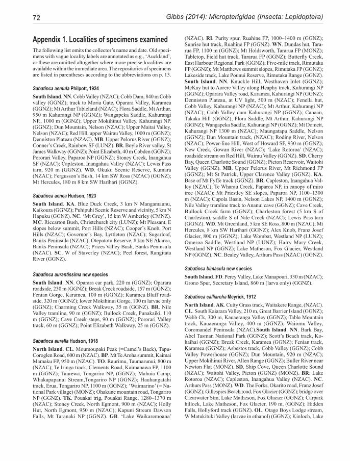
72 Gibbs (2014): Micropterigidae (Insecta: Lepidoptera)
Appendix 1. Localities of specimens examinedThe following list omits the collector’s name and date. Old speci-mens with vague locality labels are annotated as e.g., ‘Auckland’, or these are omitted altogether where more precise localities are available within the immediate area. The repositories of specimens are listed in parentheses according to the abbreviations on p. 13.
Sabatinca aemula Philpott, 1924South Island. NN. Cobb Valley (NZAC); Cobb Dam, 840 m Cobb valley (GGNZ); track to Moria Gate, Oparara Valley, Karamea (GGNZ); Mt Arthur Tableland (NZAC); Flora Saddle, Mt Arthur, 950 m Kahurangi NP (GGNZ); Wangapeka Saddle, Kahurangi NP., 1000 m (GGNZ); Upper Mokihinui Valley, Kahurangi NP. (GGNZ); Dun Mountain, Nelson (NZAC); Upper Maitai Valley, Nelson (NZAC); Red Hill, upper Wairau Valley, 1000 m (GGNZ); Denniston Plateau (NZAC). MB. Upper Pelorus River (GGNZ); Conner’s Creek, Rainbow SF (LUNZ). BR. Boyle River valley, St James Walkway (GGNZ); Point Elizabeth, 40 m Cobden (GGNZ); Pororari Valley, Paparoa NP (GGNZ); Stoney Creek, Inangahua SF (NZAC); Capleston, Inangahua Valley (NZAC); Lewis Pass tarn, 920 m (GGNZ). WD. Okuku Scenic Reserve, Kumara (NZAC); Fergusson’s Bush, 14 km SW Ross (NZAC) (GGNZ); Mt Hercules, 180 m 8 km SW Harihari (GGNZ).
Sabatinca aenea Hudson, 1923South Island. KA. Blue Duck Creek, 3 km N Mangamaunu, Kaikoura (GGNZ); Puhipuhi Scenic Reserve and vicinity, 5 km N Hapuku (GGNZ). NC. ‘Mt Grey’, 15 km W Amberley (CMNZ). MC. Riccarton Bush, Christchurch city (LUNZ); Mt Pleasant, E slopes below summit, Port Hills (NZAC); Cooper’s Knob, Port Hills (NZAC); Governor’s Bay, Lyttleton (NZAC); Sugarloaf, Banks Peninsula (NZAC); Otepatotu Reserve, 8 km NE Akaroa, Banks Peninsula (NZAC); Prices Valley Bush, Banks Peninsula (NZAC). SC. W of Staverley (NZAC); Peel forest, Rangitata River (GGNZ).
Sabatinca aurantissima new speciesSouth Island. NN. Oparara car park, 220 m (GGNZ); Oparara roadside, 230 m (GGNZ); Break Creek roadside, 157 m (GGNZ); Fenian Gorge, Karamea, 100 m (GGNZ); Karamea Bluff road-side, 320 m (GGNZ); lower Mokihinui Gorge, 100 m larvae only (GGNZ); Charming Creek Walkway, 35 m (GGNZ). BR. Nile Valley tramline, 90 m (GGNZ); Bullock Creek, Punakaiki, 110 m (GGNZ); Cave Creek steps, 90 m (GGNZ); Pororari Valley track, 60 m (GGNZ); Point Elizabeth Walkway, 25 m (GGNZ).
Sabatinca aurella Hudson, 1918North Island. CL. Moumoupaki Peak (=Camel’s Back), Tapu-Coroglen Road, 600 m (NZAC). BP. Mt Te Aroha summit, Kaimai Mamaku FP, 950 m (NZAC). TO. Raurimu, Taumarunui, 800 m (NZAC); Te Iringa track, Clements Road, Kaimanawa FP, 1100 m (GGNZ); Taurewa, Tongariro NP, (GGNZ); Mahuia Camp, Whakapapanui Stream,Tongariro NP (GGNZ); Hauhangatahi track, Erua, Tongariro NP, 1100 m (GGNZ); ‘Waimarino’ (= Na-tional Park village) (MONZ); Ohakune mountain road, Tongariro NP (GGNZ). TK. Pouakai trig, Pouakai Range, 1280–1370 m (NZAC); Stoney Creek, North Egmont, 900 m (NZAC); Holly Hut, North Egmont, 950 m (NZAC); Kapuni Stream Dawson Falls, Mt Taranaki NP (GGNZ). GB. ‘Lake Waikaremoana’
(NZAC). RI. Purity spur, Ruahine FP, 1000–1400 m (GGNZ); Sunrise hut track, Ruahine FP (GGNZ). WN. Dundas hut, Tara-rua FP, 1100 m (GGNZ); Mt Holdsworth, Tararua FP (MONZ); Tabletop, Field hut track, Tararua FP (GGNZ); Butterfly Creek, East Harbour Regional Park (GGNZ); Five-mile track, Rimutaka FP (GGNZ); Mt Matthews summit slopes, Rimutaka FP (GGNZ); Lakeside track, Lake Pounui Reserve, Rimutaka Range (GGNZ). South Island. NN. Knuckle Hill, Westhaven Inlet (GGNZ); McKay hut to Aorere Valley along Heaphy track, Kahurangi NP (GGNZ); Oparara Valley road, Karamea, Kahurangi NP (GGNZ); Denniston Plateau, at UV light, 500 m (NZAC); Fenella hut, Cobb Valley, Kahurangi NP (NZAC); Mt Arthur, Kahurangi NP (NZAC); Cobb Valley dam Kahurangi NP (GGNZ); Canaan, Takaka Hill (GGNZ); Flora Saddle, Mt Arthur, Kahurangi NP (GGNZ); Wangapeka Saddle, Kahurangi NP (GGNZ); Mt Domett, Kahurangi NP 1300 m (NZAC); Maungatapu Saddle, Nelson (GGNZ); Dun Mountain track, (NZAC); Roding River, Nelson (NZAC); Power-line Hill, West of Howard SF, 930 m (GGNZ); New Creek, Gowan River (NZAC); ‘Lake Rotoroa’ (NZAC); roadside stream on Red Hill, Wairau Valley (GGNZ). SD. Cherry Bay, Queen Charlotte Sound (GGNZ); Picton Reservoir, Waitohi Valley (GGNZ). MB. Upper Pelorus River, Mt Richmond FP (GGNZ); Mt St Patrick, Upper Clarence Valley (GGNZ). KA. Base of Mt Fyffe track (GGNZ). BR. Capleston, Inangahua Val-ley (NZAC); Te Wharau Creek, Paparoa NP, in canopy of miro tree (NZAC); Mt Priestley SE slopes, Paparoa NP, 1100–1300 m (NZAC); Cupola Basin, Nelson Lakes NP, 1400 m (GGNZ); Nile Valley tramline track to Ananui cave (GGNZ); Cave Creek, Bullock Creek farm (GGNZ); Charleston forest (5 km S of Charleston), saddle S of Nile Creek (NZAC); Lewis Pass tarn (GGNZ). WD. Mt Greenland, 5 km SE Ross, 800 m (NZAC); Mt Hercules, 8 km SW Harihari (GGNZ); Alex Knob, Franz Josef Glacier, 800 m (GGNZ); Lake Wombat, Westland NP (LUNZ); Omeroa Saddle, Westland NP (LUNZ); Hairy Mary Creek, Westland NP (GGNZ); Lake Matheson, Fox Glacier, Westland NP (GGNZ). NC. Bealey Valley, Arthurs Pass (NZAC) (GGNZ).
Sabatinca bimacula new speciesSouth Island. FD. Percy Valley, Lake Manapouri, 330 m (NZAC); Grono Spur, Secretary Island, 860 m (larva only) (GGNZ).
Sabatinca calliarcha Meyrick, 1912North Island. AK. Cutty Grass track, Waitakere Range, (NZAC). CL. South Kaiarara Valley, 210 m, Great Barrier Island (GGNZ); Webb Ck, 300 m, Kauaeranga Valley (GGNZ); Table Mountain track, Kauaeranga Valley, 400 m (GGNZ); Waiomu Valley, Coromandel Peninsula (NZAC).South Island. NN. Bark Bay, Abel Tasman National Park (GGNZ); Scott’s Beach track, Ko-haihai (GGNZ); Break Creek, Karamea (GGNZ); Fenian track, Karamea (GGNZ); Asbestos track, Cobb Valley (GGNZ); Cobb Valley Powerhouse (GGNZ); Dun Mountain, 920 m (NZAC); Upper Mokihinui River, Allen Range (GGNZ); Buller River near Newton Flat (MONZ). SD. Ship Cove, Queen Charlotte Sound (NZAC); Waitohi Valley, Picton (GGNZ) (MONZ). BR. Lake Rotoroa (NZAC); Capleston, Inangahua Valley (NZAC). NC. Arthurs Pass (MONZ). WD. The Forks, Okarito road, Franz Josef (GGNZ); Gillespies Beach road, Fox Glacier (GGNZ); bridge over Clearwater Stm, Lake Matheson, Fox Glacier (GGNZ); Carpark hillock, Lake Matheson, Fox Glacier, 190 m, (GGNZ); Hidden Falls, Hollyford track (GGNZ). OL. Otago Boys Lodge stream, W Matukituki Valley (larvae in ethanol) (GGNZ); Kinloch, Lake
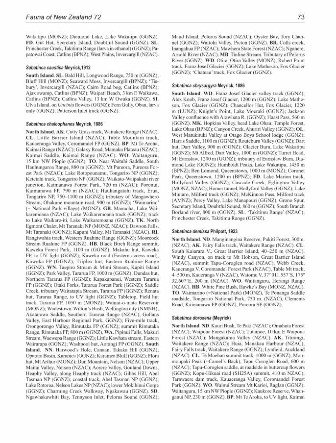
Fauna of New Zealand 72 73
Wakatipu (MONZ); Diamond Lake, Lake Wakatipu (GGNZ). FD. Gut Hut, Secretary Island, Doubtful Sound (GGNZ). SL. Princhester Creek, Takitimu Range (larva in ethanol) (GGNZ); Pa-patowai Coast, Catlins (BPNZ); West Plains, Invercargill (NZAC).
Sabatinca caustica Meyrick,1912South Island. SL. Bald Hill, Longwood Range, 750 m (GGNZ); Bluff Hill (MONZ); Seaward Moss, Invercargill (BPNZ); ‘Tis-bury’, Invercargill (NZAC); Cairn Road bog, Catlins (BPNZ); Ajax swamp, Catlins (BPNZ); Waipati Beach, 3 km E Waikawa, Catlins (BPNZ); Catlins Valley, 13 km W Owaka (GGNZ). SI. Ulva Island, on Uncinia flowers (GGNZ); Fern Gully, Oban, larva only (GGNZ); Patterson Inlet track (GGNZ).
Sabatinca chalcophanes Meyrick, 1886North Island. AK. Cutty Grass track, Waitakere Range (NZAC). CL. Little Barrier Island (NZAC); Table Mountain track, Kauaeranga Valley, Coromandel FP (GGNZ). BP. Mt Te Aroha, Kaimai Range (NZAC); Galaxy Road, Mamaku Plateau (NZAC); Kaimai Saddle, Kaimai Range (NZAC). WO. Waitanguru, 15 km NW Piopio (GGNZ). TO. Near Waituhi Saddle, South Hauhungaroa Range, 880 m (GGNZ); Mt Pureora, Pureora For-est Park (NZAC); Lake Rotopounamu, Tongariro NP (GGNZ); Ketetahi track, Tongariro NP (GGNZ); Waikato-Waipakahi river junction, Kaimanawa Forest Park, 720 m (NZAC); Poronui, Kaimanawa FP, 790 m (NZAC); Hauhangatahi track, Erua, Tongariro NP, 750–1100 m (GGNZ); tributary of Mangawhero Stream, Ohakune mountain road, 900 m (GGNZ); ‘Waimarino’ (= National Park village) (MONZ). GB. Manuoha, Lake Wai-karemoana (NZAC); Lake Waikaremoana track (GGNZ); track to Lake Waikare-iti, Lake Waikaremoana (GGNZ). TK. North Egmont Chalet, Mt Taranaki NP (MONZ, NZAC); Dawson Falls, Mt Taranaki (GGNZ); Kapuni Valley, Mt Taranaki (NZAC). RI. Rangiwahia track, Western Ruahine Range (GGNZ); Moorcock Stream Ruahine FP (GGNZ). HB. Black Birch Range summit, Kaweka Forest Park, 1100 m (GGNZ); Makahu hut, Kaweka FP, to UV light (GGNZ); Kaweka road (Eastern access road), Kaweka FP (GGNZ); Triplex hut, Eastern Ruahine Range (GGNZ). WN. Taepiro Stream & Mimi Stream, Kapiti Island (GGNZ); Park Valley, Tararua FP, 1000 m (GGNZ); Dundas hut, Northern Tararua FP (GGNZ); Kapakapanui, Western Tararua FP (GGNZ); Otaki Forks, Tararua Forest Park (GGNZ); Saddle Creek, tributary Waitatapia Stream, Tararua FP (GGNZ); Renata hut, Tararua Range, to UV light (GGNZ); Tabletop, Field hut track, Tararua FP, 1050 m (MONZ); Wainui-o-mata Reservoir (MONZ); Wadestown-Wilton’s Bush, Wellington city (NMNH); Akatarawa Saddle, Southern Tararua Range (NZAC); Gollans Valley, East Harbour Regional Park, GGNZ); Five-mile track, Orongorongo Valley, Rimutaka FP (GGNZ); summit Rimutaka Range, Rimutaka FP, 800 m (GGNZ). WA. Pipinui Falls, Makuri Stream, Waewepa Range (GGNZ); Little Kawhata stream, Eastern Wairarapa (GGNZ); Washpool hut, Aorangi FP (GGNZ). South Island. NN. Harwood’s Hole, Canaan, Takaka Hill (GGNZ); Oparara Basin, Karamea (GGNZ); Karamea Bluff (GGNZ); Flora hut, Mt Arthur (MONZ); Dun Mountain, Nelson (NZAC); Upper Maitai Valley, Nelson (NZAC); Aorere Valley, Gouland Downs, Heaphy Valley, along Heaphy track (NZAC); Gibbs Hill, Abel Tasman NP (GGNZ); coastal track, Abel Tasman NP (GGNZ); Lake Rotoroa, Nelson Lakes NP (NZAC); lower Mokihinui Gorge (GGNZ); Charming Creek Walkway, Ngakawau (GGNZ). SD. Ngawhakawhiti Bay, Tennyson Inlet, Pelorus Sound (GGNZ);
Maud Island, Pelorus Sound (NZAC); Oyster Bay, Tory Chan-nel (GGNZ); Waitohi Valley, Picton (GGNZ). BR. Colls creek, Inangahua FP (NZAC); Mawhera State Forest (NZAC); Ngahere, Arnold River (NZAC). MB. Tinline Stream. Tributary of Pelorus River (GGNZ). WD. Otira, Otira Valley (MONZ); Robert Point track, Franz Josef Glacier (GGNZ); Lake Matheson, Fox Glacier (GGNZ); ‘Chateau’ track, Fox Glacier (GGNZ).
Sabatinca chrysargyra Meyrick, 1886South Island. WD. Franz Josef Glacier valley track (GGNZ); Alex Knob, Franz Josef Glacier, 1200 m (GGNZ); Lake Mathe-son, Fox Glacier (GGNZ); Chancellor Hut, Fox Glacier, 1220 m (LUNZ); Knight’s Point, Lake Moeraki (GGNZ); Jackson Valley confluence with Arawhata R. (GGNZ); Haast Pass, 560 m (GGNZ). MK. Hopkins Valley, head Lake Ohau; Temple Forest, Lake Ohau (BPNZ); Canyon Creek, Ahuriri Valley (GGNZ); OL. West Matukituki Valley at Otago Boys School lodge (GGNZ); Harris Saddle, 1100 m (GGNZ); Routeburn Valley (GGNZ); Dart hut, Dart Valley, 900 m (GGNZ); Glacier Burn, Lake Wakatipu (GGNZ); Mt Alfred, Dart Valley, 1000 m (GGNZ); Turret Head, Mt Earnslaw, 1200 m (GGNZ); tributary of Earnslaw Burn, Dia-mond Lake (GGNZ); Humboldt Peaks, Lake Wakatipu, 1450 m (BPNZ); Ben Lomond, Queenstown, 1000 m (MONZ); Coronet Peak, Queenstown, 1200 m (BPNZ); FD. Lake Marion track, Hollyford Valley (GGNZ); Cascade Creek, Eglington Valley (MONZ, NZAC); Homer tunnel, Hollyford Valley (GGNZ); Lake Mintaro, Milford track (GGNZ); McKinnon Pass, Milford track (AMNZ); Percy Valley, Lake Manapouri (GGNZ); Grono Spur, Secretary Island, Doubtful Sound, 860 m (GGNZ); South Branch Borland river, 800 m (GGNZ). SL. ‘Takitimu Range’ (NZAC); Princhester Creek, Takitimu Range (GGNZ).
Sabatinca demissa Philpott, 1923North Island. ND. Manginangina Reserve, Pukiti Forest, 300m. (NZAC). AK. Fairy Falls track, Waitakere Range (NZAC). CL. South Kaiarara V., Great Barrier Island, 40–250 m (NZAC). Windy Canyon, on track to Mt Hobson, Great Barrier Island (NZAC), summit Tapu-Coroglen road (NZAC), Webb Creek, Kaueranga V, Coromandel Forest Park (NZAC), Table Mt track, 4–500 m, Kaueranga V (NZAC), Waiomu V, 37º 011.557´S, 175º 32.607´E, 200 m (NZAC). WO. Waitanguru, Herangi Range (NZAC). HB. White Pine Bush, Hawke’s Bay (MONZ, NZAC). TO. Waimarino (=National Park) (MONZ), Te Ponanga Saddle roadside, Tongariro National Park, 750 m. (NZAC), Clements Road, Kaimanawa FP (GGNZ), Pureora SF (GGNZ).
Sabatinca doroxena (Meyrick) North Island. ND. Kauri Bush, Te Paki (NZAC); Omahuta Forest (NZAC); Waipoua Forest (NZAC); Tutamoe, 10 km E Waipoua Forest (NZAC); Mangakahia Valley (NZAC). AK. Titirangi, Waitakere Range (NZAC); Huia, Manakau Harbour (NZAC); Fairy Falls track, Waitakere Range (GGNZ); Lynfield, Auckland (NZAC). CL. Te Moehau summit track, 1000 m (GGNZ); Mou-moupaki Peak (=Camel’s Back), Tapu-Coroglen Road, 600 m (NZAC); Tapu-Coroglen saddle, at roadside in buttercup flowers (GGNZ); Kopu-Hikuai road (SH25A) summit, 410 m NZAC); Tarawaere dam track, Kauaeranga Valley, Coromandel Forest Park (GGNZ). WO. Wainui Stream Mt Karioi, Raglan (GGNZ); Waitanguru, 15 km NW Piopio (GGNZ); Kaukore Reserve, Whan-ganui NP, 230 m (GGNZ). BP. Mt Te Aroha, to UV light, Kaimai
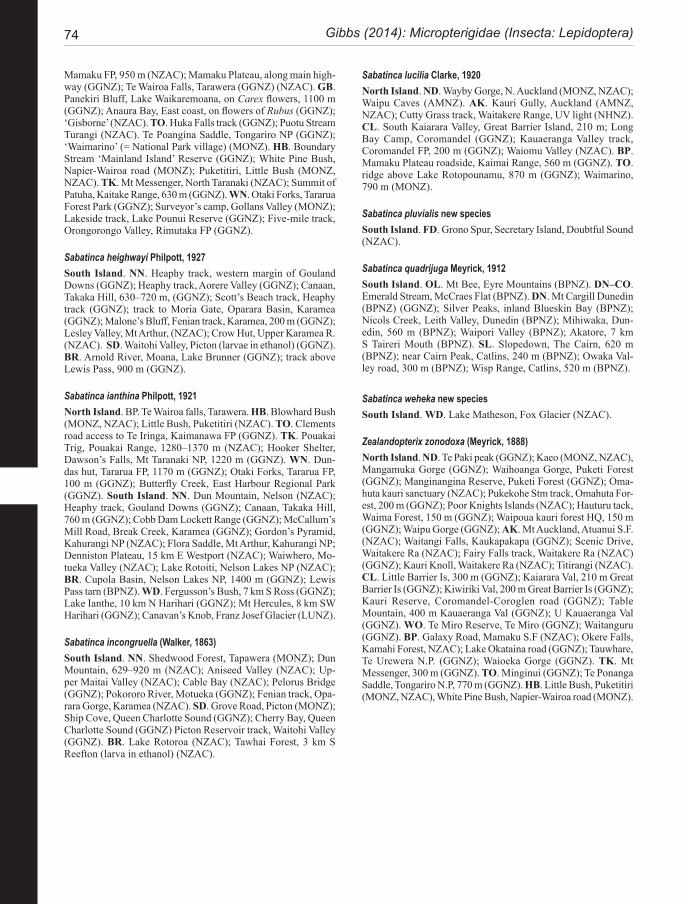
74 Gibbs (2014): Micropterigidae (Insecta: Lepidoptera)
Mamaku FP, 950 m (NZAC); Mamaku Plateau, along main high-way (GGNZ); Te Wairoa Falls, Tarawera (GGNZ) (NZAC). GB. Panekiri Bluff, Lake Waikaremoana, on Carex flowers, 1100 m (GGNZ); Anaura Bay, East coast, on flowers of Rubus (GGNZ); ‘Gisborne’ (NZAC). TO. Huka Falls track (GGNZ); Puotu Stream Turangi (NZAC). Te Poangina Saddle, Tongariro NP (GGNZ); ‘Waimarino’ (= National Park village) (MONZ). HB. Boundary Stream ‘Mainland Island’ Reserve (GGNZ); White Pine Bush, Napier-Wairoa road (MONZ); Puketitiri, Little Bush (MONZ, NZAC). TK. Mt Messenger, North Taranaki (NZAC); Summit of Patuha, Kaitake Range, 630 m (GGNZ). WN. Otaki Forks, Tararua Forest Park (GGNZ); Surveyor’s camp, Gollans Valley (MONZ); Lakeside track, Lake Pounui Reserve (GGNZ); Five-mile track, Orongorongo Valley, Rimutaka FP (GGNZ).
Sabatinca heighwayi Philpott, 1927South Island. NN. Heaphy track, western margin of Gouland Downs (GGNZ); Heaphy track, Aorere Valley (GGNZ); Canaan, Takaka Hill, 630–720 m, (GGNZ); Scott’s Beach track, Heaphy track (GGNZ); track to Moria Gate, Oparara Basin, Karamea (GGNZ); Malone’s Bluff, Fenian track, Karamea, 200 m (GGNZ); Lesley Valley, Mt Arthur, (NZAC); Crow Hut, Upper Karamea R. (NZAC). SD. Waitohi Valley, Picton (larvae in ethanol) (GGNZ). BR. Arnold River, Moana, Lake Brunner (GGNZ); track above Lewis Pass, 900 m (GGNZ).
Sabatinca ianthina Philpott, 1921North Island. BP. Te Wairoa falls, Tarawera. HB. Blowhard Bush (MONZ, NZAC); Little Bush, Puketitiri (NZAC). TO. Clements road access to Te Iringa, Kaimanawa FP (GGNZ). TK. Pouakai Trig, Pouakai Range, 1280–1370 m (NZAC); Hooker Shelter, Dawson’s Falls, Mt Taranaki NP, 1220 m (GGNZ). WN. Dun-das hut, Tararua FP, 1170 m (GGNZ); Otaki Forks, Tararua FP, 100 m (GGNZ); Butterfly Creek, East Harbour Regional Park (GGNZ). South Island. NN. Dun Mountain, Nelson (NZAC); Heaphy track, Gouland Downs (GGNZ); Canaan, Takaka Hill, 760 m (GGNZ); Cobb Dam Lockett Range (GGNZ); McCallum’s Mill Road, Break Creek, Karamea (GGNZ); Gordon’s Pyramid, Kahurangi NP (NZAC); Flora Saddle, Mt Arthur, Kahurangi NP; Denniston Plateau, 15 km E Westport (NZAC); Waiwhero, Mo-tueka Valley (NZAC); Lake Rotoiti, Nelson Lakes NP (NZAC); BR. Cupola Basin, Nelson Lakes NP, 1400 m (GGNZ); Lewis Pass tarn (BPNZ). WD. Fergusson’s Bush, 7 km S Ross (GGNZ); Lake Ianthe, 10 km N Harihari (GGNZ); Mt Hercules, 8 km SW Harihari (GGNZ); Canavan’s Knob, Franz Josef Glacier (LUNZ).
Sabatinca incongruella (Walker, 1863)South Island. NN. Shedwood Forest, Tapawera (MONZ); Dun Mountain, 629–920 m (NZAC); Aniseed Valley (NZAC); Up-per Maitai Valley (NZAC); Cable Bay (NZAC); Pelorus Bridge (GGNZ); Pokororo River, Motueka (GGNZ); Fenian track, Opa-rara Gorge, Karamea (NZAC). SD. Grove Road, Picton (MONZ); Ship Cove, Queen Charlotte Sound (GGNZ); Cherry Bay, Queen Charlotte Sound (GGNZ) Picton Reservoir track, Waitohi Valley (GGNZ). BR. Lake Rotoroa (NZAC); Tawhai Forest, 3 km S Reefton (larva in ethanol) (NZAC).
Sabatinca lucilia Clarke, 1920North Island. ND. Wayby Gorge, N. Auckland (MONZ, NZAC); Waipu Caves (AMNZ). AK. Kauri Gully, Auckland (AMNZ, NZAC); Cutty Grass track, Waitakere Range, UV light (NHNZ). CL. South Kaiarara Valley, Great Barrier Island, 210 m; Long Bay Camp, Coromandel (GGNZ); Kauaeranga Valley track, Coromandel FP, 200 m (GGNZ); Waiomu Valley (NZAC). BP. Mamaku Plateau roadside, Kaimai Range, 560 m (GGNZ). TO. ridge above Lake Rotopounamu, 870 m (GGNZ); Waimarino, 790 m (MONZ).
Sabatinca pluvialis new speciesSouth Island. FD. Grono Spur, Secretary Island, Doubtful Sound (NZAC).
Sabatinca quadrijuga Meyrick, 1912South Island. OL. Mt Bee, Eyre Mountains (BPNZ). DN–CO. Emerald Stream, McCraes Flat (BPNZ). DN. Mt Cargill Dunedin (BPNZ) (GGNZ); Silver Peaks, inland Blueskin Bay (BPNZ); Nicols Creek, Leith Valley, Dunedin (BPNZ); Mihiwaka, Dun-edin, 560 m (BPNZ); Waipori Valley (BPNZ); Akatore, 7 km S Taireri Mouth (BPNZ). SL. Slopedown, The Cairn, 620 m (BPNZ); near Cairn Peak, Catlins, 240 m (BPNZ); Owaka Val-ley road, 300 m (BPNZ); Wisp Range, Catlins, 520 m (BPNZ).
Sabatinca weheka new speciesSouth Island. WD. Lake Matheson, Fox Glacier (NZAC).
Zealandopterix zonodoxa (Meyrick, 1888)North Island. ND. Te Paki peak (GGNZ); Kaeo (MONZ, NZAC), Mangamuka Gorge (GGNZ); Waihoanga Gorge, Puketi Forest (GGNZ); Manginangina Reserve, Puketi Forest (GGNZ); Oma-huta kauri sanctuary (NZAC); Pukekohe Stm track, Omahuta For-est, 200 m (GGNZ); Poor Knights Islands (NZAC); Hauturu tack, Waima Forest, 150 m (GGNZ); Waipoua kauri forest HQ, 150 m (GGNZ); Waipu Gorge (GGNZ); AK. Mt Auckland, Atuanui S.F. (NZAC); Waitangi Falls, Kaukapakapa (GGNZ); Scenic Drive, Waitakere Ra (NZAC); Fairy Falls track, Waitakere Ra (NZAC) (GGNZ); Kauri Knoll, Waitakere Ra (NZAC); Titirangi (NZAC). CL. Little Barrier Is, 300 m (GGNZ); Kaiarara Val, 210 m Great Barrier Is (GGNZ); Kiwiriki Val, 200 m Great Barrier Is (GGNZ); Kauri Reserve, Coromandel-Coroglen road (GGNZ); Table Mountain, 400 m Kauaeranga Val (GGNZ); U Kauaeranga Val (GGNZ). WO. Te Miro Reserve, Te Miro (GGNZ); Waitanguru (GGNZ). BP. Galaxy Road, Mamaku S.F (NZAC); Okere Falls, Kamahi Forest, NZAC); Lake Okataina road (GGNZ); Tauwhare, Te Urewera N.P. (GGNZ); Waioeka Gorge (GGNZ). TK. Mt Messenger, 300 m (GGNZ). TO. Minginui (GGNZ); Te Ponanga Saddle, Tongariro N.P, 770 m (GGNZ). HB. Little Bush, Puketitiri (MONZ, NZAC), White Pine Bush, Napier-Wairoa road (MONZ).
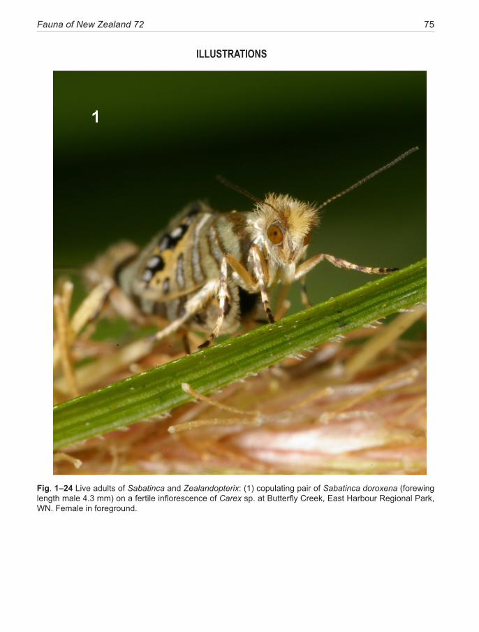
Fauna of New Zealand 72 75
ILLUSTRATIONS
Fig. 1–24 Live adults of Sabatinca and Zealandopterix: (1) copulating pair of Sabatinca doroxena (forewing length male 4.3 mm) on a fertile inflorescence of Carex sp. at Butterfly Creek, East Harbour Regional Park, WN. Female in foreground.
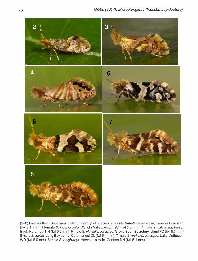
76 Gibbs (2014): Micropterigidae (Insecta: Lepidoptera)
(2–8) Live adults of Sabatinca: calliarcha-group of species: 2 female Sabatinca demissa, Pureora Forest TO (fwl 3.1 mm); 3 female S. incongruella, Waitohi Valley, Picton SD (fwl 5.0 mm); 4 male S. calliarcha, Fenian track, Karamea, NN (fwl 5.2 mm); 5 male S. pluvialis, paratype, Grono Spur, Secretary Island FD (fwl 5.3 mm); 6 male S. lucilia, Long Bay camp, Coromandel CL (fwl 5.1 mm); 7 male S. weheka, paratype, Lake Matheson, WD (fwl 6.0 mm); 8 male S. heighwayi, Harwood’s Hole, Canaan NN (fwl 6.1 mm).
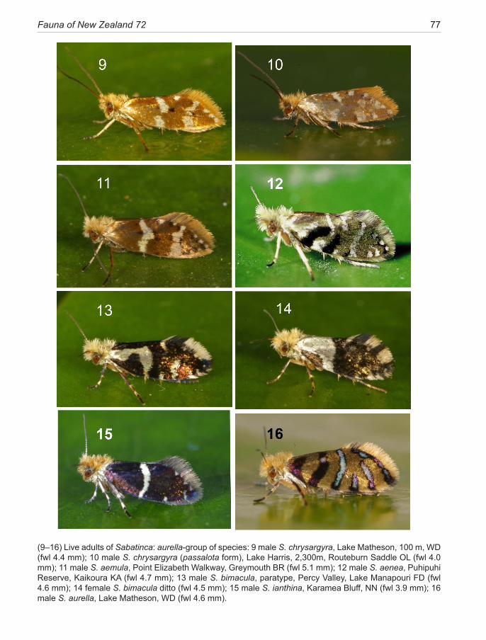
Fauna of New Zealand 72 77
(9–16) Live adults of Sabatinca: aurella-group of species: 9 male S. chrysargyra, Lake Matheson, 100 m, WD (fwl 4.4 mm); 10 male S. chrysargyra (passalota form), Lake Harris, 2,300m, Routeburn Saddle OL (fwl 4.0 mm); 11 male S. aemula, Point Elizabeth Walkway, Greymouth BR (fwl 5.1 mm); 12 male S. aenea, Puhipuhi Reserve, Kaikoura KA (fwl 4.7 mm); 13 male S. bimacula, paratype, Percy Valley, Lake Manapouri FD (fwl 4.6 mm); 14 female S. bimacula ditto (fwl 4.5 mm); 15 male S. ianthina, Karamea Bluff, NN (fwl 3.9 mm); 16 male S. aurella, Lake Matheson, WD (fwl 4.6 mm).
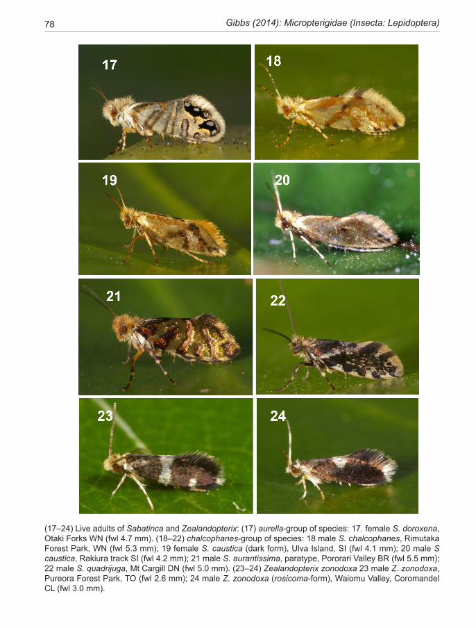
78 Gibbs (2014): Micropterigidae (Insecta: Lepidoptera)
(17–24) Live adults of Sabatinca and Zealandopterix: (17) aurella-group of species: 17. female S. doroxena, Otaki Forks WN (fwl 4.7 mm). (18–22) chalcophanes-group of species: 18 male S. chalcophanes, Rimutaka Forest Park, WN (fwl 5.3 mm); 19 female S. caustica (dark form), Ulva Island, SI (fwl 4.1 mm); 20 male S caustica, Rakiura track SI (fwl 4.2 mm); 21 male S. aurantissima, paratype, Pororari Valley BR (fwl 5.5 mm); 22 male S. quadrijuga, Mt Cargill DN (fwl 5.0 mm). (23–24) Zealandopterix zonodoxa 23 male Z. zonodoxa, Pureora Forest Park, TO (fwl 2.6 mm); 24 male Z. zonodoxa (rosicoma-form), Waiomu Valley, Coromandel CL (fwl 3.0 mm).
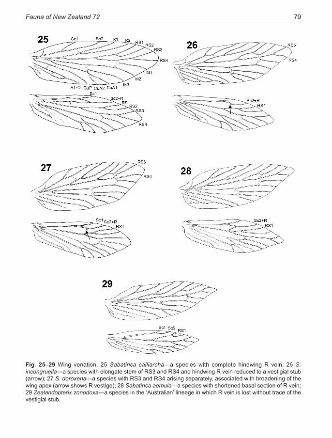
Fauna of New Zealand 72 79
Fig. 25–29 Wing venation. 25 Sabatinca calliarcha—a species with complete hindwing R vein; 26 S. incongruella—a species with elongate stem of RS3 and RS4 and hindwing R vein reduced to a vestigial stub (arrow); 27 S. doroxena—a species with RS3 and RS4 arising separately, associated with broadening of the wing apex (arrow shows R vestige); 28 Sabatinca aemula—a species with shortened basal section of R vein; 29 Zealandopterix zonodoxa—a species in the ‘Australian’ lineage in which R vein is lost without trace of the vestigial stub.
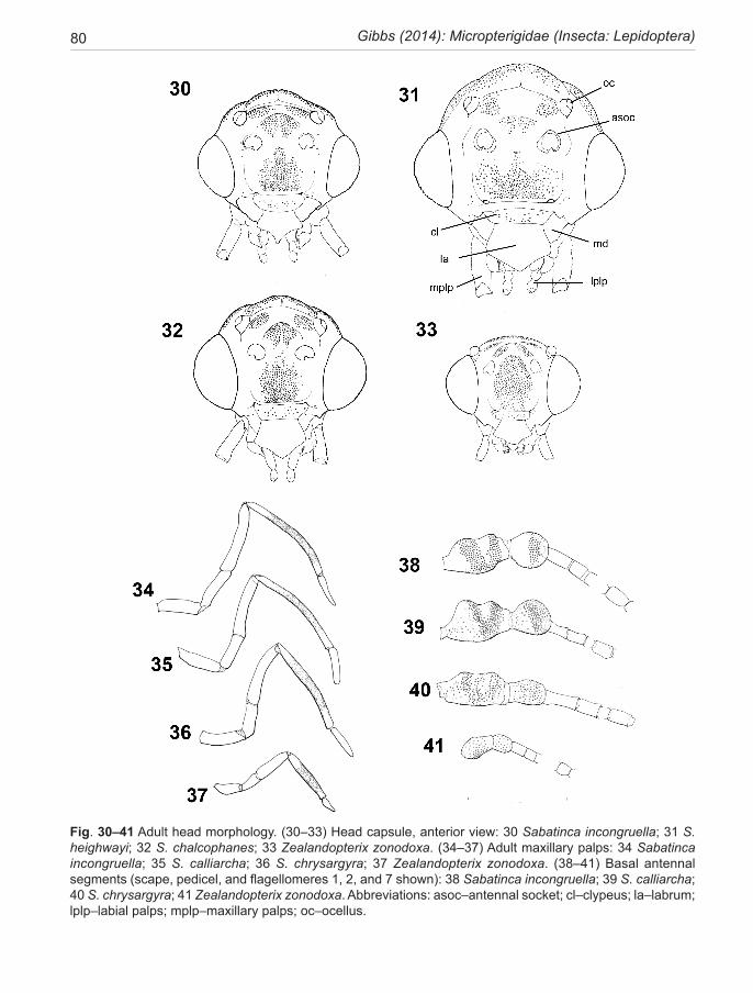
80 Gibbs (2014): Micropterigidae (Insecta: Lepidoptera)
Fig. 30–41 Adult head morphology. (30–33) Head capsule, anterior view: 30 Sabatinca incongruella; 31 S. heighwayi; 32 S. chalcophanes; 33 Zealandopterix zonodoxa. (34–37) Adult maxillary palps: 34 Sabatinca incongruella; 35 S. calliarcha; 36 S. chrysargyra; 37 Zealandopterix zonodoxa. (38–41) Basal antennal segments (scape, pedicel, and flagellomeres 1, 2, and 7 shown): 38 Sabatinca incongruella; 39 S. calliarcha; 40 S. chrysargyra; 41 Zealandopterix zonodoxa. Abbreviations: asoc–antennal socket; cl–clypeus; la–labrum; lplp–labial palps; mplp–maxillary palps; oc–ocellus.
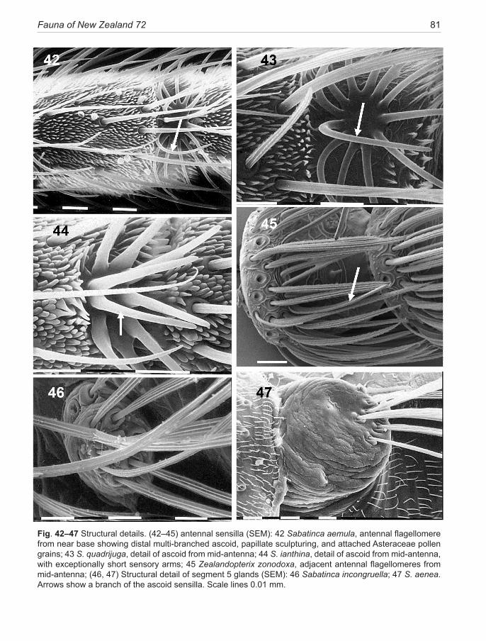
Fauna of New Zealand 72 81
Fig. 42–47 Structural details. (42–45) antennal sensilla (SEM): 42 Sabatinca aemula, antennal flagellomere from near base showing distal multi-branched ascoid, papillate sculpturing, and attached Asteraceae pollen grains; 43 S. quadrijuga, detail of ascoid from mid-antenna; 44 S. ianthina, detail of ascoid from mid-antenna, with exceptionally short sensory arms; 45 Zealandopterix zonodoxa, adjacent antennal flagellomeres from mid-antenna; (46, 47) Structural detail of segment 5 glands (SEM): 46 Sabatinca incongruella; 47 S. aenea. Arrows show a branch of the ascoid sensilla. Scale lines 0.01 mm.
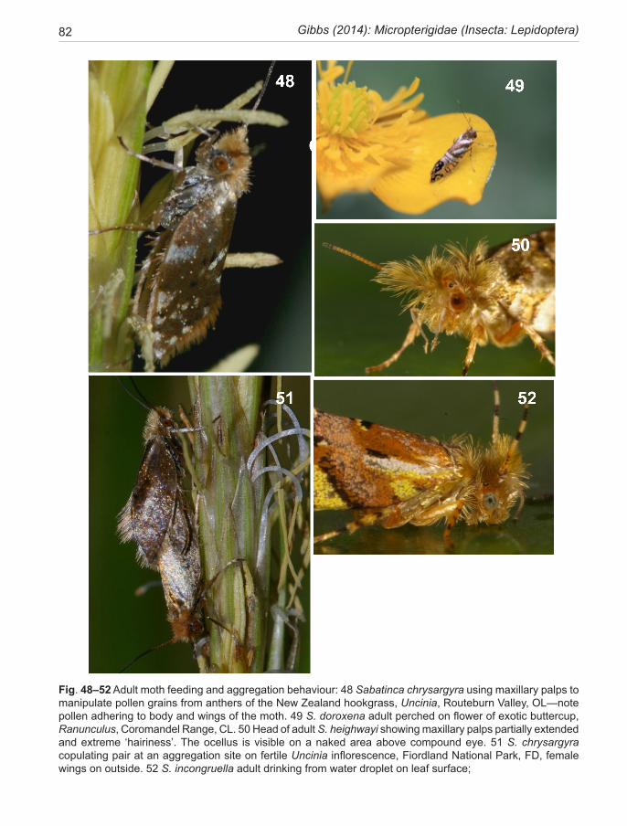
82 Gibbs (2014): Micropterigidae (Insecta: Lepidoptera)
Fig. 48–52 Adult moth feeding and aggregation behaviour: 48 Sabatinca chrysargyra using maxillary palps to manipulate pollen grains from anthers of the New Zealand hookgrass, Uncinia, Routeburn Valley, OL—note pollen adhering to body and wings of the moth. 49 S. doroxena adult perched on flower of exotic buttercup, Ranunculus, Coromandel Range, CL. 50 Head of adult S. heighwayi showing maxillary palps partially extended and extreme ‘hairiness’. The ocellus is visible on a naked area above compound eye. 51 S. chrysargyra copulating pair at an aggregation site on fertile Uncinia inflorescence, Fiordland National Park, FD, female wings on outside. 52 S. incongruella adult drinking from water droplet on leaf surface;
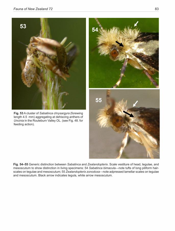
Fauna of New Zealand 72 83
Fig. 54–55 Generic distinction between Sabatinca and Zealandopterix. Scale vestiture of head, tegulae, and mesoscutum to show distinction in living specimens: 54 Sabatinca bimacula—note tufts of long piliform hair-scales on tegulae and mesoscutum; 55 Zealandopterix zonodoxa—note adpressed lamellar scales on tegulae and mesoscutum. Black arrow indicates tegula, white arrow mesoscutum.
Fig. 53 A cluster of Sabatinca chrysargyra (forewing length 4.5 mm) aggregating at dehiscing anthers of Uncinia in the Routeburn Valley OL. (see Fig. 48. for feeding action).
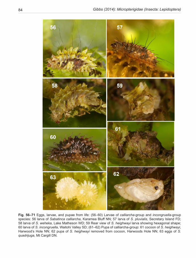
84 Gibbs (2014): Micropterigidae (Insecta: Lepidoptera)
Fig. 56–71 Eggs, larvae, and pupae from life: (56–60) Larvae of calliarcha-group and incongruella-group species: 56 larva of Sabatinca calliarcha, Karamea Bluff NN; 57 larva of S. pluvialis, Secretary Island FD; 58 larva of S. weheka, Lake Matheson WD; 59 Rear view of S. heighwayi larva showing hexagonal shape; 60 larva of S. incongruella, Waitohi Valley SD; (61–62) Pupa of calliarcha-group: 61 cocoon of S. heighwayi, Harwood’s Hole NN; 62 pupa of S. heighwayi removed from cocoon, Harwoods Hole NN; 63 eggs of S. quadrijuga, Mt Cargill DN.
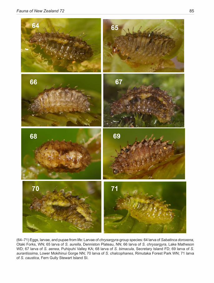
Fauna of New Zealand 72 85
(64–71) Eggs, larvae, and pupae from life: Larvae of chrysargyra-group species: 64 larva of Sabatinca doroxena, Otaki Forks, WN; 65 larva of S. aurella, Denniston Plateau, NN; 66 larva of S. chrysargyra, Lake Matheson WD; 67 larva of S. aenea, Puhipuhi Valley KA; 68 larva of S. bimacula, Secretary Island FD; 69 larva of S. aurantissima, Lower Mokihinui Gorge NN; 70 larva of S. chalcophanes, Rimutaka Forest Park WN; 71 larva of S. caustica, Fern Gully Stewart Island SI.
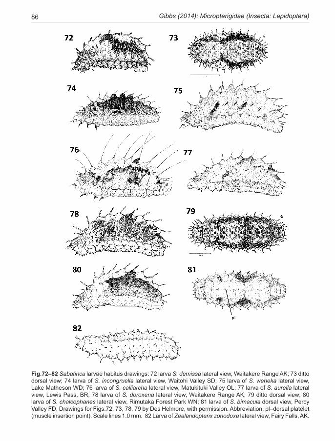
86 Gibbs (2014): Micropterigidae (Insecta: Lepidoptera)
Fig.72–82 Sabatinca larvae habitus drawings: 72 larva S. demissa lateral view, Waitakere Range AK; 73 ditto dorsal view; 74 larva of S. incongruella lateral view, Waitohi Valley SD; 75 larva of S. weheka lateral view, Lake Matheson WD; 76 larva of S. calliarcha lateral view, Matukituki Valley OL; 77 larva of S. aurella lateral view, Lewis Pass, BR; 78 larva of S. doroxena lateral view, Waitakere Range AK; 79 ditto dorsal view; 80 larva of S. chalcophanes lateral view, Rimutaka Forest Park WN; 81 larva of S. bimacula dorsal view, Percy Valley FD. Drawings for Figs.72, 73, 78, 79 by Des Helmore, with permission. Abbreviation: pl–dorsal platelet (muscle insertion point). Scale lines 1.0 mm. 82 Larva of Zealandopterix zonodoxa lateral view, Fairy Falls, AK.
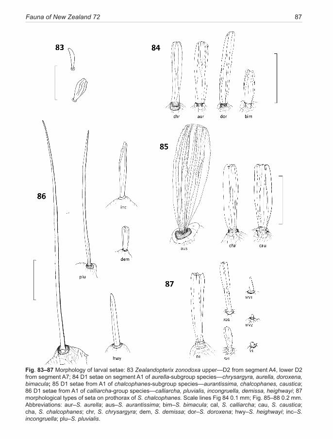
Fauna of New Zealand 72 87
Fig. 83–87 Morphology of larval setae: 83 Zealandopterix zonodoxa upper—D2 from segment A4, lower D2 from segment A7; 84 D1 setae on segment A1 of aurella-subgroup species—chrysargyra, aurella, doroxena, bimacula; 85 D1 setae from A1 of chalcophanes-subgroup species—aurantissima, chalcophanes, caustica; 86 D1 setae from A1 of calliarcha-group species—calliarcha, pluvialis, incongruella, demissa, heighwayi; 87 morphological types of seta on prothorax of S. chalcophanes. Scale lines Fig 84 0.1 mm; Fig. 85–88 0.2 mm. Abbreviations: aur–S. aurella; aus–S. aurantissima; bim–S. bimacula; cal, S. calliarcha; cau, S. caustica; cha, S. chalcophanes; chr, S. chrysargyra; dem, S. demissa; dor–S. doroxena; hwy–S. heighwayi; inc–S. incongruella; plu–S. pluvialis.
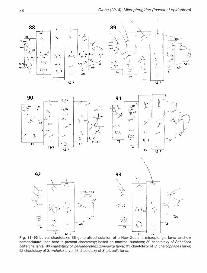
88 Gibbs (2014): Micropterigidae (Insecta: Lepidoptera)
Fig. 88–93 Larval chaetotaxy: 88 generalised setation of a New Zealand micropterigid larva to show nomenclature used here to present chaetotaxy; based on maximal numbers; 89 chaetotaxy of Sabatinca calliarcha larva; 90 chaetotaxy of Zealandopterix zonodoxa larva; 91 chaetotaxy of S. chalcophanes larva; 92 chaetotaxy of S. weheka larva; 93 chaetotaxy of S. pluvialis larva.
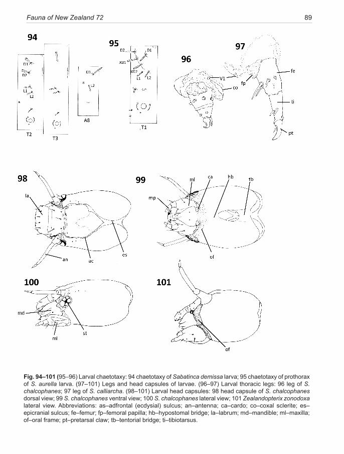
Fauna of New Zealand 72 89
Fig. 94–101 (95–96) Larval chaetotaxy: 94 chaetotaxy of Sabatinca demissa larva; 95 chaetotaxy of prothorax of S. aurella larva. (97–101) Legs and head capsules of larvae. (96–97) Larval thoracic legs: 96 leg of S. chalcophanes; 97 leg of S. calliarcha. (98–101) Larval head capsules: 98 head capsule of S. chalcophanes dorsal view; 99 S. chalcophanes ventral view; 100 S. chalcophanes lateral view; 101 Zealandopterix zonodoxa lateral view. Abbreviations: as–adfrontal (ecdysial) sulcus; an–antenna; ca–cardo; co–coxal sclerite; es–epicranial sulcus; fe–femur; fp–femoral papilla; hb–hypostomal bridge; la–labrum; md–mandible; ml–maxilla; of–oral frame; pt–pretarsal claw; tb–tentorial bridge; ti–tibiotarsus.
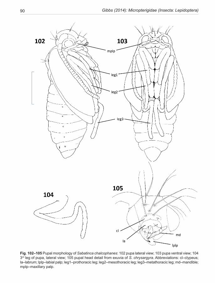
90 Gibbs (2014): Micropterigidae (Insecta: Lepidoptera)
Fig. 102–105 Pupal morphology of Sabatinca chalcophanes: 102 pupa lateral view; 103 pupa ventral view; 104 3rd leg of pupa, lateral view; 105 pupal head detail from exuvia of S. chrysargyra. Abbreviations: cl–clypeus; la–labrum; lplp–labial palp; leg1–prothoracic leg; leg2–mesothoracic leg; leg3–metathoracic leg; md–mandible; mplp–maxillary palp.
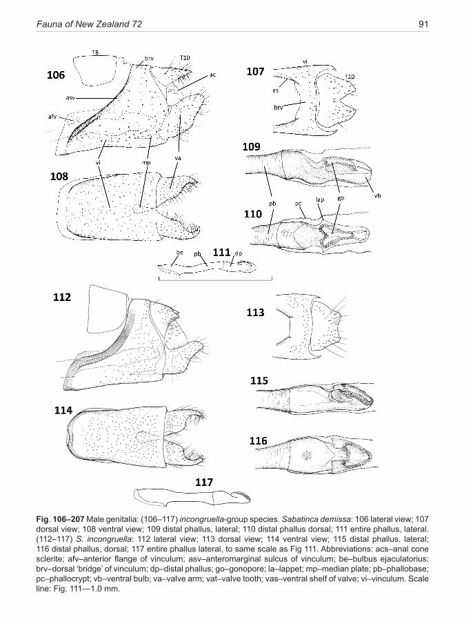
Fauna of New Zealand 72 91
Fig. 106–207 Male genitalia: (106–117) incongruella-group species. Sabatinca demissa: 106 lateral view; 107 dorsal view; 108 ventral view; 109 distal phallus, lateral; 110 distal phallus dorsal; 111 entire phallus, lateral. (112–117) S. incongruella: 112 lateral view; 113 dorsal view; 114 ventral view; 115 distal phallus, lateral; 116 distal phallus, dorsal; 117 entire phallus lateral, to same scale as Fig 111. Abbreviations: acs–anal cone sclerite; afv–anterior flange of vinculum; asv–anteromarginal sulcus of vinculum; be–bulbus ejaculatorius: brv–dorsal ‘bridge’ of vinculum; dp–distal phallus; go–gonopore; la–lappet; mp–median plate; pb–phallobase; pc–phallocrypt; vb–ventral bulb; va–valve arm; vat–valve tooth; vas–ventral shelf of valve; vi–vinculum. Scale line: Fig. 111—1.0 mm.
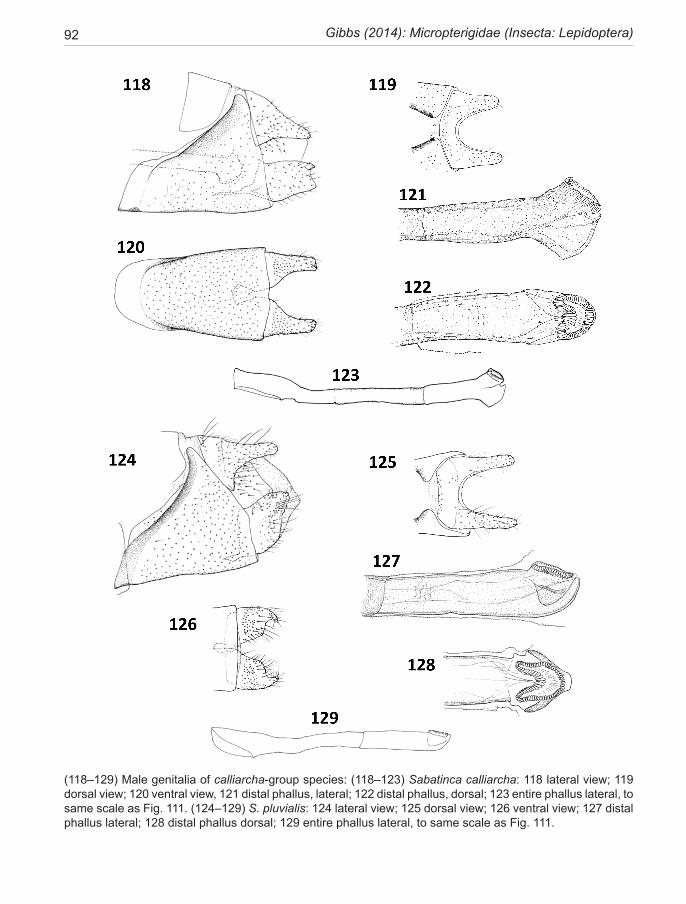
92 Gibbs (2014): Micropterigidae (Insecta: Lepidoptera)
(118–129) Male genitalia of calliarcha-group species: (118–123) Sabatinca calliarcha: 118 lateral view; 119 dorsal view; 120 ventral view, 121 distal phallus, lateral; 122 distal phallus, dorsal; 123 entire phallus lateral, to same scale as Fig. 111. (124–129) S. pluvialis: 124 lateral view; 125 dorsal view; 126 ventral view; 127 distal phallus lateral; 128 distal phallus dorsal; 129 entire phallus lateral, to same scale as Fig. 111.
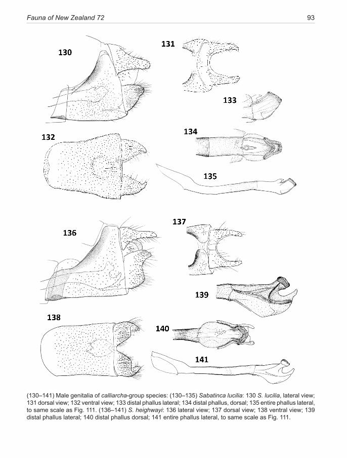
Fauna of New Zealand 72 93
(130–141) Male genitalia of calliarcha-group species: (130–135) Sabatinca lucilia: 130 S. lucilia, lateral view; 131 dorsal view; 132 ventral view; 133 distal phallus lateral; 134 distal phallus, dorsal; 135 entire phallus lateral, to same scale as Fig. 111. (136–141) S. heighwayi: 136 lateral view; 137 dorsal view; 138 ventral view; 139 distal phallus lateral; 140 distal phallus dorsal; 141 entire phallus lateral, to same scale as Fig. 111.
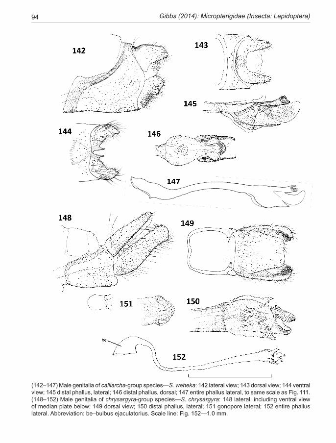
94 Gibbs (2014): Micropterigidae (Insecta: Lepidoptera)
(142–147) Male genitalia of calliarcha-group species—S. weheka: 142 lateral view; 143 dorsal view; 144 ventral view; 145 distal phallus, lateral; 146 distal phallus, dorsal; 147 entire phallus lateral, to same scale as Fig. 111. (148–152) Male genitalia of chrysargyra-group species—S. chrysargyra: 148 lateral, including ventral view of median plate below; 149 dorsal view; 150 distal phallus, lateral; 151 gonopore lateral; 152 entire phallus lateral. Abbreviation: be–bulbus ejaculatorius. Scale line: Fig. 152—1.0 mm.
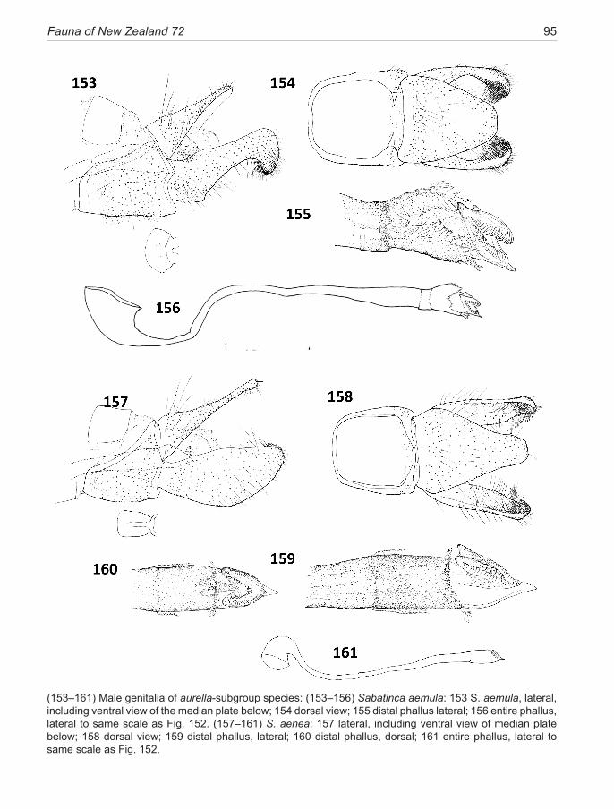
Fauna of New Zealand 72 95
(153–161) Male genitalia of aurella-subgroup species: (153–156) Sabatinca aemula: 153 S. aemula, lateral, including ventral view of the median plate below; 154 dorsal view; 155 distal phallus lateral; 156 entire phallus, lateral to same scale as Fig. 152. (157–161) S. aenea: 157 lateral, including ventral view of median plate below; 158 dorsal view; 159 distal phallus, lateral; 160 distal phallus, dorsal; 161 entire phallus, lateral to same scale as Fig. 152.
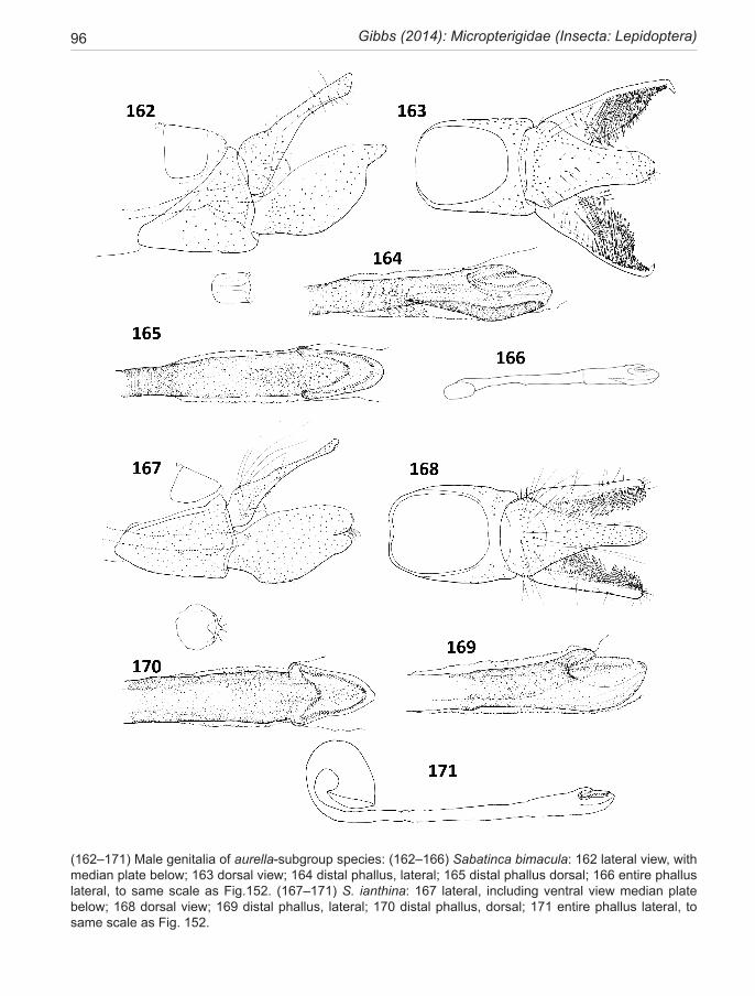
96 Gibbs (2014): Micropterigidae (Insecta: Lepidoptera)
(162–171) Male genitalia of aurella-subgroup species: (162–166) Sabatinca bimacula: 162 lateral view, with median plate below; 163 dorsal view; 164 distal phallus, lateral; 165 distal phallus dorsal; 166 entire phallus lateral, to same scale as Fig.152. (167–171) S. ianthina: 167 lateral, including ventral view median plate below; 168 dorsal view; 169 distal phallus, lateral; 170 distal phallus, dorsal; 171 entire phallus lateral, to same scale as Fig. 152.
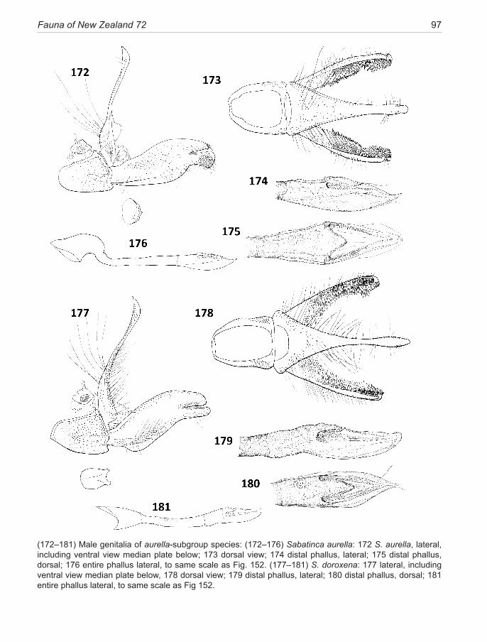
Fauna of New Zealand 72 97
(172–181) Male genitalia of aurella-subgroup species: (172–176) Sabatinca aurella: 172 S. aurella, lateral, including ventral view median plate below; 173 dorsal view; 174 distal phallus, lateral; 175 distal phallus, dorsal; 176 entire phallus lateral, to same scale as Fig. 152. (177–181) S. doroxena: 177 lateral, including ventral view median plate below, 178 dorsal view; 179 distal phallus, lateral; 180 distal phallus, dorsal; 181 entire phallus lateral, to same scale as Fig 152.
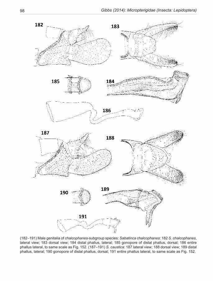
98 Gibbs (2014): Micropterigidae (Insecta: Lepidoptera)
(182–191) Male genitalia of chalcophanes-subgroup species: Sabatinca chalcophanes: 182 S. chalcophanes, lateral view; 183 dorsal view; 184 distal phallus, lateral; 185 gonopore of distal phallus, dorsal; 186 entire phallus lateral, to same scale as Fig. 152. (187–191) S. caustica: 187 lateral view; 188 dorsal view; 189 distal phallus, lateral; 190 gonopore of distal phallus, dorsal; 191 entire phallus lateral, to same scale as Fig. 152.
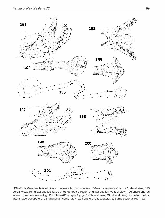
Fauna of New Zealand 72 99
(192–201) Male genitalia of chalcophanes-subgroup species: Sabatinca aurantissima: 192 lateral view; 193 dorsal view; 194 distal phallus, lateral; 195 gonopore region of distal phallus, ventral view; 196 entire phallus lateral, to same scale as Fig. 152. (197–201) S. quadrijuga: 197 lateral view; 198 dorsal view; 199 distal phallus, lateral; 200 gonopore of distal phallus, dorsal view; 201 entire phallus, lateral, to same scale as Fig. 152.
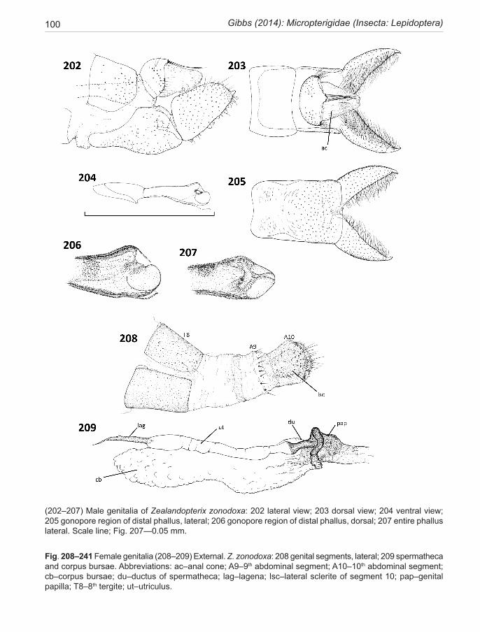
100 Gibbs (2014): Micropterigidae (Insecta: Lepidoptera)
(202–207) Male genitalia of Zealandopterix zonodoxa: 202 lateral view; 203 dorsal view; 204 ventral view; 205 gonopore region of distal phallus, lateral; 206 gonopore region of distal phallus, dorsal; 207 entire phallus lateral. Scale line; Fig. 207—0.05 mm.
Fig. 208–241 Female genitalia (208–209) External. Z. zonodoxa: 208 genital segments, lateral; 209 spermatheca and corpus bursae. Abbreviations: ac–anal cone; A9–9th abdominal segment; A10–10th abdominal segment; cb–corpus bursae; du–ductus of spermatheca; lag–lagena; lsc–lateral sclerite of segment 10; pap–genital papilla; T8–8th tergite; ut–utriculus.
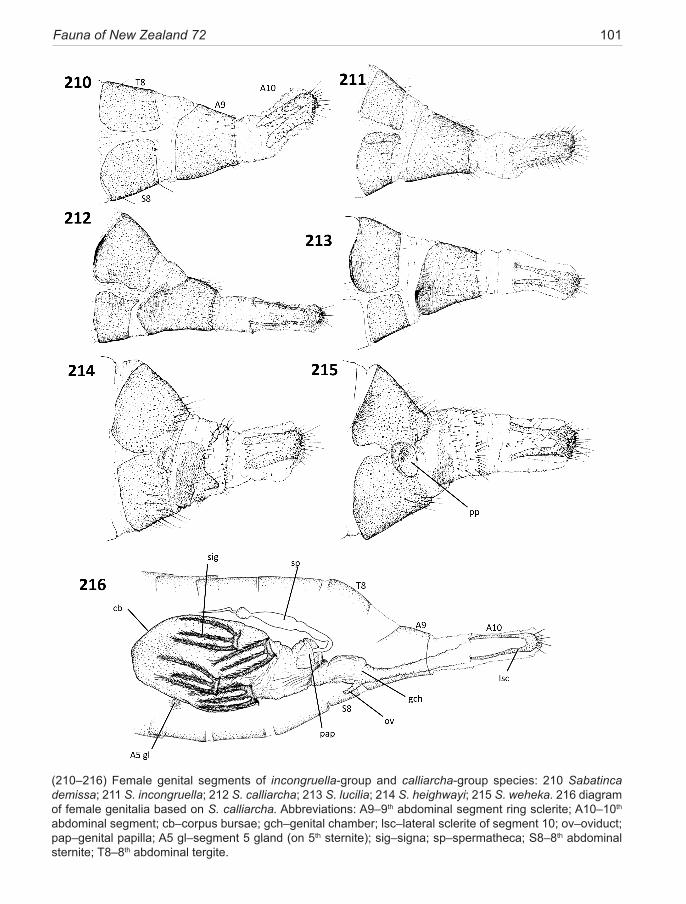
Fauna of New Zealand 72 101
(210–216) Female genital segments of incongruella-group and calliarcha-group species: 210 Sabatinca demissa; 211 S. incongruella; 212 S. calliarcha; 213 S. lucilia; 214 S. heighwayi; 215 S. weheka. 216 diagram of female genitalia based on S. calliarcha. Abbreviations: A9–9th abdominal segment ring sclerite; A10–10th abdominal segment; cb–corpus bursae; gch–genital chamber; lsc–lateral sclerite of segment 10; ov–oviduct; pap–genital papilla; A5 gl–segment 5 gland (on 5th sternite); sig–signa; sp–spermatheca; S8–8th abdominal sternite; T8–8th abdominal tergite.
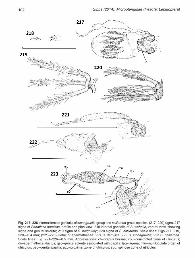
102 Gibbs (2014): Micropterigidae (Insecta: Lepidoptera)
Fig. 217–226 Internal female genitalia of incongruella-group and calliarcha-group species: (217–220) signa. 217 signa of Sabatinca demissa, profile and plan view; 218 internal genitalia of S. weheka, ventral view, showing signa and genital sclerite; 219 signa of S. heighwayi; 220 signa of S. calliarcha. Scale lines: Figs 217, 219, 220—0.4 mm; (221–226) Detail of spermathecae: 221 S. demissa; 222 S. incongruella; 223 S. calliarcha. Scale lines: Fig. 221–226—0.5 mm. Abbreviations: cb–corpus bursae; cou–constricted zone of utriculus; du–spermathecal ductus; gsc–genital sclerite associated with papilla; lag–lagena; mlu–multiloculate organ of utriculus; pap–genital papilla; pzu–proximal zone of utriculus; spu, spinose zone of utriculus.
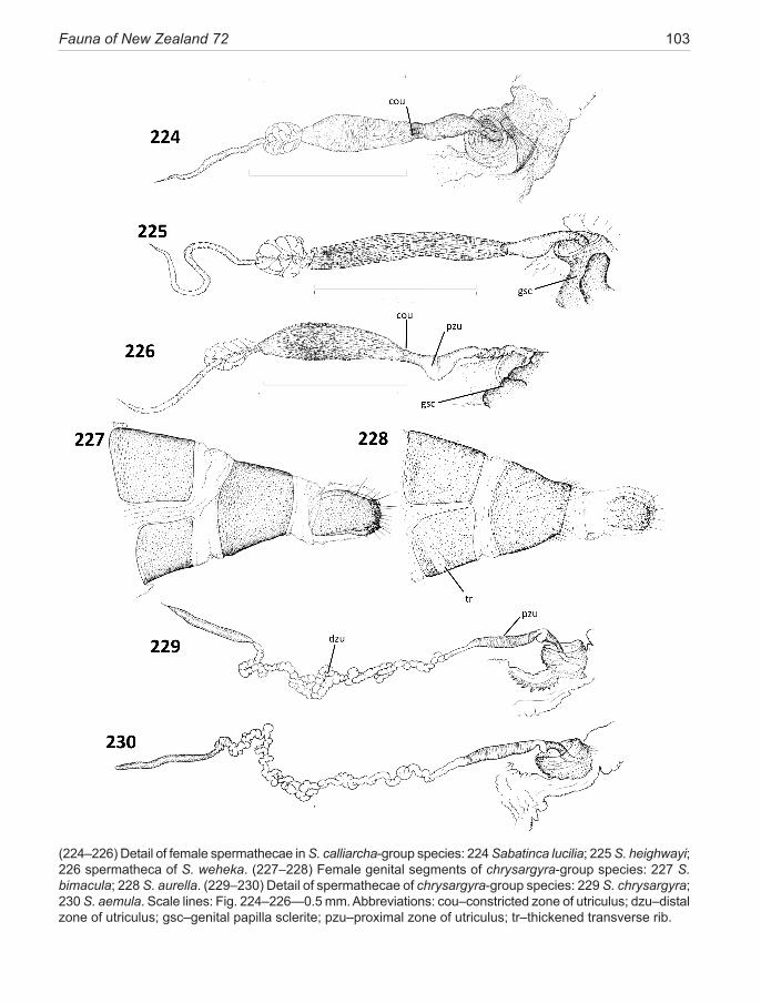
Fauna of New Zealand 72 103
(224–226) Detail of female spermathecae in S. calliarcha-group species: 224 Sabatinca lucilia; 225 S. heighwayi; 226 spermatheca of S. weheka. (227–228) Female genital segments of chrysargyra-group species: 227 S. bimacula; 228 S. aurella. (229–230) Detail of spermathecae of chrysargyra-group species: 229 S. chrysargyra; 230 S. aemula. Scale lines: Fig. 224–226—0.5 mm. Abbreviations: cou–constricted zone of utriculus; dzu–distal zone of utriculus; gsc–genital papilla sclerite; pzu–proximal zone of utriculus; tr–thickened transverse rib.
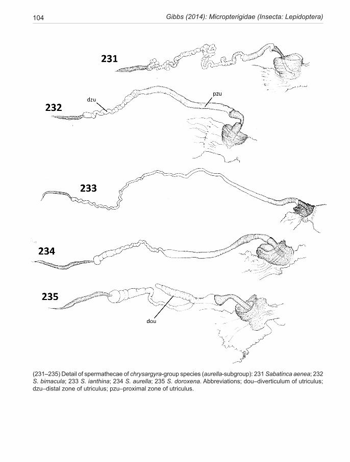
104 Gibbs (2014): Micropterigidae (Insecta: Lepidoptera)
(231–235) Detail of spermathecae of chrysargyra-group species (aurella-subgroup): 231 Sabatinca aenea; 232 S. bimacula; 233 S. ianthina; 234 S. aurella; 235 S. doroxena. Abbreviations; dou–diverticulum of utriculus; dzu–distal zone of utriculus; pzu–proximal zone of utriculus.
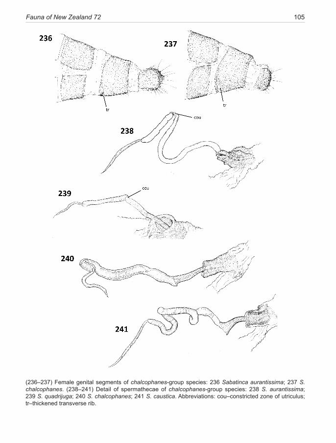
Fauna of New Zealand 72 105
(236–237) Female genital segments of chalcophanes-group species: 236 Sabatinca aurantissima; 237 S. chalcophanes. (238–241) Detail of spermathecae of chalcophanes-group species: 238 S. aurantissima; 239 S. quadrijuga; 240 S. chalcophanes; 241 S. caustica. Abbreviations: cou–constricted zone of utriculus; tr–thickened transverse rib.
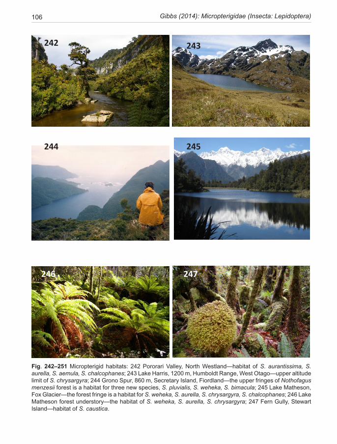
106 Gibbs (2014): Micropterigidae (Insecta: Lepidoptera)
Fig. 242–251 Micropterigid habitats: 242 Pororari Valley, North Westland—habitat of S. aurantissima, S. aurella, S. aemula, S. chalcophanes; 243 Lake Harris, 1200 m, Humboldt Range, West Otago—upper altitude limit of S. chrysargyra; 244 Grono Spur, 860 m, Secretary Island, Fiordland—the upper fringes of Nothofagus menzesii forest is a habitat for three new species, S. pluvialis, S. weheka, S. bimacula; 245 Lake Matheson, Fox Glacier—the forest fringe is a habitat for S. weheka, S. aurella, S. chrysargyra, S. chalcophanes; 246 Lake Matheson forest understory—the habitat of S. weheka, S. aurella, S. chrysargyra; 247 Fern Gully, Stewart Island—habitat of S. caustica.
242 243
244
246 247
245
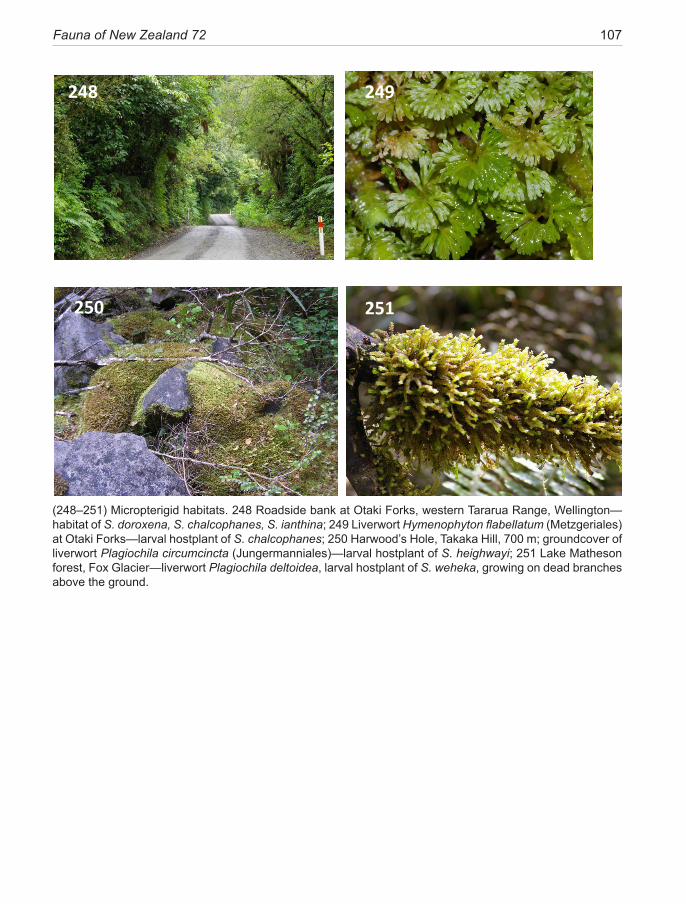
Fauna of New Zealand 72 107
(248–251) Micropterigid habitats. 248 Roadside bank at Otaki Forks, western Tararua Range, Wellington—habitat of S. doroxena, S. chalcophanes, S. ianthina; 249 Liverwort Hymenophyton flabellatum (Metzgeriales) at Otaki Forks—larval hostplant of S. chalcophanes; 250 Harwood’s Hole, Takaka Hill, 700 m; groundcover of liverwort Plagiochila circumcincta (Jungermanniales)—larval hostplant of S. heighwayi; 251 Lake Matheson forest, Fox Glacier—liverwort Plagiochila deltoidea, larval hostplant of S. weheka, growing on dead branches above the ground.
248 249
250 251
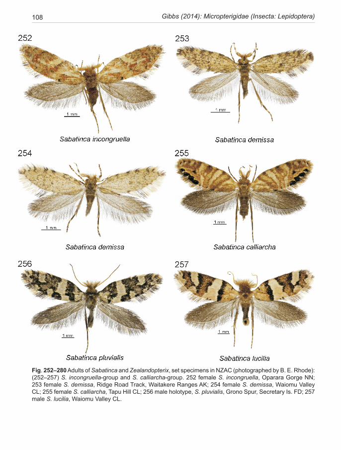
108 Gibbs (2014): Micropterigidae (Insecta: Lepidoptera)
Fig. 252–280 Adults of Sabatinca and Zealandopterix, set specimens in NZAC (photographed by B. E. Rhode): (252–257) S. incongruella-group and S. calliarcha-group. 252 female S. incongruella, Oparara Gorge NN; 253 female S. demissa, Ridge Road Track, Waitakere Ranges AK; 254 female S. demissa, Waiomu Valley CL; 255 female S. calliarcha, Tapu Hill CL; 256 male holotype, S. pluvialis, Grono Spur, Secretary Is. FD; 257 male S. lucilia, Waiomu Valley CL.
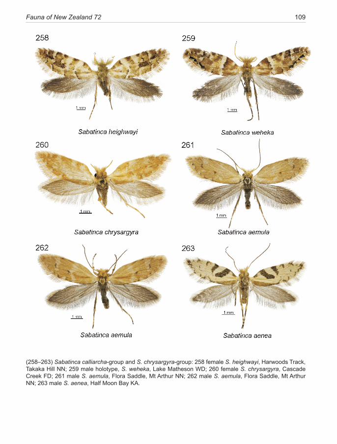
Fauna of New Zealand 72 109
(258–263) Sabatinca calliarcha-group and S. chrysargyra-group: 258 female S. heighwayi, Harwoods Track, Takaka Hill NN; 259 male holotype, S. weheka, Lake Matheson WD; 260 female S. chrysargyra, Cascade Creek FD; 261 male S. aemula, Flora Saddle, Mt Arthur NN; 262 male S. aemula, Flora Saddle, Mt Arthur NN; 263 male S. aenea, Half Moon Bay KA.
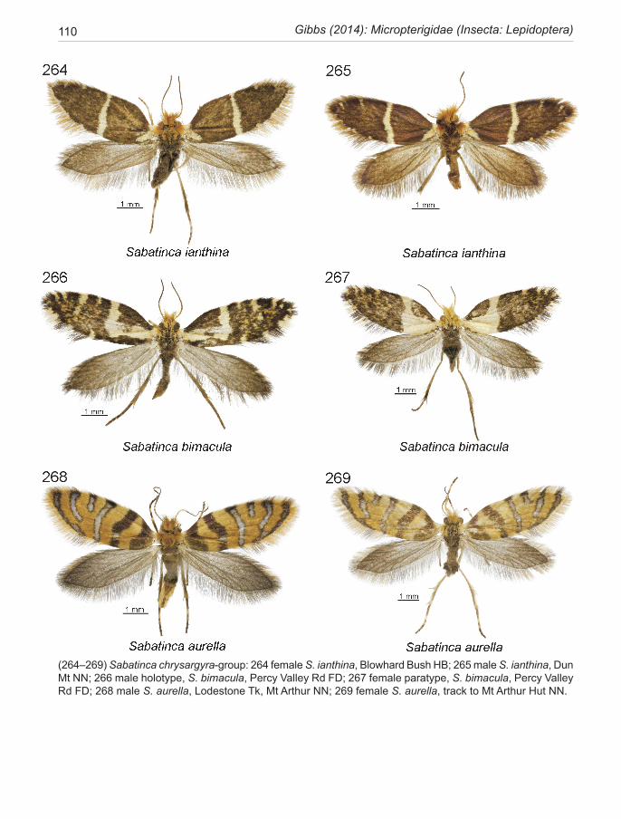
110 Gibbs (2014): Micropterigidae (Insecta: Lepidoptera)
(264–269) Sabatinca chrysargyra-group: 264 female S. ianthina, Blowhard Bush HB; 265 male S. ianthina, Dun Mt NN; 266 male holotype, S. bimacula, Percy Valley Rd FD; 267 female paratype, S. bimacula, Percy Valley Rd FD; 268 male S. aurella, Lodestone Tk, Mt Arthur NN; 269 female S. aurella, track to Mt Arthur Hut NN.
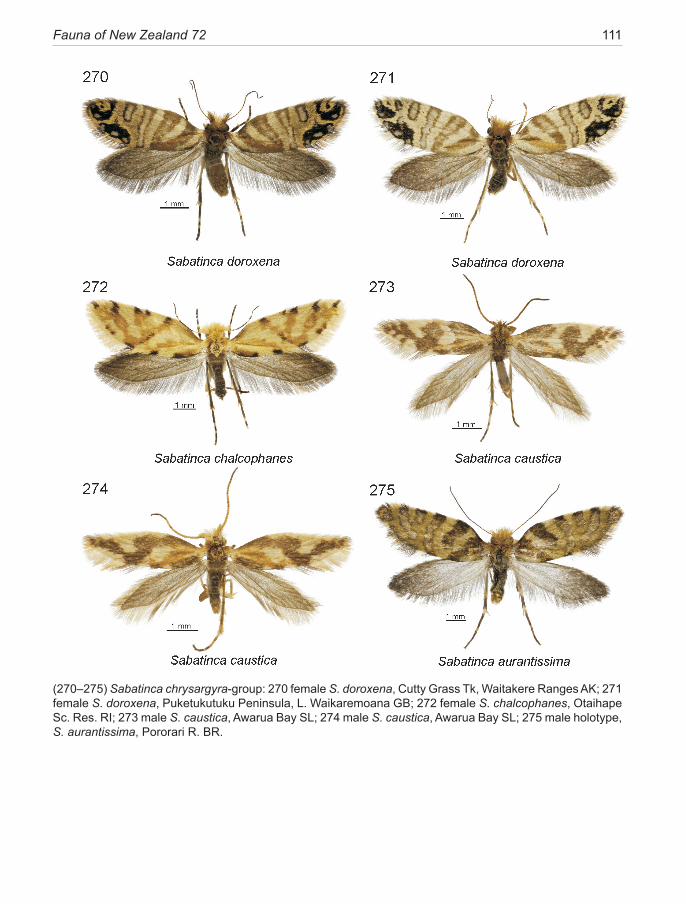
Fauna of New Zealand 72 111
(270–275) Sabatinca chrysargyra-group: 270 female S. doroxena, Cutty Grass Tk, Waitakere Ranges AK; 271 female S. doroxena, Puketukutuku Peninsula, L. Waikaremoana GB; 272 female S. chalcophanes, Otaihape Sc. Res. RI; 273 male S. caustica, Awarua Bay SL; 274 male S. caustica, Awarua Bay SL; 275 male holotype, S. aurantissima, Pororari R. BR.
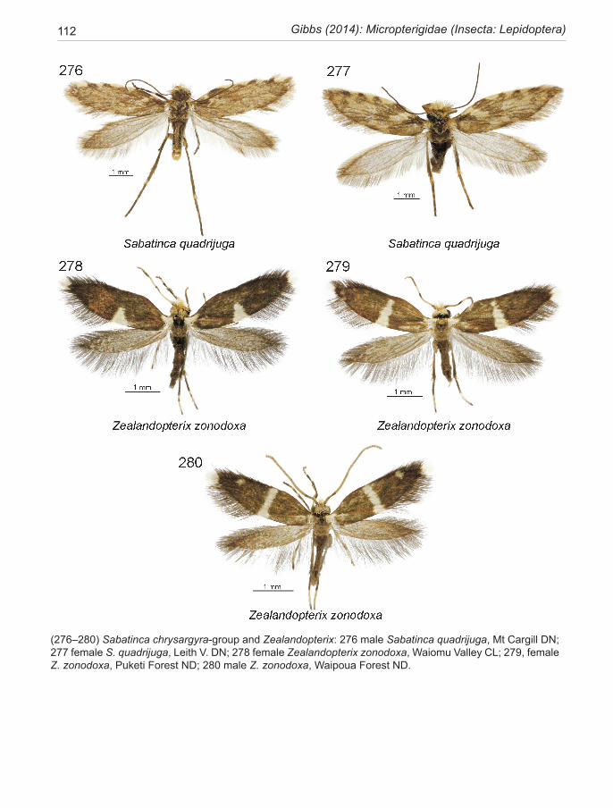
112 Gibbs (2014): Micropterigidae (Insecta: Lepidoptera)
(276–280) Sabatinca chrysargyra-group and Zealandopterix: 276 male Sabatinca quadrijuga, Mt Cargill DN; 277 female S. quadrijuga, Leith V. DN; 278 female Zealandopterix zonodoxa, Waiomu Valley CL; 279, female Z. zonodoxa, Puketi Forest ND; 280 male Z. zonodoxa, Waipoua Forest ND.
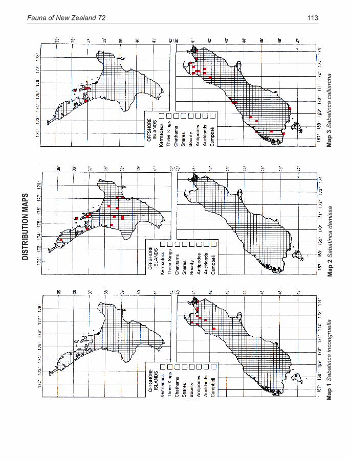
Fauna of New Zealand 72 113 DI
STRI
BUTI
ON M
APS
Map
1 S
abat
inca
inco
ngru
ella
Map
2 S
abat
inca
dem
issa
Map
3 S
abat
inca
cal
liarc
ha
114 Gibbs (2014): Micropterigidae (Insecta: Lepidoptera)
Map
4 S
abat
inca
plu
vial
isM
ap 5
Sab
atin
ca lu
cilia
Map
6 S
abat
inca
hei
ghw
ayi
Fauna of New Zealand 72 115
Map
7 S
abat
inca
weh
eka
Map
8 S
abat
inca
chr
ysar
gyra
Map
9 S
abat
inca
aem
ula

116 Gibbs (2014): Micropterigidae (Insecta: Lepidoptera)
Map
10
Sab
atin
ca a
enea
Map
11
Sab
atin
ca ia
thin
aM
ap 1
2 S
abat
inca
bim
acul
a
Fauna of New Zealand 72 117
Map
13
Sab
atin
ca a
urel
laM
ap 1
4 S
abat
inca
dor
oxen
aM
ap 1
5 S
abat
inca
cha
lcop
hane
s
118 Gibbs (2014): Micropterigidae (Insecta: Lepidoptera)
Map
16
Sab
atin
ca c
aust
ica
Map
17
Sab
atin
ca a
uran
tissi
ma
Map
18
Sab
atin
ca q
uadr
ijuga
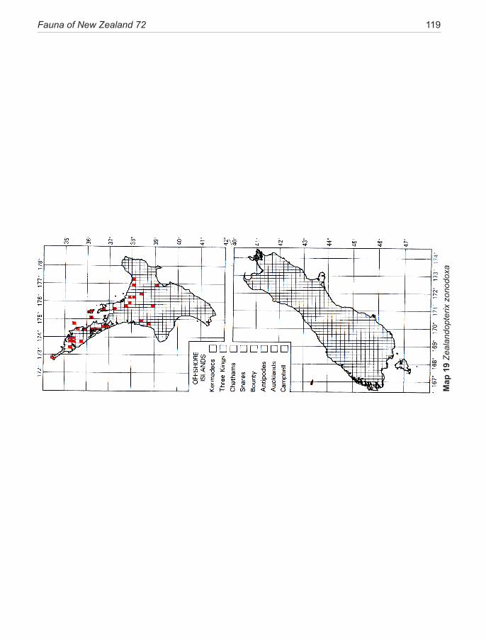
Fauna of New Zealand 72 119
Map
19
Zeal
ando
pter
ix z
onod
oxa
120 Gibbs (2014): Micropterigidae (Insecta: Lepidoptera)
TAXONOMIC INDEXPage numbers in bold type denote a description, and in italic type illustrations. A suffixed letter ‘k’ indicates a key, and ‘m’ a map.
Acanthopteroctetes 21aemula, Sabatinca 13, 15, 23, 24, 25, 28, 45, 46, 47k,
48, 49, 50, 51, 52, 72, 77, 79, 81, 95, 103, 109, 115m
aenea, Sabatinca 11, 23, 24, 29, 30, 46, 47k, 51, 54, 72, 77, 85, 95, 104, 109, 116m
Agathiphaga 21Agrionympha 10, 15, 16, 30aurantiaca, Sabatinca 49, 50aurantissima, Sabatinca 22, 23, 24, 25, 28, 45, 46,
47k, 58, 61, 63, 64, 72, 78, 85, 87, 99, 105, 106, 110, 118m
aurella, Sabatinca 6, 11, 12, 15, 16, 22, 23, 24, 27, 28, 30, 45, 46, 47k, 50, 52, 54, 55, 56, 57, 61, 63, 72, 77, 85, 86, 87, 89, 97, 103, 105, 106, 110, 117m
aurella subgroup 12, 16, 20, 26, 46, 48, 49, 77, 78, 87, 95, 96, 97, 104
Aureopterix 10, 14, 15, 16, 24, 66Austromartyria 10, 15, 24
Baltimartyria 10bimacula, Sabatinca 22, 23, 24, 25, 29, 30, 45, 46,
47k, 53, 61, 72, 77, 83, 85, 86, 87, 96, 103, 104, 106, 110, 116m
calliarcha-group 7, 12, 14, 15, 16, 17, 18, 19, 20, 21, 22, 26, 28, 30k, 31, 34, 35, 36, 37k, 39, 42, 45, 76, 84, 87, 92, 93, 94, 101, 102, 103, 108, 100
calliarcha, Sabatinca 7, 8, 13, 20, 23, 24, 26, 27, 28, 29, 36, 37, 37k, 38, 41, 57, 72, 76, 79, 80, 84, 86, 87, 88, 92, 101, 102, 108, 113m
calthella, Micropterix 22caustica, Sabatinca 7, 23, 24, 28, 29, 45, 46, 47k, 58,
59, 61, 64, 73, 78, 85, 87, 98, 106, 111, 118mchalcophanes subgroup 12, 17, 19, 20, 26, 46, 58,
87, 98, 99chalcophanes, Palaeomicra 58, 59chalcophanes, Sabatinca 7, 11, 12, 15, 21, 22, 23,
24, 28, 29, 29k, 34, 46, 47k, 56, 58, 59, 60, 61, 63, 73, 78, 80, 85, 86, 87, 88, 89, 90, 98, 105, 106, 107, 111, 117m
chrysargyra-group 12, 14, 15, 17, 18, 19, 20, 21, 26, 28, 30k, 31, 32, 44, 45, 46k, 56, 58, 59, 85, 94, 103, 104, 109, 110, 111, 112
chrysargyra, Sabatinca 7, 11, 12, 13, 15, 21, 22, 23, 24, 25, 28, 46, 47k, 48, 49, 50, 51, 52, 73, 77, 80, 82, 83, 85, 87, 90, 94, 103, 104, 106, 109, 115m
demissa, Sabatinca 14, 15, 18, 20, 23, 24, 30, 31, 32k, 34, 73, 76, 86, 87, 89, 91, 101, 102, 108, 113m
doroxena, Sabatinca 8, 11, 12, 15, 23, 24, 27, 28, 30, 45, 46, 47k, 48, 52, 55, 56, 57, 73, 75, 78, 79, 82, 85, 86, 87, 97, 104, 107, 111, 117m
eodora, Sabatinca 32Epimartyria 10, 17, 19, 26Eriocrania 21
Glyphipterix 8
heighwayi, Sabatinca 17, 21, 22, 23, 24, 28, 36, 37k, 41, 42, 43, 44, 74, 76, 80, 82, 84, 87, 93, 101, 102, 103, 107, 109, 114m
Heterobathmia 21Hypomartyria 10, 15
ianthina, Sabatinca 12, 15, 23, 24, 28, 30, 45, 46, 47k, 52, 56, 74, 77, 81, 96, 104, 107, 110, 116m
incongruella-group 12, 17, 27, 30k, 31, 32k, 46, 84, 91, 101, 102, 108
incongruella, Sabatinca 7, 10, 11, 12, 19, 20, 22, 23, 24, 27, 29, 30, 31, 32, 32k, 34, 35, 39, 58, 59, 74, 76, 79, 80, 81, 82, 84, 86, 87, 91, 101, 102, 108, 113m
Kurokopteryx 10,
lucilia, Sabatinca 12, 17, 20, 21, 23, 24, 37k, 38, 40, 41, 74, 76, 93, 101, 103, 108, 114m
matsumurana, Neomicropterix 22Micropardalis 7, 11, 15, 30, 46Micropterix calthella 22Micropterigidae 5, 6, 7, 8, 9, 10, 11, 14, 15, 16, 17,
19, 23, 23, 24, 25, 26, 27, 32, 36, 45, 46, 50, 54, 64, 65, 66
Nannopterix 15, 24Neomicropterix 10, 18, 22, 25Neomicropterix matsumurana 22Neomicropterix nipponensis 22nipponensis, Neomicropterix 22
Palaeomicra 7, 30, 46, 59Palaeomicra chalcophanes 58, 59Palaeomicra zonodoxa 66Palaeomicroides 10Paramartyria 10pluvialis, Sabatinca 7, 20, 21, 23, 24, 29, 30, 37k, 39,
74, 76, 84, 87, 88, 92, 106, 108, 114m
quadrijuga, Sabatinca 17, 22, 23, 24, 28, 29, 46k, 58, 63, 78, 81, 84, 99, 105, 112, 118m
Fauna of New Zealand 72 121
Sabatinca 7, 8, 9, 10, 11, 12, 14, 15, 16, 17, 18, 19, 20, 21, 24, 25, 26, 27, 30k, 30, 31, 32, 35, 44, 46, 57, 61, 64, 65, 75, 76–112
Sabatinca aemula 13, 15, 23, 24, 25, 28, 45, 46, 47k, 48, 49, 50, 51, 52, 72, 77, 79, 81, 95, 103, 109, 115m
Sabatinca aenea 11, 23, 24, 29, 30, 46, 47k, 51, 54, 72, 77, 85, 95, 104, 109, 116m
Sabatinca aurantissima 22, 23, 24, 25, 28, 45, 46, 47k, 58, 61, 63, 64, 72, 78, 85, 87, 99, 105, 106, 110, 118m
Sabatinca aurantiaca 49, 50Sabatinca aurella 6, 11, 12, 15, 16, 22, 23, 24, 27,
28, 30, 45, 46, 47k, 50, 52, 54, 55, 56, 57, 61, 63, 72, 77, 85, 86, 87, 89, 97, 103, 105, 106, 110, 117m
Sabatinca bimacula 22, 23, 24, 25, 29, 30, 45, 46, 47k, 53, 61, 72, 77, 83, 85, 86, 87, 96, 103, 104, 106, 110, 116m
Sabatinca calliarcha 7, 8, 13, 20, 23, 24, 26, 27, 28, 29, 36, 37, 37k, 38, 41, 57, 72, 76, 79, 80, 84, 86, 87, 88, 92, 101, 102, 108, 113m
Sabatinca caustica 7, 23, 24, 28, 29, 45, 46, 47k, 58, 59, 61, 64, 73, 78, 85, 87, 98, 106, 111, 118m
Sabatinca chrysargyra 7, 11, 12, 13, 15, 21, 22, 23, 24, 25, 28, 46, 47k, 48, 49, 50, 51, 52, 73, 77, 80, 82, 83, 85, 87, 90, 94, 103, 104, 106, 109, 115m
Sabatinca demissa 14, 15, 18, 20, 23, 24, 30, 31, 32k, 34, 73, 76, 86, 87, 89, 91, 101, 102, 108, 113m,
Sabatinca doroxena 8, 11, 12, 15, 23, 24, 27, 28, 30, 45, 46, 47k, 48, 52, 55, 56, 57, 73, 75, 78, 79, 82, 85, 86, 87, 97, 104, 107, 111, 117m
Sabatinca eodora 32Sabatinca heighwayi 17, 21, 22, 23, 24, 28, 36,
37k, 41, 42, 43, 44, 74, 76, 80, 82, 84, 87, 93, 101, 102, 103, 107, 109, 114m
Sabatinca ianthina 12, 15, 23, 24, 28, 30, 45, 46, 47k, 52, 56, 74, 77, 81, 96, 104, 107, 110, 116m
Sabatinca incongruella 7, 10, 11, 12, 19, 20, 22, 23, 24, 27, 29, 30, 31, 32, 32k, 34, 35, 39, 58, 59, 74, 76, 79, 80, 81, 82, 84, 86, 87, 91, 101, 102, 108, 113m
Sabatinca lucilia 12, 17, 20, 21, 23, 24, 37k, 38, 40, 41, 74, 76, 93, 101, 103, 108, 114m
Sabatinca munda 32Sabatinca passalota 7, 48, 49Sabatinca pluvialis 7, 20, 21, 23, 24, 29, 30, 37k,
39, 74, 76, 84, 87, 88, 92, 106, 108, 114mSabatinca quadrijuga 17, 22, 23, 24, 28, 29, 46k,
58, 63, 78, 81, 84, 99, 105, 112, 118mSabatinca rosicoma 66, 67Sabatinca weheka 7, 9, 15, 17, 20, 21, 22, 23, 24,
25, 28, 29, 31, 36, 37k, 42, 43, 44, 74, 76, 84, 86, 88, 94, 101, 102, 103, 106, 107, 109, 115m
Squamicornia 10, 15
Tasmantrix 10, 15, 16, 24, 25, 26, 65, 66
Vietomartyria 10
weheka, Sabatinca 7, 9, 15, 17, 20, 21, 22, 23, 24, 25, 28, 29, 31, 36, 37k, 42, 43, 44, 74, 76, 84, 86, 88, 94, 101, 102, 103, 106, 107, 109, 115m
Zealandopterix 7, 10, 11, 12, 14, 15, 16, 17, 18, 19, 20, 21, 24, 25, 26, 27, 30k, 53, 64, 65, 66, 75, 78, 83, 108, 112
Zealandopterix zonodoxa 10, 12, 18, 20, 22, 23, 24, 53, 64, 65, 66, 67, 74, 78, 79, 80, 81, 83, 86, 87, 88, 89, 100, 112, 119m
zonodoxa, Palaeomicra 66zonodoxa, Zealandopterix 10, 12, 18, 20, 22, 23,
24, 53, 64, 65, 66, 67, 74, 78, 79, 80, 81, 83, 86, 87, 88, 89, 100, 112, 119m
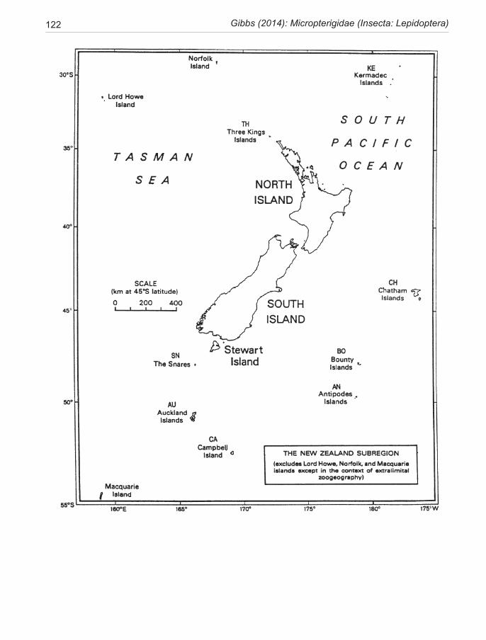
122 Gibbs (2014): Micropterigidae (Insecta: Lepidoptera)
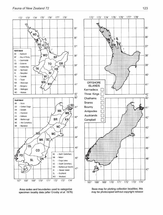
Fauna of New Zealand 72 123
124 Gibbs (2014): Micropterigidae (Insecta: Lepidoptera)
1 Terebrantia (Insecta: Thysanoptera). Laurence A. Mound & Annette K. Walker. ISBN 0-477-06687-9, 23 December 1982, 120 pp. ......................................................$29.95
2 Osoriinae (Insecta: Coleoptera: Staphylinidae). H. Pauline McColl. ISBN 0-477-06688-7, 23 December 1982, 96 pp. ...................................................................... $18.60
3 Anthribidae (Insecta: Coleoptera). B. A. Holloway. ISBN 0-477-06703-4, 23 December 1982, 272 pp. ........ ............................................................................ $41.00
4 Eriophyoidea except Eriophyinae (Arachnida: Acari). D. C. M. Manson. ISBN 0-477-06745-X,12 November 1984, 144 pp. ............................................................... $29.95
5 Eriophyinae (Arachnida: Acari: Eriophyoidea). D. C. M. Manson. ISBN 0-477-06746-8, 14 November 1984, 128 pp. .......................................................................$29.95
6 Hydraenidae (Insecta: Coleoptera). R. G. Ordish. ISBN 0-477-06747-6, 12 November 1984, 64 pp. ...... $18.60
7 Cryptostigmata (Arachnida: Acari) — a concise review. M. Luxton. ISBN 0-477-06762-X, 8 December 1985, 112 pp. ...................................................................... $29.95
8 Calliphoridae (Insecta: Diptera). James P. Dear. ISBN 0-477-06764-6. 24 February 1986, 88 pp. ......... $18.60
9 Protura (Insecta). S. L. Tuxen. ISBN 0-477-06765-4, 24 February 1986, 52 pp. ....................................... $18.60
10 Tubulifera (Insecta: Thysanoptera). Laurence A. Mound & Annette K. Walker. ISBN 0-477-06784-0, 22 September 1986, 144 pp. ..................................................... $34.65
11 Pseudococcidae (Insecta: Hemiptera). J. M. Cox. ISBN 0-477-06791-3, 7 April 1987, 232 pp. ........ $49.95
12 Pompilidae (Insecta: Hymenoptera). A. C. Harris. ISBN 0-477-02501-3, 13 November 1987, 160 pp. .... $39.95
13 Encyrtidae (Insecta: Hymenoptera). J. S. Noyes. ISBN 0-477-02517-X, 9 May 1988, 192 pp. ................ $44.95
14 Lepidoptera — annotated catalogue, and keys to family-group taxa. J. S. Dugdale. ISBN 0-477-02518-8, 23 September 1988, 264 pp. ............................. $49.95
15 Ambositrinae (Insecta: Hymenoptera: Diapriidae). I. D. Naumann. ISBN 0-477-02535-8, 30 December 1988, 168 pp. ...................................................................... $39.95
16 Nepticulidae (Insecta: Lepidoptera). Hans Donner & Christopher Wilkinson. ISBN 0-477-02538-2, 28 April 1989, 92 pp. ................................................................. $22.95
17 Mymaridae (Insecta: Hymenoptera) — introduction, and review of genera. J. S. Noyes & E. W. Valentine. ISBN 0-477-02542-0, 28 April 1989, 100 pp. .............. $24.95
18 Chalcidoidea (Insecta: Hymenoptera) — introduction, and review of genera in smaller families. J. S. Noyes & E. W. Valentine. ISBN 0-477-02545-5, 2 August 1989, 96 pp. ...................................................................... $24.95
19 Mantodea (Insecta), with a review of aspects of functional morphology and biology. G. W. Ramsay. ISBN 0-477-02581-1, 13 June 1990, 96 pp. .......................... $24.95
20 Bibionidae (Insecta: Diptera). Roy A. Harrison. ISBN 0-477-02595-1. 13 November 1990, 28 pp. ...... $14.95
21 Margarodidae (Insecta: Hemiptera). C. F. Morales. ISBN 0-477-02607-9, 27 May 1991, 124 pp. .............. $34.95
22 Notonemouridae (Insecta: Plecoptera). I. D. McLellan, ISBN 0-477-02518-8, 27 May 1991, 64 pp. ....... $24.95
23 Sciapodinae, Medeterinae (Insecta: Diptera) with a generic review of the Dolichopodidae. D. J. Bickel. ISBN 0-477-02627-3, 13 January 1992, 74 pp. .......... $27.95
24 Therevidae (Insecta: Diptera). L. Lyneborg. ISBN 0-477-02632-X, 4 March 1992, 140 pp. ....................... $34.95
25 Cercopidae (Insecta: Homoptera). K. G. A. Hamilton & C. F. Morales. ISBN 0-477-02636-2, 25 May 1992, 40 pp. ...................................................................... $17.95
26 Tenebrionidae (Insecta: Coleoptera): catalogue of types and keys to taxa. J. C. Watt. ISBN 0-477-02639-7, 13 July 1992, 70 pp. ....................................................... $27.95
27 Antarctoperlinae (Insecta: Plecoptera). I. D. McLellan. ISBN 0-477-01644-8, 18 February 1993, 70 pp. $27.95
28 Larvae of Curculionoidea (Insecta: Coleoptera): a systematic overview. Brenda M. May. ISBN 0-478-04505-0, 14 June 1993, 226 pp. ....................................... $55.00
29 Cryptorhynchinae (Insecta: Coleoptera: Curculionidae). C. H. C. Lyal. ISBN 0-478-04518-2, 2 December 1993, 308 pp. ...................................................................... $65.00
30 Hepialidae (Insecta: Lepidoptera). J. S. Dugdale. ISBN 0-478-04524-7, 1 March 1994, 164 pp. ............. $42.50
31 Talitridae (Crustacea: Amphipoda). K. W. Duncan. ISBN 0-478-04533-6, 7 October 1994, 128 pp. .......... $36.00
32 Sphecidae (Insecta: Hymenoptera). A. C. Harris, ISBN 0-478-04534-4, 7 October 1994, 112 pp. ........... $33.50
33 Moranilini (Insecta: Hymenoptera). J. A. Berry. ISBN 0-478-04538-7, 8 May 1995, 82 pp. .................. $29.95
34 Anthicidae (Insecta: Coleoptera). F. G. Werner & D. S. Chandler. ISBN 0-478-04547-6, 21 June 1995, 64 pp. . ......................................................................... $26.50
35 Cydnidae, Acanthosomatidae, and Pentatomidae (Insecta: Heteroptera): systematics, geographical distribution, and bioecology. M.-C. Larivière. ISBN 0-478-09301-2, 23 November 1995, 112 pp. ............... $42.50
36 Leptophlebiidae (Insecta: Ephemeroptera). D. R. Towns & W. L. Peters. ISBN 0-478-09303-9, 19 August 1996, 144 pp. ...................................................................... $39.50
37 Coleoptera: family-group review and keys to identification. J. Klimaszewski & J. C. Watt. ISBN 0-478-09312-8, 13 August 1997, 199 pp. ................... $49.50
38 Naturalised terrestrial Stylommatophora (Mollusca: Gastropoda). G. M. Barker. ISBN 0-478-09322-5, 25 January 1999, 253 pp. ....................................... $72.50
39 Molytini (Insecta: Coleoptera: Curculionidae: Molytinae). R. C. Craw. ISBN 0-478-09325-X, 4 February1999, 68 pp. ..................................................................... $29.50
40 Cixiidae (Insecta: Hemiptera: Auchenorrhyncha). M.-C. Larivière. ISBN 0-478-09334-9, 12 November 1999, 93 pp. ...................................................................... $37.50
FAUNA OF NEW ZEALAND PUBLICATIONS
Fauna of New Zealand 72 125
41 Coccidae (Insecta: Hemiptera: Coccoidea). C. J. Hodgson & R. C. Henderson. ISBN 0-478-09335-7, 23 February 2000, 264 pp. ..................................... $72.50
42 Aphodiinae (Insecta: Coleoptera: Scarabaeidae). Z. T. Stebnicka. ISBN 0-478-09341-1, 15 June 2001, 64 pp. ........................................................................ $29.50
43 Carabidae (Insecta: Coleoptera): catalogue. A. Larochelle & M.-C. Larivière. ISBN 0-478-09342-X, 15 June 2001, 285 pp. ............................................ $72.50
44 Lycosidae (Arachnida: Araneae). C. J. Vink. ISBN 0-478-09347-0, 23 December 2002, 94 pp. ................. $37.50
45 Nemonychidae, Belidae, Brentidae (Insecta: Coleoptera: Curculionoidea). G. Kuschel. ISBN 0-478-09348-9, 28 April 2003, 100 pp. ........................ $40.00
46 Nesameletidae (Insecta: Ephemeroptera). Terry R. Hitchings & Arnold H. Staniczek. ISBN 0-478-09349-7, 14 May 2003, 72 pp. .......................................... $32.50
47 Erotylidae (Insecta: Coleoptera: Cucujoidea): phylogeny and review. R. A. B. Leschen. ISBN 0-478-09350-0, 5 June 2003,108 pp. ...................................................... $42.50
48 Scaphidiinae (Insecta: Coleoptera: Staphylinidae). I. Löbl & R. A. B. Leschen. ISBN 0-478-09353-5,18 November 2003, 94 pp. ....................................................... $37.50
49 Lithinini (Insecta: Lepidoptera: Geometridae: Ennominae). J. D. Weintraub & M. J. Scoble. ISBN 0-478-09357-8, 29 April 2004, 48 pp. .......................... $24.50
50 Heteroptera (Insecta: Hemiptera): catalogue. M.-C. Larivière & A. Larochelle. ISBN 0-478-09358-6, 14 May 2004, 330 pp. ..................................................... $89.00
51 Coccidae (Insecta: Hemiptera: Coccoidea): adult males, pupae and prepupae of indigenous species. C. J. Hodgson & R. C. Henderson. ISBN 0-478-09360-8, 22 June 2004, 228 pp. ............................................................... $65.00
52 Raphignathoidea (Acari: Prostigmata). Qing-Hai Fan & Zhi-Qiang Zhang. ISBN 0-478-09371-3, 20 May 2005, 400 pp. ............................................................... $89.00
53 Harpalini (Insecta: Coleoptera: Carabidae: Harpalinae). A. Larochelle & M.-C. Larivière. ISBN 0-478-09369-1, 4 July 2005, 160 pp. ............................................. $55.00
54 Hierodoris (Insecta: Lepidoptera: Gelechoidea: Oeco-phoridae), and overview of Oecophoridae. Robert J. B. Hoare. ISBN 0-478-09378-0, 24 December 2005, 100 pp. ...................................................................... $40.00
55 Criconematina (Nematoda: Tylenchida). W. M. Wouts. ISBN 0-478-09381-0, 24 March 2006, 232 pp. .. $65.00
56 Tyrophagus (Acari: Astigmata: Acaridae). Qing-Hai Fan & Zhi-Qiang Zhang. ISBN 978-0-478-09386-5, 4 June 2007, 291 pp. ..................................................... $80.00
57 Apoidea (Insecta: Hymenoptera). B. J. Donovan. ISBN 978-0-478-09389-6, 7 September 2007, 295 pp . $89.00
58 Alysiinae (Insecta: Hymenoptera: Braconidae). J. A. Berry. ISBN 978-0-478-09390-2, 7 September 2007, 95 pp. ...................................................................... $45.00
59 Erotylinae (Insecta: Coleoptera: Cucujoidea: Erotylidae): taxonomy and biogeography. Paul E. Skelley & Richard A. B. Leschen. ISBN 978-0-478-09391-9, 7 September 2007, 59 pp. ....................................................... $30.00
60 Carabidae (Insecta: Coleoptera): synopsis of supraspecific taxa. A. Larochelle & M.-C. Larivière. ISBN 978-0-478-09394-0, 21 November 2007, 188 pp. $54.00
61 Lucanidae (Insecta: Coleoptera). B. A. Holloway. ISBN 978-0-478-09395-7, 21 November 2007, 254 pp. $75.00
62 Trechini (Insecta: Coleoptera: Carabidae: Trechinae). J. I. Townsend. ISBN 978-0-478-34717-9 (print), 978-0-478-34716-6 (online), 16 June 2010, 101 pp. ... $49.50
63 Auchenorrhyncha (Insecta: Hemiptera): catalogue. M.-C. Larivière, M. J. Fletcher & A. Larochelle. ISBN 978-0-478-34720-3 (print), 978-0-478-34721-0 (online), 16 June 2010, 232 pp. ....................................... $75.00
64 Pisauridae (Arachnida: Araneae). C. J. Vink & N. Dupérré. ISBN 978-0-478-34722-7 (print), 978-0-478-34723-4 (online), 13 July 2010, 60 pp. .............. $37.50
65 Izatha (Insecta: Lepidoptera: Gelechioidea: Oeco-phoridae). Robert J. B. Hoare. ISBN 978-0-478-34724-1 (print), 978-0-478-34725-8 (online), 2 September 2010, 201 pp. ............................................................... $75.00
66 Diaspididae (Insecta: Hemiptera: Coccoidea). R. C. Henderson. ISBN 978-0-478-34726-5 (print), 978-0-478-34727-2 (online), 23 May 2011, 275 pp. ............ $89.00
67 Peloridiidae (Insecta: Hemiptera: Coleorrhyncha). M.-C. Larivière, D. Burckhardt & A. Larochelle. ISBN 978-0-478-34730-2 (print), 978-0-478-34731-9 (online), 14 November 2011, 78 pp. ....................................................... $48.00
68 Simuliidae (Insecta: Diptera). Douglas A. Craig, Ruth E. G. Craig & Trevor K. Crosby. ISBN 978-0-478-34734-0 (print), 978-0-478-34735-7 (online), 29 June 2012, 336 pp. ...................................................................... $95.00
69 Carabidae (Insecta: Coleoptera): synopsis of species, Cicindelinae to Trechinae (in part) . A. Larochelle & M.-C. Larivière. ISBN 978-0-478-34738-8 (print), 978-0-478-34739-5 (online), 7 March 2013, 193 pp. .......... $75.00
70 Periegopidae (Arachnida: Araneae). C. J. Vink, N. Dupérré & J. Malumbres-Olarte. ISBN 978-0-478-34740-1 (print), 978-0-478-34741-8 (online), 7 March 2013, 41 pp. ...................................................................... $29.00
71 Fanniidae (Insecta: Diptera). M. Cecilia Domínguez & Adrian C. Pont. ISBN 978-0-478-34745-6 (print), 978-0-478-34746-3 (online), 30 June 2014, 91 pp. ..... $49.00
72 Micropterigidae (Insecta: Lepidoptera). George W. Gibbs. ISBN 978-0-478-34759-3 (print), 978-0-478-34760-9 (online), 30 June 2014, 127 pp. ...................... $59.00
Visit the Manaaki Whenua Press Website at:http://www.mwpress.co.nz/for further information.
To access on-line extracts and PDFs from this seriesvisit: http://fnz.landcareresearch.co.nz/
126 Gibbs (2014): Micropterigidae (Insecta: Lepidoptera)
Taxonomic groups covered in theFauna of New Zealand series
InsectaColeoptera Family-group review and keys to identification (J. Klimaszew-
ski & J. C. Watt, FNZ 37, 1997)Anthribidae (B. A. Holloway, FNZ 3, 1982)Anthicidae (F. G. Werner & D. S. Chandler, FNZ 34, 1995)Carabidae: catalogue (A. Larochelle & M.-C. Larivière, FNZ
43, 2001); synopsis of supraspecific taxa (A. Larochelle & M.-C. Larivière, FNZ 60, 2007); synopsis of species, Cicin-delinae to Trechinae (in part) (A. Larochelle & M.-C. Lariv-ière, FNZ xx, 2013)
Carabidae: Harpalinae: Harpalini (A. Larochelle & M.-C. Lari-vière, FNZ 53, 2005)
Carabidae: Trechinae: Trechini (J. I. Townsend, FNZ 62, 2010)
Curculionidae: Cryptorhynchinae (C. H. C. Lyal, FNZ 29, 1993)
Curculionidae: Molytinae: Molytini (R. C. Craw, FNZ 39, 1999)Curculionoidea: Nemonychidae, Belidae, Brentidae (G. Ku-
schel, FNZ 45, 2003)Curculionoidea larvae: a systematic overview (Brenda M.
May, FNZ 28, 1993)Erotylidae: phylogeny and review (Richard A. B. Leschen,
FNZ 47, 2003); Erotylinae: taxonomy and biogeography (Paul E. Skelley & Richard A. B. Leschen, FNZ 59, 2007)
Hydraenidae (R. G. Ordish, FNZ 6, 1984)Lucanidae (B. A. Holloway, FNZ 61, 2007)Scarabaeidae: Aphodiinae (Z. T. Stebnicka, FNZ 42, 2001)Staphylinidae: Osoriinae (H. Pauline McColl, FNZ 2, 1982)Staphylinidae: Scaphidiinae (I. Löbl & Richard A. B. Leschen,
FNZ 48, 2003)Tenebrionidae: catalogue of types and keys to taxa (J. C.
Watt, FNZ 26, 1992)
DipteraBibionidae (Roy A. Harrison, FNZ 20, 1990)Calliphoridae (James P. Dear, FNZ 8, 1986)Dolichopodidae: Sciapodinae, Medeterinae with a generic re-
view (D. J. Bickel, FNZ 23, 1992)Fanniidae (M. Cecilia Domínguez, Adrian C. Pont, FNZ 71,
2014)Simuliidae (Douglas A. Craig, Ruth E. G. Craig, Trevor K.
Crosby, FNZ 68, 2012)Therevidae (L. Lyneborg, FNZ 24, 1992)
EphemeropteraLeptophlebiidae (D. R. Towns & W. L. Peters, FNZ 36, 1996)Nesameletidae (Terry R. Hitchings & Arnold H. Staniczek,
FNZ 46, 2003)
HemipteraAuchenorrhyncha: catalogue (M.-C. Larivière, M. J. Fletcher
& A. Larochelle, FNZ 63, 2010)Cercopidae (K. G. A. Hamilton & C. F. Morales, FNZ 25, 1992)Cixiidae (M.-C. Larivière, FNZ 40, 1999)Coccidae (C. J. Hodgson & R. C. Henderson, FNZ 41, 2000);
adult males, pupae and prepupae of indigenous species (C. J. Hodgson & R. C. Henderson, FNZ 51, 2004)
Cydnidae, Acanthosomatidae, and Pentatomidae (M.-C. Lari-vière, FNZ 35, 1995)
Diaspididae (R. C. Henderson, FNZ 66, 2011)Heteroptera: catalogue (M.-C. Larivière & A. Larochelle, FNZ
50, 2004)Margarodidae (C. F. Morales, FNZ 21, 1991)Pseudococcidae (J. M. Cox, FNZ 11, 1987)
Peloridiidae (M.-C. Larivière, D. Burckhardt & A. Larochelle, FNZ 67, 2011).
HymenopteraApoidea (B. J. Donovan, FNZ 57, 2007)Braconidae: Alysiinae (J. A. Berry, FNZ 58, 2007)Chalcidoidea: introduction, and review of smaller families (J.
S. Noyes & E. W. Valentine, FNZ 18, 1989)Diapriidae: Ambositrinae (I. D. Naumann, FNZ 15, 1988)Encyrtidae (J. S. Noyes, FNZ 13, 1988)Mymaridae (J. S. Noyes & E. W. Valentine, FNZ 17, 1989)Pompilidae (A. C. Harris, FNZ 12, 1987)Pteromalidae: Eunotinae: Moranilini (J. A. Berry, FNZ 33,
1995)Sphecidae (A. C. Harris, FNZ 32, 1994)Lepidoptera Annotated catalogue, and keys to family-group taxa (J. S.
Dugdale, FNZ 14, 1988)Geometridae: Ennominae: Lithinini (Jason D. Weintraub &
Malcolm J. Scoble, FNZ 49, 2004)Hepialidae (J. S. Dugdale, FNZ 30, 1994)Micropterigidae (George W. Gibbs, FNZ 72, 2014)Nepticulidae (Hans Donner & Christopher Wilkinson, FNZ 16,
1989)Oecophoridae: Hierodoris (Robert J. B. Hoare, FNZ 54,
2005); Izatha (Robert J. B. Hoare, FNZ 65, 2010).Mantodea, with a review of aspects of functional morphology
and biology (G. W. Ramsay, FNZ 19, 1990)PlecopteraAntarctoperlinae (I. D. McLellan, FNZ 27, 1993)Notonemouridae (I. D. McLellan, FNZ 22, 1991)Protura (S. L. Tuxen, FNZ 9, 1986)Thysanoptera Terebrantia (Laurence A. Mound & Annette K. Walker, FNZ
1, 1982)Tubulifera (Laurence A. Mound & Annette K. Walker, FNZ 10,
1986)Arachnida
AcariAcaridae: Tyrophagus (Qing-Hai Fan & Zhi-Qiang Zhang,
FNZ 56, 2007)Cryptostigmata — a concise review (M. Luxton, FNZ 7, 1985)Eriophyoidea except Eriophyinae (D. C. M. Manson, FNZ 4,
1984)Eriophyinae (D. C. M. Manson, FNZ 5, 1984)Raphignathoidea (Qing-Hai Fan & Zhi-Qiang Zhang, FNZ 52,
2005)AraneaeLycosidae (C. J. Vink, FNZ 44, 2002)Periegopidae (C. J. Vink, N. Dupérré & Malumbres-Olarte,
FNZ 70, 2013Pisauridae (C. J. Vink & N. Dupérré, FNZ 64, 2010)
CrustaceaAmphipodaTalitridae (K. W. Duncan, FNZ 31, 1994)
MolluscaGastropodaNaturalised terrestrial Stylommatophora (G. M. Barker, FNZ
38, 1999)Nematoda
Tylenchida: Criconematina (W. M. Wouts, FNZ 55, 2006)
Fauna of New Zealand 72 127
NOTICES
This series of refereed publications has been established to encourage those with expert knowledge to publish concise yet comprehensive accounts of elements in the New Zea-land fauna. The series is professional in its conception and presentation, yet every effort is made to provide res ources for identification and information that are accessible to the non-specialist.
Fauna of N.Z. deals with non-marine invertebrates only, since the vertebrates are well documented, and marine forms are covered by the series NIWA Biodiversity Memoirs.
Contributions are invited from any person with the requisite specialist skills and resources. Material from the N.Z. Arthropod Collection is available for study.
Contributors should discuss their intentions with a member of the Editorial Board or with the Series Editor before commencing work; all necessary guidance will be given.
NGĀ PĀNUI
Kua whakatūria tēnei huinga pukapuka hei whakahauhau i ngā tohunga whai mātauranga kia whakaputa i ngā kōrero poto, engari he whaikiko tonu, e pā ana ki ngā aitanga pepeke o Aotearoa. He tōtika tonu te āhua o ngā tuhituhi, engari ko te tino whāinga, kia mārama te marea ki ngā tohu tautuhi o ia ngārara, o ia ngārara, me te roanga atu o ngā kōrero mō tēnā, mō tēnā.
He titiro whāiti tā tēnei pukapuka ki ngā mea noho whenua, kāore he tuarā; i pēnei ai i te mea kei te mōhio whānuitia ngā mea whai tuarā, ā, ko ngā mea noho moana, koirā te tino kaupapa o te huinga pukapuka NIWA Biodiversity Memoirs.
Ka āhei te tangata ki te whakauru tuhituhinga mehemea kei a ia ngā tohungatanga me ngā rauemi e tutuki pai ai tana mahi. Heoi anō, e wātea ana te Kohinga Angawaho o Aotearoa hei āta tirotiro mā te tangata mehemea he āwhina kei reira.
Me whāki te kaituhi i ōna whakaaro ki tētahi o te Kāhui Ārahi Whakarōpūtanga Tuarā-Kore, ki te Ētita rānei i mua i te tīmatanga, ā, mā rātou a ia e ārahi mō te wāhi ki tana tuhinga.


