Embed Size (px)
Citation preview
This is a n Op e n Acces s doc u m e n t dow nloa d e d fro m ORCA, Ca r diff U nive r si ty 's
ins ti t u tion al r e posi to ry: h t t p s://o rc a.c a r diff.ac.uk/134 3 5 4/
This is t h e a u t ho r’s ve r sion of a wo rk t h a t w as s u b mi t t e d to / a c c e p t e d for
p u blica tion.
Cit a tion for final p u blish e d ve r sion:
Bac hou d-Lévi, Anne-Ca t h e rin e, Sc h r a m m, Ca t h e rin e , Re my, P hilipp e, Aubin,
Ghislaine, Blond, S e r g e , Bocke t , La u r e nc e , Bru giè r e s , Pie r r e , Calvas,
Fa bie n n e, Calvier, Elisa b e t h , Ca ssim, F r a n çois, Ch alline, Dominiqu e, Ga go u,
Cla riss e Sc h e r er, Lan g ava n t , Lau r e n t Cle r e t , Collier, F r a ncis, Co t t e ncin,
Olivier, David, P hilipp e , Da mier, P hilipp e, Delliaux, M a ri e, Delm ai r e , Ch ris tine,
Delval, Arn a u d, Dé mo n e t , Jean-F r a nçois, Desc a m p s, P hilipp e, Gau r a ,
Véroniqu e, Gohier, Bén é dic t e , Gold m a n, S e r g e , H a d d a d, Bass a m, Izop e t ,
Jacq u e s, Jeny, Rola n d, Ker r-Con t e, Julie, Krys towiak, Pie r r e , Lala n n e,
Ch ris top h e, Laviss e, So nia, Lefauc h e ur, Jea n-Pasc al, Le moin e, Lau rie , Levivier,
M a r c, Lot t e rie , Jea n-Albe r t , Lun el-Fabia ni, F r a n çois e, M aison, Pa t rick,
M a s s a g er, Nicola s, M a s s a r t , Re n a u d, M e n ei, P hilipp e , Mo n t e ro-M e n ei,
Cla u dia, N eve u, Is a b elle, Pa r a n t , Olivier, Pau to t , Vivien, Payoux, Pie r r e ,
Pe r eon, Yann, Riallan d, Ama n din e, Ross er, Anne, Rou a r d, H élè n e , Sc h mi tz,
David, Simon e t t a-Mor e a u, M a rion, Si monin, Clé m e nc e, Sl a m a, Hich e m, Sol,
Jea n-Ch ris top h e, S u piot, F r é d é ric, Tang uy, Jea n-Yves, Tene n b a u m, Lilian e,
Verny, Ch ris top h e, Youssov, Katia, Pesc h a n ski, M a rc, Audu r e a u, E tie n n e, Palfi,
S t é p h a n e a n d H a n t r aye, P hilipp e 2 0 2 0. H u m a n fe t al c ell t h e r a py in
H u n ting to n ' s Dise a s e: a r a n do mize d, m ul tice n t er, p h a s e II t ri al. Move m e n t
Disor d e r s 3 5 (8) , p p . 1 3 2 3-1 3 3 5. 1 0.10 0 2/ m ds.28 2 0 1 file
P u blish e r s p a g e: h t t p://dx.doi.or g/10.10 0 2/ m d s.2 8 2 0 1
< h t t p://dx.doi.o rg/10.10 0 2/ m d s.28 2 0 1 >
Ple a s e no t e:
Ch a n g e s m a d e a s a r e s ul t of p u blishing p roc e s s e s s uc h a s copy-e di ting,
for m a t ting a n d p a g e n u m b e r s m ay no t b e r eflec t e d in t his ve r sion. For t h e
d efini tive ve r sion of t his p u blica tion, ple a s e r ef e r to t h e p u blish e d sou rc e. You
a r e a dvise d to cons ul t t h e p u blish e r’s ve r sion if you wish to ci t e t his p a p er.
This ve r sion is b ein g m a d e av ailable in a cco r d a n c e wit h p u blish e r policie s.
S e e
h t t p://o rc a .cf.ac.uk/policies.h t ml for u s a g e policies. Copyrigh t a n d m o r al r i gh t s
for p u blica tions m a d e available in ORCA a r e r e t ain e d by t h e copyrig h t
hold e r s .
1
Human fetal cell therapy in Huntington’s disease: a randomized, multicenter, phase II trial
(MIG-HD)
Prof. Anne-Catherine Bachoud-Lévi1,2,3, on behalf the MIG-HD-group
MIG-HD-group, list of authors:
• Bachoud-Lévi Anne-Catherine, MD PhD, National Reference Center for Huntington’s Disease, Neurology Department, Henri Mondor-Albert Chenevier Hospital, Assistance Publique -
Hôpitaux de Paris, France
• Schramm Catherine, PhD, National Reference Center for Huntington’s Disease, Neurology
Department, Henri Mondor Hospital, Assistance Publique - Hôpitaux de Paris, France
• Remy Philippe, MD PhD, Neurology Department, Henri Mondor Hospital, Assistance Publique
- Hôpitaux de Paris, France
• Aubin Ghislaine, MD, University Hospital Center in Angers, France
• Blond Serge, MD, University Hospital Center in Lille, France
• Bocket Laurence, PharmD, [email protected], University Hospital Center in
Lille, France
• Brugieres Pierre, MD PhD, Neuroradiology Department, Henri Mondor Hospital, Assistance
Publique - Hôpitaux de Paris, France
• Calvas Fabienne, MD, Inserm CIC 1436, University Hospital Center in Toulouse, Université
Toulouse III Paul Sabatier, Toulouse, France
• Calvier Elisabeth, MD, University Hospital Center in Nantes, France
• Cassim François, MD PhD, University Hospital Center in Lille, France
• Challine Dominique, MD, Virology Department, Henri Mondor Hospital, Assistance Publique -
Hôpitaux de Paris, France
• Cleret de Langavant Laurent, MD PhD, National Reference Center for Huntington’s Disease, Neurology Department, Henri Mondor Hospital, Assistance Publique - Hôpitaux de Paris, France
• Collier Francis, MD, University Hospital Center in Lille, France
• Cottencin Olivier, MD PhD, University Hospital Center in Lille, Lille University, F-59000 Lille,
France
• David Philippe, MD, Erasmus University Hospital, Brussels, Belgium
• Damier Philippe, MD, University Hospital Center in Nantes, France
• Delliaux Marie, Master, University Hospital Center in Lille, France
• Delmaire Christine, MD PhD, University Hospital Center in Lille, France
• Delval Arnaud, PhD, University Hospital Center in Lille, France
• Démonet Jean-François, MD, INSERM U825, Hôpital de Purpan, Toulouse, France
• Descamps Philippe, MD PhD, University Hospital Center in Angers, France
• Gaura Véronique, MD, Molecular Imaging Research Center (Mircen), Atomic Energy
Commission (CEA), Fontenay-aux-Roses, France ; Centre National de la Recherche Scientifique
(CNRS), Université Paris-Sud, Université Paris-Saclay, UMR 9199, Neurodegenerative
Diseases Laboratory, F-92260 Fontenay-aux-Roses, France ; Department of Nuclear Medicine,
CHU Tenon Hospital, Paris, France
• Gohier Bénédicte, MD PhD, University Hospital Center in Angers, France
• Goldman Serge, MD, Erasmus University Hospital, Brussels, Belgium
• Haddad Basam, MD, Gynecology Department, Intercommunal Hospital Center of Creteil,
France
2
• Izopet Jacques, PhD, University Hospital Center in Toulouse, France
• Jeny Roland, MD, Maternity Hospital Esquirol Saint Maurice, France
• Kerr-Conte Julie, PhD, University Lille, Inserm, CHU Lille, U1190 - EGID, F-59000 Lille,
France
• Krystowiak Pierre, MD PhD, Service de neurologie, CHU d’Amiens, Amiens, France ;
CHIMERE, EA 7516, Universite de Picardie Jules Verne, Amiens, France
• Lalanne Christophe, PhD, Paris Diderot University, France
• Lavisse Sonia, PhD, Molecular Imaging Research Center (Mircen), Atomic Energy Commission
(CEA), Fontenay-aux-Roses, France ; Centre National de la Recherche Scientifique (CNRS),
Université Paris-Sud, Université Paris-Saclay, UMR 9199, Neurodegenerative Diseases
Laboratory, F-92260 Fontenay-aux-Roses, France
• Lefaucheur Jean-Pascal, MD PhD, EA 4391, Faculty of Medicine, Paris-Est University, Creteil,
France; Clinical Neurophysiology Department, Henri Mondor University Hospital, APHP,
Creteil, France
• Lemoine Laurie, Psych, National Reference Center for Huntington’s Disease, Neurology Department, Henri Mondor Hospital, Assistance Publique - Hôpitaux de Paris, France
• Levivier Marc, MD PhD, Neurosurgery Service and Gamma Knife Center, Centre Hospitalier
Universitaire Vaudois, Lausanne, Switzerland; Faculty of Biology and Medicine (FBM),
University of Lausanne, Lausanne, Switzerland
• Lotterie Jean-Albert, MD, University Hospital Center in Toulouse, France
• Lunel-Fabiani Françoise, MD, University Hospital Center in Angers, France
• Maison Patrick, MD PhD, Neurology Department, Henri Mondor Hospital, Assistance Publique
- Hôpitaux de Paris, France
• Massager Nicolas, MD PhD, Erasmus University Hospital, Brussels, Belgium
• Massart Renaud, PhD, National Reference Center for Huntington’s Disease, Neurology
Department, Henri Mondor Hospital, Assistance Publique - Hôpitaux de Paris, France
• Menei Philippe, MD PhD, University Hospital Center in Angers, France
• Montero-Menei Claudia, PhD, University of Angers, UMRS 1066, 49045, Angers, France
• Neveu Isabelle, PhD, University Hospital Center in Nantes, France
• Parant Olivier, MD, University Hospital Center in Toulouse, France
• Pautot Vivien, MD, University Hospital Center in Angers, France
• Payoux Pierre, MD, University Hospital Center in Toulouse, France
• Pereon Yann, MD PhD, Reference Centre for Neuromuscular Diseases Atlantique-Occitanie-
Caraïbes, FILNEMUS, University Hospital Center in Nantes, France
• Rialland Amandine, Master, Clinical Research unit, Henri Mondor Hospital, Assistance Publique
- Hôpitaux de Paris, France
• Rosser Anne, MD PhD , Schools of Medicine and Biosciences, Cardiff University, UK
• Rouard Hélène, PharmD PhD, Cell Therapy Center (Etablissement Francais du Sang Ile
de France – site Creteil), France
• Scherer Gagou Clarisse, MD, Neurology Department, University Hospital Center in Angers, 4
rue Larrey, 49933 ANGERS cedex 9, France
• Schmitz David, Master, Clinical Research unit, Henri Mondor Hospital, Assistance Publique -
Hôpitaux de Paris, France
• Simonetta-Moreau Marion, MD PhD, University Hospital Center in Toulouse, France
• Simonin Clémence, MD PhD, University Hospital Center in Lille, France
• Slama Hichem, PhD, Erasmus University Hospital, Brussels, Belgium
• Sol Jean-Christophe, MD, University Hospital Center in Toulouse, France
• Supiot Frédéric, MD, Erasmus University Hospital, Brussels, Belgium
3
• Tanguy Jean-Yves, MD, University Hospital Center in Angers, France
• Tenenbaum Liliane, PhD, Department of Clinical Neurosciences, University Hospital of
Lausanne, Lausanne, Switzerland
• Verny Christophe, MD PhD, National Reference Center for Huntington’s Disease, Neurology Department, University Hospital Center in Angers, UMR CNRS 6015, INSERM U1083, Institut
MitoVasc, 49933 Angers, France
• Youssov Katia, MD, National Reference Center for Huntington’s Disease, Neurology Department, Henri Mondor Hospital, Assistance Publique - Hôpitaux de Paris, France
• Peschanski Marc, MD PhD, INSERM U861, I-Stem, Corbeil-Essonnes 91100 France
• Audureau Etienne, MD PhD, Clinical Research Unit, Henri Mondor Hospital, Assistance
Publique - Hôpitaux de Paris, France
• Palfi Stéphane, MD, Neurosurgery Department, Henri Mondor Hospital, Assistance Publique -
Hôpitaux de Paris, France
• Hantraye Philippe, PhD, Molecular Imaging Research Center (Mircen), Atomic Energy
Commission (CEA), Fontenay-aux-Roses, France ; Centre National de la Recherche Scientifique
(CNRS), Université Paris-Sud, Université Paris-Saclay, UMR 9199, Neurodegenerative
Diseases Laboratory, F-92260 Fontenay-aux-Roses, France
Corresponding author:
Prof. Anne-Catherine Bachoud-Lévi1,2,3, on behalf the MIG-HD-group:
1Assistance Publique-Hôpitaux de Paris, National Reference Center for Huntington’s Disease, Neurology Department, Henri Mondor-Albert Chenevier Hospital, 94000 Créteil, France 2 Equipe neuropsychologie interventionnelle, Département d’études cognitives, École normale supérieure, PSL, Research University, Institut Mondor de Recherche biomédicale, Université Paris-
Est, INSERM, 75005 Paris, and Créteil 94010, France 3Faculté de Santé, Université Paris Est, 94000 Créteil, France
Tel +33 1 49 81 23 15; Fax +33 1 49 81 23 26
Manuscript word count: 3696
Running title: MIG-HD, a phase 2 cell therapy trial in HD
Key words: Huntington’s disease, cell therapy, phase 2 trial, MIG-HD
Relevant conflicts of interest/financial disclosures: The authors have no conflict of interests to
disclose.
Funding sources for study: AOM00139 and AOM04021 “Direction de la Recherche Clinique” (Assistance Publique – Hôpitaux de Paris) and AFM.
ClinicalTrials.gov NCT00190450
4
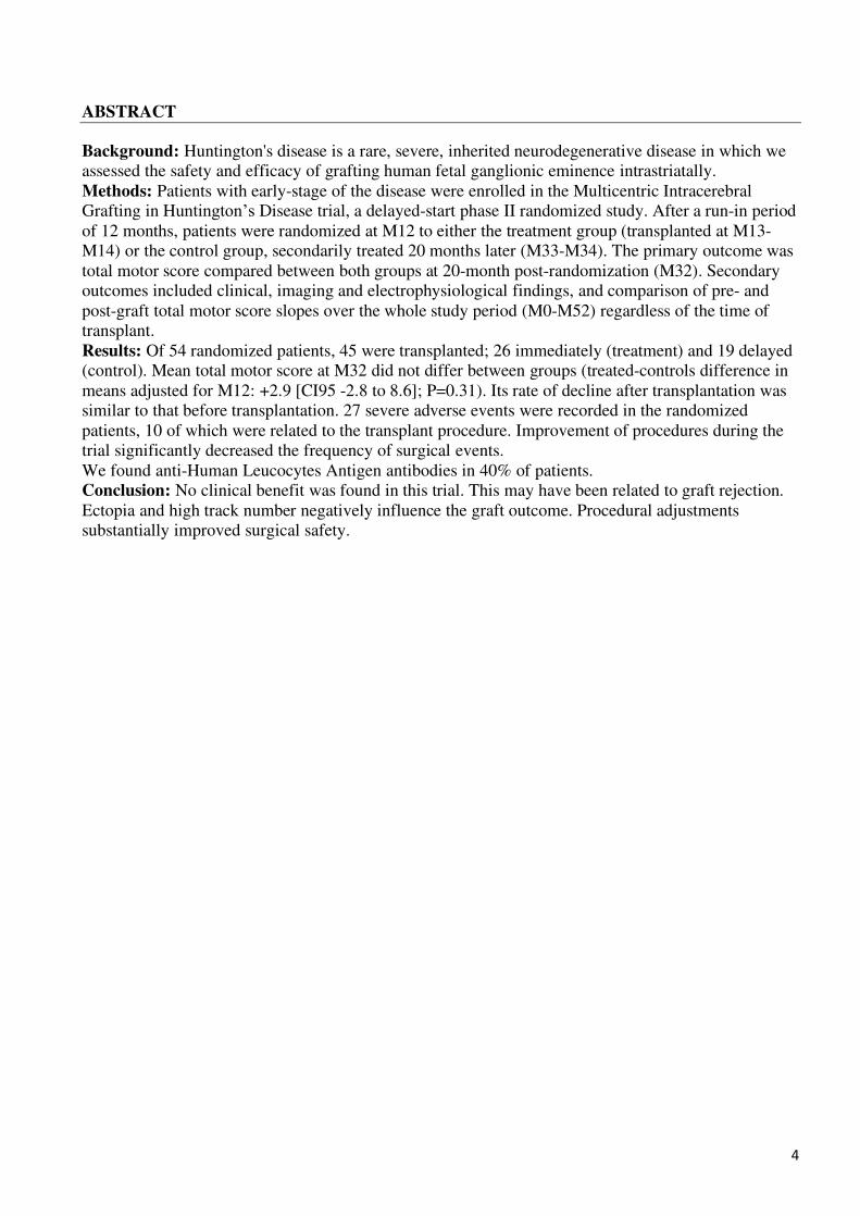
ABSTRACT
Background: Huntington's disease is a rare, severe, inherited neurodegenerative disease in which we
assessed the safety and efficacy of grafting human fetal ganglionic eminence intrastriatally.
Methods: Patients with early-stage of the disease were enrolled in the Multicentric Intracerebral
Grafting in Huntington’s Disease trial, a delayed-start phase II randomized study. After a run-in period
of 12 months, patients were randomized at M12 to either the treatment group (transplanted at M13-
M14) or the control group, secondarily treated 20 months later (M33-M34). The primary outcome was
total motor score compared between both groups at 20-month post-randomization (M32). Secondary
outcomes included clinical, imaging and electrophysiological findings, and comparison of pre- and
post-graft total motor score slopes over the whole study period (M0-M52) regardless of the time of
transplant.
Results: Of 54 randomized patients, 45 were transplanted; 26 immediately (treatment) and 19 delayed
(control). Mean total motor score at M32 did not differ between groups (treated-controls difference in
means adjusted for M12: +2.9 [CI95 -2.8 to 8.6]; P=0.31). Its rate of decline after transplantation was
similar to that before transplantation. 27 severe adverse events were recorded in the randomized
patients, 10 of which were related to the transplant procedure. Improvement of procedures during the
trial significantly decreased the frequency of surgical events.
We found anti-Human Leucocytes Antigen antibodies in 40% of patients.
Conclusion: No clinical benefit was found in this trial. This may have been related to graft rejection.
Ectopia and high track number negatively influence the graft outcome. Procedural adjustments
substantially improved surgical safety.
5
INTRODUCTION
HD is a rare inherited neurodegenerative disorder, which causes cognitive, behavioral, and motor
deficits, often beginning in early adulthood. Genetic diagnosis is unequivocal for patients with more
than 39 CAG repeats in the huntingtin gene.1 Despite intense pathophysiological research, disease-
modifying treatments remain elusive, and patients have a mean survival, with considerable dispersion,
of 20 years after motor onset.2 Gene-silencing therapies are promising, but will probably be more
effective for prevention than restoration. Multiple therapeutic strategies would presumably be required,
particularly for individuals already displaying striatal degeneration.
In HD, degeneration of neurons is particularly marked in the striatum, although not exclusive to this
region.3 Striatal quinolinic acid (QA) lesions in experimental animals indicate that massive losses of
striatal medium-sized spiny neurons, as occur in HD, can trigger progressive cortical projection neuron
degeneration. Homotopic transplantation of cells derived from the ganglionic eminence (the fetal zone
giving rise to the striatum) can replace the lost striatal neurons in rodent and non-human primate QA
lesion models, partially restoring frontostriatal connections and striatal efferent links to output nuclei,
and promoting recovery of cognitive and motor functions.4,5 Despite little neurodegeneration in R6/2
transgenic mice,6 modest improvement in locomotion was recorded after ganglionic eminence grafting.7
Functional improvement was also reported in transgenic models following stem cell-derived transplants
(e.g.8,9). Since the 1990s, 70 HD patients10 have been enrolled in open-label, non-randomized, single-
center trials (1-16 participants) of striatum-reconstructing treatments. These studies were too
heterogeneous (different cell sources, tissue preparations, and surgical protocols) and underpowered to
be conclusive or to drive improvements for future trials. Nevertheless, some patients showed clear signs
of sustained improvement.11–13 Graft-host connection was demonstrated in post mortem samples,14 with
structures resembling normal striatum in the grafted region, cortical and nigral afferents from the host,
and efferent to downstream pallidal nuclei and substantia nigra.15,16 International guidelines consider
cell transplantation into the brain to be safe17,18 despite some reports of overgrowth, graft tissues ectopic
to the target area,19,20 and subdural hematomas (SDHs).14
We set up a phase II randomized controlled trial; Multicentric Intracerebral Grafting in Huntington’s Disease (MIG-HD), to assess the safety and efficacy of human fetal cell intrastriatal transplantation in
patients with early-stage HD. This report summarizes the main study findings and key lessons learned
during the course of the trial. We identified factors that may influence transplant functionality for
consideration in future trials.
6
METHODS
Study design and oversight
MIG-HD was a multicenter randomized phase II study assessing the safety of intrastriatal human fetal
cell transplantation and its effect on motor function in patients with early-stage HD. The study was
conceived as a delayed-start design, where active treatment is sequentially provided to all participants
over time, so that all patients could eventually benefit from the transplantation procedure21. The study
was approved by the institutional review boards of Henri Mondor Hospital in France and Erasme
Hospital in Belgium. It complied with the Helsinki Declaration, current Good Clinical Practice
guidelines, and local laws and regulations. Written informed consent was obtained from patients at M0
or M1.22 An independent safety committee monitored the study conduct, the collected data and any
severe adverse events (SAEs). The protocol was registered at ClinicalTrials.gov (NCT00190450).
Methodological details are provided in the supplementary methods.
Participants
Consenting patients with genetic diagnoses of HD underwent transplantation at six French and Belgian
hospitals between 2001 and 2010; their follow-up to M52 was completed in 2013. The main inclusion
criteria were: having manifest HD for ≥1 year, >36 CAG repeats in the huntingtin gene, age 18-65 years,
total motor score (TMS) >5 on the Unified Huntington’s Disease Rating Scale (UHDRS), and total
functional capacity (TFC) score >9. The main exclusion criteria were: Mattis Dementia Rating Scale
(MDRS) score <120, and contraindication for surgery or magnetic resonance imaging (MRI)
(supplementary methods).
Randomization and masking
After a one-year run-in period, designed to verify patients’ compliance and exclude unusual patterns of
clinical deterioration, patients were randomly assigned at month 12 (M12) in a 1:1 ratio either to
treatment (receiving transplant at M13-M14) or to the (initially untreated) control group, which were
subsequently grafted 20-months later (M33-M34) (Figure S1). Randomization was computer-generated,
with centralized allocation concealment. A randomisation list prepared at the Henri Mondor Clinical
Research Unit with Nquery software (Statistical Solutions Ltd., Boston, USA) was used. Participants
and investigators responsible for clinical follow-up were not blind to treatment allocation. However, the
validity of the primary outcome (UHDRS TMS excluding rigidity) was assessed by video recordings at
M12, M32 and M52 and scored by specialists not involved in patient follow-up and recruitment and
blind to treatment allocation (Figure S2).
Procedures
Small blocks of whole ganglionic eminences from one to three 8.5- to 12-week-old fetuses (mean
±standard deviation: 1.6±0.6) per grafting session were implanted stereotactically, within 48 hours of
retrieval, into the striatum ipsilateral to the dominant hand. A mean of 2.45±3.03 months later, the
contralateral striatum was grafted (supplementary methods). Cells were injected through six tracks
(mean 4.91±1.46; range 3 to 6) within the head of the caudate nucleus (pre-commissural and
commissural) and the putamen (one in each of pre-commissural and commissural, and two in post-
commissural putamen). This totalled a volume of 206.0±43.1 µL unilaterally, distributed as 8 deposits
per track (mean 5.1±1.0 µL by deposit) with significant variations across centres. Two tracks were
omitted after the first 29 grafting sessions, to avoid SDH in patients with major striatal atrophy.
Cerebrospinal fluid leakage was limited by confinement to bed and hyperhydration for 48 h after surgery.
7
Immunosuppression was achieved with cyclosporine A, beginning three days before surgery (400
mg/day, then adjusted to maintain blood concentrations between 100 and 150 mg/L), prednisolone (0.25
mg/kg per day) and azathioprine (0.75 mg/kg per day) both initiated on the day of surgery. Cyclosporine
A was stopped six months after the second transplantation, and prednisolone and azathioprine were
stopped six months later. After the occurrence of acute graft rejection and the identification of Human
Leucocytes Antigen (HLA) antibodies in 30% of the patients tested,23 guided by international experts in
immunology, we established a new immunosuppression protocol for the last 20 patients. This involved
monitoring HLA antibodies at each centre and prolongation of full immunosuppression for up to one
year after the second graft. Azathioprine and prednisolone were continued for six additional months, and
prednisolone was withdrawn gradually. Plasma HLA antibodies were then monitored locally at each
hospital, and treatment was modified (withdrawal of cyclosporine or of prednisolone) on occurrence of
any unusual signs. Oral immunosuppressive therapy was withdrawn if no HLA antibodies against the
grafts were detected.
Short and full assessments were alternated for clinical examination (Figure S1). We used the complete
UHDRS, cognitive tasks,24 back-and-forth hand-tapping, and electrophysiological assessments. When
surgery could not be done on the scheduled date due to lack of foetus availability, preoperative
assessments were repeated if the interval between them and the transplant exceeded three months. Brain
imaging included MRI, 18F-fluorodeoxyglucose PET (FDG-PET) and, in patients not on neuroleptics,
with 11C-raclopride PET (supplementary methods).
Endpoints
The primary outcome was the UHDRS-TMS compared between treatment and control groups at 20-
month post-randomization (M32). TMS is a composite score for chorea, dystonia, oculomotor
movement, tapping, pronation/supination, palm/hand/fist sequence task, walking, tongue protrusion and
rigidity, rated from 0 to 124 points, with higher scores indicating poorer performance. Secondary
outcomes included clinical, imaging and electrophysiological findings, as well as comparison of pre-
and post-graft TMS slopes over the whole study period (M0-M52) regardless of the time of transplant.
Adverse events (AEs) were identified on clinical examination, according to the World Health
Organization checklist, at all visits and between visits if spontaneously reported by patients (Table S1).
Statistical analysis
Sample size calculation relied on data from an observational cohort of early HD patients comparable to
those included in the present trial and followed for up to 4 years,24 showing an average annual natural
progression of +13.2±14.1 for the UHDRS-TMS. Hypothesizing a stable evolution as a clinically
meaningful effect of the graft, inclusion of ≥18 subjects per group was required to achieve 80% power
at a 2-sided 5% alpha level. To account for a prespecified subgroup analysis led in graft recipients with
a metabolically active transplant based on FDG-PET imaging (60% expected like in 11), a sample size
of 60 (30 per group) was targeted.
For the primary outcome, patients were assessed according to randomized group under the modified
intent-to-treat principle, including all patients from the control group and patients from the treatment
group having received a transplant. The main planned primary endpoint analysis relied on the
comparison of the TMS at M32 between treatment and control groups using analysis of covariance
(ANCOVA) of the score at M32 with the initial value at M12 as a covariate. Supportive sensitivity
analyses of the primary endpoint included: i) ANCOVA with further adjustment for centre and other
covariates at M12 with prognostic value or showing evidence of a potential imbalance between study
arms at the time of randomization and/or transplant; ii) comparing the absolute change in TMS from
M12 to M32 between the two randomized groups and iii) assessing the graft effect on the evolution of
8
TMS over time (M0-M52) regardless of the randomized group using a piecewise two-part (before-after
the first transplant) linear mixed model.
Clinical and electrophysiological secondary endpoints were compared between randomized groups
using ANCOVA of values at M32 with values at M12 as a covariate, adjusting for similar covariates as
for the primary outcome, with the addition of the total motor score. Potential effect modifiers that could
predict improved response to intrastriatal transplant were searched for from a preselected list of 21
variables relating to patients and intervention, by testing for interactions between time after first graft
and the candidate predictors in a piecewise linear mixed model (supplementary methods).
All tests were two-tailed, with P<0.05 considered significant. Analyses were prespecified in the trial
protocol and performed with Stata v15.1 (StataCorp, College Station, USA) and R-3.6.0 (R Foundation,
Vienna, Austria).
Following the discovery of immune rejection,23 detection of antibodies directed against HLA class I and
class II antigens expressed by donor tissues was assessed in each center, using the locally available
technique.
MRI Analyses
MRI was planned as part of the study design for safety only. We conducted a retrospective volumetric
segmentation analysis using the Freesurfer software in patients scanned on the same machine for PET-
coregistration (supplementary methods).
9
Results
Between January 2001 and May 2006, 66 patients met the inclusion criteria (M0-M1), 54 were
randomized (M12), and 45 underwent transplantation (treatment group: 24 bilateral and 2 unilateral;
and controls secondarily grafted: 17 bilateral and 2 unilateral) (Figure 1). Unilateral implantations
were due to cancellation of the contralateral transplantation following serious surgical complications
after the first transplant in two patients, and to the decision of two others not having a second
transplant following several cancellations of surgery due to insufficient tissue collection. Demographic
and baseline characteristics are shown in Table 1. Patient demographic and clinical characteristics
were not significantly different between the two groups at the M12 randomization time point, except
for a longer disease duration and a more severe 1-figure cancellation task for the treatment group.
Median follow-up was 56.9 months (interquartile range [IQR] 54.5-64.1) for the treatment group, and
60.0 months (IQR 56.6-65.7) for controls.
Safety
We recorded 287 AEs from M0 to M52 in the 54 randomized patients over a period of 12 years (Table S1); 91% were not attributed to the procedure and 9% related to the procedure (immunosuppressant or
transplant). Among those, there were 27 SAEs, of which 17 were considered unrelated to the procedure:
one death by suicide, two suicide attempts, three fractures, one road accident, one acute fever, two
gastrointestinal disorders, one pulmonary embolism, and six hospitalizations for psychiatric disorders.
Ten SAEs were procedure-related: one intracranial empyema, three SDHs (two requiring surgical
drainage), one putaminal hematoma resulting in hemiparesis and aphasia, one seizure, one graft
rejection,23 and three intrastriatal cysts. Due to progressive cranial hypertension, one of these patients
with an intra-graft cyst required cauterisation of aberrant choroid plexus within the graft. Following this,
the patient improved clinically and in terms of his striatal metabolism (ipsilateral to the cyst) compared
to pre-surgery. Surgical and postoperative procedures were modified to prevent further hematomas in
the following 57 grafts, leading to significant improvement (Fisher’s test P=0.03).
Despite cyclosporine monitoring and dose titration, eighteen of the 43 patients tested (39 during the 52-
month study and four subsequently) were positive for HLA antibodies. We did not find correlation
between the clinical results and the presence of HLA antibodies.
Efficacy
M32 TMS scores did not differ significantly between treatment (50.8±17.3, N=26) and control groups
(39.0±17.0, N=26; ANCOVA adjusted for M12: P=0.31, adjusted difference in means: +2.9 [CI95 -2.8
to 8.6]). This was confirmed by supportive analyses after adjustment for disease duration (P=0.54),
center (P=0.30), or multiple adjustment for both and other potentially influent covariates (i.e.
independence scale, functional assessment scale, 1-figure cancellation, categorical fluency (1 min.);
P=0.68), and in comparisons of mean absolute TMS change from M12 to M32 (+10.3±standard error
2.3 [treatment] vs. +8.1±2.1 [controls], P=0.52, Table 2). A longitudinal analysis of graft effect on TMS,
regardless of group randomization, found no difference between the pre-graft and post-graft progression
slopes (piecewise linear mixed model, P=0.65; Figure 2A). The reliability of clinician-rated TMS,
assessed by blind scoring on the 96 exploitable videos from M12 to M52, was excellent (intraclass-
correlation coefficient=0.92 with 95% CI [0.88;0.94] and P<0.001) (Figure S2).
No significant striatal metabolic differences were observed in FDG PET-scans between M12 and M32
in either treated (N=26) or control (N=19; Figure 3) patients. At M32, eight treated patients showed a
10
non-significant lower number of hypometabolic striatal voxels compared to M12 (means M12:
1519.3±395.9 and M32: 1308.0±315.1). Their TMS (mean 49.8±10.7) was similar to that of control
patients (ANCOVA adjusted for M12: P=0.46). As for clinical and electrophysiological secondary
endpoints, no statistically significant differences were found between randomized groups between M12
and M32, adjusted for potentially confounding covariates (i.e. M12 values of total motor score, 1-figure
cancellation, categorical fluency (1 min.), independence scale, functional assessment scale and disease
duration), except for Stroop word showing a more severe decrease in the treated, than in the control
group (Table 2).
Analyses of basal ganglia MRI volumes between M12 and M32 showed a significant increase of the
striatal volume in treated patients (N=13) compared to controls (N=16, P<0.001) without correlation
with clinical scores (supplementary methods).
Exploratory analyses were performed on 10 parameters characterizing the patients’ pattern and 11
procedural aspects to identify potential predictors of transplantation outcome
(supplementary methods). Interaction analyses in the longitudinal linear mixed model detected two
detrimental predictors of steeper decline in post-graft TMS: ectopia (interaction term -0.29 [CI95% -
0.58 to -0.002], P=0.049) and a trend for high number of tracks per side ≤5.5 (-0.25 [-0.51 to 0.047],
P=0.067) (Figures 2B and 2C).
11
Discussion
This randomized multicentre delayed-start phase II trial was designed to assess the safety and efficacy
of the intrastriatal transplantation of human fetal cells in 54 patients in early to moderate stages of HD,
of whom 45 were eventually grafted. A comparison of the treatment (N=26) and control groups (N=19)
at M32 showed no improvement in TMS, even after restricting the analysis to the treated patients
identified as having an increased striatal metabolism on FDG-PET-imaging. TMS slope was unaffected
by transplantation. No benefit for secondary outcomes was observed (Table 2). We observed no increase
in raclopride binding, suggesting no/little increase in striatal-like tissue, and no metabolic improvement
in the striatum or frontal cortex post-transplantation in 80% of the grafted patients.25 This may have been
due to implantation of insufficient quantities of tissue or poor tissue survival for a range of reasons
including graft rejection, the latter according with the demonstration of transplant alloimmunogenicity23
in 41% patients tested for HLA antibodies.
Human fetal cells dissected from the developing striatum are theoretically good donor cells for
transplantation in HD patients, but their availability is limited. This limitation necessitated a long study
period (2001-2013) but did not affect the planned analyses, with repeated assessments for the
comparison of treated and control (secondarily transplanted) patients. The high degree of consistency of
blinded and investigator-attributed TMS scores demonstrates robustness (but possibly also insensitivity)
of TMS scoring. Of note, an imbalance in TMS values at M12 was apparent between controls and treated
patients, despite randomization. This observation most likely did not affect our findings based on
between-groups comparisons adjusted for M12 values, with comparable results found in the longitudinal
analysis of TMS in all grafted patients, regardless of initial group allocation.
Deaths occurred even before randomization (Figure S1), highlighting the fragility of patients with HD.
Where appropriate, protocol adaptations were made during the study to address AEs, improve patient
safety and prevent transplantation-related SAEs (see methods), without modifying the statistical validity
of the trial. The initial surgical procedure, which resulted in SDH or putaminal hematoma in 10% of
transplant recipients, compared favourably with the 43% reported in some pilot studies of fetal cell
transplantation in HD.14 This risk was eliminated by omitting the two posterior tracks in patients with
marked atrophy, hyperhydrating patients and imposing 48 hours bed rest; no such events occurred in the
subsequent 57 surgical implantations. We also successfully treated an expanding choroid cyst within the
graft by endoscopic cauterisation of choroid cells. This strategy would likely be of value for future
stereotaxic surgical trials.
Only a few studies have reported unequivocal long-lasting transplant success, and little is known about
the factors underlying graft failure.10 Graft-host connectivity has been demonstrated,15 but previous
studies in small cohort of patients were unable to identify the key factors influencing transplant
outcome.14,26–30 The MIG-HD trial, with 45 grafted patients at six centres, will help to advance cell
transplantation practices for HD by identifying some key factors that need to be considered in future
studies. The transition from single-centre to multicentre settings resulted in greater variability between
centres than anticipated, particularly for surgery-related factors, resulting in substantial graft variability
across the study. For example, larger numbers of injection tracks were expected to improve graft
function, but our results suggest in contrast that slower deterioration of the TMS was associated with
lower number of tracks. This observation might result from a combination of the number of foetuses
(from 1 to 2), presence of HLA antibodies, patients’ gender, and duration of surgery; even if not proven statistically in these few individuals. It was unclear in the study by Paganini et al.30 whether ectopic
grafts had a negative impact on graft function. In a blind analysis of MRI images, we show here that
TMS deteriorated more in patients with ectopic transplants. Whereas we did not find any correlation
12
between striatal volume change measured using MRI and clinical evolution, recent MRI techniques
should constitute a key marker in future trials.3 In contrast, given the difficulty to avoid neuroleptic
intake in HD, alternative tracers in future longitudinal long-term studies should replace 11C-raclopride
PET imaging. The number of hypometabolic striatal voxels correlated with TMS on FDG PET-scans,
without allowing us to detect clinically responsive patients. This lack of consistent correlation of imaging
and clinical response reproduces the results of other studies also reporting alloimmunisation processes
against the graft.29,31 It might be the case that chronic inflammation due to alloimmunisation and
transplant variability blurred the picture. Alloimmunization23 was unpredictable and changes in
detection techniques during MIG-HD made it impossible to model the impact of HLA antibodies.
Compared to our pilot trial,11 the use of older fetuses, the pooling of ganglionic eminences from several
fetuses to increase graft volume, and reducing the inter-graft interval from one year to about two months,
may have increased the risk of alloimmunisation. Here, 40% of patients developed HLA antibodies
against the graft. In contrast, none of our patients from the pilot trial, with one-year intervals between
transplants, had antibodies against the transplant five years after surgery (unpublished data). In two
studies with short inter-graft intervals (2-7 months), HLA antibodies were present in 50% of patients in
the German branch of MIG-HD29 and 37.5% in the Firenze study.31 The results of the MIG-HD study
suggest that better standardization and control of procedures, with improvements in atrophic structure
targeting and cell injection methods, are required for future transplant studies. It should be possible to
decrease the numbers of ectopic grafts and injection tracks, but it will be harder to control HLA antibody
development. These antibodies were also present in patients on immunosuppressants despite a correct
cyclosporine titration, suggesting suboptimal immunosuppression protocol. Yet, establishing the link
between presence of HLA antibodies against the graft and its lack of functionality is difficult because,
except in the case of acute rejection,23 alloimmunisation appears to be a long process. However,
functional impact of alloimmunization, reported in monkeys,32 justifies better procedures to avoid
alloimmunisation in future studies. The future use of stem cell-derived neural precursors should resolve
many of the critical issues highlighted here, improving surgical intervention planning and facilitating
the use of well-defined homogeneous cell therapy products effectively matched with the patient’s characteristics in advance. There are also some factors not considered here, such as tissue
preparation,33,34 which could be addressed in further studies. Besides, in retrospect, the outcome
measures lacked sensitivity (see35), which calls for new sensitive digitalised measures, as developed in
the RepairHD program.
In summary, it could be concluded that grafts cannot restore the fronto-striatal circuits despite the
positive abundant animal literature,18 but we think that it would be premature to conclude this based on
the MIG-HD study, which has highlighted many important questions that need to be addressed. It would
also be premature to disregard the results of our previous pilot study, in which striking clinical
improvement was seen in three patients across multiple outcomes analysed blindly to each other (clinics
PET, electrophysiology, and digitalized movement analysis), including an increase of the metabolism in
the frontal cortex,13,25,27 together constituting a proof of concept. We thus believe that a rational approach
is to return to the bench to solve the issues raised here; if that can be achieved there may be a place for
intracerebral transplantation, which is the only approach currently available with the potential to reverse
the loss of striatal tissue. We propose that the lessons learned from MIG-HD could guide future
transplant trials, whether for HD or other neurodegenerative diseases.
13
Acknowledgments
The sponsor was Assistance Publique – Hôpitaux de Paris (Département de la Recherche Clinique et
du Développement, Clinical Research and Development Department) and by delegation, the Clinical
Research and Development Department (DRCD), which carries out research missions in accordance
with Article L.1121-1 of the French Public Health Code. AC Bachoud-Lévi is the principal investigator
of MIG-HD. The Association Française contre les Myopathies and the Fonds National de la Recherche
Scientifique provided complementary grants for the hospital in Brussels. The work was supported by
ANR-10-LABX-0087 IEC and ANR-10-IDEX-0001-02 PSL, ANR-11-INBS-0011 - NeurATRIS and
by the National Reference Centre for Huntington’s Disease (French Ministry of Health).
We thank the members of the Data and Safety Monitoring Board: from 2001 to 2005: Jean-Thomas
Vilquin (cell therapist, Institut de Myologie), Gilles Defer (neurologist, CHU Caen), Guido Nikkhah
(neurosurgeon, Germany), Murielle Vray (methodologist – Institut Pasteur), and from 2005 to the end
2013: Henri Kreis: nephrologist/immunologist (Necker, Paris), Gilles Defer (neurologist, CHU Caen),
Marie Vidailhet (neurologist, IHU-A-ICM, Paris), Jean-Thomas Vilquin (cell therapist, Institut de
Myologie), Carole Dufouil (methodologist/statistician, Bordeaux), Emmanuel Cuny (neurosurgeon,
Hôpital Pellegrin-Bordeaux) Anne Fagot-Largeault (ethicist; Collège de France). We wish to thank our
colleagues from Angers for their participation in data collection or the procedure: Audrey Olivier
(Research assistant), Anne Clavreul and Nicole Piard (cell therapist); Brussels: Corinne Liesnard
(virologist); Créteil: Marie-Françoise Boissé (neuropsychologist), Catherine Bourdet (Psychiatrist);
Lille: Kathy Dujardin (neuropsychologist), Eric Decorte (research assistant); Bruno Quesnel (Biologist),
Bruno Lukowiak (cell therapy engineer), Gustavo Touzet (neurosurgeon); Nantes: Pierre Renou
(neuropsychologist), Philippe Naveilhan (cell therapist), Séverine Le Dily (Research Assistant), Prof.
Philip David (gynecologist), Jean-Marie Vanelle (psychiatrist), Prof. Youenn Lajat (neurosurgeon);
Toulouse: Prof. Yves Lazorthes (PI Neurosurgeon), Suzanne Jozan (cell therapist). We thank Michel
Golmann and Henri Kreis for their advice on the alloimmunization part of the study and Jean-Luc Taupin
and Cristina Sampaio for their advices concerning the manuscript. Julie Sappa from Alex Edelman and
Associates was responsible for English editing.
The MIG-HD group
Anne-Catherine Bachoud-Lévi, the principal investigator of MIG-HD, supervised all aspects of the
study and was responsible for neurological and neuropsychological training. Drs. Catherine Schramm,
Christophe Lalanne and Renaud Massart curated the data. They ran the analyses with Prof. Etienne
Audureau. The PIs at the various centers were Prof. Christophe Verny, Prof. Philippe Menei (Angers),
Dr. Clemence Simonin, Prof. Pierre Krystkowiak, Prof. Serge Blond (Lille/Amiens), Dr. Frédéric
Supiot, Prof. Marc Levivier (Brussels), Prof. Jean-François Démonet, Prof. Jean-Christophe Sol
(neurosurgeon co-PI) (Toulouse), Prof. Philippe Damier (Nantes); Dr. Marc Peschanski and Dr.
Philippe Hantraye supervised cell therapy, Prof. Stéphane Palfi supervised surgery (Créteil,
neurosurgeon PI), Prof. Philippe Remy, Dr. Véronique Gaura and Sonia Lavisse supervised the PET-
scans, Drs. Pierre Brugières and Laurent Cleret de Langavant supervised MRI and analyzed the data
obtained, Prof. Bassam Haddad and Dr. Roland Jeny were responsible for obstetric supervision, Dr.
Patrick Maison was responsible for methodology, and Prof. Jean-Pascal Lefaucheur supervised the
electrophysiology studies. Amandine Rialland and David Schmitz participated in data curation and
administrative supervision, Dr. Dominique Challine oversaw the viral work, and Prof. Anne Rosser
was responsible for blind videoscoring.
The following participated in data collection and/or in the procedure:
Angers Hospital: Dr. Clarisse Scherer Gagou (neurologist), Ghislaine Aubin (neuropsychologist), Dr.
Bénédicte Gohier (psychiatrist), Dr. Claudia Montero-Menei (cell therapist), Dr. Francoise Lunel-
Fabiani (virologist), Prof. Philippe Descamps (gynecologist), Dr. Vivien Pautot (neurophysiologist),
Dr. Jean-Yves Tanguy (brain imaging).
14
Brussels Hospital: Dr. Nicolas Massager (neurosurgeon), Hichem Slama (neuropsychologist), Liliane
Tenenbaum (cell therapist), Serge Goldman (PET imaging), Philip David (brain imaging)
Créteil Hospital – co-ordinating center: Laurie Lemoine (neuropsychologist), Dr. Hélène Rouard (cell
therapist).
Lille/Amiens Hospital: Dr. Arnaud Delval (neurologist), Ms. Marie Delliaux (neuropsychologist),
Prof. Olivier Cottencin (psychiatrist), Ms. Julie Kerr-Conte (cell therapist), Dr. Laurence Bocket
(virologist), Dr. Francis Collier (gynecologist), Dr. François Cassim (neurophysiologist), Dr. Christine
Delmaire (brain imaging). Nantes Hospital: Dr. Isabelle Neveu (cell therapist), Prof. Yan Péréon (neurophysiologist), Dr.
Elisabeth Auffray-Calvier (brain imaging).
Toulouse Hospital: Dr. Fabienne Calvas (CIC physician), Dr. Olivier Parant (gynecologist), Dr.
Marion Simonetta-Moreau (neurophysiologist), Dr. Jean-Albert Lotterie (brain imaging), Prof. Pierre
Payoux (PET imaging), Prof. Jacques Izopet (virologist).
Author’s roles
Conceptualization: ACBL MP PH PMa
Methodology: ACBL PMa MP PH SP PR JPL DC PB RJ BH
Software: CSc CL LCL VG SL
Validation: CSc CL ACBL EA
Formal analysis: CSc CL RM EA
Investigation: ACBL CV PMe CSi PK SB FS ML JFD JCS PDam MP PH SP PR VG SL PB LCL BH
RJ PMa JPL ARi DS DC ARo LL HR KY CSG GA BG CMM FLF PDe VP JYT NM HS LT SG PDav
IN YP EAC AD MD OC JKC LB FCo FCas CD FCal OP MSM JAL PP JI
Resources: ACBL
Data curation: CSc DS AR RM ACBL
Writing (original draft preparation): ACBL CSc CL SP PR LCL JPL PH RM EA
Writing (review and editing): ACBL CV PMe CSi PK SB FS ML JFD JCS PDam MP PH SP PR VG
SL PB LCL BH RJ PMa JPL ARi DS DC ARo LL HR KY CSG GA BG CMM FLF PDe VP JYT NM
HS LT SG PDav IN YP EAC AD MD OC JKC LB FCo FCas CD FCal OP MSM JAL PP JI CSc CL
RM EA
Visualization: CS ACBL
Supervision: ACBL
Project administration: ACBL DS ARi
Funding acquisition: ACBL MP

15
Financial disclosures (for the preceding 12 months)
• Bachoud-Lévi Anne-Catherine Consulting and Advisory Board Membership with honoraria: Roche
Grants and Research: investment for the future ANR grant (Neuratris, Front EUR), national center of
reference for Huntington’s disease (DGOS, ministry of Health), PHRCs (DRCI grants).
Intellectual Property Rights: Cognitive assessments (SelfCog, CATEX, CALAP)
Salary: University Hospital
• Schramm Catherine - Grants: postdoctoral fellowship FRM
• Remy Philippe No financial disclosure for the preceding 12 months.
• Gaura Véronique No financial disclosure for the preceding 12 months.
• Lavisse Sonia No financial disclosure for the preceding 12 months.
• Massart Renaud Salary: Foundation AP-HP
• Aubin Ghislaine No financial disclosure for the preceding 12 months.
• Blond Serge No financial disclosure for the preceding 12 months.
• Bocket Laurence No financial disclosure for the preceding 12 months.
• Brugieres Pierre No financial disclosure for the preceding 12 months.
• Calvas Fabienne No financial disclosure for the preceding 12 months.
• Calvier Elisabeth - Consultancies: Roche France
• Cassim François - Honoraria: Biogen
• Challine Dominique No financial disclosure for the preceding 12 months.
• Cleret Laurent - Grants: IRESP-INSERM (APP Prévention et Promotion de la Santé) LI-CLERET-AAP18-PREV-
003
• Collier Francis No financial disclosure for the preceding 12 months.
• Cottencin Olivier - Honoraria : Speaker for Janssen, Indivior, and Bouchara
- Grants : DGOS (1 PHRC) & ARS (1 AO Fonds Addiction)
• Damier Philippe - Stock Ownership: CurePark
- Honoraria for lectures: Teva, Novartis
• David Philippe No financial disclosure for the preceding 12 months.
• Delliaux Marie No financial disclosure for the preceding 12 months.
16
• Delmaire Christine No financial disclosure for the preceding 12 months.
• Delval Arnaud No financial disclosure for the preceding 12 months.
• Démonet Jean-François - Advisory Boards :Vifor Pharma (Switzerland)
- Grants : EU Eurostars, Synapsis Stifftung (Switzerland), Fondation Leenaards (Switzerland),
Fondation Empiris (Switzerland), Swiss National Foundation, Vifor Pharma (Switzerland)
• Descamps Philippe No financial disclosure for the preceding 12 months.
• Lunel-Fabiani Françoise - Partnerships: governmental institutions (INSERM, ANRS)
- Grants: ANRS
• Gohier Bénédicte No financial disclosure for the preceding 12 months.
• Goldman Serge - Employment: Université libre de Bruxelles
- Honoraria: NMEu
- Grants: Walloon Region, Fonds Erasme, AVN, FRS-FNRS
• Haddad Basam - Consultancies: Roche diagnostics France
• Izopet Jacques No financial disclosure for the preceding 12 months.
• Jeny Roland No financial disclosure for the preceding 12 months.
• Kerre-Conte Julie No financial disclosure for the preceding 12 months.
• Krystkowiak Pierre No financial disclosure for the preceding 12 months.
• Lalanne Christophe No financial disclosure for the preceding 12 months.
• Lefaucheur Jean-Pascal - Salary: University Hospital
• Lemoine Laurie Salary : AP-HP
• Levivier Marc No financial disclosure for the preceding 12 months.
• Lotterie Jean-Albert No financial disclosure for the preceding 12 months.
• Maison Patrick No financial disclosure for the preceding 12 months.
• Massager Nicolas No financial disclosure for the preceding 12 months.
• Menei Philippe - Advisory Boards: Journal of neurosurgery
- Contracts: Expression santé
- Grants: Fondation de l’avenir • Montero-Menei Claudia
- Grants: Région Pays de la Loire
17
• Neveu Isabelle No financial disclosure for the preceding 12 months.
• Parant Olivier No financial disclosure for the preceding 12 months.
• Pautot Vivien No financial disclosure for the preceding 12 months.
• Payoux Pierre No financial disclosure for the preceding 12 months.
• Pereon Yann - Advisory Boards: Avexis, PTC, Alnylam, Axelys
- Honoraria: Novartis, Sanofi, Pfizer
• Rosser Anne - Consulting and Advisory Board Membership with honoraria: NMHD-UKRI MRC, UK KMP-UKRI
MRC, Roche HD Advisory Board
- Grants and Research: Medical Research Council (MRC), Healthcare Research Wales, CHDI
Foundation, C.A.R.E., European Commission Horizon 2020
- Honoraria: NMHD-UKRI MRC, UK KMP-UKRI MRC, Roche HD Advisory Board
• Rouard Hélène - Employment: paris Est University and Etablissement Français du Sang
- Partnerships: ulm, Bergen University, UCM madrid, Universidad Autónoma de Madrid
- Grants: H2020, ANR
• Schmitz David - Salary: AP-HP
• Simonetta-Moreau Marion - Honoraria: Allergan, Merz
• Simonin Clémence - Employment: CHU Lille
- Honoraria: puntual expertises
• Slama Hichem - Employment: ULB ERASME HOSPITAL
- Contracts: ULB ERASME HOSPITAL
• Sol Jean-Christophe No financial disclosure for the preceding 12 months.
• Supiot Frédéric No financial disclosure for the preceding 12 months.
• Tanguy Jean-Yves No financial disclosure for the preceding 12 months.
• Tenenbaum Lilane - Employment: Centre hospitalier universitaire vaudois.
- Grants: Swiss national research foundation (SNF grant n°31003A_179527) ), Biosafety Advisory
Council
• Verny Christophe - Grants : Fondation Maladies Rares GROUPAMA, AAP DGOS (ministère de la Santé)
• Scherer-Gagou Clarisse No financial disclosure for the preceding 12 months.
• Youssov Katia - Employment: AP-HP
- Honoraria for consultancies: Roche
• Palfi Stéphane

18
- Consultancies: Yes
- Advisory Boards: Yes
- Contracts: Yes
- Honoraria: Yes
- Grants: Yes
• Audureau Etienne No financial disclosure for the preceding 12 months.
• Peschanski Marc No financial disclosure for the preceding 12 months.
• Hantraye Philippe No financial disclosure for the preceding 12 months.
19
References
1. A novel gene containing a trinucleotide repeat that is expanded and unstable on Huntington’s disease chromosomes. The Huntington’s Disease Collaborative Research Group. Cell. 1993;72:971–983.
2. Ross CA, Aylward EH, Wild EJ, et al. Huntington disease: natural history, biomarkers and
prospects for therapeutics. Nat Rev Neurol. 2014;10:204–216.
3. Tabrizi SJ, Scahill RI, Owen G, et al. Predictors of phenotypic progression and disease onset in
premanifest and early-stage Huntington’s disease in the TRACK-HD study: analysis of 36-month
observational data. Lancet Neurol. 2013;12:637–649.
4. Palfi S, Condé F, Riche D, et al. Fetal striatal allografts reverse cognitive deficits in a primate
model of Huntington disease. Nat Med. 1998;4:963–966.
5. Dunnett SB, Nathwani F, Björklund A. The integration and function of striatal grafts. Prog
Brain Res. 2000;127:345–380.
6. Zimmermann T, Remmers F, Lutz B, Leschik J. ESC-Derived BDNF-Overexpressing Neural
Progenitors Differentially Promote Recovery in Huntington’s Disease Models by Enhanced Striatal Differentiation. Stem Cell Reports. 2016;7:693–706.
7. Dunnett SB, Carter RJ, Watts C, et al. Striatal transplantation in a transgenic mouse model of
Huntington’s disease. Exp Neurol. 1998;154:31–40.
8. Reidling JC, Relaño-Ginés A, Holley SM, et al. Human Neural Stem Cell Transplantation
Rescues Functional Deficits in R6/2 and Q140 Huntington’s Disease Mice. Stem Cell Reports. 2018;10:58–72.
9. Al-Gharaibeh A, Culver R, Stewart AN, et al. Induced Pluripotent Stem Cell-Derived Neural
Stem Cell Transplantations Reduced Behavioral Deficits and Ameliorated Neuropathological Changes
in YAC128 Mouse Model of Huntington’s Disease. Front Neurosci. 2017;11:628.
10. Bachoud-Lévi A-C. From open to large-scale randomized cell transplantation trials in
Huntington’s disease: Lessons from the multicentric intracerebral grafting in Huntington’s disease trial (MIG-HD) and previous pilot studies. Prog Brain Res. 2017;230:227–261.
11. Bachoud-Lévi AC, Rémy P, Nguyen JP, et al. Motor and cognitive improvements in patients
with Huntington’s disease after neural transplantation. Lancet. 2000;356:1975–1979.
12. Reuter I, Tai YF, Pavese N, et al. Long-term clinical and positron emission tomography
outcome of fetal striatal transplantation in Huntington’s disease. J Neurol Neurosurg Psychiatry. 2008;79:948–951.
13. Bachoud-Lévi A-C, Gaura V, Brugières P, et al. Effect of fetal neural transplants in patients
with Huntington’s disease 6 years after surgery: a long-term follow-up study. Lancet Neurol.
2006;5:303–309.
14. Hauser RA, Furtado S, Cimino CR, et al. Bilateral human fetal striatal transplantation in
Huntington’s disease. Neurology. 2002;58:687–695.
15. Cicchetti F, Saporta S, Hauser RA, et al. Neural transplants in patients with Huntington’s disease undergo disease-like neuronal degeneration. Proc Natl Acad Sci USA. 2009;106:12483–12488.
16. Cisbani G, Saint-Pierre M, Cicchetti F. Single-cell suspension methodology favors survival and
vascularization of fetal striatal grafts in the YAC128 mouse model of Huntington’s disease. Cell Transplant. 2014;23:1267–1278.
17. Freeman TB, Cicchetti F, Bachoud-Lévi AC, Dunnett SB. Technical factors that influence
neural transplant safety in Huntington’s disease. Exp Neurol. 2011;227:1–9.
18. Rosser AE, Bachoud-Lévi A-C. Clinical trials of neural transplantation in Huntington’s disease. Prog Brain Res. 2012;200:345–371.
19. Keene CD, Chang RC, Leverenz JB, et al. A patient with Huntington’s disease and long-
surviving fetal neural transplants that developed mass lesions. Acta Neuropathol. 2009;117:329–338.
20. Gallina P, Paganini M, Lombardini L, et al. Human striatal neuroblasts develop and build a
striatal-like structure into the brain of Huntington’s disease patients after transplantation. Exp Neurol.
20
2010;222:30–41.
21. Spineli LM, Jenz E, Großhennig A, Koch A. Critical appraisal of arguments for the delayed-
start design proposed as alternative to the parallel-group randomized clinical trial design in the field of
rare disease. Orphanet J Rare Dis. 2017;12:140.
22. Cleret de Langavant L, Sudraud S, Verny C, et al. Longitudinal study of informed consent in
innovative therapy research: experience and provisional recommendations from a multicenter trial of
intracerebral grafting. PLoS ONE. 2015;10:e0128209.
23. Krystkowiak P, Gaura V, Labalette M, et al. Alloimmunisation to donor antigens and immune
rejection following foetal neural grafts to the brain in patients with Huntington’s disease. PLoS ONE. 2007;2:e166.
24. Bachoud-Lévi AC, Maison P, Bartolomeo P, et al. Retest effects and cognitive decline in
longitudinal follow-up of patients with early HD. Neurology. 2001;56:1052–1058.
25. Gaura V, Bachoud-Lévi A-C, Ribeiro M-J, et al. Striatal neural grafting improves cortical
metabolism in Huntington’s disease patients. Brain. 2004;127:65–72.
26. Kopyov OV, Jacques S, Lieberman A, Duma CM, Eagle KS. Safety of intrastriatal
neurotransplantation for Huntington’s disease patients. Exp Neurol. 1998;149:97–108.
27. Bachoud-Lévi A, Bourdet C, Brugières P, et al. Safety and tolerability assessment of
intrastriatal neural allografts in five patients with Huntington’s disease. Exp Neurol. 2000;161:194–202.
28. Rosser AE, Barker RA, Harrower T, et al. Unilateral transplantation of human primary fetal
tissue in four patients with Huntington’s disease: NEST-UK safety report ISRCTN no 36485475. J
Neurol Neurosurg Psychiatry. 2002;73:678–685.
29. Krebs SS, Trippel M, Prokop T, et al. Immune response after striatal engraftment of fetal
neuronal cells in patients with Huntington’s disease: Consequences for cerebral transplantation programs. Clinical and Experimental Neuroimmunology. 2011;2:25–32.
30. Paganini M, Biggeri A, Romoli AM, et al. Fetal striatal grafting slows motor and cognitive
decline of Huntington’s disease. J Neurol Neurosurg Psychiatry. 2014;85:974–981.
31. Porfirio B, Paganini M, Mazzanti B, et al. Donor-Specific Anti-HLA Antibodies in
Huntington’s Disease Recipients of Human Fetal Striatal Grafts. Cell Transplant. 2015;24:811–817.
32. Aron Badin R, Bugi A, Williams S, et al. MHC matching fails to prevent long-term rejection of
iPSC-derived neurons in non-human primates. Nat Commun. 2019;10:4357.
33. Harrison DJ, Roberton VH, Vinh N-N, Brooks SP, Dunnett SB, Rosser AE. The Effect of
Tissue Preparation and Donor Age on Striatal Graft Morphology in the Mouse. Cell Transplant.
2018;27:230–244.
34. Cisbani G, Freeman TB, Soulet D, et al. Striatal allografts in patients with Huntington’s disease: impact of diminished astrocytes and vascularization on graft viability. Brain. 2013;136:433–443.
35. Schobel SA, Palermo G, Auinger P, et al. Motor, cognitive, and functional declines contribute
to a single progressive factor in early HD. Neurology. 2017;89:2495–2502.
21
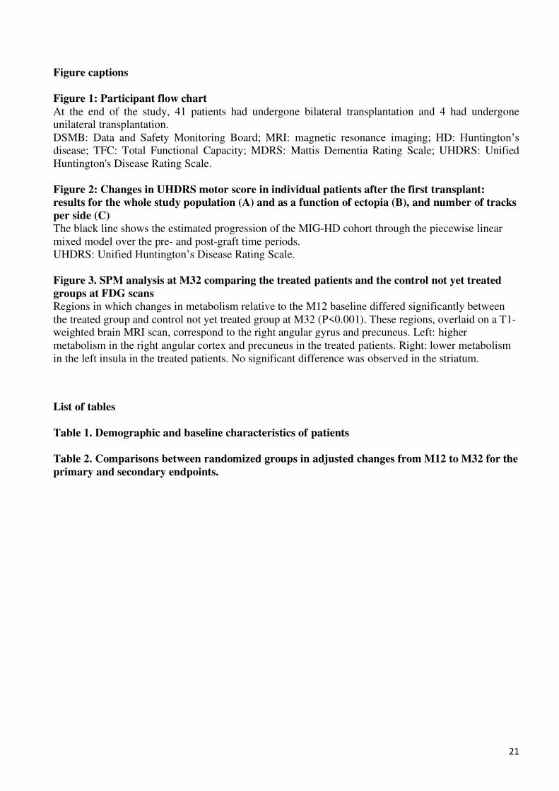
Figure captions Figure 1: Participant flow chart At the end of the study, 41 patients had undergone bilateral transplantation and 4 had undergone
unilateral transplantation.
DSMB: Data and Safety Monitoring Board; MRI: magnetic resonance imaging; HD: Huntington’s
disease; TFC: Total Functional Capacity; MDRS: Mattis Dementia Rating Scale; UHDRS: Unified
Huntington's Disease Rating Scale.
Figure 2: Changes in UHDRS motor score in individual patients after the first transplant: results for the whole study population (A) and as a function of ectopia (B), and number of tracks per side (C) The black line shows the estimated progression of the MIG-HD cohort through the piecewise linear
mixed model over the pre- and post-graft time periods.
UHDRS: Unified Huntington’s Disease Rating Scale.
Figure 3. SPM analysis at M32 comparing the treated patients and the control not yet treated groups at FDG scans Regions in which changes in metabolism relative to the M12 baseline differed significantly between
the treated group and control not yet treated group at M32 (P<0.001). These regions, overlaid on a T1-
weighted brain MRI scan, correspond to the right angular gyrus and precuneus. Left: higher
metabolism in the right angular cortex and precuneus in the treated patients. Right: lower metabolism
in the left insula in the treated patients. No significant difference was observed in the striatum.
List of tables Table 1. Demographic and baseline characteristics of patients
Table 2. Comparisons between randomized groups in adjusted changes from M12 to M32 for the primary and secondary endpoints.