Embed Size (px)
Citation preview

Phytochemistry xxx (2013) xxx–xxx
Contents lists available at ScienceDirect
Phytochemistry
journal homepage: www.elsevier .com/locate /phytochem
Steroids with anti-inflammatory activity from Vernonia nigritiana Oliv. &Hiern.
0031-9422/$ - see front matter � 2013 Elsevier Ltd. All rights reserved.http://dx.doi.org/10.1016/j.phytochem.2013.09.002
⇑ Corresponding author. Tel.: +39 040 5588836; fax: +39 040 5583215.E-mail address: [email protected] (S. Sosa).
Please cite this article in press as: Vassallo, A., et al. Steroids with anti-inflammatory activity from Vernonia nigritiana Oliv. & Hiern. Phytochemistryhttp://dx.doi.org/10.1016/j.phytochem.2013.09.002
Antonio Vassallo a, Nunziatina De Tommasi b, Irmgard Merfort c, Rokia Sanogo d, Lorella Severino e,Marco Pelin f, Roberto Della Loggia f, Aurelia Tubaro f, Silvio Sosa f,⇑a Department of Science, University of Basilicata, 85100 Potenza, Italyb Department of Pharmaceutical and Biomedical Sciences, University of Salerno, 84084 Fisciano, Salerno, Italyc Department of Pharmaceutical Biology and Biotechnology, University Freiburg, D-79104 Freiburg, Germanyd Departement de Medicine Traditionelle (DMT), INRSP, B.P. 1746 Bamako, Malie Department of Pathology and Animal Health, ‘‘Federico II’’ University of Naples, 80138 Napoli, Italyf Department of Life Sciences, University of Trieste, 34127 Trieste, Italy
a r t i c l e i n f o a b s t r a c t
Article history:Received 11 April 2013Received in revised form 27 August 2013Available online xxxx
Keywords:Vernonia nigritianaAsteraceaeAnti-inflammatory activityCroton oil ear testNF-jBSteroids
The leaves of Vernonia nigritiana Oliv. & Hiern. (Asteraceae) were investigated for their in vivo topicalanti-inflammatory properties, following a bioassay-oriented fractionation approach. Petroleum ether,chloroform and chloroform–methanol extracts inhibited the Croton oil-induced ear dermatitis in mice.The chloroform extract was only about half as active as the non steroidal anti-inflammatory drugindomethacin (ID50 = 237 and 93 lg/cm2, respectively). Phytochemical investigation of this extract ledto the isolation of nine polyhydroxylated stigmasterol glycosides and six polyhydroxylatedstigmasterols. Their structures were elucidated by NMR, MS and chemical methods. Each compoundexerted a significant anti-oedema activity, the most active being 1 (3b-O-b-D-glucopyranosyloxy-5a-stigmasta-7,9(11),24(28)Z-triene-6b,16b,26,29-tetrol) and 3 (3b-O-b-D-glucopyranosyloxy-5a-stigmas-ta-7,9(11),24(28)Z-triene-6b,16b,29-triol), only two and five fold less potent than the steroidal drughydrocortisone (ID50 = 0.10, 0.21 and 0.04 lmol/cm2, respectively). Compound 1 (50 lM) also completelyinhibited the transcription factor NF-jB in vitro.
� 2013 Elsevier Ltd. All rights reserved.
1. Introduction
Vernonia nigritiana Oliv. & Hiern. (Asteraceae) is an annual herbor shrub widely distributed in West Africa, where the leaves andother parts of the plant are traditionally used against skin inflam-mations and infections, rheumatism, fever, headache and digestiveinsufficiency (Johri and Singh, 1997; Toyang and Verpoorte, 2013).Previous studies on some Vernonia species revealed anti-inflamma-tory properties for their extracts or constituents (Cioffi et al., 2004;da Silva et al., 2011; Iwalewa et al., 2003; Latha et al., 1998; Liuet al., 2010; Mazumder et al., 2003; Ngatu et al., 2012; Rauhet al., 2011; Temponi Vdos et al., 2012; Toyang and Verpoorte,2013; Youn et al., 2012). Phytochemical studies showed the pres-ence of stigmastane glycosides, namely vernoniosides (Cioffiet al., 2004; Igile et al., 1995; Liu et al., 2009, 2010; Mazumderet al., 2003; Sanogo et al., 1998; Tchinda et al., 2002, 2003;Valverde et al., 2000) and other secondary metabolites, includingalkaloids, terpenoids, flavonoids, phenolic derivatives, isovaleryl
sucrose esters and coumarins (Johri and Singh, 1997; Ogunbinuet al., 2009; Tchinda et al., 2003; Toyang et al., 2012, 2013; Toyangand Verpoorte, 2013; Youn et al., 2012).
A study of V. nigritiana flowers led to the isolation of quercetinderivatives, while a bitter glycoside was isolated from the roots(Atindehou et al., 1993; Toyang and Verpoorte, 2013). In the pres-ent study, a phytochemical and pharmacological investigation ofV. nigritiana leaves was carried out following a bioassay-orientedfractionation procedure, to verify the rationality of their tradi-tional use in the treatment of inflammatory infections and toidentify the relevant active principles. The plant material wassubmitted to sequential extractions using solvents of increasingpolarity and the resultant extracts were evaluated for their topi-cal anti-inflammatory activity in the Croton oil-induced ear der-matitis model in mice (Tubaro et al., 1985). Bioassay-orientedfractionation of the most active extract, the chloroform one, ledto the isolation of nine new polyhydroxylated stigmasterol glyco-sides and six new stigmasterols which differ in their anti-inflam-matory activity. To verify a possible mechanism of action, in vitroinhibition of the nuclear transcription factor NF-jB was also eval-uated using the electrophoretic mobility shift assay (Lyss et al.,1997).
(2013),
*
**
*
*
*
**
** *
*
0
10
20
30
40
50
60
70
80
90
100
Petroleum ether extract
Chloroform extract
Chloroform-methanol extract
Indomethacin
% o
edem
a in
hibi
tion
30 µg 100 µg 300 µg 1000 µg
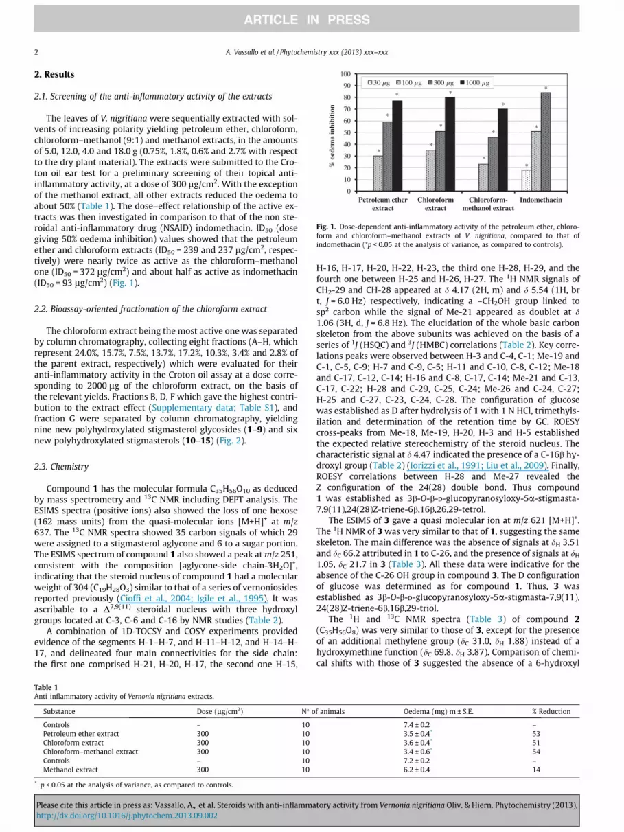
Fig. 1. Dose-dependent anti-inflammatory activity of the petroleum ether, chloro-form and chloroform–methanol extracts of V. nigritiana, compared to that ofindomethacin (⁄p < 0.05 at the analysis of variance, as compared to controls).
2 A. Vassallo et al. / Phytochemistry xxx (2013) xxx–xxx
2. Results
2.1. Screening of the anti-inflammatory activity of the extracts
The leaves of V. nigritiana were sequentially extracted with sol-vents of increasing polarity yielding petroleum ether, chloroform,chloroform–methanol (9:1) and methanol extracts, in the amountsof 5.0, 12.0, 4.0 and 18.0 g (0.75%, 1.8%, 0.6% and 2.7% with respectto the dry plant material). The extracts were submitted to the Cro-ton oil ear test for a preliminary screening of their topical anti-inflammatory activity, at a dose of 300 lg/cm2. With the exceptionof the methanol extract, all other extracts reduced the oedema toabout 50% (Table 1). The dose–effect relationship of the active ex-tracts was then investigated in comparison to that of the non ste-roidal anti-inflammatory drug (NSAID) indomethacin. ID50 (dosegiving 50% oedema inhibition) values showed that the petroleumether and chloroform extracts (ID50 = 239 and 237 lg/cm2, respec-tively) were nearly twice as active as the chloroform–methanolone (ID50 = 372 lg/cm2) and about half as active as indomethacin(ID50 = 93 lg/cm2) (Fig. 1).
2.2. Bioassay-oriented fractionation of the chloroform extract
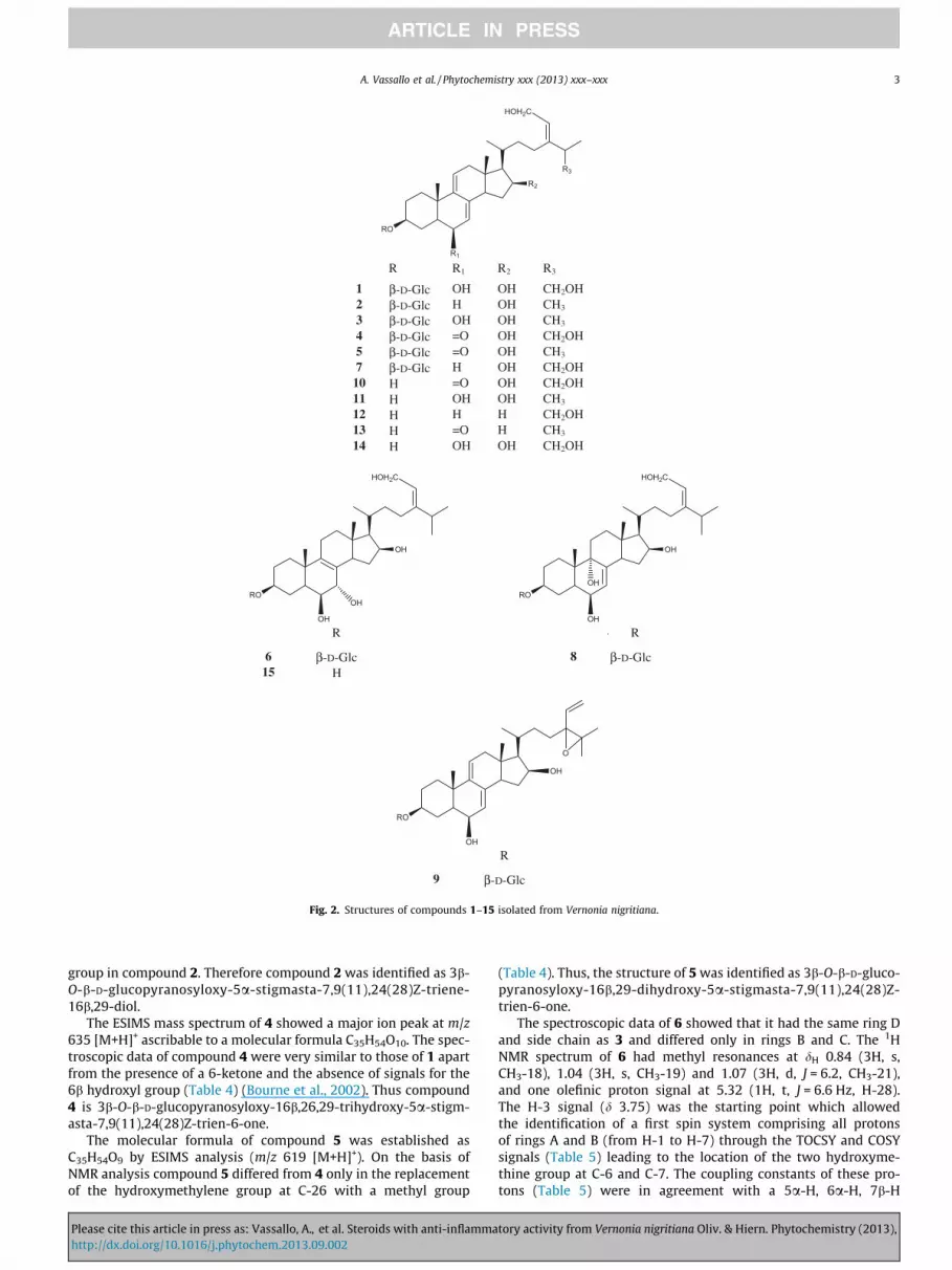
The chloroform extract being the most active one was separatedby column chromatography, collecting eight fractions (A–H, whichrepresent 24.0%, 15.7%, 7.5%, 13.7%, 17.2%, 10.3%, 3.4% and 2.8% ofthe parent extract, respectively) which were evaluated for theiranti-inflammatory activity in the Croton oil assay at a dose corre-sponding to 2000 lg of the chloroform extract, on the basis ofthe relevant yields. Fractions B, D, F which gave the highest contri-bution to the extract effect (Supplementary data; Table S1), andfraction G were separated by column chromatography, yieldingnine new polyhydroxylated stigmasterol glycosides (1–9) and sixnew polyhydroxylated stigmasterols (10–15) (Fig. 2).
2.3. Chemistry
Compound 1 has the molecular formula C35H56O10 as deducedby mass spectrometry and 13C NMR including DEPT analysis. TheESIMS spectra (positive ions) also showed the loss of one hexose(162 mass units) from the quasi-molecular ions [M+H]+ at m/z637. The 13C NMR spectra showed 35 carbon signals of which 29were assigned to a stigmasterol aglycone and 6 to a sugar portion.The ESIMS spectrum of compound 1 also showed a peak at m/z 251,consistent with the composition [aglycone-side chain-3H2O]+,indicating that the steroid nucleus of compound 1 had a molecularweight of 304 (C19H28O3) similar to that of a series of vernoniosidesreported previously (Cioffi et al., 2004; Igile et al., 1995). It wasascribable to a D7,9(11) steroidal nucleus with three hydroxylgroups located at C-3, C-6 and C-16 by NMR studies (Table 2).
A combination of 1D-TOCSY and COSY experiments providedevidence of the segments H-1–H-7, and H-11–H-12, and H-14–H-17, and delineated four main connectivities for the side chain:the first one comprised H-21, H-20, H-17, the second one H-15,
Table 1Anti-inflammatory activity of Vernonia nigritiana extracts.
Substance Dose (lg/cm2) N� o
Controls – 10Petroleum ether extract 300 10Chloroform extract 300 10Chloroform–methanol extract 300 10Controls – 10Methanol extract 300 10
* p < 0.05 at the analysis of variance, as compared to controls.
Please cite this article in press as: Vassallo, A., et al. Steroids with anti-inflammahttp://dx.doi.org/10.1016/j.phytochem.2013.09.002
H-16, H-17, H-20, H-22, H-23, the third one H-28, H-29, and thefourth one between H-25 and H-26, H-27. The 1H NMR signals ofCH2-29 and CH-28 appeared at d 4.17 (2H, m) and d 5.54 (1H, brt, J = 6.0 Hz) respectively, indicating a –CH2OH group linked tosp2 carbon while the signal of Me-21 appeared as doublet at d1.06 (3H, d, J = 6.8 Hz). The elucidation of the whole basic carbonskeleton from the above subunits was achieved on the basis of aseries of 1J (HSQC) and 3J (HMBC) correlations (Table 2). Key corre-lations peaks were observed between H-3 and C-4, C-1; Me-19 andC-1, C-5, C-9; H-7 and C-9, C-5; H-11 and C-10, C-8, C-12; Me-18and C-17, C-12, C-14; H-16 and C-8, C-17, C-14; Me-21 and C-13,C-17, C-22; H-28 and C-29, C-25, C-24; Me-26 and C-24, C-27;H-25 and C-27, C-23, C-24, C-28. The configuration of glucosewas established as D after hydrolysis of 1 with 1 N HCl, trimethyls-ilation and determination of the retention time by GC. ROESYcross-peaks from Me-18, Me-19, H-20, H-3 and H-5 establishedthe expected relative stereochemistry of the steroid nucleus. Thecharacteristic signal at d 4.47 indicated the presence of a C-16b hy-droxyl group (Table 2) (Iorizzi et al., 1991; Liu et al., 2009). Finally,ROESY correlations between H-28 and Me-27 revealed theZ configuration of the 24(28) double bond. Thus compound1 was established as 3b-O-b-D-glucopyranosyloxy-5a-stigmasta-7,9(11),24(28)Z-triene-6b,16b,26,29-tetrol.
The ESIMS of 3 gave a quasi molecular ion at m/z 621 [M+H]+.The 1H NMR of 3 was very similar to that of 1, suggesting the sameskeleton. The main difference was the absence of signals at dH 3.51and dC 66.2 attributed in 1 to C-26, and the presence of signals at dH
1.05, dC 21.7 in 3 (Table 3). All these data were indicative for theabsence of the C-26 OH group in compound 3. The D configurationof glucose was determined as for compound 1. Thus, 3 wasestablished as 3b-O-b-D-glucopyranosyloxy-5a-stigmasta-7,9(11),24(28)Z-triene-6b,16b,29-triol.
The 1H and 13C NMR spectra (Table 3) of compound 2(C35H56O8) was very similar to those of 3, except for the presenceof an additional methylene group (dC 31.0, dH 1.88) instead of ahydroxymethine function (dC 69.8, dH 3.87). Comparison of chemi-cal shifts with those of 3 suggested the absence of a 6-hydroxyl
f animals Oedema (mg) m ± S.E. % Reduction
7.4 ± 0.2 –3.5 ± 0.4* 533.6 ± 0.4* 513.4 ± 0.6* 547.2 ± 0.2 –6.2 ± 0.4 14
tory activity from Vernonia nigritiana Oliv. & Hiern. Phytochemistry (2013),
R3
HOH2C
RO
R2
R1
R R1 R2 R3
1 β-D-Glc OH OH CH2OH2 β-D-Glc H OH CH3
3 β-D-Glc OH OH CH3
4 β-D-Glc =O OH CH2OH5 β-D-Glc =O OH CH3
7 β-D-Glc H OH CH2OH10 Η =O OH CH2OH11 Η OH OH CH3
12 Η H H CH2OH13 Η =O H CH3
14 Η OH OH CH2OH
HOH2C
RO
OH
OH
OH
HOH2C
RO
OH
OH
OH
R • R
6 β-D-Glc 8 β-D-Glc15 Η
RO
OH
O
OH
R
9 β-D-Glc
Fig. 2. Structures of compounds 1–15 isolated from Vernonia nigritiana.
A. Vassallo et al. / Phytochemistry xxx (2013) xxx–xxx 3
group in compound 2. Therefore compound 2 was identified as 3b-O-b-D-glucopyranosyloxy-5a-stigmasta-7,9(11),24(28)Z-triene-16b,29-diol.
The ESIMS mass spectrum of 4 showed a major ion peak at m/z635 [M+H]+ ascribable to a molecular formula C35H54O10. The spec-troscopic data of compound 4 were very similar to those of 1 apartfrom the presence of a 6-ketone and the absence of signals for the6b hydroxyl group (Table 4) (Bourne et al., 2002). Thus compound4 is 3b-O-b-D-glucopyranosyloxy-16b,26,29-trihydroxy-5a-stigm-asta-7,9(11),24(28)Z-trien-6-one.
The molecular formula of compound 5 was established asC35H54O9 by ESIMS analysis (m/z 619 [M+H]+). On the basis ofNMR analysis compound 5 differed from 4 only in the replacementof the hydroxymethylene group at C-26 with a methyl group
Please cite this article in press as: Vassallo, A., et al. Steroids with anti-inflammahttp://dx.doi.org/10.1016/j.phytochem.2013.09.002
(Table 4). Thus, the structure of 5 was identified as 3b-O-b-D-gluco-pyranosyloxy-16b,29-dihydroxy-5a-stigmasta-7,9(11),24(28)Z-trien-6-one.
The spectroscopic data of 6 showed that it had the same ring Dand side chain as 3 and differed only in rings B and C. The 1HNMR spectrum of 6 had methyl resonances at dH 0.84 (3H, s,CH3-18), 1.04 (3H, s, CH3-19) and 1.07 (3H, d, J = 6.2, CH3-21),and one olefinic proton signal at 5.32 (1H, t, J = 6.6 Hz, H-28).The H-3 signal (d 3.75) was the starting point which allowedthe identification of a first spin system comprising all protonsof rings A and B (from H-1 to H-7) through the TOCSY and COSYsignals (Table 5) leading to the location of the two hydroxyme-thine group at C-6 and C-7. The coupling constants of these pro-tons (Table 5) were in agreement with a 5a-H, 6a-H, 7b-H
tory activity from Vernonia nigritiana Oliv. & Hiern. Phytochemistry (2013),

Table 213C and 1H NMR data (600 MHz, CH3OD) for compounds 1, 7, 12 and 14.
Positions 1 7 12 14
dC dH (J in Hz) dC dH (J in Hz) dC dC
Aglycone Aglycone
1 36.3 1.40a 35.0 1.05 35.5 36.31.99 1.45a
2 30.5 1.66 30.5 1.95 m (13.0, 7.0) 30.5 30.02.42 dt (13.0, 7.0)
3 78.7 3.71 m 78.4 3.75 m 71.0 71.04 30.6 1.31a 35.9 1.56 35.0 31.0
2.02 2.005 48.3 1.26a 41.0 1.33 41.0 48.46 69.8 3.87 br dd (6.0, 1.5) 31.4 1.86 br d (10.0, 5.0) 31.4 69.7
1.90 br d (10.0, 8.0)7 126.0 5.40 br d (1.5) 122.0 5.44 dd (6.0, 2.5) 122.0 125.98 137.0 – 136.8 – 136.8 136.89 144.5 – 145.5 – 145.3 145.010 37.0 – 37.8 – 37.5 37.311 121.2 5.59 br d (5.0) 119.0 5.52 br d (6.0) 119.0 121.312 43.9 2.14 br d (16.8) 40.5 1.30a br d (16.8) 40.5 43.4
2.36 dd (16.8, 6.0) 2.42 dd (16.8, 6.0)13 43.5 – 44.5 – 43.9 43.114 50.5 2.13 dd (11.0, 5.5) 49.5 2.53 48.3 50.615 37.2 1.53a 36.0 1.51a 25.6 36.6
2.44 dd (11.0, 8.0, 5.0) 2.4016 72.8 4.47 m 74.4 4.47 27.9 72.417 62.5 1.28a 61.2 1.28a 59.6 61.218 13.0 0.76 s 13.7 0.76 s 12.9 12.819 18.7 1.00 s 19.8 0.96 s 18.6 19.520 31.6 1.97 m 31.5 1.97 m 31.6 31.521 18.7 1.06 d (6.8) 18.8 1.04 d (6.8) 18.6 18.722 36.6 1.25a 35.7 1.39a 36.6 35.7
1.88 1.8723 28.7 1.99 28.7 1.95 28.7 28.7
2.14 2.1124 148.5 – 145.6 – 145.7 145.625 38.0 2.89 br q (13.8, 6.6) 38.2 2.90 m 38.8 38.026 66.2 3.51 m 66.2 3.49 m 66.2 66.227 21.3 1.05 d (6.1) 21.3 1.07 d (6.6) 21.3 21.328 126.6 5.54 br t (6.0) 126.6 5.53 t (6.0) 126.6 126.629 58.5 4.17 m 58.5 4.16 m 59.0 58.5
Glc Glc10 102.3 4.46 d (7.2) 102.7 4.48 d (7.8)20 75.1 3.20 br t (8.4) 75.0 3.19 br t (8.8)30 78.1 3.39 t (9.0) 77.9 3.27 t (9.0)40 71.7 3.31 t (9.0) 70.8 3.31 t (9.0)50 77.9 3.29 m 78.0 3.38 m60 62.8 3.69 dd (12.5, 5.0) 62.5 3.70 dd (12.5, 5.0)
3.90 dd (12.5, 3.0) 3.88 dd (12.5, 3.0)
Glcb-D-glucopyranosyl.a Signal overlapped.
4 A. Vassallo et al. / Phytochemistry xxx (2013) xxx–xxx
arrangement. The 13C NMR spectra for the aglycone moiety of 6showed the presence of 29 carbons, 19 of them were assignedto the steroidal nucleus including a glycosylated oxymethine (C-3, d 78.9) and two sp2 carbons to a tetrasubstituted double bondat d 129.6 and 142.7 (each quaternary carbon). The presence of atetrasubstituted double bond located between C-8 and C-9 wassupported by the HMBC correlations of C-9 with H-7 and H-11;C-8 with H-6, H-14 and H-15 and by comparison with reporteddata (Table 5) (Dellagreca et al., 2001; Qi et al., 2007). Combinedanalysis of the COSY and HSQC spectra allowed the assignment(Table 5) of the remaining protons and carbons of the aglyconemoiety, including a C10H19O side chain, and demonstrated thatthis side chain was the same as in compound 3. The NMR datafor the sugar residue linked at C-3 of the aglycone in 6 revealedthe presence of an unsubstituted b-D-glucopyranosyl unit (Sanogoet al., 1998). Thus, the structure of compound 6 was establishedas the new 3b-O-b-D-glucopyranosyloxy-5a-stigmasta-8(9),24(28)Z-diene-6b,7a,16b,29-tetrol.
The spectroscopic properties of 7 and 1 were very similar exceptfor the absence of signals for the 6-hydroxyl group (Liu et al.,
Please cite this article in press as: Vassallo, A., et al. Steroids with anti-inflammahttp://dx.doi.org/10.1016/j.phytochem.2013.09.002
2009). Thus, 7 is the 6-deoxy derivative 3b-O-b-D-glucopyranosyl-oxy-5a-stigmasta-7,9(11),24(28)Z-triene-16b,26,29-triol.
The ESIMS of 8 revealed a molecular ion at m/z 639 [M+H]+, to-gether with ions at m/z 477 and 621 corresponding to the loss of aglucose and H2O from the parent ion, respectively. This informa-tion along with the 13C NMR spectra, which sorted 35 carbons into5 methyls, 10 methylenes, 15 methines and five quaternary car-bons, allowed the determination of the molecular formula asC35H58O10. Comparison of the spectroscopic data of 8 and 3 showedthat they had identical side chains and glucose units. The assign-ment of the remaining signals in COSY and HSQC spectra estab-lished the absence of 9(11) double bound and the presence of anadditional oxygenated quaternary carbon. The HMBC correlationbetween Me-19 and C-9 indicated the presence of a hydroxyl groupat C-9 (Table 5) (Migliuolo et al., 1990). The axial disposition of thehydroxyl group at C-9 was indicated by the downfield shift of H-5(dH 1.76) with respect to compound 3 and related steroids (Cioffiet al., 2004; Igile et al., 1995; Sanogo et al., 1998). Hence, the struc-ture of compound 8 was established as 3b-O-b-D-glucopyranosyl-oxy-5a-stigmasta-7,24(28)Z-diene-6b,9a,16b,29-tetrol.
tory activity from Vernonia nigritiana Oliv. & Hiern. Phytochemistry (2013),
Table 313C and 1H NMR data (600 MHz, CH3OD) for compounds 2, 3 and 11.
Positions 2 3 11
dC dH(J in Hz) dC dH(J in Hz) dC
Aglycone Aglycone
1 36.3 1.05a 36.3 1.40a 36.41.45 1.99a
2 30.0 1.31a 30.5 1.36a 32.01.95 dt (13.0, 7.0) 2.42 dt (13.0, 7.0)
3 78.3 3.75 m 78.6 3.71 m 72.04 31.2 1.36a 30.6 1.31a 34.0
2.04 2.02a
5 41.4 1.31 48.3 1.28a 47.06 31.0 1.88a 69.8 3.87 br d (6.0, 1.5) 69.07 122.4 5.44 dd (6.0, 2.5) 126.0 5.40 br d (2.5) 126.08 137.0 – 137.0 – 136.89 144.9 – 144.5 – 144.410 37.3 – 37.0 – 37.511 120.0 5.52 br d (6.0) 121.2 5.59 br d (6.0) 121.512 40.6 2.14 br d (16.8) 40.9 2.14 br d (16.8) 43.5
2.37 dd (16.8, 6.0) 2.36 dd (16.8, 6.0)13 44.5 – 43.5 – 43.814 49.7 2.15 50.5 2.13 dd (11.0, 5.5) 50.015 36.6 1.51 37.2 1.53a 37.5
2.40 2.44a
16 73.9 4.42 72.8 4.47 m 73.017 61.0 1.29a 62.5 1.26a 62.518 13.0 0.75 s 13.0 0.76 s 13.519 18.7 1.03 s 18.7 1.00 s 17.520 31.6 1.97 m 31.6 1.97 m 31.621 18.7 1.04 d (6.8) 18.7 1.06 d (6.8) 17.522 36.2 1.26a 36.6 1.26a 37.9
1.87 1.88a
23 29.0 1.95 28.9 1.95a 28.02.11 2.11
24 145.0 – 149.5 – 148.625 30.6 2.87 br q (13.8, 6.6) 30.6 2.86 br q (6.6, 13.8) 31.026 21.5 1.03 d (6.5) 21.7 1.05 d (6.6) 21.527 21.3 1.05 d (6.5) 21.0 1.03 d (6.6) 22.028 123.6 5.31 t (6.0) 123.4 5.33 t (6.0) 123.329 58.7 4.18 d (6.0) 58.9 4.18 d (6.0) 58.3
Glc Glc10 103.0 4.48 d (7.2) 102.4 4.46 d (7.2)20 74.9 3.21 br t (8.5) 74.9 3.19 br t (8.5)30 78.0 3.38 t (9.0) 78.0 3.37 t (9.0)40 71.0 3.32 t (9.0) 71.4 3.30 t (9.0)50 77.8 3.29 m 77.9 3.30 m60 62.6 3.69 dd (12.5, 5.0) 62.8 3.70 dd (12.5, 5.0)
3.90 dd (12.5, 3.0) 3.90 dd (12.5, 3.0)
Glcb-D-glucopyranosyl.a Signal overlapped.
A. Vassallo et al. / Phytochemistry xxx (2013) xxx–xxx 5
Compound 9 (C35H54O9) showed an [M+H]+ ion at m/z 619.Comparison of its NMR spectral data with those of 3 showed iden-tical shifts for the steroid nucleus and the sugar residue, but differ-ent ones for the side chain. The NMR spectra of 9 lacked signals fora hydroxymethylene group, but exhibited signals for a monosub-stituted vinyl group (dH 5.18 and 5.16 both d J = 17.0 and10.0 Hz, respectively, and dH 5.9 8 dd J = 10.0 and 17.0 Hz) t sug-gesting different substitutions at C-24–C-29. The 1H NMR spec-trum exhibited the presence of two tertiary methyls (dH 1.20 and1.21 ppm) in the side chain with HMBC correlations to signals atd 75.2 and 81.0 ppm. Similar correlations were observed betweenthe H2-29 signal and the signals at d 81.0 and 75.2 ppm. The ter-tiary methyls were placed at C-25 on the basis of HMBC correlationbetween H-28 and C-25, C-23, and between H2-29 and C-24, C-23.This led us to locate an epoxide group at C-24–C-25 (Cioffi et al.,2004; Igile et al., 1995; Sanogo et al., 1998). From this data thestructure of compound 9 was confirmed as 24,25-epoxy-3b-O-b-D-glucopyranosyloxy-5a-stigmasta-7,9(11),28-triene-6b,16b-diol.
Compound 10 had the molecular formula C29H44O5 as deducedfrom mass spectrometry and 13C NMR. Comparison of NMR spectraof 10 and 9 revealed the absence of the glucopyranose unit in 10.
Please cite this article in press as: Vassallo, A., et al. Steroids with anti-inflammahttp://dx.doi.org/10.1016/j.phytochem.2013.09.002
On the basis of the above evidence compound 10 was establishedto be the new 3b,16b,26,29-tetrahydroxy-5a-stigmasta-7,9(11),24(28)(Z)-trien-6-one.
The ESIMS of compound 11 (C29H46O4) showed a main signal atm/z 459 ([M+H]+). Comparison of the NMR spectroscopic data(Table 3 and Section 4) of 11 with those of 3 indicated that 11differed from 3 only by the absence of the glucose unit linkedat C-3 of the aglycone. Thus, compound 11 was determined as5a-stigmasta-7,9(11),24(28)Z-triene-3b,6b,16b,29-tetrol.
Compound 12 (C29H46O3) showed an [M+H]+ ion at m/z 443.Comparison of the NMR spectral data of compound 12 with thoseof 7 showed they are identical in the side chain portion. The NMRspectra of 12 lacked the signals of the 16-hydroxyl group and theglucose moiety indicating that it was the corresponding 16-deoxyaglycone (Liu et al., 2009, 2010). Thus, 12 is 5a-stigmasta-7,9(11),24(28)Z-triene-3b,26,29-triol.
Compound 13 showed an ion peak at m/z 441 [M+H]+ in theESIMS mass spectrum, supporting the molecular formulaC29H44O3. On the basis of NMR data of 13 in comparison to thoseof 5, compound 13 displayed a methylene at C-16 instead of a sec-ondary alcoholic function and the absence of glucopyranose unit
tory activity from Vernonia nigritiana Oliv. & Hiern. Phytochemistry (2013),

Table 413C and 1H NMR data (600 MHz, CH3OD) for compounds 4, 5, 10 and 13.
Positions 4 5 10 13
dC dH(J in Hz) dC dH(J in Hz) dC dC
Aglycone
1 36.3 1.57a 36.4 1.58 36.5 36.42.01 2.01
2 30.4 1.52 a 30.0 1.52 a 30.2 30.01.92 1.90
3 78.8 3.71 m 77.9 3.71 m 71.0 71.04 31.0 1.48 a 30.0 1.45 30.1 30.0
2.12 a 2.12 a
5 52.2 2.51 dd (11.0, 3.5) 52.2 2.51 dd (11.0, 3.5) 52.0 52.26 198.2 – 198.0 – 198.2 198.07 121.0 5.69 s 121.0 5.69 s 121.0 121.08 143.8 – 143.9 – 143.8 143.99 132.0 – 132.0 – 132.0 132.010 38.1 – 38.0 – 38.1 38.011 132.4 6.25 br d (6.0) 132.0 6.25 br d (6.0) 132.4 132.012 44.0 2.29 br d (12.5) 44.0 2.29 br d (12.5) 44.0 44.0
2.61 dd (12.5, 6.0) 2.61 dd (12.5, 6.0)13 44.2 – 44.1 – 44.2 44.114 51.0 2.43 br d (11.0, 5.5) 51.3 2.45 br d (11.0, 5.5) 51.0 51.215 36.6 1.52 a 36.6 1.53 a 36.6 25.6
2.06 2.0616 72.1 4.52 m 72.2 4.52 m 72.1 28.917 62.2 1.35 62.3 1.35 62.0 58.218 12.7 0.86 s 12.8 0.86 s 12.7 12.819 18.6 1.05 s 18.6 1.05 s 18.6 18.620 31.4 1.93 m 31.5 1.93 m 31.4 31.521 15.8 1.04 d (6.6) 17.0 1.04 d (6.6) 15.8 17.022 35.7 1.26 36.3 1.26 35.7 36.3
1.87 1.8623 28.7 1.95 28.9 1.95 28.7 28.9
2.13 a 2.13 a
24 145.6 – 149.7 – 145.6 149.725 38.8 2.88 m 30.8 2.88 m 38.8 30.826 66.2 3.50 m 22.0 1.07 d (6.6) 66.2 22.027 21.0 1.06 d (6.5) 21.7 1.05 d (6.6) 21.0 21.728 126.6 5.54 br t (6.5) 123.4 5.34 br t (6.5) 126.6 123.429 58.0 4.17 m 58.9 4.16 m 58.0 58.9
Glc Glc10 102.8 4.48 d (7.8) 102.8 4.46 d (7.6)20 74.8 3.19 br t (8.4) 74.8 3.18 br t (8.4)30 77.8 3.39 t (9.0) 77.8 3.39 t (9.0)40 71.4 3.31 t (9.0) 71.4 3.34 t (9.0)50 77.8 3.30 m 77.8 3.31 m60 62.9 3.67 dd (12.5, 5.0) 62.9 3.65 dd (12.5, 5.0)
3.87 dd (12.5, 3.0) 3.86 dd (12.5, 3.5)
Glcb-D-glucopyranosyl.a Signal overlapped.
6 A. Vassallo et al. / Phytochemistry xxx (2013) xxx–xxx
linked at C-3 (Sanogo et al., 1998). Thus, the structure of compound13 was established as the new 3b,29-dihydroxy-5a-stigmasta-7,9(11),24(28)(Z)-trien-6-one.
Compound 14 displayed the molecular formula C29H46O5 as de-duced by mass spectrometry and 13C NMR data. The ESIMS spectra(positive ions) also showed a quasi-molecular ion [M+H]+ at m/z475. The analysis of the NMR data of 14 revealed the same agly-cone as in 1 (Table 2 and Section 4). Comparison of the NMR spec-tra of 14 to that of 1 showed the absence of the glucopyranose unit.Therefore, compound 14 was established to be the new 5a-stigm-asta-7,9(11),24(28)Z-triene-3b,6b,16b,26,29-pentol.
The positive ESIMS of compound 15 gave a [M+H]+ peak at m/z477 corresponding to the molecular formula C29H48O5. The 1H and13C NMR data (Table 5 and Section 4) of 15 were basically in agree-ment with those of 6 except for the absence of the glucopyranosylmoiety at C-3. Thus, compound 15 was determined as 5a-stigmas-ta-8(9),24(28)(Z)-diene-3b,6b,7a,16b,29-pentol.
Please cite this article in press as: Vassallo, A., et al. Steroids with anti-inflammahttp://dx.doi.org/10.1016/j.phytochem.2013.09.002
2.4. Anti-inflammatory activity of compounds 1–15
Screening of the anti-inflammatory activity of compounds1–15 (dose: 0.25 lmol/cm2 for compound 1; 0.5 lmol/cm2 forcompounds 2–15) showed that each steroid induced a significantoedema reduction, which ranged from 24% to 89% (Fig. 3). Themost active compounds (1 and 3) were investigated for theirdose-activity relationship, in comparison to the steroidal andnon steroidal anti-inflammatory drugs hydrocortisone andindomethacin as well as to the parent natural compounds stig-masterol and stigmastanol. A dose-dependent oedema reductionwas exerted both by compounds 1 and 3 (ID50 = 0.10 and0.21 lmol/cm2, respectively), with a potency almost three timeshigher (compound 1) or similar (compound 3) to that of indo-methacin (ID50 = 0.26 lmol/cm2), and only two and five fold lowerthan that of hydrocortisone (ID50 = 0.04 lmol/cm2). Stigmasteroland stigmastanol (0.5 lmol/cm2) were inactive (Fig. 4).
tory activity from Vernonia nigritiana Oliv. & Hiern. Phytochemistry (2013),

Table 513C and 1H NMR data (600 MHz, CH3OD) for compounds 6, 8, 9 and 15.
Positions 6 8 9 15
dC dH(J in Hz) dC dC dC
Aglycone Aglycone Aglycone
1 37.8 1.46a 32.6 36.2 36.52.04
2 28.4 1.95 31.4 30.5 30.22.37
3 78.9 3.75 m 78.5 78.3 71.04 30.7 1.31 31.4 30.0 30.1
1.68a
5 41.6 1.65a 41.2 47.8 42.06 70.1 3.53 dd (6.0, 2.6) 70.4 69.3 70.47 68.9 3.95 d (2.6) 126.4 125.6 69.08 129.6 – 141.0 137.0 130.19 142.7 – 74.5 144.8 142.810 39.7 – 41.0 37.0 39.511 24.0 2.19 27.8 121.2 24.1
2.2612 37.7 1.44a 31.0 43.2 37.9
1.9013 43.3 – 44.8 43.5 43.414 48.6 2.21 dd (11.0, 5.5) 47.7 50.0 48.715 36.8 1.44a 37.0 36.8 36.7
2.55 br dd (13.8, 7.8)16 72.9 4.44 m 72.4 72.8 72.817 61.3 1.20a 62.7 62.2 61.218 12.7 0.84 s 12.6 12.8 12.719 18.7 1.04 s 16.0 18.3 18.620 31.6 1.96 m 31.5 31.6 31.621 18.7 1.07 d (6.2) 18.8 18.2 18.722 36.7 1.25a 37.2 36.0 36.6
1.8523 28.8 1.95 28.6 30.0 28.8
2.1324 149.5 – 149.8 81.0 149.325 30.6 2.87 m 30.6 75.2 30.826 21.6 1.05 d (6.5) 21.3 25.0 21.427 22.0 1.05 d (6.5) 21.8 25.0 22.028 123.6 5.32 t (6.6) 123.5 143.8 123.329 58.9 4.16 d (6.6) 58.4 114.3 58.9
Glc Glc Glc10 102.2 4.48 d (7.8) 103.2 102.220 74.8 3.29 br t (8.4) 74.7 74.830 77.8 3.19 t (9.0) 77.9 77.840 71.4 3.39 t (9.0) 71.2 71.450 77.8 3.31 m 77.8 77.860 62.9 3.67 dd (12.5, 4.5) 64.5 62.9
3.87 dd (12.5, 3.0)
Glcb-D-glucopyranosyl.a Signal overlapped.
*
*
*
* **
*
* **
*
*
**
*
0102030405060708090
100
1 2 3 4 5 6 7 8 9 10 11 12 13 14 15
% o
edem
a in
hibi
tion
Compound
Fig. 3. Anti-inflammatory activity of V. nigritiana steroids (dose: 0.25 lmol/cm2 forcompound 1 or 0.5 lmol/cm2 for compounds 2–15; ⁄p < 0.05 at the analysis ofvariance, as compared to controls).
*
*
*
*
*
*
*
*
*
*
*
*
*
*
0
10
20
30
40
50
60
70
80
90
100
11.010.0
% o
edem
a in
hibi
tion
Dose (µmol/cm )
Compound 1Compound 3HydrocortisoneIndomethacinStigmasterolStigmastanol
Fig. 4. Dose-dependent anti-inflammatory activity of compounds 1 and 3, com-pared to that of hydrocortisone, indomethacin, stigmasterol and stigmastanol(⁄p < 0.05 at the analysis of variance, as compared to controls).
A. Vassallo et al. / Phytochemistry xxx (2013) xxx–xxx 7
2.5. ‘‘In vitro’’ inhibition of NF-jB
Compounds available in sufficient amounts (1–12) were evalu-ated for their in vitro ability to inhibit the nuclear transcription fac-tor NF-jB using Jurkat T cells. From the two most in vivo activesteroids (1 and 3), only compound 1 inhibited NF-jB, at 50 lMconcentration (Fig. 5). Compound 5 also inhibited NF-jB at a50 lM concentration, while compounds 6 and 8 exerted only anincomplete inhibition at 20 lM, which was not enhanced at higherconcentrations. The other tested steroids were inactive at concen-trations up to 200 lM. As positive control the sesquiterpene lac-tone parthenolide completely inhibited NF-jB at 20 lM (data notshown).
3. Discussion
This study demonstrates the topical anti-inflammatory proper-ties of V. nigritiana leaves, used in the West African traditionalmedicine for the treatment of inflammatory affections. Indeed,
Please cite this article in press as: Vassallo, A., et al. Steroids with anti-inflammahttp://dx.doi.org/10.1016/j.phytochem.2013.09.002
the petroleum ether, chloroform and chloroform–methanol (9:1)extracts inhibited the Croton oil-induced mouse ear oedema. Bio-assay-oriented fractionation of the chloroform extract led to theisolation of new anti-inflammatory polyhydroxylated stigmasterolglycosides (1–9) and stigmasterols (10–15) (Fig. 2). The potency ofthe most active compounds, the glucosides 1 and 3 (ID50 = 0.10 and0.21 lmol/cm2, respectively) was higher than that of the NSAIDindomethacin (ID50 = 0.26 lmol/cm2) and only two and five foldlower than that of the glucocorticoid hydrocortisone (ID50 = 0.04 -lmol/cm2).
Due to the scarcity of the available compounds, a completedose–response study for all the steroids was not possible and con-sequently also an actual structure–activity relationship evaluation.Tentatively, some structural features appear to influence the anti-inflammatory effect of some steroids belonging to the series havinga D7(8),9(11),24(28)-steroid cyclic system, such as the substituents inpositions 3 and 6. In particular, the most active compounds (1and 3), characterized by a 3-O–D-glucose moiety and a 6-hydroxygroup, were significantly more active (90% and 84% oedemareduction at 0.25 and 0.5 lmol/cm2, respectively) than the corre-sponding aglycones 14 and 11 (52% and 45% reduction at0.5 lmol/cm2, respectively). In addition, compounds 1 and 3 weremore active than their analogues 7 and 2 dehydroxylated inposition 3 (24% and 43% oedema reduction at 0.5 lmol/cm2,respectively). Similarly, oxidation of 6-hydroxyl group of com-pounds 1 and 3 to carbonyl (compounds 4 and 5; 41% and 37%oedema reduction at 0.5 lmol/cm2, respectively) was accompaniedby a decreased activity. On the other hand, dehydroxylation of the16-position in the aglycones seems to have little effect on the
tory activity from Vernonia nigritiana Oliv. & Hiern. Phytochemistry (2013),

52 10 20µM:
TNF-α:
Compound 1
1 2 3 4 5 6
+++++−
Fig. 5. Effect of compound 1 on NF-jB DNA binding in Jurkat T cells. Lane 1shows unstimulated control cells, in lane 2 cells were treated with TNF-a alone(200 IU/mL). In lanes 3–6 cells were pretreated for 1 h with various concentrationsof compound 1 and subsequently stimulated with TNF-a for 1 h. A filled arrowheadindicates the position of NF-jB DNA complexes. The open circle denotes a non-specific activity binding to the probe. The open arrowhead shows unboundoligonucleotide.
8 A. Vassallo et al. / Phytochemistry xxx (2013) xxx–xxx
activity, although a decreased activity was observed for the dehy-droxylated compound 12 with respect to its analogue 10 (29% and56% oedema reduction at 0.5 lmol/cm2, respectively).
In an attempt to enhance knowledge of the anti-inflammatorymechanism of the tested compounds, the effect on NF-jB, a tran-scription factor controlling the inducible expression of proinflam-matory mediators (Natoli et al., 2011), was evaluated sincesteroids are known to inhibit its activation (D’Acquisto et al.,2002). Only compounds 1 and 5 (50 lM) completely inhibitedNF-jB activation, while compounds 6 and 8 exerted an incompleteinhibition at 20 lM, which was not enhanced by increasing theirconcentration. Thus, NF-jB seems to be not a main target involvedin the antiphlogistic effect of these steroids. However, it has to beconsidered that the steroids may only slightly influence NF-jBDNA binding, but target NF-jB in another way, such as increasingthe biosynthesis of the inhibitory subunit IjB-a or by negativelyinfluencing the transcriptional activity of the NF-jB DNA complex(Cato and Wade, 1996). Moreover, other mechanisms involved inthe complex picture of inflammation could form the basis of theantiphlogistic action but the insufficient amount of isolated com-pounds did not allow us to exploit them.
In conclusion, this study demonstrates the topical anti-inflam-matory properties of V. nigritiana leaves, supporting their rationaluse in the West African traditional medicine for the treatment ofinflammation. The activity can be ascribed to a series of polyhydr-oxylated stigmasterol glycosides and stigmasterols which may,beconsidered as useful lead compounds for the development ofnew anti-inflammatory agents.
4. Experimental
4.1. General experimental procedures
Optical rotations were measured on a Perkin-Elmer 241 polar-imeter equipped with a sodium lamp (589 nm) and a 1 dm
Please cite this article in press as: Vassallo, A., et al. Steroids with anti-inflammahttp://dx.doi.org/10.1016/j.phytochem.2013.09.002
microcell. UV measurements were obtained on Beckman DU530UV–VIS Life Science spectrophotometer. NMR experiments wereperformed on a Bruker DRX-600 spectrometer equipped with aBruker 5 mm TCI CryoProbe at 300 K. All 2D-NMR spectra were ac-quired in CD3OD (99.95%, Sigma–Aldrich; Milan, Italy) and stan-dard pulse sequences and phase cycling were used for DQF-COSY,HSQC, HMBC, and ROESY spectra. The ROESY spectra were acquiredwith tmix = 400 ms. The NMR data were processed on a Silicon Gra-phic Indigo2 Workstation using UXNMR software. High resolutionmass spectra were acquired on a Q-Tof Premier instrument(Waters; Milford, USA), equipped with a nanospray ion source; toachieve high accuracy mass measurements, both external andinternal calibrations of the spectrometer were performed usingquercetin (molecular mass 302.0427) or amentoflavone (molecularmass 538.0900) as standards. ESIMS analyses were performedusing a ThermoFinnigan LCQ Deca XP Max ion-trap mass spec-trometer equipped with Xcalibur software. GC analysis was per-formed on a Thermo Finnigan Trace GC apparatus using a L-Chirasil-Val column (0.32 mm � 25 m). Column chromatographywas performed over Sephadex LH-20 (Amersham Biosciences;Uppsala, Sweden). Silica gel 60 (0.040–0.063 mm; Carlo Erba; Mi-lan, Italy) was used as column material. Semi-preparative RP-HPLCseparations were conducted on a Waters 590 system equippedwith a Waters R401 refractive index detector, and with a Watersl-Bondapak C18 column (300 � 7.7 mm i.d.; Waters; Milford,MA). TLC was performed on precoated Kieselgel 60 F254 plates(Merck; Darmstadt, Germany); compounds were detected byCe(SO4)2/H2SO4 (Sigma–Aldrich, Milan, Italy) solution, and reagentgrade chemicals (Carlo Erba; Milan, Italy) were used throughout.
4.2. Plant material
The leaves of V. nigritiana were collected from the Bougouni,Sikasso region of Mali, near Bandiagara, in 1999, 2003 and 2009.The plant material was identified by Prof. Seydou Dembélé, Bota-nist of DMT where a voucher specimen was deposited (vouchernumber 1396).
4.3. Chemicals
Croton oil, indomethacin, hydrocortisone, stigmasterol, stig-mastanol, parthenolide, dithiothreitol (DTT), bovine serum albu-min (BSA) and fetal calf serum were obtained from Sigma (St.Louis, USA). Ketamine hydrochloride (Inoketam100) was pur-chased from Virbac S.r.l. (Milan, Italy). RPMI 1640 was obtainedfrom Gibco (Karlsuhe, Germany), while penicillin, streptomycinand tumor necrosis factor-a (TNF-a) were from Roche Diagnostics(Mannheim, Germany). Poly(dI-dC) was purchased from Boehrin-ger (Ingelheim, Germany), nuclear factor jB (NF-jB) oligonucleo-tide from Promega (Mannheim, Germany), [c-33P] dATP fromAmersham (Braunschweig, Germany) and a T4 nucleotide kinasefrom New England Biolabs (Frankfurt, Germany). All other re-agents, of analytical grade, were Carlo Erba products (Milan, Italy).
4.4. Extraction and isolation
The leaves of V. nigritiana (660 g) were dried at 40 �C and thedried material was then powdered and submitted to sequentialextraction with petroleum ether (5.0 g), chloroform (12.0 g), chlo-roform/methanol (9:1) (4.0 g) and methanol (18.0 g) by extensivemaceration (3 times � 2 L). The extractive solutions were filteredand concentrated under vacuum, obtaining the relevant extracts.Part of the chloroform extract (6.0 g) was separated by silica gelcolumn chromatography eluting with chloroform followed byincreasing concentrations of methanol (between 1% and 100%).Fractions of 25 mL were collected, analyzed by TLC (silica gel
tory activity from Vernonia nigritiana Oliv. & Hiern. Phytochemistry (2013),

A. Vassallo et al. / Phytochemistry xxx (2013) xxx–xxx 9
plates, in CHCl3 or mixtures of CHCl3/MeOH 99:1, 98:2, 97:3, 9:1and 4:1 v/v), and grouped into 8 fractions (A 1.032 g, B 0.675 g, C0.322 g, D 0.589 g, E 0.739 g, F 0.422 g, G 0.146 g and H 0.120 g).Fraction B was subjected to RP-HPLC with MeOH/H2O (7:3 v/v) aseluent to give pure compounds 5 (5 mg, tR = 37 min), 10 (4.3 mg,tR = 33 min), 11 (7 mg, tR = 39 min), 13 (3.5 mg, tR = 43 min), and12 (4.2 mg, tR = 44 min). Fraction D, and F were separately purifiedby RP-HPLC with MeOH/H2O (65:35 v/v) as eluent to yield purecompounds 1 (5 mg, tR = 10 min), 14 (4 mg, tR = 42 min), 15(15 mg, tR = 35 min), and 8 (5.7 mg, tR = 7 min) from fraction D;compound 3 (7 mg, tR = 12 min), 4 (5.9 mg, tR = 11 min), 7(6.3 mg, tR = 15 min), and 2 (1.5 mg, tR = 26 min) from fraction F.Fraction G was subjected to RP-HPLC with MeOH/H2O (62:38 v/v)as eluent to give pure compounds 2 (5.1 mg, tR = 26 min), 6(5.3 mg, tR = 20 min) and 9 (4.1 mg, tR = 23 min).
4.4.1. 3b-O-b-D-glucopyranosyloxy-5a-stigmasta-7,9(11),24(28)Z-triene-6b,16b,26,29-tetrol (1)
An amorphous solid; [a]D25 + 18.0� (c 1.00, CHCl3); UV kmax nm
(loge): 242 (3.97), 212 (3.51); HR ESIMS m/z: 637.3949 [M+H]+
(calcd for C35H56O10, 637.3952); 1H and 13C NMR (600 MHz,CD3OD): see Table 2.
4.4.2. 3b-O-b-D-glucopyranosyloxy-5a-stigmasta-7,9(11),24(28)Z-triene-16b,29-diol (2)
An amorphous solid; [a]D25 + 21.0� (c 1.00, CHCl3); UV kmax nm
(loge): 242 (3.97), 212 (3.51); HR ESIMS m/z: 605.4052 [M+H]+
(calcd for C35H56O8, 605.4054); 1H and 13C NMR (600 MHz,CD3OD): see Table 3.
4.4.3. 3b-O-b-D-glucopyranosyloxy-5a-stigmasta-7,9(11),24(28)Z-triene-6b,16b,29-triol (3)
An amorphous solid; [a]D25 + 15.0� (c 1.00, CHCl3); UV kmax nm
(loge): 242 (3.97), 212 (3.51); HR ESIMS m/z: 621.4001 [M+H]+
(calcd for C35H56O9, 621.4003); 1H and 13C NMR (600 MHz,CD3OD): see Table 3.
4.4.4. 3b-O-b-D-glucopyranosyloxy-16b,26,29-trihydroxy-5a-stigmasta-7,9(11),24(28)Z-trien-6-one (4)
An amorphous solid; [a]D25 + 19.0� (c 1.00, CHCl3); UV kmax nm
(loge): 242 (3.93), 204 (3.87); ESIMS m/z 635 [M+H]+; MS/MS m/z473 [M+H�162]+, m/z 455 [M+H�162�18]+, m/z 285[M+H�162�18-side chain]+; HR ESIMS m/z: 635.3792 [M+H]+
(calcd for C35H54O10, 635.3796); 1H and 13C NMR (600 MHz,CD3OD): see Table 4.
4.4.5. 3b-O-b-D-glucopyranosyloxy-16b,29-dihydroxy-5a-stigmasta-7,9(11),24(28)Z-trien-6-one (5)
An amorphous solid; [a]D25 + 9.0� (c 1.00, CHCl3); UV kmax nm
(loge): 242 (3.93), 204 (3.87); HR ESIMS m/z: 619.3849 [M+H]+
(calcd for C35H54O9, 619.3847); 1H and 13C NMR (600 MHz,CD3OD): see Table 4.
4.4.6. 3b-O-b-D-glucopyranosyloxy-5a-stigmasta-8(9),24(28)Z-diene-6b,7a,16b,29-tetrol (6)
An amorphous solid; [a]D25 + 19.0� (c 1.00, CHCl3); UV kmax nm
(loge): 242 (3.36), 206 (3.93); HR ESIMS m/z: 639.4106 [M+H]+
(calcd for C35H58O10, 639.4109); 1H and 13C NMR (600 MHz, CD3-
OD): see Table 5.
4.4.7. 3b-O-b-D-glucopyranosyloxy-5a-stigmasta-7,9(11),24(28)Z-triene-16b,26,29-triol (7)
An amorphous solid; [a]D25 + 30.0� (c 1.00, CHCl3); HR ESIMS
m/z: 621.4005 [M+H]+ (calcd for C35H56O9, 621.4003); 1H and13C NMR (600 MHz, CD3OD): see Table 2.
Please cite this article in press as: Vassallo, A., et al. Steroids with anti-inflammahttp://dx.doi.org/10.1016/j.phytochem.2013.09.002
4.4.8. 3b-O-b-D-glucopyranosyloxy-5a-stigmasta-7,24(28)Z-diene-6b,9a,16b,29-tetrol (8)
An amorphous solid; [a]D25 + 26.0� (c 1.00, CHCl3); HR ESIMS
m/z: 639.4110 [M+H]+ (calcd for C35H58O10, 639.4109); 1H NMR(600 MHz, CD3OD)d: 0.82 (3H, s, Me-18), 1.00 (3H, s, Me-19),1.03 (3H, d, J = 6.5 Hz, Me-26), 1.05 (3H, d, J = 6.5 Hz, Me-27),1.06 (3H, d, J = 6.6 Hz, Me-21), 3.60 (1H, m, H-3), 3.78 (1H, br dd,J = 6.0, 2.0 Hz, H-6), 4.46 (1H, m, H-16), 5.28 (1H, br d, J = 2.0 Hz,H-7), 5.56 (1H, t, J = 6.0, H2-28); 13C NMR (600 MHz, CD3OD): seeTable 5.
4.4.9. 24,25-epoxy-3b-O-b-D-glucopyranosyloxy-5a-stigmasta-7,9(11),28-triene-6b,16b-diol (9)
An amorphous solid; [a]D25 + 24.0� (c 1.00, CHCl3); HR ESIMS
m/z: 619.3847 [M+H]+ (calcd for C35H56O10, 619.3843). 1H NMR(600 MHz, CD3OD)d: 0.74 (3H, s, Me-18), 1.00 (3H, s, Me-19),1.02 (3H, d, J = 6.0 Hz, Me-21), 1.21 (3H, s, Me-27), 1.20 (3H, s,Me-26), 3.69 (1H, m, H-3), 3.86 (1H, br dd, J = 5.5, 1.5 Hz, H-6),4.47 (1H, m, H-16), 5.37 (1H, br s, H-7), 5.98 (1H, dd, J = 10.0,17.0 Hz, H-28), 5.16 (1H, d, J = 10.0 Hz, H-29) and 5.18 (1H, d,J = 17.0 Hz, H-29), 5.59 (1H, d, J = 6.0 Hz, H-11); 13C NMR(600 MHz, CD3OD): see Table 5.
4.4.10. 3b,16b,26,29-tetrahydroxy-5a-stigmasta-7,9(11),24(28)(Z)-trien-6-one (10)
An amorphous solid; [a]D25 + 6.0� (c 1.00, CHCl3); UV kmax nm
(loge): 242 (3.93), 204 (3.87); HR ESIMS m/z: 473.3266 [M+H]+
(calcd for C29H44O5, 473.3268); 1H NMR (600 MHz, CD3OD)d:0.86 (3H, s, Me-18), 1.03 (3H, s, Me-19), 1.07 (3H, d, J = 6.5 Hz,Me-27), 3.50 (2H, m, H2-26), 3.52 (1H, m, H-3), 4.16 (1H, m,H-29), 4.52 (1H, m, H-16), 5.54 (1H, br t, J = 6.4 Hz, H-28), 5.69(1H, s, H-7), 6.25 (1H, br d, J = 6.0 Hz, H-11); 13C NMR (600 MHz,CD3OD): see Table 4.
4.4.11. 5a-stigmasta-7,9(11),24(28)Z-triene-3b,6b,16b,2 9-tetrol (11)An amorphous solid; [a]D
25 + 10.0� (c 1.00, CHCl3); UV kmax nm(loge): 280 (2.96), 204 (3.75); HR ESIMS m/z: 459.3472 [M+H]+
(calcd for C29H46O4, 459.3475); 1H NMR (600 MHz, CD3OD)d:0.82 (3H, s, Me-18), 1.04 (3H, s, Me-19), 1.07 (3H, s, Me-26), 1.06(3H, d, Me-27), 3.52 (1H, m, H-3), 3.87 (1H, br dd, J = 5.5, 1.5 Hz,H-6), 4.16 (2H, d, J = 6.4 Hz, H2-29), 4.47 (1H, m, H-16), 5.34 (1H,t, J = 6.4 Hz, H-28), 5.40 (1H, br s, H-7), 5.59 (1H, br d, J = 5.0 Hz,H-11); 13C NMR (600 MHz, CD3OD): see Table 3.
4.4.12. 5a-stigmasta-7,9(11),24(28)Z-triene-3b,26,29-triol (12)An amorphous solid; [a]D
25 + 6.0� (c 1.00, CHCl3); UV kmax nm(loge): 242 (3.93), 204 (3.87); HR ESIMS m/z: 443.3521 [M+H]+
(calcd for C29H46O3, 443.3526); 1H NMR (600 MHz, CD3OD)d:0.81 (3H, s, Me-18), 0.91 (3H, s, Me-19), 1.06 (3H, d, J = 6.0 Hz,Me-21), 1.07 (3H, d, J = 6.5 Hz, Me-27), 3.51 (2H, m, H2-26), 3.53(1H, m, H-3), 4.17 (2H, m, H2-29), 5.44 (1H, s, H-7), 5.52 (1H, brd, J = 2.0 Hz, H-11), 5.55 (1H, t, J = 6.4 Hz, H-28); 13C NMR(600 MHz, CD3OD): see Table 2.
4.4.13. 3b,29-dihydroxy-5a-stigmasta-7,9(11),24(28)(Z)-trien-6-one(13)
An amorphous solid; [a]D25 + 7.0� (c 1.00, CHCl3); UV kmax nm
(loge): 242 (3.93), 204 (3.87); HR ESIMS m/z: 441.3365 [M+H]+
(calcd for C29H44O3, 441.3369); 1H NMR (600 MHz, CD3OD)d:0.85 (3H, s, Me-18), 1.04 (3H, s, Me-19), 1.07 (1H, d, J = 6.0, Me-26), 1.06 (1H, d, J = 6.0 Hz, Me-27), 3.51 (1H, m, H-3), 4.15 (1H, d,J = 6.5 Hz, H-29), 5.37 (1H, t, J = 6.5 Hz, H-28), 5.58 (1H, s, H-7),6.24 (1H, d, J = 5.5 Hz, H-11); 13C NMR (600 MHz, CD3OD): seeTable 4.
tory activity from Vernonia nigritiana Oliv. & Hiern. Phytochemistry (2013),

10 A. Vassallo et al. / Phytochemistry xxx (2013) xxx–xxx
4.4.14. 5a-stigmasta-7,9(11),24(28)Z-triene-3b,6b,16b,26,29-pentol(14)
An amorphous solid; [a]D25 + 3.0� (c 1.00, CHCl3); UV kmax nm
(loge): 294 (2.79), 212 (3.33); HR ESIMS m/z: 475.3421 [M+H]+
(calcd for C29H46O5, 475.3424); 1H NMR (600 MHz, CD3OD)d:0.75 (3H, s, Me-18), 1.04 (3H, s, Me-19), 1.05 (3H, d, J = 6.0 Hz,Me-21), 1.07 (3H, d, J = 6.5 Hz, Me-27), 3.51 (3H, m, H-26), 3.52(1H, m, H-3), 3.87 (1H, br dd, J = 6.0, 1.5 Hz, H-6), 4.16 (2H, m,H2-29), 4.48 (1H, m, H-16), 5.40 (1H, br s, H-7), 5.52 (1H, d,J = 6.4 Hz, H-28), 5.59 (1H, d, J = 6.0 Hz, H-11); 13C NMR(600 MHz, CD3OD): see Table 2.
4.4.15. 5a-stigmasta-8(9),24(28)(Z)-diene-3b,6b,7a,16b,29-pentol(15)
An amorphous solid; [a]D25 + 4.0� (c 1.00, CHCl3); HR ESIMS
m/z: 477.3578 [M+H]+ (calcd for C29H48O5, found 477.3581); 1HNMR (600 MHz, CD3OD)d: 0.82 (3H, s, Me-18), 1.04 (1H, d,J = 6.2 Hz, Me-26), 1.05 (3H, s, Me-19), 1.06 (3H, d, J = 6.0 Hz,Me-21), 1.08 (3H, d, J = 6.2 Hz, Me-27), 3.53 (1H, dd, J = 6.0,2.0 Hz, H-6), 3.68 (1H, m, H-3), 3.94 (1H, d, J = 2.0 Hz, H-7), 4.15(2H, d, J = 6.4 Hz, H2-29), 4.44 (1H, m, H-16), 5.36 (1H, t,J = 6.4 Hz, H-28); 13C NMR (600 MHz, CD3OD): see Table 5.
4.5. Acid hydrolysis
A solution (1 mg each) of compounds 1 and 2, in 1.0 N HCl(0.5 mL) was stirred at 80 �C for 4 h. After cooling, the solutionwas concentrated by drying under N2. The residue was dissolvedin 1-(trimethylsilyl)-imidazole and pyridine (0.1 mL), and the solu-tion was stirred at 60 �C for 5 min. After drying the solution with astream of N2, the residue was partitioned between H2O and CH2Cl2
(1 mL, 1:1 v/v). The CH2Cl2 layer was analyzed by GC using an L-Chirasil-Val column
(0.32 mm � 25 m). Temperatures of the injector and detectorwere 200 �C for both. A temperature gradient system was usedfor the oven, starting at 100 �C for 1 min and increasing up to180 �C at a rate of 5 �C/min. The peak of the hydrolysate of 1 wasdetected at 14.70 min (D-glucose). Retention time for glucose afterbeing treated in the same manner with 1-(trimethylsilyl)-imidaz-ole in pyridine was detected at 14.70 min (D-glucose).
4.6. Topical anti-inflammatory activity
The topical anti-inflammatory activity was evaluated as inhibi-tion of the Croton oil-induced ear dermatitis in mice (Tubaro et al.,1985). Male CD-1 mice (28–32 g; Harlan Laboratories; Udine, Italy)were anaesthetised with ketamine hydrochloride (145 mg/kg,intraperitoneally). Skin inflammation was induced on the rightear (surface: about 1 cm2) applying 80 lg of Croton oil dissolvedin an appropriate vehicle, as reported below. The left ear remaineduntreated as preliminary experiments showed that the vehicles didnot affect the inflammatory response or induce irritation. The vehi-cles used were acetone (for the petroleum ether, chloroform andchloroform–methanol extracts, fractions A-D and H, compounds1–15, and the relevant controls) or 42% aqueous ethanol (formethanol extract, fractions F and G, and the relevant controls).Control animals received only the irritant solution, while the oth-ers received both the irritant and the substances under test. Atthe maximum of the oedematous response in control animals,6 h later, mice were killed and a punch (6 mm) was excised fromboth the treated (right) and untreated (left) ears to measure theoedematous response. Oedema was quantified as weight differencebetween the treated and the opposite ear samples. The anti-inflammatory activity was expressed as percent oedema inhibitionin mice treated with the tested substances, in comparison withcontrol mice. As reference, the anti-inflammatory drugs
Please cite this article in press as: Vassallo, A., et al. Steroids with anti-inflammahttp://dx.doi.org/10.1016/j.phytochem.2013.09.002
hydrocortisone and indomethacin were used. A total of 10 animalswere used for each group of treatment.
All animal experiments complied with the Italian Decree n. 116/1992 as well as the EU Directive 2010/63/EU) and the EuropeanConvention ETS 123.
4.7. NF-jB inhibition assay
Jurkat T cells were maintained in RPMI 1640, supplementedwith 10% foetal calf serum, penicillin (100 IU/mL) and streptomy-cin (100 lg/mL). After incubation with various concentrations ofcompounds under test, for 1 h, cells were stimulated with TNF-a.Total protein extracts were prepared and analyzed for NF-jBDNA binding in an electrophoretic mobility shift assay (Lysset al., 1997).
Total cell extracts were prepared using high salt detergent buf-fer [Totex: 20 mM Hepes, pH 7.9, 350 mM NaCl, 20% glycerol (v/v),1% (w/v) Nonidet P-40, 1 mM MgCl2, 0.5 mM EDTA, 0.1 mM ethyl-ene glycol-bis-(b-aminoethyl ether) N,N,N0,N0-tetraacetic acid,0.5 mM DTT, 0.1% phenylmethylsulfonyl fluoride, 1% aprotinin].Cells were harvested by centrifugation, washed once in ice-coldphosphate buffered saline (PBS), and re-suspended in four cell vol-umes of Totex buffer. The cell lysate was incubated on ice for30 min and centrifuged for 10 min at 13,000 rpm at 4 �C. The pro-tein content of the supernatant was determined, and equalamounts of protein (10–20 lg) were added to a reaction mixturecontaining 20 lg of BSA, 2 lg of poly(dI-dC), 2 ll of buffer D+(20 mM Hepes, pH 7.9; 20% glycerol, 100 mM KCl, 0.5 mM EDTA,0.25% Nonidet P-40, 2 mM DTT, 0.1% phenylmethylsulfonyl fluo-ride), 4 ll of buffer F (20% Ficoll 400, 100 mM Hepes, 300 mMKCl, 10 mM DTT, 0.1% phenylmethylsulfonyl fluoride) and100,000 cpm (Cerencov) of a [33P]-labelled oligonucleotide, madeup to final volume of 20 lL with distilled water. Compounds undertest or positive control (parthenolide) were incubated at roomtemperature for 25 min. NF-jB oligonucleotide was labelled using[c-33P] dATP (3000 Ci/mmol) and a T4 nucleotide kinase.
4.8. Statistical analysis
Data were analyzed by one-way analysis of variance followedby the Dunnett’s test for multiple comparisons of unpaired data,and a probability level lower than 0.05 was considered as signifi-cant. The dose giving 50% oedema inhibition (ID50) was calculatedby graphic interpolation of the logarithmic dose–effect curves.
Acknowledgements
This work was supported by the Italian Ministry of EducationUniversity and Research (PRIN).
The authors are grateful to Barbara Schuler, Department ofPharmaceutical Biology and Biotechnology, for performing theNF-jB EMSAs.
Appendix A. Supplementary data
Supplementary data associated with this article can be found, inthe online version, at http://dx.doi.org/10.1016/j.phytochem.2013.09.002.
References
Atindehou, K.K., Reynaud, J., Koukoua, G., Nguessan, Y.T., Anoma, G., Fournier, J.,1993. Contribution to the phytochemical study of Vernonia nigritiana Oliv. andHiern (Compositae). Plant. Med. Phytother. 26, 356–361.
Bourne, P., Withing, P., Dhadialla, T.S., Hormann, R.E., Girault, J.P., Harmatha, J.,Lafont, R., Dinan, L., 2002. Ecdysteroid 7,9(11)-dien-6-ones as potentialphotoaffinity labels for ecdysteroid binding proteins. J. Insect Sci. 2, 1–10.
tory activity from Vernonia nigritiana Oliv. & Hiern. Phytochemistry (2013),

A. Vassallo et al. / Phytochemistry xxx (2013) xxx–xxx 11
Cato, A.C., Wade, E., 1996. Molecular mechanisms of anti-inflammatory action ofglucocorticoids. BioEssays 18, 371–378.
Cioffi, G., Sanogo, R., Diallo, D., Romussi, G., De Tommasi, N., 2004. New compoundsfrom an extract of Vernonia colorata leaves with anti-inflammatory activity. J.Nat. Prod. 67, 389–394.
da Silva, J.B., Temponi Vdos, S., Fernandes, F.V., de Assis Dias Alves, G., de Matos,D.M., Gasparetto, C.M., Ribeiro, A., de Pinho Jde, J., Alves, M.S., de Sousa, O.V.,2011. New approaches to clarify antinociceptive and anti-inflammatory effectsof the ethanol extract from Vernonia condensata leaves. Int. J. Mol. Sci. 12, 8993–9008.
D’Acquisto, F., May, M.J., Ghosh, S., 2002. Inhibition of nuclear factor kappa B (NF-jB): an emerging theme in anti-inflammatory therapies. Mol. Interv. 2, 22–35.
Dellagreca, M., Fiorentino, A., Monaco, P., Zarrelli, A., 2001. Two newpolyhydroxylated sterols from Ruppia maritima. Nat. Prod. Lett. 15, 111–118.
Igile, G., Oleszek, W., Jurzysta, M., Aquino, R., De Tommasi, N., Pizza, C., 1995.Vernoniosides D and E, two novel saponins from Vernonia amygdalina. J. Nat.Prod. 58, 1438–1443.
Iorizzi, M., Minale, L., Riccio, R., Higa, T., Tanaka, J., 1991. Starfish saponins, part 46.Steroidal glycosides and polyhydroxysteroids from the starfish Culcitanovaeguineae. J. Nat. Prod. 54, 1254–1264.
Iwalewa, E.O., Iwalewa, O.J., Adeboye, J.O., 2003. Analgesic, antipyretic, anti-inflammatory effects of methanol, chloroform and ether extracts of Vernoniacinerea Less leaf. J. Ethnopharmacol. 86, 229–234.
Johri, R.K., Singh, C., 1997. Medicinal uses of Vernonia species. J. Med. Arom. PlantSci. 19, 744–752.
Latha, R.M., Geetha, T., Varalakshmi, P., 1998. Effect of Vernonia cinerea Less flowerextract in adjuvant-induced arthritis. Gen. Pharmacol. 31, 601–606.
Liu, J., Liu, Y., Si, Y., Yu, S., Qu, J., Xu, S., Hu, Y., Ma, S., 2009. New vernocuminosidesfrom the stem barks of Vernonia cumingiana Benth. Steroids 74, 51–61.
Liu, J., Ma, S., Yu, S., Lv, H., Li, Y., Wu, X., Liu, Y., 2010. Seven new vernocuminosidesfrom the stem bark of Vernonia cumingiana Benth. Carbohydr. Res. 345, 1156–1162.
Lyss, G., Schmidt, T.J., Merfort, I., Pahl, H.L., 1997. Helenalin, an anti-inflammatorysesquiterpene lactone from Arnica, selectively inhibits transcription factor NF-kappaB. Biol. Chem. 378, 951–961.
Mazumder, U.K., Gupta, M., Manikandan, L., Bhattacharya, S., Haldar, P.K., Roy, S.,2003. Evaluation of anti-inflammatory activity of Vernonia cinerea Less. extractin rats. Phytomedicine 10, 185–188.
Migliuolo, A., Notaro, G., Piccialli, V., Sica, D., 1990. New tetrahydroxylated sterolsfrom the marine sponge Spongia officinalis. J. Nat. Prod. 53, 1414–1424.
Natoli, G., Ghisletti, S., Barozzi, I., 2011. The genomic landscapes of inflammation.Genes Dev. 25, 101–106.
Ngatu, N.R., Okajima, M.K., Yokogawa, M., Hirota, R., Takaishi, M., Eitoku, M.,Muzembo, B.A., Sabah, A.B., Saruta, T., Miyamura, M., Kaneko, T., Sano, S.,
Please cite this article in press as: Vassallo, A., et al. Steroids with anti-inflammahttp://dx.doi.org/10.1016/j.phytochem.2013.09.002
Suganuma, 2012. Anti-allergic effects of Vernonia amygdalina leaf extracts inhapten-induced atopic dermatitis-like disease in mice. Allergol. Int. 61, 597–607.
Ogunbinu, A.O., Flamini, G., Cioni, P.L., Ogunwande, I.A., Okeniyi, S.O., 2009.Essential oil constituents of Eclipta prostrata (L.) L. and Vernonia amygdalinaDalile. Nat. Prod. Commun. 4, 421–424.
Qi, S.H., Zhang, S., Wang, Y.F., Li, M.Y., 2007. Spectral assignments and referencedata. Magn. Reson. Chem. 45, 1088–1091.
Rauh, L.K., Horinouchi, C.D., Loddi, A.M., Pietrovski, E.F., Neris, R., Souza-Fonseca-Guimarães, F., Buchi, D.F., Biavatti, M.W., Otuki, M.F., Cabrini, D.A., 2011.Effectiveness of Vernonia scoprioides ethanolic extract against skininflammatory processes. J. Ethnopharmacol. 138, 390–397.
Sanogo, R., Germanò, M.P., De Tommasi, N., Pizza, C., Aquino, R., 1998.Vernoniosides and an androstane glycoside from Vernonia kotschyana.Phytochemistry 47, 73–78.
Tchinda, T.A., Tsopmo, A., Tane, P., Ayafor, J.F., Connolly, J.D., Sterner, O., 2002.Vernoguinosterol and vernoguinoside, trypanocidal stigmastane derivativesfrom Vernonia guineensis (Asteraceae). Phytochemistry 59, 371–374.
Tchinda, A.T., Tane, P., Ayafor, J.F., Connolly, J.D., 2003. Stigmastane derivatives andisovaleryl sucrose esters from Vernonia guineensis (Asteraceae). Phytochemistry63, 841–846.
Temponi Vdos, S., da Silva, J.B., Alves, M.S., Ribeiro, A., de Jesus Ribeiro Gomes dePinho, J., Yamamoto, C.H., Pinto, M.A., Del-Vechio-Vieira, G., de Sousa, O.V.,2012. Antinociceptive and anti-inflammatory effects of etanol extract fromVernonia polyanthes leaves in rodents. Int. J. Mol. Sci. 13, 3887–3899.
Toyang, N.J., Verpoorte, R., 2013. A review of the medicinal potentials of plants ofthe genus Vernonia (Asteraceae). J. Ethnopharmacol. 146, 681–723.
Toyang, N.J., Wabo, H.K., Ateh, E.N., Davis, H., Tane, P., Kimbu, S.F., Sondengam, L.B.,Bryant, J., 2012. In-vitro anti-prostate cancer and ex vivo antiangiogenic activityof Vernonia guineensis Benth. (Asteraceae) tuber extracts. J. Ethnopharmacol.141, 866–871.
Toyang, N.J., Wabo, H.K., Ateh, E.N., Davis, H., Tane, P., Sondengam, L.B., Bryant, J.,Verpoorte, R., 2013. Cytotoxic sesquiterpene lactones from the leaves ofVernonia guineensis Benth. (Asteraceae). J. Ethnopharmacol. 146, 552–556.
Tubaro, A., Dri, P., Delbello, G., Zilli, C., Della Loggia, R., 1985. The Croton oil ear testrevised. Agents Actions 17, 347–349.
Valverde, A.L., Cardoso, G.L.C., Pereira, N.A., Silva, A.J.R., Kuster, R.M., 2000. Analgesicand antiinflammatory activities of vernonioside B2 from Vernonia condensata.Phytother. Res. 15, 263–264.
Youn, U.J., Park, E.J., Kondratyuk, T.P., Simmons, C.J., Borris, R.P., Tanamatayarat, P.,Wongwiwatthananukit, S., Toyama, O., Songsak, T., Pezzuto, J.M., Chang, L.C.,2012. Anti-inflammatory sesquiterpenes lactones from the flower of Vernoniacinerea. Bioorg. Med. Chem. Lett. 22, 5559–5562.
tory activity from Vernonia nigritiana Oliv. & Hiern. Phytochemistry (2013),


