Embed Size (px)
Citation preview
Cell Tissue Res (2015) 359:521–536DOI 10.1007/s00441-014-2040-4
REGULAR ARTICLE
Expression of pluripotency factors in echinodermregeneration
Vladimir S. Mashanov · Olga R. Zueva ·Jose E. Garcıa-Arraras
Received: 26 August 2014 / Accepted: 16 October 2014 / Published online: 3 December 2014© Springer-Verlag Berlin Heidelberg 2014
Abstract Cell dedifferentiation is an integral component ofpost-traumatic regeneration in echinoderms. As dedifferen-tiated cells become multipotent, we asked if this sponta-neous broadening of developmental potential is associatedwith the action of the same pluripotency factors (known asYamanaka factors) that were used to induce pluripotency inspecialized mammalian cells. In this study, we investigatethe expression of orthologs of the four Yamanaka factorsin regeneration of two different organs, the radial nervecord and the digestive tube, in the sea cucumber Holothuriaglaberrima. All four pluripotency factors are expressed inuninjured animals, although their expression domains donot always overlap. In regeneration, the expression levelsof the four genes were not regulated in a coordinated way,but instead showed different dynamics for individual genesand also were different between the radial nerve and the gut.SoxB1, the ortholog of the mammalian Sox2, was drasticallydownregulated in the regenerating intestine, suggesting thatthis factor is not required for dedifferentiation/regenerationin this organ. On the other hand, during the early post-injurystage, Myc, the sea cucumber ortholog of c-Myc, was signif-icantly upregulated in both the intestine and the radial nervecord and is therefore hypothesized to play a central role indedifferentiation/regeneration of various tissue types.
Keywords Regeneration · Dedifferentiation · Pluripotencyfactors · Gene expression · Echinodermata
Electronic supplementary material The online version of thisarticle (doi:10.1007/s00441-014-2040-4) contains supplementarymaterial, which is available to authorized users.
V. S. Mashanov (�) · O. R. Zueva · J. E. Garcıa-ArrarasDepartment of Biology, University of Puerto Rico, PO Box 70377,San Juan, PR 00936-8377, USAe-mail: [email protected]
Introduction
Echinoderms have emerged as important models in which tostudy cellular and molecular mechanisms of post-traumatictissue regeneration. These animals are capable of regen-erating different parts of their body after various typesof injuries. An important phylogenetic position, as a sis-ter group to chordates within the monophyletic groupDeuterostomia, makes studies on echinoderms particularlyrelevant for understanding what we can learn from spon-taneously regenerating animals to develop better treatmentoptions for human patients.
Dedifferentiation of specialized cells is a crucial step inechinoderm regeneration. Upon injury, specialized cells ofmature tissues are able to simplify their phenotype, initi-ate active mitotic divisions, and give rise to new specializedcells. There is evidence suggesting that dedifferentiatedcells become multipotent, as they not only acquire theability of giving rise to the cells of their original celltype, but can also generate other cell types. For example,radial glial cells of the injured sea cucumber central ner-vous system (CNS) give rise to both new glial cells andneurons (Mashanov et al. 2013). Mesothelial cells of theregenerating digestive tube show even deeper levels of ded-ifferentiation and can transdifferentiate across germ layerboundaries to produce cells of the luminal digestive epithe-lium (Mosher 1956; Mashanov et al. 2005). Even thoughthe ability of differentiated cells in echinoderm tissuesto acquire a broadened developmental potential has beenknown for at least a decade, the molecular mechanismsunderlying this phenomenon have remained completelyunexplored. This knowledge would not only be significantfrom the point of view of fundamental biology, but couldalso have an impact on the development of new therapeuticapproaches.
522 Cell Tissue Res (2015) 359:521–536
In mammals, including humans, the limited capacityof specialized cells to undergo in vivo dedifferentiationcorrelates with generally poor spontaneous regeneration(Mashanov et al. 2014a). However, as has been famouslyshown, forced expression of a combination of just fourtranscription factors (now known as the Yamanaka fac-tors), including Sox2, Klf4, c-Myc, and Oct4 can inducetransformation of differentiated mammalian fibroblasts intoinduced pluripotent stem (iPS) cells in vitro (Takahashi andYamanaka 2006). In spite of the great biomedical value ofthese data, it has remained unknown if these four transcrip-tion factors constitute a universal recipe for reprogrammingof all differentiated cell types. In particular, how close arethe mechanisms of experimental in vitro reprogramming tospontaneous in vivo cell dedifferentiation in regeneration-competent animals? Forced expression of Yamanaka factorswas achieved under controlled conditions in cell culture andwith very low efficiency, whereas large numbers of cells areknown to undergo dedifferentiation in vivo in injured tis-sues, and these cells remain within the tissue, being affectedby the whole gamut of signals from other cells and theextracellular matrix. Further comparative studies of the twophenomena would eventually contribute to deeper under-standing of the mechanisms underlying the naturally occur-ring in vivo dedifferentiation and promote development ofmore efficient approaches of iPS cell generation.
Expression of Yamanaka factors in post-traumatic regen-eration has been previously described in various organsof regeneration-competent vertebrates, such as fish andamphibians (Maki et al. 2009; Christen et al. 2010; Perryet al. 2013), showing variation of expression patterns in dif-ferent experimental settings. For example, Oct4 was foundto be required for fin regeneration in zebrafish (Chris-ten et al. 2010), but inhibited lens regeneration in newts(Bhavsar and Tsonis 2014). This variation can be explainedin part by the fact that regeneration-competent vertebrates,in particular amphibians, might have evolved a number oftaxon-specific regeneration mechanisms, which are absentin other animals (Garza-Garcia et al. 2010; Brockes andGates 2014). Therefore, in order to build a complete pictureof the role that pluripotency factors play in spontaneous invivo dedifferentiation and regeneration, the studies shouldbe extended to other model organisms beyond vertebrates.
Here, we describe the temporal and spatial expressionpatterns of homologs of the four Yamanaka factors in thesea cucumber Holothuria glaberrima. In order to determinewhether dedifferentiation/reprogramming processes in thesame species involve the same molecular mechanisms indifferent contexts, expression of those genes was investi-gated in two regenerating organs: the digestive tube andthe radial nerve cord. All four genes are transcriptionallyactive in the adult uninjured tissues of both organ systems,although their expression domains in the digestive tube
do not always overlap. In regenerating animals, individualgenes showed different expression dynamics, which wasalso dependent on the tissue type. Our study suggests thatSoxB1, the sea cucumber homolog of the mammalian Sox2,is not required for cell dedifferentiation and early regener-ation in the digestive tube. On the other hand, stereotypicaloverexpression of Myc in regeneration of both the gut andthe radial nerve indicates that it might play an important rolein post-traumatic response in various echinoderm tissues.
Materials and methods
Animal collection, maintenance, and sampling procedure
Adult individuals of the sea cucumber Holothuria glaber-rima Selenka, 1867 were collected from the intertidal zoneof Puerto Rico. The animals were kept in well-aeratedseawater in indoor aquaria at room temperature. To studyvisceral regeneration, autotomy of internal organs (eviscer-ation) was induced by intracoelomic injection of 2–4 mlof 0.35 M KCl. The injury paradigm in the central ner-vous system involved surgical transection of the midventralradial nerve cord at about the mid-body level, as describedpreviously (Mashanov et al. 2012b, 2013, 2014b). At differ-ent time points after evisceration or nerve cord transection,the animals were anesthetized by immersion into seawatercontaining 0.2 % solution of chlorobutanol (Sigma) for 10–30 min. Samples of normal and regenerating tissues wereexcised and used for quantitative PCR (qPCR) or in situhybridization as described below.
Sequence retrieval and analysis
The sequences of the H. glaberrima orthologs of Yamanakafactors were retrieved from the radial nerve reference tran-scriptome database (doi:10.6070/H4PN93J1) (Mashanovet al. 2014b) by local TBLASTN search run on a Bio-Linux (Field et al. 2006) (http://environmentalomics.org/bio-linux/) workstation. Protein domains were identified byPfam (http://pfam.xfam.org) and Interpro (https://www.ebi.ac.uk/interpro/) database search.
Phylogenetic analysis
Phylogenetic trees were constructed to determine the phylo-genetic relationships between the sea cucumber genes andtheir homologs from other animals. The reference homolo-gous sequences (listed in Electronic Supplementary Mate-rial, Table S1) were retrieved from the Uniprot and NCBI’snr databases by BLAST search with H. glaberrima trans-lated ORF sequences. Multiple sequence alignments wereperformed with ClustalW. These alignments then served as
Cell Tissue Res (2015) 359:521–536 523
input to construct phylogenetic trees with MEGA software(version 5 or 6) (Tamura et al. 2013) using the neighbor-joining method and 2,000 bootstrap replicates.
Quantitative real-time PCR (qPCR)
Quantitative real-time PCR was used to determine relativeexpression levels of each of the four transcription fac-tors in normal and regenerating sea cucumbers. In orderto prevent possible RNA degradation, all tissue samplingmanipulations were performed as quickly as possible. Fromnon-eviscerated animals, small pieces of about the same sizeand wet weight were taken from each of the five regions(i.e., esophagus, three regions of the intestine proper, andthe cloaca) of the digestive tube and then combined togetherprior to RNA extraction to represent the normal gut. Fromregenerating animals, the entire gut rudiment was used asthe source of total RNA. Both normal and regeneratingdigestive tubes were sampled together with the support-ing mesentery. To extract RNA from the radial nerve cord,we followed the same sampling protocol as in our pre-vious studies (Mashanov et al. 2012b, 2014b). Briefly,the tissue samples consisted of the region of the injurygap measuring 3–4 mm across plus ∼3 mm of the flank-ing stump regions on either side of the wound. Pieces ofsimilar size were also excised from uninjured animals torepresent the normal radial nerve. During dissection, everyeffort was made to surgically separate the radial nervecord from the surrounding tissues. After excision, the tissuesamples were immediately homogenized in TRIzol reagent(Sigma). Total RNA was extracted following the manufac-turer’s instructions and then treated with DNAse I (Qiagen).The RNA samples isolated from the normal and regener-ating digestive tube were directly used in the first-strandcDNA synthesis reaction. In the case of the RNA samplesderived from the radial nerve cord, we found out that wehad to perform an extra step of poly(A) RNA purificationusing the Poly(A)Purist kit (Ambion), since the samplescontained some inhibitors of downstream reactions, whichcould not be removed even after repeated rounds of phenol-chloroform extraction and ethanol precipitation. TemplatecDNA was synthesized from 1 µg of total RNA (diges-tive tube samples) or from 250 ng of poly(A) RNA (radialnerve cord samples) with random hexamer primers andSuperScript II reverse transcriptase (Invitrogen).
PCR primers were designed using Primer Premier5.0 software (PREMIER Biosoft International), and theirsequences are shown in Electronic Supplementary Mate-rial, Table S2. qPCR reactions were performed in 25-µlvolumes using Brilliant II SYBR Green QPCR MasterMix (Agilent) and were run on a Mx3005P qPCR Sys-tem (Stratagene). Each cDNA sample was assayed twice(technical replicates), and each of the conditions was
represented by three independent RNA samples (biologicalreplicates). Threshold Ct value calling, melting curve anal-ysis, and calculations of PCR efficiency were performedusing MxPro QPCR software (Stratagene) supplied with theqPCR instrument. The raw data produced by qPCR assayswere considered acceptable for further analysis if the dif-ference between technical replicates was less than 0.5×Ctand the slope values corresponding to PCR efficiencies wasbetween -3.2 and -3.5 with the R2 value above 0.98 (Nolanet al. 2006).
Statistical analysis of qPCR data was performed usingthe MCMC.qpcr R package (Matz et al. 2013; R Core Team2014) in the “classic” mode, which uses a normalizationprocedure relative to “control” genes. The following con-trol genes were used to normalize the qPCR expressionvalues in the radial nerve cord dataset: elongation factor2 (EF2), ribosomal protein rpL18a, Mn-superoxide dismu-tase (Sod), and V-type proton ATPase 16-kDa proteolipidsubunit (ATP6L). These genes have been previously shownas stably expressed in both normal and regenerating radialorgans in a high-throughput RNAseq assay (Mashanovet al. 2012b, 2014b). To normalize the expression valuesobtained for the normal and regenerating digestive tube, weused NADH dehydrogenase subunit 5 as a reference gene(Mashanov et al. 2010). p values were adjusted for multi-ple comparisons using the Benjamini & Hochberg methodsimplemented in the p.adjust function in R.
In situ hybridization
Antisense digoxigenin-labeled riboprobes for in situhybridization were transcribed from PCR-generated DNAtemplates using Roche DIG-labeling mix. The sequencesof the primers used to generate the templates are listedin Electronic Supplementary Material, Table S2. The iden-tity of all probes was confirmed by Sanger sequencingof the templates. Tissue samples were immersion fixedovernight in 4 % paraformaldehyde in 0.01 PBS (pH 7.4,1030 mOsm), cryoprotected in graded sucrose solutions andfrozen in the OCT medium (Takara). In situ hybridizationwas performed on 10-µm-thick cryosections as describedpreviously (Mashanov et al. 2010, 2012a, c).
Results
Identification and characterization of the H. glaberrimahomologs of Yamanaka factors
The sequences coding for sea cucumber homologs of thevertebrate Yamanaka factors were retrieved from the H.glaberrima reference transcriptome database (Mashanovet al. 2014b) by tblastn search. Since this database consists
524 Cell Tissue Res (2015) 359:521–536
of contigs that were derived from an automatic assembly ofnext-generation sequencing reads, the sequences were vali-dated by re-sequencing using Sanger technology. The veri-fied sequences were deposited in GenBank under accessionnumbers KM281936–KM281939.
Oct1/2/11
Oct proteins are a group of developmentally important POUdomain-containing transcription factors that recognize aspecific 8 nt DNA sequence (octamer) (Tantin 2013). Thevertebrate Oct3/4 is a core transcription factor that acti-vates protein-coding genes and non-coding RNAs requiredfor pluripotency (Shi and Jin 2010). In mammalian stemcells, Oct3/4 is co-expressed together with Oct1, a relatedtranscription factor that is also known to be involved inmaintenance of the stem cell phenotype (Maddox et al.2012; Tantin 2013). For example, Oct1 is required for radialglia formation in the CNS and is also strongly expressedin Lgr5-positive intestinal stem cells (Kiyota et al. 2008;Maddox et al. 2012).
Relatively little is known about the roles of Oct proteinsin non-mammalian animal models. Unlike in mammals,the sea urchin genome, the most thoroughly characterizedechinoderm genome so far, contains a single Oct gene,which has been shown to be most closely related to the ver-tebrate Oct1 and Oct2 genes. In early sea urchin embryogen-esis, this gene is involved in dorso-ventral axis specification(Range and Lepage 2011). The role of Oct in echinodermregeneration has not been investigated so far.
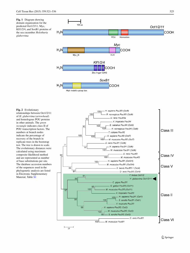
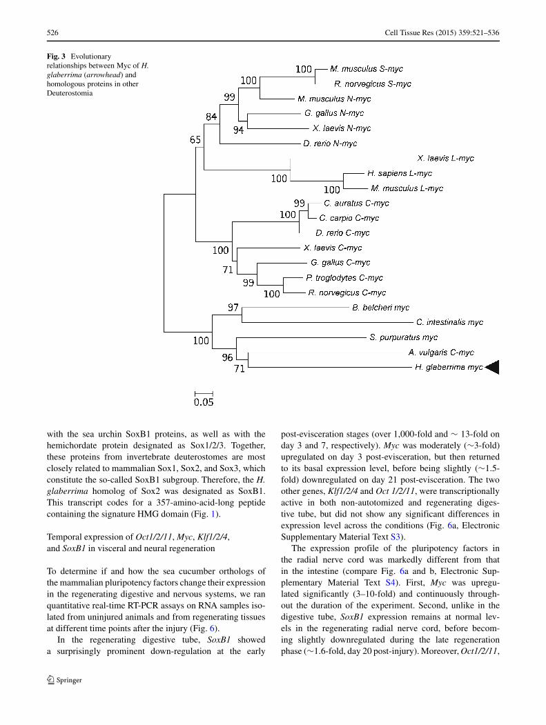
The H. glaberrima ortholog of the vertebrate Oct3/4 hasan open reading frame coding for a 779 amino acid-longprotein, which contains both motifs characteristic of Poutranscription factor proteins, a Pou domain and a homeoboxdomain, separated by a 21-amino acid linker region (Fig. 1).Phylogenetic analysis (Fig. 2) shows that H. glaberrimaOct protein belongs to class II POU proteins and clusterstogether with Oct1/2 of the sea urchin P. lividus and Pou2f1of the bivalve C. gigas to form an outgroup to vertebrateOct1, 2, and 11. Therefore, the H. glaberrima protein wasdesignated as Oct1/2/11.
Myc
Another of the four Yamanaka factors, mammalian c-Myc,is known to directly control transcription of as many as∼15 % of all protein-coding genes and therefore mediatesa wide range of biological processes. In addition to c-Myc,mammals have other functionally redundant Myc genes,including N-Myc, L-Myc, and S-Myc (Mahani et al. 2013).Unlike mammals, many invertebrates, such as tunicates,amphioxus, sea urchins, and Drosophila, have a single Mycgene in their genome (Gallant 2006; Morita et al. 2009).
Among the many biological functions of Myc proteins, thecontrol of growth and cell proliferation have been conservedbetween invertebrates and vertebrates (Gallant 2006).
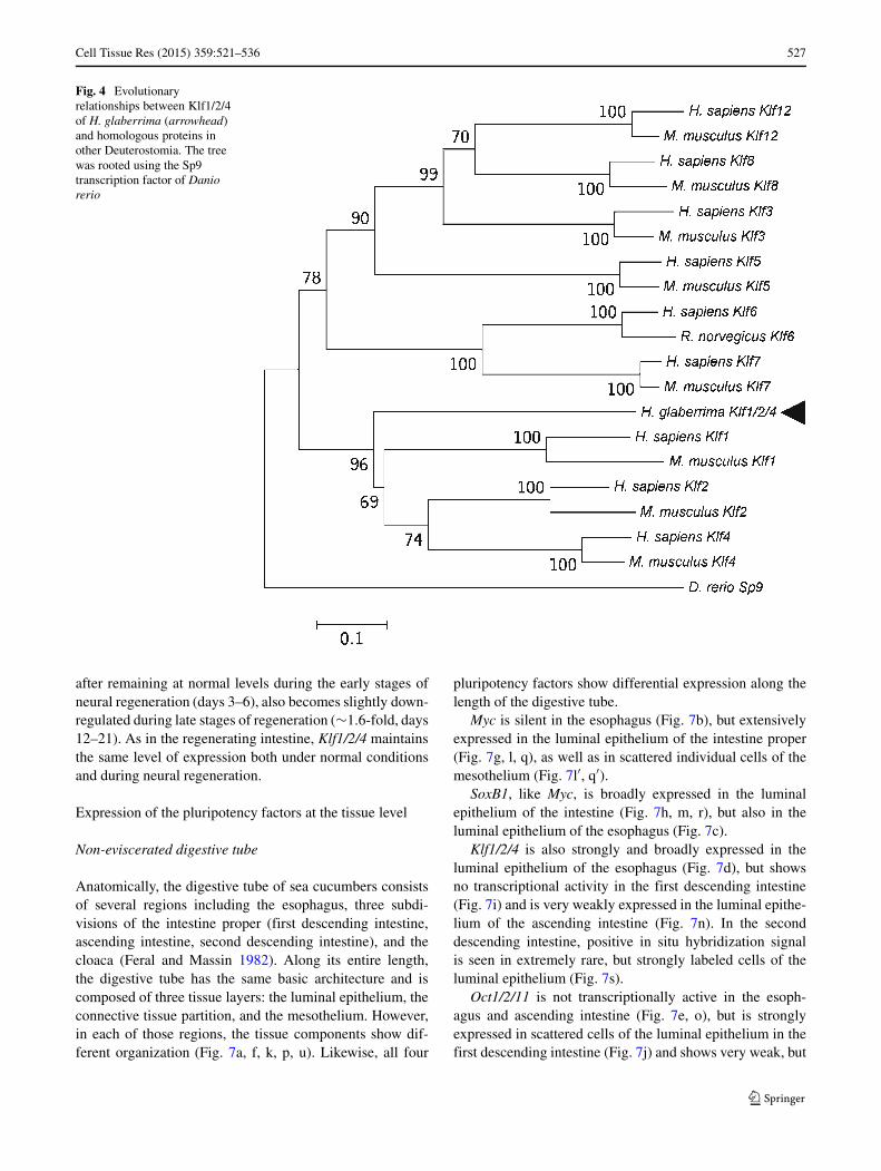
The sea cucumber homolog of Myc contains a predicted441-amino-acid-long open reading frame, which includestwo conserved motifs that define Myc proteins, the N-terminal Myc domain and the C-terminal basic helix-loop-helix domain (Fig. 1). Our phylogenetic analysis (Fig. 3)showed that the H. glaberrima Myc did not belong to anyof the vertebrate myc subfamilies, but instead clusteredtogether with Myc proteins from urochordates, cephalochor-dates, echinoids, and asteroids. This cluster received strongbootstrap support and formed an outgroup to Myc proteinsof vertebrates.
Klf1/2/4
Klf proteins are zinc-finger-containing transcription factors.Klf2 and Klf4 are known to inhibit growth, DNA synthe-sis, and cell cycle progression and are therefore regardedas tumor suppressor genes. Klf4 functions in reprogram-ming of iPS cells by promoting their survival throughinhibition of c-Myc-induced programmed cell death. Simul-taneously, c-Myc suppresses the anti-proliferation functionof Klf4 (Takahashi and Yamanaka 2006; McConnell andYang 2010).
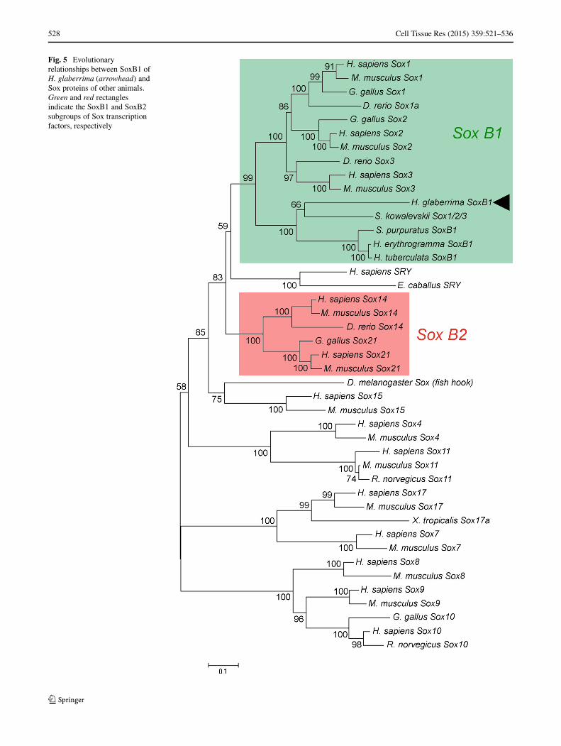
Studies of the sea urchin genome have identified severalKlf genes (Materna et al. 2006), whose functions in echino-derms remain largely unstudied. The sea cucumber homologof Klf4 codes for a 323-animo-acid-long polypeptide withthree zinc finger C2H2 motifs at the carboxy-terminal endof the protein (Fig. 1). Such organization is typical for allmembers of the Klf family (McConnell and Yang 2010).Phylogenetic analysis (Fig. 4) clusters the sea cucumberprotein as an outgroup to the clade containing mammalianKlf1, Klf2, and Klf4 with a strong bootstrap support. There-fore, this sequence was designated as Klf1/2/4.
SoxB1
Sox genes code for a diverse family of transcription fac-tors (26 members and 11 members in the human and seaurchin genome, respectively) (Howard-Ashby et al. 2006)that contain the high-mobility group (HMG) domain andare known to be involved in various aspects of develop-ment. Mammalian Sox2 protein has been shown to playa key role in acquisition and maintenance of pluripotency(Pevny and Placzek 2005; Takahashi and Yamanaka 2006;Lefebvre et al. 2007). Likewise, a related sea urchin gene,SoxB1, is thought to be involved in maintenance of theenteric neuronal precursors (Wei et al. 2011).
Our phylogenetic analysis (Fig. 5) shows that the seacucumber homolog of mammalian Sox2 clusters together
Cell Tissue Res (2015) 359:521–536 525
Fig. 1 Diagram showingdomain organization for thepredicted Oct1/2/11, Myc,Klf1/2/4, and SoxB1 proteins ofthe sea cucumber Holothuriaglaberrima
Fig. 2 Evolutionaryrelationships between Oct1/2/11of H. glaberrima (arrowhead)and homologous POU proteinsin other animals. The greenrectangle indicates class II ofPOU transcription factors. Thenumbers at branch nodesindicate the percentage ofrecovery of the branch inreplicate trees in the bootstraptest. The tree is drawn to scale.The evolutionary distances werecalculated using maximumcomposite likelihood methodand are represented as numberof base substitutions per site.The database accession numbersof the sequences used in thephylogenetic analysis are listedin Electronic SupplementaryMaterial, Table S1
526 Cell Tissue Res (2015) 359:521–536
Fig. 3 Evolutionaryrelationships between Myc of H.glaberrima (arrowhead) andhomologous proteins in otherDeuterostomia
with the sea urchin SoxB1 proteins, as well as with thehemichordate protein designated as Sox1/2/3. Together,these proteins from invertebrate deuterostomes are mostclosely related to mammalian Sox1, Sox2, and Sox3, whichconstitute the so-called SoxB1 subgroup. Therefore, the H.glaberrima homolog of Sox2 was designated as SoxB1.This transcript codes for a 357-amino-acid-long peptidecontaining the signature HMG domain (Fig. 1).
Temporal expression of Oct1/2/11, Myc, Klf1/2/4,and SoxB1 in visceral and neural regeneration
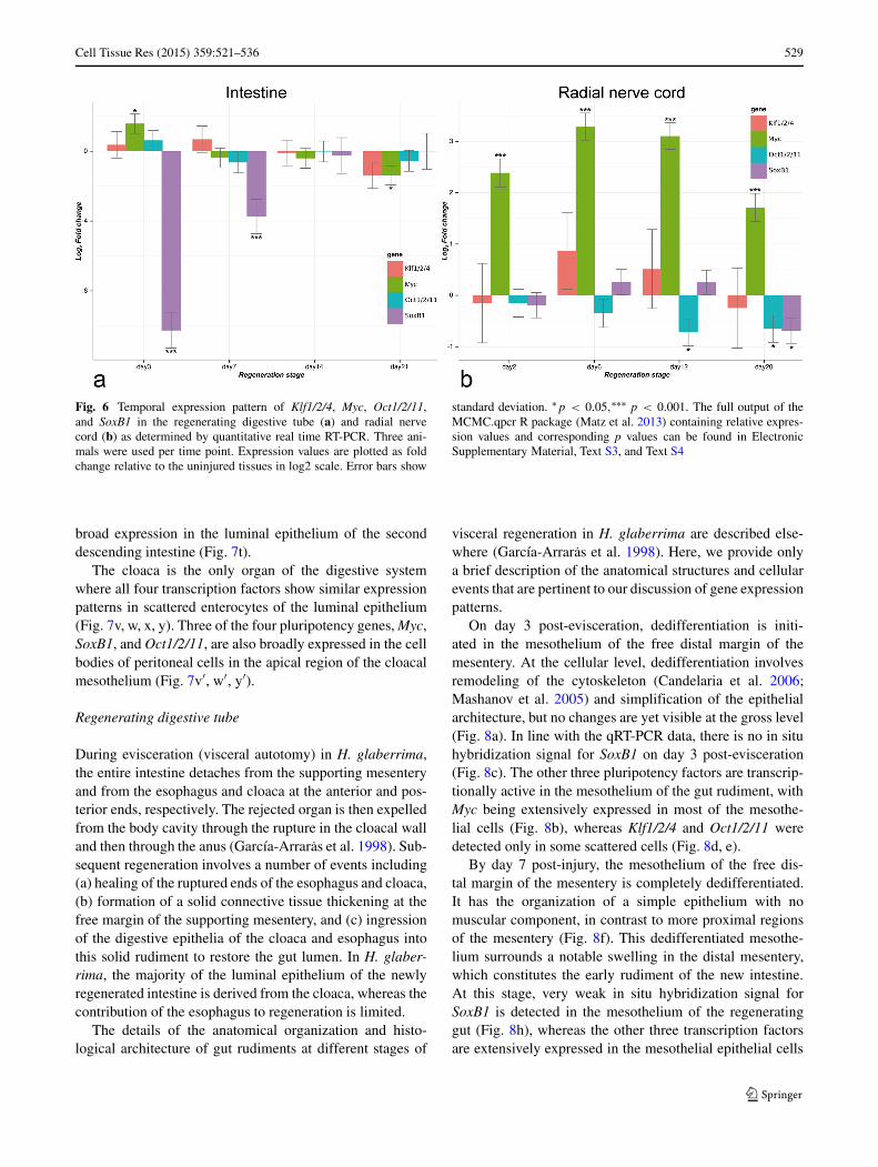
To determine if and how the sea cucumber orthologs ofthe mammalian pluripotency factors change their expressionin the regenerating digestive and nervous systems, we ranquantitative real-time RT-PCR assays on RNA samples iso-lated from uninjured animals and from regenerating tissuesat different time points after the injury (Fig. 6).
In the regenerating digestive tube, SoxB1 showeda surprisingly prominent down-regulation at the early
post-evisceration stages (over 1,000-fold and ∼ 13-fold onday 3 and 7, respectively). Myc was moderately (∼3-fold)upregulated on day 3 post-evisceration, but then returnedto its basal expression level, before being slightly (∼1.5-fold) downregulated on day 21 post-evisceration. The twoother genes, Klf1/2/4 and Oct 1/2/11, were transcriptionallyactive in both non-autotomized and regenerating diges-tive tube, but did not show any significant differences inexpression level across the conditions (Fig. 6a, ElectronicSupplementary Material Text S3).
The expression profile of the pluripotency factors inthe radial nerve cord was markedly different from thatin the intestine (compare Fig. 6a and b, Electronic Sup-plementary Material Text S4). First, Myc was upregu-lated significantly (3–10-fold) and continuously through-out the duration of the experiment. Second, unlike in thedigestive tube, SoxB1 expression remains at normal lev-els in the regenerating radial nerve cord, before becom-ing slightly downregulated during the late regenerationphase (∼1.6-fold, day 20 post-injury). Moreover, Oct1/2/11,
Cell Tissue Res (2015) 359:521–536 527
Fig. 4 Evolutionaryrelationships between Klf1/2/4of H. glaberrima (arrowhead)and homologous proteins inother Deuterostomia. The treewas rooted using the Sp9transcription factor of Daniorerio
after remaining at normal levels during the early stages ofneural regeneration (days 3–6), also becomes slightly down-regulated during late stages of regeneration (∼1.6-fold, days12–21). As in the regenerating intestine, Klf1/2/4 maintainsthe same level of expression both under normal conditionsand during neural regeneration.
Expression of the pluripotency factors at the tissue level
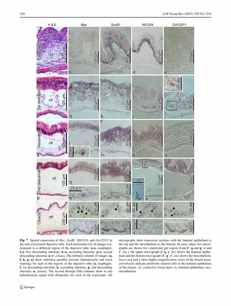
Non-eviscerated digestive tube
Anatomically, the digestive tube of sea cucumbers consistsof several regions including the esophagus, three subdi-visions of the intestine proper (first descending intestine,ascending intestine, second descending intestine), and thecloaca (Feral and Massin 1982). Along its entire length,the digestive tube has the same basic architecture and iscomposed of three tissue layers: the luminal epithelium, theconnective tissue partition, and the mesothelium. However,in each of those regions, the tissue components show dif-ferent organization (Fig. 7a, f, k, p, u). Likewise, all four
pluripotency factors show differential expression along thelength of the digestive tube.
Myc is silent in the esophagus (Fig. 7b), but extensivelyexpressed in the luminal epithelium of the intestine proper(Fig. 7g, l, q), as well as in scattered individual cells of themesothelium (Fig. 7l′, q′).
SoxB1, like Myc, is broadly expressed in the luminalepithelium of the intestine (Fig. 7h, m, r), but also in theluminal epithelium of the esophagus (Fig. 7c).
Klf1/2/4 is also strongly and broadly expressed in theluminal epithelium of the esophagus (Fig. 7d), but showsno transcriptional activity in the first descending intestine(Fig. 7i) and is very weakly expressed in the luminal epithe-lium of the ascending intestine (Fig. 7n). In the seconddescending intestine, positive in situ hybridization signalis seen in extremely rare, but strongly labeled cells of theluminal epithelium (Fig. 7s).
Oct1/2/11 is not transcriptionally active in the esoph-agus and ascending intestine (Fig. 7e, o), but is stronglyexpressed in scattered cells of the luminal epithelium in thefirst descending intestine (Fig. 7j) and shows very weak, but
528 Cell Tissue Res (2015) 359:521–536
Fig. 5 Evolutionaryrelationships between SoxB1 ofH. glaberrima (arrowhead) andSox proteins of other animals.Green and red rectanglesindicate the SoxB1 and SoxB2subgroups of Sox transcriptionfactors, respectively
Cell Tissue Res (2015) 359:521–536 529
Fig. 6 Temporal expression pattern of Klf1/2/4, Myc, Oct1/2/11,and SoxB1 in the regenerating digestive tube (a) and radial nervecord (b) as determined by quantitative real time RT-PCR. Three ani-mals were used per time point. Expression values are plotted as foldchange relative to the uninjured tissues in log2 scale. Error bars show
standard deviation. ∗p < 0.05,∗∗∗ p < 0.001. The full output of theMCMC.qpcr R package (Matz et al. 2013) containing relative expres-sion values and corresponding p values can be found in ElectronicSupplementary Material, Text S3, and Text S4
broad expression in the luminal epithelium of the seconddescending intestine (Fig. 7t).
The cloaca is the only organ of the digestive systemwhere all four transcription factors show similar expressionpatterns in scattered enterocytes of the luminal epithelium(Fig. 7v, w, x, y). Three of the four pluripotency genes, Myc,SoxB1, and Oct1/2/11, are also broadly expressed in the cellbodies of peritoneal cells in the apical region of the cloacalmesothelium (Fig. 7v′, w′, y′).
Regenerating digestive tube
During evisceration (visceral autotomy) in H. glaberrima,the entire intestine detaches from the supporting mesenteryand from the esophagus and cloaca at the anterior and pos-terior ends, respectively. The rejected organ is then expelledfrom the body cavity through the rupture in the cloacal walland then through the anus (Garcıa-Arraras et al. 1998). Sub-sequent regeneration involves a number of events including(a) healing of the ruptured ends of the esophagus and cloaca,(b) formation of a solid connective tissue thickening at thefree margin of the supporting mesentery, and (c) ingressionof the digestive epithelia of the cloaca and esophagus intothis solid rudiment to restore the gut lumen. In H. glaber-rima, the majority of the luminal epithelium of the newlyregenerated intestine is derived from the cloaca, whereas thecontribution of the esophagus to regeneration is limited.
The details of the anatomical organization and histo-logical architecture of gut rudiments at different stages of
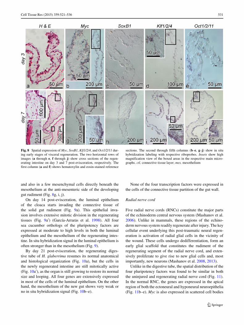
visceral regeneration in H. glaberrima are described else-where (Garcıa-Arraras et al. 1998). Here, we provide onlya brief description of the anatomical structures and cellularevents that are pertinent to our discussion of gene expressionpatterns.
On day 3 post-evisceration, dedifferentiation is initi-ated in the mesothelium of the free distal margin of themesentery. At the cellular level, dedifferentiation involvesremodeling of the cytoskeleton (Candelaria et al. 2006;Mashanov et al. 2005) and simplification of the epithelialarchitecture, but no changes are yet visible at the gross level(Fig. 8a). In line with the qRT-PCR data, there is no in situhybridization signal for SoxB1 on day 3 post-evisceration(Fig. 8c). The other three pluripotency factors are transcrip-tionally active in the mesothelium of the gut rudiment, withMyc being extensively expressed in most of the mesothe-lial cells (Fig. 8b), whereas Klf1/2/4 and Oct1/2/11 weredetected only in some scattered cells (Fig. 8d, e).
By day 7 post-injury, the mesothelium of the free dis-tal margin of the mesentery is completely dedifferentiated.It has the organization of a simple epithelium with nomuscular component, in contrast to more proximal regionsof the mesentery (Fig. 8f). This dedifferentiated mesothe-lium surrounds a notable swelling in the distal mesentery,which constitutes the early rudiment of the new intestine.At this stage, very weak in situ hybridization signal forSoxB1 is detected in the mesothelium of the regeneratinggut (Fig. 8h), whereas the other three transcription factorsare extensively expressed in the mesothelial epithelial cells
530 Cell Tissue Res (2015) 359:521–536
Fig. 7 Spatial expression of Myc, SoxB1, Klf1/2/4, and Oct1/2/11 inthe non-eviscerated digestive tube. Each horizontal row of images cor-responds to a different region of the digestive tube: a–e, esophagus;f–j, first descending intestine; k–o, ascending intestine; p–t, seconddescending intestine; u–y′ , cloaca. The leftmost column of images (a,f, k, p, u) show reference paraffin sections (hematoxylin and eosinstaining) for each of the regions of the digestive tube (a, esophagus;f, 1st descending intestine; k, ascending intestine; p, 2nd descendingintestine; u, cloaca). The second through fifth columns show in situhybridization signal with riboprobes for each of the transcripts. All
micrographs show transverse sections with the luminal epithelium tothe top and the mesothelium to the bottom. In cases when two micro-graphs are shown for a particular gut region (l and l′; q and q′; r andr′, etc.), the upper micrograph (l, q, r, etc) shows the luminal epithe-lium and the bottom micrograph (l′, q′, r′, etc) shows the mesothelium.Insets in j and s show higher magnification views of the boxed areas.arrowheads indicate positively stained cells in the luminal epitheliumof the cloaca. ctl, connective tissue layer; le, luminal epithelium; mes,mesothelium
Cell Tissue Res (2015) 359:521–536 531
Fig. 8 Spatial expression of Myc, SoxB1, Klf1/2/4, and Oct1/2/11 dur-ing early stages of visceral regeneration. The two horizontal rows ofimages (a through e, f through j) show cross sections of the regen-erating intestine on day 3 and 7 post-evisceration, respectively. Thefirst column (a and f) shows hematoxylin and eosin-stained reference
sections. The second through fifth columns (b–e, g–j) show in situhybridization labeling with respective riboprobes. Insets show highmagnification view of the boxed areas in the respective main micro-graphs. ctl, connective tissue layer; mes, mesothelium
and also in a few mesenchymal cells directly beneath themesothelium at the anti-mesenteric side of the developinggut rudiment (Fig. 8g, i, j).
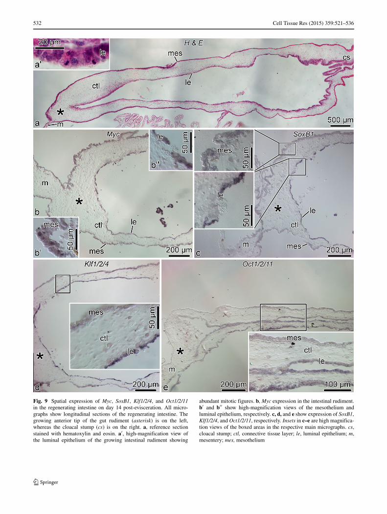
On day 14 post-evisceration, the luminal epitheliumof the cloaca starts invading the connective tissue ofthe solid gut rudiment (Fig. 9a). This epithelial inva-sion involves extensive mitotic division in the regeneratingtissues (Fig. 9a′) (Garcıa-Arraras et al. 1998). All foursea cucumber orthologs of the pluripotency factors areexpressed at moderate to high levels in both the luminalepithelium and the mesothelium of the regenerating intes-tine. In situ hybridization signal in the luminal epithelium isoften stronger than in the mesothelium (Fig. 9).
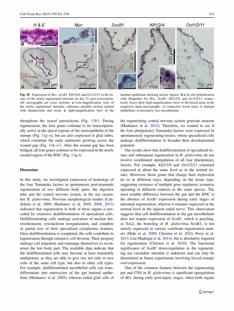
By day 21 post-evisceration, the regenerating diges-tive tube of H. glaberrima resumes its normal anatomicaland histological organization (Fig. 10a), but the cells inthe newly regenerated intestine are still mitotically active(Fig. 10a′), as the organ is still growing to restore its normalsize and looping. All four genes are extensively expressedin most of the cells of the luminal epithelium. On the otherhand, the mesothelium of the new gut shows very weak orno in situ hybridization signal (Fig. 10b–e).
None of the four transcription factors were expressed inthe cells of the connective tissue partition of the gut wall.
Radial nerve cord
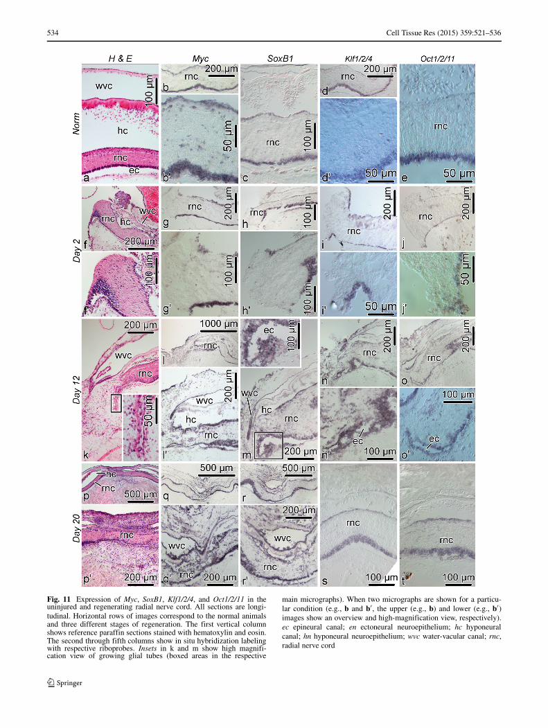
Five radial nerve cords (RNCs) constitute the major partsof the echinoderm central nervous system (Mashanov et al.2006). Unlike in mammals, these regions of the echino-derm nervous system readily regenerate after injury. The keycellular event underlying this post-traumatic neural regen-eration is activation of radial glial cells in the vicinity ofthe wound. These cells undergo dedifferentiation, form anearly glial scaffold that constitutes the rudiment of theregenerating segment of the radial nerve cord, and exten-sively proliferate to give rise to new glial cells and, mostimportantly, new neurons (Mashanov et al. 2008, 2013).
Unlike in the digestive tube, the spatial distribution of thefour pluripotency factors was found to be similar in boththe uninjured and regenerating radial nerve cord (Fig. 11).In the normal RNC, the genes are expressed in the apicalregion of both the ectoneural and hyponeural neuroepithelia(Fig. 11b–e). Myc is also expressed in scattered cell bodies
532 Cell Tissue Res (2015) 359:521–536
Fig. 9 Spatial expression of Myc, SoxB1, Klf1/2/4, and Oct1/2/11in the regenerating intestine on day 14 post-evisceration. All micro-graphs show longitudinal sections of the regenerating intestine. Thegrowing anterior tip of the gut rudiment (asterisk) is on the left,whereas the cloacal stump (cs) is on the right. a, reference sectionstained with hematoxylin and eosin. a′, high-magnification view ofthe luminal epithelium of the growing intestinal rudiment showing
abundant mitotic figures. b, Myc expression in the intestinal rudiment.b′ and b′′ show high-magnification views of the mesothelium andluminal epithelium, respectively. c, d, and e show expression of SoxB1,Klf1/2/4, and Oct1/2/11, respectively. Insets in c–e are high magnifica-tion views of the boxed areas in the respective main micrographs. cs,cloacal stump; ctl, connective tissue layer; le, luminal epithelium; m,mesentery; mes, mesothelium
Cell Tissue Res (2015) 359:521–536 533
Fig. 10 Expression of Myc, SoxB1, Klf1/2/4, and Oct1/2/11 in the tis-sues of the newly regenerated intestine on day 21 post-evisceration.All micrographs are cross sections. a Low-magnification view ofthe newly regenerated intestine; reference paraffin section stainedwith hematoxylin and eosin. a′ high-magnification view of the
luminal epithelium showing mitotic figures. b–e In situ hybridizationwith riboprobes for Myc, SoxB1, Klf1/2/4, and Oct1/2/11, respec-tively. Insets show high magnification views of the boxed areas in therespective main micrographs. ctl connective tissue layer; le luminalepithelium; m mesentery; mes mesothelium
throughout the neural parenchyma (Fig. 11b′). Duringregeneration, the four genes continue to be transcription-ally active in the apical regions of the neuroepithelia of thestumps (Fig. 11g–o), but are also expressed in glial tubes,which constitute the early rudiments growing across thewound gap (Fig. 11k–o′). After the wound gap has beenbridged, all four genes continue to be expressed in the newlycreated region of the RNC (Fig. 11q–t).
Discussion
In this study, we investigated expression of homologs ofthe four Yamanaka factors in spontaneous post-traumaticregeneration of two different body parts, the digestivetube and the central nervous system, in the sea cucum-ber H. glaberrima. Previous morphological studies (Can-delaria et al. 2006; Mashanov et al. 2005, 2008, 2013)indicated that regeneration in both of these organs is pre-ceded by extensive dedifferentiation of specialized cells.Dedifferentiating cells undergo activation of nuclear het-erochromatin, cytoskeleton reorganization, and completeor partial loss of their specialized cytoplasmic features.Once dedifferentiation is completed, the cells contribute toregeneration through extensive cell division. Their progenyundergo cell migration and rearrange themselves to recon-struct the lost body part. The available data indicate thatthe dedifferentiated cells may become at least transientlymultipotent, as they are able to give rise not only to newcells of the same cell type, but also to other cell types.For example, dedifferentiated mesothelial cells can trans-differentiate into enterocytes of the gut luminal epithe-lium (Mashanov et al. 2005), whereas radial glial cells of
the regenerating central nervous system generate neurons(Mashanov et al. 2013). Therefore, we wanted to see ifthe four pluripotency Yamanaka factors were expressed inspontaneously regenerating tissues, whose specialized cellsundergo dedifferentiation to broaden their developmentalpotential.
Our results show that dedifferentiation of specialized tis-sues and subsequent regeneration in H. glaberrima do notinvolve coordinated upregulation of all four pluripotencyfactors. For example, Klf1/2/4 and Oct1/2/11 remainedexpressed at about the same level as in the normal tis-sues. Moreover, those genes that change their expressiondo so in different ways, depending on the tissue type,suggesting existence of multiple gene regulatory scenariosoperating in different contexts in the same species. Themost notable difference between the gut and the RNC isthe absence of SoxB1 expression during early stages ofintestinal regeneration, whereas it remains expressed at thenormal level in the injured radial nerve. This observationsuggests that cell dedifferentiation in the gut mesotheliumdoes not require expression of SoxB1, which is puzzling,as Sox2, the homolog of H. glaberrima SoxB1, is notmerely expressed in various vertebrate regeneration mod-els (Maki et al. 2009; Christen et al. 2010; Perry et al.2013; Luz-Madrigal et al. 2014), but is absolutely requiredfor regeneration (Christen et al. 2010). The functionalsignificance of SoxB1 down-regulation in the regenerat-ing sea cucumber intestine is unknown and can only bedetermined in future experiments involving forced ectopicover-expression.
One of the common features between the regeneratinggut and CNS in H. glaberrima is significant upregulationof Myc during early post-injury stages, when both organs
534 Cell Tissue Res (2015) 359:521–536
Fig. 11 Expression of Myc, SoxB1, Klf1/2/4, and Oct1/2/11 in theuninjured and regenerating radial nerve cord. All sections are longi-tudinal. Horizontal rows of images correspond to the normal animalsand three different stages of regeneration. The first vertical columnshows reference paraffin sections stained with hematoxylin and eosin.The second through fifth columns show in situ hybridization labelingwith respective riboprobes. Insets in k and m show high magnifi-cation view of growing glial tubes (boxed areas in the respective
main micrographs). When two micrographs are shown for a particu-lar condition (e.g., b and b′, the upper (e.g., b) and lower (e.g., b′)images show an overview and high-magnification view, respectively).ec epineural canal; en ectoneural neuroepithelium; hc hyponeuralcanal; hn hyponeural neuroepithelium; wvc water-vacular canal; rnc,radial nerve cord
Cell Tissue Res (2015) 359:521–536 535
undergo extensive cell dedifferentiation (Candelaria et al.2006; Mashanov et al. 2013). Myc proteins play a centralrole in dedifferentiation and regeneration even in poorlyregenerating mammals. For example, it has been shownthat expression of c-Myc was required for regeneration ofthe intestinal epithelium (Ashton et al. 2010). In the mam-malian CNS, experimental forced expression of v-Myc, aviral homolog of c-Myc, stabilized the undifferentiated stateof embryonic radial glia. When injected into the injuredadult spinal cord, these cells contributed to increased neu-roprotection and axonal regrowth (Hasegawa et al. 2005).Therefore, our observations combined with known roles ofMyc proteins in regeneration and control of cell differenti-ation in various organisms (Mahani et al. 2013) make thesea cucumber Myc gene a promising candidate for furtherstudies. Our ongoing efforts are aimed at determining thefunctional role of Myc over-expression in dedifferentiatingtissues of H. glaberrima.
The fact that some pluripotency factors are present inthe regenerating sea cucumber tissues and may even be sig-nificantly up-regulated in response to injury (e.g., Myc),whereas other factors (such as SoxB1) are absent andobviously not involved in regeneration, suggests that eventhough there might be some parallels between in vivo dedif-ferentiation and in vitro iPS cell generation, the mechanismsare not exactly the same. One possible explanation whythese two processes can be different is that the cells thatdedifferentiate in vivo, unlike iPS cells, may not be pluripo-tent, but at best only multipotent (Maki et al. 2009; Christenet al. 2010). Alternatively, we cannot rule out that other, yetunknown, factors can contribute to in vivo dedifferentiationin regeneration-competent animals.
Conclusions
In the sea cucumber H. glaberrima, the orthologs ofmammalian Yamanaka pluripotency factors, Myc, SoxB1,Oct1/2/11, and Klf1/2/4, are expressed in the adult uninjuredcentral nervous system and in the digestive tube, although inthe latter, their expression domains do not overlap (exceptin the cloaca).
Expression of individual pluripotency factors is notcoordinated in injured/regenerating tissues. Some of thesegenes (Oct1/2/11 and Klf1/2/4) keep being expressed atabout the same levels as in uninjured animals, while oth-ers (SoxB1 and Myc) undergo considerable changes inexpression.
There are differences in pluripotency factor expressiondynamics between the regenerating radial nerve cord andthe regenerating intestine, suggesting that cell dedifferen-tiation in different regenerating organs may involve organ-and/or tissue-specific mechanisms.
Drastic down-regulation of SoxB1 in the regeneratingintestine during the early post-evisceration phase suggeststhat expression of this gene is not required for cell dediffer-entiation in this organ.
Consistent up-regulation of Myc in both intestinal andneural regeneration in H. glaberrima suggests that this genemay play an important role in dedifferentiation/regenerationin various tissues and organs.
Acknowledgments The study was supported by grants from theNIH (1SC1GM084770-01, 1R03NS065275-01) and the NSF (IOS-0842870, IOS-1252679), as well as by the University of PuertoRico.
References
Ashton GH, Morton JP, Myant K, Phesse TJ, Ridgway RA, MarshV, Wilkins JA, Athineos D, Muncan V, Kemp R, NeufeldK, Clevers H, Brunton V, Winton DJ, Wang X, Sears RC,Clarke AR, Frame MC, Sansom OJ (2010) Focal adhesionkinase is required for intestinal regeneration and tumorigenesisdownstream of Wnt/c-Myc signaling. Dev Cell 19(2):259–269.doi:10.1016/j.devcel.2010.07.015
Bhavsar RB, Tsonis PA (2014) Exogenous Oct-4 inhibits lens trans-differentiation in the newt Notophthalmus viridescens. PLoS One9(7):e102,510
Brockes JP, Gates PB (2014) Mechanisms underlying vertebrate limbregeneration: lessons from the salamander. Biochem Soc Trans42(3):625–630. doi:10.1042/BST20140002
Candelaria AG, Murray G, File SK, Garcıa-Arraras JE (2006) Contri-bution of mesenterial muscle dedifferentiation to intestine regen-eration in the sea cucumber Holothuria glaberrima. Cell TissueRes 325(1):55–65. doi:10.1007/s00441-006-0170-z
Christen B, Robles V, Raya M, Paramonov I, Izpisua Belmonte JC(2010) Regeneration and reprogramming compared. BMC Biol8:5. doi:10.1186/1741-7007-8-5
Feral J, Massin C (1982) Digestive system: Holothuroidea. In: JangouxM, Lawrence J (eds) Echinoderm nutrition. Balkema, Rotterdam,pp 192–212
Field D, Tiwari B, Booth T, Houten S, Swan D, Bertrand N, ThurstonM (2006) Open software for biologists: from famine to feast. NatBiotechnol 24(7):801–803. doi:10.1038/nbt0706-801
Gallant P (2006) Myc/Max/Mad in invertebrates: the evolution of theMax network. Curr Top Microbiol Immunol 302:235–253
Garcıa-Arraras JE, Estrada-Rodgers L, Santiago R, Torres II, Dıaz-Miranda L, Torres-Avillan I (1998) Cellular mechanisms of intes-tine regeneration in the sea cucumber, Holothuria glaberrimaSelenka (Holothuroidea: Echinodermata). J Exp Zool 281(4):288–304
Garza-Garcia AA, Driscoll PC, Brockes JP (2010) Evidencefor the local evolution of mechanisms underlying limbregeneration in salamanders. Integr Comp Biol 50(4):528–535. doi:10.1093/icb/icq022
Hasegawa K, Chang YW, Li H, Berlin Y, Ikeda O, Kane-Goldsmith N,Grumet M (2005) Embryonic radial glia bridge spinal cord lesionsand promote functional recovery following spinal cord injury. ExpNeurol 193(2):394–410. doi:10.1016/j.expneurol.2004.12.024
Howard-Ashby M, Materna SC, Brown CT, Chen L, Cameron RA,Davidson EH (2006) Gene families encoding transcription factorsexpressed in early development of Strongylocentrotus purpuratus.Dev Biol 300(1):90–107. doi:10.1016/j.ydbio.2006.08.033
536 Cell Tissue Res (2015) 359:521–536
Kiyota T, Kato A, Altmann CR, Kato Y (2008) The POUhomeobox protein Oct-1 regulates radial glia formationdownstream of Notch signaling. Dev Biol 315(2):579–592.doi:10.1016/j.ydbio.2007.12.013
Lefebvre V, Dumitriu B, Penzo-Mendez A, Han Y, Pallavi B (2007)Control of cell fate and differentiation by Sry-related high-mobility-group box (Sox) transcription factors. Int J Biochem CellBiol 39(12):2195–2214. doi:10.1016/j.biocel.2007.05.019
Luz-Madrigal A, Grajales-Esquivel E, McCorkle A, DiLorenzo AM,Barbosa-Sabanero K, Tsonis PA, Del Rio-Tsonis K (2014) Repro-gramming of the chick retinal pigmented epithelium after retinalinjury. BMC Biol 12(1):28. doi:10.1186/1741-7007-12-28
Maddox J, Shakya A, South S, Shelton D, Andersen JN, ChidesterS, Kang J, Gligorich KM, Jones DA, Spangrude GJ, WelmBE, Tantin D (2012) Transcription factor oct1 is a somaticand cancer stem cell determinant. PLoS Genet 8(11):e1003,048. doi:10.1371/journal.pgen.1003048
Mahani A, Henriksson J, Wright APH (2013) Origins of Myc proteins–using intrinsic protein disorder to trace distant relatives. PLoS One8(9):e75,057. doi:10.1371/journal.pone.0075057
Maki N, Suetsugu-Maki R, Tarui H, Agata K, Del Rio-Tsonis K,Tsonis PA (2009) Expression of stem cell pluripotency fac-tors during regeneration in newts. Dev Dyn 238(6):1613–1616.doi:10.1002/dvdy.21959
Mashanov V, Zueva O, Heinzeller T, Dolmatov I (2006) Ultrastruc-ture of the circumoral nerve ring and the radial nerve cordsin holothurians (Echinodermata). Zoomorphology 125(1):27–38.doi:10.1007/s00435-005-0010-9
Mashanov VS, Dolmatov IY, Heinzeller T (2005) Transdifferentiationin holothurian gut regeneration. Biol Bull 209(3):184–193
Mashanov VS, Zueva OR, Heinzeller T (2008) Regeneration of theradial nerve cord in a holothurian: a promising new model systemfor studying post-traumatic recovery in the adult nervous system.Tissue Cell 40(5):351–372. doi:10.1016/j.tice.2008.03.004
Mashanov VS, Zueva OR, Rojas-Catagena C, Garcıa-Arraras JE(2010) Visceral regeneration in a sea cucumber involves extensiveexpression of survivin and mortalin homologs in the mesothelium.BMC Dev Biol 10:117. doi:10.1186/1471-213X-10-117
Mashanov VS, Zueva OR, Garcia-Arraras JE (2012a) Expres-sion of Wnt9, TCTP, and Bmp1/Tll in sea cucumbervisceral regeneration. Gene Expr Patterns 12(1-2):24–35.doi:10.1016/j.gep.2011.10.003
Mashanov VS, Zueva OR, Garcıa-Arraras JE (2012b) Posttraumaticregeneration involves differential expression of long terminalrepeat (LTR) retrotransposons. Dev Dyn 241(10):1625–1636
Mashanov VS, Zueva OR, Garcıa-Arraras JE (2012c) Retrotrans-posons in animal regeneration: overlooked components of theregenerative machinery? Mob Genet Elements 2(5):244–247.doi:10.4161/mge.22644
Mashanov VS, Zueva OR, Garcıa-Arraras JE (2013) Radial glial cellsplay a key role in echinoderm neural regeneration. BMC Biology11(1):49. http://www.biomedcentral.com/1741-7007/11/49
Mashanov VS, Zueva O, Garcıa-Arraras JE (2014a) Chapter Seven -Postembryonic Organogenesis of the Digestive Tube: Why Does
It Occur in Worms and Sea Cucumbers but Fail in Humans?In: Galliot B (ed) Mechanisms of regeneration, current topics indevelopmental biology, vol 108. Academic Press, pp 185–216.doi:10.1016/B978-0-12-391498-9.00006-1
Mashanov VS, Zueva OR, Garcıa-Arraras JE (2014b) Tran-scriptomic changes during regeneration of the central ner-vous system in an echinoderm. BMC Genomics 15(1):357.doi:10.1186/1471-2164-15-357
Materna SC, Howard-Ashby M, Gray RF, Davidson EH (2006) TheC2H2 zinc finger genes of Strongylocentrotus purpuratus and theirexpression in embryonic development. Dev Biol 300(1):108–120.doi:10.1016/j.ydbio.2006.08.032
Matz MV, Wright RM, Scott JG (2013) No control genes required:Bayesian analysis of qRT-PCR data. PLoS One 8(8):e71,448.doi:10.1371/journal.pone.0071448
McConnell BB, Yang VW (2010) Mammalian Kruppel-like fac-tors in health and diseases. Physiol Rev 90(4):1337–1381.doi:10.1152/physrev.00058.2009
Morita M, Futami K, Zhang H, Kubokawa K, Ojima Y, Okamoto N(2009) Evolutionary analysis of amphioxus myc gene. J TokyoUniv Mar Sci Technol 5:11–16
Mosher C (1956) Observation on evisceration and visceral regenera-tion in the sea-cucumber, Actinopyga agassizi Selenka. Zoologica(NY) 41:17–26
Nolan T, Hands RE, Bustin SA (2006) Quantification ofmRNA using real-time RT-PCR. Nat Protoc 1(3):1559–1582.doi:10.1038/nprot.2006.236
Perry KJ, Thomas AG, Henry JJ (2013) Expression of pluripotencyfactors in larval epithelia of the frog Xenopus: evidence for thepresence of cornea epithelial stem cells. Dev Biol 374(2):281–294.doi:10.1016/j.ydbio.2012.12.005
Pevny L, Placzek M (2005) Sox genes and neural progenitor identity.Curr Opin Neurobiol 15(1):7–13. doi:10.1016/j.conb.2005.01.016
R Core Team (2014) R: A Language and Environment for StatisticalComputing. R Foundation for Statistical Computing, Vienna
Range R, Lepage T (2011) Maternal Oct1/2 is required for Nodaland Vg1/Univin expression during dorsal-ventral axis specifi-cation in the sea urchin embryo. Dev Biol 357(2):440–449.doi:10.1016/j.ydbio.2011.07.005
Shi G, Jin Y (2010) Role of Oct4 in maintaining and regaining stemcell pluripotency. Stem Cell Res Ther 1(5):39. doi:10.1186/scrt39
Takahashi K, Yamanaka S (2006) Induction of pluripotent stem cellsfrom mouse embryonic and adult fibroblast cultures by definedfactors. Cell 126(4):663–676. doi:10.1016/j.cell.2006.07.024
Tamura K, Stecher G, Peterson D, Filipski A, Kumar S (2013) Mega6:Molecular evolutionary genetics analysis version 6.0. Mol BiolEvol 30(12):2725–2729. doi:10.1093/molbev/mst197
Tantin D (2013) Oct transcription factors in development and stemcells: insights and mechanisms. Development 140(14):2857–2866. doi:10.1242/dev.095927
Wei Z, Angerer RC, Angerer LM (2011) Direct develop-ment of neurons within foregut endoderm of sea urchinembryos. Proc Natl Acad Sci U S A 108(22):9143–9147.doi:10.1073/pnas.1018513108
![Intrapericardial procedures for cardiac regeneration by stem cells - [Intraperikardiale Verfahren zur kardialen Regeneration durch Stammzellen]](https://img.dokumen.tips/doc/110x75/633212a24e01430403007e65/intrapericardial-procedures-for-cardiac-regeneration-by-stem-cells-intraperikardiale.jpg)


