Embed Size (px)
Citation preview

Please cite this article in press as: Emani et al., The L1TD1 Protein Interactome Reveals the Importance of Post-transcriptional Regulation inHuman Pluripotency, Stem Cell Reports (2015), http://dx.doi.org/10.1016/j.stemcr.2015.01.014
Stem Cell Reports
ResourceThe L1TD1 Protein Interactome Reveals the Importance ofPost-transcriptional Regulation in Human Pluripotency
Maheswara Reddy Emani,1,4 Elisa Narva,1,4 Aki Stubb,1 Deepankar Chakroborty,1Miro Viitala,1 Anne Rokka,1
Nelly Rahkonen,1 Robert Moulder,1 Konstantin Denessiouk,1 Ras Trokovic,2 Riikka Lund,1 Laura L. Elo,1,3
and Riitta Lahesmaa1,*1Turku Centre for Biotechnology, University of Turku and Abo Akademi University, 20521 Turku, Finland2Research Programs Unit, Molecular Neurology and Biomedicum Stem Cell Center, University of Helsinki, 00290 Helsinki, Finland3Department of Mathematics and Statistics, University of Turku, 20014 Turku, Finland4Co-first author
*Correspondence: [email protected]
http://dx.doi.org/10.1016/j.stemcr.2015.01.014
This is an open access article under the CC BY-NC-ND license (http://creativecommons.org/licenses/by-nc-nd/4.0/).
SUMMARY
The RNA-binding protein L1TD1 is one of themost specific and abundant proteins in pluripotent stem cells and is essential for themain-
tenance of pluripotency in human cells. Here, we identify the protein interaction network of L1TD1 in human embryonic stem cells
(hESCs) and provide insights into the interactome network constructed in human pluripotent cells. Our data reveal that L1TD1 has
an important role in RNA splicing, translation, protein traffic, and degradation. L1TD1 interacts withmultiple stem-cell-specific proteins,
many of which are still uncharacterized in the context of development. Further, we show that L1TD1 is a part of the pluripotency inter-
actome network of OCT4, SOX2, and NANOG, bridging nuclear and cytoplasmic regulation and highlighting the importance of RNA
biology in pluripotency.
INTRODUCTION
Pluripotent human embryonic stem cells (hESCs) can
differentiate into all somatic cells of the human body and
hold tremendous potential for developmental biology,
drug screening, and regenerative medicine (Thomson
et al., 1998). The importance of the core pluripotency tran-
scription factors OCT4, NANOG, and SOX2 in the mainte-
nance and induction of pluripotency has been well
documented (Boyer et al., 2005; Takahashi and Yamanaka,
2006; Yu et al., 2007). However, a variety of post-transcrip-
tional processes control and alter the original message after
transcription. In addition, protein degradation by protea-
somes has been suggested to have a vital role maintaining
pluripotency (Buckley et al., 2012; Vilchez et al., 2012).
The role of post-transcriptional regulators and protein net-
works in the maintenance of pluripotency is still largely
unknown, especially in human cells.
L1TD1 is highly and specifically expressed in pluripotent
cells (Iwabuchi et al., 2011; Mitsui et al., 2003; Narva et al.,
2012; Wong et al., 2011) under the control of OCT4,
NANOG, and SOX2 (Boyer et al., 2005; Narva et al., 2012;
Wong et al., 2011). We previously reported that L1TD1 is
essential for maintaining the pluripotent state in hESCs
(Narva et al., 2012). Like LIN28, which is one of the well-
characterized post-translational regulators of pluripotency
(Huang, 2012), L1TD1 is an RNA-binding protein (RBP).
RBPs have a fundamental role in a wide variety of cellular
processes, including RNA transcription, splicing, process-
ing, localization, stability, and translation. Moreover,
each RBP has unique and specific roles (Glisovic et al.,
2008). Given its highly specific expression and vitality in
pluripotent cells, we expect that L1TD1 regulates themain-
tenance of the pluripotent state.
Transcriptomic studies have provided a large amount of
data concerning genes that are specifically expressed in
the pluripotent state. However, this information can only
be used to predict the behavior of proteins that are impor-
tant for themaintenance of pluripotency. Combinations of
mass spectrometry (MS) and bioinformatics studies have
increased our understanding of the proteins and pathways
that are active in ESCs (Jadaliha et al., 2012; VanHoof et al.,
2006, 2009). Only recently, affinity purifications combined
withMS have shed light on the protein-protein interaction
networks of individual pluripotency factors and themolec-
ular networks that regulate pluripotency. The interactome
for Oct4, Sox2, and Nanog, along with other individual
transcription factors, has been reported in mouse ESCs
(mESCs) (Ding et al., 2012; Gao et al., 2012; Liang et al.,
2008; Pardo et al., 2010; van den Berg et al., 2010; Wang
et al., 2006). However, the functional protein-protein inter-
actions in hESCs have remained unexplored. Notably, the
protein networks that are distinct from transcriptional-
control networks in pluripotent cells remain unknown.
To elucidate interacting and functional components
involved in the regulation of humanpluripotency, we char-
acterized proteins associated with L1TD1. Furthermore, we
validated selected interactions in hESCs and human
induced pluripotent stem cells (hiPSCs). We show that
the pluripotency network of L1TD1 is shared with the
Stem Cell Reports j Vol. 4 j 1–10 j March 10, 2015 j ª2015 The Authors 1

(legend on next page)
2 Stem Cell Reports j Vol. 4 j 1–10 j March 10, 2015 j ª2015 The Authors
Please cite this article in press as: Emani et al., The L1TD1 Protein Interactome Reveals the Importance of Post-transcriptional Regulation inHuman Pluripotency, Stem Cell Reports (2015), http://dx.doi.org/10.1016/j.stemcr.2015.01.014

Please cite this article in press as: Emani et al., The L1TD1 Protein Interactome Reveals the Importance of Post-transcriptional Regulation inHuman Pluripotency, Stem Cell Reports (2015), http://dx.doi.org/10.1016/j.stemcr.2015.01.014
OCT4, NANOG, and SOX2 interactomes, and contains
uncharacterized protein players that are important for
maintenance of pluripotency. Our data significantly
expand the current knowledge about the biology of RBPs
and provide an important resource of protein interactions
in human pluripotent cells.
RESULTS
L1TD1 Interactome
The biological function of a protein is determined by its
structure. L1TD1 consists of a C-terminal coiled coil (CC)
domain, an RNA recognition motif (RRM) domain, a C-ter-
minal carboxyl tail domain (CTD) domain, and anN-termi-
nal CTD domain (Figure 1A). The N-terminal RRM domain
is exceptional in that only 30 of its C-terminal amino acids
(K216–D248) are typical of RRM domains. This ‘‘unique
sequence’’ possibly contains the CC domain and the rest
of the RRM domain, but it clearly differs from the se-
quences found in typical CC and RRM domains. Based on
these structural domains, we conclude that L1TD1 is an
RBP. However, the middle region of L1TD1 contains an
exceptional poly-E repeat region. Interestingly, nucleolin,
whose structure has an unusual combination of Poly-E
and RRM domains, has been shown to specifically recruit
proteins that are important for its unique function, with
the help of the acidic poly-E region (Edwards et al., 2000;
Erard et al., 1988; Lapeyre et al., 1987). On the basis of these
similarities, we speculated that by determining the proteins
that interact with L1TD1, we could identify protein
complexes that are important for the maintenance of
pluripotency.
To identify the L1TD1 protein network and to exclude
secondary partners interacting through RNA, we carried
out immunoprecipitation (IP) in the presence of RNase
with two specific anti-L1TD1 antibodies (Figure S1) that
recognize distinct regions of the L1TD1 protein (Figure 1A).
Figure 1. L1TD1-Interacting Proteins Are Involved in RNA Proces(A) Schematic diagram of the human L1TD1 protein structure: coiled ctail domain (CTD). Regions targeted by antibodies 1 and 2 used in the(2011).(B) Venn diagram of the proteins identified in the L1TD1 interactome(C) Clusters of the most enriched biological processes in the L1TD1 inet al., 2011) were visualized using Cytoscape 2.8.1 (Shannon et al., 2(D) The three most significant functions of L1TD1 interactome proteexperimentally are shown in red.(E) Western blot (WB) validations of individual protein-protein internegative control. hESC line HS360. CTNNB1 separate IP reaction, DDX(F) Protein expression of the interacting proteins after L1TD1 silencingin the Supplemental Results.See also Figure S1 and Tables S1, S2, and S3.
The availability of highly specific antibodies against L1TD1
enabled IP of the endogenous protein, excluding possible
overexpression effects. The strategy used to overexpress
tagged fusion protein was unsuccessful, as the overex-
pressed fusion protein of L1TD1 was rapidly cleaved in
living cells (data not shown). Liquid chromatography-tan-
dem MS (LC-MS/MS) analysis of three biological replicates
resulted in the detection of 306 proteins putatively
belonging to the L1TD1 interactome (Figure 1B; Table
S1). Immunoglobulin G (IgG) control IPs were used as a
negative control. In addition, we carried out a CRAPome
analysis (Mellacheruvu et al., 2013) to identify the speci-
ficity of the interactions (Table S2).
In order to identify biological processes through which
L1TD1mediates its function, we analyzed the L1TD1 inter-
actome with several of the currently available Gene
Ontology and interaction analysis tools. First, we investi-
gated known interactions within the L1TD1 interactome
based on the STRING interaction database (Szklarczyk
et al., 2011). This revealed highly connected components
of proteins involved in RNA-processing biology. In partic-
ular, RNA translation (21.6%) and splicing (10%) were
identified as key functions (Figure 1C; Table S3). Among
the other notable associated functions were intracellular
transport (9.6%), protein folding (5.6%), and the proteoso-
mal ubiquitin-dependent protein catabolic process (3%).
Ingenuity Pathway Analysis (IPA) further confirmed that
the top three functions of L1TD1-interacting proteins
were gene expression, post-transcriptional modification,
and protein synthesis (Figure 1D). Collectively, these re-
sults suggest that L1TD1 is involved in multiple steps
ranging from RNA production to the production of a func-
tional protein.
To establish the validity of the detected interaction
network, we confirmed randomly selected individual inter-
actions with specific antibodies (Figure 1E). In addition,
silencing of L1TD1 resulted in reduced expression of
most of the interacting partners (Figure 1F). Taken together
singoil (CC) domain, RNA recognition motif (RRM) domain, and carboxylIPs are shown. Protein domain borders are based on Khazina et al.
(shaded area) based on MS analysis of three biological replicates.teractome. Data from the STRING interaction database (Szklarczyk003).ins based on Ingenuity Pathway Analysis (IPA). Proteins validated
actions in L1TD1, IgG control, and unbound (UB) IP reactions. NC,3X and PABPC1 separate IP reaction, hESC line H9.. NT, non-targeting. hESC line HS360. Quantifications can be found
Stem Cell Reports j Vol. 4 j 1–10 j March 10, 2015 j ª2015 The Authors 3

Figure 2. The L1TD1 Interactome Uncovers the Importance of RBPs and the Potential Role of L1TD1 in Nuclear Pore Traffic(A) RBPs that were both identified here in the L1TD1 interactome in hESCs and previously reported in mESCs (Kwon et al., 2013). Proteinswere grouped according to the CRAPome percentages (Workflow 1). Proteins with 0.8-1 SAINT probability are shown in bold (Workflow 3).(B) WB validation of SRSF3 and U2AF1 interactions in L1TD1, IgG control, and UB IP reactions. hIPSC line HEL 11.4.(C) Subcellular location of L1TD1-interacting proteins based on IPA software.
(legend continued on next page)
4 Stem Cell Reports j Vol. 4 j 1–10 j March 10, 2015 j ª2015 The Authors
Please cite this article in press as: Emani et al., The L1TD1 Protein Interactome Reveals the Importance of Post-transcriptional Regulation inHuman Pluripotency, Stem Cell Reports (2015), http://dx.doi.org/10.1016/j.stemcr.2015.01.014

Please cite this article in press as: Emani et al., The L1TD1 Protein Interactome Reveals the Importance of Post-transcriptional Regulation inHuman Pluripotency, Stem Cell Reports (2015), http://dx.doi.org/10.1016/j.stemcr.2015.01.014
with the previous observation that the silencing of L1TD1
has a rapid effect on pluripotency (Narva et al., 2012), our
results suggest that interacting proteins that respond to
L1TD1 silencing participate in the maintenance of plurip-
otency along with L1TD1.
L1TD1 Interacts with Multiple RBPs
To gain further insight into the network revealed by these
measurements, we classified the L1TD1 interactome on
the basis of the structural domains of the interaction part-
ners (Table S4). Based on this, 58 proteins (19%) were clas-
sified as ribosomal proteins, providing further evidence
that L1TD1 is involved in translation. Interestingly,
another major class (10%, 31 proteins) identified was pro-
teins with the RNA-binding domain RRM or KH (K homol-
ogy), suggesting that L1TD1 performs its function in a
complex with other RBPs.
Recently, Kwon et al. (2013) reported an RNA-binding
repertoire of mESCs including 283 RBPs. A comparison of
these RBPs with the L1TD1 interactome resulted in the
identification of 89 shared RBPs, 72 of which had high
significance based on the CRAPome (Figure 2A). Based on
this comparison, 29% of the L1TD1-interacting proteins
bind RNA. Moreover, two RBPs that interact with L1TD1,
U2af1, and Srsf3 (Figure 2B) were recently shown to be
vital for somatic cell reprogramming in mESCs (Ohta
et al., 2013).
L1TD1 Traffics between the Nucleus and Cytoplasm
Most of the L1TD1-interacting proteins identified were
annotated as cytoplasmic in location (Figure 2C), although
more than 30% were nuclear proteins. The presence of nu-
clear proteins in the L1TD1 interactome led us to reanalyze
the cellular location of L1TD1. In a previous study,we local-
ized L1TD1 in the cytoplasmic processing bodies (P-bodies)
based on its co-location with AGO2 and LIN28 (Narva
et al., 2012). However, both of these proteins are able to
traffic between the cytoplasm and nucleus (Balzer and
Moss, 2007; Robb et al., 2005). In addition, a recent MS
analysis identified L1TD1 in the nuclear fraction (Sarkar
et al., 2012). To test whether L1TD1 is found in the nucleus,
we carried out cell fractionation experiments. The results
showed the presence of L1TD1 and LIN28 in both the nu-
clear and cytoplasmic fractions, whereas OCT4, NANOG,
(D) Expression of L1TD1, KPNA2, LIN28, OCT4, NANOG, SOX2, GAPDHcytoplasmic (C), and nuclear (N) cell fractions of hESCs (H9), 2102EP(E) Immunostaining of L1TD1 (red), DNA (DAPI staining, blue), NANO(F) WB validation of KPNA2, L1TD1, and RAE1 interactions in L1TD1, Kthe experiment shown in Figure 1E. KPNA2 IP hIPSC line HEL24.3.(G) Effect of L1TD1 silencing on the expression of KPNA2 and OCT4. hResults.See also Table S2.
and SOX2 showed extensive nuclear expression (Fig-
ure 2D). The location was also validated in cancer cell lines
expressing endogenous L1TD1 (Figure 2D).
Based on the immunostainings, we concluded that most
of the L1TD1 complexes localize adjacent to the nuclear
membrane (Figure 2E). This led us to analyze the L1TD1 in-
teractome for the presence of possible binding partners
associated with the nuclear membrane and nuclear pore
traffic. In addition to identifyingmultipleproteins involved
in cellular transport, we identified key factors in nuclear
pore traffic, such as KPNA2, RAN, and RAE1. Interestingly,
karyopherin KPNA2 is enriched in undifferentiated ESCs
(Van Hoof et al., 2006) and has been shown to be respon-
sible for the nuclear import of OCT4 (Young et al., 2011).
Furthermore, RAE1 is a componentof thenuclear pore com-
plex involved in RNA export (Murphy et al., 1996). We
validated the cytoplasmic and nuclear location of KPNA2
(Figure 2D), as well as interactions of KPNA2 and RAE1
with L1TD1 (Figure 2F). Further, by reciprocal pull-down
of KPNA2, we were able to validate the interaction with
L1TD1 andOCT4 (Figure 2F). In addition,we demonstrated
that silencing of L1TD1 results in decreased KPNA2 and
OCT4 levels (Figure 2G). These findings strongly suggest
that L1TD1 is able to traffic between the nucleus and cyto-
plasm and participate in nuclear pore traffic.
L1TD1 Levels Are Rapidly Decreased by Proteasome
Inhibitor
The LC-MS/MS analysis resulted in the identification of
multiplemembers of the ubiquitin-dependent proteasome,
such as PSMD1, PSMD2, PSMD7, PSMD8, PSMD11, and
PSMD14. Of particular interest, PSMD11 was recently re-
ported to be responsible for the increased proteasome activ-
ity in hESCs, which is important for the maintenance of
pluripotency (Vilchez et al., 2012). The interaction be-
tween L1TD1 and PSMD11 was validated with immuno-
blotting (Figure 3A). Moreover, silencing of L1TD1 resulted
in decreased levels of PSMD11 (Figure 3B).
To further study the role of L1TD1 in the proteasome
complex, we inhibited proteasome activity with MG-132,
which is known to decrease the expression of SOX2 (Vil-
chez et al., 2012). Treatment with MG-132 resulted in an
immediate reduction of L1TD1 and SOX2, but did not in-
fluence the levels of OCT4 or LIN28 at the early time points
(cytoplasmic control), and PARP1 (nuclear control) in total (T),embryoteratocarcinoma, and TCAM2 seminoma cell lines.G, Phalloidin (actin), and secondary controls in hIPSCs (HEL 11.4).PNA2, IgG control, and UB IP reactions. Additional detections from
ESC line HS360. Quantifications can be found in the Supplemental
Stem Cell Reports j Vol. 4 j 1–10 j March 10, 2015 j ª2015 The Authors 5

Figure 3. L1TD1 Participates in the HighProteasome Activity of Pluripotency(A) WB detection of PSMD11 in L1TD1, IgGcontrol, and UB IP reactions. hESC lineHS360.(B) Protein expression of PSMD11 afterL1TD1 silencing. hESC line HS360. NT, non-targeting. Quantifications can be found inthe Supplemental Results.(C) Protein expression of L1TD1, OCT4,LIN28, SOX2, and GAPDH after inhibition ofproteosomal activity with MG-132 inhibitor(10 mM) versus DMSO control. hESC line H9.
Please cite this article in press as: Emani et al., The L1TD1 Protein Interactome Reveals the Importance of Post-transcriptional Regulation inHuman Pluripotency, Stem Cell Reports (2015), http://dx.doi.org/10.1016/j.stemcr.2015.01.014
studied (Figure 3C). These results indicate that the protein
levels of L1TD1 correlate with proteasome activity and that
reduction of L1TD1 expression is an immediate indicator of
a decrease in hESC proteasome activity and pluripotency.
L1TD1 Is a Member of the Pluripotency Network
To understand the role of L1TD1 in regulating pluripo-
tency, we compared the L1TD1 interactome with lists of
proteins (Jadaliha et al., 2012; Van Hoof et al., 2006) and
genes (Assou et al., 2007) that are enriched in hESCs
compared with their differentiated counterparts. Based
on this analysis, 30% of the L1TD1 interaction partners
are associated with pluripotency (Figures 4A and S2A).
Nineteen of the interacting proteins had previously been
shown to be enriched in hESCs at both the gene and pro-
tein levels. Interestingly, the panel contains several factors,
such as DPPA4 (developmental pluripotency associated 4),
RFC5 (replication factor C subunit 5), and TOP2A (topo-
isomerase-IIa), whose role in the regulation of pluripotency
remains to be investigated. The interaction of L1TD1 with
TOP2A was validated (Figure S2B). In addition, silencing of
L1TD1 led to decreased TOP2A levels (Figure 1F).
L1TD1 belongs to the interactomes of Nanog (Nitzsche
et al., 2011) and Oct4 (Ding et al., 2012; van den Berg
et al., 2010) in mESCs. However, NANOG and OCT4 were
not detected as L1TD1-interacting proteins in our MS
data. It has proved to be challenging to detect these plurip-
otency factors withMS (Ding et al., 2012; Liang et al., 2008;
Pardo et al., 2010; van den Berg et al., 2010). Therefore, we
tested the presence of these proteins by immunoblotting of
the L1TD1 IPs. This approach led to the detection of OCT4,
NANOG, and SOX2 as L1TD1-interacting proteins (Fig-
ure 4B). Successful optimization of SOX2-IP further verified
L1TD1 interaction with SOX2 (Figure S2C). In addition to
these transcription factors, we demonstrated that L1TD1
interacts with the pluripotency-related proteins DNMT3B
and TRIM28 (Figure S2D). These interactions were initially
identified only in individual MS replicates. In addition,
silencing of L1TD1 led to decreased TRIM28 levels
(Figure 1F).
6 Stem Cell Reports j Vol. 4 j 1–10 j March 10, 2015 j ª2015 The Authors
Finally, we sought to determine which components of
the L1TD1 interactome were also present in the mESC in-
teractomes of OCT4, NANOG, and SOX2 (Figure 4C)
(Ding et al., 2012; Nitzsche et al., 2011; van den Berg
et al., 2010). We applied IPA and identified 45, 25, and 8
proteins that interact with OCT4, NANOG, and SOX2,
respectively.We then analyzed the gene expression of these
factors in human undifferentiated, early differentiated
(SSEA3�), and differentiated (embryoid bodies [EBs], fibro-
blasts, and keratinocytes) cells (Figure 4D). This analysis re-
sulted in the identification of proteins that form the key
core of pluripotency at the protein level. In addition to
NANOG, L1TD1, LIN28A, SOX2, and OCT4, this analysis
suggested that MCM5 (minichromosome maintenance
complex component 5), PARP1 (poly ADP-ribose polymer-
ase 1), CDK1 (cyclin-dependent kinase 1), RFC4, and
TOP2A could have important roles in the regulation of plu-
ripotency. To evaluate the importance of these factors in
terms of pluripotency, we selected one of them to deter-
mine the influence of its silencing. Depletion of RFC4 led
to an immediate reduction of OCT4 and L1TD1 levels (Fig-
ure 4E), demonstrating that this factor is required for the
maintenance of pluripotency.
To confirm our results, we repeated selected L1TD1 pro-
tein interactions in hiPSCs (Figure S2E) and tested the spec-
ificity of the L1TD1-bead-antibody complex by performing
IP indifferentiatedHeLacontrol cells comparedwithplurip-
otent cells, including the RFC4 interaction (Figure S2F).
DISCUSSION
RBPs are engaged in every aspect of RNA biology and are
essential for carrying specific RNA products from the trans-
lationmachinery to pre-mRNA splicing, transport, localiza-
tion, translation, and turnover (Glisovic et al., 2008). Our
study indicates that L1TD1 is a functional RBP in human
pluripotent cells that is associatedwith proteins that partic-
ipate in cellular RNA processes, thus shedding light on the
RNA regulation of pluripotent cells.

Figure 4. L1TD1 Connects Uncharacterized Factors to Core Pluripotency Regulation(A) hESC-enriched genes (Assou et al., 2007) and proteins (Jadaliha et al., 2012; Van Hoof et al., 2006) in the L1TD1 interactome.Overlapping hits are listed.(B) WB detection of OCT4, SOX2, and NANOG in L1TD1, IgG control, and UB IP reactions. hESC line HS360.(C) Proteins that interact with L1TD1 and OCT4 (n = 45), NANOG (n = 25), or SOX2 (n = 8) (Ding et al., 2012; Nitzsche et al., 2011; van denBerg et al., 2010). The figure was constructed with the use of IPA software.(D) RNA expression of proteins represented in Figure 4C in hESCs and differentiated cells. EB, embryoid body; DIFF.K.CYTES, differentiatedkeratinocytes. Microarray data collected from previous studies (Enver et al., 2005; Golan-Mashiach et al., 2005; Skottman et al., 2005). Thefigure was generated from normalized raw files using GeneSpring GX Software (Agilent Technologies Genomics). The top ten differenti-ation-responsive genes are highlighted in yellow.(E) Effect of RFC4 silencing (siRNA1) on the expression of L1TD1 and OCT4. hESC line HS360. Quantifications can be found in the Sup-plemental Results.See also Figure S2.
Stem Cell Reports j Vol. 4 j 1–10 j March 10, 2015 j ª2015 The Authors 7
Please cite this article in press as: Emani et al., The L1TD1 Protein Interactome Reveals the Importance of Post-transcriptional Regulation inHuman Pluripotency, Stem Cell Reports (2015), http://dx.doi.org/10.1016/j.stemcr.2015.01.014

Please cite this article in press as: Emani et al., The L1TD1 Protein Interactome Reveals the Importance of Post-transcriptional Regulation inHuman Pluripotency, Stem Cell Reports (2015), http://dx.doi.org/10.1016/j.stemcr.2015.01.014
Several observations support the idea that L1TD1 shuttles
between the nucleus and cytoplasm. Based on our results
and consistent with a previous report on the subcellular
proteome of hESCs (Sarkar et al., 2012), we conclude that
L1TD1 (like LIN28) can be found in cytoplasmic, nuclear,
and membrane fractions. Based on the NetNES 1.1 server,
the amino acids L811–L814 could be responsible for the nu-
clear location of L1TD1. As RBPs are known to participate in
RNA traffic through nuclear pores, it is possible that L1TD1
has a role in that process. The interaction of L1TD1 with
OCT4, SOX2, NANOG, and the nuclear pore traffic protein
karyopherin KPNA2 suggests the intriguing possibility that
L1TD1 is responsible for the nuclear import of these tran-
scription factors after their translation to protein in the
cytoplasm. Highly stem-cell-specific L1TD1 could be
responsible for thenuclear localization, thereby controlling
these transcription factors and providing an additional
checkpoint for the maintenance of pluripotency.
The network shared among L1TD1, OCT4, SOX2, and
NANOG is highly interesting. Our analysis suggests that
the nuclear epigenetic regulator PARP1 and CDK1 are key
proteins in the core pluripotency network. Recently,
PARP1 expression and PARylation activity were shown to
be increased in undifferentiated mESCs and to regulate nu-
clear reprogramming (Chiou et al., 2013; Doege et al., 2012;
Lai et al., 2012). In addition, CDK1 was shown to affect the
differentiation of mESCs through an interplay with Oct4
(Li et al., 2012). Our results also revealed a panel of proteins
that were not previously reported in the context of self-
renewal but may be involved in the regulation of pluripo-
tency. Although the role of TOP2A, RCF4, and MCM5 in
pluripotency remains uncharacterized, their contribution
to this network and function in other cell types suggest
they may have a role in regulation of the cell cycle and
DNA replication in pluripotent cells. We chose to further
investigate the role of RFC4 and demonstrated that it is
vital for maintaining pluripotency.
Post-transcriptional regulators and protein turnover
determine which factors are active in pluripotency.
Recently, the deubiquitinating enzyme Psmd14 was shown
to be essential for cellular reprogramming (Buckley et al.,
2012), andPSMD11was shownto regulate increasedprotea-
some activity in hESCs (Vilchez et al., 2012). However, it is
still unclear how proteins are selected for ubiquitination
in pluripotent cells. The presence of multiple proteasome
subunits in the highly pluripotent-specific L1TD1 interac-
tomesuggests thatL1TD1couldparticipate in this selection.
One of the central functional categories detected in the
L1TD1 interactome was splicing (Figure 1C). Cell-type-spe-
cific RBPs are able to regulate tissue-specific splicing (Licata-
losi and Darnell, 2010), which has been shown to be
unique in mESCs and reorganized during reprogramming
by RBPs U2af1 and Srsf3 (Ohta et al., 2013). The interaction
8 Stem Cell Reports j Vol. 4 j 1–10 j March 10, 2015 j ª2015 The Authors
between L1TD1 (identified here in hESCs) and these two
factors implies that U2AF1 and SRSF3 also have an impor-
tant role in human pluripotency, and L1TD1 may play a
role in stem-cell-specific splicing.
In addition to regulating pluripotency, L1TD1 may have
an important function in cancer. We previously showed
that L1TD1 is expressed in selected cancer types, such as
colorectal carcinoma, ovarian germ cell tumors, and testis
seminomas and non-seminomas (Narva et al., 2012). Inter-
estingly, the L1TD1 interactome revealsmany proteins that
have an important role or diagnostic value in cancer. For
example, KPNA2has been shown to affectmalignant trans-
formation and its elevated expression is correlated with
poor diagnosis in multiple forms of cancer (Christiansen
and Dyrskjot, 2012).
Of all the factors in the interactome, L1TD1 has the fast-
est kinetics in response to differentiation. This implies that
L1TD1 has a regulatory role in the maintenance of plurip-
otency. Moreover, interaction with transcriptional, cell
cycle, epigenetic, proteosomal, protein traffic, and splicing
regulators further supports the regulatory role of L1TD1.
The protein interactome constructed here can be used as
an important resource for elucidating individual protein
interactions in pluripotent stem cells and possibly in
cancer stem cells. Moreover, this analysis sheds light on
post-translational regulation in pluripotency and the
importance of RBPs in pluripotent cells.
EXPERIMENTAL PROCEDURES
All experiments were performed in feeder-free culture conditions
on Matrigel (BD Biosciences) in mTeSR1 media (Stem Cell
Technologies).
For IP, cells were lysed into NP-40 buffer. Lysates were preincu-
bated with 10 mg/ml RNase A (QIAGEN) at +4�C. IP was carried
out using M-280 sheep anti-rabbit IgG Dynabeads (11203D;
Invitrogen) with L1TD1 HPA030064 (Ab1), HPA028501 (Ab2)
(Sigma), SOX-2 #5024 (Cell Signaling), and normal rabbit IgG
12-370 (Millipore).
IP proteins were gel separated, digested, and submitted for LC-
MS/MS analysis. The criteria used for inclusion as an L1TD1-inter-
acting protein were (1) the presence of the protein in two out of
three replicates, (2) more than one unique peptide was identified
in the L1TD1 IP analysis, and (3) on/off or R3-fold enrichment
of the identified peptides compared with the control IgG IP
reaction.
Detailed methods are described in the Supplemental Experi-
mental Procedures.
SUPPLEMENTAL INFORMATION
Supplemental Information includes Supplemental Results, Supple-
mental Experimental Procedures, two figures, and four tables and
can be found with this article online at http://dx.doi.org/10.
1016/j.stemcr.2015.01.014.
Please cite this article in press as: Emani et al., The L1TD1 Protein Interactome Reveals the Importance of Post-transcriptional Regulation inHuman Pluripotency, Stem Cell Reports (2015), http://dx.doi.org/10.1016/j.stemcr.2015.01.014
AUTHOR CONTRIBUTIONS
M.R.E. and E.N. conceived and designed the study, performed
experimental work, analyzed data, and wrote the manuscript.
A.S., M.V., and N.R. performed experimental work. K.D. and D.C.
analyzed data. A.R. performed mass spectrometry. R.M. provided
scientific support and language editing. R.T. provided supervision
for hIPSC cultures. R. Lund and L.L.E. performed data analysis
and provided supervision. R. Lahesmaa was the leader and supervi-
sor of the project from conception and design of the study to
writing of the manuscript.
ACKNOWLEDGMENTS
We thankOuti Hovatta (Karolinska Institute, Sweden) for the hESC
lines and Timo Otonkoski (University of Helsinki) for hiPSC lines.
We also thank Marjo Hakkarainen, Paivi Junni, Bogata Fezazi, and
Krista Maurinen for excellent technical assistance, and Omid Ra-
sool and Juha-Pekka Pursiheimo for fruitful discussions. We are
grateful to the Turku Proteomics Facility, Cell Imaging Core, Turku
Centre for Biotechnology (Turku, Finland), and Biomedicum Stem
Cell Center (Helsinki, Finland) for their assistance. This study was
supported by funding for the Juvenile Diabetes Research Founda-
tion (JDRF), The Academy of Finland (the Centre of Excellence in
Molecular Systems Immunology and Physiology Research, 2012-
2017, Decision No. 250114 and Postdoctoral Researcher Decision
No. 265723), the Finnish Cancer Organizations, the Sigrid Juselius
Foundation, the Turku Graduate School of Biomedical Sciences,
the Finnish Cultural Foundation, European Commission Seventh
Framework grant EC-FP7-SYBILLA-201106, and the Finnish Can-
cer Institute.
Received: July 24, 2014
Revised: January 16, 2015
Accepted: January 16, 2015
Published: February 19, 2015
REFERENCES
Assou, S., Le Carrour, T., Tondeur, S., Strom, S., Gabelle, A., Marty,
S., Nadal, L., Pantesco, V., Reme, T., Hugnot, J.P., et al. (2007). A
meta-analysis of human embryonic stem cells transcriptome inte-
grated into a web-based expression atlas. Stem Cells 25, 961–973.
Balzer, E., and Moss, E.G. (2007). Localization of the develop-
mental timing regulator Lin28 to mRNP complexes, P-bodies and
stress granules. RNA Biol. 4, 16–25.
Boyer, L.A., Lee, T.I., Cole, M.F., Johnstone, S.E., Levine, S.S.,
Zucker, J.P., Guenther, M.G., Kumar, R.M., Murray, H.L., Jenner,
R.G., et al. (2005). Core transcriptional regulatory circuitry in hu-
man embryonic stem cells. Cell 122, 947–956.
Buckley, S.M., Aranda-Orgilles, B., Strikoudis, A., Apostolou, E.,
Loizou, E., Moran-Crusio, K., Farnsworth, C.L., Koller, A.A., Das-
gupta, R., Silva, J.C., et al. (2012). Regulation of pluripotency and
cellular reprogramming by the ubiquitin-proteasome system.
Cell Stem Cell 11, 783–798.
Chiou, S.H., Jiang, B.H., Yu, Y.L., Chou, S.J., Tsai, P.H., Chang,
W.C., Chen, L.K., Chen, L.H., Chien, Y., and Chiou, G.Y. (2013).
Poly(ADP-ribose) polymerase 1 regulates nuclear reprogramming
and promotes iPSC generation without c-Myc. J. Exp. Med. 210,
85–98.
Christiansen, A., andDyrskjot, L. (2012). The functional role of the
novel biomarker karyopherin a 2 (KPNA2) in cancer. Cancer Lett.
331, 18–23.
Ding, J., Xu, H., Faiola, F., Ma’ayan, A., and Wang, J. (2012). Oct4
links multiple epigenetic pathways to the pluripotency network.
Cell Res. 22, 155–167.
Doege, C.A., Inoue, K., Yamashita, T., Rhee, D.B., Travis, S., Fujita,
R., Guarnieri, P., Bhagat, G., Vanti, W.B., Shih, A., et al. (2012).
Early-stage epigeneticmodification during somatic cell reprogram-
ming by Parp1 and Tet2. Nature 488, 652–655.
Edwards, T.K., Saleem, A., Shaman, J.A., Dennis, T., Gerigk, C., Oli-
veros, E., Gartenberg, M.R., and Rubin, E.H. (2000). Role for nucle-
olin/Nsr1 in the cellular localization of topoisomerase I. J. Biol.
Chem. 275, 36181–36188.
Enver, T., Soneji, S., Joshi, C., Brown, J., Iborra, F., Orntoft, T., Thyk-
jaer, T., Maltby, E., Smith, K., Abu Dawud, R., et al. (2005). Cellular
differentiation hierarchies in normal and culture-adapted human
embryonic stem cells. Hum. Mol. Genet. 14, 3129–3140.
Erard, M.S., Belenguer, P., Caizergues-Ferrer, M., Pantaloni, A., and
Amalric, F. (1988). A major nucleolar protein, nucleolin, induces
chromatin decondensation by binding to histone H1. Eur. J.
Biochem. 175, 525–530.
Gao, Z., Cox, J.L., Gilmore, J.M., Ormsbee, B.D., Mallanna, S.K.,
Washburn, M.P., and Rizzino, A. (2012). Determination of protein
interactome of transcription factor Sox2 in embryonic stem cells
engineered for inducible expression of four reprogramming fac-
tors. J. Biol. Chem. 287, 11384–11397.
Glisovic, T., Bachorik, J.L., Yong, J., and Dreyfuss, G. (2008). RNA-
binding proteins and post-transcriptional gene regulation. FEBS
Lett. 582, 1977–1986.
Golan-Mashiach, M., Dazard, J.E., Gerecht-Nir, S., Amariglio, N.,
Fisher, T., Jacob-Hirsch, J., Bielorai, B., Osenberg, S., Barad, O.,
Getz, G., et al. (2005). Design principle of gene expression used
by human stem cells: implication for pluripotency. FASEB J. 19,
147–149.
Huang, Y. (2012). Amirror of two faces: Lin28 as amaster regulator
of both miRNA and mRNA. Wiley Interdiscip. Rev. RNA 3,
483–494.
Iwabuchi, K.A., Yamakawa, T., Sato, Y., Ichisaka, T., Takahashi, K.,
Okita, K., and Yamanaka, S. (2011). ECAT11/L1td1 is enriched in
ESCs and rapidly activated during iPSC generation, but it is
dispensable for the maintenance and induction of pluripotency.
PLoS ONE 6, e20461.
Jadaliha, M., Lee, H.J., Pakzad, M., Fathi, A., Jeong, S.K., Cho, S.Y.,
Baharvand, H., Paik, Y.K., and Salekdeh, G.H. (2012). Quantitative
proteomic analysis of human embryonic stem cell differentiation
by 8-plex iTRAQ labelling. PLoS ONE 7, e38532.
Khazina, E., Truffault, V., Buttner, R., Schmidt, S., Coles, M., and
Weichenrieder, O. (2011). Trimeric structure and flexibility of the
L1ORF1 protein in human L1 retrotransposition. Nat. Struct.
Mol. Biol. 18, 1006–1014.
Kwon, S.C., Yi, H., Eichelbaum, K., Fohr, S., Fischer, B., You, K.T.,
Castello, A., Krijgsveld, J., Hentze, M.W., and Kim, V.N. (2013).
Stem Cell Reports j Vol. 4 j 1–10 j March 10, 2015 j ª2015 The Authors 9

Please cite this article in press as: Emani et al., The L1TD1 Protein Interactome Reveals the Importance of Post-transcriptional Regulation inHuman Pluripotency, Stem Cell Reports (2015), http://dx.doi.org/10.1016/j.stemcr.2015.01.014
The RNA-binding protein repertoire of embryonic stem cells. Nat.
Struct. Mol. Biol. 20, 1122–1130.
Lai, Y.S., Chang, C.W., Pawlik, K.M., Zhou, D., Renfrow, M.B., and
Townes, T.M. (2012). SRY (sex determining region Y)-box2 (Sox2)/
poly ADP-ribose polymerase 1 (Parp1) complexes regulate pluripo-
tency. Proc. Natl. Acad. Sci. USA 109, 3772–3777.
Lapeyre, B., Bourbon, H., and Amalric, F. (1987). Nucleolin, the
major nucleolar protein of growing eukaryotic cells: an unusual
protein structure revealed by the nucleotide sequence. Proc. Natl.
Acad. Sci. USA 84, 1472–1476.
Li, L.,Wang, J., Hou, J.,Wu, Z., Zhuang, Y., Lu,M., Zhang, Y., Zhou,
X., Li, Z., Xiao, W., and Zhang, W. (2012). Cdk1 interplays with
Oct4 to repress differentiation of embryonic stem cells into tro-
phectoderm. FEBS Lett. 586, 4100–4107.
Liang, J., Wan, M., Zhang, Y., Gu, P., Xin, H., Jung, S.Y., Qin, J.,
Wong, J., Cooney, A.J., Liu, D., and Songyang, Z. (2008). Nanog
and Oct4 associate with unique transcriptional repression com-
plexes in embryonic stem cells. Nat. Cell Biol. 10, 731–739.
Licatalosi, D.D., and Darnell, R.B. (2010). RNA processing and its
regulation: global insights into biological networks. Nat. Rev.
Genet. 11, 75–87.
Mellacheruvu, D., Wright, Z., Couzens, A.L., Lambert, J.P., St-
Denis, N.A., Li, T., Miteva, Y.V., Hauri, S., Sardiu, M.E., Low, T.Y.,
et al. (2013). The CRAPome: a contaminant repository for affinity
purification-mass spectrometry data. Nat. Methods 10, 730–736.
Mitsui, K., Tokuzawa, Y., Itoh, H., Segawa, K., Murakami, M., Taka-
hashi, K.,Maruyama,M.,Maeda,M., and Yamanaka, S. (2003). The
homeoprotein Nanog is required for maintenance of pluripotency
in mouse epiblast and ES cells. Cell 113, 631–642.
Murphy, R., Watkins, J.L., andWente, S.R. (1996). GLE2, a Saccha-
romyces cerevisiae homologue of the Schizosaccharomyces pombe
export factor RAE1, is required for nuclear pore complex structure
and function. Mol. Biol. Cell 7, 1921–1937.
Narva, E., Rahkonen, N., Emani, M.R., Lund, R., Pursiheimo, J.P.,
Nasti, J., Autio, R., Rasool, O., Denessiouk, K., Lahdesmaki, H.,
et al. (2012). RNA-binding protein L1TD1 interacts with LIN28
via RNA and is required for human embryonic stem cell self-
renewal and cancer cell proliferation. Stem Cells 30, 452–460.
Nitzsche, A., Paszkowski-Rogacz, M., Matarese, F., Janssen-Megens,
E.M., Hubner, N.C., Schulz, H., de Vries, I., Ding, L., Huebner, N.,
Mann, M., et al. (2011). RAD21 cooperates with pluripotency tran-
scription factors in the maintenance of embryonic stem cell iden-
tity. PLoS ONE 6, e19470.
Ohta, S., Nishida, E., Yamanaka, S., and Yamamoto, T. (2013).
Global splicing pattern reversion during somatic cell reprogram-
ming. Cell Rep. 5, 357–366.
Pardo, M., Lang, B., Yu, L., Prosser, H., Bradley, A., Babu,M.M., and
Choudhary, J. (2010). An expanded Oct4 interaction network: im-
plications for stem cell biology, development, and disease. Cell
Stem Cell 6, 382–395.
Robb, G.B., Brown, K.M., Khurana, J., and Rana, T.M. (2005). Spe-
cific and potent RNAi in the nucleus of human cells. Nat. Struct.
Mol. Biol. 12, 133–137.
10 Stem Cell Reports j Vol. 4 j 1–10 j March 10, 2015 j ª2015 The Authors
Sarkar, P., Collier, T.S., Randall, S.M., Muddiman, D.C., and Rao,
B.M. (2012). The subcellular proteome of undifferentiated human
embryonic stem cells. Proteomics 12, 421–430.
Shannon, P.,Markiel, A., Ozier, O., Baliga, N.S.,Wang, J.T., Ramage,
D., Amin, N., Schwikowski, B., and Ideker, T. (2003). Cytoscape: a
software environment for integratedmodels of biomolecular inter-
action networks. Genome Res. 13, 2498–2504.
Skottman, H., Mikkola, M., Lundin, K., Olsson, C., Stromberg,
A.M., Tuuri, T., Otonkoski, T., Hovatta, O., and Lahesmaa, R.
(2005). Gene expression signatures of seven individual human em-
bryonic stem cell lines. Stem Cells 23, 1343–1356.
Szklarczyk, D., Franceschini, A., Kuhn, M., Simonovic, M., Roth,
A., Minguez, P., Doerks, T., Stark, M., Muller, J., Bork, P., et al.
(2011). The STRING database in 2011: functional interaction net-
works of proteins, globally integrated and scored. Nucleic Acids
Res. 39, D561–D568.
Takahashi, K., and Yamanaka, S. (2006). Induction of pluripotent
stem cells from mouse embryonic and adult fibroblast cultures
by defined factors. Cell 126, 663–676.
Thomson, J.A., Itskovitz-Eldor, J., Shapiro, S.S., Waknitz, M.A.,
Swiergiel, J.J., Marshall, V.S., and Jones, J.M. (1998). Embryonic
stem cell lines derived from human blastocysts. Science 282,
1145–1147.
van den Berg, D.L., Snoek, T., Mullin, N.P., Yates, A., Bezstarosti, K.,
Demmers, J., Chambers, I., and Poot, R.A. (2010). An Oct4-
centered protein interaction network in embryonic stem cells.
Cell Stem Cell 6, 369–381.
Van Hoof, D., Passier, R., Ward-Van Oostwaard, D., Pinkse, M.W.,
Heck, A.J., Mummery, C.L., and Krijgsveld, J. (2006). A quest for
human and mouse embryonic stem cell-specific proteins. Mol.
Cell. Proteomics 5, 1261–1273.
Van Hoof, D., Munoz, J., Braam, S.R., Pinkse, M.W., Linding, R.,
Heck, A.J., Mummery, C.L., and Krijgsveld, J. (2009). Phosphoryla-
tion dynamics during early differentiation of human embryonic
stem cells. Cell Stem Cell 5, 214–226.
Vilchez, D., Boyer, L., Morantte, I., Lutz, M., Merkwirth, C., Joyce,
D., Spencer, B., Page, L., Masliah, E., Berggren, W.T., et al. (2012).
Increased proteasome activity in human embryonic stem cells is
regulated by PSMD11. Nature 489, 304–308.
Wang, J., Rao, S., Chu, J., Shen, X., Levasseur, D.N., Theunissen,
T.W., andOrkin, S.H. (2006). A protein interactionnetwork for plu-
ripotency of embryonic stem cells. Nature 444, 364–368.
Wong, R.C., Ibrahim, A., Fong, H., Thompson, N., Lock, L.F., and
Donovan, P.J. (2011). L1TD1 is a marker for undifferentiated hu-
man embryonic stem cells. PLoS ONE 6, e19355.
Young, J.C., Major, A.T., Miyamoto, Y., Loveland, K.L., and Jans,
D.A. (2011). Distinct effects of importin a2 and a4 onOct3/4 local-
ization and expression inmouse embryonic stem cells. FASEB J. 25,
3958–3965.
Yu, J., Vodyanik, M.A., Smuga-Otto, K., Antosiewicz-Bourget, J.,
Frane, J.L., Tian, S., Nie, J., Jonsdottir, G.A., Ruotti, V., Stewart,
R., et al. (2007). Induced pluripotent stem cell lines derived from
human somatic cells. Science 318, 1917–1920.

Stem Cell Reports
Supplemental Information
The L1TD1 Protein Interactome Reveals
the Importance of Post-transcriptional
Regulation in Human Pluripotency
Maheswara Reddy Emani, Elisa Närvä, Aki Stubb, Deepankar Chakroborty, Miro Viitala,
Anne Rokka, Nelly Rahkonen, Robert Moulder, Konstantin Denessiouk, Ras Trokovic,
Riikka Lund, Laura L. Elo, and Riitta Lahesmaa

Supplemental Information
Figure S1, Related to Figure 1. IP with L1TD1 antibodies. Coomassie stained SDS-gel
and WB detection of L1TD1 in IP reactions with IgG, L1TD1 HPA030064 (Ab1) and
HPA028501 (Ab2), hESC line HS360, (UB) unbound fraction.

Figure S2, Related to Figure 4. Additional validations. A) hESC enriched genes (Assou
et al., 2007) (red) and proteins (yellow) (Jadaliha et al., 2012; Van Hoof et al., 2006) in
L1TD1 interactome. Present in both (orange). B) WB detection of TOP2A, RBBP4 and
CGN in IP reactions with IgG and L1TD1. Additional detections from the experiment shown
in Figure 3A. hESC line HS360. C) WB detection of L1TD1, OCT4 and SOX2

immunoprecipitated with SOX2 antibody. This data is an additional line of experiment
shown in main Figure 4B. hESC line HS360. D) WB detection of L1TD1, DNMT3B (hESC
line H9) and TRIM28 (hESC line HS360, additional data from the experiment shown in
main Figure 1E) in IP reactions with IgG, and L1TD1. E) WB validation of interactions in
human induced pluripotent stem cells (hiPSCs), hiPSC line HEL24.3, IP reactions with IgG
and L1TD1. F) Validation specificity of key interactions in Hela cells grown on matrigel
compared to hIPSCs. Immunoprecipitation reactions with IgG and L1TD1. UB (unbound
fraction), NC (negative control).
Table S1, Related to Figure 1, L1TD1 interactome. Final L1TD1 interactome and
individual three replicates presented in separate excel sheets. (Excel File)
Table S2, Related to Figure 1, CRAPome analysis. Workflow 1 (Percentage found in
other studies) and Workflow 3 (Saint Probability) for L1TD1 interactome proteins. (Excel
File)
Table S3, Related to Figure 1, STRING interactions. STRING9.0 and DAVID pathway
analysis. (Excel File)
Table S4, Related to Figure 2. Protein classifications based on protein domains.
(Excel File)

Supplemental Results
Western Blot quantification for siRNA experiments, Related to Figures 1F, 2G, 3B
and 4E. Data is expressed as fold changes compared with non-targeting siRNA,
normalized to GAPDH and are representative of three biological replicates. (Statistical
analyses: The statistical analysis was performed using Graph Pad Prism 6, results were
evaluated using unpaired Student’s t test. (Statistical significance was accepted at
*p<0.05, **p<0.01 and ***p<0.001, **** p<0.0001)

Supplemental Experimental Procedures
Cell Culture
Human ESC line HS360 was obtained from Outi Hovatta (Karolinska Institutet, Sweden)
and H9 from WiCell Research Institute (Madison, WI). All experiments were performed in
feeder-free culture conditions on Matrigel (BD Biosciences) in mTeSR1 media (Stem Cell
Technologies) passaged with dispase (Stem cell technologies). Human iPSC lines used in
this study HEL11.4 and HEL24.3 were generated using Sendai viruses (SeV, Cytotune,
Lifetechnologies) and were thoroughly characterized as previously described (Mikkola et
al., 2013; Toivonen et al., 2013). Hela control cells were grown on matrigel in mTeSR1
media for IP reactions.
Immunoprecipitation
For one IP reaction 3-4 70 % confluent 10 cm plates were washed twice with cold PBS
and lysed into NP-40 -buffer (20–50 mM Tris, 150 mM NaCl, 0.5% sodium deoxylate, 0.5%
NP-40) containing PhosSTOP and complete EDTA-free inhibitors (Roche). Lysates were
preincubated with 10 µg/ml RNaseA (Qiagen) and pre-washed uncoupled 40 μL
Dynabeads® Sheep anti-Rabbit Dynabeads® at +4°C for 1 hour to eliminate proteins that
bind nonspecifically to the beads. IP was carried out using M-280 Sheep Anti-Rabbit IgG
Dynabeads® (Invitrogen – 11203D) according to the manufacturer’s protocol. Briefly, 80 μl
of μL Dynabeads® Sheep anti-Rabbit Dynabeads® were pre-washed per 2 ug of antibody.
IPs were performed with L1TD1 HPA030064 (Ab1), HPA028501 (Ab2) (Sigma), SOX-2
#5024 (Cell signaling) and Normal Rabbit IgG 12-370 (Millipore). 4 ug of antibody was
Incubated with gentle tilting and rotation for 2 hours at 4°C with beads. Unbound antibody
was washed 8 times with NP-40 buffer. 2 mg of RNAse treated protein lysate was added

to the IgG and L1TD1 coupled beads and incubated o/n with gentle tilting and rotation at
4°C. Unbound proteins were washed off 8 times with NP-40 buffer. Elution was done with
lower pH PAG Elution buffer 10701 (Admetech).
In case of KPNA2, IP was carried out using Dynabeads® protein G (Invitrogen – 10004D)
according to the manufacturer’s protocol. 80 μl of Dynabeads® protein G per 4 ug of
antibody were pre-washed with NP-40 buffer. IPs were performed with KPNA2 sc-55538
(Sigma) and Normal Mouse IgG 12-371 (Millipore). 4 ug of antibody was Incubated with
gentle tilting and rotation for 2hours at 4°C with beads. Unbound antibody was washed 8
times with NP-40 buffer. Beads were then crosslinked to antibodies using DMP (dimethyl
pimelimidate dihydrochloride). First bead - antibody complexes were washed with 0.2 M
triethanolamine, pH 8.2 (Sigma) and then resuspended to 20 mM DMP (Sigma) in 0.2 M
triethanolamine, pH 8.2. Reaction was stopped by resuspending beads in to 50 mM Tris,
pH 7.7. After reaction, complexes were washed using PBST (0,01% tween-20). RNAse
treated lysate was added on the IgG and KPNA2 coupled beads and incubated o/n with
gentle tilting and rotation at +4°C. Unbound proteins were washed off 8 times with NP-40
buffer. Elution was done with lower pH PAG Elution buffer 10701 (Admetech).
Mass Spectrometry
IP proteins were separated on Criterion XT 12% Bis-Tris gel (BioRad) and stained with
PageBlue (Thermo Scientific). Lanes were cut to 11-12 pieces and destained with 50%
methanol. Proteins were in-gel digested as described earlier (Shevchenko et al., 1996).
Tryptic peptides were dissolved in formic acid (0.1%) and submitted for LC-MS/MS
analysis.

The LC-MS/MS system consisted of a nanoflow HPLC system (Ultimate 3000, Dionex,
Sunnyvale, CA) coupled to a QSTAR Elite mass spectrometer (Applied Biosystems/MDS
Sciex, Canada) equipped with a nano-electrospray ionization source (Proxeon, Odense,
Denmark). Each sample was injected as one technical replicate. Peptides were first loaded
on a trapping column (0.3 x 5 mm PepMap C18, LC Packings) and subsequently
separated on a 15 cm C18 column (75 m x 15 cm, Magic 5 μm 200 Å C18, Michrom
BioResources Inc., Sacramento, CA, USA). Peptide separation was achieved using a
gradient from 2 to 35% B at a mobile phase in flow rate of 200 nl/min. The mobile phase
consisted of water/acetonitrile (98:2 (v/v)) with 0.2% formic acid (solvent A) or
acetonitrile/water (95:5 (v/v)) with 0.2% formic acid (solvent B). Two different methods,
including either 20 or 45 min gradient, were used depending on the sample complexity.
LC-MS/MS data acquisition and analysis
Data dependent acquisition was performed using Analyst QS 2.0 software (Applied
Biosystems/MDS Sciex, Canada). The two or three of the most intensive precursor ions
from the survey scan (350 - 1500 m/z) were selected for fragmentation with a dynamic
exclusion for 60 seconds (for the 20 and 45 minute methods, respectively). MS/MS peak
lists were created with Analyst QS 2.0. and database searches were performed using the
Mascot search engine (version 2.2.06, Matrix Sciences, London, UK) against the
SwissProt database (release 2011_08 containing 20245 sequence entries). The search
results from samples cut from the same gel lane were merged. The Mascot search
settings included a taxonomy filter 'human', trypsin as an enzyme, one missed trypsin
cleavage, precursor-ion mass tolerance of 0.2 Da, fragment-ion mass tolerance of 0.3 Da,

variable modifications of carbamidomethylation of cysteine and methionine oxidation. A
significance threshold of p<0.05 was used.
Scaffold 3.00.03 (Proteome Software Inc., Portland, OR) was used to combine and control
the false discovery rates form MS/MS based peptide and protein identifications. Protein
identifications were accepted if they could be established at greater than 99.0% probability
and contained at least 2 identified peptides. Peptide identifications were accepted if they
could be established at greater than 95.0% probability.
The criteria used for inclusion as a L1TD1-interacting protein were presence in two out of
three replicates, more than one unique peptide identified in the L1TD1 IP-analysis and
on/off or ≥ 3 fold enrichment of identified peptides compared to control IgG IP-reaction.
To investigate which proteins in the MS data were potential contaminants we used the
CRAPome analysis tool (Mellacheruvu et al., 2013). The L1TD1 interactome was uploaded
to CRAPome and the analysis was run following the CRAPome Workflow 1. To perform
the significance analysis of the interactome SAINT (Choi et al., 2011; Mellacheruvu et al.,
2013), the spectral count data for three L1TD1 and three IgG control replicates were
uploaded to the CRAPome tool. The analysis was run following the CRAPome Workflow 3
using the default parameters for the scoring functions. As the CRAPome data differed
from the IP conditions used in the L1TD1 interactome, the obtained results were not used
as an exclusion criterion, but as an estimate of probability and significance for each
interacting protein.
Previously reported protein interactions between the L1TD1 interacting proteins were
downloaded from the STRING interaction database (Szklarczyk et al., 2011). Only high

confidence (>0.7) interactions derived from experimental data or curated databases were
considered. The resulting network was visualized using Cytoscape 2.8.1 (Shannon et al.,
2003). The force-directed layout was applied and clusters were identified using the Markov
Clustering algorithm (MCL) implemented in the Cytoscape plug-in clusterMaker v. 1.9 with
default settings (Morris et al., 2011).
Enriched biological pathways (KEGG), Gene Ontology (GO) terms, and disease
associations (OMIM and Genetic Association Database) were identified using the
functional annotation tool DAVID 6.7 (Database for Annotation, Visualization and
Integrated Discovery)(Huang da et al., 2009a; Huang da et al., 2009b). For the GO
analysis, the GO FAT terms were considered.
Western Blotting
In silencing, cell fractioning and proteosomal activity assays cells were lysed in lysis buffer
(50 mM Tris-HCl pH 7.5, 150 mM NaCl, 0.5% TX-100, 5% glycerol, 1% SDS, 1 mM
Na3VO4, 10 mM NaF, and 1 mM phenylmethanesulfonyl fluoride). Protein concentrations
were determined with DC Protein Assay (Bio-Rad) and 6xSDS sample buffer (0.5 M Tris-
HCl pH 6.8, 28% glycerol, 9% SDS, 5% 2-mercaptoethanol, 0.01% bromphenol blue) was
added. Lysates were electrophoresed on a 10% SDSPAGE gel and transferred to a
nitrocellulose membrane. Membranes were incubated overnight at +4 ºC with primary
antibodies: L1TD1 HPA028501 (Sigma), L1TD1 HPA030064 (Sigma), OCT-4 (sc-9081) or
(sc-5279) (Santa Cruz), NANOG AF1997 (R&D Systems), SOX-2 MAB2018 (R&D
Systems), SOX-2 #5024 (Cell signaling), TIF1 beta MA1-2023 (Thermo Scientific), KPNA2
Sc-55538 (Santa Cruz), LIN28 ab46020 (Abcam), RbAp48 sc-8270 (Santa Cruz), IGF2BP-
3 sc-376067 (Santa Cruz), IGF2BP-1 sc-166344 (Santa Cruz), DDX3 sc-81247 (Santa
Cruz), KHDRBS1 sc-136062 (Santa Cruz), ELAVL1 sc-56709 (Santa Cruz), Cingulin
(CGN) ab117796 (Abcam), DNMT3B ab13604 (Abcam), PSMD11 FZ10R-1150 (EUROPA
bioproducts), CTNNB1 sc-59737 (Santa Cruz), TOPO1 sc-10783 (Santa Cruz), TOPO2A
sc-365918 (Santa Cruz), PARP1 #9532 (Cell signaling), Mrnp41 (RAE1) sc-374261 (Santa
Cruz), PABP sc-32318 (Santa Cruz), DHX9 ab26271 (Abcam), DHX9 ab54593 (Abcam),
Smad4 sc-7966 (Santa Cruz), HDAC1 sc-8410 (Santa Cruz), Nodal (H-110) SC-28913
(Santa Cruz), GAPDH 5G4 (HyTest Ltd), β-actin A5441 (Sigma), U2AF1 SAB1402953
(Sigma), SRSF3 (SRp20) sc-73059 (Santa Cruz), anti-rabbit-HRP 554021 (BD
Pharmingen), Histone H2B sc-10808 (Santa Cruz), RFC4 (C-9) sc-28301 (Santa Cruz).
Secondary antibodies were anti-mouse-HRP sc-2005 (Santa Cruz), anti-goat-HRP sc-
2020 (Santa Cruz Biotechnology). Signal was detected with enhanced chemiluminescence
reagent (Amersham Biosciences) or Pierce developing solution (Pierce [Thermo Fisher
Scientific]).
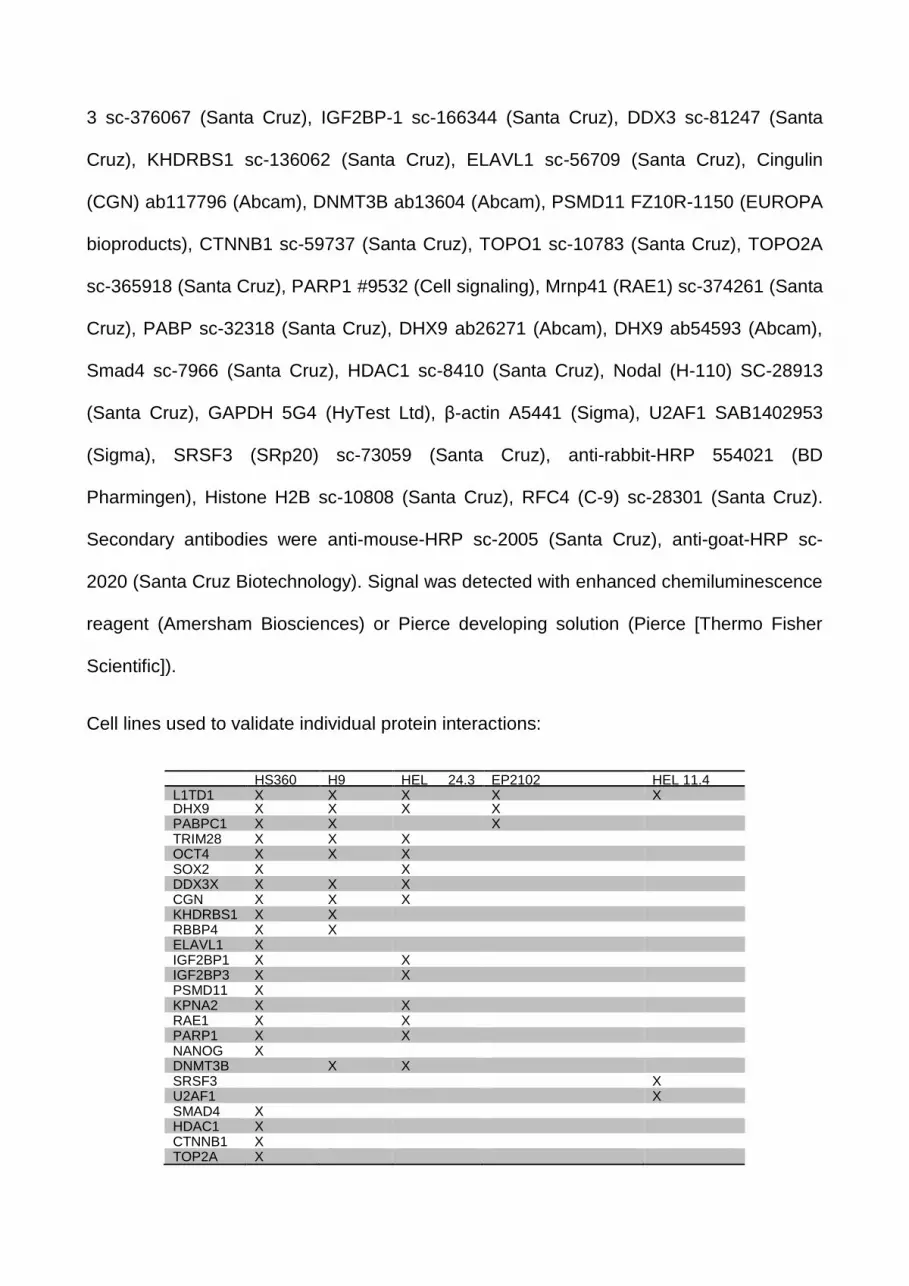
Cell lines used to validate individual protein interactions:
HS360
hESC
H9
hESC
HEL 24.3
hIPSC
EP2102
embryoteratocarcinoma
HEL 11.4
hIPSC
L1TD1 X X X X X DHX9 X X X X PABPC1 X X X TRIM28 X X X OCT4 X X X SOX2 X X DDX3X X X X CGN X X X KHDRBS1 X X RBBP4 X X ELAVL1 X IGF2BP1 X X IGF2BP3 X X PSMD11 X KPNA2 X X RAE1 X X PARP1 X X NANOG X DNMT3B X X SRSF3 X U2AF1 X SMAD4 X HDAC1 X CTNNB1 X TOP2A X

RFC4 X
Immunofluorescence
Cells were fixed with 4 % paraformaldehyde for 15 min and permeabilized for 15 min using
0.5% Triton X-100. Cells were stained with primary antibodies for 2 h to o/n: L1TD1
antibody (Ab2) 1:100, Atto 647N-Phalloidin 65906 (Sigma) 1:200 and NANOG AF1997
1:100 (R&D Systems) in 40 % horse serum. Secondary antibody staining was performed
with Alexa-488 and 555 (Invitrogen).
Cell fractioning
Cell fractioning was done with commercial kit (Pierce kit 78833). After collecting
cytoplasmic fraction excessive washings with 1x PBS was included in the protocol.
RNA Interference and hESC Transfection
RNA interference was performed with siRNA oligonucleotides (Sigma). The sequences of
siRNAs are as follows siL1TD1: GAGATGAGTCATGATGAGCATA; NTsiRNA control scr3:
CCUACAUCCCGAUCGAUGAUG (Berra et al., 2003); siRFC4 (Arai et al., 2009): RFC4#1
GACGUACCAUGGAGAAGGAGUCGAA, RFC4#2
CAAGGAUCGAGGAGUAGCUGCCAGT. Transfections were performed as previously
described (Narva et al., 2012).
Ethical Consideration
Ethics Committee of South-West Finland Hospital District provided the permission to
culture the human ESC lines used in this study. Research was carried out following the
good scientific practice and guidelines of the National Advisory Board on Research Ethics.

Supplemental References
Arai, M., Kondoh, N., Imazeki, N., Hada, A., Hatsuse, K., Matsubara, O., and Yamamoto, M. (2009). The knockdown of endogenous replication factor C4 decreases the growth and enhances the chemosensitivity of hepatocellular carcinoma cells. Liver Int. 29, 55-62.
Berra, E., Benizri, E., Ginouves, A., Volmat, V., Roux, D., and Pouyssegur, J. (2003). HIF prolyl-hydroxylase 2 is the key oxygen sensor setting low steady-state levels of HIF-1alpha in normoxia. EMBO J. 22, 4082-4090.
Choi, H., Larsen, B., Lin, Z.Y., Breitkreutz, A., Mellacheruvu, D., Fermin, D., Qin, Z.S., Tyers, M., Gingras, A.C., and Nesvizhskii, A.I. (2011). SAINT: probabilistic scoring of affinity purification-mass spectrometry data. Nat. Methods 8, 70-73.
Huang da, W., Sherman, B.T., and Lempicki, R.A. (2009a). Bioinformatics enrichment tools: paths toward the comprehensive functional analysis of large gene lists. Nucleic Acids Res. 37, 1-13.
Huang da, W., Sherman, B.T., and Lempicki, R.A. (2009b). Systematic and integrative analysis of large gene lists using DAVID bioinformatics resources. Nat. Protoc. 4, 44-57.
Mikkola, M., Toivonen, S., Tamminen, K., Alfthan, K., Tuuri, T., Satomaa, T., Natunen, J., Saarinen, J., Tiittanen, M., Lampinen, M., et al. (2013). Lectin from Erythrina cristagalli supports undifferentiated growth and differentiation of human pluripotent stem cells. Stem Cells Dev. 22, 707-716.
Morris, J.H., Apeltsin, L., Newman, A.M., Baumbach, J., Wittkop, T., Su, G., Bader, G.D., and Ferrin, T.E. (2011). clusterMaker: a multi-algorithm clustering plugin for Cytoscape. BMC Bioinformatics 12, 436-2105-12-436.
Shevchenko, A., Wilm, M., Vorm, O., and Mann, M. (1996). Mass spectrometric sequencing of proteins silver-stained polyacrylamide gels. Anal. Chem. 68, 850-858.
Toivonen, S., Ojala, M., Hyysalo, A., Ilmarinen, T., Rajala, K., Pekkanen-Mattila, M., Aanismaa, R., Lundin, K., Palgi, J., Weltner, J., et al. (2013). Comparative analysis of targeted differentiation of human induced pluripotent stem cells (hiPSCs) and human embryonic stem cells reveals variability associated with incomplete transgene silencing in retrovirally derived hiPSC lines. Stem Cells Transl. Med. 2, 83-93.


