Embed Size (px)
Citation preview
Send Orders for Reprints to [email protected] Current Neuropharmacology, 2021, 19, 957-989 957
REVIEW ARTICLE
1570-159X/21 $65.00+.00 ©2021 Bentham Science Publishers
Expanding the Arsenal Against Huntington's Disease-Herbal Drugs and Their Nanoformulations
Sukriti Vishwas1, Monica Gulati1, Bhupinder Kapoor1, Saurabh Gupta2,*, Sachin Kumar Singh1,*, Ankit Awasthi1, Arzoo Khan2, Aditya Goyal2, Anil Bansal2, Suman Baishnab2, Thakur Gurjeet Singh2, Sandeep Arora2, Omji Porwal3, Ankit Kumar1 and Vijay Kumar4
1School of Pharmaceutical Sciences, Lovely Professional University, Phagwara, Punjab-144411, India; 2Chitkara College of Pharmacy, Chitkara University, Punjab-140401, India; 3Department of Pharmacognosy, Faculty of Pharmacy, Tishk International University, Erbil, Kurdistan, Iraq; 4Department of Biotechnology, School of Bioengineering and Biosciences, Faculty of Technology and Sciences, Lovely Professional University, Phagwara-144411, Punjab, India
Abstract: Huntington’s disease (HD) is an autosomal fatal genetic disease in which degeneration of neuronal cells occurs in the central nervous system (CNS). Commonly used therapeutics are clude-monoamine depletors, antipsychotics, antidepressants, and tranquilizers. However, these drugs can-not prevent the psychotic, cognitive, and behavioral dysfunctions associated with HD. In addition to this, their chronic use is limited by their long-term side effects. Herbal drugs offer a plausible alter-native to this and have shown substantial therapeutic effects against HD. Moreover, their safety profile is better in terms of side effects. However, due to limited drug solubility and permeability to reach the target site, herbal drugs have not been able to reach the stage of clinical exploration. In recent years, the paradigm of research has been shifted towards the development of herbal drugs based nanoformulations that can enhance their bioavailability and blood-brain barrier permeability. The present review covers the pathophysiology of HD, available biomarkers, phytomedicines ex-plored against HD, ongoing clinical trials on herbal drugs exclusively for treating HD and their nanocarriers, along with their potential neuroprotective effects.
Keywords: Huntington's disorder, oxidative stress, herbal medicine, neuroprotective effects, blood-brain barrier, nanocarriers.
1. INTRODUCTION
The term HD was coined by Ohio based physician George Huntington in 1872, who described this disease for the first time [1]. HD is an autosomal fatal genetic disorder which is a progressive, genetically programmed ND that leads to depletion of psychological, cognitive, and motor functions. As per Huntington’s disease Society of America (HDSA), HD patients show symptoms similar to those of Alzheimer’s disease (AD), Parkinson’s disease (PD), and amyotrophic lateral sclerosis (ALS) [2-7]. The symptoms associated with HD are “chorea” (abnormal autonomic movements), loss of rational abilities, and psychological disturbances. This abnormality occurs due to the mutation of the Huntingtin genes. Healthy neurons contain 6-35 repeats of units of cytosine-adenine-guanine (CAG) trinucleotide, while accumulation of mutant Huntingtin (mHTT) genes changes the translation process (more than 36 CAG repeats) [8, 9]. This process may lead to neuronal cell death and
*Address correspondence to these authors at the School of Pharmaceutical Sciences, Lovely Professional University, Phagwara Punjab-144411, India; Tel: +919888720835; Fax: +91 1824501900; E-mails: [email protected], [email protected], [email protected] and Department of Pharmacology, Chitkara College of Pharmacy, Chitkara University, Rajpura, Punjab, India; Tel: +919644065606; E-mail: [email protected]
cause degeneration of neurotransmitters within the central nervous system (CNS) [10]. After the first appearance of symptoms in an affected person, death usually occurs within 15 to 20 years [11]. Various biochemical alterations such as downregulation of γ-aminobutyric acid (GABA) and acetyl-choline (ACh), along with a decrease in their production enzymes, glutamate decarboxylase (GAD) and choline-acetyl transferase (CAT), respectively are seen in patients with HD [11-13].
Globally, 5 to 8 people in a population of 0.1 million are diagnosed with HD [14, 15]. The disease is reported to be more prevalent in Europe as compared to that in the USA, China, and India. A number of patients diagnosed with HD are extrapolated to increase from 58,176 in 2019 to 60,743 in 2024 [14].
2. ETIOPATHOGENESIS 2.1. Neuropsychiatric Disturbance There is a broad range of HD neuropsychiatric symp-toms, involving irritation, obsessive- compulsive behavior, depression, psychosis, and apathy. Prior to the knowledge of HD, this disease was categorized under psychiatric disorder because its symptoms were similar to psychiatric diseases. Later on, based on mechanistic studies, it was understood
A R T I C L E H I S T O R Y
Received: June 23, 2020 Revised: October 07, 2020 Accepted: November 02, 2020
DOI: 10.2174/1570159X18666201109090824
958 Current Neuropharmacology, 2021, Vol. 19, No. 7 Vishwas et al.
that the pathology of HD is associated with neurodegenera-tion in the brain [16, 17]. Irritation, depression, and apathy are neuropsychiatric symptoms that continuously manifest and get advanced with the progress of the disease [18].
2.2. Neurodegeneration
NDs can be classified by extrapyramidal and pyramidal motor disturbances that can lead to cognitive or behavioral changes in the body [19]. Neurodegeneration is a process that involves the degeneration of neurons due to aging of the brain or the influence of pathological factors that can damage the neurons. It has been seen that the loss of neurons in the brain is one of the significant health hazards. Cerebral mal-functioning occurs due to various NDs like AD, PD, HD, ALS, and multiple sclerosis [20-22]. Moreover, activation of excitatory neurotransmitters receptors such as N-methyl-D-aspartate (NMDA) and α-amino-3-hydroxy-5-methyl-4-isoxazolepropionic acid (AMPA) is also a leading cause of excitotoxicity and apoptosis of the neurons. These excitatory receptors can lead to excitotoxicity as observed in chronic NDs such as PD and HD. AMPA and NMDA receptors limit the neuronal entry of calcium ions by regulating calcium ions-permeability in the brain and CNS [23]. The factors that can cause degeneration of neurons are shown in Fig. (1).
2.3. Genetic Factors
mHTT genes work at the molecular level of cells. They are located in chromosome 4p16.3, 67 exons, and 3144 amino acids. Healthy human genes contain 5 to 35 CAG triplet
genes in rRNA exons. mHTTs protein causes a genetic muta-tion in cells and changes the translation process. Hence, CAG repeat increases from 36 to 121. A number of repeats of CAG depend on the age of onset of the disease [24-26].
2.4. Mitochondrial Dysfunction
Mitochondria play a crucial role in storing maximal bio-energy, adenosine triphosphate (ATP) in the body (eukary-otic cells). They regulate intracellular calcium homeostasis, which can lead to diminishment in the production of free radicals in the endoplasmic reticulum and reduces the apop-tosis process. Indeed, mitochondrial dysfunction has been affected by an earlier pathological manifestation of HD. In HD, an increase in the level of polyglutamate occurs in the striatum and cerebral cortex parts of the brain. The mHTT protein is known to cause mitochondrial dysfunction in Huntington’s patients. This mHTT protein binds with mito-chondrial transporter II receptors and causes oxidative dam-age as well as mitochondrial dysfunction. The dysfunction of mitochondria results in lower intake of glucose metabolism and mitochondrial oxidation in cerebrospinal fluid (CSF), which has been clearly seen in post-mortem reports of brains of HD’s patients [27-30]. It also increases lactate levels in both the CSF and cerebral cortical tissue [31, 32]. Deregula-tion of mitochondrial function by 3-nitropropionic acid (3-NP) mitochondrial toxin has been observed in various stud-ies in which metabolic impairment occurred due to defi-ciency of energy, excitotoxicity, and oxidative stress (OS) [33-36].
Fig. (1). Factors that cause degeneration of neurons in HD. (A higher resolution / colour version of this figure is available in the electronic copy of the article).
Expanding the Arsenal Against Huntington's Disease-Herbal Drugs Current Neuropharmacology, 2021, Vol. 19, No. 7 959
2.5. Oxidative Stress (OS)
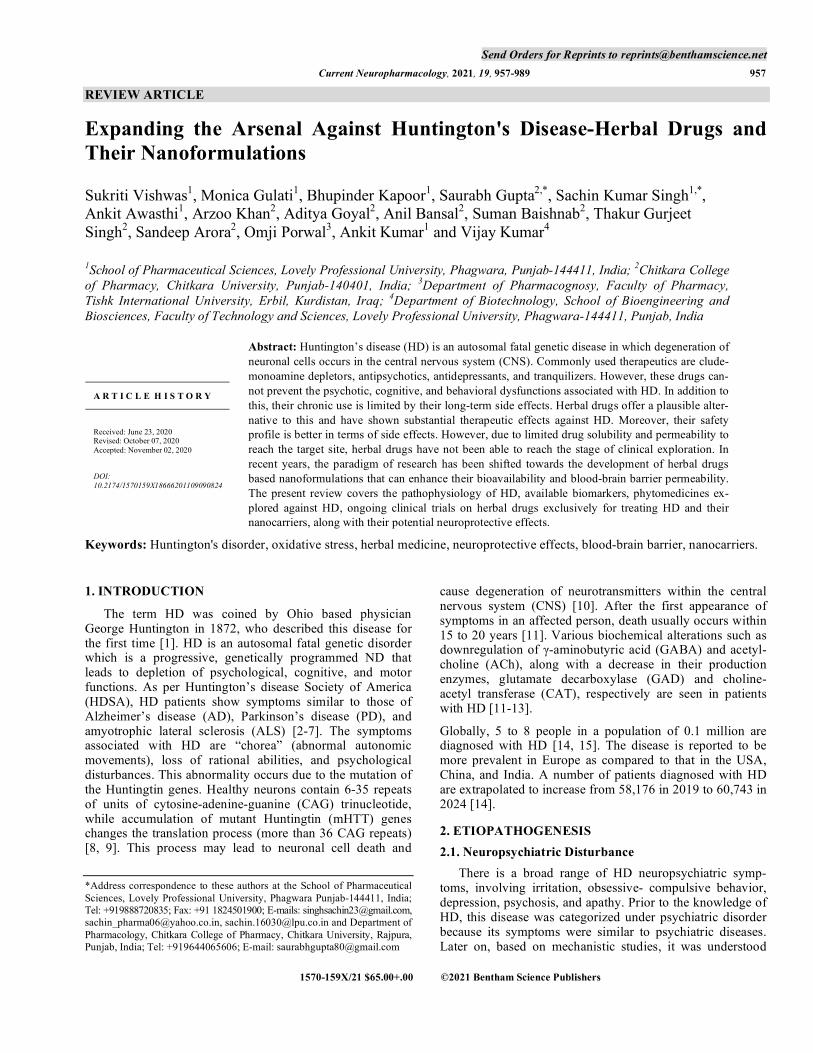
The mechanism of OS in HD is still not clear. Some mo-lecular hypotheses state that increase in the level of ROS, lipid peroxidation, and chromosomal mutation can be major factors for disease manifestation [37, 38]. Impairment in the electron transport chain, oxidative damage, and mitochon-drial dysfunction can increase OS. 8-Hydroxy-2-deoxygonosine (OH8dG) is a biomarker of oxidative damage in DNA. As reported by Bogdanovet et al. (2001), the enhanced concen-tration of OH8dG may increase oxidative damage [39]. Ac-cumulation of mHTT protein has been observed in HD’s patients; hence, it may be implicated in the increase of OS [40, 41]. Increased concentrations of free radicals can pre-dispose to excitotoxicity that can cause impairment of the mitochondrial functions, energy production, and metabolic inhibition [42, 43]. These mechanisms of OS are presented in Fig. (2).
3. BIOMARKERS INVOLVED IN THE PATHO- GENESIS
Biomarkers play an important role in evaluating and measuring pathogenic as well as biological processes and
pharmacological responses against HD. The ideal biomarker must be reliable, accurate, and specific. For understanding new clinical strategies, understanding the biomarkers of a disease is very important. In order to assess the treatment response and monitor the progression of the disease, the Unified Huntington's Disease Rating Scale is currently in use [11].
Based on the method of identification, biomarkers of HD are divided into three categories. These include clinical, bio-fluid, and imaging biomarkers. Clinical biomarkers are used to measure the motor, cognitive and psychotic abnormalities related to HD. Neurotransmitters, microglial toxins, and mHTT protein are listed under the category of bio-fluid bio-markers. Various techniques that are used to quantify their levels in blood and CSF include high-performance liquid chromatography (HPLC), mass spectrometry(MS), time-resolved fluorescence energy transfer (TR-FRET), homoge-neous time-resolved fluorescence (HTRF), and enzyme-linked immune sorbent assay (ELISA) [45]. Imaging bio-markers are used for the detection of structural changes in brain with the help of imaging techniques such as MRI and [18F] MNI-659 PET [46, 47]. Various applications of these biomarkers are listed in Table 1.
Fig. (2). Pathophysiological mediators that are responsible for OS and HD. Mitochondrial dysfunction: Mitochondria are widely known as the powerhouse of cells as they generate energy in the form of adenosine triphosphate (ATP). mHTT genes bind with transporter II in mitochondria and cause mDNA damage and bioenergy failure. mHTT proteins increase the influx of Ca2+ in cytoplasm in mitochondria which leads to excitotoxicity and bioenergy failure, and ATP formation reduces. As a result of this, mitochondrial dysfunction and the gen-eration of ROS take place. Neuro-inflammation: Microglia and astrocytes in the presence of ATP chemokines activate Toll-like receptor (TLR) and m1 receptor protein inflammation cytokines, which, in turn, increase the intracellular Ca2+ entry and ROS levels. M1 receptor protein inflammation cytokines also increase inflammatory mediators (IL6, TNFα) and OS, which give rise to neuroinflammation and degen-eration of the neuronal cell. Accumulation of mHTT genes: The normal base DNA pair contains 5-35 repeated units of CAG chain in exon 1 cytoplasm. When alteration in base DNA pair occurs, mHTT genes bind with exon 1 and increase the CAG units from 36 to 121, which is responsible for OS. This leads to the misfolding of mHtt and the formation of their aggregates in neuronal nuclei and neuropils in the brains of HD patients. This misfolded mHtt exerts its neurotoxicity by disturbing a wide range of cellular functions due to its interaction with a variety of proteins, thus interrupting their function [44]. Increase ROS: Due to mHTT gene, the intracellular influx of Ca2+ increases. This process can enhance excitotoxicity and cause oxidative damage and OS. OS: The factors like accumulation of mHTT genes, neuroinflamma-tion, high lipid concentration, and mitochondrial dysfunction can increase OS, and that is responsible for the progression of the disease. (A higher resolution / colour version of this figure is available in the electronic copy of the article).

960 Current Neuropharmacology, 2021, Vol. 19, No. 7 Vishwas et al.
4. TREATMENT STRATEGIES
Till now, none of the available drugs has been able to show complete relief in symptoms of the disease. Tetra-benazine, however, has been reported to show the most sig-nificant response in terms of reducing symptoms of motor abnormality (chorea) [59]. A combination of antipsychotic, anti-depressant, and anti-AD medicines is reported to reduce
cognitive, psychotic, and motor abnormalities [60-62]. Tetrabenazine is a monoamine enzyme inhibitor that pre-vents the loss of adrenergic neurotransmitters in the synapse. It has been found to be useful in the treatment of hyperki-netic movement disorder. The major side effects of this drug are depression, exacerbation of depression, akathisia, rest-lessness, and psychotic problems [59]. Haloperidol is an an-
Table 1. Biomarker of HD.
S. No. Biomarkers Mediator Molecule Sample Methods Comments Refs.
1. Clinical Motor -- -- Anti-saccade error
rate Understanding genetic and environmental
factor for disease [48]
- - -- -- Digitomotography Assessment of quantitative motor by finger
tapping. [46, 49]
Cognitive - - SDMT - [46]
2. Bio-fluid Immune system IL-6,IL-8, IL-1β CSF, Blood
MSD immunoassay, ELISA
Leukotriene inflammatory mediator activate NFκB and cause neuroinflammation
[50]
- - TNF-α CSF MSD antibody-
based tetraplex array
Tumour necrosis factor α inflammatory mediator activate NFκB and cause neuroin-
flammation [51]
- Genetic HTT
mutation HTT Protein Blood
TR-FRET, HTRF, ELISA
- [45]
- - mHTT protein CSF, Blood
IP-FCM, ELISA, HTRF
mHTT protein can increase OS. [52]
- Microglial markers YKL-40, MCP1, Chi-
totriosidase CSF ELISA - [53]
- Microglial toxins 3-HK, QUIN, ROS - - - -
- Neurodegeneration neurofilament light (NfL) CSF, Blood
ELISA Analyse premanifest and manifest
Huntington's pateints [54]
- - GABA CSF Radioreceptor
assay, Ion-exchange fluorometry
Diminution of inhibitory neurotransmitter GABA
- - - Blood
Ion-exchange chromatography, High resolution
proton NMR spectroscopy and
HPLC
-
[55]
- - Choline CSF Radiochemical micro-method
- [56]
- - Dopamine CSF - - -
- Transglutaminase Nε-(γ-l-glutamyl)-l-lysine
(GGEL) CSF MS - [57]
- - γ-glutamylspermidine, γ-glutamylputrescine, bis-γ glutamylputrescine
CSF HPLC - [58]
3. Imaging Structural loss -- -- MRI Neurodegeneration seen in the brain [46]
- PDE10 uptake -- -- [18F]MNI-659 PET - [47]
Expanding the Arsenal Against Huntington's Disease-Herbal Drugs Current Neuropharmacology, 2021, Vol. 19, No. 7 961
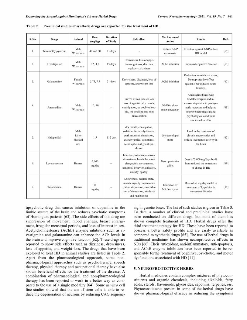
tipsychotic drug that causes inhibition of dopamine in the limbic system of the brain and reduces psychotic symptoms of Huntington patients [63]. The side effects of this drug are suppression of movement, mood changes, breast enlarge-ment, irregular menstrual periods, and loss of interest in sex. Acetylcholinesterase (AChE) enzyme inhibitors such as ri-vastigmine and galantamine can enhance the ACh levels in the brain and improve cognitive function [62]. These drugs are reported to show side effects such as dizziness, drowsiness, loss of appetite, and weight loss. The drugs that have been explored to treat HD in animal studies are listed in Table 2. Apart from the pharmacological approach, some non-pharmacological approaches such as psychotherapy, speech therapy, physical therapy and occupational therapy have also shown beneficial effects for the treatment of the disease. A combination of pharmacological and non-pharmacological therapy has been reported to work in a better way as com-pared to the use of a single modality [64]. Some in vitro cell line studies showed that the use of stem cells is able to re-duce the degeneration of neurons by reducing CAG sequenc-
ing in genetic bases. The list of such studies is given in Table 3. To date, a number of clinical and preclinical studies have been conducted on different drugs, but none of them has shown complete treatment of HD. Herbal drugs offer the third treatment strategy for HD. These have been reported to possess a better safety profile and are easily available as compared to synthetic drugs [65]. The use of herbal drugs in traditional medicines has shown neuroprotective effects in NDs [66]. Their antioxidant, anti-inflammatory, anti-apoptosis, and AChE enzyme inhibition have been reported to be re-sponsible forthe treatment of cognitive, psychotic, and motor dysfunctions associated with HD [11].
5. NEUROPROTECTIVE HERBS
Herbal medicines contain complex mixtures of phytocon-stituents and organic chemicals, including alkaloids, fatty acids, sterols, flavonoids, glycosides, saponins, terpenes, etc. Phytoconstituents present in some of the herbal drugs have shown pharmacological efficacy in reducing the symptoms
Table 2. Preclinical studies of synthetic drugs are reported for the treatment of HD.
S. No. Drugs Animal Dose
(mg/kg) Duration of Study
Side effect Mechanism of
Action Results Refs.
1. Tetramethylpyrazine Male
Wistar rats 40 and 80 21 days -
Reduce 3-NP neurotoxin
Effective against 3-NP induce HD model
[67]
2. Rivastigmine Male
Wistar rats 0.5, 1,2 15 days
Drowsiness, loss of appe-tite/weight loss, diarrhea,
weakness, dizziness AChE inhibitor Improved cognitive function [61]
3. Galantamine Female
Wistar rats 3.75, 7.5 21 days
Drowsiness, dizziness, loss of appetite, and weight loss
AChE inhibitor
Reduction in oxidative stress, Neuroprotective effect
against 3-NP induced neuro-toxicity.
[62]
4.
Amantadine Male
Wistar rats 10, 40
--
Blurred vision, nausea, and loss of appetite, dry mouth,
constipation, or trouble sleep-ing, leg swelling and skin
discoloration
NMDA gluta-mate antagonist
Amantadine binds with NMDA receptor and in-
creases dopamine in postsyn-aptic receptors and helps to improve neurological and psychological conditions
associated in NDs.
[68]
5. Haloperidol
Male Lister
Hooded rats
1.5 112 day
dry mouth, constipation, sedation, tardive dyskinesia, parkinsonism, depression, extrapyramidal symptoms, neuroleptic malignant syn-
drome
decrease dopa-mine
Used in the treatment of chronic neuroleptics and
reduce locomotors activity in the brain
[63]
6. Leveteracetam Human 3,000
mg/day --
Infection, asthenia, neurosis, drowsiness, headache, naso-
pharyngitis, nervousness, abnormal behavior, agitation,
anxiety, apathy.
Neuroprotective effect
Dose of 3,000 mg/day for 48 hour reduced the symptoms
of chorea in HD [69]
7. Terabenzine Human 50
mg/day --
Drowsiness, sedated state, muscle rigidity, depersonal-ization depression, exacerba-tion of depression, akathisia,
and restlessness.
Inhibition of MAO enzyme
Dose of 50 mg/day useful in treatment of hyperkinetic
movement disorder [59]

962 Current Neuropharmacology, 2021, Vol. 19, No. 7 Vishwas et al.
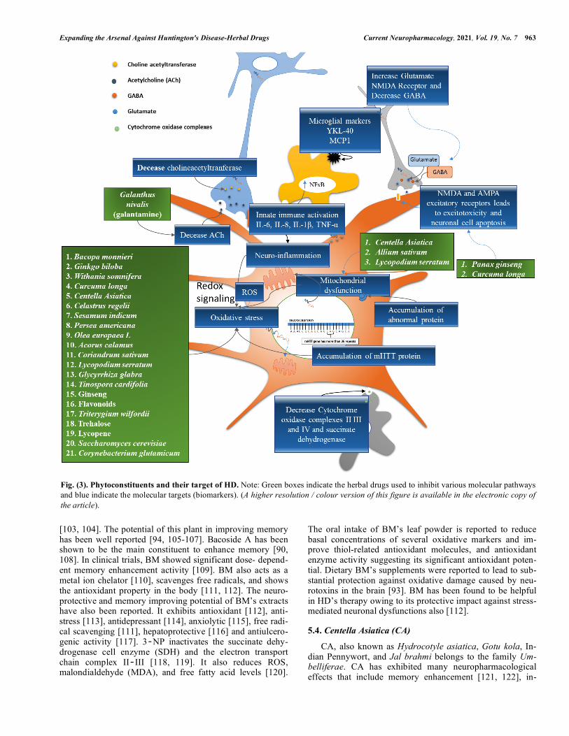
of HD. In India, traditional herbal plants have been used for a number of diseases afflicting the nervous system. VataVy-adh is a Sanskrit word that means disease related to the nervous system. Vata represents energy around the body, and disturbance of this process is called as VataVyadh. It ulti-mately leads to weakness, hypersensitivity, dementia, and chorea [77]. Certain herbal drugs possess phytoconstituents that enhance ACh levels in post synaptic neurons by inhibit-ing AChE in synapse and enhancing cognitive function [78, 79]. The in vivo studies have shown that certain herbal drugs and their phytochemicals exhibit a significant response against 3‐NP neurotoxin. Several other pathways are also crucial in HD. Based on the concept of multi targets, net-work pharmacology-based analysis is employed to find out related proteins in disease networks. The network targeting method aims to find out the related mechanism of efficacious substances in a rational design way. Traditional Chinese medicine (TCM) prescriptions would be used for research and development against HD [80]. Virtual screening is per-formed to obtain drug molecules with high binding capacity from TCM. Mechanism of action and beneficial effects of herbal drugs are shown in Fig. (3).
5.1. Acorus Calamus (AC)
AC, also known as the Sweet flag, belongs to Araceae family. It acts as a brain and nervous system rejuvenator with beneficial memory-enhancing properties. It also im-proves learning efficiency, and reduces behavioral alteration. The major constituents of the plant are α-and β-asarone. β-Asarone has the ability to suppress beta-amyloid-induced neuronal apoptosis in the hippocampus through reversal down-regulation of Bcl-2, Bcl-w, caspase-3 activation, and phosphorylation of c-Jun terminal kinase (JNK) [81]. It has the potential to enhance dopaminergic nerve function. There-fore, it can play a key role in PD by increasing the amount of striatal extracellular dopamine and the expression of tyrosine hydroxylase in substantia nigra. It also improves the expres-sion of DJ-1 genes in the striatum and thus acts as PD neuro-
protective [82]. The treatment of PD using AC indicates its neuroprotective action; hence, it could be used for the treat-ment of HD.
5.2. Allium Sativum (AS)
AS is one of the most widely researched herbs found in the ancient medical literature [83, 84]. AS belongs to the family Amaryllidaceae. The main bioactive compounds of AS are allicin (allyl2-propene thiosulfinate or diallyl-thiosulfinate) and alliin. S-allyl cysteine (SAC) is the major component of the extensively studied aged garlic extract (AGE) [85, 86]. SAC exerts antioxidant activity, both di-rectly and indirectly. It also decreases protein oxidation and nitration. In addition to this, it is reported to reduce lipid peroxidation and DNA fragmentation. Dopamine levels, oxi-dative damage, and lipid peroxidation in 1-methyl-4-phenyl pyridinium and 6-hydroxydopamine (6-OHDA) models of PD were found to be downregulated by SAC. It decreased lipid peroxidation and mitochondrial dysfunction in 3-nitro propionic acid and quinolinic acid animal models of HD. It also increased the dismutase activity of manganese and su-peroxide copper/zinc and prevented changes in behavior. AGE activates the expression of significant genes required for neuronal survival, both directly and indirectly [87, 88].
5.3. Bacopa Monnieri (BM)
BM or Herpestis monniera, commonly referred to as Brahmi, belongs to the family scrophulariaceae. It is found throughout the Indian subcontinent and is categorized in Ayurveda as Medhya Rasayana [89-92]. It is used to treat epilepsy, insomnia, anxiety and is a memory enhancer [93, 94]. The significant chemical components present in the plant are tri-terpenoid saponins like dammarane, bacosides A and B [90, 95]. In addition to these significant components, it also contains other saponins, including bacopa saponin A‐G [96-98], along with pseudo-jujubogenin, jujubogenin [99], bacopaside I‐V, X, and N1 and N2 [100-102]. Brah-mine, herpestine, and monnierin are also present in the plant
Table 3. Cell line studies of HD.
Disease Cell line Test Results Refs.
HD LUMCi007-A, LUMCi007-B,
LUMCi008-A, LUMCi008-B, LUMCi008C -- Reducing CAG repeats [70]
ICGi018-A (iHD38Q-3) DNA fragment analysis of
PCR-product In vitro cell line studies reduce CAG18 and
38 repeats by PBMCs and iPSC line [71]
CSSi006-A (3681) Sequencing Reducing CAG repeats in fibroblasts (17 ± 2 and 46 ± 3 CAG repeats) [72]
CSSi004-A (2962) Sequencing Reducing CAG repeats in fibroblasts (17 ± 1 and 43 ± 2 CAG repeats) [73]
Genea090 human embryonic stem cell line Sterility The cell line is tested and found negative for Mycoplasma and any
visible contamination [74]
Genea017 human embryonic stem cell line Sterility The cell line is tested and found negative for Mycoplasma and any visible contamination. Mycoplasma and any visible contamination
Herbal formula B401 -- Neuroprotective and angiogenesis effects in R6/2 mouse model of HD [75]
CurcuminSolid lipid nanoparticles (SLNs)
(C-SLNs) SDH Staining, Mitochondrial Oxidative Stress Parameters
Reduce ROS, mitochondrial dysfunction and lipid preroxidation. [76]
Expanding the Arsenal Against Huntington's Disease-Herbal Drugs Current Neuropharmacology, 2021, Vol. 19, No. 7 963
[103, 104]. The potential of this plant in improving memory has been well reported [94, 105-107]. Bacoside A has been shown to be the main constituent to enhance memory [90, 108]. In clinical trials, BM showed significant dose- depend-ent memory enhancement activity [109]. BM also acts as a metal ion chelator [110], scavenges free radicals, and shows the antioxidant property in the body [111, 112]. The neuro-protective and memory improving potential of BM’s extracts have also been reported. It exhibits antioxidant [112], anti-stress [113], antidepressant [114], anxiolytic [115], free radi-cal scavenging [111], hepatoprotective [116] and antiulcero-genic activity [117]. 3‐NP inactivates the succinate dehy-drogenase cell enzyme (SDH) and the electron transport chain complex II‐III [118, 119]. It also reduces ROS, malondialdehyde (MDA), and free fatty acid levels [120].
The oral intake of BM’s leaf powder is reported to reduce basal concentrations of several oxidative markers and im-prove thiol-related antioxidant molecules, and antioxidant enzyme activity suggesting its significant antioxidant poten-tial. Dietary BM’s supplements were reported to lead to sub-stantial protection against oxidative damage caused by neu-rotoxins in the brain [93]. BM has been found to be helpful in HD’s therapy owing to its protective impact against stress-mediated neuronal dysfunctions also [112].
5.4. Centella Asiatica (CA)
CA, also known as Hydrocotyle asiatica, Gotu kola, In-dian Pennywort, and Jal brahmi belongs to the family Um-belliferae. CA has exhibited many neuropharmacological effects that include memory enhancement [121, 122], in-
Fig. (3). Phytoconstituents and their target of HD. Note: Green boxes indicate the herbal drugs used to inhibit various molecular pathways and blue indicate the molecular targets (biomarkers). (A higher resolution / colour version of this figure is available in the electronic copy of the article).
964 Current Neuropharmacology, 2021, Vol. 19, No. 7 Vishwas et al.
creased neurite elongation, and nerve regeneration accelera-tion [123]. It has also been reported for its anti-oxidant prop-erties [124, 125]. Triterpenoid saponins, including asiatico-side, Asian acid, madecassoside, and madecassic acid, are the most significant chemical constituents of CA [126, 127]. Other minor saponins present in CA are brahmoside and brahminoside [126, 128]. Various acids that are present in the plant are triterpene acids, betullic acid, brahmic acid, and isobrahmic acid [126, 128]. The essential oils that are pre-sent in plant leaves include monoterpenes such as bornyl acetate, α-pinene, β-pinene, and π-pinene [129]. In addition to these constituents, CA is also reported to contain flavones, sterols, and lipids. Attenuation of 3-NP-induced depletion of GSH, total thiols, and endogenous antioxidants level by CA has been reported in the striatum and other brain regions [130]. It also displayed protection against 3-NP-induced mi-tochondrial dysfunctions, viz., reduced SDH activity, en-zymes in the electron transport chain, and reduced mito-chondrial viability [130].
5.5. Coriandrum Sativum (CS)
CS, commonly known as coriander, belongs to Apiaceae family. It contains a number of flavonoids. The major phyto-constituents include glucoronides such as quercetin and polyphenols such as caffeic acid, protocatechinic acid, and glycitin. The flavonoid content of the plants is reported to be equivalent to 12.6 quercetin equivalents per gram, while polyphenolic content is equivalent to 12.2 gallic acid equiva-lents per gram [131, 132]. A study showed that the CS’s ex-tract enhanced concentrations of superoxide dismutase (SOD), glutathione, CAT, and total protein in the animal model. It also reduced the levels of cerebral infarction, lipid peroxidation (LPO), and calcium in the rats [133]. Scopola-mine and diazepam-induced memory deficits were found to be reversed by leaf extracts of CS. It reduced reactive modi-fications in brain histology such as gliosis, lymphocytic infil-tration, and cellular edema. It showed protective function in the states of cerebrovascular insufficiency. The leaves also demonstrated antioxidant properties in terms of free radical scavenging activity by 2, 2-diphenyl-1-picrylhydrazyl and lipoxygenase inhibition [134-136].
5.6. Curcuma Longa (CL)
The common name used for the CL is turmeric. It is a perennial herb and belongs to the family Zingiberaceae. It is used throughout the world, mainly in China, Japan, and In-dia, as a pharmacotherapeutic [137]. It has a long history of use as a spice and household remedy to treat inflammation, skin diseases, wounds, as well as antibacterial and antiseptic agent [138]. CL contains different curcuminoids, sesquiter-penes, essential oil, and starch. Most of the curcuminoids are diarylheptanoid, curcumin being the most prevalent. Des-methoxycurcumin and bis-desmethoxycurcumin are the other two curcuminoids [138, 139]. CL shows a number of pharmacological actions such as antioxidant [140], anti-inflammatory [141], choleretic, hepatoprotective, analgesic, antifungal, free radical scavenging, antiparasitic, antiviral, antibacterial [138, 142], and anti-mutagenic [143]. The anti-oxidant properties of turmeric are attributed to its direct scavenging of superoxide radicals, chelating action [140,
144, 145], and by induction of antioxidant enzymes such as glutathione‐S‐transferase, glutathione peroxidase, catalase, superoxide dismutase, and hemeoxygenase [145]. It shows anti-inflammatory action by restricting cyclooxygenase-2 pathway (COX-2).In various neurological disorders, it is reported to show neuroprotective action [146]. Curcumin alone or along with manganese complex provides protective action against vascular dementia due to its antioxidant activ-ity [147-149], and it is also helpful in treating aging and memory dysfunction [150]. In one of the studies, it has been reported that chronic administration of curcumin enhanced body weight continuously and increased SDH activity in rats treated with 3‐NP [150]. The reversed 3‐NP‐induced motor and cognitive impairment, along with a powerful antioxidant property, indicate that curcumin may be helpful in treating HD [150].
5.7. Galanthus Nivalis (GN)
GN, commonly known as snowdrop, belongs to the fam-ily Amaryllidacea. Galantamine, a tertiary isoquinoline alka-loid, is the main ingredient found in bulbs and flowers of GN. The neuroprotective activity of galantamine is due to this alkaloid. It is a reversible carbamates AChE inhibitor. Galantamine is an FDA approved drug that is used to treat AD. It can stimulate nicotinic receptors that further improve memory and cognition [151]. The drug allosterically modu-lates nicotinic receptors of ACh, particularly subtypes α7 and α3β4, to increase the release of ACh on cholinergic cells [152].
5.8. Ginkgo Biloba (GB)
GB is an ancient Chinese herbal plant having neuropro-tective properties [153]. The active phytoconstituents are mainly obtained from the leaves and flowers of the plant. These include flavonoids (quercetin, isorhamnetins and kaempferol), bioflavonoids (bilobetin, sciadopitysin, 5-methoxybilobetol, isoginkgetin, ginkgetin and aimoflavone), proanthocyanidins, Trilactonic diterpenes (A-C ginkgolide and J-M ginkgolide), and sesquiterpenes (bilobalide) [154-156].
This plant’s leaf extract has been reported to be effective against dementia, cardiovascular diseases, stress, tumor and led to increased peripheral and central blood flow [157]. It also showed numerous pharmacotherapeutic activities due to its antioxidant effect [158], anti-platelet activating factor activity, and inhibition of amyloid-beta (Aβ) peptide aggre-gation [159, 160]. In one of the studies, the extract of GB (100 mg / kg, i.p. for 15 days) reversed neurobehavioral deficits induced by 3‐NP and also reduced striatal MDA [161]. The standardized extract of Ginkgo Biloba (EGb 761) also caused up and downregulation of the expression of Bcl-xl and striatal glyceraldehyde-3-phosphate dehydrogenase concentrations, respectively. These biochemical findings suggested the neuroprotective function of EGb 761 in HD [161]. The extracts of GB have been reported to commonly induce biphasic dose responses in a range of cell types and endpoints (e.g., cochlea neural stem cells, cell viability, cell proliferation) [162]. The magnitude and width of the low dose stimulation of these biphasic dose responses are similar to those reported for hormetic dose responses. These hor-
Expanding the Arsenal Against Huntington's Disease-Herbal Drugs Current Neuropharmacology, 2021, Vol. 19, No. 7 965
metic dose responses occur within direct stimulatory re-sponses as well as in preconditioning experimental protocols, displaying acquired resistance within an adaptive homeody-namic and temporal framework and repeated measurement protocols. The demonstrated GB’s dose responses further reflect the general occurrence of hormetic dose responses that consistently appear to be independent of the biological model, endpoint, inducing agent, and/or mechanism. These findings have important implications for consideration(s) of study designs involving dose selection, dose spacing, sample size, and statistical power [163].
5.9. Glycyrrhiza Glabra (G. glabra)
G. glabra, commonly referred to as Yashti-madhuh, be-longs to the family Leguminosae. G. galabra contains an isoflavane glabridin. It has been reported to exert various pharmacological activities such as antiviral, anticancer, anti-ulcer, anti-diabetic, antioxidant, immunomodulatory, anti-inflammatory, and anticonvulsant effects. Glabridin reduces the amount of MDA and glutathione, and increases the amount of SOD in the brain [164, 165]. G. glabra lowers the brain concentrations of neurotransmitters such as glutamate and dopamine and reduces the activity of AChE [166].
5.10. Lycopodium Serratum (LS)
LS belongs to the family lycopodiaceae. Alkaloid hu-perzine is obtained from LS extract [167]. As per the litera-ture, this alkaloid shows AChE inhibition activity. Therefore, it increases the level of ACh in post synaptic receptors in the brain. LS elicits the same kind of effects as AChE inhibitor drugs [168]. The component huperzine is also used for the treatment of AD because it inhibits the ACh enzyme, acts as an antioxidant, and possesses anti-inflammatory properties [169]. Huperzine has been reported to have several neuropro-tective effects such as apoptosis, the rectification of mito-chondrial dysfunction, and anti-inflammatory effects [170].
5.11. Olea Europaea (OE)
It is commonly known as olive oil and belongs to the family Oleaceae [171]. Fruits’ oils contain many nutritious chemical constituents such as triacylglycerols, glycerol, free fatty acid, pigments, phosphatides, and flavor compounds [172]. Olive oil is very nutritious to the health and is also used as cooking oil. Some of the pharmacological studies have reported the potential effects of olive oil and extrava-gant olive oil against cardiovascular diseases [173], AD [174, 175], PD [176, 177], MS, and cancer [178, 179]. In studies conducted by Visioli et al. (1998) and Tasset et al. (2011), potential effects of extravagant olive oil have been reported against HD due to its antioxidant property [180] and neuroprotective effects [181]. In pharmaceutical formula-tions such as emulsions, olive oil acts as a solubilizer. In one of the studies, Guo el al., have used olive oil as a solubilizer for lycopene. The lycopene loaded microemulsions (LME) were prepared in which lycopene has been dissolved in olive oil. The potentials of microemulsion in improving bioavail-ability and brain-targeting efficiency following oral admini-stration were investigated [182]. The pharmacokinetics and tissue distributions of optimized LME were evaluated in rats
and mice, respectively. The pharmacokinetic study revealed a dramatic 2.10-folds enhancement of relative bioavailability with LME against the control lycopene dissolved in olive oil (LOO) dosage form in rats. Moreover, LME showed a pref-erential targeting distribution of lycopene toward the brain in mice, with the value of drug targeting index (DTI) up to 3.45 [182]. 5.12. Plants Containing Trehalose
Trehalose was subsequently found in mosses, ferns, green algae, and liverworts [183]. It is found in many plants that grow in low and high altitudes, as well as in many or-ganisms like bacteria, yeast, fungi, insects, invertebrates [184, 185]. It has been found in the literature that trehalose inhibits the formation of amyloid [186, 187]. Besides these, it also helps in inhibiting polyglutamine (polyQ) 3-mediated protein aggregation and reduced toxicity caused by Huntington's aggregates. Tanaka et al. (2004) and Sarkar et al. (2007) conducted a study on HD using HD R6/2 mouse model. It was found that trehalose helped in the inhibition of polyQ-induced pathology by stabilizing the partly unfolded mutant proteins [188, 189]. It has also been reported that, by offering neuroprotective activity against HD, trehalose in-creases autophagic activity against multiple aggregations of proteins such as mHTT [188].
5.13. Panax Ginseng (PG)
PG root is a well-known herb used in China, Japan, and Korea as a tonic to revitalize and restore adequate body me-tabolism for over more than 2,000 years [190]. The most prevalent species of PG are Asian ginseng and American ginseng (Panax1 quinquefolium L.) from the Araliaceae family. PG is a neuroprotective herb, and its neuroprotective potential can be used to prevent and treat neurodegenerative diseases such as AD, PD, HD, depression symptoms, and strokes [163, 191]. The major constituent that is responsible for the neuroprotective action of PG is ginsenoside. In recent years a number of studies have been reported on the role of ginsenosides in the prevention of NDs [192]. Moreover, the results of some of the clinical trials conducted on PG and its constituents, ginsenosides, and gintonin, revealed that they are safe [192]. PG contains tetracyclic dammarane, triterpe-noids, saponin glycosides, and ginsenosides as their active constituents [193, 194]. Different studies (in vitro and in vivo) have shown positive results of ginseng in various pathological conditions such as cardiovascular diseases, CNS disorders, cancer, immune deficiency, and hepatotoxic-ity [194, 195]. It also has antioxidant [196], anti-apoptotic [196], anti-inflammatory [197], and immune-stimulating functions [195]. It helps in decreasing lipid peroxidation by inhibiting excitotoxicity and over-influx of Ca2+ into neu-rons. It retains concentrations of cellular ATP, preserves neuronal structural integrity, which helps in increasing cog-nitive performance [195]. Ginsenoside Rb1 and Rg3 are re-ported to exhibit protective effects by preventing Ca2 + influx through glutamate receptors on cortical neurons against glu-tamate-induced cell death [198]. Ginseng contains saponins that are NMDA glutamate antagonists. They reduce intracel-lular Ca2+ influx in the hippocampus; hence glutamate type NMDA receptors get inhibited, and this results in reduction
966 Current Neuropharmacology, 2021, Vol. 19, No. 7 Vishwas et al.
of the symptoms of HD [199]. Ginsenosides Rb1, Rb3, and Rd showed a neuroprotective impact on striatal neuronal harm caused by 3‐NP [200-202].
5.14. Sesamum Indicum (SI)
Sesamol is obtained from the plant SI, frequently referred to as sesame, belonging to family Pedaliaceae. It is used in India and other East Asian nations as a healthy food [203]. The oil obtained from sesame is responsible for its pharma-cological activities. Its active component, sesamol, is ac-countable for its antioxidant activity [204]. It helps in reduc-ing hyperlipidemia, blood pressure, and lipid peroxidation by diminishing enzymatic and non-enzymatic oxidants stress. It also has tumour suppressant action [205]. Sesamol has been reported to have its protective effect against HD through suppression of the expression of nitric oxide (NOS) [206]. It is also reported to attenuate behavioral, biochemical, and cellular changes in 3‐NP‐induced animals [207]. It has been reported to protect the brain against memory impairment caused by 3‐NP, OS, neuroinflammation in the neurons of the hippocampus, and thus increases synaptic plasticity and neurotransmission [208].
5.15. Solanum Lycopersicum (SL)
SL is commonly referred to as tomato and belongs to the family Solanaceae. Lycopene is a well-known carotenoid found in tomatoes and tomato-based goods in considerable quantities [209]. It has been reported to possess powerful neuroprotective [210], antioxidant [211, 212], antiprolifera-tive, anticancer [213], anti-inflammatory [214], memory enhancing [215], and hypocholesterolemic properties [216]. It is a stronger singlet oxygen carotenoid quencher for vita-min E and glutathione [216]. Treatment with lycopene con-siderably helps in the reduction of multiple behavioral and biochemical changes induced by 3‐NP, indicating its thera-peutic potential against HD’s symptoms [217].
5.16. Tinospora Cordifolia (TC)
TC belongs to the family Menispermaceae and is fre-quently known as Giloy. Phytochemical constituents such as alkaloids, steroids, diterpenoid lactones, aliphatics, and gly-cosides are present in giloy extract [218]. TC has been re-ported for memory enhancing property, immunostimulation, and enhancement of ACh synthesis [219]. It has powerful free radical scavenging characteristics and also reduces ROS and reactive nitrogen species as studied by paramagnetic resonance electron spectroscopy [220]. It also reduces the level of glutathione, gamma-glutamyl-cysteine ligase expres-sion, copper-zinc superoxide dismutase genes, owing to which it can be used for the treatment of hypoxia, ischemia, and neuronal injury [220]. Additionally, TC is helpful in enhancing dopamine levels in the brain and improving cog-nitive and psychotic function [219].
5.17. Tripterygium Wilfordii (TW)
The root extract of TW has been extensively used as tra-ditional Chinese medicine for the treatment of inflammation and autoimmune diseases such as rheumatic arthritis [221]. TW’s root extract also showed neurotropic and neuroprotec-
tive effects [222]. Celastrol and Triptolide are the two major neuroprotective phytoconstituents that are isolated from the root extract of TW. It has many therapeutic potentials such as antioxidant [223], anti-inflammatory [221], anticancer [224], and insecticidal activity [225]. A pro-inflammatory study conducted on animals using 1‐methyl‐4‐phenyl‐1, 2, 3, 6-tetrahydropyridine (MTPT) indicated that celastrol helped in improving the functions of dopaminergic cells, increasing the dopaminergic level [222]. By controlling the expression of the thermal shock protein gene in dopaminer-gic cells, it has also provided protection against 3-NP-induced striatal damage [222, 226].
5.18. Withania Somnifera (WS)
WS is also known by the common name Ashwagandha. It belongs to the family Solanaceae. For centuries, it has been used in Ayurvedic medicine [227]. Ashwagandha’s root extract has been reported to possess antioxidant [228, 229], memory enhancing [230], anti-inflammatory [231], immu-nomodulatory [232], anti-stress [233], and anti-convulsant characteristics [234]. As an antioxidant, WS and its active ingredients (sitoindosides VII‐X and withaferin A) increase catalase, ascorbic acid, endogenous superoxide dismutase, and reduce lipid peroxidation [235-237]. It functions as an anti-inflammatory agent through complement inhibition, the proliferation of lymphocytes, and delayed hypersensitivity. Different trials have shown that WS increases cortisol circu-lation, decreases tiredness, increases physical performance, and decreases refractory stress depression [238]. It also modulates different receptor systems for neurotransmitters in the CNS. Major active constituents of WS include steroidal lactones and alkaloids (collectively referred to as witha-nolides). Withaferin A, withanolide A, withanolide D‐P, withanone, sitoindoside VII‐X are the major isolated witha-nolides from WS. WS inhibits AChE and increases the level of ACh in the brain. The beneficial effects of herbal drugs against HD are listed in Table 4.
6. CURRENT ONGOING CLINICAL TRIALS
A limited number of clinical trials have been reported for the treatment of HD. This could be attributed to a lack of complete understanding of the underlying mechanism of the disease as the drugs used so far have been unable to provide complete relief to patients. Some of the trials that have been completed or are ongoing, are listed in Table 5.
7. HORMESIS AND REDOX ASPECTS OF HERBAL DRUGS AND THEIR POTENTIAL CHEMICAL CONSTITUENTS
Hormesis is a biological process that has found its appli-cation in drug development, drug designing, and toxicologi-cal studies. It helps to rationalize the dose-response relation-ships [80, 256]. Hormetins are the chemical inducers of hormesis and possess a range of therapeutic applications, including protection against stress, toxin, and aging-related diseases [257, 258]. They have a protective effect at a low level and show deleterious effects at higher levels due to the narrow therapeutic window [259, 260]. Hormesis describes the phenomenon of pharmacological conditioning of the heart and brain where a low dose of pharmacological agents
Expanding the Arsenal Against Huntington's Disease-Herbal Drugs Current Neuropharmacology, 2021, Vol. 19, No. 7 967
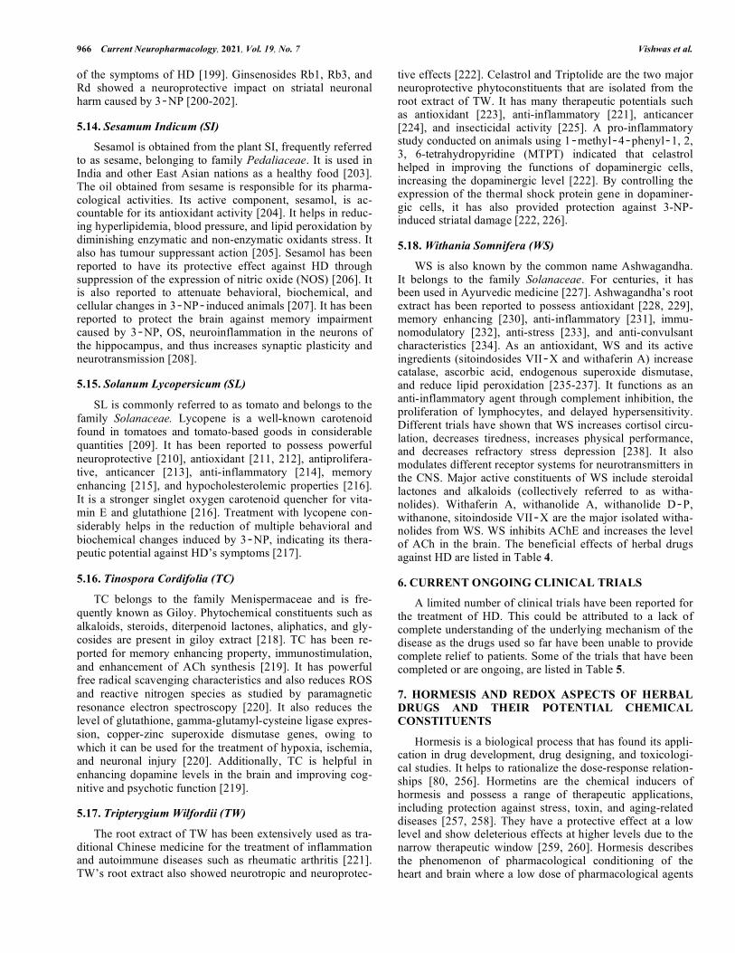
Table 4. Effect of herbal drugs and phytoconstituents.
Name of Herbal
Medicine Synonyms Source
Bioactive
Component Structure Animal Models Effects Refs.
AC Sweet flag α-and
β-Asarone --
Antioxidant, anti-
inflammatory [239]
AG Ginseng Whose
root Ginsenosides
O
OH
OH
OH
O
OH
HOH
O
HH
OH
OOH
OH
OH
HO
H
H
--
Antioxidant, anti-
apoptotic, anti-
inflammatory, and
immune-stimulating
functions
[195-
197]
WS
Withania root,
asgandh,
winter cherry.
Dried
Roots Withaferin A
O O
OHCH3
H3C
CH3
CH3O
CH3O
3-nitropropionic
acid model
Reduce oxida-
tive/nitrosative
stress,
inhibits complex II
of the mitochondrial
electron transport
chain
[240]
BM
Kapotvadka,
somvalli and
saraswati
Aerial
parts
Bacoside A,
O OH
O O
O
H3C
CH3
CH3
OH
OH
OH
CH2OH
HO
CH3
CH3
OH
OH
OH
3-nitropropionic
acid induce
model
Memory enhancer [241]
Bacoside B,
O OH
O O
O
HO
CH3
CH3
OH
OH
OH
CH2OH
HO
CH3
CH3
CH3
OH
OH
3-nitropropionic
acid induce
model
Facilitates an-
terograde memory
CR Celastrol
(tripterine) -- Celastrol
O
OH CH3
CH3
HO
CH3
O
OH
--
Anti-inflammatory,
anti-oxidant, and
inhibition of Pro-
inflammatory
cytokines.
[221]
CL
Indian
saffron,
curcuma,
Turmeric,
Haldi
Fresh
rhizomes Curcumin
O O
R2
OH
R1
HO
3-nitropropionic
acid-induced
HD rat model
and inhibitory
response against
AMPA receptor
Anti-oxidant, Anti-
inflammatory and
reduce excitotoxic-
ity
[242]
(Table 4) contd….
968 Current Neuropharmacology, 2021, Vol. 19, No. 7 Vishwas et al.
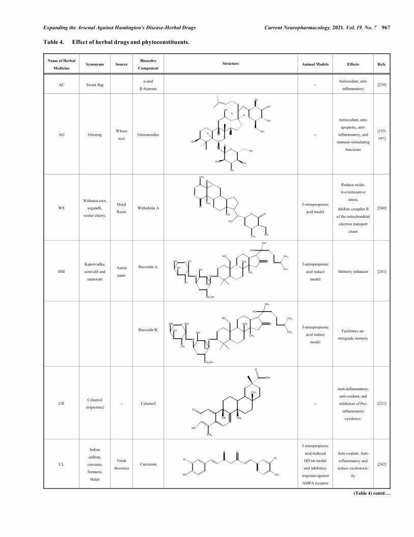
Name of Herbal
Medicine Synonyms Source
Bioactive
Component Structure Animal Models Effects Refs.
Demethoxy
curcumin --
Rotenone-induced
PD in rats
Anti-oxidant and
Anti-inflammatory
CS
Coriander Leaves
Coriandrum
sativum
extract
Ischemic reperfusion
insult in brain
Neuroprotective
effects [133]
GN Snowdrop Bulbs and
flowers
Galantamine
0.1 mg/kg, s.c.
O
N
HO
OH
Scopolamine-
induced amnesia
model in mice
Reduce AChE
enzyme and
increase ACh in
postsynaptic
receptor
[243]
Ginkgo (GB)
Ginnan,
maidenhair
tree.
Leaf Gingkolides
A,B,C,J and M --
3-nitropropionic
acid model
Memory enhancer
property and Anti-
Platelet Activating
Factor (Anti-PAF)
[161]
GG
Yashti-
madhuh or
liquorice
Stems Glabridin -- - Antioxidant [244]
CA
Spade leaf,
Indian Pen-
nywort,
Mandukaparni
Asiatic Acid HO
HO
CH3
OH
CH3 CH3
H3C
CH3
OR
OCH3
--
Neuroprotective
effect against harm
caused by OS and
Mitochondrial
dysfunction
[245]
LS
Ground pines
or creeping
cedar, Qian
Ceng Ta.
Leaves Huperzine A
HN
H2N
O
--
Antioxidant and
anti-inflammatory
and reduce mito-
chondrial dysfunc-
tion
[170]
Persea
Americana
Avocado
Peel, seed
coat and
seeds
Persea major
methanolic extract
(0.5 mg/ml)
-- Cellular viability
assay, Glutamate
uptake assay
Antioxidant capac-
ity, increased
glutamate uptake
[246]
OE Olive-
growing Oil
Olive oil, Extravir-
gin olive oil
(20 mg/kg ip)
O
O OO
O R2
O R3
R1
3-nitropropionic
acid-induced HD-
like rat model
Reduces oxidative
damage
[181]
Sesamum
indicum
Sesame,
benne
Oil Sesamol
O
O
OH
-- Neuroprotective
effect [247]
TC
Giloe Stem
Tinospora cordifo-
lia-stem methano-
lic extract
--
6-hydroxy dopamine
(6-OHDA) lesion rat
model, Cadmium-
induced OS in
Wistar rats
Anti-OS, Memory
enhance and
Increase dopamine
level in to brain.
[248]
(Table 4) contd….
Expanding the Arsenal Against Huntington's Disease-Herbal Drugs Current Neuropharmacology, 2021, Vol. 19, No. 7 969
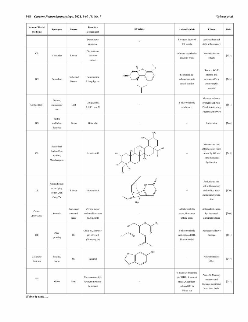
Name of Herbal
Medicine Synonyms Source
Bioactive
Component Structure Animal Models Effects Refs.
TW Thunder
god vine
Root
extracts
Celastrol,
Triptolide
H
O OH
HO
O
-- Antioxidant effects [249]
Fruits,
vegetables, tea,
cocoa and wine
-- -- Flavonoids O
O
--
Effects against OS
and Inflammation
[250]
Mosses ferns,
green algae, and
liverworts
-- -- Trehalose
O
OHO
O
OH
OHHO
OH
OHHO
OH
induced damage in
bovine spermatozoa
Antioxidant effects [183,
251]
SL Tomato Hole
fruits Lycopene
H3C
CH3
H3C CH3
CH3 CH3CH3
CH3
CH3CH3
3-nitropropionic acid-
induced HD rat model
Inhibition of cogni-
tive dysfunction and
motor abnormality
and antioxidant
effects
[252,
253]
Saccharomyces
cerevisiae and
Corynebacterium
glutamicum
Rasberry Fruit Salidroside
O
OH
OHO
HO OH
OH
Inhibit the SOD1 and
HTT genes and also
show anti-
inflammatory effects.
Reduce the symp-
toms of HD by acting
oxidative stress and
inflammation, and
HTT genes.
[254]
Table 5. Ongoing clinical trial of HD.
Disease Drug Sample Size Purpose Phase Status Design Study State Study end
HD THC, CBD 21 Treatment Phase 2 Completed DB, R, CO December 30, 2011 February 1, 2013
EGCG 54 Treatment Phase 1 Completed R May 23, 2011 June 16, 2015
PBT2 109 Treatment Phase 2 Completed DB, R May 3, 2012 July 18, 2016
DM/Q 22 Treatment Phase 3 Recruiting R February 26, 2019 April 19, 2019
Triheptanoin 10 Treatment Phase 2 Completed June 20, 2013 March 24, 2016
SD-809 90 Treatment Phase 3 Completed R, DB January 2, 2006 September 20, 2017
Digoxin, Dimebon 12 Treatment Phase 1 Completed R January 29, 2009 June 12, 2009
Chorea SD-809 90 Treatment Phase 3 Completed R, DB February 21, 2013 August 11, 2017
Amantadine sulphate 30 Treatment Phase 4 Completed NR July 31, 2009 June 28, 2011
HMD Tetrabenazine -- -- -- Available -- March 24, 2008 February 26, 2020
Abbreviations: CO; Cross Over, CBC, Cannabidiol, DB; Double Blind, DM/Q; Dextromethorphan/quinidine, EGCG; (2)-epigallocatechin-3-gallate, NR, Non-Randomized , R; Randomized, THC; Delta-9-tetrahydrocannabinol, HMD; Hyperkinetic Movement Disorders Based on search of clinicaltrial.gov (https://clinicaltrials.gov/ct2/ results?cond=huntington+disease&term=&cntry=&state=&city=&dist=) [255] [Accessed May 26, 2020].
970 Current Neuropharmacology, 2021, Vol. 19, No. 7 Vishwas et al.
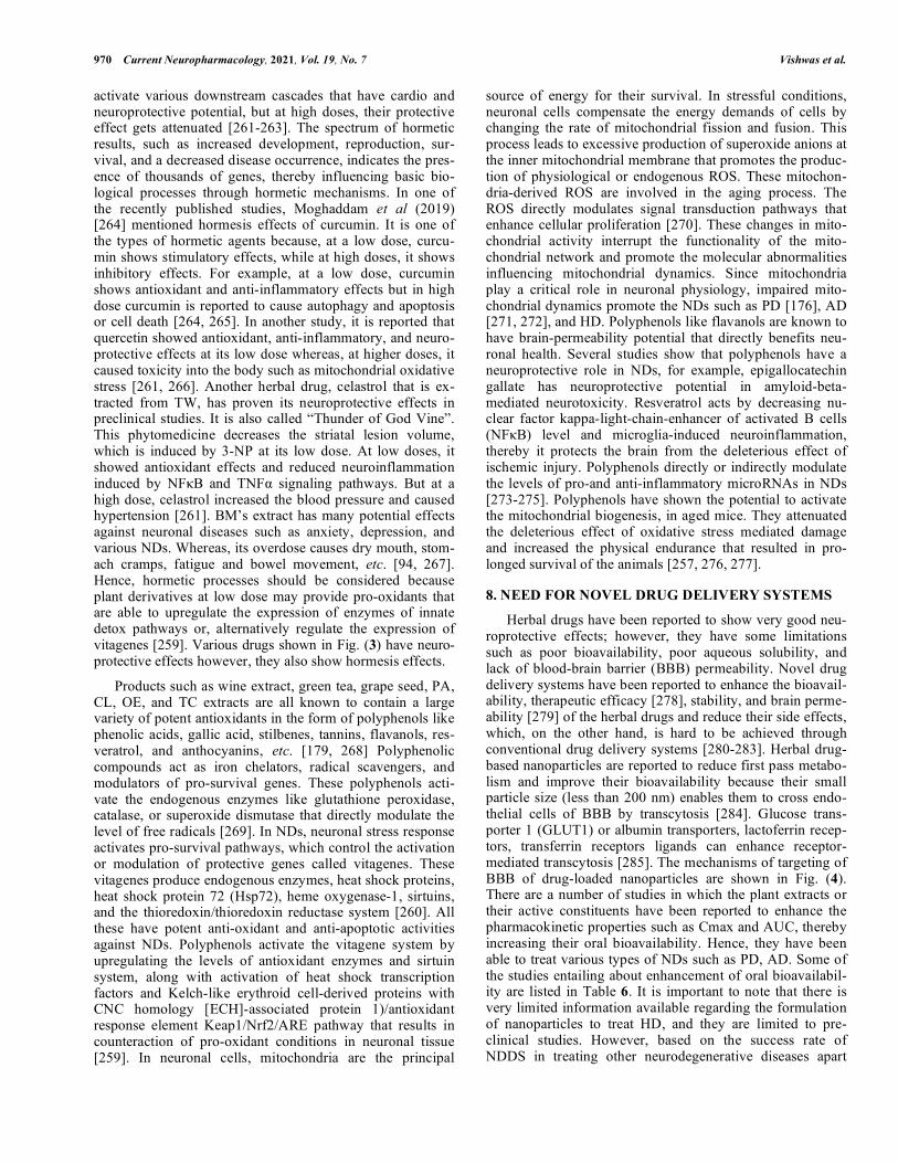
activate various downstream cascades that have cardio and neuroprotective potential, but at high doses, their protective effect gets attenuated [261-263]. The spectrum of hormetic results, such as increased development, reproduction, sur-vival, and a decreased disease occurrence, indicates the pres-ence of thousands of genes, thereby influencing basic bio-logical processes through hormetic mechanisms. In one of the recently published studies, Moghaddam et al (2019) [264] mentioned hormesis effects of curcumin. It is one of the types of hormetic agents because, at a low dose, curcu-min shows stimulatory effects, while at high doses, it shows inhibitory effects. For example, at a low dose, curcumin shows antioxidant and anti-inflammatory effects but in high dose curcumin is reported to cause autophagy and apoptosis or cell death [264, 265]. In another study, it is reported that quercetin showed antioxidant, anti-inflammatory, and neuro-protective effects at its low dose whereas, at higher doses, it caused toxicity into the body such as mitochondrial oxidative stress [261, 266]. Another herbal drug, celastrol that is ex-tracted from TW, has proven its neuroprotective effects in preclinical studies. It is also called “Thunder of God Vine”. This phytomedicine decreases the striatal lesion volume, which is induced by 3-NP at its low dose. At low doses, it showed antioxidant effects and reduced neuroinflammation induced by NFκB and TNFα signaling pathways. But at a high dose, celastrol increased the blood pressure and caused hypertension [261]. BM’s extract has many potential effects against neuronal diseases such as anxiety, depression, and various NDs. Whereas, its overdose causes dry mouth, stom-ach cramps, fatigue and bowel movement, etc. [94, 267]. Hence, hormetic processes should be considered because plant derivatives at low dose may provide pro-oxidants that are able to upregulate the expression of enzymes of innate detox pathways or, alternatively regulate the expression of vitagenes [259]. Various drugs shown in Fig. (3) have neuro-protective effects however, they also show hormesis effects.
Products such as wine extract, green tea, grape seed, PA, CL, OE, and TC extracts are all known to contain a large variety of potent antioxidants in the form of polyphenols like phenolic acids, gallic acid, stilbenes, tannins, flavanols, res-veratrol, and anthocyanins, etc. [179, 268] Polyphenolic compounds act as iron chelators, radical scavengers, and modulators of pro-survival genes. These polyphenols acti-vate the endogenous enzymes like glutathione peroxidase, catalase, or superoxide dismutase that directly modulate the level of free radicals [269]. In NDs, neuronal stress response activates pro-survival pathways, which control the activation or modulation of protective genes called vitagenes. These vitagenes produce endogenous enzymes, heat shock proteins, heat shock protein 72 (Hsp72), heme oxygenase-1, sirtuins, and the thioredoxin/thioredoxin reductase system [260]. All these have potent anti-oxidant and anti-apoptotic activities against NDs. Polyphenols activate the vitagene system by upregulating the levels of antioxidant enzymes and sirtuin system, along with activation of heat shock transcription factors and Kelch-like erythroid cell-derived proteins with CNC homology [ECH]-associated protein 1)/antioxidant response element Keap1/Nrf2/ARE pathway that results in counteraction of pro-oxidant conditions in neuronal tissue [259]. In neuronal cells, mitochondria are the principal
source of energy for their survival. In stressful conditions, neuronal cells compensate the energy demands of cells by changing the rate of mitochondrial fission and fusion. This process leads to excessive production of superoxide anions at the inner mitochondrial membrane that promotes the produc-tion of physiological or endogenous ROS. These mitochon-dria-derived ROS are involved in the aging process. The ROS directly modulates signal transduction pathways that enhance cellular proliferation [270]. These changes in mito-chondrial activity interrupt the functionality of the mito-chondrial network and promote the molecular abnormalities influencing mitochondrial dynamics. Since mitochondria play a critical role in neuronal physiology, impaired mito-chondrial dynamics promote the NDs such as PD [176], AD [271, 272], and HD. Polyphenols like flavanols are known to have brain-permeability potential that directly benefits neu-ronal health. Several studies show that polyphenols have a neuroprotective role in NDs, for example, epigallocatechin gallate has neuroprotective potential in amyloid-beta-mediated neurotoxicity. Resveratrol acts by decreasing nu-clear factor kappa-light-chain-enhancer of activated B cells (NFκB) level and microglia-induced neuroinflammation, thereby it protects the brain from the deleterious effect of ischemic injury. Polyphenols directly or indirectly modulate the levels of pro-and anti-inflammatory microRNAs in NDs [273-275]. Polyphenols have shown the potential to activate the mitochondrial biogenesis, in aged mice. They attenuated the deleterious effect of oxidative stress mediated damage and increased the physical endurance that resulted in pro-longed survival of the animals [257, 276, 277].
8. NEED FOR NOVEL DRUG DELIVERY SYSTEMS
Herbal drugs have been reported to show very good neu-roprotective effects; however, they have some limitations such as poor bioavailability, poor aqueous solubility, and lack of blood-brain barrier (BBB) permeability. Novel drug delivery systems have been reported to enhance the bioavail-ability, therapeutic efficacy [278], stability, and brain perme-ability [279] of the herbal drugs and reduce their side effects, which, on the other hand, is hard to be achieved through conventional drug delivery systems [280-283]. Herbal drug-based nanoparticles are reported to reduce first pass metabo-lism and improve their bioavailability because their small particle size (less than 200 nm) enables them to cross endo-thelial cells of BBB by transcytosis [284]. Glucose trans-porter 1 (GLUT1) or albumin transporters, lactoferrin recep-tors, transferrin receptors ligands can enhance receptor-mediated transcytosis [285]. The mechanisms of targeting of BBB of drug-loaded nanoparticles are shown in Fig. (4). There are a number of studies in which the plant extracts or their active constituents have been reported to enhance the pharmacokinetic properties such as Cmax and AUC, thereby increasing their oral bioavailability. Hence, they have been able to treat various types of NDs such as PD, AD. Some of the studies entailing about enhancement of oral bioavailabil-ity are listed in Table 6. It is important to note that there is very limited information available regarding the formulation of nanoparticles to treat HD, and they are limited to pre-clinical studies. However, based on the success rate of NDDS in treating other neurodegenerative diseases apart
Expanding the Arsenal Against Huntington's Disease-Herbal Drugs Current Neuropharmacology, 2021, Vol. 19, No. 7 971
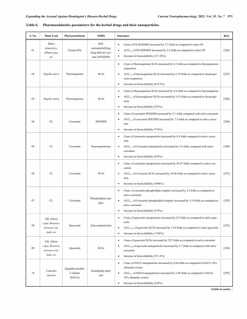
Table 6. Pharmacokinetics parameters for the herbal drugs and their nanoparticles.
S. No. Plant Used Phytoconstituent NDDS Outcomes Refs.
01
Malus domestica,
Allium cepa, etc
Fisetin (FS)
Self-nanoemulsifying drug delivery sys-tem (SNEDDS)
• Cmax of FS SNEDDS increased by 3.7 folds as compared to naïve FS
• AUC0-∞ of FS SNEDDS increased by 1.5 folds as compared to naïve FS
• Increase in bioavailability (151.58%)
[286]
02 Nigella sativa Thymoquinone SLNs
• Cmax of thymoquinone SLNs increased by 4.3 folds as compared to thymoquinone suspension
• AUC0-∞ of thymoquinone SLNs increased by 6.19 folds as compared to thymoqui-none suspension
• Increase in bioavailability (619.3%)
[287]
03 Nigella sativa Thymoquinone SLNs
• Cmax of thymoquinone SLNs increased by 4.8 folds as compared to thymoquinone
• AUC0-∞ of thymoquinone SLNs increased by 5.53 folds as compared to thymoqui-none
• Increase in bioavailability (553%)
[288]
04 CL Curcumin SNEDDS
• Cmax of curcumin SNEDDS increased by 9.1 folds compared with naïve curcumin
• AUC0-∞ of curcumin SNEDDS increased by 7.5 folds as compared to naïve curcu-min
• Increase in bioavailability (754%)
[289]
05 CL Curcumin Nanosuspensions
• Cmax of curcumin nanoparticles increased by 4.8 folds compared to naïve curcu-min.
• AUC0-∞ of Curcumin nanoparticles increased by 5.5 folds compared with naïve curcumin.
• Increase in bioavailability (558%)
[290]
06 CL Curcumin SLNs
• Cmax of curcumin nanoparticles increased by 49.27 folds compared to naïve cur-cumin.
• AUC0-∞ of Curcumin SLNs increased by 39.06 folds as compared to naïve curcu-min.
• Increase in bioavailability (3906%)
[291]
07 CL Curcumin Phospholipid com-
plex
• Cmax of curcumin phospholipid complex increased by 2.4 folds as compared to naïve curcumin
• AUC0-∞ of Curcumin phospholipid complex increased by 5.19 folds as compared to naïve curcumin
• Increase in bioavailability (519%)
[292]
08
GB, Allium cepa, Brassica oleracea var.
italic etc
Quercetin Zein nanoparticles
• Cmax of quercetin nanoparticles increased by 8.5 folds as compared to naïve quer-cetin
• AUC0-120 of quercetin SLNs increased by 13.9 folds as compared to naïve quercetin
• Increase in bioavailability (1396%)
[293]
09
GB, Allium cepa, Brassica oleracea var.
italic etc
Quercetin SLNs
• Cmax of quercetin SLNs increased by 2.07 folds as compared to naïve curcumin
• AUC0-48 of quercetin nanoparticles increased by 5.7 folds as compared with naïve curcumin
• Increase in bioavailability (571.4%)
[294]
10 Camellia sinensis
Epigallocatechin-3-Gallate (EGCG)
Nanolipidic parti-cles
• Cmax of EGCG nanoparticles increased by 6.04 folds as compared to EGCG 10% ethanolic extract
• AUC0-∞ of EGCG nanoparticles increased by 2.49 folds as compared to EGCG 10% ethanolic extract
• Increase in bioavailability (249%)
[295]
(Table 6) contd….
972 Current Neuropharmacology, 2021, Vol. 19, No. 7 Vishwas et al.
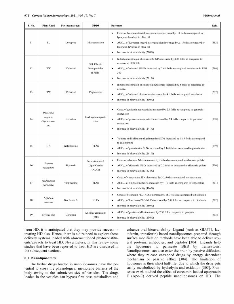
S. No. Plant Used Phytoconstituent NDDS Outcomes Refs.
11 SL Lycopene Microemulsion
• Cmax of lycopene-loaded microemulsion increased by 1.8 folds as compared to lycopene devolved in olive oil
• AUC0-∞ of lycopene-loaded microemulsion increased by 2.1 folds as compared to lycopene devolved in olive oil
• Increase in bioavailability (210%)
[182]
12 TW Celastrol Silk Fibroin
Nanoparticles (SFNPs)
• Initial concentration of celastrol SFNPs increased by 4.36 folds as compared to celastrol in PEG 300
• AUC0-∞ of celastrol SFNPs increased by 2.61 folds as compared to celastrol in PEG 300
• Increase in bioavailability (261%)
[296]
13 TW Celastrol Phytosomes
• Initial concentration of celastrol phytosomes increased by 5 folds as compared to celastrol
• AUC0-∞ of celastrol phytosomes increased by 4.1 folds as compared to celastrol
• Increase in bioavailability (410%)
[297]
14
Phaseolus vulgaris,
Glycine max, etc
Genistein Eudragit nanoparti-
cles
• Cmax of genistein nanoparticles increased by 2.4 folds as compared to genistein suspension
• AUC0-∞ of genistein nanoparticles increased by 2.4 folds compared to genistein suspension
• Increase in bioavailability (241%)
[298]
15 GN Galantamine SLNs
• Volume of distribution of galantamine SLNs increased by 1.15 folds as compared to galantamine
• AUC0-∞ of galantamine SLNs increased by 2.14 folds as compared to galantamine
• Increase in bioavailability (261%)
[299]
16 Silybum
marianum Silymarin
Nanostructured Lipid Carrier
(NLCs)
• Cmax of silymarin NLCs increased by 3.4 folds as compared to silymarin pellets
• AUC0-∞ of silymarin NLCs increased by 2.2 folds as compared to silymarin pellets
• Increase in bioavailability (224%)
[300]
17 Medagascar periwinkle
Vinpocetine SLNs
• Cmax of vinpocetine SLNs increased by 3.2 folds as compared to vinpocetine
• AUC0-∞ of vinpocetine SLNs increased by 4.16 folds as compared to vinpocetine
• Increase in bioavailability (416%)
[301]
18 Trifolium pratense
Biochanin A NLCs
• Cmax of biochanin PEG-NLCs increased by 15.74 folds as compared to biochanin
• AUC0-∞ of biochanin PEG-NLCs increased by 2.89 folds as compared to biochanin
• Increase in bioavailability (289%)
[302]
19 Glycine max Genistein Micellar emulsions
(ME)
• AUC0-∞ of genistein MEs increased by 2.36 folds compared to genistein
• Increase in bioavailability (236%) [303]
from HD, it is anticipated that they may provide success in treating HD also. Hence, there is a dire need to explore those delivery systems loaded with aforementioned phytoconstitu-ents/extracts to treat HD. Nevertheless, in this review some studies that have been reported to treat HD are discussed in the subsequent sections.
8.1. Nanoliposomes The herbal drugs loaded in nanoliposomes have the po-tential to cross the physiological membrane barriers of the body owing to the submicron size of vesicles. The drugs loaded in the vesicles can bypass first pass metabolism and
enhance oral bioavailability. Ligand (such as GLUT1, lac-toferrin, transferrin) based nanoliposomes prepared through surface modification methods have been able to deliver sev-eral proteins, antibodies, and peptides [304]. Ligands help the liposomes to permeate BBB by transcytosis. Nanoliposomes can also enter the brain by passive diffusion, where they release entrapped drugs by energy dependent mechanism or passive efflux [304]. The limitation of liposomes is their short half-life, due to which the drug gets easily metabolized by hydrolysis and oxidation [305]. Fran-cesca et al. studied the effect of curcumin-loaded apoprotein E (Apo-E) derived peptide nanoliposomes on HD. The

Expanding the Arsenal Against Huntington's Disease-Herbal Drugs Current Neuropharmacology, 2021, Vol. 19, No. 7 973
liposomes were prepared by loading Apo-E in the dispersion of bovine brain sphingomyelin (Sm), cholesterol (Chol), and 1,2-stearoyl-sn-glycero-3-phosphoethanolamine-N-[maleimide (poly(ethylene glycol)-2000)] (mal-PEG-PhoEth) using thin film hydration method (Fig. 5A). The prepared liposomes showed particle size, PDI, and zeta potential of 132 ± 10 nm, 0.187, and −19.41 ± 0.09 mV, respectively. The ratio of Apo-E to Chol and Sm was kept constant. However, compo-sition of Apo-E and mal-PEG-PhoEth were varied in the ratio of 1.2:1 and 1:5. During ApoE-liposome coupling, it was observed that when the ratio of peptide and mal-PEG-PhoEth was changed from 1:5 to 1.2:1, it resulted in in-creased density of ApoE on the surface of the liposome (Fig. 5B). It was observed that around 70,000 molecules of lipids were on the surface of the liposome, having a particle size of 140 nm and consisted of reactive mol-PEG-PhoEth (2.5 mol). Coupling efficiency was found to be 70%. The molar ratio of peptide and mol-PEG- PhoEth (1:5 to 1:2:1) after the incubation period showed a high density of 1200 and low density of 400 peptide molecules per single nanoliposomes particle (Fig. 5C). The in vitro cell line study was done on rat’s brain endothelial cells. The curcumin-nanoliposomes did not show any cytotoxicity as confirmed by 3-(4,5-dimethylthiazol-2-yl)-2,5-diphenyl tetrazolium bromide (MTT) assay. The liposomes were labeled by using a fluo-rescent dye and cellular uptake of the fluorescence labeled liposome was detected by using confocal laser scanning mi-
croscopy (CLSM). It was found that liposomes in the ab-sence of surface functionalization did not show any mem-brane accretion and cellular uptake of fluorescence. The Rat Brain Endothelial cells (RBE4) showed very less fluores-cence at high and low density of peptides. It was observed that the green fluorescence of cells got increased with an increase in the high density of peptides. The green spots were near the nucleus below the plasma membrane. The liposomes coupled with peptide mApoE displayed effective uptake (Fig. 5D). The curcumin-nanoliposomes helped in treating HD by their interaction with low-density lipoprotein receptors via special Apo-E sequence amino acid and pene-trated curcumin across the BBB through transcytosis without getting affected by lysosomal degradation. Hence, the ob-tained results revealed that ligand-based nanoliposomes suc-cessfully targeted BBB and protected the drug from degrada-tion [306].
8.2. Solid Lipid Nanoparticles (SLNs) SLNs contain a solid lipid matrix stabilized by lipid molecules and physiological emulsifiers. Homogenization is used for the preparation of SLNs, where high temperature and high pressure provided by thermodynamic and mechani-cal stress causes size reduction of drug particles [307, 308]. SLNs are excellent nanocarriers to enhance the bioavailability of drugs and are highly biocompatible. SLNs of size ranging from 0 to 1000nm can be prepare during high pressure
Fig. (4). Mechanism of BBB permeability of the nanoparticles. (A higher resolution / colour version of this figure is available in the elec-tronic copy of the article).
974 Current Neuropharmacology, 2021, Vol. 19, No. 7 Vishwas et al.
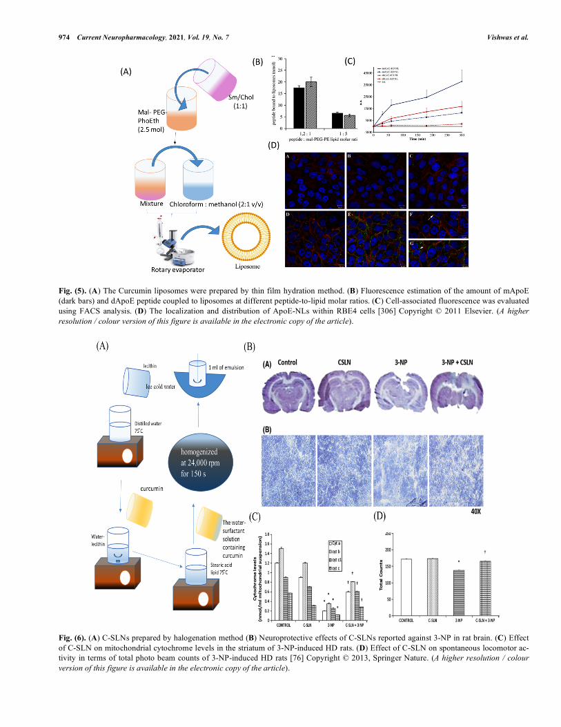
Fig. (5). (A) The Curcumin liposomes were prepared by thin film hydration method. (B) Fluorescence estimation of the amount of mApoE (dark bars) and dApoE peptide coupled to liposomes at different peptide-to-lipid molar ratios. (C) Cell-associated fluorescence was evaluated using FACS analysis. (D) The localization and distribution of ApoE-NLs within RBE4 cells [306] Copyright © 2011 Elsevier. (A higher resolution / colour version of this figure is available in the electronic copy of the article).
Fig. (6). (A) C-SLNs prepared by halogenation method (B) Neuroprotective effects of C-SLNs reported against 3-NP in rat brain. (C) Effect of C-SLN on mitochondrial cytochrome levels in the striatum of 3-NP-induced HD rats. (D) Effect of C-SLN on spontaneous locomotor ac-tivity in terms of total photo beam counts of 3-NP-induced HD rats [76] Copyright © 2013, Springer Nature. (A higher resolution / colour version of this figure is available in the electronic copy of the article).
Expanding the Arsenal Against Huntington's Disease-Herbal Drugs Current Neuropharmacology, 2021, Vol. 19, No. 7 975
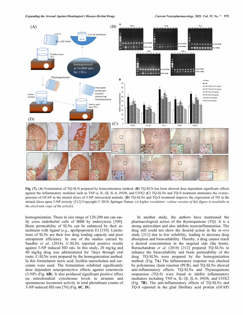
Fig. (7). (A) Formulation of TQ-SLN prepared by homosimeation method. (B) TQ-SLN has been showed dose dependent significant effects against the inflammatory mediator such as TNF-α, IL-1β, IL-6, iNOS, and COX2 (C) TQ-SLNs and TQ-S treatment attenuates the overex-pression of GFAP in the striatal slices of 3-NP intoxicated animals. (D) TQ-SLNs and TQ-S treatment improve the expression of TH in the striatal slices upon 3-NP toxicity [312] Copyright © 2018, Springer Nature. (A higher resolution / colour version of this figure is available in the electronic copy of the article).
homogenization. Those in size range of 120-200 nm can eas-ily cross endothelial cells of BBB by endocytosis [309]. Brain permeability of SLNs can be enhanced by their at-tachment with ligand (e.g., apolipoprotein E) [310]. Limita-tions of SLNs are their low drug loading capacity and poor entrapment efficiency. In one of the studies carried by Sandhir et al. (2014), C-SLNs reported positive results against 3-NP induced HD rats. In this study, 20 mg/kg and 40 mg/kg drug was administered for 7days through oral route. C-SLNs were prepared by the homogenization method. In this formulation steric acid, lecithin taurocholate and cur-cumin were used. The formulation exhibited significantly dose dependent neuroprotective effects against neurotoxin (3-NP) (Fig. 6B). It also produced significant positive effect on mitochondrial cytochrome levels in striatum and spontaneous locomotor activity in total photobeam counts of 3-NP-induced HD rats [76] (Fig. 6C, D).
In another study, the authors have mentioned the pharmacological action of the thymoquinone (TQ). It is a strong antioxidant and also inhibits neuroinflammation. The drug still could not show the desired action in the in-vivo study [311] due to low solubility, leading to decrease drug absorption and bioavailability. Thereby, a drug cannot reach a desired concentration in the targeted side (the brain). Ramachandran et al. (2018) [312] prepared TQ-SLNs to enhance the bioavailability and brain permeability of the drug. TQ-SLNs were prepared by the homogenization method. (Fig. 7A) The inflammatory response was checked by polymerase chain reaction (PCR), and TQ-SLNs showed anti-inflammatory effects. TQ-SLNs and Thymoquinone suspension (TQ-S) were found to inhibit inflammatory mediators including TNF-α, IL-1β, IL-6, iNOS, and COX2 (Fig. 7B). The anti-inflammatory effects of TQ-SLNs and TQ-S reported in the glial fibrillary acid protein (GFAP)
976 Current Neuropharmacology, 2021, Vol. 19, No. 7 Vishwas et al.
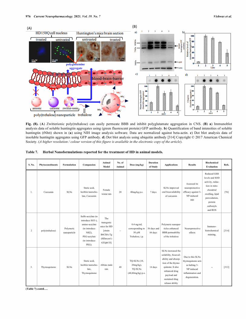
Fig. (8). (A) Zwitterionic poly(trehalose) can easily permeate BBB and inhibit polyglutamate aggregation in CNS. (B) a) Immunoblot analysis data of soluble huntingtin aggregates using (green fluorescent protein) GFP antibody. b) Quantification of band intensities of soluble huntingtin (tNhtt) shown in (a) using NIH image analysis software. Data are normalized against beta-actin. c) Dot blot analysis data of insoluble huntingtin aggregates using GFP antibody. d) Dot blot analysis using ubiquitin antibody [314] Copyright © 2017 American Chemical Society. (A higher resolution / colour version of this figure is available in the electronic copy of the article).
Table 7. Herbal Nanoformulations reported for the treatment of HD in animal models.
S. No. Phytoconstituents Formulation Compassion Animal
Model
No. of
Animal Dose (mg/kg)
Duration
of Study Applications Results
Biochemical
Evaluation Refs.
1. Curcumin SLNs
Steric acid,
lecithin taurocho-
late, Curcumin
Female
wistar rats 20 40mg/kg p.o. 7 days
SLNs improved
oral bioavailability
of curcumin
Assessed its
neuroprotective
efficacy against 3-NP-induced
HD
Reduced GSH
levels and SOD
activity, reduc-tion in mito-
chondrial
swelling, lipid peroxidation,
protein
carbonyls and ROS
[76]
2 poly(trehalose) Polymeric
nanoparticle
Sulfo-acrylate (to
introduce SO3-), amino-acrylate
(to introduce
NH2), PEG-acrylate
(to introduce
PEG).
The
transgenic mice for HD
[strain
B6CBA-Tg (HDexon1)
62Gpb/3J]
--
0.4 mg/mL
corresponding to 50 µM
Trehalose, i.p.
56 days and
84 days
Polymeric nanopar-
ticles enhanced BBB permeability
of the trehalose
Neuroprotective
effects
Immuno-
histochemical staining,
[314]
3. Thymoquinone SLNs
Steric acid,
lecithin taurocho-
late,
Thymoquinone
Albino male
rats 48
TQ-SLNs (10,
20mg/kg),
TQ-SLNs
(40,80mg/kg) p.o.
14 days
SLNs increased the
solubility, bioavail-
ability and absorp-
tion of the thymo-quinone. It also
enhanced drug
payload and sustained drug
release ability
Due to this SLNs
thymoquinone acts
as halting 3-
NP induced inflammation and
degeneration.
-- [312]
(Table 7) contd….
Expanding the Arsenal Against Huntington's Disease-Herbal Drugs Current Neuropharmacology, 2021, Vol. 19, No. 7 977
S. No. Phytoconstituents Formulation Compassion Animal
Model
No. of
Animal Dose (mg/kg)
Duration
of Study Applications Results
Biochemical
Evaluation Refs.
4. Cholesterol Nanaolipos omes -- Mice 3
Chol-D6-loaded
liposomes (200
µg/mouse)
2 days
Nanoliposomes
enhanced brain
delivery of cholesterol
Brain cholesterol
(Chol) synthesis,
which is essential for optimalsynap-
tic transmission
-- [315]
5. Selenium Nanoliposomes -- Worms -- -- --
Selenium Nanopar-
ticles enhanced
bioavailability and
therapeutic efficacy with low toxicity
--
20µM, Nano-Se
played a dosage-
dependent
protective effect on the viability
after the
exposure to both stress stimuli
[316]
6. Lithium Microemulsion
(NP03) --
YAC128
mouse
20 µg Li/kg
or 40 µg Li/kg
body weigh i.e.
0.03 and 0.06 mEq/kg)
2 months
Microemulsion of
lithium (NP03)
reduced the toxicity
of lithium and increased the
absorptions at
targeted site
NP03 improves
motor function
and rescues striatal
pathology and testicular atrophy
in YAC128 mice.
-- [317]
7. Coenzyme Q10
(CoQ10)
Coenzyme Q10 --
R6/2 transgenic
mouse 110
(CoQ10) 1000, 5000,
10000, or 20000
mg/kg/day and (HydroQ-sorb)
400, 1000, and
2000 mg/ kg/day
150 days -- Showed neuropro-
tective effects -- [318]
and tyrosine hydroxylase (TH) against 3-NP induced animals model [312] are shown in Fig. 7C and D.
8.3. Polymeric Nanoparticles
The particle size of polymeric nanoparticles is approxi-mately in the range of 10 nm to 1000 nm. These can be for-mulated in the form of nanospheres and nanocapsules. Nano-spheres are made up of a matrix system. In nanocapsules, the drug is loaded into the cavity, which is made up of a polym-eric membrane [313]. Debnath et al. (2017) reported the suc-cessful delivery of trehalose by enhancing its BBB perme-ability through poly(trehalose) nanoparticles. Poly(trehalose) nanoparticles were reported to be more potent as compared to trehalose molecules. They were found to inhibit polyglu-tamine aggregation in HD150Q cell in in vitro study. (Fig. 8A) Immunoblot analysis and Dot blot analysis of poly(trehalose) nanoparticles reduced polyglutamine levels and amyloid aggregation and also suppressed mHTT genes [314]. (Fig. 8B) Herbal drug nanoparticle formulation, ani-mal models, and beneficial results are enlisted in Table 7.
CONCLUSION
The prevention of NDs is essential for the aged popula-tion globally. Phytoconstituents serve as novel medicinal therapies in the present scenario. Herbal drugs are reported to have multiple actions such as antioxidant, anti-inflammatory, anti-proliferative and anti-apoptotic. Many of them are also reported to reduce AChE levels in synapses.
Hence, they could offer a pragmatic alternative to the current synthetic drugs that are being used to treat HD. Various pre-clinical and clinical studies have been highlighted in the manuscript indicating a significant positive response against symptoms of HD. Despite having such therapeutic potential, the efficacy of herbal drugs has not been widely explored due to their poor solubility and pharmacokinetic properties. Herbal drugs incorporated in various nanocarriers such as nanoliposomes, microemulsions, SLNs, and polymeric nanoparticles have shown very good efficacy to treat HD due to the enhancement in their bioavailability ordirect targeting to specific cells. This has further helped in the reduction of their dose as well as toxicity. The major challenges associ-ated with the formulation of herbal drug-loaded nanoparti-cles include the poor loading of drugs in the formulation, low stability of herbal drugs during their processing, diffi-culty during scale-up of the process, and low stability of nanoformulations. Hence, it is important to look at these issues prior to the start of pre-clinical studies. Upon getting successful pre-clinical reports, a thorough clinical study is required for their positioning into the market.
LIST OF ABBREVIATIONS
3-NP = Nitropropionic acid
6-OHDA = 6-hydroxydopamine
AC = Acorus calamus
ACh = Acetylcholine
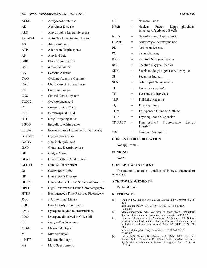
978 Current Neuropharmacology, 2021, Vol. 19, No. 7 Vishwas et al.
AChE = Acetylcholinesterase
AD = Alzheimer Disease
ALS = Amyotrophic Lateral Sclerosis
Anti-PAF = Anti-Platelet Activating Factor
AS = Allium sativum
ATP = Adenosine Triphosphate
Aβ = Amyloid beta
BBB = Blood Brain Barrier
BM = Bacopa monnieri
CA = Centella Asiatica
CAG = Cytisine-Adenine-Guanine
CAT = Choline-Acetyl Transferase
CL = Curcuma Longa
CNS = Central Nerves System
COX-2 = Cyclooxygenase-2
CS = Coriandrum sativum
CSF = Cerebrospinal Fluid
DTI = Drug Targeting Index
EGCG = Epigallocatechin gallate
ELISA = Enzyme-Linked Immune Sorbent Assay
G. glabra = Glycyrrhiza glabra
GABA = γ-aminobutyric acid
GAD = Glutamate Decarboxylase
GB = Ginkgo biloba
GFAP = Glial Fibrillary Acid Protein
GLUT1 = Glucose Transporter1
GN = Galanthus nivalis
HD = Huntington's Disease
HDSA = Huntington’s Disease Society of America
HPLC = High-Performance Liquid Chromatography
HTRF = Homogeneous Time Resolved Fluorescenc
JNK = c-Jun terminal kinase
LDL = Low Density Lipoprotein
LME = Lycopene loaded microemulsions
LOO = Lycopene dissolved in Olive Oil
LS = Lycopodium Serratum
MDA = Malondialdehyde
ME = Microemulsion
mHTT = Mutant Huntingtin
MS = Mass Spectrometry
NE = Nanoemulsions
NFκB = Nuclear Factor kappa-light-chain-enhancer of activated B cells
NLCs = Nanostructured Lipid Carrier
OH8dG = 8-hydroxy-2-deoxygonosine
PD = Parkinson Disease
PG = Panax Ginseng
RNS = Reactive Nitrogen Species
ROS = Reactive Oxygen Species
SDH = Succinate dehydrogenase cell enzyme
SI = Sedamim Indicum
SLNs = Solid Lipid Nanoparticles
TC = Tinospora cordifolia
TH = Tyrosine Hydroxylase
TLR = Toll-Like Receptor
TQ = Thymoquinone
TQM = Triterpenoid Quinone Methide
TQ-S = Thymoquinone Suspension
TR-FRET = Time-resolved Fluorescence Energy Transfer
WS = Withania Somnifera
CONSENT FOR PUBLICATION
Not applicable.
FUNDING None.
CONFLICT OF INTEREST
The authors declare no conflict of interest, financial or otherwise.
ACKNOWLEDGEMENTS
Declared none.
REFERENCES [1] Walker, F.O. Huntington’s disease. Lancet, 2007, 369(9557), 218-
228. http://dx.doi.org/10.1016/S0140-6736(07)60111-1 PMID:
17240289 [2] Medicalnewstoday, what you need to know about Huntington's
disease. https://www.medicalnewstoday.com/articles/159552 [3] Dey, A.; Bhattacharya, R.; Mukherjee, A.; Pandey, D.K. Natural
products against Alzheimer’s disease: Pharmaco-therapeutics and biotechnological interventions. Biotechnol. Adv., 2017, 35(2), 178-216.
http://dx.doi.org/10.1016/j.biotechadv.2016.12.005 PMID: 28043897
[4] Uddin, M.S.; Tewari, D.; Mamun, A.A.; Kabir, M.T.; Niaz, K.; Wahed, M.I.I.; Barreto, G.E.; Ashraf, G.M. Circadian and sleep dysfunction in Alzheimer’s disease. Ageing Res. Rev., 2020, 60, 101046.
Expanding the Arsenal Against Huntington's Disease-Herbal Drugs Current Neuropharmacology, 2021, Vol. 19, No. 7 979
http://dx.doi.org/10.1016/j.arr.2020.101046 PMID: 32171783 [5] Uddin, M.S.; Kabir, M.T.; Tewari, D.; Mamun, A.A.; Mathew, B.;
Aleya, L.; Barreto, G.E.; Bin-Jumah, M.N.; Abdel-Daim, M.M.; Ashraf, G.M. Revisiting the role of brain and peripheral Aβ in the pathogenesis of Alzheimer’s disease. J. Neurol. Sci., 2020, 416, 116974.
http://dx.doi.org/10.1016/j.jns.2020.116974 PMID: 32559516 [6] Uddin, M.S.; Kabir, M.T.; Tewari, D.; Mathew, B.; Aleya, L.
Emerging signal regulating potential of small molecule biflavon-oids to combat neuropathological insults of Alzheimer’s disease. Sci. Total Environ., 2020, 700, 134836.
http://dx.doi.org/10.1016/j.scitotenv.2019.134836 PMID: 31704512
[7] Anand, A.; Patience, A.A.; Sharma, N.; Khurana, N. The present and future of pharmacotherapy of Alzheimer’s disease: A compre-hensive review. Eur. J. Pharmacol., 2017, 815, 364-375.
http://dx.doi.org/10.1016/j.ejphar.2017.09.043 PMID: 28978455 [8] Szlachcic, W.J.; Switonski, P.M.; Krzyzosiak, W.J.; Figlerowicz,
M.; Figiel, M. Huntington disease iPSCs show early molecular changes in intracellular signaling, the expression of oxidative stress proteins and the p53 pathway. Dis. Model. Mech., 2015, 8(9), 1047-1057.
http://dx.doi.org/10.1242/dmm.019406 PMID: 26092128 [9] Warby, S.C.; Montpetit, A.; Hayden, A.R.; Carroll, J.B.; Butland,
S.L.; Visscher, H.; Collins, J.A.; Semaka, A.; Hudson, T.J.; Hay-den, M.R. CAG expansion in the Huntington disease gene is asso-ciated with a specific and targetable predisposing haplogroup. Am. J. Hum. Genet., 2009, 84(3), 351-366.
http://dx.doi.org/10.1016/j.ajhg.2009.02.003 PMID: 19249009 [10] Li, X.; Valencia, A.; Sapp, E.; Masso, N.; Alexander, J.; Reeves,
P.; Kegel, K.B.; Aronin, N.; Difiglia, M. Aberrant Rab11-dependent trafficking of the neuronal glutamate transporter EAAC1 causes oxidative stress and cell death in Huntington’s disease. J. Neurosci., 2010, 30(13), 4552-4561.
http://dx.doi.org/10.1523/JNEUROSCI.5865-09.2010 PMID: 20357106
[11] Choudhary, S.; Kumar, P.; Malik, J. Plants and phytochemicals for Huntington’s disease. Pharmacogn. Rev., 2013, 7(14), 81-91.
http://dx.doi.org/10.4103/0973-7847.120505 PMID: 24347915 [12] Dey, A.; Nandy, S.; Mukherjee, A.; Pandey, D.K. Plant Natural
Products as Neuroprotective Nutraceuticals: Preclinical and Clini-cal Studies and Future Implications. Proc. Natl. Acad. Sci. India Sect B: Biol. Sci. 2020, pp. 1-15.
[13] Pareek, H.; Thakur, P.; Ray, D. Modeling and docking studies of 4-aminobutyrate aminotransferase for huntington’s disease. Int. J. Pharma Bio Sci., 2011, 2, 539-549.
[14] Kay, C.; Hayden, M.R.; Leavitt, B.R. Epidemiology of Huntington disease. Handbook of Clinical Neurology; Elsevier, 2017, Vol. 144, pp. 31-46.
[15] Uddin, M.S.; Tewari, D.; Sharma, G.; Kabir, M.T.; Barreto, G.E.; Bin-Jumah, M.N.; Perveen, A.; Abdel-Daim, M.M.; Ashraf, G.M. Molecular mechanisms of ER stress and UPR in the pathogenesis of Alzheimer’s disease. Mol. Neurobiol., 2020, 57(7), 2902-2919.
http://dx.doi.org/10.1007/s12035-020-01929-y PMID: 32430843 [16] Craufurd, D.; Thompson, J.C.; Snowden, J.S. Behavioral changes
in Huntington disease. Neuropsychiatry Neuropsychol. Behav. Neurol., 2001, 14(4), 219-226.
PMID: 11725215 [17] Tabrizi, S.J.; Langbehn, D.R.; Leavitt, B.R.; Roos, R.A.; Durr, A.;
Craufurd, D.; Kennard, C.; Hicks, S.L.; Fox, N.C.; Scahill, R.I.; Borowsky, B.; Tobin, A.J.; Rosas, H.D.; Johnson, H.; Reilmann, R.; Landwehrmeyer, B.; Stout, J.C. TRACK-HD investigators. Bio-logical and clinical manifestations of Huntington’s disease in the longitudinal TRACK-HD study: cross-sectional analysis of base-line data. Lancet Neurol., 2009, 8(9), 791-801.
http://dx.doi.org/10.1016/S1474-4422(09)70170-X PMID: 19646924
[18] Tabrizi, S.J.; Scahill, R.I.; Owen, G.; Durr, A.; Leavitt, B.R.; Roos, R.A.; Borowsky, B.; Landwehrmeyer, B.; Frost, C.; Johnson, H.; Craufurd, D.; Reilmann, R.; Stout, J.C.; Langbehn, D.R. TRACK-HD Investigators. Predictors of phenotypic progression and disease onset in premanifest and early-stage Huntington’s disease in the TRACK-HD study: analysis of 36-month observational data. Lan-cet Neurol., 2013, 12(7), 637-649.
http://dx.doi.org/10.1016/S1474-4422(13)70088-7 PMID: 23664844
[19] Dugger, B.N.; Dickson, D.W. Pathology of neurodegenerative diseases. Cold Spring Harb. Perspect. Biol., 2017, 9(7), a028035.
http://dx.doi.org/10.1101/cshperspect.a028035 PMID: 28062563 [20] Clinart, M.E.N.A. Rare Disease Clinical Research Untapped poten-
tial in MENA. https://longtaal.com/wp-content/uploads/2019/05/Rare-Disease-Clinical-Research-Untapped-potential-in-MENA.pdf2020.
[21] Sheikh, S.; Haque, E.; Mir, S.S. Neurodegenerative diseases: multi-factorial conformational diseases and their therapeutic interven-tions. J. Neurodegen. Dis., 2013, 2013,563481.
http://dx.doi.org/10.1155/2013/563481 [22] Bawari, S.; Tewari, D.; Argüelles, S.; Sah, A.N.; Nabavi, S.F.; Xu,
S.; Vacca, R.A.; Nabavi, S.M.; Shirooie, S. Targeting BDNF sig-naling by natural products: Novel synaptic repair therapeutics for neurodegeneration and behavior disorders. Pharmacol. Res., 2019, 148, 104458.
http://dx.doi.org/10.1016/j.phrs.2019.104458 PMID: 31546015 [23] Jayakar, S.S.; Dikshit, M. AMPA receptor regulation mechanisms:
future target for safer neuroprotective drugs. Int. J. Neurosci., 2004, 114(6), 695-734.
http://dx.doi.org/10.1080/00207450490430453 PMID: 15204061 [24] Rubinsztein, D.C.; Leggo, J.; Coles, R.; Almqvist, E.; Biancalana,
V.; Cassiman, J-J.; Chotai, K.; Connarty, M.; Crauford, D.; Curtis, A.; Curtis, D.; Davidson, M.J.; Differ, A.M.; Dode, C.; Dodge, A.; Frontali, M.; Ranen, N.G.; Stine, O.C.; Sherr, M.; Abbott, M.H.; Franz, M.L.; Graham, C.A.; Harper, P.S.; Hedreen, J.C.; Hayden, M.R. Phenotypic characterization of individuals with 30-40 CAG repeats in the Huntington disease (HD) gene reveals HD cases with 36 repeats and apparently normal elderly individuals with 36-39 repeats. Am. J. Hum. Genet., 1996, 59(1), 16-22.
PMID: 8659522 [25] Gusella, J.F.; MacDonald, M.E.; Lee, J.M. Genetic modifiers of
Huntington’s disease. Mov. Disord., 2014, 29(11), 1359-1365. http://dx.doi.org/10.1002/mds.26001 PMID: 25154728 [26] Rosenblatt, A.; Kumar, B.V.; Mo, A.; Welsh, C.S.; Margolis, R.L.;
Ross, C.A. Age, CAG repeat length, and clinical progression in Huntington’s disease. Mov. Disord., 2012, 27(2), 272-276.
http://dx.doi.org/10.1002/mds.24024 PMID: 22173986 [27] Leenders, K.L.; Frackowiak, R.S.; Quinn, N.; Marsden, C.D. Brain
energy metabolism and dopaminergic function in Huntington’s dis-ease measured in vivo using positron emission tomography. Mov. Disord., 1986, 1(1), 69-77.
http://dx.doi.org/10.1002/mds.870010110 PMID: 2973559 [28] Stahl, W.L.; Swanson, P.D. Biochemical abnormalities in
Huntington’s chorea brains. Neurology, 1974, 24(9), 813-819. http://dx.doi.org/10.1212/WNL.24.9.813 PMID: 4277376 [29] Cramer, H.; Warter, J.M.; Renaud, B. Analysis of neurotransmitter
metabolites and adenosine 3′,5′-monophosphate in the CSF of pa-tients with extrapyramidal motor disorders. Adv. Neurol., 1984, 40, 431-435.
PMID: 6198888 [30] Gines, S.; Seong, I.S.; Fossale, E.; Ivanova, E.; Trettel, F.; Gusella,
J.F.; Wheeler, V.C.; Persichetti, F.; MacDonald, M.E. Specific pro-gressive cAMP reduction implicates energy deficit in presympto-matic Huntington’s disease knock-in mice. Hum. Mol. Genet., 2003, 12(5), 497-508.
http://dx.doi.org/10.1093/hmg/ddg046 PMID: 12588797 [31] Koroshetz, W.J.; Jenkins, B.G.; Rosen, B.R.; Beal, M.F. Energy
metabolism defects in Huntington’s disease and effects of coen-zyme Q10. Ann. Neurol., 1997, 41(2), 160-165.
http://dx.doi.org/10.1002/ana.410410206 PMID: 9029064 [32] Jenkins, B.G.; Koroshetz, W.J.; Beal, M.F.; Rosen, B.R. Evidence
for impairment of energy metabolism in vivo in Huntington’s dis-ease using localized 1H NMR spectroscopy. Neurology, 1993, 43(12), 2689-2695.
http://dx.doi.org/10.1212/WNL.43.12.2689 PMID: 8255479 [33] Fatokun, A.A.; Smith, R.A.; Stone, T.W. Resistance to kynurenic
acid of the NMDA receptor-dependent toxicity of 3-nitropropionic acid and cyanide in cerebellar granule neurons. Brain Res., 2008, 1215, 200-207.
http://dx.doi.org/10.1016/j.brainres.2008.04.013 PMID: 18486115 [34] Huang, Q-Y.; Yu, L.; Ferrante, R.J.; Chen, J-F. Mutant
SOD1G93A in bone marrow-derived cells exacerbates 3-
980 Current Neuropharmacology, 2021, Vol. 19, No. 7 Vishwas et al.
nitropropionic acid induced striatal damage in mice. Neurosci. Lett., 2007, 418(2), 175-180.
http://dx.doi.org/10.1016/j.neulet.2007.03.038 PMID: 17418947 [35] Alexi, T.; Borlongan, C.V.; Faull, R.L.; Williams, C.E.; Clark,
R.G.; Gluckman, P.D.; Hughes, P.E. Neuroprotective strategies for basal ganglia degeneration: Parkinson’s and Huntington’s diseases. Prog. Neurobiol., 2000, 60(5), 409-470.
http://dx.doi.org/10.1016/S0301-0082(99)00032-5 PMID: 10697073
[36] Alexi, T.; Hughes, P.E.; Faull, R.L.; Williams, C.E. 3-Nitropropionic acid’s lethal triplet: cooperative pathways of neu-rodegeneration. Neuroreport, 1998, 9(11), R57-R64.
http://dx.doi.org/10.1097/00001756-199808030-00001 PMID: 9721909
[37] Farooqui, T.; Farooqui, A.A. Aging: an important factor for the pathogenesis of neurodegenerative diseases. Mech. Ageing Dev., 2009, 130(4), 203-215.
http://dx.doi.org/10.1016/j.mad.2008.11.006 PMID: 19071157 [38] Dong, X.X.; Wang, Y.; Qin, Z.H. Molecular mechanisms of excito-
toxicity and their relevance to pathogenesis of neurodegenerative diseases. Acta Pharmacol. Sin., 2009, 30(4), 379-387.
http://dx.doi.org/10.1038/aps.2009.24 PMID: 19343058 [39] Bogdanov, M.B.; Andreassen, O.A.; Dedeoglu, A.; Ferrante, R.J.;
Beal, M.F. Increased oxidative damage to DNA in a transgenic mouse model of Huntington’s disease. J. Neurochem., 2001, 79(6), 1246-1249.
http://dx.doi.org/10.1046/j.1471-4159.2001.00689.x PMID: 11752065
[40] Chen, C-M. Mitochondrial dysfunction, metabolic deficits, and increased oxidative stress in Huntington’s disease. Chang Gung Med. J., 2011, 34(2), 135-152.
PMID: 21539755 [41] Browne, S.E.; Ferrante, R.J.; Beal, M.F. Oxidative stress in
Huntington’s disease. Brain Pathol., 1999, 9(1), 147-163. http://dx.doi.org/10.1111/j.1750-3639.1999.tb00216.x PMID:
9989457 [42] Sawa, A.; Tomoda, T.; Bae, B-I. Mechanisms of neuronal cell
death in Huntington’s disease. Cytogenet. Genome Res., 2003, 100(1-4), 287-295.
http://dx.doi.org/10.1159/000072864 PMID: 14526190 [43] Túnez, I.; Tasset, I.; Pérez-De La Cruz, V.; Santamaría, A. 3-
Nitropropionic acid as a tool to study the mechanisms involved in Huntington’s disease: past, present and future. Molecules, 2010, 15(2), 878-916.
http://dx.doi.org/10.3390/molecules15020878 PMID: 20335954 [44] Shirasaki, D.I.; Greiner, E.R.; Al-Ramahi, I.; Gray, M.; Boonthe-
ung, P.; Geschwind, D.H.; Botas, J.; Coppola, G.; Horvath, S.; Loo, J.A.; Yang, X.W. Network organization of the huntingtin proteo-mic interactome in mammalian brain. Neuron, 2012, 75(1), 41-57.
http://dx.doi.org/10.1016/j.neuron.2012.05.024 PMID: 22794259 [45] Weiss, A.; Träger, U.; Wild, E.J.; Grueninger, S.; Farmer, R.;
Landles, C.; Scahill, R.I.; Lahiri, N.; Haider, S.; Macdonald, D.; Frost, C.; Bates, G.P.; Bilbe, G.; Kuhn, R.; Andre, R.; Tabrizi, S.J. Mutant huntingtin fragmentation in immune cells tracks Huntington’s disease progression. J. Clin. Invest., 2012, 122(10), 3731-3736.
http://dx.doi.org/10.1172/JCI64565 PMID: 22996692 [46] Weir, D.W.; Sturrock, A.; Leavitt, B.R. Development of biomark-
ers for Huntington’s disease. Lancet Neurol., 2011, 10(6), 573-590. http://dx.doi.org/10.1016/S1474-4422(11)70070-9 PMID:
21601164 [47] Russell, D.S.; Jennings, D.L.; Barret, O.; Tamagnan, G.D.; Carroll,
V.M.; Caillé, F.; Alagille, D.; Morley, T.J.; Papin, C.; Seibyl, J.P.; Marek, K.L. Change in PDE10 across early Huntington disease as-sessed by [18F]MNI-659 and PET imaging. Neurology, 2016, 86(8), 748-754.
http://dx.doi.org/10.1212/WNL.0000000000002391 PMID: 26802091
[48] Tabrizi, S.J.; Reilmann, R.; Roos, R.A.; Durr, A.; Leavitt, B.; Owen, G.; Jones, R.; Johnson, H.; Craufurd, D.; Hicks, S.L.; Ken-nard, C.; Landwehrmeyer, B.; Stout, J.C.; Borowsky, B.; Scahill, R.I.; Frost, C.; Langbehn, D.R. TRACK-HD investigators. Poten-tial endpoints for clinical trials in premanifest and early Huntington’s disease in the TRACK-HD study: analysis of 24 month observational data. Lancet Neurol., 2012, 11(1), 42-53.
http://dx.doi.org/10.1016/S1474-4422(11)70263-0 PMID: 22137354
[49] Braisch, U.; Muche, R.; Rothenbacher, D.; Landwehrmeyer, G.B.; Long, J.D.; Orth, M. REGISTRY Investigators of the European Huntington’s Disease Network and COHORT Investigators of the Huntington Study Group. Identification of symbol digit modality test score extremes in Huntington’s disease. Am. J. Med. Genet. B. Neuropsychiatr. Genet., 2019, 180(3), 232-245.
http://dx.doi.org/10.1002/ajmg.b.32719 PMID: 30788902 [50] Björkqvist, M.; Wild, E.J.; Thiele, J.; Silvestroni, A.; Andre, R.;
Lahiri, N.; Raibon, E.; Lee, R.V.; Benn, C.L.; Soulet, D.; Magnus-son, A.; Woodman, B.; Landles, C.; Pouladi, M.A.; Hayden, M.R.; Khalili-Shirazi, A.; Lowdell, M.W.; Brundin, P.; Bates, G.P.; Leavitt, B.R.; Möller, T.; Tabrizi, S.J. A novel pathogenic pathway of immune activation detectable before clinical onset in Huntington’s disease. J. Exp. Med., 2008, 205(8), 1869-1877.
http://dx.doi.org/10.1084/jem.20080178 PMID: 18625748 [51] Rodrigues, F.B.; Byrne, L.M.; McColgan, P.; Robertson, N.;
Tabrizi, S.J.; Zetterberg, H.; Wild, E.J. Cerebrospinal fluid inflam-matory biomarkers reflect clinical severity in Huntington’s disease. PLoS One, 2016, 11(9), e0163479.
http://dx.doi.org/10.1371/journal.pone.0163479 PMID: 27657730 [52] Southwell, A.L.; Smith, S.E.; Davis, T.R.; Caron, N.S.; Villanueva,
E.B.; Xie, Y.; Collins, J.A.; Ye, M.L.; Sturrock, A.; Leavitt, B.R.; Schrum, A.G.; Hayden, M.R. Ultrasensitive measurement of hunt-ingtin protein in cerebrospinal fluid demonstrates increase with Huntington disease stage and decrease following brain huntingtin suppression. Sci. Rep., 2015, 5(1), 12166.
http://dx.doi.org/10.1038/srep12166 PMID: 26174131 [53] Vinther-Jensen, T.; Budtz-Jørgensen, E.; Simonsen, A.H.; Nielsen,
J.E.; Hjermind, L.E. YKL-40 in cerebrospinal fluid in Huntington’s disease--a role in pathology or a nonspecific response to inflamma-tion? Parkinsonism Relat. Disord., 2014, 20(11), 1301-1303.
http://dx.doi.org/10.1016/j.parkreldis.2014.08.011 PMID: 25219973
[54] Constantinescu, R.; Romer, M.; Oakes, D.; Rosengren, L.; Kie-burtz, K. Levels of the light subunit of neurofilament triplet protein in cerebrospinal fluid in Huntington’s disease. Parkinsonism Relat. Disord., 2009, 15(3), 245-248.
http://dx.doi.org/10.1016/j.parkreldis.2008.05.012 PMID: 19056308
[55] Nicoli, F.; Vion-Dury, J.; Maloteaux, J-M.; Delwaide, C.; Confort-Gouny, S.; Sciaky, M.; Cozzone, P.J. CSF and serum metabolic profile of patients with Huntington’s chorea: a study by high reso-lution proton NMR spectroscopy and HPLC. Neurosci. Lett., 1993, 154(1-2), 47-51.
http://dx.doi.org/10.1016/0304-3940(93)90168-K PMID: 8361646 [56] Saelens, J.K.; Allen, M.P.; Simke, J.P. Determination of acetylcho-
line and choline by an enzymatic assay. Arch. Int. Pharmacodyn. Ther., 1970, 186(2), 279-286.
PMID: 4331694 [57] Jeitner, T.M.; Bogdanov, M.B.; Matson, W.R.; Daikhin, Y.;
Yudkoff, M.; Folk, J.E.; Steinman, L.; Browne, S.E.; Beal, M.F.; Blass, J.P.; Cooper, A.J. N(ε)-(γ-L-glutamyl)-L-lysine (GGEL) is increased in cerebrospinal fluid of patients with Huntington’s dis-ease. J. Neurochem., 2001, 79(5), 1109-1112.
http://dx.doi.org/10.1046/j.1471-4159.2001.00673.x PMID: 11739625
[58] Jeitner, T.M.; Matson, W.R.; Folk, J.E.; Blass, J.P.; Cooper, A.J. Increased levels of γ-glutamylamines in Huntington disease CSF. J. Neurochem., 2008, 106(1), 37-44.
http://dx.doi.org/10.1111/j.1471-4159.2008.05350.x PMID: 18422943
[59] Guay, D.R. Tetrabenazine, a monoamine-depleting drug used in the treatment of hyperkinetic movement disorders. Am. J. Geriatr. Pharmacother., 2010, 8(4), 331-373.
http://dx.doi.org/10.1016/j.amjopharm.2010.08.006 PMID: 20869622
[60] Pidgeon, C.; Rickards, H. The pathophysiology and pharmacologi-cal treatment of Huntington disease. Behav. Neurol., 2013, 26(4), 245-253.
http://dx.doi.org/10.1155/2013/705373 PMID: 22713409 [61] Kumar, P.; Kumar, A. Protective effect of rivastigmine against 3-
nitropropionic acid-induced Huntington’s disease like symptoms:

Expanding the Arsenal Against Huntington's Disease-Herbal Drugs Current Neuropharmacology, 2021, Vol. 19, No. 7 981
possible behavioural, biochemical and cellular alterations. Eur. J. Pharmacol., 2009, 615(1-3), 91-101.
http://dx.doi.org/10.1016/j.ejphar.2009.04.058 PMID: 19445928 [62] Park, J-E.; Lee, S-T.; Im, W.S.; Chu, K.; Kim, M. Galantamine
reduces striatal degeneration in 3-nitropropionic acid model of Huntington’s disease. Neurosci. Lett., 2008, 448(1), 143-147.
http://dx.doi.org/10.1016/j.neulet.2008.10.020 PMID: 18938211 [63] Besret, L.; Page, K.J.; Dunnett, S.B. The problem of antipsychotic
treatment for functional imaging in Huntington’s disease: receptor binding, gene expression and locomotor activity after sub-chronic administration and wash-out of haloperidol in the rat. Brain Res., 2000, 853(1), 125-135.
http://dx.doi.org/10.1016/S0006-8993(99)02311-2 PMID: 10627316
[64] Wyant, K.J.; Ridder, A.J.; Dayalu, P. Huntington’s disease—update on treatments. Curr. Neurol. Neurosci. Rep., 2017, 17(4), 33.
http://dx.doi.org/10.1007/s11910-017-0739-9 PMID: 28324302 [65] Iriti, M.; Vitalini, S.; Fico, G.; Faoro, F. Neuroprotective herbs and
foods from different traditional medicines and diets. Molecules, 2010, 15(5), 3517-3555.
http://dx.doi.org/10.3390/molecules15053517 PMID: 20657497 [66] Ratheesh, G.; Tian, L.; Venugopal, J.R.; Ezhilarasu, H.; Sadiq, A.;
Fan, T-P.; Ramakrishna, S. Role of medicinal plants in neurode-generative diseases. Biomanufacturing Rev., 2017, 2(1), 2.
http://dx.doi.org/10.1007/s40898-017-0004-7 [67] Danduga, R.C.S.R.; Dondapati, S.R.; Kola, P.K.; Grace, L.; Ta-
digiri, R.V.B.; Kanakaraju, V.K. Neuroprotective activity of tetramethylpyrazine against 3-nitropropionic acid induced Huntington’s disease-like symptoms in rats. Biomed. Pharma-cother., 2018, 105, 1254-1268.
http://dx.doi.org/10.1016/j.biopha.2018.06.079 PMID: 30021362 [68] Nikolaus, S.; Wittsack, H-J.; Beu, M.; Antke, C.; Hautzel, H.;
Wickrath, F.; Müller-Lutz, A.; De Souza Silva, M.A.; Huston, J.P.; Antoch, G.; Müller, H.W. Amantadine enhances nigrostriatal and mesolimbic dopamine function in the rat brain in relation to motor and exploratory activity. Pharmacol. Biochem. Behav., 2019, 179, 156-170.
http://dx.doi.org/10.1016/j.pbb.2018.12.010 PMID: 30639878 [69] Zesiewicz, T.A.; Sullivan, K.L.; Hauser, R.A.; Sanchez-Ramos, J.
Open-label pilot study of levetiracetam (Keppra) for the treatment of chorea in Huntington’s disease. Mov. Disord., 2006, 21(11), 1998-2001.
http://dx.doi.org/10.1002/mds.21061 PMID: 16941461 [70] Khursheed, R.; Singh, S.K.; Wadhwa, S.; Gulati, M.; Awasthi, A.
Enhancing the potential preclinical and clinical benefits of quer-cetin through novel drug delivery systems. Drug Discov. Today, 2019.
PMID: 31707120 [71] Malakhova, A.A.; Grigor’eva, E.V.; Malankhanova, T.B.; Pavlova,
S.V.; Valetdinova, K.R.; Abramycheva, N.Y.; Vetchinova, A.S.; Il-larioshkin, S.N.; Zakian, S.M. Generation of induced pluripotent stem cell line ICGi018-A from peripheral blood mononuclear cells of a patient with Huntington’s disease. Stem Cell Res. (Amst.), 2020, 44, 101743.
http://dx.doi.org/10.1016/j.scr.2020.101743 PMID: 32179492 [72] Rotundo, G.; Bidollari, E.; Ferrari, D.; Spasari, I.; Bernardini, L.;
Consoli, F.; De Luca, A.; Santimone, I.; Lamorte, G.; Migliore, S.; Squitieri, F.; Vescovi, A.L.; Rosati, J. Generation of the induced pluripotent stem cell line CSSi006-A (3681) from a patient affected by advanced-stage Juvenile Onset Huntington’s Disease. Stem Cell Res. (Amst.), 2018, 29, 174-178.
http://dx.doi.org/10.1016/j.scr.2018.04.008 PMID: 29704769 [73] Bidollari, E.; Rotundo, G.; Ferrari, D.; Candido, O.; Bernardini, L.;
Consoli, F.; De Luca, A.; Santimone, I.; Lamorte, G.; Ilari, A.; Squitieri, F.; Vescovi, A.L.; Rosati, J. Generation of induced pluri-potent stem cell line, CSSi004-A (2962), from a patient diagnosed with Huntington’s disease at the presymptomatic stage. Stem Cell Res. (Amst.), 2018, 28, 145-148.
http://dx.doi.org/10.1016/j.scr.2018.02.014 PMID: 29486399 [74] Dumevska, B.; Schaft, J.; McKernan, R.; Hu, J.; Schmidt, U. Deri-
vation of Huntington Disease affected Genea090 human embryonic stem cell line. Stem Cell Res. (Amst.), 2016, 16(2), 519-521.
http://dx.doi.org/10.1016/j.scr.2016.02.024 PMID: 27346026
[75] Wang, S-E.; Lin, C-L.; Hsu, C-H.; Sheu, S-J.; Chien, C-T.; Wu, C-H. Treatment with a herbal formula B401 enhances neuroprotection and angiogenesis in the R6/2 mouse model of Huntington’s disease. Drug Des. Devel. Ther., 2015, 9, 887-900.
PMID: 25733809 [76] Sandhir, R.; Yadav, A.; Mehrotra, A.; Sunkaria, A.; Singh, A.;
Sharma, S. Curcumin nanoparticles attenuate neurochemical and neurobehavioral deficits in experimental model of Huntington’s disease. Neuromol. Med., 2014, 16(1), 106-118.
http://dx.doi.org/10.1007/s12017-013-8261-y PMID: 24008671 [77] Rao, R.V.; Descamps, O.; John, V.; Bredesen, D.E. Ayurvedic
medicinal plants for Alzheimer’s disease: a review. Alzheimers Res. Ther., 2012, 4(3), 22.
http://dx.doi.org/10.1186/alzrt125 PMID: 22747839 [78] Wu, P.F.; Zhang, Z.; Wang, F.; Chen, J.G. Natural compounds
from traditional medicinal herbs in the treatment of cerebral ische-mia/reperfusion injury. Acta Pharmacol. Sin., 2010, 31(12), 1523-1531.
http://dx.doi.org/10.1038/aps.2010.186 PMID: 21127495 [79] Sandhya, S.; Vinod, K.; Kumar, S. Herbs used for brain disorders.
Hygeia J Drugs Med, 2010, 2, 38-45. [80] Wang, D.; Calabrese, E.J.; Lian, B.; Lin, Z.; Calabrese, V. Horme-
sis as a mechanistic approach to understanding herbal treatments in traditional Chinese medicine. Pharmacol. Ther., 2018, 184, 42-50.
http://dx.doi.org/10.1016/j.pharmthera.2017.10.013 PMID: 29080703
[81] Geng, Y.; Li, C.; Liu, J.; Xing, G.; Zhou, L.; Dong, M.; Li, X.; Niu, Y. Beta-asarone improves cognitive function by suppressing neu-ronal apoptosis in the beta-amyloid hippocampus injection rats. Biol. Pharm. Bull., 2010, 33(5), 836-843.
http://dx.doi.org/10.1248/bpb.33.836 PMID: 20460763 [82] Paterna, J-C.; Leng, A.; Weber, E.; Feldon, J.; Büeler, H. DJ-1 and
Parkin modulate dopamine-dependent behavior and inhibit MPTP-induced nigral dopamine neuron loss in mice. Mol. Ther., 2007, 15(4), 698-704.
http://dx.doi.org/10.1038/sj.mt.6300067 PMID: 17299411 [83] Bayan, L.; Koulivand, P.H.; Gorji, A. Garlic: a review of potential
therapeutic effects. Avicenna J. Phytomed., 2014, 4(1), 1-14. PMID: 25050296 [84] Chauhan, N.B. Effect of aged garlic extract on APP processing and
tau phosphorylation in Alzheimer’s transgenic model Tg2576. J. Ethnopharmacol., 2006, 108(3), 385-394.
http://dx.doi.org/10.1016/j.jep.2006.05.030 PMID: 16842945 [85] Tatara, M.R.; Sliwa, E.; Dudek, K.; Mosiewicz, J.; Studzinski, T.
Effect of aged garlic extract and allicin administration to sows dur-ing pregnancy and lactation on body weight gain and gastrointesti-nal tract development of piglets. PART I. Bull. Vet. Inst. Pulawy, 2005, 49(3), 349.
[86] Pérez-Torres, I.; Torres-Narváez, J.C.; Pedraza-Chaverri, J.; Rubio-Ruiz, M.E.; Díaz-Díaz, E.; Del Valle-Mondragón, L.; Martínez-Memije, R.; Varela, L.E.; Guarner-Lans, V. Effect of the aged gar-lic extract on cardiovascular function in metabolic syndrome rats. Molecules, 2016, 21(11), 1425.
http://dx.doi.org/10.3390/molecules21111425 PMID: 27792195 [87] Medina-Campos, O.N.; Barrera, D.; Segoviano-Murillo, S.; Rocha,
D.; Maldonado, P.D.; Mendoza-Patiño, N.; Pedraza-Chaverri, J. S-allylcysteine scavenges singlet oxygen and hypochlorous acid and protects LLC-PK(1) cells of potassium dichromate-induced toxic-ity. Food Chem. Toxicol., 2007, 45(10), 2030-2039.
http://dx.doi.org/10.1016/j.fct.2007.05.002 PMID: 17576034 [88] Roy, S.; Awasthi, H. Herbal medicines as neuroprotective agent: A
mechanistic approach. Int. J. Pharm. Pharm. Sci., 2017, 9, 1-7. http://dx.doi.org/10.22159/ijpps.2017v9i11.19444 [89] Gohil, K.J.; Patel, J.A. A review on Bacopa monniera: current
research and future prospects. Intl. J. Green Pharmacy, 2010, 4(1). http://dx.doi.org/10.4103/0973-8258.62156 [90] Russo, A.; Borrelli, F. Bacopa monniera, a reputed nootropic plant:
an overview. Phytomedicine, 2005, 12(4), 305-317. http://dx.doi.org/10.1016/j.phymed.2003.12.008 PMID: 15898709 [91] Nandy, S.; Mukherjee, A.; Pandey, D.K.; Dey, A. Bacopa mon-
nieri: The neuroprotective elixir from the East—Phytochemistry, pharmacology, and biotechnological improvement. Bioactive Natu-ral products in Drug Discovery; Springer, 2020, pp. 97-126.
http://dx.doi.org/10.1007/978-981-15-1394-7_2
982 Current Neuropharmacology, 2021, Vol. 19, No. 7 Vishwas et al.
[92] Dey, A.; Hazra, A.; Nongdam, P.; Nandy, S.; Tikendra, L.; Muk-herjee, A.; Banerjee, S.; Mukherjee, S.; Pandey, D. Enhanced baco-side content in polyamine treated in-vitro raised Bacopa monnieri (L.) Wettst. S. Afr. J. Bot., 2019, 123, 259-269.
http://dx.doi.org/10.1016/j.sajb.2019.03.012 [93] Shinomol, G.K.; Muralidhara, Bacopa monnieri modulates endoge-
nous cytoplasmic and mitochondrial oxidative markers in prepuber-tal mice brain. Phytomedicine, 2011, 18(4), 317-326.
http://dx.doi.org/10.1016/j.phymed.2010.08.005 PMID: 20850955 [94] Calabrese, C.; Gregory, W.L.; Leo, M.; Kraemer, D.; Bone, K.;
Oken, B. Effects of a standardized Bacopa monnieri extract on cognitive performance, anxiety, and depression in the elderly: a randomized, double-blind, placebo-controlled trial. J. Altern. Com-plement. Med., 2008, 14(6), 707-713.
http://dx.doi.org/10.1089/acm.2008.0018 PMID: 18611150 [95] Bammidi, S.R.; Volluri, S.S.; Chippada, S.C.; Avanigadda, S.;
Vangalapati, M. A review on pharmacological studies of Bacopa monniera. J. Chem. Biol. Phys. Sci., 2011, 1(2), 250.
[96] Hou, C-C.; Lin, S-J.; Cheng, J-T.; Hsu, F-L. Bacopaside III, baco-pasaponin G, and bacopasides A, B, and C from Bacopa monniera. J. Nat. Prod., 2002, 65(12), 1759-1763.
http://dx.doi.org/10.1021/np020238w PMID: 12502309 [97] Mahato, S.B.; Garai, S.; Chakravarty, A.K. Bacopasaponins E and
F: two jujubogenin bisdesmosides from Bacopa monniera. Phyto-chemistry, 2000, 53(6), 711-714.
http://dx.doi.org/10.1016/S0031-9422(99)00384-2 PMID: 10746885
[98] Garai, S.; Mahato, S.B.; Ohtani, K.; Yamasaki, K. Dammarane-type triterpenoid saponins from Bacopa monniera. Phytochemistry, 1996, 42(3), 815-820.
http://dx.doi.org/10.1016/0031-9422(95)00936-1 PMID: 8768327 [99] Garai, S.; Mahato, S.B.; Ohtani, K.; Yamasaki, K. Bacopasaponin
D--a pseudojujubogenin glycoside from Bacopa monniera. Phyto-chemistry, 1996, 43(2), 447-449.
http://dx.doi.org/10.1016/0031-9422(96)00250-6 PMID: 8862037 [100] Kawai, K-I.; Shibata, S. Pseudojujubogenin, a new sapogenin from
Bacopa monniera. Phytochemistry, 1978, 17(2), 287-289. http://dx.doi.org/10.1016/S0031-9422(00)94166-9 [101] Murthy, P.B.S.; Raju, V.R.; Ramakrisana, T.; Chakravarthy, M.S.;
Kumar, K.V.; Kannababu, S.; Subbaraju, G.V. Estimation of twelve bacopa saponins in Bacopa monnieri extracts and formula-tions by high-performance liquid chromatography. Chem. Pharm. Bull. (Tokyo), 2006, 54(6), 907-911.
http://dx.doi.org/10.1248/cpb.54.907 PMID: 16755069 [102] Chakravarty, A.K.; Garai, S.; Masuda, K.; Nakane, T.; Kawahara,
N. Bacopasides III-V: three new triterpenoid glycosides from Ba-copa monniera. Chem. Pharm. Bull. (Tokyo), 2003, 51(2), 215-217.
http://dx.doi.org/10.1248/cpb.51.215 PMID: 12576661 [103] Chakravarty, A.K.; Sarkar, T.; Masuda, K.; Shiojima, K.; Nakane,
T.; Kawahara, N. Bacopaside I and II: two pseudojujubogenin gly-cosides from Bacopa monniera. Phytochemistry, 2001, 58(4), 553-556.
http://dx.doi.org/10.1016/S0031-9422(01)00275-8 PMID: 11576596 [104] Kapoor, R.; Srivastava, S.; Kakkar, P. Bacopa monnieri modulates
antioxidant responses in brain and kidney of diabetic rats. Environ. Toxicol. Pharmacol., 2009, 27(1), 62-69.
http://dx.doi.org/10.1016/j.etap.2008.08.007 PMID: 21783922 [105] Vollala, V.R.; Upadhya, S.; Nayak, S. Learning and memory-
enhancing effect of Bacopa monniera in neonatal rats. Bratisl. Lek Listy, 2011, 112(12), 663-669.
PMID: 22372329 [106] Roodenrys, S.; Booth, D.; Bulzomi, S.; Phipps, A.; Micallef, C.;
Smoker, J. Chronic effects of Brahmi (Bacopa monnieri) on human memory. Neuropsychopharmacology, 2002, 27(2), 279-281.
http://dx.doi.org/10.1016/S0893-133X(01)00419-5 PMID: 12093601
[107] Singh, H.; Dhawan, B. Neuropsychopharmacological effects of the Ayurvedic nootropic Bacopa monniera Linn. (Brahmi). Indian J. Pharmacol., 1997, 29(5), 359.
PMID: 31831931 [108] Vohora, D.; Pal, S.N.; Pillai, K.K. Protection from phenytoin-
induced cognitive deficit by Bacopa monniera, a reputed Indian nootropic plant. J. Ethnopharmacol., 2000, 71(3), 383-390.
http://dx.doi.org/10.1016/S0378-8741(99)00213-5 PMID: 10940574
[109] Stough, C.; Lloyd, J.; Clarke, J.; Downey, L.A.; Hutchison, C.W.; Rodgers, T.; Nathan, P.J. The chronic effects of an extract of Ba-copa monniera (Brahmi) on cognitive function in healthy human subjects. Psychopharmacology (Berl.), 2001, 156(4), 481-484.
http://dx.doi.org/10.1007/s002130100815 PMID: 11498727 [110] Tripathi, Y.B.; Chaurasia, S.; Tripathi, E.; Upadhyay, A.; Dubey,
G.P. Bacopa monniera Linn. as an antioxidant: mechanism of ac-tion. Indian J. Exp. Biol., 1996, 34(6), 523-526.
PMID: 8792640 [111] Russo, A.; Izzo, A.A.; Borrelli, F.; Renis, M.; Vanella, A. Free
radical scavenging capacity and protective effect of Bacopa mon-niera L. on DNA damage. Phytother. Res., 2003, 17(8), 870-875.
http://dx.doi.org/10.1002/ptr.1061 PMID: 13680815 [112] Bhattacharya, S.K.; Bhattacharya, A.; Kumar, A.; Ghosal, S. Anti-
oxidant activity of Bacopa monniera in rat frontal cortex, striatum and hippocampus. Phytother. Res., 2000, 14(3), 174-179.
http://dx.doi.org/10.1002/(SICI)1099-1573(200005)14:3<174::AID-PTR624>3.0.CO;2-O PMID: 10815010
[113] Chowdhuri, D.K.; Parmar, D.; Kakkar, P.; Shukla, R.; Seth, P.K.; Srimal, R.C. Antistress effects of bacosides of Bacopa monnieri: modulation of Hsp70 expression, superoxide dismutase and cyto-chrome P450 activity in rat brain. Phytother. Res., 2002, 16(7), 639-645.
http://dx.doi.org/10.1002/ptr.1023 PMID: 12410544 [114] Sairam, K.; Dorababu, M.; Goel, R.K.; Bhattacharya, S.K. RK.
Antidepressant activity of standardized extract of Bacopa monniera in experimental models of depression in rats. Phytomedicine, 2002, 9(3), 207-211.
http://dx.doi.org/10.1078/0944-7113-00116 PMID: 12046860 [115] Shanker, G.; Singh, H. Anxiolytic profile of standardized Brahmi
extract. Indian J. Pharmacol., 2000, 32(152), 5. [116] Sumathy, T.; Subramanian, S.; Govindasamy, S.; Balakrishna, K.;
Veluchamy, G. Protective role of Bacopa monniera on morphine induced hepatotoxicity in rats. Phytother. Res., 2001, 15(7), 643-645.
http://dx.doi.org/10.1002/ptr.1007 PMID: 11746853 [117] Sairam, K.; Rao, C.V.; Babu, M.D.; Goel, R.K. Prophylactic and
curative effects of Bacopa monniera in gastric ulcer models. Phy-tomedicine, 2001, 8(6), 423-430.
http://dx.doi.org/10.1078/S0944-7113(04)70060-4 PMID: 11824516
[118] Andreassen, O.A.; Ferrante, R.J.; Hughes, D.B.; Klivenyi, P.; Dedeoglu, A.; Ona, V.O.; Friedlander, R.M.; Beal, M.F. Malonate and 3-nitropropionic acid neurotoxicity are reduced in transgenic mice expressing a caspase-1 dominant-negative mutant. J. Neuro-chem., 2000, 75(2), 847-852.
http://dx.doi.org/10.1046/j.1471-4159.2000.0750847.x PMID: 10899963
[119] Coles, C.J.; Edmondson, D.E.; Singer, T.P. Inactivation of succi-nate dehydrogenase by 3-nitropropionate. J. Biol. Chem., 1979, 254(12), 5161-5167.
PMID: 447637 [120] Kim, G.W.; Copin, J-C.; Kawase, M.; Chen, S.F.; Sato, S.; Gobbel,
G.T.; Chan, P.H. Excitotoxicity is required for induction of oxida-tive stress and apoptosis in mouse striatum by the mitochondrial toxin, 3-nitropropionic acid. J. Cereb. Blood Flow Metab., 2000, 20(1), 119-129.
http://dx.doi.org/10.1097/00004647-200001000-00016 PMID: 10616800
[121] Veerendra Kumar, M.H.; Gupta, Y.K. Effect of different extracts of Centella asiatica on cognition and markers of oxidative stress in rats. J. Ethnopharmacol., 2002, 79(2), 253-260.
http://dx.doi.org/10.1016/S0378-8741(01)00394-4 PMID: 11801389
[122] Wijeweera, P.; Arnason, J.T.; Koszycki, D.; Merali, Z. Evaluation of anxiolytic properties of Gotukola--(Centella asiatica) extracts and asiaticoside in rat behavioral models. Phytomedicine, 2006, 13(9-10), 668-676.
http://dx.doi.org/10.1016/j.phymed.2006.01.011 PMID: 16488124 [123] Soumyanath, A.; Zhong, Y.P.; Gold, S.A.; Yu, X.; Koop, D.R.;
Bourdette, D.; Gold, B.G. Centella asiatica accelerates nerve re-generation upon oral administration and contains multiple active fractions increasing neurite elongation in-vitro. J. Pharm. Pharma-col., 2005, 57(9), 1221-1229.
Expanding the Arsenal Against Huntington's Disease-Herbal Drugs Current Neuropharmacology, 2021, Vol. 19, No. 7 983
http://dx.doi.org/10.1211/jpp.57.9.0018 PMID: 16105244 [124] Hussin, M.; Abdul-Hamid, A.; Mohamad, S.; Saari, N.; Ismail, M.;
Bejo, M.H. Protective effect of Centella asiatica extract and pow-der on oxidative stress in rats. Food Chem., 2007, 100(2), 535-541.
http://dx.doi.org/10.1016/j.foodchem.2005.10.022 [125] Kaur, P.; Pandey, D.K.; Dey, A.; Dwivedi, P.; Malik, T.; Gupta, R.
Swertia spp.: A potential source of high-value bioactive compo-nents, pharmacology, and analytical techniques. Bioactive Natural products in Drug Discovery; Springer, 2020, pp. 165-213.
http://dx.doi.org/10.1007/978-981-15-1394-7_4 [126] Singh, B.; Rastogi, R. A reinvestigation of the triterpenes of Cen-
tella asiatica. Phytochemistry, 1969, 8(5), 917-921. http://dx.doi.org/10.1016/S0031-9422(00)85884-7 [127] Randriamampionona, D.; Diallo, B.; Rakotoniriana, F.; Rabeman-
antsoa, C.; Cheuk, K.; Corbisier, A-M.; Mahillon, J.; Ratsi-mamanga, S.; El Jaziri, M. Comparative analysis of active constitu-ents in Centella asiatica samples from Madagascar: application for ex situ conservation and clonal propagation. Fitoterapia, 2007, 78(7-8), 482-489.
http://dx.doi.org/10.1016/j.fitote.2007.03.016 PMID: 17560738 [128] Singh, B.; Rastogi, R. Chemical examination of Centella asiatica
linn—III: Constitution of brahmic acid. Phytochemistry, 1968, 7(8), 1385-1393.
http://dx.doi.org/10.1016/S0031-9422(00)85642-3 [129] Asakawa, Y.; Matsuda, R.; Takemoto, T. Mono-and sesquiterpe-
noids from Hydrocotyle and Centella species. Phytochemistry, 1982, 21(10), 2590-2592.
http://dx.doi.org/10.1016/0031-9422(82)85264-3 [130] Shinomol, G.K.; Muralidhara, Prophylactic neuroprotective prop-
erty of Centella asiatica against 3-nitropropionic acid induced oxi-dative stress and mitochondrial dysfunctions in brain regions of prepubertal mice. Neurotoxicology, 2008, 29(6), 948-957.
http://dx.doi.org/10.1016/j.neuro.2008.09.009 PMID: 18930762 [131] Bhat, S.; Kaushal, P.; Kaur, M.; Sharma, H. Coriander (Corian-
drum sativum L.): Processing, nutritional and functional aspects. Afr. J. Plant Sci., 2014, 8(1), 25-33.
http://dx.doi.org/10.5897/AJPS2013.1118 [132] Deepa, B.; Anuradha, C.V. Antioxidant potential of Coriandrum
sativum L. seed extract. Indian J. Exp. Biol., 2011, 49(1), 30-38. PMID: 21365993 [133] Vekaria, R.H.; Patel, M.N.; Bhalodiya, P.N.; Patel, V.; Desai, T.R.;
Tirgar, P.R. Evaluation of neuroprotective effect of Coriandrum Sativum linn. against ischemicreperfusion insult in brain. Intl. J. Phytopharmacol., 2012, 2, 186-193.
[134] Liu, Q.F.; Jeong, H.; Lee, J.H.; Hong, Y.K.; Oh, Y.; Kim, Y-M.; Suh, Y.S.; Bang, S.; Yun, H.S.; Lee, K.; Cho, S.M.; Lee, S.B.; Jeon, S.; Chin, Y.W.; Koo, B.S.; Cho, K.S. Coriandrum sativum suppresses Aβ42-induced ROS increases, glial cell proliferation, and ERK activation. Am. J. Chin. Med., 2016, 44(7), 1325-1347.
http://dx.doi.org/10.1142/S0192415X16500749 PMID: 27776428 [135] Singh, A.; Kaur, M.; Choudhary, A.; Kumar, B. Effect of Butea
monosperma leaf extracts on cyclophosphamide induced clasto-genicity and oxidative stress in mice. Pharmacognosy Res., 2015, 7(1), 85-91.
http://dx.doi.org/10.4103/0974-8490.147215 PMID: 25598640 [136] Habibyar, A.F.; Sharma, N.; Khurana, N. PASS assisted prediction
and pharmacological evaluation of hesperidin against scopolamine induced amnesia in mice. Eur. J. Pharmacol., 2016, 789, 385-394.
http://dx.doi.org/10.1016/j.ejphar.2016.07.013 PMID: 27397428 [137] Jain, S.; Shrivastava, S.; Nayak, S.; Sumbhate, S. Recent trends in
Curcuma longa Linn. Pharmacogn. Rev., 2007, 1(1). [138] Joe, B.; Vijaykumar, M.; Lokesh, B.R. Biological properties of
curcumin-cellular and molecular mechanisms of action. Crit. Rev. Food Sci. Nutr., 2004, 44(2), 97-111.
http://dx.doi.org/10.1080/10408690490424702 PMID: 15116757 [139] Akram, M.; Shahab-Uddin, A.A.; Usmanghani, K.; Hannan, A.;
Mohiuddin, E.; Asif, M. Curcuma longa and curcumin: a review article. Rom. J. Biol. Plant Biol., 2010, 55(2), 65-70.
[140] Kunchandy, E.; Rao, M. Oxygen radical scavenging activity of curcumin. Int. J. Pharm., 1990, 58(3), 237-240.
http://dx.doi.org/10.1016/0378-5173(90)90201-E [141] Chainani-Wu, N. Safety and anti-inflammatory activity of curcu-
min: a component of tumeric (Curcuma longa). J. Altern. Comple-ment. Med., 2003, 9(1), 161-168.
http://dx.doi.org/10.1089/107555303321223035 PMID: 12676044
[142] Chattopadhyay, I.; Biswas, K.; Bandyopadhyay, U.; Banerjee, R.K. Turmeric and curcumin: Biological actions and medicinal applica-tions. Curr. Sci. Bangalore, 2004, 87, 44-53.
[143] Simon, A.; Allais, D.P.; Duroux, J.L.; Basly, J.P.; Durand-Fontanier, S.; Delage, C. Inhibitory effect of curcuminoids on MCF-7 cell proliferation and structure-activity relationships. Can-cer Lett., 1998, 129(1), 111-116.
http://dx.doi.org/10.1016/S0304-3835(98)00092-5 PMID: 9714342 [144] Sreejayan, ; Rao, M.N. Nitric oxide scavenging by curcuminoids. J.
Pharm. Pharmacol., 1997, 49(1), 105-107. http://dx.doi.org/10.1111/j.2042-7158.1997.tb06761.x PMID:
9120760 [145] Daniel, S.; Limson, J.L.; Dairam, A.; Watkins, G.M.; Daya, S.
Through metal binding, curcumin protects against lead- and cad-mium-induced lipid peroxidation in rat brain homogenates and against lead-induced tissue damage in rat brain. J. Inorg. Biochem., 2004, 98(2), 266-275.
http://dx.doi.org/10.1016/j.jinorgbio.2003.10.014 PMID: 14729307 [146] Camacho-Barquero, L.; Villegas, I.; Sánchez-Calvo, J.M.; Talero,
E.; Sánchez-Fidalgo, S.; Motilva, V.; Alarcón de la Lastra, C. Cur-cumin, a Curcuma longa constituent, acts on MAPK p38 pathway modulating COX-2 and iNOS expression in chronic experimental colitis. Int. Immunopharmacol., 2007, 7(3), 333-342.
http://dx.doi.org/10.1016/j.intimp.2006.11.006 PMID: 17276891 [147] Vajragupta, O.; Boonchoong, P.; Watanabe, H.; Tohda, M.; Kum-
masud, N.; Sumanont, Y. Manganese complexes of curcumin and its derivatives: evaluation for the radical scavenging ability and neuroprotective activity. Free Radic. Biol. Med., 2003, 35(12), 1632-1644.
http://dx.doi.org/10.1016/j.freeradbiomed.2003.09.011 PMID: 14680686
[148] Thiyagarajan, M.; Sharma, S.S. Neuroprotective effect of curcumin in middle cerebral artery occlusion induced focal cerebral ischemia in rats. Life Sci., 2004, 74(8), 969-985.
http://dx.doi.org/10.1016/j.lfs.2003.06.042 PMID: 14672754 [149] Anand, A.; Khurana, P.; Chawla, J.; Sharma, N.; Khurana, N.
Emerging treatments for the behavioral and psychological symp-toms of dementia. CNS Spectr., 2018, 23(6), 361-369.
http://dx.doi.org/10.1017/S1092852917000530 PMID: 28911339 [150] Frautschy, S.A.; Hu, W.; Kim, P.; Miller, S.A.; Chu, T.; Harris-
White, M.E.; Cole, G.M. Phenolic anti-inflammatory antioxidant reversal of Abeta-induced cognitive deficits and neuropathology. Neurobiol. Aging, 2001, 22(6), 993-1005.
http://dx.doi.org/10.1016/S0197-4580(01)00300-1 PMID: 11755008
[151] Wattmo, C.; Jedenius, E.; Blennow, K.; Wallin, Å.K. Dose and plasma concentration of galantamine in Alzheimer’s disease - clini-cal application. Alzheimers Res. Ther., 2013, 5(1), 2.
http://dx.doi.org/10.1186/alzrt156 PMID: 23286718 [152] Prvulovic, D.; Hampel, H.; Pantel, J. Galantamine for Alzheimer’s
disease. Expert Opin. Drug Metab. Toxicol., 2010, 6(3), 345-354. http://dx.doi.org/10.1517/17425251003592137 PMID: 20113148 [153] Nakanishi, K. Terpene trilactones from Gingko biloba: from an-
cient times to the 21st century. Bioorg. Med. Chem., 2005, 13(17), 4987-5000.
http://dx.doi.org/10.1016/j.bmc.2005.06.014 PMID: 15990319 [154] McKenna, D.J.; Jones, K.; Hughes, K. Efficacy, safety, and use of
ginkgo biloba in clinical and preclinical applications. Altern. Ther. Health Med., 2001, 7(5), 70-86, 88-90.
PMID: 11565403 [155] Smith, J.V.; Luo, Y. Studies on molecular mechanisms of Ginkgo
biloba extract. Appl. Microbiol. Biotechnol., 2004, 64(4), 465-472. http://dx.doi.org/10.1007/s00253-003-1527-9 PMID: 14740187 [156] DeFeudis, F.V.; Drieu, K. Ginkgo biloba extract (EGb 761) and
CNS functions: basic studies and clinical applications. Curr. Drug Targets, 2000, 1(1), 25-58.
http://dx.doi.org/10.2174/1389450003349380 PMID: 11475535 [157] Ramassamy, C.; Longpré, F.; Christen, Y. Ginkgo biloba extract
(EGb 761) in Alzheimer’s disease: is there any evidence? Curr. Alzheimer Res., 2007, 4(3), 253-262.
http://dx.doi.org/10.2174/156720507781077304 PMID: 17627482 [158] Pietri, S.; Maurelli, E.; Drieu, K.; Culcasi, M. Cardioprotective and
anti-oxidant effects of the terpenoid constituents of Ginkgo biloba extract (EGb 761). J. Mol. Cell. Cardiol., 1997, 29(2), 733-742.
http://dx.doi.org/10.1006/jmcc.1996.0316 PMID: 9140830
984 Current Neuropharmacology, 2021, Vol. 19, No. 7 Vishwas et al.
[159] Mahadevan, S.; Park, Y. Multifaceted therapeutic benefits of Ginkgo biloba L.: chemistry, efficacy, safety, and uses. J. Food Sci., 2008, 73(1), R14-R19.
http://dx.doi.org/10.1111/j.1750-3841.2007.00597.x PMID: 18211362
[160] Amri, H.; Ogwuegbu, S.O.; Boujrad, N.; Drieu, K.; Papadopoulos, V. In vivo regulation of peripheral-type benzodiazepine receptor and glucocorticoid synthesis by Ginkgo biloba extract EGb 761 and isolated ginkgolides. Endocrinology, 1996, 137(12), 5707-5718.
http://dx.doi.org/10.1210/endo.137.12.8940403 PMID: 8940403 [161] Mahdy, H.M.; Tadros, M.G.; Mohamed, M.R.; Karim, A.M.;
Khalifa, A.E. The effect of Ginkgo biloba extract on 3-nitropropionic acid-induced neurotoxicity in rats. Neurochem. Int., 2011, 59(6), 770-778.
http://dx.doi.org/10.1016/j.neuint.2011.07.012 PMID: 21827809 [162] Zhao, B. Natural antioxidants for neurodegenerative diseases. Mol.
Neurobiol., 2005, 31(1-3), 283-293. http://dx.doi.org/10.1385/MN:31:1-3:283 PMID: 15953828 [163] Calabrese, E.J.; Calabrese, V.; Tsatsakis, A.; Giordano, J.J. Horme-
sis and Ginkgo biloba (GB): Numerous biological effects of GB are mediated via hormesis. Ageing Res. Rev., 2020, 101019.
http://dx.doi.org/10.1016/j.arr.2020.101019 PMID: 31931153 [164] Teltumbde, A.; Wahurwagh, A.; Lonare, M.; Nesari, T. Effect of
Yashtimadhu (Glycyrrhiza glabra) on intelligence and memory function in male adolescents. Sch. J. App. Med. Sci, 2013, 1(2), 90-95.
[165] Yu, X-Q.; Xue, C.C.; Zhou, Z-W.; Li, C-G.; Du, Y-M.; Liang, J.; Zhou, S-F. In vitro and in vivo neuroprotective effect and mecha-nisms of glabridin, a major active isoflavan from Glycyrrhiza glabra (licorice). Life Sci., 2008, 82(1-2), 68-78.
http://dx.doi.org/10.1016/j.lfs.2007.10.019 PMID: 18048062 [166] Balamurugan, G.; Muralidharan, P.; Babu, V. Cerebroprotective
effect of Glycyrrhiza glabra Linn. root extract on hypoxic rats. Bangladesh J. Pharmacol., 2009, 4(1), 60-64.
[167] Ding, R.; Fu, J-G.; Xu, G-Q.; Sun, B-F.; Lin, G-Q. Divergent total synthesis of the Lycopodium alkaloids huperzine A, huperzine B, and huperzine U. J. Org. Chem., 2014, 79(1), 240-250.
http://dx.doi.org/10.1021/jo402419h PMID: 24299147 [168] Chu, D.; Tian, J.; Liu, W.; Li, Z.; Li, Y. Poly(lactic-co-glycolic
acid) microspheres for the controlled release of huperzine A: in vi-tro and in vivo studies and the application in the treatment of the impaired memory of mice. Chem. Pharm. Bull. (Tokyo), 2007, 55(4), 625-628.
http://dx.doi.org/10.1248/cpb.55.625 PMID: 17409558 [169] Zangara, A. The psychopharmacology of huperzine A: an alkaloid
with cognitive enhancing and neuroprotective properties of interest in the treatment of Alzheimer’s disease. Pharmacol. Biochem. Be-hav., 2003, 75(3), 675-686.
http://dx.doi.org/10.1016/S0091-3057(03)00111-4 PMID: 12895686
[170] Zhang, H.Y.; Zheng, C.Y.; Yan, H.; Wang, Z.F.; Tang, L.L.; Gao, X.; Tang, X.C. Potential therapeutic targets of huperzine A for Alzheimer’s disease and vascular dementia. Chem. Biol. Interact., 2008, 175(1-3), 396-402.
http://dx.doi.org/10.1016/j.cbi.2008.04.049 PMID: 18565502 [171] Boskou, D. Olive oil: chemistry and technology; Elsevier, 2015. [172] Boskou, D.; Blekas, G.; Tsimidou, M. Olive oil composition. Olive
Oil; Elsevier, 2006, pp. 41-72. [173] Covas, M-I.; Konstantinidou, V.; Fitó, M. Olive oil and cardiovas-
cular health. J. Cardiovasc. Pharmacol., 2009, 54(6), 477-482. http://dx.doi.org/10.1097/FJC.0b013e3181c5e7fd PMID: 19858733 [174] Abuznait, A.H.; Qosa, H.; Busnena, B.A.; El Sayed, K.A.; Kad-
doumi, A. Olive-oil-derived oleocanthal enhances β-amyloid clear-ance as a potential neuroprotective mechanism against Alzheimer’s disease: in vitro and in vivo studies. ACS Chem. Neurosci., 2013, 4(6), 973-982.
http://dx.doi.org/10.1021/cn400024q PMID: 23414128 [175] Batarseh, Y.S.; Kaddoumi, A. Oleocanthal-rich extra-virgin olive
oil enhances donepezil effect by reducing amyloid-β load and re-lated toxicity in a mouse model of Alzheimer’s disease. J. Nutr. Biochem., 2018, 55, 113-123.
http://dx.doi.org/10.1016/j.jnutbio.2017.12.006 PMID: 29413486 [176] Brunetti, G.; Di Rosa, G.; Scuto, M.; Leri, M.; Stefani, M.;
Schmitz-Linneweber, C.; Calabrese, V.; Saul, N. Healthspan main-tenance and prevention of parkinson’s-like phenotypes with hy-
droxytyrosol and oleuropein aglycone in C. elegans. Int. J. Mol. Sci., 2020, 21(7), 2588.
http://dx.doi.org/10.3390/ijms21072588 PMID: 32276415 [177] Di Rosa, G.; Brunetti, G.; Scuto, M.; Trovato Salinaro, A.; Cala-
brese, E.J.; Crea, R.; Schmitz-Linneweber, C.; Calabrese, V.; Saul, N. Healthspan enhancement by olive polyphenols in C. elegans wild type and Parkinson’s models. Int. J. Mol. Sci., 2020, 21(11), 3893.
http://dx.doi.org/10.3390/ijms21113893 PMID: 32486023 [178] Materljan, E.; Materljan, M.; Materljan, B.; Vlacić, H.; Barićev-
Novaković, Z.; Sepcić, J. Multiple sclerosis and cancers in Croatia--a possible protective role of the “Mediterranean diet”. Coll. Antro-pol., 2009, 33(2), 539-545.
PMID: 19662776 [179] Leri, M.; Scuto, M.; Ontario, M.L.; Calabrese, V.; Calabrese, E.J.;
Bucciantini, M.; Stefani, M. Healthy effects of plant polyphenols: molecular mechanisms. Int. J. Mol. Sci., 2020, 21(4), 1250.
http://dx.doi.org/10.3390/ijms21041250 PMID: 32070025 [180] Visioli, F.; Bellomo, G.; Galli, C. Free radical-scavenging proper-
ties of olive oil polyphenols. Biochem. Biophys. Res. Commun., 1998, 247(1), 60-64.
http://dx.doi.org/10.1006/bbrc.1998.8735 PMID: 9636654 [181] Tasset, I.; Pontes, A.J.; Hinojosa, A.J.; de la Torre, R.; Túnez, I.
Olive oil reduces oxidative damage in a 3-nitropropionic acid-induced Huntington’s disease-like rat model. Nutr. Neurosci., 2011, 14(3), 106-111.
http://dx.doi.org/10.1179/1476830511Y.0000000005 PMID: 21756531
[182] Guo, Y.; Mao, X.; Zhang, J.; Sun, P.; Wang, H.; Zhang, Y.; Ma, Y.; Xu, S.; Lv, R.; Liu, X. Oral delivery of lycopene-loaded micro-emulsion for brain-targeting: preparation, characterization, phar-macokinetic evaluation and tissue distribution. Drug Deliv., 2019, 26(1), 1191-1205.
http://dx.doi.org/10.1080/10717544.2019.1689312 PMID: 31738085
[183] Lunn, J.E.; Delorge, I.; Figueroa, C.M.; Van Dijck, P.; Stitt, M. Trehalose metabolism in plants. Plant J., 2014, 79(4), 544-567.
http://dx.doi.org/10.1111/tpj.12509 PMID: 24645920 [184] Chen, Q.; Haddad, G.G. Role of trehalose phosphate synthase and
trehalose during hypoxia: from flies to mammals. J. Exp. Biol., 2004, 207(Pt 18), 3125-3129.
http://dx.doi.org/10.1242/jeb.01133 PMID: 15299033 [185] Kandror, O.; Bretschneider, N.; Kreydin, E.; Cavalieri, D.; Gold-
berg, A.L. Yeast adapt to near-freezing temperatures by STRE/Msn2,4-dependent induction of trehalose synthesis and cer-tain molecular chaperones. Mol. Cell, 2004, 13(6), 771-781.
http://dx.doi.org/10.1016/S1097-2765(04)00148-0 PMID: 15053871
[186] Arora, A.; Ha, C.; Park, C.B. Inhibition of insulin amyloid forma-tion by small stress molecules. FEBS Lett., 2004, 564(1-2), 121-125.
http://dx.doi.org/10.1016/S0014-5793(04)00326-6 PMID: 15094052
[187] Liu, R.; Barkhordarian, H.; Emadi, S.; Park, C.B.; Sierks, M.R. Trehalose differentially inhibits aggregation and neurotoxicity of beta-amyloid 40 and 42. Neurobiol. Dis., 2005, 20(1), 74-81.
http://dx.doi.org/10.1016/j.nbd.2005.02.003 PMID: 16137568 [188] Sarkar, S.; Davies, J.E.; Huang, Z.; Tunnacliffe, A.; Rubinsztein,
D.C. Trehalose, a novel mTOR-independent autophagy enhancer, accelerates the clearance of mutant huntingtin and α-synuclein. J. Biol. Chem., 2007, 282(8), 5641-5652.
http://dx.doi.org/10.1074/jbc.M609532200 PMID: 17182613 [189] Tanaka, M.; Machida, Y.; Niu, S.; Ikeda, T.; Jana, N.R.; Doi, H.;
Kurosawa, M.; Nekooki, M.; Nukina, N. Trehalose alleviates poly-glutamine-mediated pathology in a mouse model of Huntington disease. Nat. Med., 2004, 10(2), 148-154.
http://dx.doi.org/10.1038/nm985 PMID: 14730359 [190] Nah, S.Y.; Kim, D.H.; Rhim, H. Ginsenosides: are any of them
candidates for drugs acting on the central nervous system? CNS Drug Rev., 2007, 13(4), 381-404.
http://dx.doi.org/10.1111/j.1527-3458.2007.00023.x PMID: 18078425
[191] Flanagan, E.; Lamport, D.; Brennan, L.; Burnet, P.; Calabrese, V.; Cunnane, S.C.; de Wilde, M.C.; Dye, L.; Farrimond, J.A.; Emerson Lombardo, N.; Hartmann, T.; Hartung, T.; Kalliomäki, M.; Kuhnle,
Expanding the Arsenal Against Huntington's Disease-Herbal Drugs Current Neuropharmacology, 2021, Vol. 19, No. 7 985
G.G.; La Fata, G.; Sala-Vila, A.; Samieri, C.; Smith, A.D.; Spencer, J.P.E.; Thuret, S.; Tuohy, K.; Turroni, S.; Vanden Berghe, W.; Verkuijl, M.; Verzijden, K.; Yannakoulia, M.; Geurts, L.; Vauzour, D. Nutrition and the ageing brain: Moving towards clinical applica-tions. Ageing Res. Rev., 2020, 62, 101079.
http://dx.doi.org/10.1016/j.arr.2020.101079 PMID: 32461136 [192] Rajabian, A.; Rameshrad, M.; Hosseinzadeh, H. Therapeutic poten-
tial of Panax ginseng and its constituents, ginsenosides and gin-tonin, in neurological and neurodegenerative disorders: a patent re-view. Expert Opin. Ther. Pat., 2019, 29(1), 55-72.
http://dx.doi.org/10.1080/13543776.2019.1556258 PMID: 30513224
[193] Liu, C.X.; Xiao, P.G. Recent advances on ginseng research in China. J. Ethnopharmacol., 1992, 36(1), 27-38.
http://dx.doi.org/10.1016/0378-8741(92)90057-X PMID: 1501490 [194] Rausch, W-D.; Liu, S.; Gille, G.; Radad, K. Neuroprotective effects
of ginsenosides. Acta Neurobiol. Exp. (Warsz.), 2006, 66(4), 369-375.
PMID: 17265697 [195] Radad, K.; Gille, G.; Liu, L.; Rausch, W-D. Use of ginseng in
medicine with emphasis on neurodegenerative disorders. J. Phar-macol. Sci., 2006, 100(3), 175-186.
http://dx.doi.org/10.1254/jphs.CRJ05010X PMID: 16518078 [196] Keum, Y-S.; Park, K-K.; Lee, J-M.; Chun, K-S.; Park, J.H.; Lee,
S.K.; Kwon, H.; Surh, Y-J. Antioxidant and anti-tumor promoting activities of the methanol extract of heat-processed ginseng. Can-cer Lett., 2000, 150(1), 41-48.
http://dx.doi.org/10.1016/S0304-3835(99)00369-9 PMID: 10755385
[197] Li, X.; Li, S. Effects of total saponins of Sanchi (Panax pseudo-ginseng var. notoginseng) on TNF, NO and its mechanisms. Chin. Tradit. Herbal Drugs, 1999, 30(7), 514-517.
[198] Kim, Y.; Kim, S.; Markelonis, G.; Oh, T. Ginsenosides Rb1 and Rg3 protect cultured rat cortical cells from glutamate‐induced neu-rodegeneration. J. Neurosci. Res., 53: 426‐432. J. Neurosci. Res., 1998, 54(1), 123-123.
http://dx.doi.org/10.1002/(SICI)1097-4547(19981001)54:1<123::AID-JNR13>3.0.CO;2-8 PMID: 9778146
[199] Kim, S.; Ahn, K.; Oh, T.H.; Nah, S-Y.; Rhim, H. Inhibitory effect of ginsenosides on NMDA receptor-mediated signals in rat hippo-campal neurons. Biochem. Biophys. Res. Commun., 2002, 296(2), 247-254.
http://dx.doi.org/10.1016/S0006-291X(02)00870-7 PMID: 12163009
[200] Lian, X.Y.; Zhang, Z.; Stringer, J.L. Protective effects of ginseng components in a rodent model of neurodegeneration. Ann. Neurol., 2005, 57(5), 642-648.
http://dx.doi.org/10.1002/ana.20450 PMID: 15852378 [201] Kim, J-H.; Kim, S.; Yoon, I-S.; Lee, J-H.; Jang, B-J.; Jeong, S.M.;
Lee, J-H.; Lee, B-H.; Han, J-S.; Oh, S.; Kim, H.C.; Park, T.K.; Rhim, H.; Nah, S.Y. Protective effects of ginseng saponins on 3-nitropropionic acid-induced striatal degeneration in rats. Neuro-pharmacology, 2005, 48(5), 743-756.
http://dx.doi.org/10.1016/j.neuropharm.2004.12.013 PMID: 15814108
[202] Wu, J.; Jeong, H.K.; Bulin, S.E.; Kwon, S.W.; Park, J.H.; Bezproz-vanny, I. Ginsenosides protect striatal neurons in a cellular model of Huntington’s disease. J. Neurosci. Res., 2009, 87(8), 1904-1912.
http://dx.doi.org/10.1002/jnr.22017 PMID: 19185022 [203] Sankar, D.; Sambandam, G.; Ramakrishna Rao, M.; Pugalendi,
K.V. Modulation of blood pressure, lipid profiles and redox status in hypertensive patients taking different edible oils. Clin. Chim. Acta, 2005, 355(1-2), 97-104.
http://dx.doi.org/10.1016/j.cccn.2004.12.009 PMID: 15820483 [204] Baba, N.H.; Antoniades, K.; Habbal, Z. Effects of dietary canola,
olive, and linolenic acid enriched olive oils on plasma lipids, lipid peroxidation and lipoprotein lipase activity in rats. Nutr. Res., 1999, 19(4), 601-612.
http://dx.doi.org/10.1016/S0271-5317(99)00025-1 [205] Kapadia, G.J.; Azuine, M.A.; Tokuda, H.; Takasaki, M.; Muka-
inaka, T.; Konoshima, T.; Nishino, H. Chemopreventive effect of resveratrol, sesamol, sesame oil and sunflower oil in the Epstein-Barr virus early antigen activation assay and the mouse skin two-stage carcinogenesis. Pharmacol. Res., 2002, 45(6), 499-505.
http://dx.doi.org/10.1006/phrs.2002.0992 PMID: 12162952 [206] Hsu, D-Z.; Chen, K-T.; Li, Y-H.; Chuang, Y-C.; Liu, M-Y.
Sesamol delays mortality and attenuates hepatic injury after cecal ligation and puncture in rats: role of oxidative stress. Shock, 2006, 25(5), 528-532.
http://dx.doi.org/10.1097/01.shk.0000209552.95839.43 PMID: 16680019
[207] Kumar, P.; Kalonia, H.; Kumar, A. Sesamol attenuate 3-nitropropionic acid-induced Huntington-like behavioral, biochemi-cal, and cellular alterations in rats. J. Asian Nat. Prod. Res., 2009, 11(5), 439-450.
http://dx.doi.org/10.1080/10286020902862194 PMID: 19504387 [208] Hsu, D-Z.; Wan, C-H.; Hsu, H-F.; Lin, Y-M.; Liu, M-Y. The pro-
phylactic protective effect of sesamol against ferric-nitrilotriacetate-induced acute renal injury in mice. Food Chem. Toxicol., 2008, 46(8), 2736-2741.
http://dx.doi.org/10.1016/j.fct.2008.04.029 PMID: 18539378 [209] Visioli, F.; Riso, P.; Grande, S.; Galli, C.; Porrini, M. Protective
activity of tomato products on in vivo markers of lipid oxidation. Eur. J. Nutr., 2003, 42(4), 201-206.
http://dx.doi.org/10.1007/s00394-003-0415-5 PMID: 12923651 [210] Hsiao, G.; Fong, T.H.; Tzu, N.H.; Lin, K.H.; Chou, D.S.; Sheu, J.R.
A potent antioxidant, lycopene, affords neuroprotection against mi-croglia activation and focal cerebral ischemia in rats. in vivo, 2004, 18(3), 351-356.
[211] Rafi, M.M.; Yadav, P.N.; Reyes, M. Lycopene inhibits LPS-induced proinflammatory mediator inducible nitric oxide synthase in mouse macrophage cells. J. Food Sci., 2007, 72(1), S069-S074.
http://dx.doi.org/10.1111/j.1750-3841.2006.00219.x PMID: 17995901
[212] Vishwas, S.; Awasthi, A.; Corrie, L.; Kumar Singh, S.; Gulati, M. Multiple target-based combination therapy of galantamine, meman-tine and lycopene for the possible treatment of Alzheimer’s disease. Med. Hypotheses, 2020, 143, 109879.
http://dx.doi.org/10.1016/j.mehy.2020.109879 PMID: 32474382 [213] Gunasekera, R.S.; Sewgobind, K.; Desai, S.; Dunn, L.; Black, H.S.;
McKeehan, W.L.; Patil, B. Lycopene and lutein inhibit prolifera-tion in rat prostate carcinoma cells. Nutr. Cancer, 2007, 58(2), 171-177.
http://dx.doi.org/10.1080/01635580701328339 PMID: 17640163 [214] Kuhad, A.; Sethi, R.; Chopra, K. Lycopene attenuates diabetes-
associated cognitive decline in rats. Life Sci., 2008, 83(3-4), 128-134.
http://dx.doi.org/10.1016/j.lfs.2008.05.013 PMID: 18585396 [215] Akbaraly, N.T.; Faure, H.; Gourlet, V.; Favier, A.; Berr, C. Plasma
carotenoid levels and cognitive performance in an elderly popula-tion: results of the EVA Study. J. Gerontol. A Biol. Sci. Med. Sci., 2007, 62(3), 308-316.
http://dx.doi.org/10.1093/gerona/62.3.308 PMID: 17389729 [216] Atessahin, A.; Yilmaz, S.; Karahan, I.; Ceribasi, A.O.; Karaoglu,
A. Effects of lycopene against cisplatin-induced nephrotoxicity and oxidative stress in rats. Toxicology, 2005, 212(2-3), 116-123.
http://dx.doi.org/10.1016/j.tox.2005.04.016 PMID: 15946783 [217] Kumar, P.; Kalonia, H.; Kumar, A. Lycopene modulates nitric
oxide pathways against 3-nitropropionic acid-induced neurotoxic-ity. Life Sci., 2009, 85(19-20), 711-718.
http://dx.doi.org/10.1016/j.lfs.2009.10.001 PMID: 19822156 [218] Rawal, A.K.; Muddeshwar, M.G.; Biswas, S.K. Rubia cordifolia,
Fagonia cretica linn and Tinospora cordifolia exert neuroprotec-tion by modulating the antioxidant system in rat hippocampal slices subjected to oxygen glucose deprivation. BMC Complement. Al-tern. Med., 2004, 4(1), 11.
http://dx.doi.org/10.1186/1472-6882-4-11 PMID: 15310392 [219] Kosaraju, J.; Chinni, S.; Roy, P.D.; Kannan, E.; Antony, A.S.;
Kumar, M.N. Neuroprotective effect of Tinospora cordifolia etha-nol extract on 6-hydroxy dopamine induced Parkinsonism. Indian J. Pharmacol., 2014, 46(2), 176-180.
http://dx.doi.org/10.4103/0253-7613.129312 PMID: 24741189 [220] Bairy, K.L.; Rao, Y.; Kumar Das, S.; Kumar, K.B. Efficacy of
Tinospora cordifolia on learning and memory in healthy volun-teers: A double-blind, randomized, placebo controlled study. Ira-nian J. Pharmacol. Therap.,2004, 3(2), 57-50.
[221] Kim, D.H.; Shin, E.K.; Kim, Y.H.; Lee, B.W.; Jun, J.G.; Park, J.H.; Kim, J.K. Suppression of inflammatory responses by celastrol, a
986 Current Neuropharmacology, 2021, Vol. 19, No. 7 Vishwas et al.
quinone methide triterpenoid isolated from Celastrus regelii. Eur. J. Clin. Invest., 2009, 39(9), 819-827.
http://dx.doi.org/10.1111/j.1365-2362.2009.02186.x PMID: 19549173
[222] Cleren, C.; Calingasan, N.Y.; Chen, J.; Beal, M.F. Celastrol pro-tects against MPTP- and 3-nitropropionic acid-induced neurotoxic-ity. J. Neurochem., 2005, 94(4), 995-1004.
http://dx.doi.org/10.1111/j.1471-4159.2005.03253.x PMID: 16092942
[223] Allison, A.C.; Cacabelos, R.; Lombardi, V.R.; Álvarez, X.A.; Vigo, C. Celastrol, a potent antioxidant and anti-inflammatory drug, as a possible treatment for Alzheimer’s disease. Prog. Neuropsycho-pharmacol. Biol. Psychiatry, 2001, 25(7), 1341-1357.
http://dx.doi.org/10.1016/S0278-5846(01)00192-0 PMID: 11513350
[224] Lee, J-H.; Choi, K.J.; Seo, W.D.; Jang, S.Y.; Kim, M.; Lee, B.W.; Kim, J.Y.; Kang, S.; Park, K.H.; Lee, Y-S.; Bae, S. Enhancement of radiation sensitivity in lung cancer cells by celastrol is mediated by inhibition of Hsp90. Int. J. Mol. Med., 2011, 27(3), 441-446.
PMID: 21249311 [225] Avilla, J.; Teixidò, A.; Velázquez, C.; Alvarenga, N.; Ferro, E.;
Canela, R. Insecticidal activity of Maytenus species (Celastraceae) nortriterpene quinone methides against codling moth, Cydia pomo-nella (L.) (Lepidoptera: tortricidae). J. Agric. Food Chem., 2000, 48(1), 88-92.
http://dx.doi.org/10.1021/jf990008w PMID: 10637057 [226] Zhang, Y-Q.; Sarge, K.D. Celastrol inhibits polyglutamine aggre-
gation and toxicity though induction of the heat shock response. J. Mol. Med. (Berl.), 2007, 85(12), 1421-1428.
http://dx.doi.org/10.1007/s00109-007-0251-9 PMID: 17943263 [227] Winters, M. Ancient medicine, modern use: Withania somnifera
and its potential role in integrative oncology. Altern. Med. Rev., 2006, 11(4), 269-277.
PMID: 17176166 [228] Yadava, S.A.; Hakkim, L.; Sathishkumar, F.; Sathishkumar, R.
Antioxidant activity of Withania somnifera (L.) Dunal by different solvent extraction methods. J. Pharm. Res., 2011, 4(5), 1428-1430.
[229] Bhattacharya, S.K.; Satyan, K.S.; Ghosal, S. Antioxidant activity of glycowithanolides from Withania somnifera. Indian J. Exp. Biol., 1997, 35(3), 236-239.
PMID: 9332168 [230] Kulkarni, S.; Verma, A. Aswagandha and Brahmi: Nootropic and
de-addiction profile of psychotropic indigenous plants. Drugs To-day (Barc), 1993, 29, 257-257.
[231] Davis, L.; Kuttan, G. Effect of Withania somnifera on cell medi-ated immune responses in mice. J. Exper. Clin. Cancer Res., (East Lansing Mich.), 2002, 21(4), 585-590.
[232] Davis, L.; Kuttan, G. Immunomodulatory activity of Withania somnifera. J. Ethnopharmacol., 2000, 71(1-2), 193-200.
http://dx.doi.org/10.1016/S0378-8741(99)00206-8 PMID: 10904163
[233] Bhattacharya, S.K.; Goel, R.K.; Kaur, R.; Ghosal, S. Anti�stress activity of sitoindosides VII and VIII, new acylsterylglucosides from Withania somnifera. Phytother. Res., 1987, 1(1), 32-37.
http://dx.doi.org/10.1002/ptr.2650010108 [234] Kulkarni, S.; Akula, K.K.; Dhir, A. Effect of Withania somnifera
Dunal root extract against pentylenetetrazol seizure threshold in mice: possible involvement of GABAergic system. Indian J. Exp. Biol., 2008, 46(6), 465-9.
[235] Bhatnagar, M.; Sisodia, S.S.; Bhatnagar, R. Antiulcer and antioxi-dant activity of Asparagus racemosus Willd and Withania somnif-era Dunal in rats. Ann. N. Y. Acad. Sci., 2005, 1056(1), 261-278.
http://dx.doi.org/10.1196/annals.1352.027 PMID: 16387694 [236] Gupta, S.K.; Dua, A.; Vohra, B.P. Withania somnifera (Ashwa-
gandha) attenuates antioxidant defense in aged spinal cord and in-hibits copper induced lipid peroxidation and protein oxidative modifications. Drug Metabol. Drug Interact., 2003, 19(3), 211-222.
http://dx.doi.org/10.1515/DMDI.2003.19.3.211 PMID: 14682611 [237] Bhattacharya, S.K.; Bhattacharya, D.; Sairam, K.; Ghosal, S. Effect
of Withania somnifera glycowithanolides on a rat model of tardive dyskinesia. Phytomedicine, 2002, 9(2), 167-170.
http://dx.doi.org/10.1078/0944-7113-00089 PMID: 11995951 [238] Singh, B.; Saxena, A.K.; Chandan, B.K.; Gupta, D.K.; Bhutani,
K.K.; Anand, K.K. Adaptogenic activity of a novel, withanolide-
free aqueous fraction from the roots of Withania somnifera Dun. Phytother. Res., 2001, 15(4), 311-318.
http://dx.doi.org/10.1002/ptr.858 PMID: 11406854 [239] Patel, V.; Jivani, N.; Patel, S. Medicinal plants with potential
nootropic activity: a review. Res. J. Pharm. Biol. Chem. Sci., 2014, 5(1), 1-11.
[240] Kumar, P.; Kumar, A. Possible neuroprotective effect of Withania somnifera root extract against 3-nitropropionic acid-induced behav-ioral, biochemical, and mitochondrial dysfunction in an animal model of Huntington’s disease. J. Med. Food, 2009, 12(3), 591-600.
http://dx.doi.org/10.1089/jmf.2008.0028 PMID: 19627208 [241] Kumar, N.; Abichandani, L.; Thawani, V.; Gharpure, K.; Naidu,
M.; Venkatm R,G. Efficacy of standardized extract of Bacopa monnieri (Bacognize®) on cognitive functions of medical students: a six-week, randomized placebo-controlled trial. Evid-Based Com-plement Alternat. Med.,2016, 2016, 4103423.
[242] Singh, S.; Jamwal, S.; Kumar, P. Piperine enhances the protective effect of curcumin against 3-NP induced neurotoxicity: possible neurotransmitters modulation mechanism. Neurochem. Res., 2015, 40(8), 1758-1766.
http://dx.doi.org/10.1007/s11064-015-1658-2 PMID: 26160706 [243] Busquet, P.; Capurro, V.; Cavalli, A.; Piomelli, D.; Reggiani, A.;
Bertorelli, R. Synergistic effects of galantamine and memantine in attenuating scopolamine-induced amnesia in mice. J. Pharmacol. Sci., 2012, 120(4), 305-9.
http://dx.doi.org/10.1254/jphs.12166SC [244] Damle, M. Glycyrrhiza glabra (Liquorice)-a potent medicinal herb.
International Journal of Herbal Medicine, 2014, 2(2), 132-136. [245] Prakash, A.; Kumar, A. Mitoprotective effect of Centella asiatica
against aluminum-induced neurotoxicity in rats: possible relevance to its anti-oxidant and anti-apoptosis mechanism. Neurol. Sci., 2013, 34(8), 1403-1409.
http://dx.doi.org/10.1007/s10072-012-1252-1 PMID: 23224641 [246] Fedalto, M.L.; Ludka, F.K.; Tasca, C.I.; Molz, S. Neuroprotection
of Persea major extract against oxygen and glucose deprivation in hippocampal slices involves increased glutamate uptake and modu-lation of A1 and A2A adenosine receptors. Rev. Bras. Farmacogn., 2013, 23(5), 789-795.
http://dx.doi.org/10.1590/S0102-695X2013000500011 [247] Ren, B.; Yuan, T.; Diao, Z.; Zhang, C.; Liu, Z.; Liu, X. Protective
effects of sesamol on systemic oxidative stress-induced cognitive impairments via regulation of Nrf2/Keap1 pathway. Food Funct., 2018, 9(11), 5912-5924.
http://dx.doi.org/10.1039/C8FO01436A PMID: 30375618 [248] Padma, V.V.; Baskaran, R.; Divya, S.; Priya, L.B.; Saranya, S.
Modulatory effect of Tinospora cordifolia extract on Cd-induced oxidative stress in Wistar rats. Integr. Med. Res., 2016, 5(1), 48-55.
http://dx.doi.org/10.1016/j.imr.2015.12.005 PMID: 28462097 [249] Zhou, J.; Xi, C.; Wang, W.; Fu, X.; Jinqiang, L.; Qiu, Y.; Jin, J.;
Xu, J.; Huang, Z. Triptolide-induced oxidative stress involved with Nrf2 contribute to cardiomyocyte apoptosis through mitochondrial dependent pathways. Toxicol. Lett., 2014, 230(3), 454-466.
http://dx.doi.org/10.1016/j.toxlet.2014.08.017 PMID: 25169008 [250] Suen, J.; Thomas, J.; Kranz, A.; Vun, S.; Miller, M. Effect of fla-
vonoids on oxidative stress and inflammation in adults at risk of cardiovascular disease: a systematic review: Healthcare, 2016, 4(3),69.
[251] Mizunoe, Y.; Kobayashi, M.; Sudo, Y.; Watanabe, S.; Yasukawa, H.; Natori, D.; Hoshino, A.; Negishi, A.; Okita, N.; Komatsu, M.; Higami, Y. Trehalose protects against oxidative stress by regulating the Keap1-Nrf2 and autophagy pathways. Redox Biol., 2018, 15, 115-124.
http://dx.doi.org/10.1016/j.redox.2017.09.007 PMID: 29241092 [252] Tvrdá, E.; Kováčik, A.; Tušimová, E.; Paál, D.; Mackovich, A.;
Alimov, J.; Lukáč, N. Antioxidant efficiency of lycopene on oxida-tive stress - induced damage in bovine spermatozoa. J. Anim. Sci. Biotechnol., 2016, 7(1), 50.
http://dx.doi.org/10.1186/s40104-016-0113-9 PMID: 27602206 [253] Chen, D.; Huang, C.; Chen, Z. A review for the pharmacological
effect of lycopene in central nervous system disorders. Biomed. Pharmacother., 2019, 111, 791-801.
http://dx.doi.org/10.1016/j.biopha.2018.12.151 PMID: 30616078 [254] Kallscheuer, N.; Menezes, R.; Foito, A.; da Silva, M.H.; Braga, A.;
Dekker, W.; Sevillano, D.M.; Rosado-Ramos, R.; Jardim, C.;
Expanding the Arsenal Against Huntington's Disease-Herbal Drugs Current Neuropharmacology, 2021, Vol. 19, No. 7 987
Oliveira, J.; Ferreira, P.; Rocha, I.; Silva, A.R.; Sousa, M.; All-wood, J.W.; Bott, M.; Faria, N.; Stewart, D.; Ottens, M.; Naesby, M.; Nunes Dos Santos, C.; Marienhagen, J. Identification and mi-crobial production of the raspberry phenol salidroside that is active against Huntington’s disease. Plant Physiol., 2019, 179(3), 969-985.
http://dx.doi.org/10.1104/pp.18.01074 PMID: 30397021 [255] ClinicalTrials.gov. Huntington’s Disease.
https://clinicaltrials.gov/ct2/results?cond=huntington+disease&term=&cntry=&state=&city=&dist=
[256] Calabrese, E.J.; Baldwin, L.A.; Holland, C.D. Hormesis: a highly generalizable and reproducible phenomenon with important impli-cations for risk assessment. Risk Anal., 1999, 19(2), 261-281.
http://dx.doi.org/10.1111/j.1539-6924.1999.tb00404.x PMID: 10765404
[257] Valerio, A.; D’Antona, G.; Nisoli, E. Branched-chain amino acids, mitochondrial biogenesis, and healthspan: an evolutionary perspec-tive. Aging (Albany NY), 2011, 3(5), 464-478.
http://dx.doi.org/10.18632/aging.100322 PMID: 21566257 [258] Calabrese, V.; Santoro, A.; Trovato Salinaro, A.; Modafferi, S.;
Scuto, M.; Albouchi, F.; Monti, D.; Giordano, J.; Zappia, M.; Franceschi, C.; Calabrese, E.J. Hormetic approaches to the treat-ment of Parkinson’s disease: Perspectives and possibilities. J. Neu-rosci. Res., 2018, 96(10), 1641-1662.
http://dx.doi.org/10.1002/jnr.24244 PMID: 30098077 [259] Calabrese, V.; Cornelius, C.; Dinkova-Kostova, A.T.; Iavicoli, I.;
Di Paola, R.; Koverech, A.; Cuzzocrea, S.; Rizzarelli, E.; Cala-brese, E.J. Cellular stress responses, hormetic phytochemicals and vitagenes in aging and longevity. Biochim. Biophys. Acta, 2012, 1822(5), 753-783.
http://dx.doi.org/10.1016/j.bbadis.2011.11.002 PMID: 22108204 [260] Bellia, F.; Vecchio, G.; Cuzzocrea, S.; Calabrese, V.; Rizzarelli, E.
Neuroprotective features of carnosine in oxidative driven diseases. Mol. Aspects Med., 2011, 32(4-6), 258-266.
http://dx.doi.org/10.1016/j.mam.2011.10.009 PMID: 22020110 [261] Calabrese, V.; Cornelius, C.; Dinkova-Kostova, A.T.; Calabrese,
E.J.; Mattson, M.P. Cellular stress responses, the hormesis para-digm, and vitagenes: novel targets for therapeutic intervention in neurodegenerative disorders. Antioxid. Redox Signal., 2010, 13(11), 1763-1811.
http://dx.doi.org/10.1089/ars.2009.3074 PMID: 20446769 [262] Calabrese, V.; Santoro, A.; Monti, D.; Crupi, R.; Di Paola, R.;
Latteri, S.; Cuzzocrea, S.; Zappia, M.; Giordano, J.; Calabrese, E.J.; Franceschi, C. Aging and Parkinson’s Disease: Inflammaging, neu-roinflammation and biological remodeling as key factors in patho-genesis. Free Radic. Biol. Med., 2018, 115, 80-91.
http://dx.doi.org/10.1016/j.freeradbiomed.2017.10.379 PMID: 29080843
[263] Peters, V.; Calabrese, V.; Forsberg, E.; Volk, N.; Fleming, T.; Baelde, H.; Weigand, T.; Thiel, C.; Trovato, A.; Scuto, M.; Modaf-feri, S.; Schmitt, C.P. Protective actions of anserine under diabetic conditions. Int. J. Mol. Sci., 2018, 19(9), 2751.
http://dx.doi.org/10.3390/ijms19092751 PMID: 30217069 [264] Moghaddam, N.S.A.; Oskouie, M.N.; Butler, A.E.; Petit, P.X.;
Barreto, G.E.; Sahebkar, A. Hormetic effects of curcumin: What is the evidence? J. Cell. Physiol., 2019, 234(7), 10060-10071.
http://dx.doi.org/10.1002/jcp.27880 PMID: 30515809 [265] Siracusa, R.; Scuto, M.; Fusco, R.; Trovato, A.; Ontario, M.L.;
Crea, R.; Di Paola, R.; Cuzzocrea, S.; Calabrese, V. Anti-inflammatory and Anti-oxidant Activity of Hidrox® in Rotenone-Induced Parkinson’s Disease in Mice. Antioxidants, 2020, 9(9), 824.
http://dx.doi.org/10.3390/antiox9090824 PMID: 32899274 [266] Costa, L.G.; Garrick, J.M.; Roquè, P.J.; Pellacani, C. Mechanisms
of neuroprotection by quercetin: counteracting oxidative stress and more. Oxid. Med. Cell. Longev., 2016. 2016, 2986796.
http://dx.doi.org/10.1155/2016/2986796 [267] Peth-Nui, T.; Wattanathorn, J.; Muchimapura, S.; Tong-Un, T.;
Piyavhatkul, N.; Rangseekajee, P.; Ingkaninan, K.; Vittaya-areekul, S. Effects of 12-week Bacopa monnieri consumption on attention, cognitive processing, working memory, and functions of both cho-linergic and monoaminergic systems in healthy elderly volunteers. Evid. Based Complement. Alternat. Med., 2012, 2012, 606424.
[268] Ulusoy, H.G.; Sanlier, N. A minireview of quercetin: from its me-tabolism to possible mechanisms of its biological activities. Crit. Rev. Food Sci. Nutr., 2019, 1-14.
http://dx.doi.org/10.1080/10408398.2019.1683810 PMID: 31680558
[269] Lotito, S.B.; Frei, B. Relevance of apple polyphenols as antioxi-dants in human plasma: contrasting in vitro and in vivo effects. Free Radic. Biol. Med., 2004, 36(2), 201-211.
PMID: 14744632 [270] Dutson, A.J.; Todd, R.H.; Magleby, S.P.; Sorensen, C.D. A review
of literature on teaching engineering design through pro-ject�oriented capstone courses. J. Eng. Educ., 1997, 86(1), 17-28.
http://dx.doi.org/10.1002/j.2168-9830.1997.tb00260.x [271] Miquel, S.; Champ, C.; Day, J.; Aarts, E.; Bahr, B.A.; Bakker, M.;
Bánáti, D.; Calabrese, V.; Cederholm, T.; Cryan, J.; Dye, L.; Far-rimond, J.A.; Korosi, A.; Layé, S.; Maudsley, S.; Milenkovic, D.; Mohajeri, M.H.; Sijben, J.; Solomon, A.; Spencer, J.P.E.; Thuret, S.; Vanden Berghe, W.; Vauzour, D.; Vellas, B.; Wesnes, K.; Wil-latts, P.; Wittenberg, R.; Geurts, L. Poor cognitive ageing: Vulner-abilities, mechanisms and the impact of nutritional interventions. Ageing Res. Rev., 2018, 42, 40-55.
http://dx.doi.org/10.1016/j.arr.2017.12.004 PMID: 29248758 [272] Pilipenko, V.; Narbute, K.; Amara, I.; Trovato, A.; Scuto, M.;
Pupure, J.; Jansone, B.; Poikans, J.; Bisenieks, E.; Klusa, V.; Cala-brese, V. GABA-containing compound gammapyrone protects against brain impairments in Alzheimer’s disease model male rats and prevents mitochondrial dysfunction in cell culture. J. Neurosci. Res., 2019, 97(6), 708-726.
http://dx.doi.org/10.1002/jnr.24396 PMID: 30742328 [273] Tili, E.; Michaille, J-J. Promiscuous effects of some phenolic natu-
ral products on inflammation at least in part arise from their ability to modulate the expression of global regulators, namely microR-NAs. Molecules, 2016, 21(9), 1263.
http://dx.doi.org/10.3390/molecules21091263 PMID: 27657035 [274] Scuto, M.; Di Mauro, P.; Ontario, M.L.; Amato, C.; Modafferi, S.;
Ciavardelli, D.; Trovato, S.; A.; Maiolino, L.; Calabrese, V. Nutri-tional mushroom treatment in meniere’s disease with Coriolus ver-sicolor: A Rationale for therapeutic intervention in neuroinflamma-tion and antineurodegeneration. Int. J. Mol. Sci., 2019, 21(1), 284.
http://dx.doi.org/10.3390/ijms21010284 PMID: 31906226 [275] Amara, I.; Scuto, M.; Zappalà, A.; Ontario, M.L.; Petralia, A.;
Abid-Essefi, S.; Maiolino, L.; Signorile, A.; Trovato Salinaro, A.; Calabrese, V. Hericium Erinaceus Prevents DEHP-induced mito-chondrial dysfunction and apoptosis in PC12 Cells. Int. J. Mol. Sci., 2020, 21(6), 2138.
http://dx.doi.org/10.3390/ijms21062138 PMID: 32244920 [276] Mishra, A.; Patni, P.; Hegde, S.; Aleya, L.; Tewari, D. Neuroplas-
ticity and environment: A pharmacotherapeutic approach towards preclinical and clinical understanding; Current Opinion in Envi-ronmental Science & Health, 2020.
[277] Sirangelo, I.; Borriello, M.; Vilasi, S.; Iannuzzi, C. Hydroxytyrosol inhibits protein oligomerization and amyloid aggregation in human insulin. Int. J. Mol. Sci., 2020, 21(13), 4636.
http://dx.doi.org/10.3390/ijms21134636 PMID: 32629793 [278] Kamble, B.; Talreja, S.; Gupta, A.; Patil, D.; Pathak, D.;
Moothedath, I.; Duraiswamy, B. Development and biological evaluation of Gymnema sylvestre extract-loaded nonionic surfac-tant-based niosomes. Nanomedicine (Lond.), 2013, 8(8), 1295-1305.
http://dx.doi.org/10.2217/nnm.12.162 PMID: 23259778 [279] Lockman, P.R.; Mumper, R.J.; Khan, M.A.; Allen, D.D. Nanopar-
ticle technology for drug delivery across the blood-brain barrier. Drug Dev. Ind. Pharm., 2002, 28(1), 1-13.
http://dx.doi.org/10.1081/DDC-120001481 PMID: 11858519 [280] Garbayo, E.; Ansorena, E.; Blanco-Prieto, M.J. Brain drug delivery
systems for neurodegenerative disorders. Curr. Pharm. Biotechnol., 2012, 13(12), 2388-2402.
http://dx.doi.org/10.2174/138920112803341761 PMID: 23016644 [281] Ou, H.; Cheng, T.; Zhang, Y.; Liu, J.; Ding, Y.; Zhen, J.; Shen, W.;
Xu, Y.; Yang, W.; Niu, P.; Liu, J.; An, Y.; Liu, Y.; Shi, L. Surface-adaptive zwitterionic nanoparticles for prolonged blood circulation time and enhanced cellular uptake in tumor cells. Acta Biomater., 2018, 65, 339-348.
http://dx.doi.org/10.1016/j.actbio.2017.10.034 PMID: 29079515 [282] Zhang, Y.; Walker, J.B.; Minic, Z.; Liu, F.; Goshgarian, H.; Mao,
G. Transporter protein and drug-conjugated gold nanoparticles ca-pable of bypassing the blood-brain barrier. Sci. Rep., 2016, 6, 25794.
988 Current Neuropharmacology, 2021, Vol. 19, No. 7 Vishwas et al.
http://dx.doi.org/10.1038/srep25794 PMID: 27180729 [283] Wang, S.; Su, R.; Nie, S.; Sun, M.; Zhang, J.; Wu, D.; Moustaid-
Moussa, N. Application of nanotechnology in improving bioavail-ability and bioactivity of diet-derived phytochemicals. J. Nutr. Bio-chem., 2014, 25(4), 363-376.
http://dx.doi.org/10.1016/j.jnutbio.2013.10.002 PMID: 24406273 [284] Betzer, O.; Shilo, M.; Opochinsky, R.; Barnoy, E.; Motiei, M.;
Okun, E.; Yadid, G.; Popovtzer, R. The effect of nanoparticle size on the ability to cross the blood-brain barrier: an in vivo study. Nanomedicine (Lond.), 2017, 12(13), 1533-1546.
http://dx.doi.org/10.2217/nnm-2017-0022 PMID: 28621578 [285] Wiley, D.T.; Webster, P.; Gale, A.; Davis, M.E. Transcytosis and
brain uptake of transferrin-containing nanoparticles by tuning avid-ity to transferrin receptor. Proc. Natl. Acad. Sci. USA, 2013, 110(21), 8662-8667.
http://dx.doi.org/10.1073/pnas.1307152110 PMID: 23650374 [286] Kumar, R.; Kumar, R.; Khurana, N.; Singh, S.K.; Khurana, S.;
Verma, S.; Sharma, N.; Kapoor, B.; Vyas, M.; Khursheed, R.; Awasthi, A.; Kaur, J.; Corrie, L. Enhanced oral bioavailability and neuroprotective effect of fisetin through its SNEDDS against rote-none-induced Parkinson’s disease rat model. Food Chem. Toxicol., 2020, 144, 111590.
http://dx.doi.org/10.1016/j.fct.2020.111590 PMID: 32710995 [287] Singh, A.; Ahmad, I.; Akhter, S.; Jain, G.K.; Iqbal, Z.; Tale-
gaonkar, S.; Ahmad, F.J. Nanocarrier based formulation of Thy-moquinone improves oral delivery: stability assessment, in vitro and in vivo studies. Colloids Surf. B Biointerfaces, 2013, 102, 822-832.
http://dx.doi.org/10.1016/j.colsurfb.2012.08.038 PMID: 23104039 [288] Surekha, R.; Sumathi, T. An efficient encapsulation of thymoqui-
none using solid lipid nanoparticle for brain targeted drug delivery: physicochemical characterization, pharmacokinetics and bio-distribution studies. Intl. J. Pharm. Clin. Res., 2016, 8(12), 1616-1624.
[289] Kumar, B.; Singh, S.K.; Prakash, T.; Bhatia, A.; Gulati, M.; Garg, V.; Pandey, N.K.; Singh, S.; Melkani, I. Pharmacokinetic and pharmacodynamic evaluation of Solid self-nanoemulsifying deliv-ery system (SSNEDDS) loaded with curcumin and duloxetine in at-tenuation of neuropathic pain in rats. Neurol. Sci., 2020, 1-13.
PMID: 32885394 [290] Ravichandran, R. Pharmacokinetic study of nanoparticulate curcu-
min: oral formulation for enhanced bioavailability. 2013. http://dx.doi.org/10.4236/jbnb.2013.43037 [291] Kakkar, V.; Singh, S.; Singla, D.; Kaur, I.P. Exploring solid lipid
nanoparticles to enhance the oral bioavailability of curcumin. Mol. Nutr. Food Res., 2011, 55(3), 495-503.
http://dx.doi.org/10.1002/mnfr.201000310 PMID: 20938993 [292] Maiti, K.; Mukherjee, K.; Gantait, A.; Saha, B.P.; Mukherjee, P.K.
Curcumin-phospholipid complex: Preparation, therapeutic evalua-tion and pharmacokinetic study in rats. Int. J. Pharm., 2007, 330(1-2), 155-163.
http://dx.doi.org/10.1016/j.ijpharm.2006.09.025 PMID: 17112692 [293] Penalva, R.; González-Navarro, C.J.; Gamazo, C.; Esparza, I.;
Irache, J.M. Zein nanoparticles for oral delivery of quercetin: Pharmacokinetic studies and preventive anti-inflammatory effects in a mouse model of endotoxemia. Nanomedicine (Lond.), 2017, 13(1), 103-110.
http://dx.doi.org/10.1016/j.nano.2016.08.033 PMID: 27615118 [294] Li, H.; Zhao, X.; Ma, Y.; Zhai, G.; Li, L.; Lou, H. Enhancement of
gastrointestinal absorption of quercetin by solid lipid nanoparticles. J. Control. Release, 2009, 133(3), 238-244.
http://dx.doi.org/10.1016/j.jconrel.2008.10.002 PMID: 18951932 [295] Smith, A.; Giunta, B.; Bickford, P.C.; Fountain, M.; Tan, J.; Shytle,
R.D. Nanolipidic particles improve the bioavailability and α-secretase inducing ability of epigallocatechin-3-gallate (EGCG) for the treatment of Alzheimer’s disease. Int. J. Pharm., 2010, 389(1-2), 207-212.
http://dx.doi.org/10.1016/j.ijpharm.2010.01.012 PMID: 20083179 [296] Onyeabor, F.; Paik, A.; Kovvasu, S.; Ding, B.; Lin, J.; Wahid,
M.A.; Prabhu, S.; Betageri, G.; Wang, J. Optimization of prepara-tion and preclinical pharmacokinetics of celastrol-encapsulated silk fibroin nanoparticles in the rat. Molecules, 2019, 24(18), 3271.
http://dx.doi.org/10.3390/molecules24183271 PMID: 31500392 [297] Freag, M.S.; Saleh, W.M.; Abdallah, O.Y. Self-assembled phos-
pholipid-based phytosomal nanocarriers as promising platforms for
improving oral bioavailability of the anticancer celastrol. Int. J. Pharm., 2018, 535(1-2), 18-26.
http://dx.doi.org/10.1016/j.ijpharm.2017.10.053 PMID: 29102699 [298] Tang, J.; Xu, N.; Ji, H.; Liu, H.; Wang, Z.; Wu, L. Eudragit
nanoparticles containing genistein: formulation, development, and bioavailability assessment. Int. J. Nanomed., 2011, 6, 2429-2435.
PMID: 22072878 [299] Misra, S.; Chopra, K.; Sinha, V.R.; Medhi, B. Galantamine-loaded
solid-lipid nanoparticles for enhanced brain delivery: preparation, characterization, in vitro and in vivo evaluations. Drug Deliv., 2016, 23(4), 1434-1443.
http://dx.doi.org/10.3109/10717544.2015.1089956 PMID: 26405825
[300] Shangguan, M.; Lu, Y.; Qi, J.; Han, J.; Tian, Z.; Xie, Y.; Hu, F.; Yuan, H.; Wu, W. Binary lipids-based nanostructured lipid carriers for improved oral bioavailability of silymarin. J. Biomater. Appl., 2014, 28(6), 887-896.
http://dx.doi.org/10.1177/0885328213485141 PMID: 24008629 [301] Luo, Y.; Chen, D.; Ren, L.; Zhao, X.; Qin, J. Solid lipid nanoparti-
cles for enhancing vinpocetine’s oral bioavailability. J. Control. Release, 2006, 114(1), 53-59.
http://dx.doi.org/10.1016/j.jconrel.2006.05.010 PMID: 16828192 [302] Wang, L.; Luo, Q.; Lin, T.; Li, R.; Zhu, T.; Zhou, K.; Ji, Z.; Song,
J.; Jia, B.; Zhang, C.; Chen, W.; Zhu, G. PEGylated nanostructured lipid carriers (PEG-NLC) as a novel drug delivery system for bio-chanin A. Drug Dev. Ind. Pharm., 2015, 41(7), 1204-1212.
http://dx.doi.org/10.3109/03639045.2014.938082 PMID: 25010850 [303] Zhang, T.; Wang, H.; Ye, Y.; Zhang, X.; Wu, B. Micellar emul-
sions composed of mPEG-PCL/MCT as novel nanocarriers for sys-temic delivery of genistein: a comparative study with micelles. Int. J. Nanomedicine, 2015, 10, 6175-6184.
PMID: 26491290 [304] Noble, G.T.; Stefanick, J.F.; Ashley, J.D.; Kiziltepe, T.; Bilgicer,
B. Ligand-targeted liposome design: challenges and fundamental considerations. Trends Biotechnol., 2014, 32(1), 32-45.
http://dx.doi.org/10.1016/j.tibtech.2013.09.007 PMID: 24210498 [305] Himanshu, A.; Sitasharan, P.; Singhai, A. Liposomes as drug carri-
ers. IJPLS, 2011, 2(7), 945-951. [306] Re, F.; Cambianica, I.; Zona, C.; Sesana, S.; Gregori, M.; Rigolio,
R.; La Ferla, B.; Nicotra, F.; Forloni, G.; Cagnotto, A.; Salmona, M.; Masserini, M.; Sancini, G. Functionalization of liposomes with ApoE-derived peptides at different density affects cellular uptake and drug transport across a blood-brain barrier model. Nanomedi-cine (Lond.), 2011, 7(5), 551-559.
http://dx.doi.org/10.1016/j.nano.2011.05.004 PMID: 21658472 [307] Battaglia, L.; Gallarate, M. Lipid nanoparticles: state of the art,
new preparation methods and challenges in drug delivery. Expert Opin. Drug Deliv., 2012, 9(5), 497-508.
http://dx.doi.org/10.1517/17425247.2012.673278 PMID: 22439808 [308] Mishra, V.; Bansal, K.K.; Verma, A.; Yadav, N.; Thakur, S.; Sud-
hakar, K.; Rosenholm, J.M. Solid lipid nanoparticles: Emerging colloidal nano drug delivery systems. Pharmaceutics, 2018, 10(4), 191.
http://dx.doi.org/10.3390/pharmaceutics10040191 PMID: 30340327
[309] Gastaldi, L.; Battaglia, L.; Peira, E.; Chirio, D.; Muntoni, E.; So-lazzi, I.; Gallarate, M.; Dosio, F. Solid lipid nanoparticles as vehi-cles of drugs to the brain: current state of the art. Eur. J. Pharm. Biopharm., 2014, 87(3), 433-444.
http://dx.doi.org/10.1016/j.ejpb.2014.05.004 PMID: 24833004 [310] Michaelis, K.; Hoffmann, M.M.; Dreis, S.; Herbert, E.; Alyautdin,
R.N.; Michaelis, M.; Kreuter, J.; Langer, K. Covalent linkage of apolipoprotein e to albumin nanoparticles strongly enhances drug transport into the brain. J. Pharmacol. Exp. Ther., 2006, 317(3), 1246-1253.
http://dx.doi.org/10.1124/jpet.105.097139 PMID: 16554356 [311] Odeh, F.; Ismail, S.I.; Abu-Dahab, R.; Mahmoud, I.S.; Al Bawab,
A. Thymoquinone in liposomes: a study of loading efficiency and biological activity towards breast cancer. Drug Deliv., 2012, 19(8), 371-377.
http://dx.doi.org/10.3109/10717544.2012.727500 PMID: 23043626 [312] Ramachandran, S.; Thangarajan, S. Thymoquinone loaded solid
lipid nanoparticles counteracts 3-Nitropropionic acid induced mo-tor impairments and neuroinflammation in rat model of Huntington’s disease. Metab. Brain Dis., 2018, 33(5), 1459-1470.
Expanding the Arsenal Against Huntington's Disease-Herbal Drugs Current Neuropharmacology, 2021, Vol. 19, No. 7 989
http://dx.doi.org/10.1007/s11011-018-0252-0 PMID: 29855977 [313] Jawahar, N.; Meyyanathan, S. Polymeric nanoparticles for drug
delivery and targeting: A comprehensive review. Int. J. Health Al-lied Sci., 2012, 1(4), 217.
http://dx.doi.org/10.4103/2278-344X.107832 [314] Debnath, K.; Pradhan, N.; Singh, B.K.; Jana, N.R.; Jana, N.R. Poly
(trehalose) nanoparticles prevent amyloid aggregation and suppress polyglutamine aggregation in a Huntington’s Disease model mouse. ACS Appl. Mater. Interfaces, 2017, 9(28), 24126-24139.
http://dx.doi.org/10.1021/acsami.7b06510 PMID: 28632387 [315] Passoni, A.; Favagrossa, M.; Colombo, L.; Bagnati, R.; Gobbi, M.;
Diomede, L.; Birolini, G.; Di Paolo, E.; Valenza, M.; Cattaneo, E.; Salmona, M. Efficacy of Cholesterol Nose-to-Brain Delivery for Brain Targeting in Huntington’s Disease. ACS Chem. Neurosci., 2020, 11(3), 367-372.
http://dx.doi.org/10.1021/acschemneuro.9b00581 PMID: 31860272 [316] Cong, W.; Bai, R.; Li, Y-F.; Wang, L.; Chen, C. Selenium
Nanoparticles as an efficient nanomedicine for the therapy of
Huntington’s Disease. ACS Appl. Mater. Interfaces, 2019, 11(38), 34725-34735.
http://dx.doi.org/10.1021/acsami.9b12319 PMID: 31479233 [317] Pouladi, M.A.; Brillaud, E.; Xie, Y.; Conforti, P.; Graham, R.K.;
Ehrnhoefer, D.E.; Franciosi, S.; Zhang, W.; Poucheret, P.; Compte, E.; Maurel, J.C.; Zuccato, C.; Cattaneo, E.; Néri, C.; Hayden, M.R. NP03, a novel low-dose lithium formulation, is neuroprotective in the YAC128 mouse model of Huntington disease. Neurobiol. Dis., 2012, 48(3), 282-289.
http://dx.doi.org/10.1016/j.nbd.2012.06.026 PMID: 22796360 [318] Smith, K.M.; Matson, S.; Matson, W.R.; Cormier, K.; Del Signore,
S.J.; Hagerty, S.W.; Stack, E.C.; Ryu, H.; Ferrante, R.J. Dose rang-ing and efficacy study of high-dose coenzyme Q10 formulations in Huntington’s disease mice. Biochim. Biophys. Acta, 2006, 1762(6), 616-626.
http://dx.doi.org/10.1016/j.bbadis.2006.03.004 PMID: 16647250