Embed Size (px)
Citation preview
Evidence for a Retroviral Insertion in TRPM1 as the Causeof Congenital Stationary Night Blindness and LeopardComplex Spotting in the HorseRebecca R. Bellone1*, Heather Holl2, Vijayasaradhi Setaluri3, Sulochana Devi3, Nityanand Maddodi3,Sheila Archer4, Lynne Sandmeyer5, Arne Ludwig6, Daniel Foerster6, Melanie Pruvost6,7, MonikaReissmann8, Ralf Bortfeldt8, David L. Adelson9, Sim Lin Lim9, Janelle Nelson1, Bianca Haase10, MartinaEngensteiner11, Tosso Leeb11, George Forsyth12, Michael J. Mienaltowski13, Padmanabhan Mahadevan1,Michael Hofreiter14, Johanna L. A. Paijmans14, Gloria Gonzalez-Fortes14, Bruce Grahn5, Samantha A.Brooks2
1 Department of Biology, University of Tampa, Tampa, Florida, United States of America, 2 Department of Animal Science, Cornell University, Ithaca, New York,United States of America, 3 Department of Dermatology, School of Medicine and Public Health, University of Wisconsin, Madison, Wisconsin, United States ofAmerica, 4 Quill Lake, Saskatchewan, Canada, 5 Department of Small Animal Clinical Sciences, Western College of Veterinary Medicine, University ofSaskatchewan, Saskatoon, Saskatchewan, Canada, 6 Department of Evolutionary Genetics, Leibniz Institute for Zoo and Wildlife Research, Berlin, Germany,7 Epigenomic and Palaeogenomic Group, Institut Jacques Monod, Paris, France, 8 Department of Breeding Biology and Molecular Genetics, HumboldtUniversity Berlin, Berlin, Germany, 9 School of Molecular and Biomedical Science, the University of Adelaide, South Australia, Australia, 10 Faculty of VeterinaryScience, University of Sydney, Sydney, New South Wales, Australia, 11 Institute of Genetics, University of Bern, Bern, Switzerland, 12 Department of VeterinaryBiomedical Sciences, Western College of Veterinary Medicine, University of Saskatchewan, Saskatoon, Saskatchewan, Canada, 13 Department of MolecularPharmacology & Physiology, College of Medicine, University of South Florida, Tampa, Florida, United States of America, 14 Department of Biology, TheUniversity of York, York, United Kingdom
Abstract
Leopard complex spotting is a group of white spotting patterns in horses caused by an incompletely dominant gene(LP) where homozygotes (LP/LP) are also affected with congenital stationary night blindness. Previous studiesimplicated Transient Receptor Potential Cation Channel, Subfamily M, Member 1 (TRPM1) as the best candidategene for both CSNB and LP. RNA-Seq data pinpointed a 1378 bp insertion in intron 1 of TRPM1 as the potentialcause. This insertion, a long terminal repeat (LTR) of an endogenous retrovirus, was completely associated with LP,testing 511 horses (χ2=1022.00, p<<0.0005), and CSNB, testing 43 horses (χ2=43, p<<0.0005). The LTR was shownto disrupt TRPM1 transcription by premature poly-adenylation. Furthermore, while deleterious transposable elementinsertions should be quickly selected against the identification of this insertion in three ancient DNA samplessuggests it has been maintained in the horse gene pool for at least 17,000 years. This study represents the firstdescription of an LTR insertion being associated with both a pigmentation phenotype and an eye disorder.
Citation: Bellone RR, Holl H, Setaluri V, Devi S, Maddodi N, et al. (2013) Evidence for a Retroviral Insertion in TRPM1 as the Cause of CongenitalStationary Night Blindness and Leopard Complex Spotting in the Horse. PLoS ONE 8(10): e78280. doi:10.1371/journal.pone.0078280
Editor: Michael G Anderson, University of Iowa, United States of America
Received August 30, 2013; Accepted September 10, 2013; Published October 22, 2013
Copyright: © 2013 Bellone et al. This is an open-access article distributed under the terms of the Creative Commons Attribution License, which permitsunrestricted use, distribution, and reproduction in any medium, provided the original author and source are credited.
Funding: This study was supported by the L. David Dube and Heather Ryan Veterinary Health Research Fund from the University of Saskatchewan and aDana Faculty Development Grant from the University of Tampa. This study was also supported by grant no. 31003A-133034 from the Swiss NationalScience Foundation to TL. Melanocyte culture work was supported by NIH Grant AR AR056087 to VS. Ancient DNA work was supported by the DFG grantLU 852/7-4 and the NSF grant 1132229. The funders had no role in study design, data collection and analysis, decision to publish, or preparation of themanuscript.
Competing interests: The authors have declared that no competing interests exist.
* E-mail: [email protected]
Introduction
Domestic mammals are selectively bred for variability in coatcolor and pattern, making pigmentation an attractive system tostudy. In addition, mutant genes controlling pigmentation inboth domestic and experimental mammals have been shown to
have pleotropic effects leading to vision, hearing, and neuronaldeficits, among others, and have become important means tostudy human disorders [1-5].
Complete congenital stationary night blindness (CSNB) isone such example, where discovery of causal mutations inhumans was aided by pigmentation studies in the horse [6-9].
PLOS ONE | www.plosone.org 1 October 2013 | Volume 8 | Issue 10 | e78280

Specifically, horses homozygous for a white spottingphenotype, known as leopard complex spotting, are affected byCSNB [10,11]. CSNB is characterized by a congenital and non-progressive impaired vision in dark condition. It is diagnosed bydark adapted “negative” electroretinography (ERG) where theb-wave (arising from ON-bipolar cell activation) is absent andthe a-wave has increased amplitude (arising from photoreceptor activation; Figure 1) [12,13].
Leopard complex spotting (LP) is found in several breeds ofhorse and is characterized by the absence of pigment (whitespotting) in the coat (Figure 1B and C), and associatedpigmentation characteristics (Figure 2) [14]. These whitespotting patterns tend to be symmetrical and centered over thehips. However, the amount of white on the coat varies and canrange from a few white flecks on the rump to horses that are
almost completely white (Figure 1B and C). LP associatedcharacteristics include white sclera, striped hooves, mottling,and progressive roaning, known as varnish roan, as shown inFigure 2. While all horses, like other mammals have a sclera,the white surrounding the eyeball, in LP horses the sclera ismore visible due to a lack of perilimbal bulbar conjunctivalpigmentation. LP spotted horses also have vertical bands ofpigment on their hooves known as stripped hooves. Mottling ischaracterized by pink skin with patches of pigmented skinoccurring around the eye, muzzle, anus, and genitalia of thehorse. Finally varnish roaning is the progressive loss ofpigment in the coat, as the horse ages, while maintaining fullpigment on the bony surfaces of the face, hips, and, lower legs.While these LP associated characteristics all appear to resultfrom a reduction in pigmentation, work is ongoing to determine
Figure 1. Congenital stationary night blindness is associated with homozygosity for leopard complex spotting inhorses. (A) lp/lp horses do not display a leopard complex spotting pattern and are not night blind as evidenced by normal ERGs.(B) LP/lp horses display one of several characteristic spotting patterns that vary in the amount of un-pigmented hairs in the coat andhave pigmented spots (“leopard spots”) in the un-pigmented area. These horses are not night blind as evidenced by normal ERGs.(C) LP/LP horses displaying the characteristic homozygous patterns with varying amounts of white on the coat with few to no“leopard spots”. These horses all have CSNB as evidenced by the “negative” ERG in which the b wave is absent.doi: 10.1371/journal.pone.0078280.g001
LTR in TRPM1 Associated with CSNB and LP in Horse
PLOS ONE | www.plosone.org 2 October 2013 | Volume 8 | Issue 10 | e78280

if these characteristics are caused by a reduction inmelanocyte number or loss of melanocyte function.
Leopard complex spotting is inherited as a singleincompletely dominant trait (LP), where homozygotes inaddition to being night blind have few or no spots of pigmentknown as “leopard spots” (Figure 1C) [14,15]. LP was mappedto ECA 1; positional and functional candidate genes wereinvestigated by qRT-PCR and transient receptor potentialcation channel subfamily M member 1 (TRPM1) was implicatedas the cause for both CSNB and LP [16,17]. Following thisdiscovery, investigation of human complete-CSNB with no
known genetic cause led to the identification of mutations inTRPM1 as the most frequent causal mutations of autosomalrecessive CSNB [6-9]. Concurrently, work in mice localizedTRPM1 to the ON-bipolar cell dendrites and identified TRPM1as the non-selective ion channel that generates thedepolarizing current in ON bipolar cells [18-21]. In terms ofpigmentation, TRPM1 was first identified in melanoma cells asRNA levels were inversely correlated with melanomaprogression and tumor thickness [22] and has since beencorrelated with melanin content, and implicated in pigmentstorage and UV regulated calcium homeostasis in human
Figure 2. LP-associated characteristics. In addition to coat color spotting patterns LP also causes four other associatedpigmentation characteristics, readily visible white sclera (as shown in A) striped hooves, which are bands of pigment on the hoofalternating with unpigmented bands (as shown in B), mottling, which is pink skin with spots of pigment that occurs around the anus,genitalia (C), muzzle (D), and eyes (E), and varnish roaning, the progressive loss of pigment throughout the coat while retainingpigment on boney surfaces; as shown here on the face, hips, and lower legs (E).doi: 10.1371/journal.pone.0078280.g002
LTR in TRPM1 Associated with CSNB and LP in Horse
PLOS ONE | www.plosone.org 3 October 2013 | Volume 8 | Issue 10 | e78280
melanocytes [23,24]. TRPM1 therefore has potential roles inCa2+-dependent signaling related to melanocyte proliferation,differentiation, and/or survival.
DNA sequencing of annotated TRPM1 exons did not identifya causative mutation, however fine mapping detected a single173 kb haplotype (ECA1: 108,197,355- 108,370,150)associated with LP and CSNB [25]. Targeted DNA re-sequencing of a 300 kb region surrounding this haplotype intwo homozygotes (one Knabstrupper and one Appaloosa; twobreeds famous for their leopard complex spotting patterns)identified six polymorphisms near this gene for furtherinvestigation. Three of these were perfectly associated with LPphenotype in 7 breeds of horses and with CSNB in 2 breeds(ECA1 g.108281765TC, ECA1 g.108288853CT, and ECA1g.108337089TG) [11,25,26]. Additionally, splice variantsidentified two novel exons in human TRPM1 (termed exon 0and 1’) [23]. Therefore, we utilized RNA-Seq technologies toinvestigate the hypothesis that these three associated SNPswere within unannotated TRPM1 exons in the horse and todetermine if these or other mutations identified by RNA-Seqwere causative for LP and CSNB. Furthermore, it was unknownif LP pigmentation characteristics are caused by absence ofmelanocytes or non-functioning melanocytes. Thus, here wealso sought to isolate, culture, and characterize epidermalmelanocytes in LP/LP and lp/lp horses.
Results
RNA-sequencingIllumina RNA-Seq from five samples (three from skin and two
from retina) generated over 43,000,000 reads of which anaverage of 69.32 % of these reads mapped to the EquCab2genome assembly. Summary statistics for this sequencing canbe found in Table 1. RNA sequencing determined that the threeassociated SNPs were not detected in a yet uncharacterizedexon in horse TRPM1, as no reads were identified coveringthese SNPs in the five horses sequenced (Figure S1). BothECA1 g.108281765TC (Figure S1A) and ECA1 g.108288853CT (Figure S1B) are located in introns of TRPM1(as defined by human, rat and mouse RefSeq genes) and werenot detected in RNA-Seq reads from either skin or retina.ECA1g.108337089TG (Figure S1C) is also not in a transcribedregion, as no expression was detected and no known RefSeqgene has been mapped to this region. From these data we alsodetermined that exon 0, detected in human melanocyte, brain,and retina [23] was also present in the horse retina samplesbut not in skin (Figure 3). Additionally, exon 1’, expressed atlow levels in human melanocytes, brain, and retina (<2.5, 5,and 4 % respectively) [23], was not detected in either skin orretina of the horse (Figure 3).
Polymorphism Detection and AnalysisPrevious gene expression analyses [17] did not detect
expression of TRPM1 in the skin and retina of LP/LP CSNBindividuals, suggesting that transcription of this gene wasdisrupted. However, when comparing raw read counts, weidentified novel expression within intron 1, having 85 reads inthe skin and 460 reads in the retina of the LP/LP horse
compared to 0 and 4 reads (in the skin and retina respectively)of the lp/lp horse (Figure 3). De novo assembly of thistranscript followed by PCR and genomic DNA sequencingidentified a 1378 bp insertion in intron 1 (ECA1g.108,297,929_108,297,930 ins1378) of TRPM1. In order tocharacterize this insertion we performed a blast analysis usingRepeatMasker [27] (Figure S2). This insertion is a long terminalrepeat (LTR) of an endogenous retrovirus (ERV) class II retro-element and is characterized by flanking regions of micro-homology, indicative of non-homologous end joining doublestranded break repair as the mechanism of insertion (Figure 4Aand Figure S2). Detection of this insertion by allele specificPCR confirmed complete association with the LP genotype (aspredicted by phenotype) in tests of 511 horses (χ2=1022.00,p<<0.0005) and CSNB status in tests of 43 horses (χ2=43,p<<0.0005) (Table 2).
ERVs are remnants of infectious retroviruses that integratedinto the genome and are now stably inherited. ERV LTRs havea central role in viral replication, integration, and in geneexpression as they contain transcriptional regulatory elements[28-31]. Novel transcription, in the vicinity of the insertion,supports use of this LTR as alternative promoter in LP horsesas normalized RPKM from the retina samples showed 131times greater coverage surrounding the insertion than in the 5’exons. However, lack of TRPM1 exon expression downstreamof the LTR insertion was evident in both skin and retina LPsamples (Figure 3A), consistent with previous qRT-PCR data[17], indicating that this LTR does not cause LP and CSNB byacting as an alternative promoter. Instead, we hypothesizedthat because this LTR insertion contains four canonical poly-adenylation signal sequences (3 AAUAAA and 1 AGUAAA;Figure 4A) [28], it disrupts normal TRPM1 expression bypremature poly-adenylation, similar to a mechanismdemonstrated in mouse endogenous retroviruses [32]. 3’ RACEfollowed by sequencing of RNA from LP/LP CSNB affectedsamples supports the use of one of the poly-adenylationsignals (AGUAAA) 19 nucleotides from a novel poly(A) (Figure4B-C). Thus, premature poly-adenylation explains the dramaticloss of TRPM1 expression 3’ of the insertion (Figure 3).
Bioinformatic analysis of the LTR insertion transcriptidentified nine possible open reading frames with the longestpredicted protein being 90 amino acids. Performing a BLASTsearch with these ORFs (http://blast.ncbi.nlm.nih.gov/Blast.cgi)did not identify any putative conserved domains or proteins
Table 1. Summary of Illumina RNA-Seq data from the 5samples used to identify the causative structural variant forLP and CSNB.
RNA Sample Reads % Mapped LengthLP/LP pigmented skin 7,982,171 66.02 78 bplp/lp pigmented skin 7,111,639 62.25 78 bpLP/lp pigmented skin 8,392,169 72.13 81 bpCSNB affected retina 10,261,636 66.14 81 bpCSNB Unaffected retina 10,109,154 80.08 81 bp
Totals 43,856,769
doi: 10.1371/journal.pone.0078280.t001
LTR in TRPM1 Associated with CSNB and LP in Horse
PLOS ONE | www.plosone.org 4 October 2013 | Volume 8 | Issue 10 | e78280

with significant homology. However predicting protein functionof the translated ORFs using the PFP and ESG servers [33,34]yielded several putative Gene Ontology (GO) molecularfunctions and biological processes (Table S1). For the longestORF (90 amino acids) the PFP server predicted a molecularfunction of protein kinase CK2 activity with a probability of 83%and a biological process of regulation of retinal programmedcell death with a probability of 80%, among others. The ESGserver did not produce predictions greater than the cutoff of60% probability. For the second longest ORF (70 amino acid),PFP predicted a molecular function of dolichyl-phosphate beta-D-mannosyltransferase activity with a probability of 90% and abiological process of cofactor metabolism with a probability of82%, among others. The ESG server predicted a molecularfunction of RNA binding for this ORF with a probability of 60%and a biological process of RNA-dependent DNA replicationwith a probability of 60%.
Investigating the LP/CSNB associated haplotype region(ECA1: 108,197,355- 108,370,150) for additional variants in theLP samples identified four SNPs (Table 3). Two of these SNPswere previously identified (ECA1g.108, 298, 921A>T andECA1g.108, 309,573T>G) [25] while two additional SNPs arenovel discoveries (ECA1g.108,299,069GA and ECA1g.108,308,005CA). The regions harboring the SNPs were notexpressed in the lp/lp samples suggesting that they are specificto the LTR insertion transcript resulting from premature poly-adenylation and were therefore not investigated further.
Characterizing the LTR in the horse genomeComparison of the LP LTR sequence against all known LTR
sequences in the horse revealed high sequence homologyacross the LTRs (Figure S3). Therefore, with these data it is
not possible to calculate an exact age as most of these LTRare highly identical in sequence (172/197 having 94% or betteridentity). To gain a better approximation of dating the insertionevent, eight ancient DNA samples that either previously typedpositive for a SNP associated with LP [35] or were consideredto be well preserved specimens were genotyped for this LTRinsertion using single molecule capture coupled to Illuminasequencing (Table 4). Genotyping for large insertions inancient DNA is difficult due to poor quality of the DNA, howeverhere we used NGS technologies to identify the presence of thisinsertion. The LTR insertion was definitively detected in threesamples. One of these samples dates to 15-14,000 BC (Table4 and S2).
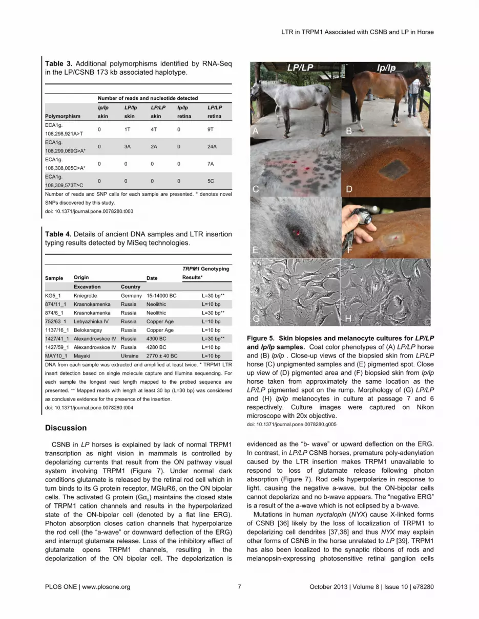
Melanocyte CulturesTo begin to characterize LP melanocytes we cultured
epidermal melanocytes from lp/lp pigmented skin and bothpigmented and non-pigmented LP/LP skin (Figure 5). Wesuccessfully isolated and established melanocyte cultures forboth the pigmented LP/LP and lp/lp skin (Figure 5). However,the non-pigmented area of the LP/LP horse skin did not yieldany viable melanocytes that could be propagated. Interestingly,transmission electron microscopy of cultured melanocytesshowed spherical melanosomes in wild type lp/lp melanocytes,but irregularly shaped melanosomes in LP/LP melanocytes(Figure 6A). LP genotypes of melanocytes cultures wereconfirmed by allele specific PCR, as described in the methods,using DNA from pelleted cultured cells. Genotypes were asexpected; specifically cells isolated from the pigmented LP/LPsample genotyped homozygous for the LTR insertion.Differential gene expression between LP/LP and lp/lp cells,originally detected in skin samples [17], was confirmed by qRT-
Figure 3. RNA sequencing reads mapped to TRPM1. RNA-Seq reads from LP/LP CSNB affected and lp/lp CSNB unaffectedretina RNA, as well as, from LP/LP, LP/lp and lp/lp skin RNA. ECA1 Mb, TRPM1 exon position, location of the insertion, and thenumber of reads for each sample are presented. (A) exon 0-27 illustrating that in LP/LP samples transcripts were not detected inexons 3’ of the insertion in intron 1. (B) exon 0, 1, 1’, 2 and 3 of TRPM1 illustrating that transcripts from exon 0 (yellow highlight)were detected in retinal samples but not skin while transcripts from exon 1’ (green highlight) were not detected in any of the horsesamples, and finally transcripts were detected that mapped to intron 1 of LP/LP and LP/lp samples allowing for the identification ofthe insertion (denoted by red line).doi: 10.1371/journal.pone.0078280.g003
LTR in TRPM1 Associated with CSNB and LP in Horse
PLOS ONE | www.plosone.org 5 October 2013 | Volume 8 | Issue 10 | e78280

Figure 4. LTR insertion in TRPM1 causes premature poly-adenylation. (A) The 1378bp LTR insertion (ECA1g.108,297,929_108,297,930 ins1378) is flanked by 6 bp regionsof micro-homology which are highlighted in yellow. Poly-adenylation signal sequences are highlighted in green. The CAdinucleotide cleavage and poly-adenylation site is highlightedin red. (B) poly(A) tail detected by 3’RACE in LP/LP skin and(C) CSNB LP/LP retina samples. Sequence represented isfrom poly-adenylation signal sequence AGUAAA throughpoly(A) tail.doi: 10.1371/journal.pone.0078280.g004
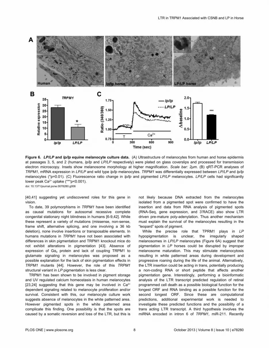
PCR in our cultured cells (p=0.01; Figure 6B). Furthermore,ratiometric imaging showed lower peak [Ca2+] uptake in LP/LPmelanocytes compared to lp/lp cells, consistent with lack ofTRPM1 functioning in these cells (Figure 6C).
Table 2. Complete association between CSNB, leopardcomplex spotting phenotype, and 1.4 kb insertion in intron 1of TRPM1.
Genotype for Insertion
Breed Phenotype/Genotype I/I I/- -/-CSNB Appaloosa CSNB 13 0 0 Unaffected 0 6 6American Miniature Horse CSNB 3 0 0 Unaffected 0 5 4Knabstrupper CSNB 1 0 0 Unaffected 0 1 0Quarter Horse* CSNB 0 0 0 Unaffected 0 0 4
Total (N=43) χ2=57,p<<0.0005
CSNB 17 0 0
Unaffected 0 12 14LP Appaloosa LPLP/LPLP 67 0 0 LPLP/LPlp 0 84 0 LPlp/LPlp 0 0 29Knabstrupper LPLP/LPLP 27 0 0 LPLP/LPlp 0 32 0 LPlp/LPlp 0 0 5Noriker LPLP/LPLP 2 0 0 LPLP/LPlp 0 58 0 LPlp/LPlp 0 0 52American Miniature Horse LPLP/LPLP 20 0 0 LPLP/LPlp 0 32 0 LPlp/LPlp 0 0 11Pony of the Americas LPLP/LPLP 4 0 0 LPLP/LPlp 0 14 0 LPlp/LPlp 0 0 2British Spotted Pony LPLP/LPLP 5 0 0 LPLP/LPlp 0 19 0 LPlp/LPlp 0 0 1Australian Spotted Pony LPLP/LPLP 1 0 0 LPLP/LPlp 0 9 0 LPlp/LPlp 0 0 0Thoroughbred* LPLP/LPLP 0 0 0 LPLP/LPlp 0 0 0 LPlp/LPlp 0 0 37
Totals (N=511) χ2=1022.00,p<<0.0005
LPLP/LPLP 126 0 0
LPLP/LPlp 0 248 0 LPlp/LPlp 0 0 137*. breeds not segregating for the LP mutation; I, presence of insertion; - absence ofinsertiondoi: 10.1371/journal.pone.0078280.t002
LTR in TRPM1 Associated with CSNB and LP in Horse
PLOS ONE | www.plosone.org 6 October 2013 | Volume 8 | Issue 10 | e78280
Discussion
CSNB in LP horses is explained by lack of normal TRPM1transcription as night vision in mammals is controlled bydepolarizing currents that result from the ON pathway visualsystem involving TRPM1 (Figure 7). Under normal darkconditions glutamate is released by the retinal rod cell which inturn binds to its G protein receptor, MGluR6, on the ON bipolarcells. The activated G protein (Gαo) maintains the closed stateof TRPM1 cation channels and results in the hyperpolarizedstate of the ON-bipolar cell (denoted by a flat line ERG).Photon absorption closes cation channels that hyperpolarizethe rod cell (the “a-wave” or downward deflection of the ERG)and interrupt glutamate release. Loss of the inhibitory effect ofglutamate opens TRPM1 channels, resulting in thedepolarization of the ON bipolar cell. The depolarization is
Table 3. Additional polymorphisms identified by RNA-Seqin the LP/CSNB 173 kb associated haplotype.
Number of reads and nucleotide detected
Polymorphismlp/lpskin
LP/lpskin
LP/LPskin
lp/lpretina
LP/LPretina
ECA1g.108,298,921A>T
0 1T 4T 0 9T
ECA1g.108,299,069G>A*
0 3A 2A 0 24A
ECA1g.108,308,005C>A*
0 0 0 0 7A
ECA1g.108,309,573T>C
0 0 0 0 5C
Number of reads and SNP calls for each sample are presented. * denotes novelSNPs discovered by this study.doi: 10.1371/journal.pone.0078280.t003
Table 4. Details of ancient DNA samples and LTR insertiontyping results detected by MiSeq technologies.
Sample Origin Date
TRPM1 GenotypingResults*
Excavation Country KG5_1 Kniegrotte Germany 15-14000 BC L=30 bp**874/11_1 Krasnokamenka Russia Neolithic L=10 bp874/6_1 Krasnokamenka Russia Neolithic L=30 bp**752/63_1 Lebyazhinka IV Russia Copper Age L=10 bp1137/16_1 Belokaragay Russia Copper Age L=10 bp1427/41_1 Alexandrovskoe IV Russia 4300 BC L=30 bp**1427/59_1 Alexandrovskoe IV Russia 4280 BC L=10 bpMAY10_1 Mayaki Ukraine 2770 ± 40 BC L=10 bp
DNA from each sample was extracted and amplified at least twice. * TRPM1 LTRinsert detection based on single molecule capture and Illumina sequencing. Foreach sample the longest read length mapped to the probed sequence arepresented. ** Mapped reads with length at least 30 bp (L=30 bp) was consideredas conclusive evidence for the presence of the insertion.doi: 10.1371/journal.pone.0078280.t004
evidenced as the “b- wave” or upward deflection on the ERG.In contrast, in LP/LP CSNB horses, premature poly-adenylationcaused by the LTR insertion makes TRPM1 unavailable torespond to loss of glutamate release following photonabsorption (Figure 7). Rod cells hyperpolarize in response tolight, causing the negative a-wave, but the ON-bipolar cellscannot depolarize and no b-wave appears. The “negative ERG”is a result of the a-wave which is not eclipsed by a b-wave.
Mutations in human nyctalopin (NYX) cause X-linked formsof CSNB [36] likely by the loss of localization of TRPM1 todepolarizing cell dendrites [37,38] and thus NYX may explainother forms of CSNB in the horse unrelated to LP [39]. TRPM1has also been localized to the synaptic ribbons of rods andmelanopsin-expressing photosensitive retinal ganglion cells
Figure 5. Skin biopsies and melanocyte cultures for LP/LPand lp/lp samples. Coat color phenotypes of (A) LP/LP horseand (B) lp/lp . Close-up views of the biopsied skin from LP/LPhorse (C) unpigmented samples and (E) pigmented spot. Closeup view of (D) pigmented area and (F) biopsied skin from lp/lphorse taken from approximately the same location as theLP/LP pigmented spot on the rump. Morphology of (G) LP/LPand (H) lp/lp melanocytes in culture at passage 7 and 6respectively. Culture images were captured on Nikonmicroscope with 20x objective.doi: 10.1371/journal.pone.0078280.g005
LTR in TRPM1 Associated with CSNB and LP in Horse
PLOS ONE | www.plosone.org 7 October 2013 | Volume 8 | Issue 10 | e78280
[40,41] suggesting yet undiscovered roles for this gene invision.
To date, 39 polymorphisms in TRPM1 have been identifiedas causal mutations for autosomal recessive completecongenital stationary night blindness in humans [6-9,42]. Whilethese represent a variety of mutations (missense, non-sense,frame shift, alternative splicing, and one involving a 36 kbdeletion), none involve insertions or transposable elements. Inhumans mutations in TRPM1 have not been associated withdifferences in skin pigmentation and TRPM1 knockout mice donot exhibit alterations in pigmentation [43]. Absence ofexpression of Gα0 protein and lack of coupling TRPM1 toglutamate signaling in melanocytes was proposed as apossible explanation for the lack of skin pigmentation effects inTRPM1 mutants [44]. However, the role of this TRPM1structural variant in LP pigmentation is less clear.
TRPM1 has been shown to be involved in pigment storageand UV regulated calcium homeostasis in human melanocytes[23,24] suggesting that this gene may be involved in Ca2+
dependent signaling related to melanocyte proliferation and/orsurvival. Consistent with this, our melanocyte culture worksuggests absence of melanocytes in the white patterned area.However pigmented spots in the white patterned areacomplicate this finding. One possibility is that the spots arecaused by a somatic reversion and loss of the LTR, but this is
not likely because DNA extracted from the melanocytesisolated from a pigmented spot were confirmed to have theinsertion and data from RNA analysis of pigmented spots(RNA-Seq, gene expression, and 3’RACE) also show LTRdriven pre-mature poly-adenylation. Thus another mechanismmust explain the survival of the melanocytes resulting in the“leopard” spots of pigment.
While the precise role that TPRM1 plays in LPhypopigmentation is unclear, the irregularly shapedmelanosomes in LP/LP melanocytes (Figure 6A) suggest thatpigmentation in LP horses could be disrupted by impropermelanosome maturation. This may stimulate melanocytosisresulting in white patterned areas during development andprogressive roaning during the life of the animal. Alternatively,the LTR insertion could be acting in trans, potentially producinga non-coding RNA or short peptide that affects anotherpigmentation gene. Interestingly, performing a bioinformaticanalysis of the LTR transcript predicted regulation of retinalprogrammed cell death as a possible biological function for thelongest ORF and RNA binding as a possible function for thesecond longest ORF. Since these are computationalpredictions, additional experimental work is needed toinvestigate these predicted functions and the possibility of atrans acting LTR transcript. A third hypothesis involves themiRNA encoded in intron 6 of TRPM1, miR-211. Recently
Figure 6. LP/LP and lp/lp equine melanocyte culture data. (A) Ultrastructure of melanocytes from human and horse epidermisat passages 3, 5, and 2 (humans, lp/lp and LP/LP respectively) were plated on glass coverslips and processed for transmissionelectron microscopy. Insets show melanosome morphology at higher magnification. Scale bar: 2μm. (B) qRT-PCR analyses ofTRPM1, mRNA expression in LP/LP and wild type lp/lp melanocytes. TRPM1 was differentially expressed between LP/LP and lp/lpmelanocytes (*p=0.01). (C) Fluorescence ratio change in lp/lp and pigmented LP/LP melanocytes. LP/LP cells had significantlylower peak Ca2+ uptake (***p<0.001).doi: 10.1371/journal.pone.0078280.g006
LTR in TRPM1 Associated with CSNB and LP in Horse
PLOS ONE | www.plosone.org 8 October 2013 | Volume 8 | Issue 10 | e78280

miR-211, which is cotranscribed with TRPM1, has been shownto be expressed in normal melanocytes but reduced inmelanoma [45,46]. miR211 has also been shown to regulatetransforming growth factor B2R and TGF-B signaling has a rolein maintenance of melanocyte stem cells [47]. Thus it ispossible that premature poly-adenylation of TRPM1 in intron 1completely abolishes miR-211 causing an increased level ofTGF B signaling and resulting in either depleting themelanocyte stem cell population by induced apoptosis orinhibiting pigment production by down regulating melanogenicgenes. The latter two hypotheses might help explain whyCSNB is recessive while LP spotting is dominant. All of thesehypotheses remain to be further investigated.
We have identified a 1378 bp LTR insertion with the TRPM1gene that is associated with both a pigmentation phenotype(LP) and an eye disorder (CSNB). It is believed that LTRs maycontribute to species evolution through gene regulatorychanges mainly by cis-acting sequences that function asalternative promoters or novel poly-adenylation signals[29,30,48-52]. Moreover, recent retrotransposon events,involving LINE-1 elements, have been shown to disrupt normalgene function and cause disease and approximately 65 suchdisease causing mutations have been identified in humansalone [53]. None of those described to date involve an LTR.Thus LTR transposable element insertions creating diseasephenotypes, like CSNB, are likely under negative selection.Yet, this LTR insertion was definitively detected in three ancientsamples suggesting that this structural variation has beenmaintained in the horse population long before domesticationand may have provided some selective advantage. While theexact timing of the integration of this structural variant, it’s
selective advantage, and the physiological mechanism ofhypopigmentation remain to be determined, we propose thatthis insertion disrupts normal TRPM1 transcription bypremature poly-adenylation and that this abnormal transcriptionresults in both CSNB and LP spotting.
Materials and Methods
Ethics StatementAnimals were sampled with the appropriate consent from
horse owners. Those horses sacrificed were humanelyeuthanized following The Canadian Council on Animal CareGuidelines for Experimental Animal Use and approved by theUniversity of Saskatchewan Animal Care Committee.
SamplesHorses were categorized according to genotype and
phenotype for LP, which was evaluated by coat colorassessment and breeding records. Those horses utilized in thestudies of CSNB were also examined by a scotopic electro-retinogram (ERG) to determine CSNB status. For the RNAsequencing experiments total RNA was isolated from three(LP/LP, LP/lp, lp/lp) full thickness pigmented skin biopsies fromlive animals and from retina (one CSNB affected LP/LP andone CSNB unaffected lp/lp). To control for changes in geneexpression related to sex and pigmentation differences thesesamples were matched for sex (male) and base coat color(chestnut). Additional RNA samples from pigmented skin (threeLP/LP and one lp/lp) and retina (two CSNB affected LP/LP)were utilized for 3’ RACE experiments. A total of 511 horsesfrom seven breeds segregating for LP spotting and one breed
Figure 7. The role of TRPM1 in night vision. Night vision is controlled by ON visual pathway which comprises both retinal rodand ON-bipolar cells and involves the neurotransmitter glutamate, its receptor mGluR6, the G protein Gαo, TRPM1, and Ca2+.Photon absorption by the rod cell causes the rod cell to hyperpolarize (resulting in the “a” wave of the ERG) and decreases theconcentration of glutamate being released by that cell. This leads to the opening of the calcium ion channel, TRPM1, and results inthe depolarization of the ON bipolar cell (“b” wave on the ERG). In the absence of photon activation, low light conditions, the rod cellis depolarized leading to the release of glutamate and the closing of TRPM1. This results in the hyperpolarization of the ON-bipolarcell. In contrast, in LP/LP CSNB horses TRPM1 is unavailable to respond to changes in glutamate concentration and thus the ON-bipolar cells remain hyperpolarized in response to light. The b-wave is absent, so the ERG appears “negative” as it is only the a-wave (the rod cell response) that is detected.doi: 10.1371/journal.pone.0078280.g007
LTR in TRPM1 Associated with CSNB and LP in Horse
PLOS ONE | www.plosone.org 9 October 2013 | Volume 8 | Issue 10 | e78280

not segregating for LP as well as 43 horses phenotyped forCSNB were utilized to genotype for the discovered insertion.
RNA sequencing and read mappingTotal RNA was isolated from 0.5 g of skin tissue in a buffer of
4 M guanidinium isothiocyanate, 0.1 M Tris–HCl, 25 mM EDTA(pH 7.5), and 1% (v/v) 2-mercaptoethanol, followed bydifferential alcohol and salt precipitations as previouslydescribed [54,55]. Retinal RNA was isolated as previouslydescribed using Trizol (Invitrogen) [17]. RNA was quantifiedusing a NanoDrop spectrophotometer (NanoDropTechnologies) and assayed for quality using a 2100Bioanalyzer (AgilentTechnologies). Library preparation andIllumina sequencing was performed by Cornell University's LifeSciences Core Laboratory Center. RNA-Seq reads werealigned to the horse reference genome (equCab2) withBurrows-Wheeler Aligner (BWA) [56] with the default options.BAM Files generated using SAMtools [57] allowed forvisualization of output alignments using the UCSC GenomeBrowser (http://genome-preview.ucsc.edu/cgi-bin/hgGateway?hgsid=2633482&clade=mammal&org=Horse&db=0). RNA-Seqdata was submitted as studies to the Sequence Read Archive(SRA at the European Nucleotide Archive http://www.ebi.ac.uk/ena/ and can be found at http://www.ebi.ac.uk/ena/data/view/ERP001524 and http://www.ebi.ac.uk/ena/data/view/ERP001525 (Accession numbers. ERP001524 andERP001525).
Insertion discoveryRNA-Seq analysis identified expression of a region in intron
1 of TRPM1 in LP/LP and LP/lp samples but not lp/lp skin andretina samples. This region was investigated by genomic PCRto test for structural variations by using two different sets ofprimers (Table S3). One primer combination (108297763F and108299526 R) resulted in correct size product for LP/lp andlp/lp horses but only non-specific amplification in the LP/LPhorses while amplicons involving primers spanning ECA1:108,298,197-108,300,248 resulted in the correct size productfor all genotypes, thus suggesting the presence of a structuralvariant not allowing for amplification from ECA1: 108,297,763-108,298,197. Genomic sequence from the region(ECA1: 108, 297,763-108,298,197) was used for de novoassembly of RNA-Seq reads from the LP/LP CSNB retinasample using cross_match and visualized with consed (Version20) [58]. This enabled the identification of the precise locationof the insertion and identified the first 200 bp of the insertion.The full length insertion was discovered in 4 horses from 3different breeds (2 American Miniature horses, 1 Appaloosa,and 1 Knabstrupper) by genomic PCR amplification followed bysequence walking using Sanger technology. Long range PCRusing 200 ng of DNA, 2.5 pmol of each primer (108, 297,763Fand108,298,197R, Table S3) and Platinum PCR SuperMixHigh Fidelity (Invitrogen at Life Technologies, Grand Island,NY, USA) was performed. Amplicons were gel purified usingthe QIAquick gel extraction kit (Qiagen Sciences, Valencia, CA,USA) and subsequently sequenced using BigDye Terminatorv1.1 and ABI 310 Genetic Analyzer (Applied Biosystems, atLife Technologies, Grand Island, NY, USA) and 4 different
primers (5.0 pmol) to obtain sequence information for the entireinsertion (Table S3). Sequences were assembled usingContigExpress from the Vector NTI Advance 11 softwarepackage (Invitrogen at Life Technologies, Grand Island, NY,USA).
LTR analysisThe LP insertion element was identified as a ERV2-1N-
EC_LTR using RepeatMasker [27]. To determine therelationship of this LTR to other LTRs in the equine genome,the insertion element was used to identify all similar elementsin the equine genome EquCab2.0 using LASTZ[59] based oncriteria of ≥80% sequence identity and ≥90% length. Anadditional 202 full length LTR were identified in this fashion andtheir sequences, along with 50 bp of flanking sequences, wereextracted from the equine genome assembly. The retrievedLTR sequences were then aligned using MUSCLE [60,61] anda phylogenetic tree computed using FastTree [62,63] with theGeneral Time Reversible model of nucleotide substitution andGamma20. LTR88, a mammalian LTR sequence was includedto provide an outgroup for the tree. Flanking sequences wereused as input for TSDscan [64] to determine if TSDs could beused to predict relative insertion age.
Genotyping modern horsesWhole blood or mane hair was collected by one of the
authors or submitted by owners for this study. DNA from bloodsamples was extracted either using the Puregene whole-bloodextraction kit (Qiagen Inc., Valencia, CA, USA) or NucleonBacc2 kit (GE Healthcare Bio-Sciences Corp., Piscataway, NJ,USA) according to the manufacturer’s protocol. Hair sampleswere processed using 5–7 hair bulbs according to the methoddescribed previously [65]. Standard PCR protocol using allelicspecific primers allowed for genotyping of this insertion. 2.0pmol of two forward primers 108,297,821 F (to amplify thewildtype allele 186 bp) and TRPM1 ins F” (to amplify the insertallele 225 bp) and one reverse primer (108,297,987R, TableS3) were used in the same reaction with 1 uL of DNA from hairlysis extraction or 25 ng of DNA isolated from blood, 1X PCRbuffer with 2.0mM MgCl2, 100 µM of each dNTP, and 0.1UFastStartTaq DNA polymerase (Roche Applied ScienceIndianapolis, IN, USA). Association of insertion with LPgenotype and CSNB status were analyzed by chi-square tests.
3’ RACE experiments3’ RACE (Rapid Amplification of cDNA ends) ready cDNA
was synthesized from 187.5 ng of total RNA (3 LP/LP skin and3 LP/LP CSNB affected retina samples as well as 1 lp/lp skinand 1 lp/lp unaffected retina as negative controls) using 3’CDSPrimer A (supplied with SMARTerTM RACE cDNA amplificationkit) and SMARTScribeTM Reverse Transcriptase followingmanufactures instructions (SMARTerTM RACE cDNAamplification kit Clontech Laboratories, Inc., Mountain View,CA, USA). Following cDNA synthesis 1.25 µl of diluted 3’RACE ready cDNA was utilized as a template for 3’RACE PCRusing UPM (supplied with SMARTerTM RACE cDNAamplification kit) and TRPM1 lp RACE 3’ RACE F-1(Table S3).PCR was performed following manufactures instructions using
LTR in TRPM1 Associated with CSNB and LP in Horse
PLOS ONE | www.plosone.org 10 October 2013 | Volume 8 | Issue 10 | e78280

the Advantage 2 PCR KIT and reducing final reaction volumeto 25 µl. (Clontech Laboratories, Inc., Mountain View, CA,USA). To increase specificity, a semi-nested PCR wasperformed using 2.5 µl diluted PCR product (5 µl of PCRproduct diluted in 245 µl of Tris-EDTA buffer, pH= 8.0), UPM,TRPM1 ins TRANS R-6 (Table S3), and the same Advantage 2PCR KIT again following manufacturer’s instructions. Productswere visualized on 1% EtBr agarose gel and amplicons weregel purified using the QIAquick gel extraction kit (QiagenSciences, Valencia, CA, USA) and subsequently sequencedusing BigDye Terminator v1.1 and ABI 310 Genetic Analyzer(Applied Biosystems, at Life Technologies, Grand Island, NY,USA). Each sample was sequenced with 4 different primers (5pmol) to obtain full amplicon sequence information fromTRPM1 gene specific sequence (TRPM1 ins TRANS R-6) tothe poly(A) tail (Table S3).
Bioinformatics analysis of ORFsThe transcript sequence from the LP/LP pigmented skin
sample was translated using the Sequence Manipulation Suite[66]. All identified ORFs were analyzed using BLASTP againstthe nr database at www.ncbi.nlm.nih.gov. These ORFs werefurther analyzed using the Protein Function Prediction server[33] available at http://kiharalab.org/web/pfp.php and theExtended Similarity Group server [34] available at http://kiharalab.org/web/esg.php. Only probability scores greater thanor equal to 60% were retained for the predicted molecularfunction and biological process.
Melanocyte Culture ExperimentsIsolation and culture of equine melanocytes. Full
thickness skin biopsies (3-6mm) were collected in DMEMmedium with 2% FBS and supplemented with standard tissueculture grade antibiotic and antimycotic mixture. The tissueswere sliced into small pieces and incubated in 0.25% trypsin-EDTA in 6 well plates overnight at 4°C. Next day, epidermiswas separated using sterile forceps and cells were dissociatedby vigorous pipetting. The tissue pieces and cells were pelletedby centrifuging at 1500 rpm for 10 minutes and re-suspendedin melanocyte medium, plated in 6-well plates and cultured at37°C with 5% CO2. The medium was changed 24 hours afterplating and after every three days [24].
Transmission electronmicroscopy. Cells on coverslipswere washed with phosphate-buffered saline, fixed with 2.5%glutaraldehyde, and processed for transmission electronmicroscopy. Ultrathin sections were observed andphotographed using the Jeol 100 electron microscope (Tokyo,Japan) at the UW Electron Microscope Facility.
Quantitative RT-PCR. Total RNA was isolated usingRNeasy mini kit (Qiagen, Valencia, CA). Total RNA (1–3 µg)was reverse transcribed using the SuperScript III First-StrandSynthesis system for RT-PCR (Invitrogen). Primers for TRPM1and β-actin were designed using PrimerExpress software(Applied Biosystems, Foster City, CA) and obtained from IDTInc. (Coralville, IA). The primer sequences and the calculatedamplicon length are represented in Table S4. qPCR wasperformed in triplicate with SYBR Green PCR core reagents(Applied Biosystems), in 20 µl of reactions containing 50-150ng
cDNA and 10µM each primer. β-actin was used as thehousekeeping gene control. PCR was performed using ABI7000 and cycle parameters: denaturation at 95°C, followed by40 cycles of 95°C for 15 s, 57°C for 15 s, and 72°C for 15 s.Relative quantification (RQ) was calculated using 2- ΔΔC
T
method qRT-PCR data was analyzed according Schmittgenand Livak [67].
Immunofluorescence. Melanocytes were confirmed byimmunostaining for the melanocyte-specific markers namelyTYRP1 and MITF (Figure S4). Melanocytes grown on chamberslides (Nalge Nunc International, Naperville, IL) were fixed in4% freshly prepared paraformaldehyde for 15 minutes andpermeabilized for 5 minutes in 0.1% Triton X-100. The cellswere washed, blocked for 30 minutes in 10% goat serum andincubated for 1 h with primary antibodies (anti-TYRP1mAbTA99 at 1:1000 and anti-MITF antibody SC- 56725 at1:100) followed by FITC-conjugated anti-mouse rabbit IgGdiluted in 10% goat serum for 1 h. After washing with PBS,coverslips were mounted using a mounting medium containinganti-fade reagent (Molecular Probes, Invitrogen).
Calcium imaging. Melanocytes plated on glass chamberslides were washed in buffer D (153mM NaCl, 5.4mM KCl,1.8mM CaCl2, 10mM N-2-hydroxy ethylpiperazine-N’-2-ethanesulfonic acid (HEPES), 25mM glucose, 0.8mM MgSO4,0.9mM NaH2PO4 and 2.4mM NaHCO3, pH 7.4), incubated in 2µM Fura-2 (Invitrogen) in buffer D at 37°C for 60 minutes,washed three times with buffer D without calcium and de-esterified for 30 minutes at 37°C. Cells were imaged and ratioof fluorescence emission was monitored using Nikon Tifluorescence microscope and NIS-Elements software. In a 20xfield with well separated cells (n=8-10), baseline wasestablished in buffer D without Ca2+ and change in F340/380was monitored after addition of various reagents or Ca2+ for atotal of 15 minutes with no cell exposed to light for longer than20 min. Results were expressed as ratio value afterbackground fluorescence subtraction and normalization tobaseline fluorescence.
Ancient DNA typingDNA was extracted from eight ancient samples (Table S2)
following a previously established protocol [68] in dedicatedancient DNA facilities at the Humboldt-University of Berlin,Germany and at the University of York, UK. In addition to theancient samples, two modern samples (1 LP/lp and 1 LP/LP)were included to serve as positive controls for the enrichmentstrategy (Table S1). Sequencing libraries suitable for theIllumina platform were prepared following an establishedprotocol [69] with some minor modifications: the Illumina P5adapter sequences were modified to include a 8 bp index at the3' end, effectively double-indexing the libraries without the needfor an extra sequence read. To enrich the libraries for the DNAsequences required to detect the LTR insertion, probes weredesigned that overlap the boundary between TRPM1 intron 1and the start of the insert as well as intron 1 and the end of theinsert (Table S5). The target regions for capture weresynthesized as biotinylated DNA oligos and pooled in equalconcentrations for in-solution capture following Horn (2012)[70] in two consecutive capture rounds. After enrichment,
LTR in TRPM1 Associated with CSNB and LP in Horse
PLOS ONE | www.plosone.org 11 October 2013 | Volume 8 | Issue 10 | e78280

samples were pooled in equimolar amounts and 12.5 pM of thelibrary pool was sequenced using the MiSeq System (Illumina,San Diego, CA, USA). 100 bp paired-end sequencing wascarried out; i.e. 2x101 sequencing cycles (indexing was carriedout using 9 sequencing cycles) using the Illumina MiSeqReagent Kit v2 (300 cycles). The lower concentration and poorquality of DNA in ancient samples resulted in lower sequencecoverage compared to the control samples. Generated readswere cleaned from index and adapter sequences usingcutadapt (v1.1) [71] with default parameters and differentstringencies for the minimal required read length after trimming(10-30 nt). Trimmed reads were further filtered according tobase quality using fastx toolkit (http://hannonlab.cshl.edu/fastx_toolkit/) requiring a quality score of a minimum of 30 overat least 50% of the read length. Final reads were mapped tothe LTR probe sequences using Bowtie [72] to identify the LPinsert and additionally mapped to the horse reference genome(equCab2) to confirm location in the TRPM1 intron 1. Mappedreads with a minimum length of 30 nt were required forconclusive LTR insert detection.
Supporting Information
Figure S1. Investigating the expression of 3 SNPspreviously identified as completely associated with LP andCSNB. BAM files for each of the RNA samples utilized in thisRNA-Seq experiment were aligned to the equCab2 referencegenome using the UCSC Genome Browser. RNA isolated fromlp/lp skin is denoted by 46-lp-skin, while that from LP/LP skin isdenoted by 54-LP-Skin, and LP/lp skin is denoted by 49-LP-lp-pigmented. Retina RNA samples are denoted by 45-lp-retina(CSNB unaffected) and 54-LP retina (CSNB affected). 100 bpflanking each SNP is shown as represented by the coordinatesalong with any non-horse RefSeq genes. Both ECA1 g.108281765T>C (A) and ECA1 g.108288853C>T (B) arelocated in introns of TRPM1 as defined by human, rat andmouse RefSeq genes and this is a confirmed intron in thehorse as no expression of this region was detected in eitherskin or retina. While ECA1g.108337089T>G (C) is also not inan exon, as no expression was detected, however no knownRefSeq gene has been mapped to this region.(TIF)
Figure S2. RepeatMasker blast analysis summary of 1.4 kbLP/CSNB insertion. Version 3.2.9 of RepeatMaster wasutilized for this blast analysis. The LP/CSNB insertion wasidentified as an LTR ERV Class II.(TIFF)
Figure S3. The relationship of the LP/CSNB LTR insertioncompared to all other equine LTRs. The LTR in intron 1 ofTRPM1 causing both CSNB and LP is denoted by a red box.The inset, boxed in blue, shows a more detailed version of thebranch of the tree containing the newly discovered insertion(red branch).(TIFF)
Figure S4. Expression of TYRP1 and MITF in melanoctyes.Cells isolated from lp/lp horse cultures were compared tohuman cells to confirm melanocytes identity. Cells cultured onglass coverslips were fixed with paraformaldehyde andpermeabilized with methanol and incubated with anti-TYRP1and anti-MITF antibodies followed by FITC-conjugated anti-mouse IgG. Images were captured on Nikon microscope with20x objective.(TIF)
Table S1. Predicted protein functions for LP insertiontranscript. Nine potential ORF of the LTR insertion transcriptwere investigated using the PFP and ESG protein predictionservers. GO molecular functions and biological processes withprobabilities equal to or greater than 60% are reported.(XLSX)
Table S2. TRPM1 insert detection in ancient DNA samples.Genotyping was based on single molecule capture and Illuminasequencing. Data presented are MiSeq read depth aftermapping with Bowtie and utilizing different filter settings forread lengths. A modern homozygote (LP/LP) and heterozygote(LP/lp) were included for validation of the data.(XLSX)
Table S3. Primers utilized in the discovery and theinvestigation of the 1.4 kb insertion causing LP and CSNB.Primers were used for identification and sequencing of fulllength LP/CSNB insertion, genotyping for LP and CSNB, andfor performing 3’ RACE and sequencing RACE products.(XLSX)
Table S4. Primers used for qRT-PCR of melanocytecultures.(XLSX)
Table S5. Probes sequences used to genotype ancientDNA for the LP/CSNB insertion. Probes were used to enrichlibraries for the target regions via hybridization capture usingbiotin-streptavidin binding coupled to Illumina sequencing.(XLSX)
Acknowledgements
The authors wish to thank all of the horse owners who providedhair and skin samples for nucleic acid extraction and formelanocyte culture work. We would especially like to thankthose owners who donated their horses for retinal researchpurposes. We are grateful to Dr. Jay Clifford and Mr. RichardGrist for their assistance with skin biopsy collections. We thankKarla Brown, Cathryn Cuka, Taryn Cranford, ElizabethKowalski, and Scott Lawson for technical assistance as part oftheir Independent Study Courses at the University of Tampa.
Author Contributions
Prepared the manuscript with contribution and input from allauthors: RRB. Conceived and designed the experiments: RRB
LTR in TRPM1 Associated with CSNB and LP in Horse
PLOS ONE | www.plosone.org 12 October 2013 | Volume 8 | Issue 10 | e78280

SA GF SAB VS SD NM DLA SLL AL MP DF RB MH JP GGMR. Performed the experiments: RRB GF MJM VS SD NM LSBG SA BH ME TL JN DLA SLL AL MP DF RB MH JP GG MRPM. Analyzed the data: RRB SAB HH VS SD NM DLA SLL AL
MP DF RB MH JP GG MR PM. Contributed reagents/materials/analysis tools: RRB SA GF MJM LS BG ME TL AL MR PM.Wrote the manuscript: RRB.
References
1. Duhl DM, Stevens ME, Vrieling H, Saxon PJ, Miller MW et al. (1994)Pleiotropic effects of the mouse lethal yellow (Ay) mutation explainedby deletion of a maternally expressed gene and the simultaneousproduction of agouti fusion RNAs. Development 120(6): 1695-1708.PubMed: 8050375.
2. Barsh GS (1995) Pigmentation, pleiotropy, and genetic pathways inhumans and mice. Am J Hum Genet 57(4): 743-747. PubMed:7573031.
3. Bellone RR (2010) Pleiotropic effects of pigmentation genes in horses.Anim Genet 41 Suppl 2: 100-110. doi:10.1111/j.1365-2052.2010.02116.x. PubMed: 21070283.
4. Bennett DC, Lamoreux ML (2003) The Color Loci of Mice – A GeneticCentury. Pigment Cell Res 16: 333–344. doi:10.1034/j.1600-0749.2003.00067.x. PubMed: 12859616.
5. Reissmann M, Ludwig A (2013) Pleiotropic effects of coat colour-associated mutations in humans, mice and other mammals. Semin CellDev Biol 24: 576-586. doi:10.1016/j.semcdb.2013.03.014. PubMed:23583561.
6. Audo I, Kohl S, Leroy BP, Munier FL, Guillonneau X et al. (2009)TRPM1 is mutated in patients with autosomal-recessive completecongenital stationary night blindness. Am J Hum Genet 85(5): 720-729.doi:10.1016/j.ajhg.2009.10.013. PubMed: 19896113.
7. Li Z, Sergouniotis PI, Michaelides M, Mackay DS, Wright GA et al.(2009) Recessive mutations of the gene TRPM1 abrogate ON bipolarcell function and cause complete congenital stationary night blindnessin humans. Am J Hum Genet 85(5): 711-719. doi:10.1016/j.ajhg.2009.10.003. PubMed: 19878917.
8. van Genderen MM, Bijveld MM, Claassen YB, Florijn RJ, Pearring JNet al. (2009) Mutations in TRPM1 are a common cause of completecongenital stationary night blindness. Am J Hum Genet 85(5): 730-736.doi:10.1016/j.ajhg.2009.10.012. PubMed: 19896109.
9. Nakamura M, Sanuki R, Yasuma TR, Onishi A, Nishiguchi KM et al.(2010) TRPM1 mutations are associated with the complete form ofcongenital stationary night blindness. Mol Vis 16: 425-437. PubMed:20300565.
10. Sandmeyer LS, Breaux CB, Archer S, Grahn BH (2007) Clinical andelectroretinographic characteristics of congenital stationary nightblindness in the Appaloosa and the association with the leopardcomplex. Vet Ophthalmol 10(6): 368-375. doi:10.1111/j.1463-5224.2007.00572.x. PubMed: 17970998.
11. Sandmeyer LS, Bellone RR, Archer S, Bauer BS, Nelson J et al. (2012)Congenital stationary night blindness is associated with the leopardcomplex in the miniature horse. Vet Ophthalmol 15: 18-22. doi:10.1111/j.1463-5224.2011.00903.x. PubMed: 22051042.
12. Witzel DA, Joyce JR, Smith EL (1977) Electroretinography ofcongenital night blindness in an Appaloosa filly. J Eq Med Surg. 1:226-229.
13. Witzel DA, Smith EL, Wilson RD, Aguirre GD (1978) Congenitalstationary night blindness: an animal model. Invest Ophthalmol Vis Sci17(8): 788-795. PubMed: 308060.
14. Sponenberg DP (2009) Equine color genetics. xiii. 3rd ed. Ames, IA:Wiley-Blackwell. 277 pp.
15. Sponenberg DP, Carr G, Simak E, Schwink K (1990) The inheritance ofthe leopard complex of spotting patterns in horses. J Hered 81(4):323-331. PubMed: 2177073.
16. Terry RB, Archer S, Brooks S, Bernoco D, Bailey E (2004) Assignmentof the appaloosa coat colour gene (LP) to equine chromosome 1. AnimGenet 35(2): 134-137. doi:10.1111/j.1365-2052.2004.01113.x.PubMed: 15025575.
17. Bellone RR, Brooks SA, Sandmeyer L, Murphy BA, Forsyth G et al.(2008) Differential gene expression of TRPM1, the potential cause ofcongenital stationary night blindness and coat spotting patterns (LP) inthe Appaloosa horse (Equus caballus). Genetics 179(4): 1861-1870.doi:10.1534/genetics.108.088807. PubMed: 18660533.
18. Shen Y, Heimel JA, Kamermans M, Peachey NS, Gregg RG et al.(2009) A transient receptor potential-like channel mediates synaptictransmission in rod bipolar cells. J Neurosci 29(19): 6088-6093. doi:10.1523/JNEUROSCI.0132-09.2009. PubMed: 19439586.
19. Morgans CW, Zhang J, Jeffrey BG, Nelson SM, Burke NS et al. (2009)TRPM1 is required for the depolarizing light response in retinal ON-
bipolar cells. Proc Natl Acad Sci U S A 106(45): 19174-19178. doi:10.1073/pnas.0908711106. PubMed: 19861548.
20. Morgans CW, Brown RL, Duvoisin RM (2010) TRPM1: the endpoint ofthe mGluR6 signal transduction cascade in retinal ON-bipolar cells.Bioessays 32(7): 609-614. doi:10.1002/bies.200900198. PubMed:20544736.
21. Koike C, Obara T, Uriu Y, Numata T, Sanuki R et al. (2010) TRPM1 isa component of the retinal ON bipolar cell transduction channel in themGluR6 cascade. Proc Natl Acad Sci U S A 107(1): 332-337. doi:10.1073/pnas.0912730107. PubMed: 19966281.
22. Duncan LM, Deeds J, Hunter J, Shao J, Holmgren LM et al. (1998)Down-regulation of the novel gene melastatin correlates with potentialfor melanoma metastasis. Cancer Res 58(7): 1515-1520. PubMed:9537257.
23. Oancea E, Vriens J, Brauchi S, Jun J, Splawski I et al. (2009) TRPM1forms ion channels associated with melanin content in melanocytes.Sci Signal 2(70): ra21. PubMed: 19436059.
24. Devi S, Kedlaya R, Maddodi N, Bhat KM, Weber CS et al. (2009)Calcium homeostasis in human melanocytes: role of transient receptorpotential melastatin 1 (TRPM1) and its regulation by ultraviolet light.Am J Physiol Cell Physiol 297(3): C679-C687. doi:10.1152/ajprenal.90680.2008. PubMed: 19587221.
25. Bellone RR, Forsyth G, Leeb T, Archer S, Sigurdsson S et al. (2010)Fine-mapping and mutation analysis of TRPM1: a candidate gene forleopard complex (LP) spotting and congenital stationary night blindnessin horses. Brief Funct Genomics 9(3): 193-207. doi:10.1093/bfgp/elq002. PubMed: 20353955.
26. Bellone RR, Archer S, Wade CM, Cuka-Lawson C, Haase B et al.(2010) Association analysis of candidate SNPs in TRPM1 with leopardcomplex spotting (LP ) and congenital stationary night blindness(CSNB) in horses. Anim Genet 41: 207-207. doi:10.1111/j.1365-2052.2010.02119.x.
27. Smit AFA, Hubley R, Green P (1996-2004) RepeatMasker Open-3.0.Repeat Masker website. Available: http://www.repeatmasker.org.Accessed 2013 September 26.
28. Guntaka RV (1993) Transcription termination and polyadenylation inretroviruses. Microbiol Rev 57(3): 511-521. PubMed: 7902524.
29. Dunn CA, Medstrand P, Mager DL (2003) An endogenous retrovirallong terminal repeat is the dominant promoter for human beta1,3-galactosyltransferase 5 in the colon. Proc Natl Acad Sci U S A 100(22):12841-12846. doi:10.1073/pnas.2134464100. PubMed: 14534330.
30. Cohen CJ, Lock WM, Mager DL (2009) Endogenous retroviral LTRs aspromoters for human genes: a critical assessment. Gene 448(2):105-114. doi:10.1016/j.gene.2009.06.020. PubMed: 19577618.
31. Chuong EB, Rumi MA, Soares MJ, Baker JC. (2013) Endogenousretroviruses function as species-specific enhancer elements in theplacenta. Nat Genet 45(3): 325-329. doi:10.1038/ng.2553. PubMed:23396136.
32. Maksakova IA, Romanish MT, Gagnier L, Dunn CA, van de LagemaatLN et al. (2006) Retroviral elements and their hosts: insertionalmutagenesis in the mouse germ line. PLOS Genet 2(1): e2. doi:10.1371/journal.pgen.0020002. PubMed: 16440055.
33. Chitale M, Hawkins T, Park C, Kihara D (2009) ESG: extendedsimilarity group method for automated protein function prediction.Bioinformatics 25(14): 1739-1745. doi:10.1093/bioinformatics/btp309.PubMed: 19435743.
34. Hawkins T, Chitale M, Luban S, Kihara D (2009) PFP: Automatedprediction of gene ontology functional annotations with confidencescores using protein sequence data. Proteins 74(3): 566-582. doi:10.1002/prot.22172. PubMed: 18655063.
35. Pruvost M, Bellone R, Benecke N, Sandoval-Castellanos E, Cieslak Met al. (2011) Genotypes of predomestic horses match phenotypespainted in Paleolithic works of cave art. Proc Natl Acad Sci U S A108(46): 18626-18630. doi:10.1073/pnas.1108982108. PubMed:22065780.
36. Bech-Hansen NT, Naylor MJ, Maybaum TA, Sparkes RL, Koop B et al.(2000) Mutations in NYX, encoding the leucine-rich proteoglycannyctalopin, cause X-linked complete congenital stationary nightblindness. Nat Genet 26(3): 319-323. doi:10.1038/81619. PubMed:11062471.
LTR in TRPM1 Associated with CSNB and LP in Horse
PLOS ONE | www.plosone.org 13 October 2013 | Volume 8 | Issue 10 | e78280

37. Cao Y, Posokhova E, Martemyanov KA (2011) TRPM1 formscomplexes with nyctalopin in vivo and accumulates in postsynapticcompartment of ON-bipolar neurons in mGluR6-dependent manner. JNeurosci 31(32): 11521-11526. doi:10.1523/JNEUROSCI.1682-11.2011. PubMed: 21832182.
38. Pearring JN, Bojang P Jr, Shen Y, Koike C, Furukawa T et al. (2011) Arole for nyctalopin, a small leucine-rich repeat protein, in localizing theTRP melastatin 1 channel to retinal depolarizing bipolar cell dendrites.J Neurosci 31(27): 10060-10066. doi:10.1523/JNEUROSCI.1014-11.2011. PubMed: 21734298.
39. Nunnery C, Pickett JP, Zimmerman KL (2005) Congenital stationarynight blindness in a Thoroughbred and a Paso Fino. Vet Ophthalmol8(6): 415-419. doi:10.1111/j.1463-5224.2005.00416.x. PubMed:16359365.
40. Klooster J, Blokker J, Ten Brink JB, Unmehopa U, Fluiter K et al.(2011) Ultrastructural localization and expression of TRPM1 in thehuman retina. Invest Ophthalmol Vis Sci 52(11): 8356-8362. doi:10.1167/iovs.11-7575. PubMed: 21896854.
41. Hughes S, Pothecary CA, Jagannath A, Foster RG, Hankins MW et al.(2012) Profound defects in pupillary responses to light in TRPM-channel null mice: a role for TRPM channels in non-image-formingphotoreception. Eur J Neurosci 35(1): 34-43. doi:10.1111/j.1460-9568.2011.07944.x. PubMed: 22211741.
42. Audo I, Bujakowska KM, Léveillard T, Mohand-Saïd S, Lancelot ME etal. (2012) Development and application of a next-generation-sequencing (NGS) approach to detect known and novel gene defectsunderlying retinal diseases. Orphanet J Rare Dis 7(1): 8. doi:10.1186/1750-1172-7-8. PubMed: 22277662.
43. Koike C, Numata T, Ueda H, Mori Y, Furukawa T (2010) TRPM1: avertebrate TRP channel responsible for retinal ON bipolar function. CellCalcium 48(2-3): 95-101. doi:10.1016/j.ceca.2010.08.004. PubMed:20846719.
44. Devi S, Markandeya Y, Maddodi N, Anuradha D, Vardi N et al. (2013)Metabotropic glutamate receptor 6 signaling enhancesTRPM1 calciumchannel function and increases melanincontent in human melanocytes.Pigment Cell. Melanoma Res 26 (3): 348–356.
45. Levy C, Khaled M, Iliopoulos D, Janas MM, Schubert S et al. (2010)Intronic miR-211 assumes the tumor suppressive function of its hostgene in melanoma. Mol Cell 40(5): 841-849. doi:10.1016/j.molcel.2010.11.020. PubMed: 21109473.
46. Mazar J, DeYoung K, Khaitan D, Meister E, Almodovar A et al. (2010)The regulation of miRNA-211 expression and its role in melanoma cellinvasiveness. PLOS ONE 5(11): e13779. doi:10.1371/journal.pone.0013779. PubMed: 21072171.
47. Nishimura EK, Suzuki M, Igras V, Du J, Lonning S et al. (2010) Keyroles for transforming growth factor beta in melanocyte stem cellmaintenance. Cell Stem Cell 6(2): 130-140. doi:10.1016/j.stem.2009.12.010. PubMed: 20144786.
48. Kazazian HH Jr (2004) Mobile elements: drivers of genome evolution.Science 303(5664): 1626-1632. doi:10.1126/science.1089670.PubMed: 15016989.
49. Mager DL, Hunter DG, Schertzer M, Freeman JD (1999) Endogenousretroviruses provide the primary polyadenylation signal for two newhuman genes (HHLA2 and HHLA3). Genomics 59(3): 255-263. doi:10.1006/geno.1999.5877. PubMed: 10444326.
50. Dunn CA, Romanish MT, Gutierrez LE, van de Lagemaat LN, MagerDL (2006) Transcription of two human genes from a bidirectionalendogenous retrovirus promoter. Gene 366(2): 335-342. doi:10.1016/j.gene.2005.09.003. PubMed: 16288839.
51. Piriyapongsa J, Polavarapu N, Borodovsky M, McDonald J (2007)Exonization of the LTR transposable elements in human genome. BMCGenomics 8: 291. doi:10.1186/1471-2164-8-291. PubMed: 17725822.
52. Conley AB, Piriyapongsa J, Jordan IK (2008) Retroviral promoters inthe human genome. Bioinformatics 24(14): 1563-1567. doi:10.1093/bioinformatics/btn243. PubMed: 18535086.
53. Beck CR, Garcia-Perez JL, Badge RM, Moran JV (2011) LINE-1elements in structural variation and disease. Annu Rev Genomics Hum
Genet 12: 187-215. doi:10.1146/annurev-genom-082509-141802.PubMed: 21801021.
54. Chomczynski P, Sacchi N (1987) Single-step method of RNA isolationby acid guanidinium thiocyanate-phenol-chloroform extraction. AnalBiochem 162(1): 156-159. doi:10.1006/abio.1987.9999. PubMed:2440339.
55. MacLeod JN, Burton-Wurster N, Gu DN, Lust G (1996) FibronectinmRNA splice variant in articular cartilage lacks bases encoding the V,III-15, and I-10 protein segments. J Biol Chem 271(31): 18954-18960.doi:10.1074/jbc.271.31.18954. PubMed: 8702559.
56. Li H, Durbin R (2009) Fast and accurate short read alignment withBurrows-Wheeler transform. Bioinformatics 25(14): 1754-1760. doi:10.1093/bioinformatics/btp324. PubMed: 19451168.
57. Li H, Handsaker B, Wysoker A, Fennell T, Ruan J et al. (2009) TheSequence Alignment/Map format and SAMtools. Bioinformatics 25(16):2078-2079. doi:10.1093/bioinformatics/btp352. PubMed: 19505943.
58. Gordon D, Abajian C, Green P (1998) Consed: a graphical tool forsequence finishing. Genome Res 8(3): 195-202. doi:10.1101/gr.8.3.195. PubMed: 9521923.
59. Harris R, Riemer C, LASTZ. Available: http://www.bx.psu.edu/miller_lab/dist/README.lastz-1.02.00/README.lastz-1.02.00a.html#references. Accessed 2013 September26.
60. Edgar RC (2004) MUSCLE: multiple sequence alignment with highaccuracy and high throughput. Nucleic Acids Res 32(5): 1792-1797.doi:10.1093/nar/gkh340. PubMed: 15034147.
61. Edgar RC (2004) MUSCLE: a multiple sequence alignment methodwith reduced time and space complexity. BMC Bioinformatics 5: 113.doi:10.1186/1471-2105-5-113. PubMed: 15318951.
62. Price MN, Dehal PS, Arkin AP (2010) FastTree 2--approximatelymaximum-likelihood trees for large alignments. PLOS ONE 5(3):e9490. doi:10.1371/journal.pone.0009490. PubMed: 20224823.
63. Price MN, Dehal PS, Arkin AP (2009) FastTree: computing largeminimum evolution trees with profiles instead of a distance matrix. MolBiol Evol 26(7): 1641-1650. doi:10.1093/molbev/msp077. PubMed:19377059.
64. Terai G, Yoshizawa A, Okida H, Asai K, Mituyama T (2010) Discoveryof short pseudogenes derived from messenger RNAs. Nucleic AcidsRes 38(4): 1163-1171. doi:10.1093/nar/gkp1098. PubMed: 19965772.
65. Locke MM, Penedo MC, Bricker SJ, Millon LV, Murray JD (2002)Linkage of the grey coat colour locus to microsatellites on horsechromosome 25. Anim Genet 33(5): 329-337. doi:10.1046/j.1365-2052.2002.00885.x. PubMed: 12354140.
66. Stothard P (2000) The sequence manipulation suite: JavaScriptprograms for analyzing and formatting protein and DNA sequences.BioTechniques 28(6): 1102-1104. PubMed: 10868275.
67. Schmittgen TD, Livak KJ (2008) Analyzing real-time PCR data by thecomparative C(T) method. Nat Protoc 3(6): 1101-1108. doi:10.1038/nprot.2008.73. PubMed: 18546601.
68. Rohland N, Siedel H, Hofreiter M (2010) A rapid column-based ancientDNA extraction method for increased sample throughput. Mol EcolResour 10(4): 677-683. PubMed: 21565072.
69. Meyer M, Kircher M (2010) Illumina sequencing library preparation forhighly multiplexed target capture and sequencing. Cold Spring HarbProtoc (6): pdb Prot5448: pdb.prot5448 PubMed: 20516186.
70. Horn S (2012) Target enrichment via DNA hybridization capture.Methods Mol Biol 840: 177-188. doi:10.1007/978-1-61779-516-9_21.PubMed: 22237535.
71. Martin M (2011) Cutadapt removes adapter sequences from high-throughput sequencing reads. Bnet:Education Minnesota. p. journal17
72. Langmead B, Trapnell C, Pop M, Salzberg SL (2009) Ultrafast andmemory-efficient alignment of short DNA sequences to the humangenome. Genome Biol 10(3): R25. doi:10.1186/gb-2009-10-3-r25.PubMed: 19261174.
LTR in TRPM1 Associated with CSNB and LP in Horse
PLOS ONE | www.plosone.org 14 October 2013 | Volume 8 | Issue 10 | e78280