Embed Size (px)
Citation preview

JOURNAL OF VIROLOGY,0022-538X/98/$04.0010
July 1998, p. 5955–5966 Vol. 72, No. 7
Copyright © 1998, American Society for Microbiology. All Rights Reserved.
Retroviral Diversity and Distribution in VertebratesELISABETH HERNIOU,1† JOANNE MARTIN,1 KAREN MILLER,1 JAMES COOK,1
MARK WILKINSON,2† AND MICHAEL TRISTEM1*
Department of Biology, Imperial College, Ascot, Berkshire SL5 7PY,1 and School of BiologicalSciences, University of Bristol, Bristol, Avon BS8 1UG,2 United Kingdom
Received 8 October 1997/Accepted 10 February 1998
We used the PCR to screen for the presence of endogenous retroviruses within the genomes of 18 vertebrateorders across eight classes, concentrating on reptilian, amphibian, and piscine hosts. Thirty novel retroviralsequences were isolated and characterized by sequencing approximately 1 kb of their encoded protease andreverse transcriptase genes. Isolation of novel viruses from so many disparate hosts suggests that retrovirusesare likely to be ubiquitous within all but the most basal vertebrate classes and, furthermore, gives a goodindication of the overall retroviral diversity within vertebrates. Phylogenetic analysis demonstrated thatviruses clustering with (but not necessarily closely related to) the spumaviruses and murine leukemia virusesare widespread and abundant in vertebrate genomes. In contrast, we were unable to identify any viruses fromhosts outside of mammals and birds which grouped with the other five currently recognized retroviral genera:the lentiviruses, human T-cell leukemia-related viruses, avian leukemia virus-related retroviruses, type Dretroviruses, and mammalian type B retroviruses. There was also some indication that viruses isolated fromindividual vertebrate classes tended to cluster together in phylogenetic reconstructions. This implies that thehorizontal transmission of at least some retroviruses, between some vertebrate classes, occurs relativelyinfrequently. It is likely that many of the retroviral sequences described here are distinct enough from thoseof previously characterized viruses to represent novel retroviral genera.
Vertebrate genomes contain numerous parasitic genetic el-ements, many of which undergo vertical germ line transmissionand are capable of remaining in the same locus for millions ofyears (40, 45). Some of the best studied of these elements aremembers of the Retroviridae which, as exogenous infectiousviruses, cause neurological and immunological diseases, malig-nancies, and immunodeficiencies (4). Prior to integrationwithin their host’s genome, retroviruses use virally encodedreverse transcriptase to copy their RNA genome into DNA.The low fidelity of this enzyme results in a high rate of muta-tion, with the subsequent result that retroviruses have highlydivergent nucleotide sequences (36). However, there are re-gions of the retroviral genome, especially within the polymer-ase gene, which are reasonably well conserved when differentisolates are compared, and this has enabled phylogenetic treesof the retroviruses to be constructed (6, 8, 45). The latestclassification, based at least partly on this type of analysis,placed retroviruses into seven genera: the spumaviruses, mu-rine leukemia-related viruses (MLVs), lentiviruses, human T-cell leukemia-related viruses (HTLVs), avian leukosis viruses(ALVs), type D viruses, and mammalian type B viruses (4, 9).With few exceptions, all of the retroviruses from these generawere isolated from mammalian (and a small number of avian)hosts, leaving questions pertaining to their origin, evolution,and distribution within other vertebrates that remain largelyunanswered. Other questions of interest concern the hostrange boundary of the Retroviridae and whether additionalretroviral genera remain to be discovered. With regard to thelatter point, several recently reported novel retroviruses in
reptiles, amphibians, and fish have suggested that this mayindeed be the case (13, 15, 39).
One approach to answering these questions involves theamplification of endogenous retroviral sequences by PCR (34,43), as there are several highly conserved motifs within retro-viral proteins against which degenerate oligonucleotide prim-ers can be designed (37, 41, 45). It should therefore be possibleto obtain a good idea of the retroviral diversity within a rangeof organisms by using this methodology, in conjunction withmultiple phylogenetic analyses of the resultant sequences.
Here we use this procedure to isolate and characterize novelendogenous retroviral sequences from a wide range of verte-brate hosts and to examine their relationships with previouslydescribed retroviruses.
MATERIALS AND METHODS
Nomenclature. The term retrovirus is currently used to describe two different(but overlapping) sets of retroelements: (i) a member of the Retroviridae, in thesense that this family is monophyletic with respect to other retroelements, and(ii) an infectious retroelement, in the sense that (in addition to many membersof the Retroviridae) the gypsy long terminal repeat (LTR) retrotransposon inDrosophila melanogaster can also be transmitted horizontally (16). For purposesof clarity we use the former definition in this report.
Primer design and amplification. Four degenerate oligonucleotide primerswere used in this study. One was designed against the active-site motif presentwithin the protease protein and three were designed against the active site of thereverse transcriptase protein as follows: protease (PRO) 59GTT/GTTIG/TTIGAT/CACIGGIG/TC39 and reverse transcriptase, designated CT, 59AGIAGGTCA/GTCIACA/GTAC/GTG39, JO 59ATIAGIAG/TA/GTCA/GTCIACA/GTA39, and EM 59ATIAGIAG/TA/GTCA/GTCCATA/GTA39, where I 5 ino-sine. PCR conditions, which have been previously described in detail (37), con-sisted of 2 min at 80°C followed by 35 cycles of a 45 to 50°C annealing step for30 s, polymerization at 74°C for 60 to 70 s, and denaturation at 94°C for 30 s andfinally one cycle at 45 to 50°C for 3 min and 74°C for 10 min. Reaction conditionswere as follows: 50 mM KCl, 10 mM Tris-HCl (pH 8.3), 1.5 mM MgCl2, 200 mM(each) deoxynucleoside triphosphate, 150 pmol of each primer, 100 to 500 ng oftemplate DNA, and 2 U of Taq polymerase.
Sequencing and sequence analysis. Amplification products were electropho-resed through 1.3% agarose gels, and products of between 600 and 1,200 bp wereexcised, purified, and cloned into the vector pCRII (Invitrogen). Cloned insertswere sequenced in both directions, either manually using a Sequenase kit (U.S.Biochemicals) or with an automated DNA sequencer (ABI 373 Stretch) and a
* Corresponding author. Mailing address: Department of Biology,Imperial College, Silwood Park, Ascot, Berkshire SL5 7PY, UnitedKingdom. Phone: (1344) 294 373. Fax: (1344) 294 339. E-mail: [email protected].
† Present address: Department of Zoology, The Natural HistoryMuseum, London SW7 5BD, United Kingdom.
5955

dye-terminator kit (Perkin-Elmer Cetus). Retroviral sequences were initiallyidentified by BLAST searches (1) or by screening CD-ROM sequence libraries.
Phylogenetic analysis. The data matrix consisted of 190 amino acid residues,160 derived from the reverse transcriptase protein (aligned as described inreference 45) and 30 from the protease [from 15 residues 59 to 12 residues 39 ofthe well-conserved GR(D/N) motif]. Phylogenetic trees were constructed byusing the program PAUP4d-56–4d61 (written by D. L. Swofford) and utilizedboth the neighbor-joining and maximum parsimony approaches. All trees, exceptwhere stated, were generated by using amino acids and an unordered matrix. Sixequally parsimonious trees were identified after the data set was subjected to aheuristic search comprising 100 random-addition replicates. The robustness ofeach node was assessed by bootstrap resampling with 1,000 replicates usingneighbor joining or with 100 replicates using maximum parsimony (each of 25random-addition sequences with all characters unordered). The group III se-quences were investigated further by using a longer sequence alignment of 257amino acid residues. The phylogeny of these sequences was reconstructed usingthe PROTPARS matrix and was subjected to 1,000 bootstrap replicates withneighbor-joining analyses or 100 bootstrap replicates (each with five randomadditions) in the case of maximum parsimony. The level of clustering of thegroup III sequences into clades derived from a single vertebrate class was ex-amined by using the program MacClade (20). A multistate character represent-ing the host class was scored for each taxon, and the minimum number ofcharacter state changes (i.e., shifts between host classes) required by the phy-logeny was then calculated. This number was then compared to those obtainedfor each of 100 test replicates in which the host class characters were shuffledrandomly between the viral taxa while maintaining the same tree. A smallernumber of steps required to generate the fit of the real associations, whencompared to the fit of the random associations, indicates that the virus-hostassociations are phylogenetically correlated.
Southern hybridization. Southern hybridization analysis was performed by themethod of Sambrook et al. (32). Hybridization of each of the fragments to 10 mgof host genomic DNA was carried out at 65°C, and the filters were washed downto 0.53 SSC (13 SSC is 0.15 M NaCl plus 0.015 M sodium citrate)–0.5% sodiumdodecyl sulfate at 65°C.
Sequence sources. Sequences were obtained from sequence databases with theoriginal sources as described in a previous report (38), in the text, or as follows:micropia (18), feline syncytial foamy virus (FeSFV) (14), human endogenousretrovirus HERV.L (5), murine endogenous retrovirus MuERV.L (2), endoge-nous retrovirus type 9 (ERV-9) (17), HERV.I (21), human cosmid U85A3 [H-cosmid(U85A3)] (25), jaagsiekte (46), HERV.K10 (26), and RSV (33). The se-quences described here have been submitted to the EMBL/GenBank/DDLB data-bases with the following accession numbers: RV-common possum, AJ225211;RV-stripe faced dunnartI, AJ225230; RV-stripe faced dunnartII, AJ225231; RV-bower bird, AJ225208; RV-tinamou, AJ225235; RV-pit viper, AJ225222; RV-tuatara, AJ225236; RV-gharial, AJ225215; RV-slider turtleI, AJ225227; RV-slider turtleII, AJ225228; RV-palmate newtI, AJ225220; RV-palmate newtII,AJ225221; RV-tiger salamanderI, AJ225232; RV-tiger salamanderII, AJ225233;RV-tiger salamanderIII, AJ225234; RV-rhinatremid caecilianI, AJ225224; RV-rhinatremid caecilianII, AJ225225; RV-rocket frog, AJ225226; RV-edible frog,AJ225212; RV-leopard frog, AJ225218; RV-Iberian frog, AJ225217; RV-Eu-ropean frog, AJ225213; RV-African clawed toad, AJ225207; RV-painted frog,AJ225219; RV-stickleback, AJ225229; RV-brook trout, AJ225209; RV-browntrout, AJ225210; RV-freshwater houting, AJ225214; RV-puffer fish, AJ225223.
RESULTS
Retroviruses have highly variable nucleotide sequences (6,45), and therefore, to ensure PCR amplification of as wide arange of retroviral sequences as possible, several degenerateoligonucleotide primers were used in this study. Table 1 showsthe amino acid motifs on which the primers were based andthose retroviral genera which they were predicted to amplify.The PRO primer, based on the protease protein, was used incombination with either CT, JO, or EM, which are comple-mentary to the conserved motif within domain 5 of the reversetranscriptase protein. Virtually all described retroviruses en-code either YVDD or YMDD in this position (Table 1), andthus, together, the reverse transcriptase primers are suitablefor amplifying retroviruses from all seven genera. This is alsothe case for the protease primer, as all known retroviral pro-teases encode the motifs DTGA or DSGA (41), against whichthe PRO primer was designed. Furthermore, because manyelements from a separate retroelement family, the gypsy LTRretrotransposons, also contain identical or very similar se-quences, it seemed probable that even highly divergent retro-viruses would be amplified by a PCR-based approach utilizing
these primers. Although CT, JO, and EM were designed toamplify specific retroviral subgroups or genera (Table 1), somecross-amplification of nontarget genera was observed. Table 2shows the reverse transcriptase primer used in each successfulamplification, the host species, and the name of the isolate.
PCRs were performed on genomic DNA samples from morethan 50 taxa (which were either obtained from other research-ers or prepared from tissue samples). The taxa included mem-bers of eight vertebrate and three nonvertebrate classes (Fig.1), but were mainly derived from reptiles, amphibians, andbony fish, as there have been numerous reports of retroviralparticles in these organisms (24, 30). Retroviral sequenceswere identified in each of the vertebrate classes surveyed, withthe exception of the lampreys (Cephalaspidomorphi, such asthe river lamprey Lampetra fluviatilis) and the hagfish (Myxini,such as the Taiwanease hagfish Myxine yangi). Furthermore, noretroviral sequences were amplified from nonvertebrate hosts:we examined the lancelet Amphioxus floridae (Cephalochor-data), the sea squirt Ciona intestinalis (Urochordata: Ascidia-cea), and nine species of mollusc (including the soft-shelledclam Mya arenaria). Almost all the amplifications were per-formed in duplicate, using both the CT and JO primers,whereas the EM primer was used (generally in parallel with theother primers) with a somewhat smaller number (approximate-ly 40) of taxa.
Amplification often resulted in the isolation of more thanone clone with homology to reverse transcriptase. However,these clones were usually very similar or identical to each
FIG. 1. Distribution of retroviral sequences within vertebrates and otherMetazoa. The phylogeny is based on that described by Young (47). Endogenousretroviral sequences identified by PCR screening are indicated by a 1, whereasa 2 represents screened taxa from which retroviral sequences have yet to beidentified.
TABLE 1. Oligonucleotide primer motifs and target genera
Primer Target site Target genera
PROa 59V(F/L)(F/L/V)DTG(S/A)39(protease)
All
JOa 59YVDD(L/I/M)LI39(RT)b SpumavirusesMLV-related viruses
CT 59(H/Q)YVDDLL39(RT) MLV-related virusesEM 59YMDD(L/I/M)L(I/M)39(RT) HTLV-BLV group
ALV-related virusesType D virusesMammalian type B virusesLentiviruses
a PRO and JO are also capable of amplifying gypsy and copia LTR retrotrans-posons.
b RT, reverse transcriptase.
5956 HERNIOU ET AL. J. VIROL.
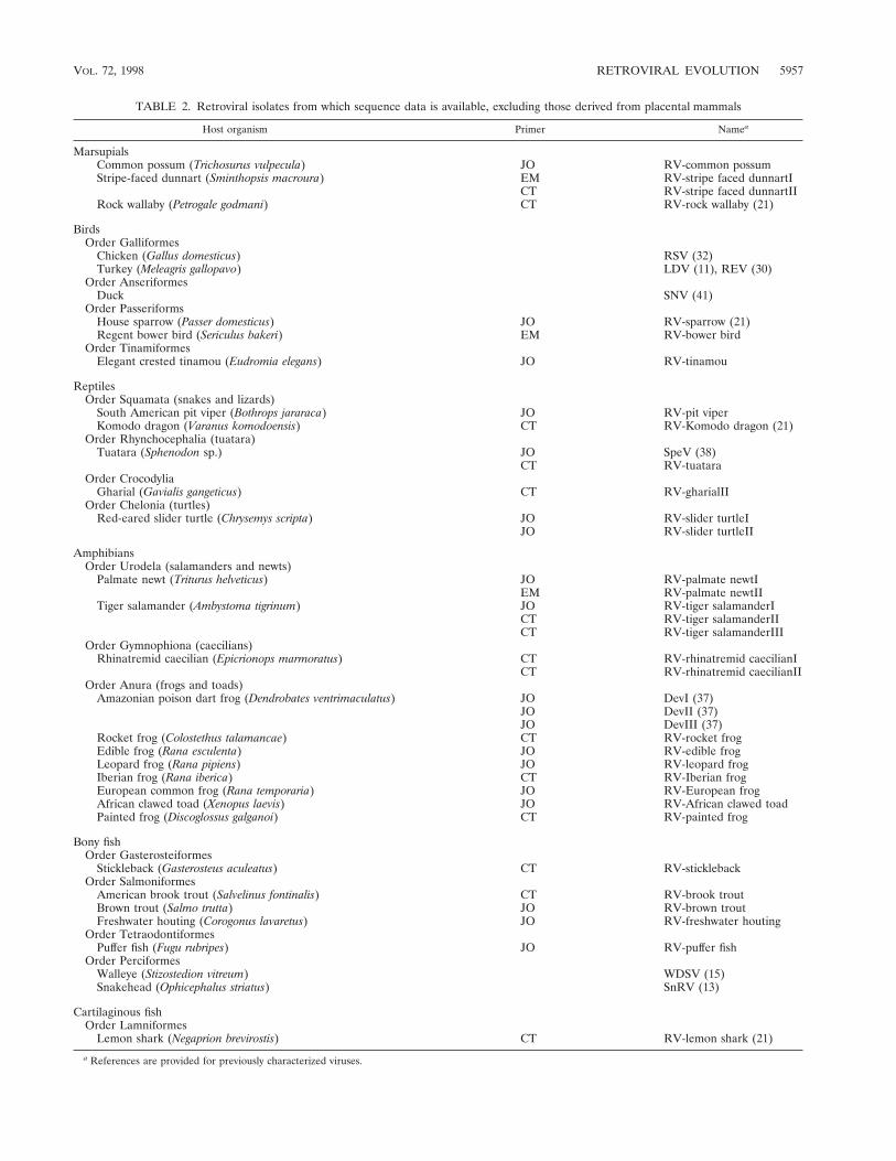
TABLE 2. Retroviral isolates from which sequence data is available, excluding those derived from placental mammals
Host organism Primer Namea
MarsupialsCommon possum (Trichosurus vulpecula) JO RV-common possumStripe-faced dunnart (Sminthopsis macroura) EM RV-stripe faced dunnartI
CT RV-stripe faced dunnartIIRock wallaby (Petrogale godmani) CT RV-rock wallaby (21)
BirdsOrder Galliformes
Chicken (Gallus domesticus) RSV (32)Turkey (Meleagris gallopavo) LDV (11), REV (30)
Order AnseriformesDuck SNV (41)
Order PasseriformsHouse sparrow (Passer domesticus) JO RV-sparrow (21)Regent bower bird (Sericulus bakeri) EM RV-bower bird
Order TinamiformesElegant crested tinamou (Eudromia elegans) JO RV-tinamou
ReptilesOrder Squamata (snakes and lizards)
South American pit viper (Bothrops jararaca) JO RV-pit viperKomodo dragon (Varanus komodoensis) CT RV-Komodo dragon (21)
Order Rhynchocephalia (tuatara)Tuatara (Sphenodon sp.) JO SpeV (38)
CT RV-tuataraOrder Crocodylia
Gharial (Gavialis gangeticus) CT RV-gharialIIOrder Chelonia (turtles)
Red-eared slider turtle (Chrysemys scripta) JO RV-slider turtleIJO RV-slider turtleII
AmphibiansOrder Urodela (salamanders and newts)
Palmate newt (Triturus helveticus) JO RV-palmate newtIEM RV-palmate newtII
Tiger salamander (Ambystoma tigrinum) JO RV-tiger salamanderICT RV-tiger salamanderIICT RV-tiger salamanderIII
Order Gymnophiona (caecilians)Rhinatremid caecilian (Epicrionops marmoratus) CT RV-rhinatremid caecilianI
CT RV-rhinatremid caecilianIIOrder Anura (frogs and toads)
Amazonian poison dart frog (Dendrobates ventrimaculatus) JO DevI (37)JO DevII (37)JO DevIII (37)
Rocket frog (Colostethus talamancae) CT RV-rocket frogEdible frog (Rana esculenta) JO RV-edible frogLeopard frog (Rana pipiens) JO RV-leopard frogIberian frog (Rana iberica) CT RV-Iberian frogEuropean common frog (Rana temporaria) JO RV-European frogAfrican clawed toad (Xenopus laevis) JO RV-African clawed toadPainted frog (Discoglossus galganoi) CT RV-painted frog
Bony fishOrder Gasterosteiformes
Stickleback (Gasterosteus aculeatus) CT RV-sticklebackOrder Salmoniformes
American brook trout (Salvelinus fontinalis) CT RV-brook troutBrown trout (Salmo trutta) JO RV-brown troutFreshwater houting (Corogonus lavaretus) JO RV-freshwater houting
Order TetraodontiformesPuffer fish (Fugu rubripes) JO RV-puffer fish
Order PerciformesWalleye (Stizostedion vitreum) WDSV (15)Snakehead (Ophicephalus striatus) SnRV (13)
Cartilaginous fishOrder Lamniformes
Lemon shark (Negaprion brevirostis) CT RV-lemon shark (21)
a References are provided for previously characterized viruses.
VOL. 72, 1998 RETROVIRAL EVOLUTION 5957

other, and only in those cases where homology was less than90% at the nucleotide level were both clones investigated fur-ther. Thirty novel endogenous retroviral fragments were char-acterized by sequencing the entire length of the amplified andcloned fragment. Of these, 3 were derived from nonplacentalmammals (marsupials and monotremes), 2 were from birds, 5were from reptiles, 14 were from amphibians, and 5 were frombony fish (Table 2). The retroviral isolates included represen-tatives from each of the four orders of reptiles and the threeorders of amphibians. Southern hybridization (32) of each ofthe fragments was performed to confirm its species of origin(data not shown). The lack of sufficient genomic DNA meantthat it was not possible to accurately determine the copy num-ber of many of the isolates.
Conceptual translations of the DNA sequence derived from
each isolate were performed, and a 160-amino-acid-residueregion of the reverse transcriptase protein was then aligned tothat of other previously described retroviruses, as described byXiong and Eickbush (45). Many of the sequences containedstop codons and frameshifts, which required the insertion ofone to several nucleotides (coded as unknown) in order tomaintain the reading frame (Fig. 2). This alignment was usedas the basis for the phylogenetic analyses. The majority of se-quence information from outside of this region was not suit-able for phylogenetic reconstruction due to the difficulty ofaligning homologous amino acid positions, although it waspossible to include a 30-amino-acid-residue region from theprotease protein. Both neighbor-joining and maximum parsi-mony trees were generated from the alignment, with supportfor individual branches investigated by bootstrap analysis.
FIG. 2. Amino acid alignment derived from retroviral reverse transcriptase proteins, based on that described by Xiong and Eickbush (45). Sequences identified inthis study are indicated in bold type. The underlined regions of the alignment were used in subsequent phylogenetic analysis. Asterisks represent in-frame stop codons,and missing data (due to frameshifts or deletions) are indicated by question marks. Three isolates, RV-brook trout, RV-brown trout, and RV-tiger salamanderI,contained large deletions and are not shown.
5958 HERNIOU ET AL. J. VIROL.

Figure 3a shows an unrooted, bootstrapped neighbor-joiningtree of the isolates shown in Fig. 2 (groups not recovered in atleast 50% of the bootstrap replicates were collapsed and arerepresented by polytomies). It was apparent from this analysisthat the retroviral sequences clustered into three main groups,each with bootstrap support greater than 80%.
One group (group I) contained the spumaviruses (humanspumavirus [HSV], simian foamy virus type 1 [SFV1], SFVL3,and FeSFV), the previously described Sphenodon endogenousretrovirus (SpeV, isolated from a reptile), snakehead fish ret-rovirus (SnRV), HERV.L (a recently identified endogenoushuman retrovirus), and MuERV.L (an endogenous murineretrovirus closely related to HERV.L), as well as several othernovel sequences derived from marsupials, birds, and amphib-ians. In contrast, group II contained viruses derived exclusivelyfrom mammalian and avian hosts and included all members offive of the seven currently recognized retroviral genera (thelentiviruses, the HTLV-bovine leukemia virus [BLV] group,
the ALVs, and the type B and D retroviruses), as well asseveral novel endogenous viruses. Investigation of 30 otheramphibian, reptilian, and piscine hosts with the EM primerfailed to reveal retroviral sequences related to this group.Group III contained the majority of the endogenous retroviralsequences isolated by our PCR screening, as well as all mem-bers of the murine leukemia virus (MLV) genus, exemplifiedby the feline leukemia virus (FeLV), gibbon ape leukemia virus(GaLV), human endogenous retrovirus type E (HERV.E), andbovine endogenous virus (BoEV). Members of the MLV genushave previously been identified in several species of birds andreptiles (4, 19, 29, 35, 42, 48) and, consistent with this, we alsoidentified elements clustering with the FeLV/GaLV/BoEV/HERV.E isolates in both of these vertebrate classes (data notshown). Several other previously described viruses were alsopresent within this lineage. These included the human endog-enous retroviruses ERV-9 and HERV.I (44), the recently de-scribed walleye dermal sarcoma virus (WDSV) from fish, and
FIG. 2—Continued.
VOL. 72, 1998 RETROVIRAL EVOLUTION 5959

the Dendrobates elements (DevI, DevII, and DevIII) from apoison dart frog. A BLAST search revealed that one furtherretroviral sequence should also be included. This element waspresent in a cosmid (accession no. U85A3) sequenced as partof the human genome mapping project (25). One further pointto note is that the entire nucleotide sequence of the WDSVisolate has already been determined and contains at least twonovel accessory genes (15). This, and other evidence, has led tothe suggestion that it probably represents a novel retroviralsubfamily or genus (15, 30).
Several gypsy LTR retrotransposon sequences (gypsy, del,Ty3, and micropia) were added to the alignment and used asoutgroups to root the retrovirus tree. In rooted phylogeneticanalyses, the group II sequences were recovered in a well-supported clade that is the sister group to all other retroviruses(Fig. 3b). Group III sequences also comprised a well-sup-ported clade. However, the addition of the outgroup sequencesabolished the bootstrap support for the group I spumaviruslikesequences shown in the unrooted analyses, breaking them up
into three well-supported clades that form a polytomy with thegroup III sequences. Support for this basal polytomy (59%) isalso uncompelling. Varying the composition of the outgroupsometimes resulted in weak bootstrap support (about 60%) forthe monophyly of group I or an association of the spumaviruseswith the SpeV, SnRV, RV-painted frog subgroup. Individualrooted neighbor-joining and parsimony trees did usually in-clude the group I sequences as a clade (Fig. 3c and d), but thismonophyly was also dependent on the composition of theoutgroup. The uncertainty of the relationships of the group Ispumaviruslike sequences close to the base of the retrovirustree and the weak support for the clade comprising group I andgroup III sequences must also limit confidence in the place-ment of the root of the Retroviridae.
The complex and diverse nature of retroviruses clusteringwith the MLV genus is emphasized in Fig. 3b. Most of therelationships within this lineage were not well supported bybootstrap analysis, and the reason for this lack of resolution isapparent from Fig. 3c and d: the distance from the base of the
FIG. 3. Phylogenetic trees of the retroviruses based on the alignment shown in Fig. 2, with the addition of 30 residues derived from the protease protein. Exogenousisolates are indicated by bold type. The genera to which particular retroviruses have been assigned are also shown. (a) Unrooted neighbor-joining bootstrap tree. Thefigures on each branch represent bootstrap support (from 1,000 replicates), with percentage values on only those branches with support greater than 50 being shown.Unresolved branches are shown as polytomies. (b) Neighbor-joining tree rooted on several gypsy LTR retrotransposon sequences (gypsy, Del, Ty3, and micropia), withfigures on each branch again showing percentage bootstrap support (1,000 replicates). (c) Rooted neighbor-joining tree with branch lengths proportional to the geneticdistance between the sequences. The tree is rooted as described in the legend to panel a. (d) Strict consensus of six equal-length trees identified by maximum parsimony.Bootstrap values were generated from 100 replicates (each of 25 random-sequence additions) by using the unordered matrix. The tree is rooted as described in thelegend to panel a.
5960 HERNIOU ET AL. J. VIROL.

group to the separation points of many of the taxa is smallwhen compared to their overall diversity. Despite this, it didappear that there were several well-supported subgroups whichcontained members derived from only a single vertebrate classor order. For example, many of the fish isolates clusteredtogether and groups of amphibian viruses were also observed.To determine whether this clustering was statistically signifi-cant and to try and increase the resolution of the various taxa,the group III viruses were then analyzed separately from theother retroviral sequences. This enabled trees to be generatedfrom an extended sequence alignment (257 as opposed to 190amino acids), as shown in Fig. 4. The degree to which virusesclustered on the basis of their host class of origin was assessedusing the program MacClade (20). The number of steps re-quired to generate the actual host class-virus associations
shown in Fig. 4 (nine) was always significantly lower than thatobserved for each of 100 test replicates in which the host classcharacters were shuffled randomly between the viral taxa(range, 18 to 24 steps; P , 0.01).
DISCUSSION
Retroviral phylogeny and taxonomy has for a long time beenbased almost exclusively on viruses within mammalian andavian hosts, and with the exception of several MLV-relatedretroviruses within reptiles (for which no sequence data havebeen reported), all members of the seven currently recognizedretroviral genera are present within one or both of these ver-tebrate classes (4, 9, 19, 48). We, and others, have previouslyobtained molecular data from a small number of reptilian,
FIG. 3—Continued.
VOL. 72, 1998 RETROVIRAL EVOLUTION 5961

amphibian, and piscine retroviruses (13, 15, 22, 38, 39); thosesequences, together with the ones in this report, create anemerging picture of the distribution and diversity of retrovi-ruses within lower vertebrate taxa. Endogenous retroviral se-quences have now been identified in more than 25 vertebrateorders across six classes, and this suggests that retroviruses maywell be ubiquitous in many vertebrate taxa, although theirexact host range remains to be determined. The most basalvertebrate from which a retroviral sequence has so far beenidentified is the lemon shark Negaprion brevirostris (22). Wethink it unlikely that this element represents a particularlyprimitive or basal virus for two reasons. First, it is closely
related to viruses within several different vertebrate classes,including HERV.I within humans. Second, rooted topologicalconstraint trees which placed RV-lemon shark (or all of theHERV.I-related retroviruses) basal to the other retrovirusesrequired at least an additional 17 steps over the shortest tree inmaximum parsimony analysis (data not shown). These pointsimply that the presence of HERV.I-related viruses in sharksmay be the result of horizontal transfer from another verte-brate class. However, numerous additional members of thisgroup will have to be identified and characterized before theirmode of dispersal within vertebrates can be definitively an-swered.
FIG. 3—Continued.
5962 HERNIOU ET AL. J. VIROL.

We screened four other (more primitive) vertebrate andchordate classes for the presence of endogenous retroviruseswithout success (data not shown). However, the number oftaxa investigated has so far been small; DNA samples wereavailable from only two species of lamprey (Cephalaspidomor-phi, Petromyzoniformes), one hagfish (Myxini, Myxiniformes),one lancelet (Cephalochordata, Branchiostomidae), and onesea squirt (Urochordata, Ascidiacea), and investigation of amuch larger number of primitive chordates is required beforethe absence of retroviruses in these groups can be consideredprobable. Furthermore, there is some evidence to suggest thatretroviruses may be present in nonvertebrate/chordate taxa:seasonal neoplasm in the soft-shelled clam (Mya arenaria) has
been linked to exogenous type B retroviruslike particles iden-tified in this species (27, 28). However, PCR screening of ninespecies of mollusc (including the soft-shelled clam) failed toisolate any endogenous retroviral sequences (data not shown).
Retroviral phylogenies have been reported previously by anumber of workers (6–8, 45). Many were constructed usingamino acid data sets derived from the 59 end of the reversetranscriptase protein, and the PCR-amplified retroviral frag-ments described here contain most of this region. Phylogeneticreconstruction has generally resulted (when the trees arerooted) in the placement of the spumaviruses (here shownwithin group I) and MLVs (within group III) as the sister taxonto a (group II) lineage containing the other five retroviral
FIG. 3—Continued.
VOL. 72, 1998 RETROVIRAL EVOLUTION 5963

genera (lentiviruses, HTLV-BLV group, ALVs, and type B andD retroviruses). Our phylogenetic analyses are consistent withthis view of retroviral phylogeny but highlight the uncertaintyof the interrelationships of the group I spumaviruslike se-quences and the root of the retrovirus tree.
Despite screening a large number of lower vertebrate taxa,no endogenous retroviral sequences were identified which clus-tered with the lentiviruses (equine infectious anemia virus[EIAV], ovine maedi-visna virus [OMVV], and simian immu-nodeficiency virus SIVmac), the HTLV-BLV group, or theALVs (Rous sarcoma virus [RSV]). Indeed, the only sequenceswhich were placed with other group II viruses were basal to themurine type B (mouse mammary tumor virus [MMTV]) andmammalian type D (jaagsiekte and simian retrovirus type 1[SRV1]) retroviruses, being placed in a polytomy with HERV.K10, RSV, lymphoproliferative disease virus (LDV), and in-tracisternal type-A retroviral particle (IAP-hamster). The ab-sence of isolates derived from nonmammalian or avian hostssuggests that endogenous retroviruses related to these fivegenera are likely to be rare in lower vertebrate genomes. Wethink it unlikely that this distribution is simply the result oftarget sequences being missed during amplification for severalreasons. (i) The EM primer was designed against a sequencemotif conserved in each of the five genera, (ii) negative resultsusing EM were obtained with more than 30 species of reptiles,amphibians, and fish, (iii) the EM primer gave positive resultswith mammalian and avian taxa, and (iv) the JO and CTprimers have previously cross-amplified group II-related vi-ruses from mammalian genomes (37) but failed to do so whenused against lower vertebrate taxa. The distribution of thegroup II sequences in mammals and birds is interesting, par-ticularly given their apparent absence from reptilian relativesof birds. This suggests that their distribution may involve eitherdispersal across significant taxonomic distances or possibly theextinction of some taxa.
The spumaviruses (human spumavirus [HSV], two simianfoamy viruses [SFV and SFVL3], and FeSFV) are exogenous
viruses of primates and other mammals (4). There have beenprevious reports of two endogenous retroviruses which clusterwith this genus, SpeV from the reptile tuatara (39) andHERV.L, a recently identified human isolate (5). It has beensuggested that HERV.L represents a possible ancestor of theexogenous mammalian spumaviruses (5). Our phylogeneticanalyses suggest that viruses distantly related to the spumavi-ruses may be widespread in vertebrates: unrooted trees (Fig.3a) showed strong support for a group containing the spuma-viruses and novel isolates derived from birds, reptiles, amphib-ians, and fish. However, the spumaviruses are unlikely to haveemerged from HERV.L or a closely related endogenous virus,because this element appears more closely related to severalviruses derived from nonmammalian hosts. The piscine mem-ber of the spumaviruslike lineage, SnRV, is an exogenousretrovirus which was originally isolated from striped snakeheadfish (10). It has previously been reported to group (albeitdistantly) with the MLVs (13), but the inclusion of additionalretroviral isolates (which were not available to previous work-ers) suggests this may not be correct. It is our opinion, how-ever, that none of the viruses described here should be as-signed to the spumavirus genus. Although a spumaviruslikegroup was well supported by bootstrap analysis in unrootedphylogenies, this was not the case when several gypsy LTRretrotransposon sequences were included for rooting purposes(Fig. 3b). Furthermore, the monophyly of this group withinindividual rooted neighbor-joining and maximum parsimonytrees (Fig. 3c and d) was outgroup dependent; varying thecomposition of the outgroup taxa sometimes resulted in theplacement of the spumaviruslike viruses into two or three sep-arate groups. These factors suggest that the spumavirus genusis only distantly related to the other group I viruses describedhere. Consistent with this, SnRV, HERV.L, and the spumavi-ruses are known to have significant differences in genomicorganization when compared to each other and to other ret-roviral genera (5, 13). We were also unable to detect obvious
FIG. 4. Unrooted maximum parsimony tree of the group III viruses shown in Fig. 3. Branch lengths are proportional to the number of changes required to generatethe observed variation. Numbers on each branch reflect percentage bootstrap support using maximum parsimony (upper value) or neighbor-joining (lower value).
5964 HERNIOU ET AL. J. VIROL.

sequence homology between the accessory proteins of SnRVand those encoded by the spumaviruses (data not shown).
The vast majority of endogenous lower vertebrate virusescharacterized during this study (group III) clustered with theMLV-related genus. MLV-like viruses are present in numer-ous mammalian species and have also been described in birds(such as the spleen necrosis virus [SNV] and reticuloendothe-liosis virus [REV] of domestic fowl) and several reptiles, in-cluding the corn snake and Russell’s viper (3, 19, 29, 31, 48).Several other groups of endogenous viruses (such as theHERV.I-related viruses and ERV-9 [also present within hu-mans]) cluster with these viruses (17, 21, 44). Thus, membersof this retroviral genus are already known to be present withinseveral vertebrate classes. However, the phylogenetic treesshown in Fig. 3 demonstrate that the lineage containing theMLV genus is extremely complex. A polytomy, consisting of 13lineages (one of which contained MLV/ERV-9/HERV.I-re-lated viruses), was left unresolved by bootstrap analyses. Sev-eral of these lineages contained a single retroviral sequence,such as the previously described WDSV of fish (15), whereasothers contained groups with several members. We believethat many of these lineages are likely to represent novel ret-roviral genera. Although other factors, in addition to sequencedivergence (such as the presence of novel genes and alternativeprimer binding site homology), are required for genus-levelclassification (4), these may well become apparent when thecomplete nucleotide sequences of these viruses are deter-mined. For example, it has already been suggested that WDSVrepresents a novel retroviral genus because it encodes twoaccessory proteins which are absent from members of theMLV genus and (unusually) utilizes a primer binding site ho-mologous to tRNAHis (15, 23).
Although most of the retroviruses clustering in this region ofthe tree (excluding those present within the MLV genus) werederived from reptiles, amphibians, and fish, it was apparentthat two novel mammalian viruses were also present. We orig-inally isolated the first of these (RV-horse) when screeningmammalian genomes but were unable at the time to determineits phylogenetic relationship to other retroviruses. The secondelement, H-cosmid(U85A3), was identified from a BLASTsearch of the EMBL/GenBank/DDBJ data banks and was orig-inally sequenced as part of the human genome project (25).Our sequence analysis of this element suggests it contains afull-length polymerase gene, part of the envelope region, andthat the gag gene has probably been replaced by a later inser-tion (in the reverse orientation) of an HERV-H-like element(unpublished results). We also identified a second element (byBLAST searches), closely related to H-cosmid(U85A3) on theX chromosome within cosmid HS49L23 (12). These resultsimply that other highly divergent endogenous retroviruses re-main to be discovered in both humans and other mammals.
Finally, the distribution and phylogeny of the sequencesshown in Fig. 3 (especially those within group III) is intriguingas it suggests that some of the retroviral groups may be re-stricted to particular vertebrate classes, indicating that inter-class horizontal transmission may be a rare event for certaintypes of retrovirus. Although interpretation of these treesshould be cautious (due to the lack of robust support for manyof the relationships), the overall tree topologies are consistentwith this possibility. For example, the majority of endogenouspiscine retroviruses appear to be monophyletic, even thoughtheir hosts are classified into several different taxonomic or-ders. This also appears to be the case for many of the reptilianand amphibian retroviruses. Tests using the program Mac-Clade also demonstrated that the host class-virus associationsshown in Fig. 4 are phylogenetically correlated. We are cur-
rently trying to obtain a better idea of interclass transmissionrates by looking in detail at the phylogeny and distribution ofthe MLV genus.
ACKNOWLEDGMENTS
We are indebted to D. Swofford for permission to publish resultsusing PAUP4. We thank M. Clark, A. Fergusson, A. Flavell, J. Gatesy,P. Holland, R. Kusmierski, D. R. Martin, N. Okada, G. Olbricht,J. Pino-Perez, and R. Waugh-O’Neill for providing some of the geno-mic DNA samples used in this study.
E.H. thanks Universite Paris XI for a DEA scholarship. M.T. andJ.C. are supported by the NERC Taxonomy Initiative and the RoyalSociety. This work was supported in part by an NERC grant (GST/02/852) to M.W.
REFERENCES1. Altschul, S. F., W. Gish, W. Miller, E. W. Myers, and D. L. Lipman. 1990.
Basic alignment search tool. J. Mol. Biol. 215:403–410.2. Benit, L., N. de Parseval, J. F. Casella, I. Callebaut, A. Cordonnier, and T.
Heidmann. 1997. Cloning of a new murine endogenous retrovirus, MuERV-L, with strong similarity to the human HERV-L element and with a gagcoding sequence closely related to the Fv1 restriction gene. J. Virol. 71:5652–5657.
3. Chen, Y. C., Z. Cui, L. F. Lee, and R. L. Witter. 1987. Serological differencesamong non-defective reticuloendotheliosis viruses. Arch. Virol. 93:233–245.
4. Coffin, J. M. 1992. Structure and classification of retroviruses, p. 19–49. InJ. A. Levy (ed.), The Retroviridae, vol. 1. Plenum Press, New York, N.Y.
5. Cordonnier, A., J.-P. Casella, and T. Heidmann. 1995. Isolation of novelhuman endogenous retrovirus-like elements with foamy virus-related polsequence. J. Virol. 69:5890–5897.
6. Doolittle, R. F., D.-F. Feng, M. S. Johnson, and M. A. McClure. 1989. Originsand evolutionary relationships of retroviruses. Quart. Rev. Biol. 64:1–30.
7. Doolittle, R. F., D.-F. Feng, M. A. McClure, and M. S. Johnson. 1990.Retrovirus phylogeny and evolution. Curr. Top. Microbiol. Immunol. 157:1–18.
8. Eickbush, T. H. 1994. Origin and evolutionary relationships of retroele-ments, p. 121–157. In S. S. Morse (ed.), The evolutionary biology of viruses.Raven Press, New York, N.Y.
9. Francki, R. I. B., C. M. Fauquet, D. L. Knudson, and F. Brown (ed.). 1991.Classification and nomenclature of viruses. Fifth report of the InternationalCommittee on Taxonomy of Viruses. Arch. Virol. 1991(Suppl. 2):290–298.
10. Frerichs, G. N., D. Morgan, D. Hart, C. Skerrow, R. J. Roberts, and D. E.Onions. 1991. Spontaneously productive C-type retrovirus infection of fishcell lines. J. Gen. Virol. 72:2537–2539.
11. Gak, E., A. Yanev, L. Sherman, M. Ianconescu, S. R. Tronick, and A. Gazit.1991. Lymphoproliferative disease virus of turkeys: sequence analysis andtranscriptional activity of the long terminal repeat. Gene 99:157–162.
12. Grafham, D. Unpublished data.13. Hart, D., N. Frerichs, A. Rambaut, and D. E. Onions. 1996. Complete
nucleotide sequence and transcriptional analysis of the snakehead fish ret-rovirus. J. Virol. 70:3606–3616.
14. Helps, C. R., and D. A. Harbour. Unpublished data.15. Holzschu, D. L., D. Martineau, S. K. Fodor, V. M. Vogt, P. R. Bowser, and
J. W. Casey. 1995. Nucleotide sequence and protein analysis of a complexpiscine retrovirus, walleye dermal sarcoma virus. J. Virol. 69:5320–5331.
16. Kim, A., C. Terzian, P. Santamaria, A. Pelisson, N. Prud’homme, and A.Bucheton. 1994. Retroviruses in invertebrates: the gypsy retrotransposon isapparently an infectious retrovirus of Drosophila melanogaster. Proc. Natl.Acad. Sci. USA 91:1265–1269.
17. La Mantia, G., D. Maglione, G. Pengue, A. Di Cristofano, A. Simeone, L.Lanfrancone, and L. Lania. 1991. Identification and characterization ofnovel human endogenous retroviral sequences preferentially expressed inundifferentiated embryonal carcinoma cells. Nucleic Acids Res. 19:1513–1520.
18. Lankenau, D. H., P. Huijser, E. Jansen, K. Miedema, and W. Hennig. 1990.DNA sequence comparison of micropia transposable elements from Dro-sophila hydei and Drosophila melanogaster. Chromosoma 99:111–117.
19. Lunger, P. D., W. D. Hardy, and H. F. Clark. 1974. C type virus particles ina reptilian tumor. J. Natl. Cancer Inst. 52:1231–1235.
20. Maddison, W. P., and D. R. Maddison. 1992. MacClade: analysis of phylog-eny and character evolution, version 3.0. Sinauer Associates, Sunderland,Mass.
21. Maeda, N. 1985. Nucleotide sequence of the haptoglobin and haptoglobin-related gene pair. J. Biol. Chem. 260:6698–6709.
22. Martin, J., E. Herniou, J. Cook, R. Waugh O’Neill, and M. Tristem. 1997.Human endogenous retrovirus type I-related viruses have an apparentlywidespread distribution within vertebrates. J. Virol. 71:437–443.
23. Martineau, D., P. R. Bowser, R. R. Renshaw, and J. W. Casey. 1992. Mo-lecular characterization of a unique retrovirus associated with a fish tumor.J. Virol. 66:596–599.
VOL. 72, 1998 RETROVIRAL EVOLUTION 5965

24. Masahito, P., M. Nishioka, H. Ueda, Y. Kato, I. Yamazaki, K. Nomura, H.Sugano, and T. Kitagawa. 1995. Frequent development of pancreatic carci-nomas in the Rana nigromaculata group. Cancer Res. 55:3781–3784.
25. Odell, C. Unpublished data.26. Ono, M., T. Yasunaga, T. Miyata, and H. Ushikubo. 1986. Nucleotide se-
quence of human endogenous retrovirus genome related to the mouse mam-mary tumor virus genome. J. Virol. 60:589–598.
27. Oprandy, J. J., and P. W. Chang. 1983. 5-Bromodeoxyuridine induction ofhematopoietic neoplasia and retrovirus activation in the soft-shelled clam,Mya arenaria. J. Invertebr. Pathol. 42:196–206.
28. Oprandy, J. J., P. W. Chang, A. D. Pronovost, K. R. Cooper, R. S. Brown, andV. J. Yates. 1981. Isolation of a viral agent causing hematopoietic neoplasiain the soft-shelled clam, Mya arenaria. J. Invertebr. Pathol. 38:45–51.
29. Payne, L. N. 1992. Biology of avian retroviruses, p. 299–404. In J. A. Levy(ed.), The Retroviridae, vol. 1. Plenum Press, New York, N.Y.
30. Poulet, F. M., P. R. Bowser, and J. W. Casey. 1994. Retroviruses of fish,reptiles and molluscs, p. 1–38. In J. A. Levy (ed.), The Retroviridae, vol. 3.Plenum Press, New York, N.Y.
31. Purchase, H. G., C. Ludford, K. Nazerian, and H. W. Cox. 1973. A new groupof oncogenic viruses: reticuloendotheliosis viruses, chick syncytial, duck in-fectious anemia, and spleen necrosis viruses. J. Natl. Cancer Inst. 51:489–499.
32. Sambrook, J., E. Fritsch, and T. Maniatis. 1989. Molecular cloning: a lab-oratory manual, 2nd ed. Cold Spring Harbor Laboratory Press, New York,N.Y.
33. Schwartz, D. E., R. Tizard, and W. Gilbert. 1983. Nucleotide sequence ofRous sarcoma virus. Cell 32:853–869.
34. Shih, A., R. Misra, and M. G. Rush. 1989. Detection of multiple, novelreverse transcriptase coding sequences in human nucleic acids: relation toprimate retroviruses. J. Virol. 63:64–75.
35. Stephens, R. M., N. R. Rice, R. R. Hiebsch, H. R. Bose, and R. V. Gilden.1983. Nucleotide sequence of v-rel—the oncogene of reticuloendotheliosisvirus. Proc. Natl. Acad. Sci. USA 80:6229–6233.
36. Temin, H. M. 1989. Retrovirus variation and evolution. Genome 31:17–22.37. Tristem, M. 1996. Amplification of divergent retroelements by PCR. Bio-
techniques 20:608–612.38. Tristem, M., E. Herniou, K. Summers, and J. Cook. 1996. Three retroviral
sequences in amphibians are distinct from those in mammals and birds.J. Virol. 70:4864–4870.
39. Tristem, M., T. Myles, and F. Hill. 1995. A highly divergent retroviralsequence in the tuatara (Sphenodon). Virology 210:206–211.
40. Varmus, H., and P. Brown. 1989. Retroviruses, p. 53–108. In D. E. Berg andM. M. Howe (ed.), Mobile DNA. American Society for Microbiology, Wash-ington, D.C.
41. Wain-Hobson, S. 1994. Is antigenic variation of HIV important for AIDS,and what might be expected in the future, p. 185–209. In S. S. Morse (ed.),The evolutionary biology of viruses. Raven Press, New York, N.Y.
42. Weaver, T. A., K. J. Talbot, and A. T. Panganiban. 1990. Spleen necrosisvirus Gag polyprotein is necessary for particle assembly and release but notfor proteolytic processing. J. Virol. 64:2642–2652.
43. Wichman, H. A., and R. A. Van den Bussche. 1992. In search of retrotrans-posons—exploring the potential of the PCR. Biotechniques 13:258–264.
44. Wilkinson, D. A., D. L. Mager, and J.-A. C. Leong. 1994. Endogenous humanretroviruses, p. 465–535. In J. A. Levy (ed.), The Retroviridae, vol. 3. PlenumPress, New York, N.Y.
45. Xiong, Y., and T. H. Eickbush. 1990. Origin and evolution of retroelementsbased upon their reverse transcriptase sequences. EMBO J. 9:3353–3362.
46. York, D. F., R. Vigne, D. W. Verwoerd, and G. Querat. 1992. Nucleotidesequence of the jaagsiekte retrovirus, an exogenous and endogenous type Dand B retrovirus of sheep and goats. J. Virol. 66:4930–4939.
47. Young, A. Z. 1981. The life of vertebrates, 3rd ed. Clarendon Press, Oxford,United Kingdom.
48. Ziegel, R. F., and H. F. Clark. 1969. Electron microscopic observations on a“C”-type virus in cell cultures derived from a tumor bearing viper. J. Natl.Cancer Inst. 43:1097–1101.
5966 HERNIOU ET AL. J. VIROL.


