Embed Size (px)
Citation preview
Epstein–Barr Virus (EBV) Rta-Mediated EBV and Kaposi’sSarcoma-Associated Herpesvirus Lytic Reactivations in293 CellsYen-Ju Chen1,2, Wan-Hua Tsai2, Yu-Lian Chen2, Ying-Chieh Ko2, Sheng-Ping Chou2, Jen-Yang Chen1,2*,
Su-Fang Lin2*
1 College of Medicine, Graduate Institute of Microbiology, National Taiwan University, Taipei, Taiwan, 2 National Institute of Cancer Research, National Health Research
Institutes, Miaoli County, Taiwan
Abstract
Epstein–Barr virus (EBV) Rta belongs to a lytic switch gene family that is evolutionarily conserved in all gamma-herpesviruses. Emerging evidence indicates that cell cycle arrest is a common means by which herpesviral immediate-earlyprotein hijacks the host cell to advance the virus’s lytic cycle progression. To examine the role of Rta in cell cycle regulation,we recently established a doxycycline (Dox)-inducible Rta system in 293 cells. In this cell background, inducible Rtamodulated the levels of signature G1 arrest proteins, followed by induction of the cellular senescence marker, SA-b-Gal. Todelineate the relationship between Rta-induced cell growth arrest and EBV reactivation, recombinant viral genomes weretransferred into Rta-inducible 293 cells. Somewhat unexpectedly, we found that Dox-inducible Rta reactivated both EBV andKaposi’s sarcoma-associated herpesvirus (KSHV), to similar efficacy. As a consequence, the Rta-mediated EBV and KSHV lyticreplication systems, designated as EREV8 and ERKV, respectively, were homogenous, robust, and concurrent with cell deathlikely due to permissive lytic replication. In addition, the expression kinetics of EBV lytic genes in Dox-treated EREV8 cellswas similar to that of their KSHV counterparts in Dox-induced ERKV cells, suggesting that a common pathway is used todisrupt viral latency in both cell systems. When the time course was compared, cell cycle arrest was achieved between 6 and48 h, EBV or KSHV reactivation was initiated abruptly at 48 h, and the cellular senescence marker was not detected until120 h after Dox treatment. These results lead us to hypothesize that in 293 cells, Rta-induced G1 cell cycle arrest couldprovide (1) an ideal environment for virus reactivation if EBV or KSHV coexists and (2) a preparatory milieu for cellsenescence if no viral genome is available. The latter is hypothetical in a transient-lytic situation.
Citation: Chen Y-J, Tsai W-H, Chen Y-L, Ko Y-C, Chou S-P, et al. (2011) Epstein–Barr Virus (EBV) Rta-Mediated EBV and Kaposi’s Sarcoma-Associated HerpesvirusLytic Reactivations in 293 Cells. PLoS ONE 6(3): e17809. doi:10.1371/journal.pone.0017809
Editor: Micah Luftig, Duke University Medical Center, United States of America
Received November 10, 2010; Accepted February 11, 2011; Published March 10, 2011
Copyright: � 2011 Chen et al. This is an open-access article distributed under the terms of the Creative Commons Attribution License, which permitsunrestricted use, distribution, and reproduction in any medium, provided the original author and source are credited.
Funding: This study was supported by Taiwan NHRI CA-099-PP-17 and Department of Health DOH99-TD-C-111-004 to S.-F. Lin; NHRI CA-099-PP-13, NationalScience Council NSC98-3112-B-400-002, and NSC99-3112-B-400-009 to J.-Y. Chen. The funders had no role in study design, data collection and analysis, decision topublish, or preparation of the manuscript.
Competing Interests: The authors have declared that no competing interests exist.
* E-mail: [email protected] (S-FL); [email protected] (J-YC)
Introduction
Human oncogenic herpesviruses such as Epstein–Barr virus
(EBV) and Kaposi’s sarcoma-associated herpesvirus (KSHV) are
closely linked to a variety of malignancies including nonkeratiniz-
ing nasopharyngeal carcinoma, gastric adenocarcinoma, Burkitt’s
lymphoma, Kaposi’s sarcoma, primary effusion lymphoma,
multicentric Castleman’s disease, and various forms of lympho-
proliferative disorders. Both EBV and KSHV are latent residents
in B lymphocytes and show sporadic reactivation in lymphoepi-
thelial tissues such as tonsils [1,2,3]. Lytic reactivation of EBV or
KSHV in epithelial cells of the nasopharynx is strongly influenced
by the state of differentiation [4,5,6]. In addition, XBP-1s, a
product of the master gene responsible for B cell differentiation,
was recently suggested to be one of the physiological stimuli that
trigger the lytic switch of EBV and KSHV in latently infected B
cells [7,8].
For a cycling cell, growth arrest in the G1 phase implies one of the
following fates to choose: quiescence (re-enters proliferation at a
later time), apoptosis, differentiation or senescence [9,10]. Among
these four outcomes, differentiation and senescence share two
features in common: dramatic chromosome remodeling [11] and
lengthy development time (usually days). Cell senescence is a
biochemical process exhibited by metabolically active cells whose
cell cycles are frozen beyond the restriction point in G1 phase. First
identified in in vitro cultured cells, cellular senescence occurs both in
primary and cancer cell lines [12,13]. In addition, the limit in
proliferative capacity triggered by aberrant mitogenic signals of
oncogenes, known as oncogene-induced senescence (OIS), is an
alternative tumor suppressive mechanism that has been recently
validated in vivo [14,15]. Senescence not only occurs in pre-
malignant cells, but also appears in malignant tumors. In the latter
case, senescence was usually produced by the removal of an essential
oncogenic stimulus or the restoration of a tumor suppressor. For
example, ablation of c-Myc in transgenic mouse models induced
rapid tumor regression associated with senescence [16]; systemic
expression of a dominant-interfering Myc mutant in a preclinical
mouse model with Ras-initiated lung adenocarcinoma triggered
rapid tumor regression accompanied by senescence [17].
PLoS ONE | www.plosone.org 1 March 2011 | Volume 6 | Issue 3 | e17809

Lytic replication of herpesvirus occurs preferentially in the G1
phase of the cell cycle [18]. Accumulating evidence indicates that a
number of viral immediate-early proteins actively exert a growth-
arrest function by which the virus induces a less competitive
environment for resources required for viral DNA replication. In
this regard, when EBV undergoes lytic replication, host cells are
protected from apoptosis and the DNA-synthetic machinery is
blocked, although the activities of certain S-phase regulators
increase [19,20]. In addition, upon viral infection, ICP0 of herpes
simplex virus induces cell cycle arrest in G1 by both p53-mediated
and p53-independent pathways [21]. Through the up-regulation
of cyclins E and D, the IE2 protein of human cytomegalovirus
potently arrests U373 cells and simultaneously blocks cellular
DNA synthesis [22]. Conceivably, G1 phase is not only a pivotal
stage for cell fate determination [9,10], but also is critical for virus
fate, namely to maintain latency or to initiate a lytic replication
episode.
Among the identified immediate-early molecules, RTA (repli-
cation and transcription activator) is positionally and structurally
conserved among the genomes of all gamma-herpesviruses.
Ectopic expression of the EBV Rta in epithelial or B cells is
capable of efficiently inducing the lytic cycle of EBV [23,24].
Similarly, ectopic expression of KSHV RTA (K-RTA) in B or
endothelial cells latently infected with KSHV leads to the
successive expression of KSHV early and late genes [25,26]. We
recently established doxycycline-inducible system of Rta in 293
cells, nasopharyngeal carcinoma cells (NPC-TW01), and laryngeal
carcinoma cells (HEp-2). We found that, in the absence of BZLF1
and other EBV viral proteins, Rta alone can promote irreversible
G1 arrest followed by cellular senescence in these epithelial cells
([27] and unpublished). In the present study, we further
demonstrate that in 293 cells, the doxycycline-inducible Rta not
only reactivates EBV but also KSHV, to similar efficacy. While the
precise mechanism of Rta-mediated KSHV reactivation is
currently not resolved, results from comparative kinetics studies
strongly indicate a casual role of Rta-induced G1 arrest in EBV
and KSHV reactivations.
Results
EBV Rta alone is sufficient to initiate and complete lyticEBV replication in EREV8 cells
EBV Rta alone is known to disrupt EBV latency in both
epithelial and B cells [23,24]. 293TetER is a recently established
293 cell line that displays doxycycline (Dox) -controlled,
conditional expression of EBV Rta [27]. To confirm whether
the expression of EBV Rta in 293TetER cells is sufficient to
promote EBV lytic replication from the latent stage, rAkata-G418
EBV genome was transferred into 293TetER cells, yielding an
EREV8 derivative line [28]. To measure the induction rate of the
EBV lytic cycle triggered by EBV Rta, Dox (50 ng/ml)-treated
EREV8 cells were analyzed using an immunofluorescence assay
and flow cytometry. As expected, the immunofluorescence assay
showed a very high and homogenous expression of transgene Flag-
EBV Rta (Figure 1A), and flow cytometry showed that <76% of
the cells were positive. Similarly, considerable expression of
immediate-early protein BZLF1 (<82%) and late glycoprotein
protein BALF4/gB (<50%) were detected in the Dox-treated
EREV8 cells (Figure 1A). Next, the expression kinetics of a panel
of lytic proteins including BZLF1, BMRF1, BHRF1, and
membrane protein gp350/220 in EREV8 cells were compared
in parallel using western blot analysis. Although the degree of
antibody affinity may vary, the overall kinetics of the different
proteins was distinguishable (Figure 1B). The optimal expressions
for each protein were in a hierarchical order: namely Flag-Rta
(24–48 h), EBV immediate-early protein BZLF1 (48–72 h), early
proteins BMRF1 and BHRF1 (72–144 h), and late membrane
protein gp350/220 (120–144 h). Finally, the quantity of EBV
genome equivalents encapsidated in the viral particles released
from Dox-treated EREV8 cells at each time points were
determined by comparative quantitative PCR (q-PCR). As shown
in Figure 1C, the Dox-treated EREV8 cells continued to produce
viral particles in an increasing manner until 144 h. Noticeably, at
96 h after induction, a subpopulation of cells started to round up
and seemed to be full of granules. Anoikis-like detachment of cells
from the petri dish was observed at later times (detailed below).
These observations are reminiscent of proficient permissive
replication of bacteriophage in E. coli.
To compare the ‘‘throughput’’ of lytic cycle replications
initiated by the present system with that by conventional method
(20 ng/ml 12-O-tetradecanoylphorbol-13-acetate (TPA) plus
3 mM butyrate), EREV8 cells treated with either method were
performed simultaneously for a course of 96 h, a time when most
of the chemical-treated cells were dead. Of note, in an
immunofluorescence assay, while closed to 70% of Dox-treated
cells expressed Flag-Rta at 24 h, only 43% of the chemical-treated
EREV8 cells responded to TPA/butyrate (i.e. BZLF1 positive),
indicating the presence of a considerable refractory subpopulation
in the traditional induction system. For each time point, the
expressions of EBV immediate-early protein Rta, BZLF1, and
early protein BMRF1 were revealed by western blot analysis
(Figure 2A) and the yields of encapsidated viral particles were
determined by comparative q-PCR (Figure 2B). Interestingly,
while Rta was detectable as early as 6 h and increasingly
augmented until 48 h in the Dox-treated cells, Rta was only
transiently upregulated at 24 h in the TPA/butyrate-treated cells.
By contrast, the expressions of BZLF1 and BMRF1 were
significantly augmented in the chemical-treated cells than in the
Dox-treated group by 48 h. Furthermore, although TPA/
butyrate-induced EREV8 cells appeared to shed EBV particles
at earlier time (24 h), the overall viral yields in the Dox-treated cell
were <1.5-higher than that in the chemical-treated cells at 96 h
(Figure 2B). In addition, since the Dox-treated cells may continue
to shed viral particles at an increasingly manner until 196 h
(Figure 1C), it is estimated that from same number of EREV8
cells, the viral particles produced by Dox treatment will be at least
4-fold more than that by chemical induction. Taken together,
these results indicated that although TPA/butyrate provided a
faster and stronger stimulus for EBV reactivation in the EREV8
cells, yet the induction rate was poorer, the cells were sicker and
the viral yield was lower than those triggered by Dox-inducible
Rta.
EBV Rta alone is sufficient to initiate and complete lyticKSHV replication in ERKV cells
In the course of establishing EREV8, a control experiment was
performed in which rKSHV.219 [29] was used to infect
293TetER cells and served to differentiate the specificity of EBV
Rta for its cognate viral genome, as described previously [25].
rKSHV.219 carries a genetic cassette that can be used to
distinguish the stages of viral infection in the host cells: latent
(green fluorescence) and lytic (red fluorescence) [29]. At first, we
expected that Dox-induced EBV Rta would reactivate EBV but
not KSHV genomes residing in the 293TetER cells. Somewhat
surprisingly, upon the administration of Dox, a number of
293TetER cell clones harboring rKSHV.219 genomes exhibited
strong red fluorescence, indicating lytic replication (Figure 3A). By
contrast, only a low percentage of Dox-treated control 293Tet cells
EBV Rta-Mediated EBV and KSHV Reactivation
PLoS ONE | www.plosone.org 2 March 2011 | Volume 6 | Issue 3 | e17809
harboring rKSHV.219 genome exhibited red fluorescence
(Figure 3A, 293Tet_rKSHV_C1), suggesting that EBV Rta was
the determinant that triggered rKSHV.219 lytic cycle replication.
To verify these observations further, five 293_TetER_rKSHV.219
cell clones were expanded, pooled, and collectively designated as
ERKV. Stable latent infection of rKSHV.219 in ERKV cells was
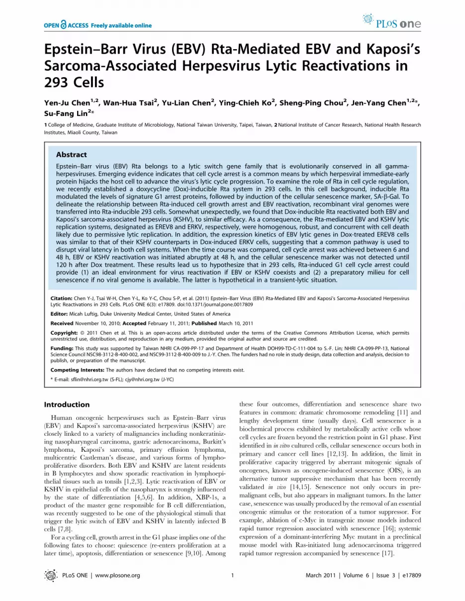
Figure 1. Reactivation of EBV lytic replication by EBV Rta in EREV8 cells. (A) Individual induction efficiency of Flag-EBV Rta (Flag), BZLF1, andlate glycoprotein BALF4/gB were shown by an immunofluorescence assay. Untreated (–) or EREV8 cells treated with doxycycline (Dox) for 48 h wereanalyzed in parallel. Cells with immuno-positivity were quantified by flow cytometry and indicated in percentages for each detection. (B) Expressionkinetics of EBV lytic proteins including BZLF1, BMRF1, BHRF1, and gp350/220 in Dox-induced EREV8 cells for the indicated times were analyzed bywestern blot analysis. b-actin served as a loading control. C6 and C72 indicate untreated cells at 6 h and 72 h, respectively. (C) Titration of viralparticles released from 72–144 h Dox-treated EREV8 cells. Copy numbers of DNase I-resistant, encapsidated viral DNAs were determined bycomparative quantitative PCR of EBV DNA polymerase gene (BALF5) using serial dilutions of Raji DNA as standards. Data are presented as means6SDfrom six independent PCR assays.doi:10.1371/journal.pone.0017809.g001
Figure 2. Comparative studies of EBV reactivation induced by Dox (50 ng/ml) vs. conventional chemical method (20 ng/ml TPAplus 3 mM butyrate) in EREV8 cells. (A) Expression kinetics of EBV Rta, BZLF1 at 6, 24, and 48 h in cells treated with indicated inducers. b-actinserved as a loading control. (B)Viral particles released from Dox-treated or TPA/butyrate-treated EREV8 cells. Data are presented as means6SD fromfour independent PCR assays.doi:10.1371/journal.pone.0017809.g002
EBV Rta-Mediated EBV and KSHV Reactivation
PLoS ONE | www.plosone.org 3 March 2011 | Volume 6 | Issue 3 | e17809
achieved by puromycin selection using previously described
procedures [29]. Next, the expression kinetics of KSHV
immediate-early protein K-RTA, early protein K-bZIP, and late
protein K8.1 were studied by western blot analysis. Again, the
overall expression pattern of these four molecules could be
arranged in a cascade manner by their respective peak times:
namely Flag-EBV Rta (24–48 h), immediate-early K-RTA and K-
bZIP (48–72 h), and late glycoprotein K8.1 (72–96 h) (Figure 3B).
Interestingly, expression of the three KSHV lytic proteins was
extinguished at 168 h, suggesting no resources were available for
virus multiplication. The titers of KSHV particles released into the
culture medium at different time points were determined by
comparative q-PCR of cell-free, encapsidated KSHV genome
equivalents. The results showed that viral particles manufactured
in ERKV cells were about 3-fold to that produced by EREV8 cells
at 96 h (18 vs. 6 millions/ml), however, the production was
plateaued afterwards (Figure 3C), reinforcing cellular resources for
KSHV replication were exhausted after 96 h.
To determine the infectivity of these viral particles, an aliquot of
the filtrated supernatant was used to infect fresh 293 cells, and the
green fluorescence-glowing cells, dubbed as ‘‘green 293 units’’
were determined by fluorescence microscopy (Figure 3D). The
highest titer produced at Dox 96 h, 1.86105 units/ml, is <30-fold
higher than that induced by the combination of sodium butyrate
and K-RTA in the same 293 background described previously
(Fig. 8D in [29]), indicating that this new system to induce lytic
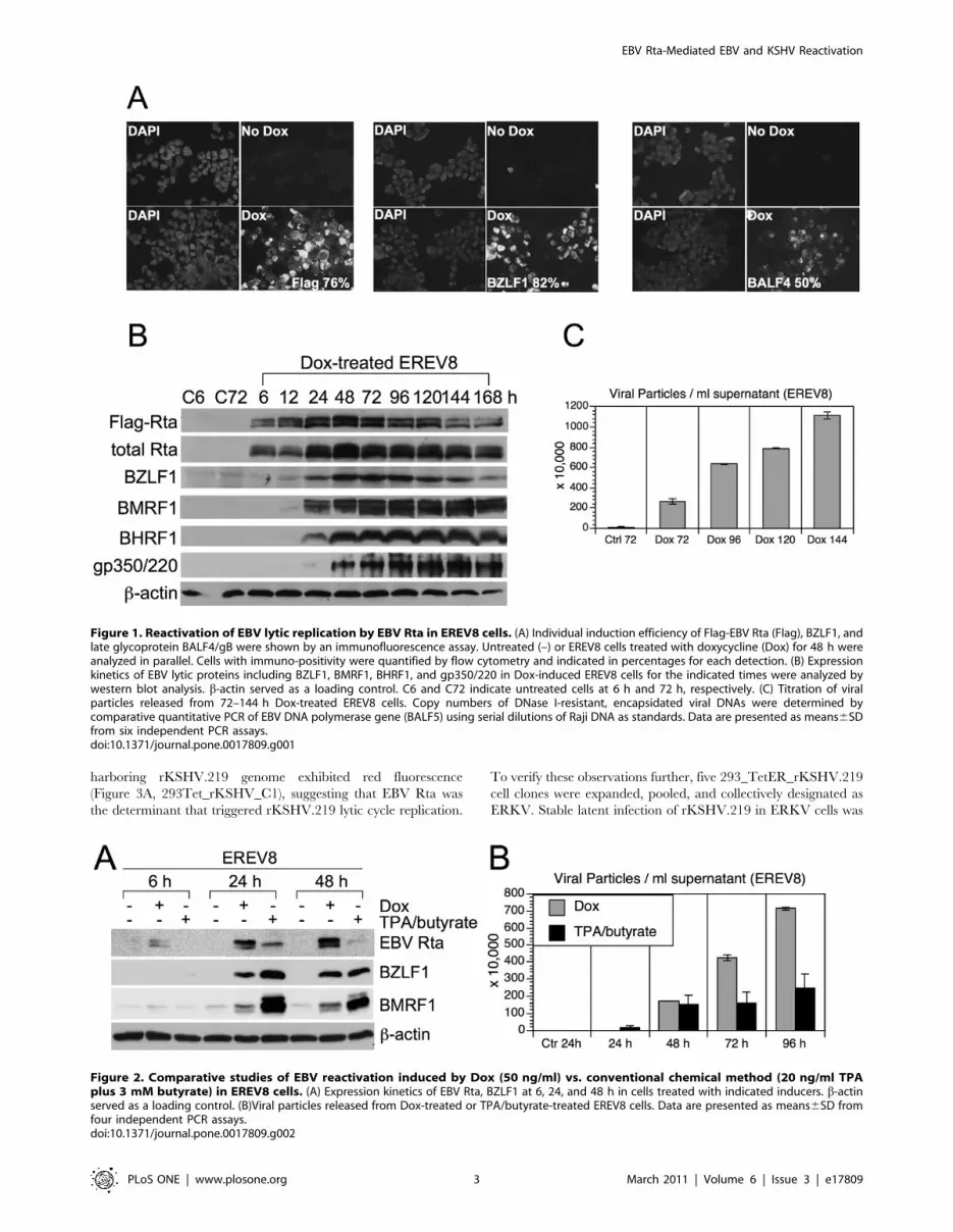
Figure 3. Reactivation of KSHV lytic replication by EBV Rta in ERKV cells. (A) Latent and lytic infections of rKSHV.219, indicated by GFP andRFP, respectively, were inspected in control 293Tet (293Tet_rKSHV_C1) and 293TetER cells (293TetER_rKSHV_E2 and E10) treated with doxycycline(Dox) for 48 h. R/G represents the fraction of RFP-expressing cells in the population determined by using Image J (NIH). Five 293TetER_rKSHVsubclones with R/G .70% were pooled and collectively referred to as ERKV. (B) The expression kinetics of KSHV lytic proteins (K-RTA, K-bZIP, K8.1) incontrol (C6 and C72) and Dox-treated (6–168 h) ERKV cells were examined by western blot analysis. (C) Titration of KSHV particles released from Dox-treated ERKV cells. Copy numbers of DNase I-resistant, encapsidated viral DNAs in each filtrated (0.45 mm) viral supernatants were determined bycomparative quantitative PCR of KSHV DNA polymerase gene (ORF9) using serial dilutions of cosmid GB11 DNA as standards. Data are presented asmeans6SD from six independent PCR assays. (D) Titration of infectious KSHV particles from Dox-treated ERKV cells. Aliquots of filtrated supernatantswere used to infect fresh 293 cells. Two days after infection, the numbers of GFP-positive cells, designated as ‘‘green 293 units’’, in each infection werecounted under a fluorescence microscope. Error bars depict standard deviations of three independent counts. Two independent experiments wereperformed, one set of results is shown. (E) A luciferase reporter gene assay was used to screen the responsiveness of various viral and cellularpromoters to EBV Rta (ER) and K-RTA (KR) in 293 cells. The error bars of each column indicate the standard deviation of each set of triplicate wells. Thetransfection efficiency of each sample was validated by Western blot analysis using M2 Flag monoclonal antibodies.doi:10.1371/journal.pone.0017809.g003
EBV Rta-Mediated EBV and KSHV Reactivation
PLoS ONE | www.plosone.org 4 March 2011 | Volume 6 | Issue 3 | e17809
KSHV replication is very robust. Furthermore, since K-RTA is by
far the only known immediate-early protein that is required and
sufficient to complete a lytic cycle replication, to confirm whether
K-RTA is the only gene activated by EBV Rta in ERKV cells,
luciferase reporter gene assays were used to analyze the
responsiveness to the cotransfected EBV Rta proteins of a panel
of KSHV viral promoters. Two known responders of EBV Rta,
namely promoters of EBV BGLF5 and cellular p21, were included
as controls. As shown in Figure 3E, the EBV BGLF5 and cellular
p21 promoter sequences were responsive to EBV Rta, whereas the
three KSHV lytic promoters (PANp, ORF57p, and K-bZIPp)
were preferentially responsive to K-RTA, as expected. These
results established the respective specificity of EBV Rta and K-
RTA for their cognate responsive elements in the present assay.
Intriguingly, when the promoter of K-RTA was considered, even
though the expression of EBV Rta was much less than that of K-
RTA, the K-RTA promoter exhibited a significantly stronger
responsiveness to EBV Rta than to K-RTA. Taken together, these
results suggest that in Dox-treated ERKV cells, EBV Rta
efficiently up-regulates the expression of K-RTA, followed by
the activation of numerous KSHV lytic promoters and DNA
replication elicited by K-RTA itself. In summary, unexpectedly,
we found that EBV Rta alone is also sufficient for initiating and
completing the lytic replication of KSHV in 293 cells. Similar to
EREV8, ERKV cells consistently became aggregated and
disrupted on the fourth day after Dox induction, suggestive of
permissive viral replication in these cells.
Long-term Dox-treated EREV8 and ERKV cells displayedgrowth arrest followed by cell death
We demonstrated previously that EBV Rta can initiate a
sustained and irreversible G1 arrest, a hallmark of cellular
senescence in both 293 and NPC cells [27]. In the current study,
we observed that EBV Rta alone is sufficient to induce and
complete the lytic cycle of EBV and KSHV latent genomes in
293TetER cells. To characterize further the molecular phenotypes
imposed by EBV Rta-mediated processes, the growth curves and
metabolic activities of Dox-treated and -untreated 293TetER,
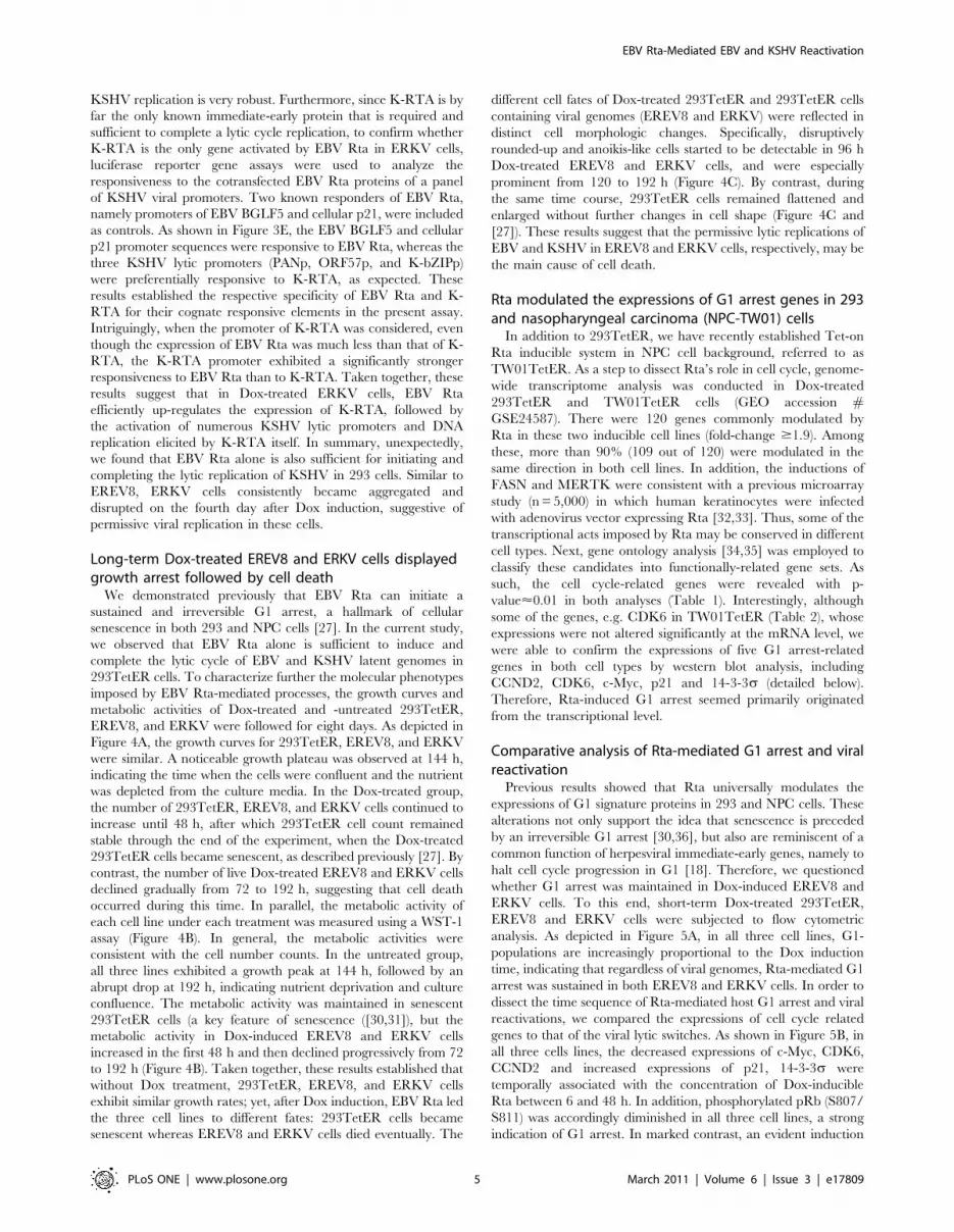
EREV8, and ERKV were followed for eight days. As depicted in
Figure 4A, the growth curves for 293TetER, EREV8, and ERKV
were similar. A noticeable growth plateau was observed at 144 h,
indicating the time when the cells were confluent and the nutrient
was depleted from the culture media. In the Dox-treated group,
the number of 293TetER, EREV8, and ERKV cells continued to
increase until 48 h, after which 293TetER cell count remained
stable through the end of the experiment, when the Dox-treated
293TetER cells became senescent, as described previously [27]. By
contrast, the number of live Dox-treated EREV8 and ERKV cells
declined gradually from 72 to 192 h, suggesting that cell death
occurred during this time. In parallel, the metabolic activity of
each cell line under each treatment was measured using a WST-1
assay (Figure 4B). In general, the metabolic activities were
consistent with the cell number counts. In the untreated group,
all three lines exhibited a growth peak at 144 h, followed by an
abrupt drop at 192 h, indicating nutrient deprivation and culture
confluence. The metabolic activity was maintained in senescent
293TetER cells (a key feature of senescence ([30,31]), but the
metabolic activity in Dox-induced EREV8 and ERKV cells
increased in the first 48 h and then declined progressively from 72
to 192 h (Figure 4B). Taken together, these results established that
without Dox treatment, 293TetER, EREV8, and ERKV cells
exhibit similar growth rates; yet, after Dox induction, EBV Rta led
the three cell lines to different fates: 293TetER cells became
senescent whereas EREV8 and ERKV cells died eventually. The
different cell fates of Dox-treated 293TetER and 293TetER cells
containing viral genomes (EREV8 and ERKV) were reflected in
distinct cell morphologic changes. Specifically, disruptively
rounded-up and anoikis-like cells started to be detectable in 96 h
Dox-treated EREV8 and ERKV cells, and were especially
prominent from 120 to 192 h (Figure 4C). By contrast, during
the same time course, 293TetER cells remained flattened and
enlarged without further changes in cell shape (Figure 4C and
[27]). These results suggest that the permissive lytic replications of
EBV and KSHV in EREV8 and ERKV cells, respectively, may be
the main cause of cell death.
Rta modulated the expressions of G1 arrest genes in 293and nasopharyngeal carcinoma (NPC-TW01) cells
In addition to 293TetER, we have recently established Tet-on
Rta inducible system in NPC cell background, referred to as
TW01TetER. As a step to dissect Rta’s role in cell cycle, genome-
wide transcriptome analysis was conducted in Dox-treated
293TetER and TW01TetER cells (GEO accession #GSE24587). There were 120 genes commonly modulated by
Rta in these two inducible cell lines (fold-change $1.9). Among
these, more than 90% (109 out of 120) were modulated in the
same direction in both cell lines. In addition, the inductions of
FASN and MERTK were consistent with a previous microarray
study (n = 5,000) in which human keratinocytes were infected
with adenovirus vector expressing Rta [32,33]. Thus, some of the
transcriptional acts imposed by Rta may be conserved in different
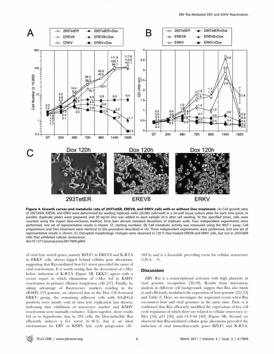
cell types. Next, gene ontology analysis [34,35] was employed to
classify these candidates into functionally-related gene sets. As
such, the cell cycle-related genes were revealed with p-
value<0.01 in both analyses (Table 1). Interestingly, although
some of the genes, e.g. CDK6 in TW01TetER (Table 2), whose
expressions were not altered significantly at the mRNA level, we
were able to confirm the expressions of five G1 arrest-related
genes in both cell types by western blot analysis, including
CCND2, CDK6, c-Myc, p21 and 14-3-3s (detailed below).
Therefore, Rta-induced G1 arrest seemed primarily originated
from the transcriptional level.
Comparative analysis of Rta-mediated G1 arrest and viralreactivation
Previous results showed that Rta universally modulates the
expressions of G1 signature proteins in 293 and NPC cells. These
alterations not only support the idea that senescence is preceded
by an irreversible G1 arrest [30,36], but also are reminiscent of a
common function of herpesviral immediate-early genes, namely to
halt cell cycle progression in G1 [18]. Therefore, we questioned
whether G1 arrest was maintained in Dox-induced EREV8 and
ERKV cells. To this end, short-term Dox-treated 293TetER,
EREV8 and ERKV cells were subjected to flow cytometric
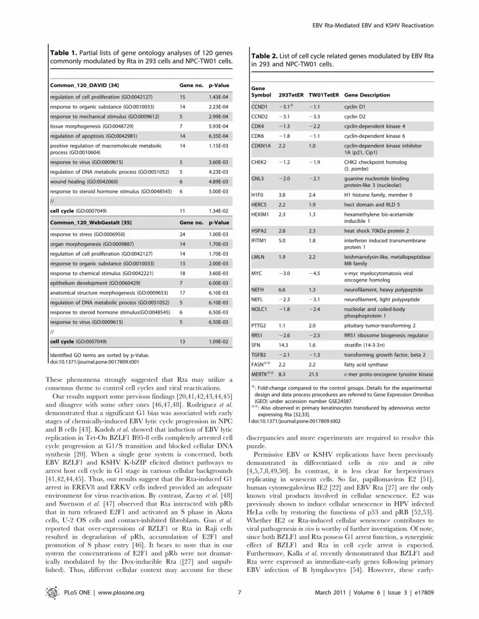
analysis. As depicted in Figure 5A, in all three cell lines, G1-
populations are increasingly proportional to the Dox induction
time, indicating that regardless of viral genomes, Rta-mediated G1
arrest was sustained in both EREV8 and ERKV cells. In order to
dissect the time sequence of Rta-mediated host G1 arrest and viral
reactivations, we compared the expressions of cell cycle related
genes to that of the viral lytic switches. As shown in Figure 5B, in
all three cells lines, the decreased expressions of c-Myc, CDK6,
CCND2 and increased expressions of p21, 14-3-3s were
temporally associated with the concentration of Dox-inducible
Rta between 6 and 48 h. In addition, phosphorylated pRb (S807/
S811) was accordingly diminished in all three cell lines, a strong
indication of G1 arrest. In marked contrast, an evident induction
EBV Rta-Mediated EBV and KSHV Reactivation
PLoS ONE | www.plosone.org 5 March 2011 | Volume 6 | Issue 3 | e17809
of viral lytic switch genes, namely BZLF1 in EREV8 and K-RTA
in ERKV cells, always lagged behind cellular gene alterations,
suggesting that Rta-mediated host G1 arrest preceded the onset of
viral reactivation. It is worth noting that the decrement of c-Myc
before induction of K-RTA (Figure 5B, ERKV) agrees with a
recent report in which elimination of c-Myc led to KSHV
reactivation in primary effusion lymphoma cells [37]. Finally, by
taking advantage of fluorescence markers residing in the
rKSHV.219 genome, we observed that in the Dox 120 h-treated
ERKV group, the remaining adherent cells with SA-b-Gal
positivity were mostly void of virus lytic replication (not shown),
indicating that exhibition of senescence marker and KSHV
reactivation were mutually exclusive. Taken together, these results
led us to hypothesize that in 293 cells, the Dox-inducible Rta
efficiently induces a G1 arrest (6–48 h) that is an ideal
environment for EBV or KSHV lytic cycle progression (48–
168 h) and is a favorable preceding event for cellular senescence
(120 h – ‘).
Discussion
EBV Rta is a transcriptional activator with high plasticity in
viral genome recognition [38,39]. Results from microarray
analysis in different cell backgrounds suggest that Rta also binds
to and efficiently modulates the expression of host genome ([32,33]
and Table 2). Here, we investigate the sequential events when Rta
encounters host and viral genomes at the same time. First, it is
confirmed that Rta efficiently modified the expressions of key cell
cycle regulators of which three are related to cellular senescence (c-
Myc [16], p21 [30], and 14-3-3s [40]) (Figure 5B). Second, we
observed that Rta-mediated cellular gene alterations preceded the
induction of viral immediate-early genes BZLF1 and K-RTA.
Figure 4. Growth curves and metabolic rate of 293TetER, EREV8, and ERKV cells with or without Dox treatment. (A) Cell growth ratesof 293TetER, EREV8, and ERKV were determined by seeding triplicate wells (20,000 cells/well) in a 24-well tissue culture plate for each time point. Inparallel, duplicate plates were prepared, and 50 ng/ml Dox was added to each sample 24 h after cell seeding. At the specified times, cells werecounted using the trypan blue-exclusion method. Error bars denote standard deviations of triplicate wells. Four independent experiments wereperformed, one set of representative results is shown. ST, starting numbers. (B) Cell metabolic activity was measured using the WST-1 assay. Cellpreparations and Dox treatment were identical to the procedure described in (A). Three independent experiments were performed, and one set ofrepresentative results is shown. (C) Disrupted morphologic changes were observed in 120 h Dox-treated EREV8 and ERKV cells, but not in 293TetERcells that exhibited cellular senescence.doi:10.1371/journal.pone.0017809.g004
EBV Rta-Mediated EBV and KSHV Reactivation
PLoS ONE | www.plosone.org 6 March 2011 | Volume 6 | Issue 3 | e17809
These phenomena strongly suggested that Rta may utilize a
consensus theme to control cell cycles and viral reactivations.
Our results support some previous findings [20,41,42,43,44,45]
and disagree with some other ones [46,47,48]. Rodriguez et al.
demonstrated that a significant G1 bias was associated with early
stages of chemically-induced EBV lytic cycle progression in NPC
and B cells [43]. Kudoh et al. showed that induction of EBV lytic
replication in Tet-On BZLF1 B95-8 cells completely arrested cell
cycle progression at G1/S transition and blocked cellular DNA
synthesis [20]. When a single gene system is concerned, both
EBV BZLF1 and KSHV K-bZIP elicited distinct pathways to
arrest host cell cycle in G1 stage in various cellular backgrounds
[41,42,44,45]. Thus, our results suggest that the Rta-induced G1
arrest in EREV8 and ERKV cells indeed provided an adequate
environment for virus reactivation. By contrast, Zacny et al. [48]
and Swenson et al. [47] observed that Rta interacted with pRb
that in turn released E2F1 and activated an S phase in Akata
cells, U-2 OS cells and contact-inhibited fibroblasts. Guo et al.
reported that over-expressions of BZLF1 or Rta in Raji cells
resulted in degradation of pRb, accumulation of E2F1 and
promotion of S phase entry [46]. It bears to note that in our
system the concentrations of E2F1 and pRb were not dramat-
ically modulated by the Dox-inducible Rta ([27] and unpub-
lished). Thus, different cellular context may account for these
discrepancies and more experiments are required to resolve this
puzzle.
Permissive EBV or KSHV replications have been previously
demonstrated in differentiated cells in vivo and in vitro
[4,5,7,8,49,50]. In contrast, it is less clear for herpesviruses
replicating in senescent cells. So far, papillomavirus E2 [51],
human cytomegalovirus IE2 [22] and EBV Rta [27] are the only
known viral products involved in cellular senescence. E2 was
previously shown to induce cellular senescence in HPV infected
HeLa cells by restoring the functions of p53 and pRB [52,53].
Whether IE2 or Rta-induced cellular senescence contributes to
viral pathogenesis in vivo is worthy of further investigation. Of note,
since both BZLF1 and Rta possess G1 arrest function, a synergistic
effect of BZLF1 and Rta in cell cycle arrest is expected.
Furthermore, Kalla et al. recently demonstrated that BZLF1 and
Rta were expressed as immediate-early genes following primary
EBV infection of B lymphocytes [54]. However, these early-
Table 1. Partial lists of gene ontology analyses of 120 genescommonly modulated by Rta in 293 cells and NPC-TW01 cells.
Common_120_DAVID [34] Gene no. p-Value
regulation of cell proliferation (GO:0042127) 15 1.43E-04
response to organic substance (GO:0010033) 14 2.23E-04
response to mechanical stimulus (GO:0009612) 5 2.99E-04
tissue morphogenesis (GO:0048729) 7 5.93E-04
regulation of apoptosis (GO:0042981) 14 6.35E-04
positive regulation of macromolecule metabolicprocess (GO:0010604)
14 1.15E-03
response to virus (GO:0009615) 5 3.60E-03
regulation of DNA metabolic process (GO:0051052) 5 4.23E-03
wound healing (GO:0042060) 6 4.89E-03
response to steroid hormone stimulus (GO:0048545) 6 5.00E-03
//
cell cycle (GO:0007049) 11 1.34E-02
Common_120_WebGestalt [35] Gene no. p-Value
response to stress (GO:0006950) 24 1.00E-03
organ morphogenesis (GO:0009887) 14 1.70E-03
regulation of cell proliferation (GO:0042127) 14 1.70E-03
response to organic substance (GO:0010033) 13 2.00E-03
response to chemical stimulus (GO:0042221) 18 3.60E-03
epithelium development (GO:0060429) 7 6.00E-03
anatomical structure morphogenesis (GO:0009653) 17 6.10E-03
regulation of DNA metabolic process (GO:0051052) 5 6.10E-03
response to steroid hormone stimulus(GO:0048545) 6 6.50E-03
response to virus (GO:0009615) 5 6.50E-03
//
cell cycle (GO:0007049) 13 1.09E-02
Identified GO terms are sorted by p-Value.doi:10.1371/journal.pone.0017809.t001
Table 2. List of cell cycle related genes modulated by EBV Rtain 293 and NPC-TW01 cells.
GeneSymbol 293TetER TW01TetER Gene Description
CCND1 23.1# 21.1 cyclin D1
CCND2 23.1 23.3 cyclin D2
CDK4 21.3 22.2 cyclin-dependent kinase 4
CDK6 21.8 21.1 cyclin-dependent kinase 6
CDKN1A 2.2 1.0 cyclin-dependent kinase inhibitor1A (p21, Cip1)
CHEK2 21.2 21.9 CHK2 checkpoint homolog(S. pombe)
GNL3 22.0 22.1 guanine nucleotide bindingprotein-like 3 (nucleolar)
H1F0 3.8 2.4 H1 histone family, member 0
HERC5 2.2 1.9 hect domain and RLD 5
HEXIM1 2.3 1.3 hexamethylene bis-acetamideinducible 1
HSPA2 2.8 2.3 heat shock 70kDa protein 2
IFITM1 5.0 1.8 interferon induced transmembraneprotein 1
LMLN 1.9 2.2 leishmanolysin-like, metallopeptidaseM8 family
MYC 23.0 24.5 v-myc myelocytomatosis viraloncogene homolog
NEFH 6.6 1.3 neurofilament, heavy polypeptide
NEFL 22.3 23.1 neurofilament, light polypeptide
NOLC1 21.8 22.4 nucleolar and coiled-bodyphosphoprotein 1
PTTG2 1.1 2.0 pituitary tumor-transforming 2
RRS1 22.6 22.5 RRS1 ribosome biogenesis regulator
SFN 14.3 1.6 stratifin (14-3-3s)
TGFB2 22.1 21.3 transforming growth factor, beta 2
FASN## 2.2 2.2 fatty acid synthase
MERTK## 8.3 21.5 c-mer proto-oncogene tyrosine kinase
#: Fold-change compared to the control groups. Details for the experimentaldesign and data process procedures are referred to Gene Expression Omnibus(GEO) under accession number GSE24587.
##: Also observed in primary keratinocytes transduced by adenovirus vectorexpressing Rta [32,33].
doi:10.1371/journal.pone.0017809.t002
EBV Rta-Mediated EBV and KSHV Reactivation
PLoS ONE | www.plosone.org 7 March 2011 | Volume 6 | Issue 3 | e17809
expressed BZLF1 and Rta failed to initiate the EBV lytic cycle
owing to the intruding viral genome was in an un-methylated
status [54]. Therefore, we hypothesize that in such a transient-lytic
phase where only the host genome is accessible, Rta (and BZLF1)
may exert to trigger a cell senescence process.
Among genes modulated by Rta depicted in Figure 5B, the
sharply decreased expression of c-Myc by EBV Rta has two
implications. First, one oncogenic role of c-Myc was suggested to
be a repressor of cellular senescence [16,55]. In our previous
report, we demonstrated that EBV Rta efficiently induces cellular
senescence in 293, NPC-TW01 and HONE-1 cells [27]. Here we
further confirm that in these senescent cells the decrement of c-
Myc was one of the earliest events modulated by EBV Rta. Thus,
decreased expression of c-Myc via Rta seems to participate in Rta-
induced cellular senescence. Second, c-Myc is a negative regulator
of KSHV lytic cycle replication [37,56]. RNAi-mediated knock-
down of c-Myc resulted in disruption of KSHV latency and
increment in mRNA and protein levels of K-RTA [37]. Consistent
with these results, we observed that the reactivation of KSHV
latent genome was preceded by a gradual decrement of c-Myc in
the ERKV cells (Figure 5B). In addition, in a luciferase-reporting
assay, the promoter sequences of K-RTA, but not those of K-
bZIP, PAN, and ORF57 were preferentially activated by ectopic
expression of Rta (Figure 3E). Thus, we hypothesize that either a
direct act from Rta alone, or via down-regulated c-Myc, or both,
are attributable to Rta-induced K-RTA synthesis in Dox-treated
ERKV cells.
EBV Rta is not the only variant that cross-reactivates KSHV;
other viral factors including the HCMV UL112-113 locus [57] and
HIV-1 tat protein [58] have also been ascribed to possess such
functionality, suggesting that other viral infections may also
participate in KSHV pathogenesis. Further, although permissive
EBV or KSHV lytic replication were detectable in vivo, but a
homogenous and thorough lysis of host cell by viral lytic
replication is still lacking in vitro. Here, we have produced a model
that provides a nearly permissive replication system for both EBV
and KSHV that is controlled directly by EBV Rta. This system
offers two advantages over the conventional approaches. First, the
stimulus, 50 ng/ml Dox, is a very dilute, physiologically neutral
compound. Compared with the conventional sodium butyrate or
phorbol ester, Dox elicits far fewer, possibly no, undesirable effects
on the treated cells. Second, the treatment produces homogenous
results. Routinely, Flag-tagged EBV Rta and BZLF1 were
detected in close to 80% of the 48 h Dox-treated EREV8 cells
when assessed by an immunofluorescence assay. Similarly, more
than 80% of the treated ERKV cells produced red fluorescence
48 h after induction. Our newly established EREV and ERKV
cells thus provide a feasible system for elucidating host factors and
viral determinants that contribute to regulate the EBV and KSHV
reactivations.
Figure 5. Rta-mediated cell cycle arrest precedes the expressions of viral immediate-early genes. (A) Dox-treated 293TetER, EREV8, andERKV cells cultured for 24 and 48 h were subjected to flow cytometry analysis to quantify the cellular DNA content. The distributions of cells residingin the G2/M, S, G1, and subG1 stages at each time are shown. The results of three independent experiments were similar, and one representativedataset is shown. (B) Comparative expression kinetics (0–48 h) of cell cycle regulators or viral immediate-early proteins in Dox treated 293TetER,EREV8 and ERKV cells. Down-regulation of cell cycle activators (c-Myc, CDK6, CCND2, phosphorylated pRb) and up-regulation of cell cycle inhibitors(p21, 14-3-3s) are temporally associated with the expression of Rta in all three cell lines. In comparison, EBV BZLF1 and KSHV K-RTA are notsignificantly augmented until 48 h, a time that alterations of cell cycle gene are nearly completed. a-tubulin served as a loading control.doi:10.1371/journal.pone.0017809.g005
EBV Rta-Mediated EBV and KSHV Reactivation
PLoS ONE | www.plosone.org 8 March 2011 | Volume 6 | Issue 3 | e17809

Materials and Methods
Cell culture293TetER is a doxycycline inducible, EBV Rta conditional
expression cell lines created by Virapower systemTM (Invitrogen,
Carlsbad, CA) [27]. Same procedures were carried out to establish
TW01TetER in which inducible Rta was expressed in a
nasopharyngeal carcinoma cell line, NPC-TW01 [59]. EREV8 is
an EBV positive 293TetER derivative line generated by using cell-
to-cell infection method [28]. ERKV is a KSHV positive
293TetER derivative line that was stably infected with
rKSHV.219 [29]. Specifically, 293TetER cells incubated with
rKSHV.219 viral sup for 48 h were selected with 660 mg/ml
puromycin for three weeks to obtain green fluorescent clones.
Twelve such cell colonies were isolated, expanded, and deter-
mined for inducibility of KSHV lytic replication (red fluorescence)
by 50 ng/ml doxycycline treatment. Five clones with high
inducibility (70–90%) were pooled and used to compose the first
generation of ERKV. 293TetER, EREV8 and ERKV cells were
maintained in DMEM containing 10% Tet System Approved FBS
(Clontech Laboratories, Mountain View, CA), 5 mg/ml blastici-
din-S-HCl (Invitrogen) and 200 mg/ml zeocin (Invitrogen). To
maintain the latently infected viral genomes, EREV8 and ERKV
cultures were further supplemented with 400 mg/ml G418 and
660 mg/ml puromycin, respectively.
PlasmidspLenti4-Flag-CPO is a modified expression plasmid derived
from pLenti4/TO/V5-DEST (Invitrogen). In brief, the original
attR1 site to V5 epitope region (nt 2405–4203) in pLenti4/TO/
V5-DEST was replaced with an in-frame DNA fragment encoding
Kozak sequence, ATG, FLAG tag and a rare cutter CPO I site
(59CGGTCCG). Accordingly, the cDNAs of EBV Rta (M-ABA
strain) and KSHV RTA (Genebank: U71367.1) were PCR-
amplified with CPO I sites flanking at both ends, and subcloned
into pLenti4-Flag-CPO. The resulting plasmids, namely pLenti4-
Flag-ER and pLenti4-Flag-KR, were propagated in DH5a and
used in further studies. The upstream sequences of p21 (2.4 kb),
EBV BGLF5 (nt 108641 to 110053 of NC_007605), K-RTA (nt
70240 to 71597 of U75698), PAN (nt 28159 to 28660), ORF57 (nt
81556 to 82005), and K-bZIP (74619 to 74849), were cloned in
front of luciferase gene located in pGL3-Basic (Promega, Madison,
WI), yielding pGL3-Basic-p21p, -BGLF5p, K-RTAp, -PANp,
-ORF57p and -K-bZIPp, respectively.
Transfection and luciferase reporter assayTransfection was performed in 24-well plates. The next day
when the cultured 293 cells were 90% confluent, appropriate
amount of indicated plasmids were transfected into cells by using
LipofectamineTM 2000 (Invitrogen) according to the manufactur-
er’s instructions. Twenty-four hr after transfection, cells were
harvested for luciferase activity assay by using Dual-Glo lucifearse
assay kit (Promega). In addition, an aliquot of cell lysates was
subjected to western blot analysis for the normalization of each
transfection efficiency.
Titration of EBV and KSHV viral particlesFiltrated (0.45 mm) viral supernatant (160 ml) was incubated
with 2 U DNase I (Invitrogen) at 37uC for 30 min followed by
extraction of encapsidated EBV DNA using QIAamp MinElute
virus spin kit (QIAGEN). Each comparative quantitative PCR
reaction was composed of 4 ml diluted viral DNA, 5 ml Power
SYBR Green Master Mix (Applied Biosystems, Foster City, CA),
and 1 ml primer mix (2 mM). The primers used in the present study
were as follows: detection of EBV genome, BALF5-forward (59-
CGGAGTTGTTATCAAAGAGGC-39) and BALF5-reverse (59-
CGAGAAAGACGGAGATGGC-39); detection of KSHV ge-
nome, ORF9-forward (59-CCAACATCATCCAATGCCTC-39)
and ORF9-reverse (59-GGGAAAAGTCACGGGAATG-39).
Known copy numbers of serially diluted EBV genome from Raji
cellular DNA (50 copies/cell) were used as standards in titrating
EBV viral particles. Known copy numbers of serially diluted
cosmid GB11 DNA encompassing KSHV genome nt 1–35,022
(U75698) were used as standards in titrating KSHV viral particles.
The reaction was conducted and detected by StepOnePlusTM
Real-Time PCR system (Applied Biosystems).
Infectivity assay of KSHV particlesWe followed the EBV infection procedure described by Hutt-
Fletcher and colleagues with minor modification [60]. Specifically,
293 cells were seeded onto 12-well plates at 1.26105 cells/well that
produced >30% confluent monolayer 24 h later. Two hundred-ml
undiluted, filtrated viral supernatant was gently applied onto the
surface of cells. After 2 h of incubation on cells, 1.5 ml growth
medium was added and the cells were reincubated for 48 h. To
score the infectious units in each well, the culture supernatant was
removed, cells were trypsinized and subjected to visual inspection
for GFP expression under a fluorescence microscope (OLYMPUS
BX51, Olympus UK Ltd, Essex SS2 5QH, UK).
Western blot analysisCell lysates extracted by RIPA buffer were subjected to SDS-
PAGE separation and transferred onto nitrocellulose membranes.
The membranes were blocked for 1 h in 16TBST containing 5%
non-fat milk and then incubated with the indicated primary
antibody overnight at 4uC. The blots were washed three times
with 16 TBST for 5 min each. The blots were incubated with
peroxidase-conjugated secondary antibody in blocking buffer for
1 h at room temperature. Blots were washed three times with 16TBST for 5 min each and developed by SuperSignal West Pico
chemiluminescent substrate kit (Pierce).
Cell proliferation assayCell proliferation was determined by using a WST-1 kit (Roche,
Indianapolis, IN) or by trypan blue exclusion method as described
previously [27].
Immunofluorescence assayCells were resuspended in PBS and dropped onto multiple-well
diagnostic microscope slides and fixed in methanol/acetone (1:1)
at 220uC for 20 min. Cells were permeabilized with 0.1% Triton-
X 100 at room temperature for 20 min. The slide was incubated
with indicated primary antibody at room temperature for 1 h,
washed three times in PBS for 5 min each, and incubated with
FITC-conjugated secondary antibody at room temperature for
1 h. After PBS wash, the slide was incubated with Hoechst 33258
at room temperature for 20 min, washed with PBS, mounted in
VECTASHIELD TMmedium and inspected by fluorescence
microscopy. To quantitate the percentage of positively immuno-
reactive cells in the immunofluorescence assay, an aliquot of cells
were analyzed in parallel by flow cytometric analysis.
Flow cytometric analysisCells were harvested by centrifugation, washed with phosphate-
buffered saline (PBS), fixed in ice-cold 75% ethanol and stored at
220uC until all samples from different time points were collected.
Of note, to quench the green and red fluorescence in ERKV cells,
EBV Rta-Mediated EBV and KSHV Reactivation
PLoS ONE | www.plosone.org 9 March 2011 | Volume 6 | Issue 3 | e17809

the fixation reagent was replaced with 95% methanol. Prior to
flow cytometer analysis, the fixed cells were repelleted by
centrifugation, permeabilized in PBS containing 0.1% Triton X-
100 at room temperature for 30 min, and resuspended in PBS
containing 50 mg/ml propidium iodide and 50 mg/ml RNaseA.
After the cells were incubated in dark for 30 min, cell cycle profile
analysis was carried out on 5,000 cells with a fluorescence
activated cell sorter (FACSCalibur, Becton Dickinson, Franklin
Lakes, NJ). The results were analyzed by using WinMDI v2.8
software.
AntibodiesMouse monoclonal antibodies of EBV proteins were: Rta (467),
BZLF1 (4F10), BMRF1 (88A9), BALF4/gB (L2), BHRF1 (3E8),
and gp350/220 (72A1). Anti-KSHV RTA was provided by Dr.
Keiji Ueda (Osaka University Medical School, Japan). Anti-
KSHV K-bZIP was provided by Dr. Mengtao Li (University of
Kentucky College of Dentistry, USA). All other antibodies were
commercially available: KSHV K8.1 (ABI, Columbia, MD);
CDK6, pRb/S807/S811 and p21 (Cell Signaling Technology,
Danvers, MA); c-Myc (Santa Cruz Biotechnology, Santa Cruz,
CA); 14-3-3s (GeneTex, Irvine, CA); CCND2 (BD Pharmingen,
Franklin Lakes, NJ); a-tubulin (Millipore, Billerica, MA); b-actin
and M2-FLAG (Sigma-Aldrich, St. Louis, MO).
Acknowledgments
We are indebted to Ching-Hwa Tsai, Mei-Ru Chen (National Taiwan
University, Taiwan) and Shih-Tung Liu (Chang Gung University, Taiwan)
for providing various antibodies of EBV proteins; Dr. Keiji Ueda (Osaka
University Medical School, Japan) for anti-K-RTA. Dr. Mengtao Li
(University of Kentucky College of Dentistry, USA) for anti-K-bZIP;
Jeffrey Vieira (University of Washington, Seattle, USA) for rKSHV.219.
We also acknowledge Mr. Shu-Wei Nien for technical support on
microarray analysis, and the array services provided by Microarray Core
Laboratory of National Health Research Institutes, Taiwan.
Author Contributions
Conceived and designed the experiments: YJC WHT JYC SFL. Performed
the experiments: YJC WHT YLC YCK SPC. Analyzed the data: YJC
WHT YLC YCK JYC SFL. Wrote the paper: YJC SFL.
References
1. Pauk J, Huang ML, Brodie SJ, Wald A, Koelle DM, et al. (2000) Mucosal
shedding of human herpesvirus 8 in men. N Engl J Med 343: 1369–1377.
2. Sixbey JW, Nedrud JG, Raab-Traub N, Hanes RA, Pagano JS (1984) Epstein-
Barr virus replication in oropharyngeal epithelial cells. N Engl J Med 310:
1225–1230.
3. Thorley-Lawson DA (2005) EBV the prototypical human tumor virus–just how
bad is it? J Allergy Clin Immunol 116: 251–261; quiz 262.
4. Feederle R, Neuhierl B, Bannert H, Geletneky K, Shannon-Lowe C, et al.
(2007) Epstein-Barr virus B95.8 produced in 293 cells shows marked tropism for
differentiated primary epithelial cells and reveals interindividual variation in
susceptibility to viral infection. Int J Cancer 121: 588–594.
5. Johnson AS, Maronian N, Vieira J (2005) Activation of Kaposi’s sarcoma-
associated herpesvirus lytic gene expression during epithelial differentiation.
J Virol 79: 13769–13777.
6. Young LS, Lau R, Rowe M, Niedobitek G, Packham G, et al. (1991)
Differentiation-associated expression of the Epstein-Barr virus BZLF1 transacti-
vator protein in oral hairy leukoplakia. J Virol 65: 2868–2874.
7. Sun CC, Thorley-Lawson DA (2007) Plasma cell-specific transcription factor
XBP-1s binds to and transactivates the Epstein-Barr virus BZLF1 promoter.
J Virol 81: 13566–13577.
8. Wilson SJ, Tsao EH, Webb BL, Ye H, Dalton-Griffin L, et al. (2007) X box
binding protein XBP-1s transactivates the Kaposi’s sarcoma-associated herpes-
virus (KSHV) ORF50 promoter, linking plasma cell differentiation to KSHV
reactivation from latency. J Virol 81: 13578–13586.
9. Blomen VA, Boonstra J (2007) Cell fate determination during G1 phase
progression. Cell Mol Life Sci 64: 3084–3104.
10. Pfeuty B, David-Pfeuty T, Kaneko K (2008) Underlying principles of cell fate
determination during G1 phase of the mammalian cell cycle. Cell Cycle 7:
3246–3257.
11. Sekeri-Pataryas KE, Sourlingas TG (2007) The differentiation-associated linker
histone, H1.0, during the in vitro aging and senescence of human diploid
fibroblasts. Ann N Y Acad Sci 1100: 361–367.
12. Hwang ES (2002) Replicative senescence and senescence-like state induced in
cancer-derived cells. Mech Ageing Dev 123: 1681–1694.
13. Serrano M, Lin AW, McCurrach ME, Beach D, Lowe SW (1997) Oncogenic ras
provokes premature cell senescence associated with accumulation of p53 and
p16INK4a. Cell 88: 593–602.
14. Collado M, Serrano M (2010) Senescence in tumours: evidence from mice and
humans. Nat Rev Cancer 10: 51–57.
15. Mooi WJ, Peeper DS (2006) Oncogene-induced cell senescence–halting on the
road to cancer. N Engl J Med 355: 1037–1046.
16. Wu CH, van Riggelen J, Yetil A, Fan AC, Bachireddy P, et al. (2007) Cellular
senescence is an important mechanism of tumor regression upon c-Myc
inactivation. Proc Natl Acad Sci U S A 104: 13028–13033.
17. Soucek L, Whitfield J, Martins CP, Finch AJ, Murphy DJ, et al. (2008)
Modelling Myc inhibition as a cancer therapy. Nature 455: 679–683.
18. Flemington EK (2001) Herpesvirus lytic replication and the cell cycle: arresting
new developments. J Virol 75: 4475–4481.
19. Inman GJ, Binne UK, Parker GA, Farrell PJ, Allday MJ (2001) Activators of the
Epstein-Barr virus lytic program concomitantly induce apoptosis, but lytic gene
expression protects from cell death. J Virol 75: 2400–2410.
20. Kudoh A, Fujita M, Kiyono T, Kuzushima K, Sugaya Y, et al. (2003)
Reactivation of lytic replication from B cells latently infected with Epstein-Barr
virus occurs with high S-phase cyclin-dependent kinase activity while inhibiting
cellular DNA replication. J Virol 77: 851–861.
21. Hobbs WE, 2nd, DeLuca NA (1999) Perturbation of cell cycle progression and
cellular gene expression as a function of herpes simplex virus ICP0. J Virol 73:
8245–8255.
22. Noris E, Zannetti C, Demurtas A, Sinclair J, De Andrea M, et al. (2002) Cell
cycle arrest by human cytomegalovirus 86-kDa IE2 protein resembles premature
senescence. J Virol 76: 12135–12148.
23. Ragoczy T, Heston L, Miller G (1998) The Epstein-Barr virus Rta protein
activates lytic cycle genes and can disrupt latency in B lymphocytes. J Virol 72:
7978–7984.
24. Zalani S, Holley-Guthrie E, Kenney S (1996) Epstein-Barr viral latency is
disrupted by the immediate-early BRLF1 protein through a cell-specific
mechanism. Proc Natl Acad Sci U S A 93: 9194–9199.
25. Sun R, Lin SF, Gradoville L, Yuan Y, Zhu F, et al. (1998) A viral gene that
activates lytic cycle expression of Kaposi’s sarcoma- associated herpesvirus. Proc
Natl Acad Sci U S A 95: 10866–10871.
26. Lukac DM, Kirshner JR, Ganem D (1999) Transcriptional activation by the
product of open reading frame 50 of Kaposi’s sarcoma-associated herpesvirus is
required for lytic viral reactivation in B cells. J Virol 73: 9348–9361.
27. Chen YL, Chen YJ, Tsai WH, Ko YC, Chen JY, et al. (2009) The Epstein-Barr
virus replication and transcription activator, Rta/BRLF1, induces cellular
senescence in epithelial cells. Cell Cycle 8: 58–65.
28. Lee CP, Huang YH, Lin SF, Chang Y, Chang YH, et al. (2008) Epstein-Barr
virus BGLF4 kinase induces disassembly of the nuclear lamina to facilitate virion
production. J Virol 82: 11913–11926.
29. Vieira J, O’Hearn PM (2004) Use of the red fluorescent protein as a marker of
Kaposi’s sarcoma-associated herpesvirus lytic gene expression. Virology 325:
225–240.
30. Blagosklonny MV (2006) Cell senescence: hypertrophic arrest beyond the
restriction point. J Cell Physiol 209: 592–597.
31. Demidenko ZN, Blagosklonny MV (2008) Growth stimulation leads to cellular
senescence when the cell cycle is blocked. Cell Cycle 7: 3355–3361.
32. Li Y, Mahajan NP, Webster-Cyriaque J, Bhende P, Hong GK, et al. (2004) The
C-mer gene is induced by Epstein-Barr virus immediate-early protein BRLF1.
J Virol 78: 11778–11785.
33. Li Y, Webster-Cyriaque J, Tomlinson CC, Yohe M, Kenney S (2004) Fatty
acid synthase expression is induced by the Epstein-Barr virus immediate-early
protein BRLF1 and is required for lytic viral gene expression. J Virol 78:
4197–4206.
34. Huang da W, Sherman BT, Lempicki RA (2009) Systematic and integrative
analysis of large gene lists using DAVID bioinformatics resources. Nat Protoc 4:
44–57.
35. Zhang B, Kirov S, Snoddy J (2005) WebGestalt: an integrated system for
exploring gene sets in various biological contexts. Nucleic Acids Res 33:
W741–748.
36. Campisi J, d’Adda di Fagagna F (2007) Cellular senescence: when bad things
happen to good cells. Nat Rev Mol Cell Biol 8: 729–740.
37. Li X, Chen S, Feng J, Deng H, Sun R (2010) Myc is required for the
maintenance of Kaposi’s sarcoma-associated herpesvirus latency. J Virol.
38. Chen LW, Chang PJ, Delecluse HJ, Miller G (2005) Marked variation in
response of consensus binding elements for the Rta protein of Epstein-Barr virus.
J Virol 79: 9635–9650.
EBV Rta-Mediated EBV and KSHV Reactivation
PLoS ONE | www.plosone.org 10 March 2011 | Volume 6 | Issue 3 | e17809
39. Gruffat H, Sergeant A (1994) Characterization of the DNA-binding site
repertoire for the Epstein-Barr virus transcription factor R. Nucleic Acids Res22: 1172–1178.
40. Schultz J, Ibrahim SM, Vera J, Kunz M (2009) 14-3-3sigma gene silencing
during melanoma progression and its role in cell cycle control and cellularsenescence. Mol Cancer 8: 53.
41. Cayrol C, Flemington EK (1996) The Epstein-Barr virus bZIP transcriptionfactor Zta causes G0/G1 cell cycle arrest through induction of cyclin-dependent
kinase inhibitors. Embo J 15: 2748–2759.
42. Izumiya Y, Lin SF, Ellison TJ, Levy AM, Mayeur GL, et al. (2003) Cell cycleregulation by Kaposi’s sarcoma-associated herpesvirus K-bZIP: direct interac-
tion with cyclin-CDK2 and induction of G1 growth arrest. J Virol 77:9652–9661.
43. Rodriguez A, Jung EJ, Flemington EK (2001) Cell cycle analysis of Epstein-Barrvirus-infected cells following treatment with lytic cycle-inducing agents. J Virol
75: 4482–4489.
44. Wu FY, Chen H, Wang SE, ApRhys CM, Liao G, et al. (2003) CCAAT/enhancer binding protein alpha interacts with ZTA and mediates ZTA-induced
p21(CIP-1) accumulation and G(1) cell cycle arrest during the Epstein-Barr viruslytic cycle. J Virol 77: 1481–1500.
45. Wu FY, Tang QQ, Chen H, ApRhys C, Farrell C, et al. (2002) Lytic replication-
associated protein (RAP) encoded by Kaposi sarcoma-associated herpesviruscauses p21CIP-1-mediated G1 cell cycle arrest through CCAAT/enhancer-
binding protein-alpha. Proc Natl Acad Sci U S A 99: 10683–10688.46. Guo Q, Qian L, Guo L, Shi M, Chen C, et al. (2010) Transactivators Zta and
Rta of Epstein-Barr virus promote G0/G1 to S transition in Raji cells: a novelrelationship between lytic virus and cell cycle. Mol Immunol 47: 1783–1792.
47. Swenson JJ, Mauser AE, Kaufmann WK, Kenney SC (1999) The Epstein-Barr
virus protein BRLF1 activates S phase entry through E2F1 induction. J Virol 73:6540–6550.
48. Zacny VL, Wilson J, Pagano JS (1998) The Epstein-Barr virus immediate-earlygene product, BRLF1, interacts with the retinoblastoma protein during the viral
lytic cycle. J Virol 72: 8043–8051.
49. Greenspan JS, Greenspan D, Lennette ET, Abrams DI, Conant MA, et al.(1985) Replication of Epstein-Barr virus within the epithelial cells of oral ‘‘hairy’’
leukoplakia, an AIDS-associated lesion. N Engl J Med 313: 1564–1571.
50. Hadinoto V, Shapiro M, Sun CC, Thorley-Lawson DA (2009) The dynamics of
EBV shedding implicate a central role for epithelial cells in amplifying viral
output. PLoS Pathog 5: e1000496.
51. Wells SI, Francis DA, Karpova AY, Dowhanick JJ, Benson JD, et al. (2000)
Papillomavirus E2 induces senescence in HPV-positive cells via pRB- and
p21(CIP)-dependent pathways. Embo J 19: 5762–5771.
52. Psyrri A, DeFilippis RA, Edwards AP, Yates KE, Manuelidis L, et al. (2004)
Role of the retinoblastoma pathway in senescence triggered by repression of the
human papillomavirus E7 protein in cervical carcinoma cells. Cancer Res 64:
3079–3086.
53. Wells SI, Aronow BJ, Wise TM, Williams SS, Couget JA, et al. (2003)
Transcriptome signature of irreversible senescence in human papillomavirus-
positive cervical cancer cells. Proc Natl Acad Sci U S A 100: 7093–7098.
54. Kalla M, Schmeinck A, Bergbauer M, Pich D, Hammerschmidt W (2009) AP-1
homolog BZLF1 of Epstein-Barr virus has two essential functions dependent on
the epigenetic state of the viral genome. Proc Natl Acad Sci U S A 107:
850–855.
55. Zhuang D, Mannava S, Grachtchouk V, Tang WH, Patil S, et al. (2008) C-
MYC overexpression is required for continuous suppression of oncogene-
induced senescence in melanoma cells. Oncogene 27: 6623–6634.
56. Liu J, Martin HJ, Liao G, Hayward SD (2007) The Kaposi’s sarcoma-associated
herpesvirus LANA protein stabilizes and activates c-Myc. J Virol 81:
10451–10459.
57. Wells R, Stensland L, Vieira J (2009) The human cytomegalovirus UL112-113
locus can activate the full Kaposi’s sarcoma-associated herpesvirus lytic
replication cycle. J Virol 83: 4695–4699.
58. Harrington W, Jr., Sieczkowski L, Sosa C, Chan-a-Sue S, Cai JP, et al. (1997)
Activation of HHV-8 by HIV-1 tat. Lancet 349: 774–775.
59. Lin CT, Chan WY, Chen W, Huang HM, Wu HC, et al. (1993)
Characterization of seven newly established nasopharyngeal carcinoma cell
lines. Lab Invest 68: 716–727.
60. Turk SM, Jiang R, Chesnokova LS, Hutt-Fletcher LM (2006) Antibodies to
gp350/220 enhance the ability of Epstein-Barr virus to infect epithelial cells.
J Virol 80: 9628–9633.
EBV Rta-Mediated EBV and KSHV Reactivation
PLoS ONE | www.plosone.org 11 March 2011 | Volume 6 | Issue 3 | e17809