Embed Size (px)
Citation preview
Note: Within nine months of the publication of the mention of the grant of the European patent in the European PatentBulletin, any person may give notice to the European Patent Office of opposition to that patent, in accordance with theImplementing Regulations. Notice of opposition shall not be deemed to have been filed until the opposition fee has beenpaid. (Art. 99(1) European Patent Convention).
Printed by Jouve, 75001 PARIS (FR)
(19)E
P1
636
375
B1
(Cont. next page)
��&������������(11) EP 1 636 375 B1
(12) EUROPEAN PATENT SPECIFICATION
(45) Date of publication and mention of the grant of the patent: 13.08.2008 Bulletin 2008/33
(21) Application number: 04743090.5
(22) Date of filing: 25.06.2004
(51) Int Cl.: �C12Q 1/48 (2006.01) G01N 33/68 (2006.01)
C12Q 1/42 (2006.01)
(86) International application number: PCT/GB2004/002739
(87) International publication number: WO 2005/001114 (06.01.2005 Gazette 2005/01) �
(54) Method for screening for substances capable of modulating the phosphorylation of Tau protein.�Screening- �Verfahren zur Identifizierung von Tau- �Protein modulierenden Substanzen. �Procédé de criblage pour l’identification de substance pouvant moduler la phosphorylation de la protéine Tau.�
(84) Designated Contracting States: AT BE BG CH CY CZ DE DK EE ES FI FR GB GR HU IE IT LI LU MC NL PL PT RO SE SI SK TR
(30) Priority: 25.06.2003 GB 0314943
(43) Date of publication of application: 22.03.2006 Bulletin 2006/12
(73) Proprietors: • PROTEOME SCIENCES PLC
Cobham, �Surrey KT11 3EP (GB) �
• KING’S COLLEGE LONDONLondon WC2R 2LS (GB) �
(72) Inventors: • Anderton, Brian, �
c/o King’s College LondonLondon SE5 8AF (GB) �
• Hanger, Diane, �c/o King’s College LondonLondon SE5 8AF (GB) �
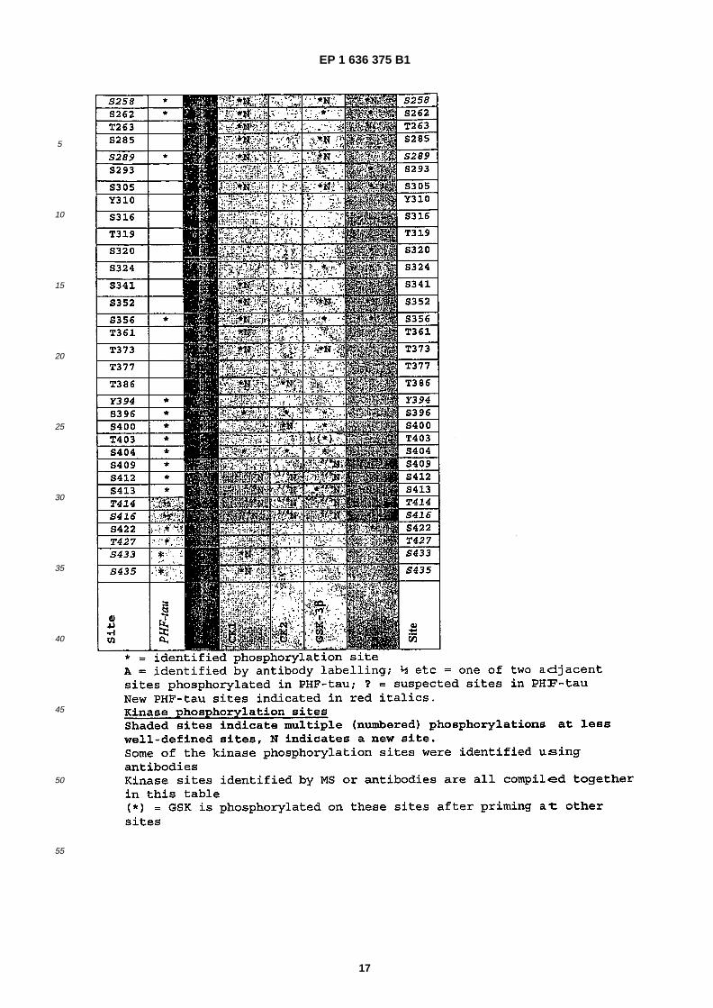
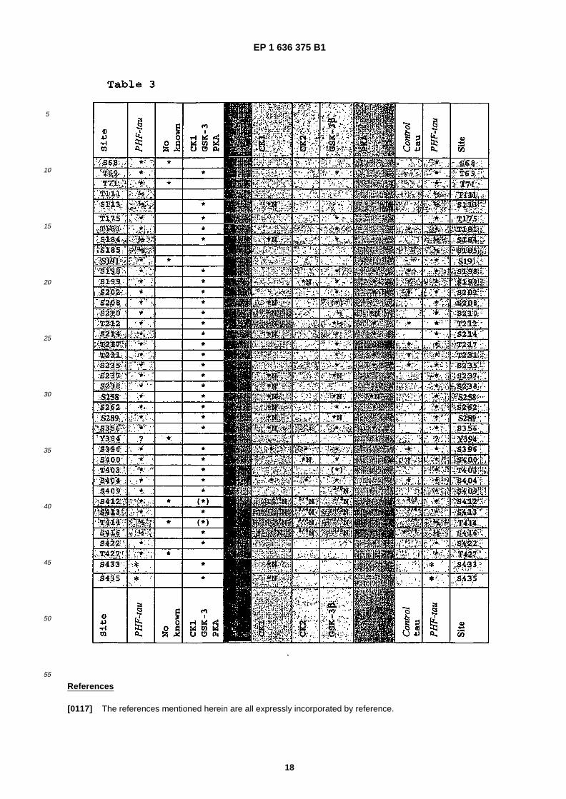
• Ward, Malcolm, �Proteome Sciences plcCobham, �Surrey KT11 3EP (GB) �
• Byers, Helen, �Proteome Sciences plc. �Cobham, �Surrey KT11 3EP (GB) �
(74) Representative: Kiddle, Simon JohnMewburn Ellis LLP York House, 23 KingswayLondon WC2B 6HP (GB) �
(56) References cited: WO- �A- �95/19178 US-�A- 6 057 117
• MEIJER L ET AL: "INHIBITION OF CYCLIN- �DEPENDENT KINASES, GSK- �3SS AND CK1 BY HYMENIALDISINE, A MARINE SPONGE CONSTITUENT" CHEMISTRY AND BIOLOGY, CURRENT BIOLOGY, LONDON, GB, vol. 7, no. 1, January 2000 (2000-01), pages 51-63, XP000901413 ISSN: 1074-5521
• KURET JEFF ET AL: "Casein kinase 1 is tightly associated with paired- �helical filaments isolated from Alzheimer’s disease brain" JOURNAL OF NEUROCHEMISTRY, vol. 69, no. 6, December 1997 (1997-12), pages 2506-2515, XP002308829 ISSN: 0022-3042
• SINGH T J ET AL: "PHOSPHORYLATION OF TAU PROTEIN BY CASEIN KINASE- �1 CONVERTS IT TO ANABNORMAL ALZHEIMER- �LIKE STATE" JOURNAL OF NEUROCHEMISTRY, NEW YORK, NY, US, vol. 64, no. 3, 1995, pages 1420-1423, XP000886450 ISSN: 0022-3042
• LEE G ET AL: "Tyrosine phosphorylation of tau" SOCIETY FOR NEUROSCIENCE ABSTRACTS, vol. 27, no. 1, 2001, page 1436, XP002308830 & 31ST ANNUAL MEETING OF THE SOCIETY FOR NEUROSCIENCE; SAN DIEGO, CALIFORNIA, USA; NOVEMBER 10-15, 2001 ISSN: 0190-5295
2
EP 1 636 375 B1
• TROJANOWSKI JOHN Q ET AL: "Phosphorylation of paired helical filament tau in Alzheimer’s disease neurofibrillary lesions: Focusing on phosphatases" FASEB JOURNAL, vol. 9, no. 15, 1995, pages 1570-1576, XP002308831 ISSN: 0892-6638
• LARNER A J: "Tau protein as a therapeutic target in Alzheimer’s disease and other neurodegenerative disorders" EXPERT OPINION ON THERAPEUTIC PATENTS 1999 UNITED KINGDOM, vol. 9, no. 10, 1999, pages 1359-1370, XP002308832 ISSN: 1354-3776
• CASTRO A ET AL: "Inhibition of tau phosphorylation: A new therapeutic strategy for the treatment of Alzheimer’s disease and other neurodegenerative disorders" EXPERT OPINION ON THERAPEUTIC PATENTS 2000 UNITED KINGDOM, vol. 10, no. 10, 2000, pages 1519-1527, XP008039626 ISSN: 1354-3776
• SINGH TOOLSSE J ET AL: "Non- �proline- �dependent protein kinases phosphorylate several sites found in tau from Alzheimer disease brain" MOLECULAR AND CELLULAR BIOCHEMISTRY, vol. 154, no. 2, 1996, pages 143-151, XP008039649 ISSN: 0300-8177
• RODER HANNO M: "Prospect of therapeutic approaches to tauopathies." JOURNAL OF MOLECULAR NEUROSCIENCE, vol. 20, no. 2, April 2003 (2003-04), pages 197-201, XP008039739 ISSN: 0895-8696
EP 1 636 375 B1
3
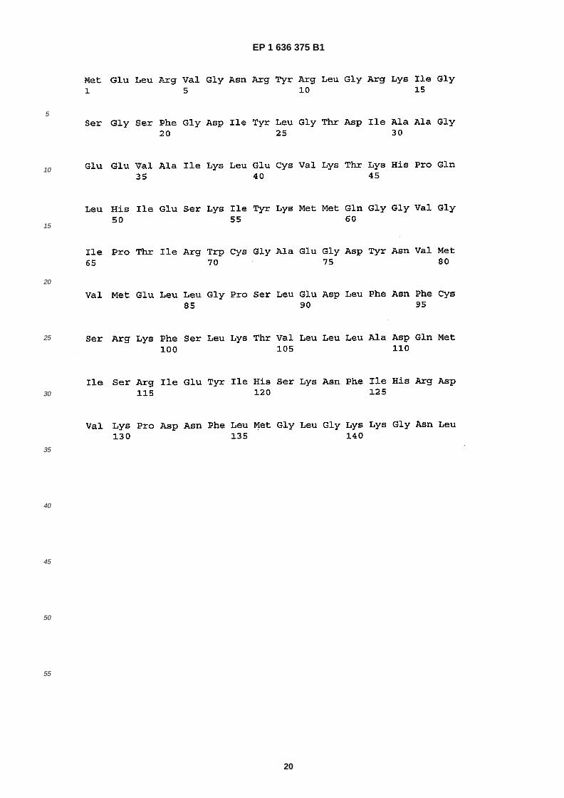
5
10
15
20
25
30
35
40
45
50
55
Description
Field of the Invention
�[0001] The present invention relates to methods for screening for substances capable of modulating the phosphor-ylation of tau protein, and in particular paired helical filament (PHF) tau. The assays and screening methods are basedon the identification of new phosphorylation sites in PHF tau and new kinases and combinations of kinases as therapeutictargets.
Background of the Invention
�[0002] Alzheimer’s disease (AD) is a neurodegenerative disease characterised by the presence of senile plaques andneurofibrillary tangles in the brain. The degree of dementia at death correlates better with neurofibrillary tangle numbersthan with senile plaques counts. The presence of neurofibrillary tangles in neurons results in the death of those neurons,implying that prevention of tangle formation is an important therapeutic goal. The principal protein that forms the neu-rofibrillary tangle is the microtubule-�associated protein, tau, which assembles into filaments that have the appearanceof twisting about each other in pairs and are referred to as paired helical filaments (PHF). PHF are present in differentlocations in degenerating neurons in the Alzheimer brain and when many aggregate in the neuronal cell body, theyproduce the neurofibrillary tangle (Lee et al, 2001).�[0003] Senile plaques have an extracellular central deposit of amyloid β-�peptide (Aβ), which is surrounded by dystrophicneurites to form the senile or neuritic plaque. In vitro and in vivo Aβ has been shown to be neurotoxic. Aβ is derived byproteolytic processing of the larger amyloid precursor protein (APP). Much attention has been focused on Aβ productionas a therapeutic target because its production is believed to be an early event in AD pathogenesis. This is becausemutations in the APP gene, which give rise to autosomal dominant AD, result in either increased overall production ofAβ or in a relative increase in the slightly longer Aβ42 over Aβ40 the former being more amyloidogenic; Aβ42 has twoadditional hydrophobic amino acids at the C-�terminus of 40-�residue Aβ40 thereby endowing the peptide with an increasedtendency to aggregate and form amyloid fibres. Mutations in two other genes that also cause autosomal dominant AD,presenilin-�1 and presenilin-�2 (PS1 & PS2) also result in an increase in the ratio of Aβ42 to Aβ40. The belief that Aβdeposition in the brain precedes the appearance of neurofibrillary tangles has been the basis of the amyloid cascadehypothesis but it has been uncertain whether tangles are important in pathogenesis or are only an unimportant epiphe-nomenon. This has been changed by the discovery of mutations in the gene for tau in some other related neurodegen-erative diseases.�[0004] The mechanism by which Aβ kills neurons in the brain has still to be established. Many studies of Aβ toxicityhave been conducted in tissue culture using rat brain neuronal cultures. We have shown that exposure of both foetalrat and human brain neuronal cultures to aggregated Aβ induces within 2 to 10 minutes increases in the phosphotyrosinecontent of several proteins but also including tau (Williamson et al 2002). We have also shown that this treatment resultsin activation of the tyrosine kinase fyn, a member of the src family of tyrosine kinases. This tyrosine phosphorylation oftau was prevented by inhibitors of the src family of tyrosine kinases.�[0005] It has previously been reported that increased levels of fyn are associated with neurons containing abnormallyphosphorylated tau in AD brain (Shirazi et al, 1993) and we have demonstrated using antibodies that recognise phos-photyrosine that PHF-�tau from AD brain contains phosphotyrosine (Williamson et al 2002). We have shown in vitro thatfyn and Lck, both src family kinases, phosphorylate recombinant human tau and phosphotyrosines 18, 310 and 394were positively identified in one or more of their respective tryptic peptides, from sequence information of fragmentedpeptides. In addition, phosphotyrosine at position 197 was inferred from peptide masses in the survey scan (Scales etal, 2002).�[0006] Intraneuronal deposits of tau in the form of typical neurofibrillary tangles of AD or other morphologically distincttau aggregates in a number of other neurodegenerative diseases, is the basis for grouping these conditions as tauopa-thies. Thus, in addition to AD, the main examples of the tauopathies are frontotemporal dementia with Parkinsonismlinked to chromosome 17 (FTDP-�17), progressive supranuclear palsy (PSP), Pick’s disease, corticobasal degeneration,and multisystem atrophy (MSA). The intracellular tau deposits (usually neuronal but can also be glial) are all filamentousand mostly in a hyperphosphorylated state compared to the level of phosphorylation of tau from control human brain.In the case of AD, this hyperphosphorylated tau is often referred to as PHF-�tau because it is derived from the PHF.�[0007] Other than for AD, deposits of Aβ in the brain are either absent or minimal in these other tauopathies. Thereare some tauopathy pedigrees with autosomal dominant disease in which the causative gene has been identified as thetau gene and although cases with the same mutation may present with apparently different diseases, they invariablyhave tau deposits in the brain and are mostly of the FTDP-�17 variety. Thus, the finding of mutations in the tau genewhich result in disease and deposition of tau aggregates in neurons is compelling evidence for the primary pathogenicimportance of tau deposition in all of these conditions, including AD, whatever the primary cause of disease. Therefore,
EP 1 636 375 B1
4
5
10
15
20
25
30
35
40
45
50
55
the amyloid cascade hypothesis is borne out by the discovery of tau mutations and confirms that indeed neurofibrillarytangle formation is almost certainly subservient to Aβ deposition in AD, but that in the other tauopathies lacking Aβdeposits, then some other primary event must trigger the tau pathology. Tau abnormalities and deposition are thereforeimportant therapeutic targets for all tauopathies, including AD.�[0008] Tau is a phosphoprotein, the function of phosphorylation remaining to be unequivocally established. However,increased phosphorylation of tau on multiple serine and threonine residues reduces the ability of tau to promote micro-tubule assembly and to stabilise assembled microtubules, effects that have been demonstrated both in vitro and in cells.Many studies have shown that PHF-�tau from AD brain is more heavily phosphorylated on serine and threonine than taufrom control brain. This has been demonstrated partly by protein sequencing and partly by demonstrating that certainmonoclonal antibodies only label either PHF-�tau or non- �phosphorylated tau and not PHF- �tau; the epitopes for many ofthese antibodies have been mapped to particular phosphorylated residues present in PHF-�tau and absent from controlbrain tau. The pathological tau from most other cases of other tauopathies seems to be similarly hyperphosphorylatedto PHF-�tau.�[0009] These findings strongly imply that similar abnormalities in regulating phosphorylation of tau are shared by allthe tauopathies including AD. Since phosphorylation of proteins is effected by protein kinases and dephosphorylationby protein phosphatases, identifying the protein kinases and phosphatases for tau is important because they are poten-tially therapeutic targets for these diseases.�[0010] It remains a considerable problem in the art in identifying the enzymes responsible for causing phosphorylationof paired helical filament tau and the sites phosphorylated by those enzymes.�[0011] Meijer at al (CHEMISTRY AND BIOLOGY, CURRENT BIOLOGY, vol. 7, no. 1, January 2000, pages 51-63)disclosed a marine sponge constituent which is a potent inhibitor of cyclin-�dependent kinases, glycogen synthase kinase-3beta and casein kinase 1. The constituent, hymenialdisine, also blocks the in vivo phosphorylation of the tau protein,at sites which are hyperphosphorylated by GSK- �3 and CDK5/p35 in Alzheimer’s disease.�[0012] US 6,057,117 discusses the use of selective inhibitors of GSK3 for the treatment of diseases that are mediatedby GSK3 activity, including non-�insulin dependent diabetes mellitus and Alzheimer’s disease. Methods of identifyingthese inhibitors are also described.�[0013] Lee G et al ("Tyrosine phosphorylation of tau", SOCIETY FOR NEUROSCIENCE ABSTRACTS, vol. 27, no.1, 2001, page 1436) examined the tyrosine phophorylation of tau protein, and found that in non-�neuronal cells tau istyrosine phophorylated when co-�transfected with fyn. They also found that in vitro, tau can be tyrosine phophorylatedby fyn and speculated that tyrosine phosphorylated tau has a role in signal transduction in neuronal cells.�[0014] WO 95/19178 concerns methods for treating Alzheimer’s disease and related disorders by increasing theactivity of a phosphatase towards abnormal hyperphosphorylated tau present in neurofibrillary tangles of paired helicalfilaments in patients.�[0015] Trojanowski et al (FASEB JOURNAL, vol. 9, no. 15, 1995, pages 1570-1576) concerns the phosphorylation ofpaired helical filament tau in Alzheimer’s disease neurofibrillary lesions.�[0016] Singh et al (JOURNAL OF NEUROCHEMISTRY, NEW YORK, NY, US, vol. 64, no. 3, 1995, pages 1420-1423)disclosed that the phosphorylation of tau prtein by casein dependent kinase 1 converts it to an abnormal Alzheimer-�likestate. The same group later disclosed that non-�proline-�dependent protein kinases phosphorylate several sites found intau protein from Alzheimer diseased brains (MOLECULAR AND CELLULAR BIOCHEMISTRY, vol. 154, no. 2, 1996,pages 143-151).
Summary of the Invention
�[0017] Broadly, the present invention relates to the modulation of the phosphorylation of tau protein through its inter-action with kinases and phosphatases. In particular, it is based on the identification of new sites in tau protein that aresusceptible to phosphorylation by kinases and to the identification of kinases and combinations of kinases that arecapable of phosphorylating new and known phosphorylation sites in tau protein. Importantly, many of the newly identifiedsites are present in paired helical filament (PHF) tau, and not in control tau or fetal tau.�[0018] The present invention is based on the analysis by mass spectrometry PHF-�tau and tau from control adult andfoetal rat brain, and identifies 12 new sites in PHF- �tau, bringing the total to 37 phosphorylation sites (1 site is tyrosine394 and the other 36 are either serine or threonine residues) and this with >90% sequence coverage. Of these 12 sites,11 have not been found in tau from normal human brain.�[0019] A number of protein kinases have been demonstrated to phosphorylate tau in vitro, including glycogen synthasekinase- �3α (GSK- �3α), glycogen synthase kinase-�3β (GSK-�3β), MAP kinases (ERKs 1 & 2), cdk5, cdc2 kinase, JNK,several members of the SAP kinases (1γ, 2a, 2b, 3, 4), p38MAP kinase, calmodulin- �dependent kinase, protein kinaseA (PKA), protein kinase C (PKC), casein kinase 1 (CK1), casein kinase 2 (CK2), MARK, PKN, PKB, TTK, DYRK, Rhokinase and phosphorylase kinase. Of these kinases, GSK-�3 has been demonstrated to phosphorylate the greatestnumber of identified sites in PHF- �tau, this being 25 sites, including 2 sites that are generated by GSK- �3 only when tau
EP 1 636 375 B1
5
5
10
15
20
25
30
35
40
45
50
55
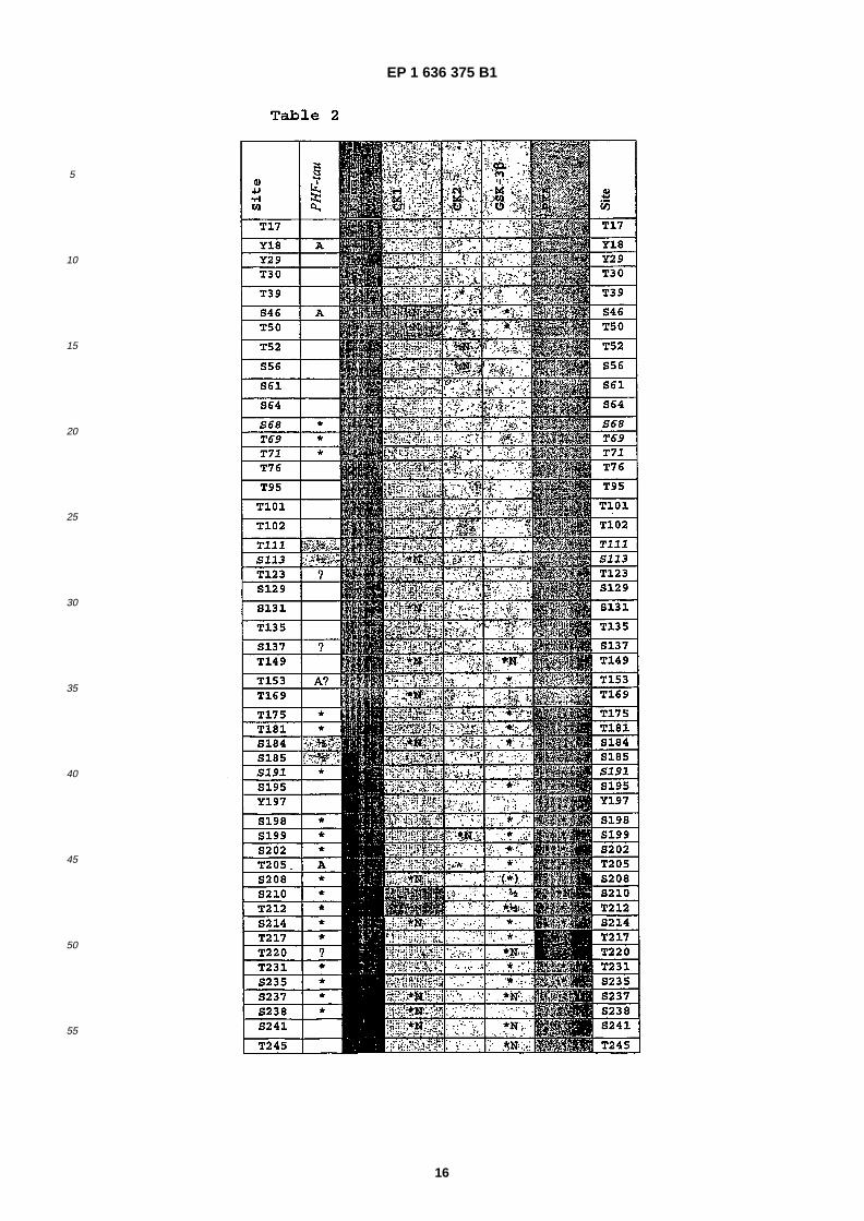
is already phosphorylated and PKA phosphorylates 16 sites in PHF-�tau. We have now shown also by in vitro phospho-rylation that CK1 is also a candidate kinase for 6 of the 12 newly identified sites, GSK-�3 phosphorylates 4 of these andPKA phosphorylates 2 of the new PHF-�tau sites. This brings the total number of sites in PHF-�tau that can be phospho-rylated by CK1 to 17 sites.�[0020] The MAP kinases (ERKs 1 & 2), cdk5, cdc2 kinase, JNK, several members of the SAP kinases (1γ, 2a, 2b, 3,4),� and p38MAP kinase are similar in specificity to GSK- �3, being essentially proline-�directed protein kinases and theyall phosphorylate most of the sites phosphorylated by GSK-�3. Thus, after these proline-�directed protein kinases, CK1is now the most conspicuous kinase as a candidate for contributing to generating the phosphorylation state of PHF-�tau,with only 18 CK1 sites definitely shared by GSK-�3. Therefore, of the 36 ser/thr sites in PHF-�tau, 31 could potentially bephosphorylated by a combination of GSK-�3, CK1, and PKA, and the additional 5 sites remain as orphan sites with nokinase known to phosphorylate these residues. It is possible that GSK-�3, CK1 or PKA could phosphorylate some or allof these orphan sites or indeed that one or more of the other potential tau kinases listed above could be responsibleand the phosphorylated sites have not been detected. However, the data disclosed herein imply that CK1 should beconsidered as a strong candidate for generating hyperphosphorylated tau in AD and the other tauopathies and henceis a potential therapeutic target.�[0021] Of the known phosphorylation sites in PHF- �tau, several are considered to be particularly important. Monoclonalantibody, AT100, of all such antibodies is the most specific for PHF- �tau since it does not recognise normal brain tau norfoetal tau; as such it is considered to be diagnostic for pathological hyperphosphorylated tau in the tauopathies. TheAT100 epitope requires phosphorylation of both T212 and S214. It is known that T212 and S214 can be phosphorylatedby GSK-�3 and it has been reported that phosphorylation T212 by GSK-�3 primes tau for phosphorylation at S214 by PKA(Singh et al, 1995a,�b). We have found that CK1 is also able to phosphorylate S214, thereby further implicating CK1 inpathological phosphorylation.�[0022] One other site in tau, S262, has been shown to be important in regulating the binding of tau to microtubulessuch that phosphorylation causes dissociation of tau. A novel kinase, MARK, that phosphorylates tau at this site wasisolated from brain and proposed as the responsible kinase. We have found that CK1 is also able to phosphorylate S262and S356, the latter being an homologous residue that may behave like S262 in contributing to regulating binding of tauto microtubules and we have found that both S262 and S356 are phosphorylated in PHF-�tau.�[0023] Thus, the above two classes of phosphorylation of tau that are considered to be important could be regulatedby CK1. Furthermore, it has been reported that CK1, particularly the CK1δ isoform, is elevated in brain extracts fromAD cases compared to controls, which adds to the potential importance of CK1 in pathogenesis (Ghoshal et al, 1999).�[0024] With respect to tyrosine phosphorylation, PHF- �tau is phosphorylated on tyrosine 394 and fyn is the strongestcandidate although other src family kinases may also phosphorylate tau in brain.�[0025] Accordingly, in one aspect, the present invention proposes that CK1 is a novel therapeutic target for treatingAD and other related tauopathies.�[0026] In a further aspect, the present invention proposes that fyn and related src family kinases are novel therapeutictargets for treating AD and other related tauopathies, in particular for tyrosine phosphorylation sites disclosed herein.�[0027] In a further aspect, the present invention proposes new phosphorylation sites in tau protein for use in screeningfor inhibitors of phosphorylation or promoters of dephosphorylation, optionally used in combination with the kinasesidentified herein as being capable of phosphorylating the sites.�[0028] As a consequence of these findings, the new sites and kinases can be used as the basis of assays and assaymethods for screening for modulators of the phosphorylation of the sites in tau protein for use or development astherapeutics for the treatment of tauopathies. Preferred modulators are capable of inhibiting the phosphorylation of tauto produce a phosphorylated state similar or identical to that of PHF-�tau and/or promoting the dephosphorylation ofphosphorylated forms of PHF-�tau.�[0029] Eleven of the new phosphorylation sites in tau protein are shown in Table 2 in red type in the left hand column.They are the serine and threonine residues at positions S68, T69, T71, (T111/S113), S191, S258, S289, (T414/S416),T427, S433 and S435. A further tyrosine site at position 394 (Y394) has also been identified (e.g. phosphorylated bytyrosine kinases and dephosphorylated by tyrosine phosphatases). Of the 12 sites, 10 are only found in PHF-�tau, seeTable 2 comparing the PHF tau and control tau columns.�[0030] Accordingly, in a further aspect, the present invention provides the use of a tau protein comprising one or moreof these phosphorylation sites as defined herein for screening for candidate substances which are capable of inhibitingphosphorylation at the site�(s) by a kinase.�[0031] In the present invention, the tau protein comprising the phosphorylation sites may be substantially full lengthand/or wild type tau or PHF- �tau protein, or may be a fragment, active portion or sequence variant thereof. In otherembodiments, the present invention may employ a corresponding nucleic acid molecule encoding the tau protein. Wherea tau protein which is a fragment, active portion or sequence variant is employed, the phosphorylation site�(s) may bepresent with surrounding amino acids from the tau protein sequence. Preferably, the present invention employs PHF-tau protein. In the present invention the numbering of tau and PHF-�tau is according to the sequence of the longest brain
EP 1 636 375 B1
6
5
10
15
20
25
30
35
40
45
50
55
isoform of human tau (441 amino acids) disclosed in Goedert et al (1989) EMBO J. 1989 Feb;�8 �(2): �393-9. Cloning andsequencing of the cDNA encoding an isoform of microtubule- �associated protein tau containing four tandem repeats:differential expression of tau protein mRNAs in human brain. Goedert M, Spillantini MG, Potier MC, Ulrich J, CrowtherRA; or Goedert M, Jakes R. (1990) Expression of separate isoforms of human tau protein: correlation with the tau patternin brain and effects on tubulin polymerization. EMBO J., 9, 4225-30.�[0032] Alternatively or additionally, any of the above defined tau proteins may possess phosphorylation at one or moreof the phosphorylation sites. This enables the effects of cooperative phosphorylation of the protein to be studied, thatis, where the phosphorylation of one site is dependent in changes to the tau protein caused by one or more precedingor simultaneous phosphorylation steps. Thus, in some embodiments of the present invention, the tau protein may includeone or more of the known tau phosphorylation sites, for example those set out in Table 2, left hand column in black type,in addition to one or more of the newly found sites, and optionally have phosphorylation at one or more of those additionalsites.�[0033] In a further aspect, the present invention provides a method of screening for substances which are capable ofinhibiting phosphorylation by a kinase at one or more of the site �(s) of a tau protein selected from the group consistingof S68, T69, T7, (T111/S113), S191, S258, S289, (T414/S416), T427, S433, S435 and Y394, the method comprising:�
(a) contacting at least one candidate substance, the tau protein as defined herein and a kinase which is capable ofphosphorylating the tau protein under conditions in which the kinase is capable of phosphorylating the site�(s) of thetau protein in the absence of the candidate substance, wherein the kinase is selected from CK1, CK2, PKA, GSK-3 beta and fyn,(b) determining whether, and optionally the extent to which, the candidate substance inhibits the phosphorylationof the tau protein at one or more sites of the tau protein; and,(c) selecting the candidate substance which inhibits phosphorylation of the tau protein at one or more of the sites.
�[0034] In some embodiments, the method may comprise, having identified a candidate substance according to oneof the methods disclosed herein, the further step�(s) of optimising the candidate substance to improve one or more of itsproperties and/or formulating it as a pharmaceutical.�[0035] In the methods and uses disclosed herein, the kinase is selected from casein kinase 1 (CK1), casein kinase 2(CK2), protein kinase A (PKA), glycogen synthase kinase 3α (GSK-�3α), and glycogen synthase kinase 3β (GSK-�3β).More preferably, the kinase is CK1 or a combination (either simultaneously or sequentially applied) of CK1, PKA andGSK-�3β.�[0036] In the present invention, preferably the step of detecting the presence and extent of phosphorylation anddephosphorylation in the tau protein can be carried out using mass spectroscopy as described in detail below. Alterna-tively, or additionally, site specific recognition agents which are capable of distinguishing between a site which is phos-phorylated and one which is not may be used. Examples of such agents known in the art are site specific antibodiessuch as monoclonal antibody AT100.�[0037] Substances obtainable from one of the methods disclosed herein are capable of inhibiting the phosphorylationor promoting the dephosphorylation of a tau protein at one or more of the above defined sites.�[0038] A further aspect of the present invention is based on the finding that casein kinase 1 is capable of phosphorylatinga tau protein at previously unknown positions. Some of the positions are known or suspected in the art of being phos-phorylation sites, while others are among the phosphorylation sites identified herein for the first time. The sites of PHF-tau protein that are phosphorylated by CK1 include (S46/T50), S113, S131, T149, T169, S184, S208, (S210/T212),S214, S237, S238, S241, S258, S262, T263, S285, S289, S305, S341, S352, S356, T361, T373, T386,(S412/S413/T414/S416 -two of these four), S416, S433 and S435. Of these sites, S113, 184, 208, (210/212), 214, 237,238, S258, S289, S433 and S435 are disclosed as phosphorylation sites of PHF-�tau protein for the first time herein. Thesequence of casein kinase 1 is provided in J Biol Chem. 1993 Mar 25;�268 �(9): �6394-401. Molecular cloning, expression,and characterization of a 49-�kilodalton casein kinase I isoform from rat testis. Graves PR, Haas DW, Hagedorn CH,DePaoli-�Roach AA, Roach PJ.�[0039] In a further aspect, the invention provides a method of screening for candidate substances which are capableof (a) inhibiting the activity of casein kinase 1 in phosphorylating a tau protein at one or more sites; or (b) binding tocasein kinase 1 to inhibit its interaction with a tau protein at one or more sites, the method comprising the step ofdetermining whether, and optionally the extent to which, the candidate substances have the property of (a) or (b) underconditions in which the casein kinase 1 is capable of (a) or (b) in the absence of the candidate substance, wherein theone or more sites are selected from the group consisting of (S46/T50) S113, S131, T149, T169, S184, S208, (S210/T212),S214, S237, S238, S241, S258, S262, T263, S285, S289, S305, S341, S352, S356, T361, T373, T386,(S412/S413/T414), S416, S433 and S435 of tau protein.�[0040] Preferably, the above method comprises the steps of:�
EP 1 636 375 B1
7
5
10
15
20
25
30
35
40
45
50
55
(a) contacting at least one candidate substance, the tau protein and casein kinase 1 under conditions in which thecasein kinase 1 is capable of phosphorylating the site�(s) of the tau protein in the absence of the candidate substance;(b) determining whether, and optionally the extent to which, the candidate substance inhibits the phosphorylationof the tau protein at the one or more sites of the tau protein by casein kinase 1; and,(c) selecting the candidate substance which inhibits phosphorylation of the tau protein at the one or more of the sites.
�[0041] In a further aspect, the present application also discloses that a combination of kinases is required to phos-phorylate the majority of the phosphorylation sites disclosed herein or in the prior art. In the experiments disclosed herein,a combination of casein kinase 1 (CK1), protein kinase A (PKA) and glycogen synthase kinase 3β (GSK-�3β) was foundto be capable, either alone or in cooperation, of phosphorylating the majority of the phosphorylation sites of tau proteinand in particular PHF- �tau protein. This combination of kinases can be used simultaneously or sequentially to screen formodulators of tau phosphorylation, in contrast to prior art proposals that have focussed on screening using a single kinase.�[0042] Accordingly, the present invention provides methods involving the use of a casein kinase 1 (CK1), proteinkinase A (PKA) and glycogen synthase kinase 3β (GSK-�3β) (including fragments, active portions or sequence variants),or a corresponding nucleic acid molecule, for screening for candidate compounds which are capable of (a) inhibiting theactivity of casein kinase 1 in phosphorylating a tau protein or (b) binding to casein kinase 1 to inhibit its interaction with a tau.�[0043] In a further aspect, the present invention provides a method of screening for substances which are capable ofinhibiting the phosphorylation of a tau protein by casein kinase 1 (CK1), protein kinase A (PKA) and glycogen synthasekinase 3β (GSK-�3β), wherein the tau protein comprises one or more phosphorylation sites selected from the goupconsisting of S68, T69, T7 (T111/S113), S191, S258, S289, (T414/S416), T427, S43, S435 and Y394 the methodcomprising: �
(a) contacting at least one candidate substance, the tau protein as defined herein and casein kinase 1 (CK1), proteinkinase A (PKA) and glycogen synthase kinase 3β (GSK-�3β) under conditions in which the kinases are capable ofphosphorylating the site �(s) of the tau protein in the absence of the candidate substance;(b) determining whether, and optionally the extent to which, the candidate substance inhibits the phosphorylationof the tau protein at one or more sites of the tau protein by the kinases; and,(c) selecting the candidate substance which inhibits phosphorylation of the tau protein at one or more of the sites.
�[0044] Embodiments of the present invention will now be discussed in more detail by way of example and not limitationwith reference to the accompanying tables.
Tables
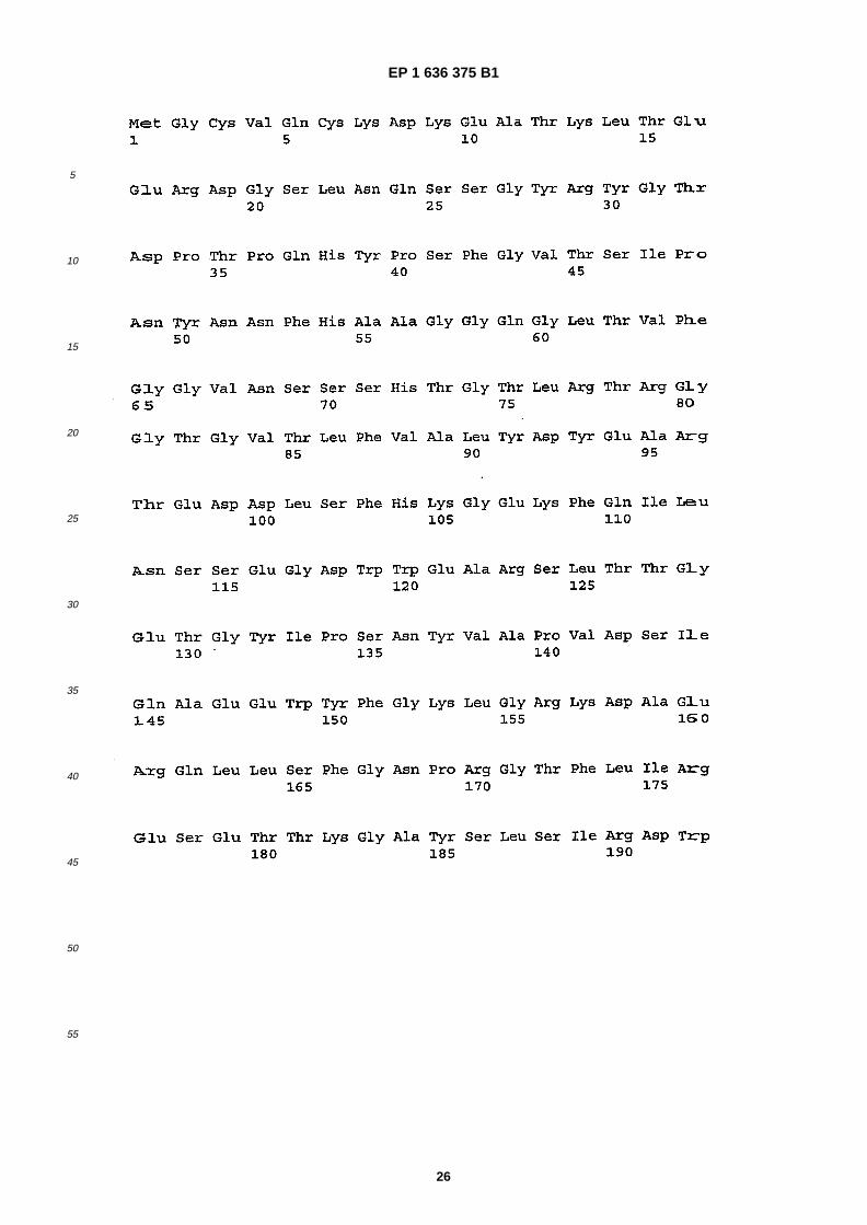
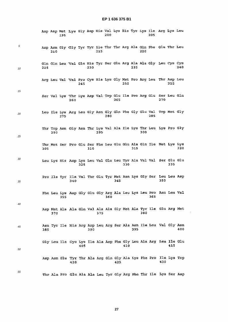
�[0045] Table 1 summarises the new sites found in the work leading to the present invention and the kinases capableof acting at those sites. Tables 2 and 3 present this data in more detail.�[0046] SEQ ID NO: 1 shows the amino acid sequence of rat casein kinase 1, a 428 amino acid protein.�[0047] SEQ ID NO: 2 shows the amino acid sequence of the long form of human tau protein, a 441 amino acid protein.�[0048] SEQ ID NO: 3 shows the amino acid sequence of human fyn kinase, a 537 amino acid protein.
Detailed Description
Tau proteins
�[0049] The assays and assay methods disclosed herein can employ wild-�type or full length tau proteins, kinases orphosphatases or fragments, active portions or derivatives thereof. In the case of tau proteins, the materials used in theassays may be unphosphorylated or partially phosphorylated as discussed above.�[0050] In the present invention, derivatives of the tau proteins, kinases (especially CK1 kinase or fyn kinase) orphosphatases have an amino acid sequence which differs by one or more amino acid residues from the wild-�type aminoacid sequence, by one or more of addition, insertion, deletion and substitution of one or more amino acids. Thus, variants,derivatives, alleles, mutants and homologues, e.g. from other organisms, are included. Thus, a derivative of tau proteinor CK1 kinase or fyn kinase may include 1, 2, 3, 4, 5, greater than 5, or greater than 10 amino acid alterations such assubstitutions with respect to the wild-�type sequence.�[0051] Preferably, a fragment or derivative of a protein used in the assays disclosed herein preferably shares sequenceidentity with the corresponding portion of the relevant wild-�type sequence of the protein, and preferably has at leastabout 60%, or 70%, or 75%, or 80%, or 85%, 90% or 95% sequence identity. As is well-�understood, identity at the aminoacid level is generally in terms of amino acid identity which may be defined and determined by the TBLASTN program,of Altschul et al. (1990) J. Mol. Biol. 215: 403-10, which is in standard use in the art. Identity may be over the full- �length
EP 1 636 375 B1
8
5
10
15
20
25
30
35
40
45
50
55
of the relevant peptide or over a contiguous sequence of about 5, 10, 15, 20, 25, 30, 35, 50, 75, 100 or more aminoacids, compared with the relevant wild-�type amino acid sequence. Alternatively, nucleic acid encoding a fragment orderivative may hybridise to the corresponding wild type nucleic acid under stringent conditions, for example as disclosedin textbooks such as Ausubel, Short Protocols in Molecular Biology, 1992 or Sambrook et al, Molecular Cloning, ALaboratory Manual, Cold Spring Harbor Laboratory Press, 1989, using a hybridization solution comprising: 5X SSC, 5XDenhardt’s reagent, 0.5-1.0% SDS, 100 Pg/ml denatured, fragmented salmon sperm DNA, 0.05% sodium pyrophosphateand up to 50% formamide. Hybridization is carried out at 37-42°C for at least six hours. Following hybridization, filtersare washed as follows: (1) 5 minutes at room temperature in 2X SSC and 1% SDS; (2) 15 minutes at room temperaturein 2X SSC and 0.1% SDS; (3) 30 minutes-�1 hour at 37°C in 1X SSC and 1% SDS; (4) 2 hours at 42-65°C in 1X SSCand 1% SDS, changing the solution every 30 minutes.�[0052] One common formula for calculating the stringency conditions required to achieve hybridization between nucleicacid molecules of a specified sequence homology is (Sambrook et al., 1989):�Tm = 81.5°C + 16. 6Log [Na+] + 0. 41 �(% G+C) - 0.63 (% formamide) - 600/ �#bp in duplex�[0053] As an illustration of the above formula, using [Na+] = [0.368] and 50% formamide, with GC content of 42% andan average probe size of 200 bases, the Tm is 57°C. The Tm of a DNA duplex decreases by 1 - 1.5°C with every 1%decrease in homology. Thus, targets with greater than about 75% sequence identity would be observed using a hybrid-ization temperature of 42°C. Such a sequence would be considered substantially homologous to the nucleic acid se-quence of the present invention.
Methods of Screening for Inhibitors and Enhancers
�[0054] It is well known that pharmaceutical research leading to the identification of a new drug may involve the screeningof very large numbers of candidate substances, both before and even after a lead compound has been found. This isone factor which makes pharmaceutical research very expensive and time- �consuming. Means for assisting in the screen-ing process can have considerable commercial importance and utility.�[0055] As detailed above, methods of screening for a substance which are inhibitors of phosphorylation of tau proteinor promoters of dephosphorylation of tau protein can be carried out by contacting one or more test substances with thetau protein and kinase or phosphatase (as defined herein) in a suitable reaction medium, and determining the presenceor extent of phosphorylation of dephosphorylation in the presence and absence of the candidate substance. A differencein activity in the presence and absence of the candidate substance is indicative of a modulating effect.�[0056] Preliminary assays in vitro may be followed by, or run in parallel with, in vivo assays.�[0057] Of course, the person skilled in the art will design any appropriate control experiments with which to compareresults obtained in test assays.�[0058] Performance of an assay method according to the present invention may be followed by isolation and/ormanufacture and/or use of a compound, substance or molecule which tests positive for ability to modulate interactionbetween one of the phosphorylation sites of tau protein (as defined herein) and a kinase (such as CK1 or a combinationof CK1, PKA and GSK-�3β) or a phosphatase.�[0059] The precise format of an assay of the invention may be varied by those of skill in the art using routine skill andknowledge. For example, interaction between substances may be studied in vitro by labelling one with a detectable labeland bringing it into contact with the other which has been immobilised on a solid support. Suitable detectable labels,especially for peptidyl substances include 35S-�methionine which may be incorporated into recombinantly produced pep-tides and polypeptides. Recombinantly produced peptides and polypeptides may also be expressed as a fusion proteincontaining an epitope which can be labelled with an antibody.�[0060] The protein which is immobilized on a solid support may be immobilized using an antibody against that proteinbound to a solid support or via other technologies which are known per se. A preferred in vitro interaction may utilise afusion protein including glutathione-�S-�transferase (GST). This may be immobilized on glutathione agarose beads. In anin vitro assay format of the type described above a test compound can be assayed by determining its ability to diminishthe amount of labelled peptide or polypeptide which binds to the immobilized GST-�fusion polypeptide. This may bedetermined by fractionating the glutathione-�agarose beads by SDS- �polyacrylamide gel electrophoresis. �Alternatively, the beads may be rinsed to remove unbound protein and the amount of protein which has bound can bedetermined by counting the amount of label present in, for example, a suitable scintillation counter.�[0061] The amount of a candidate substance which may be added to an assay of the invention will normally bedetermined by trial and error depending upon the type of compound used. Typically, from about 0.001 nM to 1mM ormore concentrations of putative inhibitor compound may be used, for example from 0.01 nM to 100PM, e.g. 0.1 to 50PM, such as about 10 PM. Greater concentrations may be used when a peptide is the test substance. Even a moleculewhich has a weak effect may be a useful lead compound for further investigation and development.�[0062] Combinatorial library technology provides an efficient way of testing a potentially vast number of differentsubstances for ability to modulate activity of a polypeptide. Such libraries and their use are known in the art. Compounds
EP 1 636 375 B1
9
5
10
15
20
25
30
35
40
45
50
55
which may be used may be natural or synthetic chemical compounds used in drug screening programmes. Extracts ofplants which contain several characterised or uncharacterised components may also be used.�[0063] Antibodies directed to the site of interaction in either protein form a further class of putative inhibitor compounds.Candidate inhibitor antibodies may be characterised and their binding regions determined to provide single chain anti-bodies and fragments thereof which are responsible for disrupting the interaction. Antibodies may also be employed assite specific recognition agents for determining whether phosphorylation of a site in tau protein has occurred during anassay.�[0064] Antibodies may be obtained using techniques which are standard in the art. Methods of producing antibodiesinclude immunising a mammal (e.g. mouse, rat, rabbit, horse, goat, sheep or monkey) with the protein or a fragmentthereof. Antibodies may be obtained from immunised animals using any of a variety of techniques known in the art, andscreened, preferably using binding of antibody to antigen of interest. For instance, Western blotting techniques or im-munoprecipitation may be used (Armitage et al., 1992, Nature 3 57: 80-82). Isolation of antibodies and/or antibody-producing cells from an animal may be accompanied by a step of sacrificing the animal.�[0065] As an alternative or supplement to immunising a mammal with a peptide, an antibody specific for a protein maybe obtained from a recombinantly produced library of expressed immunoglobulin variable domains, e.g. using lambdabacteriophage or filamentous bacteriophage which display functional immunoglobulin binding domains on their surfaces;for instance see WO 92/01047. The library may be naive, that is constructed from sequences obtained from an organismwhich has not been immunised with any of the proteins (or fragments), or may be one constructed using sequencesobtained from an organism which has been exposed to the antigen of interest.�[0066] Antibodies may be modified in a number of ways. Indeed the term "antibody" should be construed as coveringany binding substance having a binding domain with the required specificity. Thus the invention covers antibody frag-ments, derivatives, functional equivalents and homologues of antibodies, including synthetic molecules and moleculeswhose shape mimicks that of an antibody enabling it to bind an antigen or epitope.�[0067] Example antibody fragments, capable of binding an antigen or other binding partner are the Fab fragmentconsisting of the VL, VH, Cl and CH1 domains; the Fd fragment consisting of the VH and CH1 domains; the Fv fragmentconsisting of the VL and VH domains of a single arm of an antibody; the dAb fragment which consists of a VH domain;isolated CDR regions and F �(ab’)�2 fragments, a bivalent fragment including two Fab fragments linked by a disulphidebridge at the hinge region. Single chain Fv fragments are also included.�[0068] A hybridoma producing a monoclonal antibody may be subject to genetic mutation or other changes. It willfurther be understood by those skilled in the art that a monoclonal antibody can be subjected to the techniques ofrecombinant DNA technology to produce other antibodies or chimeric molecules which retain the specificity of the originalantibody. Such techniques may involve introducing DNA encoding the immunoglobulin variable region, or the comple-mentarity determining regions (CDRs), of an antibody to the constant regions, or constant regions plus frameworkregions, of a different immunoglobulin. See, for instance, EP 0 184 187 A, GB 2 188 638 A or EP 0 239 400 A. Cloningand expression of chimeric antibodies are described in EP 0 120 694 A and EP 0 125 023 A.�[0069] Hybridomas capable of producing antibody with desired binding characteristics which can be used in the meth-ods of the invention are host cells, eukaryotic or prokaryotic, containing nucleic acid encoding antibodies (includingantibody fragments) and capable of their expression. The invention also describes methods of production of the antibodiesincluding growing a cell capable of producing the antibody under conditions in which the antibody is produced, andpreferably secreted.�[0070] The reactivities of antibodies on a sample may be determined by any appropriate means. Tagging with individualreporter molecules is one possibility. The reporter molecules may directly or indirectly generate detectable, and preferablymeasurable, signals. The linkage of reporter molecules may be directly or indirectly, covalently, e.g. via a peptide bondor non-�covalently. Linkage via a peptide bond may be as a result of recombinant expression of a gene fusion encodingantibody and reporter molecule. The mode of determining binding is not a feature of the present invention and thoseskilled in the art are able to choose a suitable mode according to their preference and general knowledge.
Mass Spectroscopy
�[0071] An LC/MS/MS based strategy was used to discover new phosphorylation sites within tau protein isolated fromAD brain. So called PHF-�tau was initially extracted from a heat-�stable preparation of human AD brain material andsubsequently further purified by ion exchange chromatography. Having been separated using SDS-�PAGE, phospho-peptide mapping was then undertaken. Coomassie stained bands are excised, reduced, alkylated and enzymaticallydigested using a suite of proteases such as trypsin, chymotrypsin and endoproteinase Asp-�N. Resulting peptide mixturesare then analysed by LC/MS/MS using a Q-�TOF micro instrument with peptide separation achieved using a 75 micronID PepMap reversed phase column with peptides eluted using a gradient of acetonitrile at a flowrate of 200nl/min.�[0072] Database searching against bespoke index files is performed utilising the Mascot algorithm (Matrix Science).All MS/MS spectra relating to phosphopeptides are then subsequently visually verified to check the result.
EP 1 636 375 B1
10
5
10
15
20
25
30
35
40
45
50
55
�[0073] Tandem MS/MS of peptides may be used to provide sequence information by virtue of the fragment ionsproduced. Fragmentation occurs generally across the peptide bond leading to a ladder of sequence ions that are diag-nostic of the amino acid sequence. The difference between consecutive ions in a series indicates the mass of the aminoacid at that position in the peptide. The most common ion types are b and y ions. The C-�terminal containing fragmentsare designated y-�ions and the N-�terminal containing fragments are designated b-�ions (Roepstorff, P., Fohlman, J. J.Biomed. Mass Spectrom. 1984, 11, 601). Peptides created by trypsin proteolysis and ionised by electrospray generallyform ions that are doubly charged. This stems from the presence of basic groups within the peptide, namely, the alphaamino group at the N-�terminus and the side chain of the C- �terminal lysine or arginine. MS/MS spectra of such peptidesgenerally yield a prominent y-�type ion series in the high mass end of the spectrum (Bonner, R., Shushan, B. RapidCommun. Mass Spectrom. 1995, 9, 1067-1076). Ideally, for de novo sequencing purposes, a complete set of comple-mentary b and y ions will ensure a high confidence level for the proposed peptide sequence. Moreover, if fragment ionsrepresenting the complete sequence of the peptide are present, the site of attachment of the phosphate group can bededuced from the position and pattern of these fragment ions. Therefore, it is possible in most instances to discover theexact site of phosphorylation in each phosphopeptide. In some instances we have even found MS/MS spectra to beheterogeneous. Here two (or more) distinct phosphopeptides are represented in the same spectrum. This is becauseeach phosphopeptide form has the same molecule weight and the same number of phosphate groups, but these areattached to different amino acids within the peptide. Therefore, both forms give rise to precursor ions of the same m/zratio, which are then selected simultaneously by the mass spectrometer during the MS/MS experiment. In such cases,we refer to the phosphopeptides concerned as "regiomers"
Multiplex Assays for Screening Compounds
�[0074] In drug development it is desirable to develop rapid high throughput assays with simple read out to showwhether a compound has an effect on the proposed target. In the case of compounds inhibiting an enzyme function,such as a kinase, it is possible to develop an artificial substrate for the target enzyme that is modified by the enzyme ina way that the level of modification can be readily detected. In the presence of an inhibitory compound, the substrate isnot modified and this can also be readily detected.�[0075] In the case of inhibitors of tau phosphorylation, it is necessary to monitor the effect of inhibiting specific proteinkinases on the phosphorylation status of a large number of sites. In one aspect, it is possible to prepare artificial substratescorresponding to each of the phosphorylation sites on tau and assess each compound for their ability to inhibit thephosphorylation of each site independent of the other sites. In such a system, each compound would be added to multiplewells each well containing the proposed kinase target, one of the phosphorylation site-�specific artificial substrates anda reporter system to show phosphorylation, such as a monoclonal antibody that binds specifically to the substrate ineither the phosphorylated or unphosphorylated form, and which antibody is labelled with a fluorescent marker, an enzymethat converts a colour less substrate into a coloured product, or an enzyme that promotes the production of a luminescentsignal. In such an assay, it is desirable that the artificial substrate for the target is immobilised on a solid surface suchthat as part of the assay procedure any unreacted antibody is removed from the system by washing before the result isread. Such assays may be run in microtitre wells of varying formats of typically 96, or more typically 384, or even moretypically 1536 wells, or alternatively may be run on a microarray based on a solid support such as glass.�[0076] Alternatively, the effect of different kinase inhibitors on the global phosphorylation status of tau may be designed.In such an assay, full length recombinant tau protein carrying no phosphorylations, or one or more desirable phospho-rylations may be used as the substrate. Alternatively, a mixture of equal amounts of all of the artificial substrates repre-senting single phosphorylation sites may be used. Each screening assay will determine the effect of compounds on theinhibition of one, two or more protein kinases with known activitity for the phosphorylation of tau. As with the more simpleassays described above substrate, target kinase and compound are added to a well of a microtitre plate and incubatedwith appropriate buffers and other constituents that permit the phosphorylation of substrate in the absence of an inhibitorycompound. The phosphorylation status of the substrate may then be determined using a mixture of antibodies or othermolecules with specificity for individual phosphorylation sites on tau, wherein such antibodies or other molecules areeach labelled with a unique reporter such as a fluorescent dye or compounds with unique spectral properties in infra-red, visible or ultraviolet spectra. After removal of antibodies that remain unbound to the phosphorylated substrate �(s),levels of each specific reporter are determined using an appropriate reading device, and the levels of phosphorylationat each specific site in tau is revealed by comparison with a control where no kinase inhibitor was added.�[0077] In a preferred embodiment of such a multiplex screening assay, the substrate is dephosphorylated recombinanttau protein and the kinase is selected from CK1, CK2, GSK-�3a, GSK-�3b, PKA, CDK5, ERK1/2, SAPK1g, SAPK2a,SAPK2b, SAPK3, SAPK4, stress activated protein kinase family kinases (SAPKs) such as p38MAPK and JNK, MARKfamily kinases such as 110K, cdc2, cdk2, PKC, PKN, TTK, PKB, DYRK, PK, CaMKII, PKD, Rho kinase, or a mixture ofone of more these kinases. Reporter systems are preferably labelled antibodies, typically monoclonal antibodies, forexample those that can be obtained from rabbits or mice using techniques well known in the art. Labels are preferably
EP 1 636 375 B1
11
5
10
15
20
25
30
35
40
45
50
55
fluorescent or colorimetric compounds that are covalently attached to antibodies, more preferably fluorescent or color-imetric nanoparticles and are most preferably nanoparticles with unique Raman spectra.
Development of Mimetic Substances
�[0078] Other candidate inhibitor compounds may be based on modelling the 3-�dimensional structure of a polypeptideor peptide fragment and using rational drug design to provide potential inhibitor compounds with particular molecularshape, size and charge characteristics.�[0079] Once candidate substance have been found in the assays and screens according to the present invention,they may be used to design mimetic compounds for development as drugs. The designing of mimetics to a knownpharmaceutically active compound is a known approach to the development of pharmaceuticals based on a "lead"compound. This might be desirable where the active compound is difficult or expensive to synthesise or where it isunsuitable for a particular method of administration, e.g. peptides are unsuitable active agents for oral compositions asthey tend to be quickly degraded by proteases in the alimentary canal. Mimetic design, synthesis and testing is generallyused to avoid randomly screening large number of molecules for a target property.�[0080] There are several steps commonly taken in the design of a mimetic from a compound having a given targetproperty. Firstly, the particular parts of the compound that are critical and/or important in determining the target propertyare determined. In the case of a peptide, this can be done by systematically varying the amino acid residues in thepeptide, e.g. by substituting each residue in turn. These parts or residues constituting the active region of the compoundare known as its "pharmacophore".�[0081] Once the pharmacophore has been found, its structure is modelled to according its physical properties, e.g.stereochemistry, bonding, size and/or charge, using data from a range of sources, eg spectroscopic techniques, X- �raydiffraction data and NMR. Computational analysis, similarity mapping (which models the charge and/or volume of apharmacophore, rather than the bonding between atoms) and other techniques can be used in this modelling process.�[0082] In a variant of this approach, the three- �dimensional structure of the ligand and its binding partner are modelled.This can be especially useful where the ligand and/or binding partner change conformation on binding, allowing themodel to take account of this in the design of the mimetic.�[0083] A template molecule is then selected onto which chemical groups which mimic the pharmacophore can begrafted. The template molecule and the chemical groups grafted on to it can conveniently be selected so that the mimeticis easy to synthesise, is likely to be pharmacologically acceptable, and does not degrade in vivo, while retaining thebiological activity of the lead compound. The mimetic or mimetics found by this approach can then be screened to seewhether they have the target property, or to what extent they exhibit it. Further optimisation or modification can then becarried out to arrive at one or more final mimetics for in vivo or clinical testing.
Pharmaceutical Compositions
�[0084] Following identification of a substance which modulates or affects phosphorylation or dephosphorylation of tauprotein, the substance may be investigated further. Furthermore, it may be manufactured and/or used in preparation,i.e. manufacture or formulation, of a composition such as a medicament, pharmaceutical composition or drug. Thesemay be administered to individuals.�[0085] The substances identified as kinase inhibitors or phosphatase promoters in the assays and assay methods ofthe present invention, or compounds or substances arising from further development or optimisation, may be formulatedin pharmaceutical compositions. These compositions may be employed for the treatment of tauopathies, that is conditionswhich are characterised by neurofibrillary tangles or aggregates of tau protein. Tauopathies are a recognised class ofconditions known to those skilled in the art and include Alzheimer’s disease,� frontotemporal dementia with Parkinsonismlinked to chromosome 17 (FTDP-�17), progressive supranuclear palsy (PSP), Pick’s disease, corticobasal degeneration,multisystem atrophy (MSA), neurobasal degeneration with iron accumulation, type 1 (Hallervorden-�Spatz), argyrophilicgrain dementia, Down’s syndrome, diffuse neurofibrillary tangles with calcification, dementia pugilistica, Gerstmann-Sträussler-�Scheinker disease, myotonic dystrophy, Niemann-�Pick disease type C, progressive subcortical gliosis, prionprotein cerebral amyloid angiopathy, tangle only dementia, postencephalitic parkinsonism, subacute sclerosing panen-cephalitis, Creutzfeldt-�Jakob disease, amyotrophic lateral sclerosis/ �parkinsonism- �dementia complex, non-�Guamanianmotor neuron disease with neurofibrillary tangles/�dementia, and Parkinson’s disease. The intracellular tau deposits areusually neuronal or glial and are filamentous and generally in a hyperphosphorylated state as compared to the level ofphosphorylation in tau from control human brain. In the case of AD, this hyperphosphorylated tau is often referred to apaired helical filament tau (PHF) tau because it is derived from the PHF.�[0086] These compositions may comprise, in addition to one of the above substances, a pharmaceutically acceptableexcipient, carrier, buffer, stabiliser or other materials well known to those skilled in the art. Such materials should benon-�toxic and should not interfere with the efficacy of the active ingredient. The precise nature of the carrier or other
EP 1 636 375 B1
12
5
10
15
20
25
30
35
40
45
50
55
material may depend on the route of administration, e.g. oral, intravenous, cutaneous or subcutaneous, nasal, intramus-cular, intraperitoneal routes.�[0087] Pharmaceutical compositions for oral administration may be in tablet, capsule, powder or liquid form. A tabletmay include a solid carrier such as gelatin or an adjuvant. Liquid pharmaceutical compositions generally include a liquidcarrier such as water, petroleum, animal or vegetable oils, mineral oil or synthetic oil. Physiological saline solution,dextrose or other saccharide solution or glycols such as ethylene glycol, propylene glycol or polyethylene glycol maybe included.�[0088] For intravenous, cutaneous or subcutaneous injection, or injection at the site of affliction, the active ingredientwill be in the form of a parenterally acceptable aqueous solution which is pyrogen-�free and has suitable pH, isotonicityand stability. Those of relevant skill in the art are well able to prepare suitable solutions using, for example, isotonicvehicles such as Sodium Chloride Injection, Ringer’s Injection, Lactated Ringer’ s Injection. Preservatives, stabilisers,buffers, antioxidants and/or other additives may be included, as required.�[0089] Whether it is a polypeptide, antibody, peptide, nucleic acid molecule, small molecule or other pharmaceuticallyuseful compound that is to be given to an individual, administration is preferably in a "prophylactically effective amount"or a "therapeutically effective amount" (as the case may be, although prophylaxis may be considered therapy), this beingsufficient to show benefit to the individual. The actual amount administered, and rate and time-�course of administration,will depend on the nature and severity of what is being treated. Prescription of treatment, e.g.� decisions on dosage etc,is within the responsibility of general practitioners and other medical doctors, and typically takes account of the disorderto be treated, the condition of the individual patient, the site of delivery, the method of administration and other factorsknown to practitioners. Examples of the techniques and protocols mentioned above can be found in Remington’s Phar-maceutical Sciences, 20th Edition, 2000, pub. Lippincott, Williams & Wilkins. A composition may be administered aloneor in combination with other treatments, either simultaneously or sequentially, dependent upon the condition to be treated.
Materials and Methods
Mass Spectrometry
Data Acquisition
�[0090] Following SDS-�PAGE the gel bands relating to PHF-�tau were excised, reduced, alkylated and digested withtrypsin. Peptides were extracted from the gel pieces by a series of acetonitrile and aqueous washes. The extract waspooled with the initial supernatant and lyophilised. Each sample was then resuspended in 6 ml of 50mM ammoniumbicarbonate and analysed by LC/MS/MS. Chromatographic separations were performed using an Ultimate LC system(Dionex, UK). Peptides were resolved by reverse phase chromatography on a 75 mm C18 PepMap column. A gradientof acetonitrile in 0.05% formic acid was delivered to elute the peptides at a flow rate of 200 nl/min. �Peptides were ionised by electrospray ionisation using a Z-�spray source fitted to a Q-�Tofmicro (Micromass, UK). Theinstrument was set to run in automated switching mode, selecting precursor ions based on their intensity, for sequencingby collision-�induced fragmentation.
Data Analysis
�[0091] The mass spectral data was processed into peak lists and searched against the full- �length sequence of Tau-6 (441 amino acids; mw 45847) using Mascot software (Matrix Science, UK). Phosphorylated peptides were identifiedby selecting phosphate as a variable modification within the searching parameters. Serine, threonine and tyrosinephosphorylation were all considered. The exact location of the modification within each peptide was determined by thepattern of fragment ions produced (see below for further explanation).
Tandem Mass Spectrometry
�[0092] To obtain definitive evidence and determine the exact site of phosphorylation, peptides were separated byreversed-�phase chromatography and sequenced by tandem MS/MS. In these experiments precursor ions relating toeach of the phosphopeptides are individually selected and subjected to collision induced dissociation (CID). Fragmentions so produced are indicative of the sequence of the phosphopeptide and the site of modification is determined by themolecular weight of the relevant fragment ions. Conversely, other potential sites of phosphorylation within particularphosphopeptides can also be ruled out by the presence of other fragment ions within the MS/MS spectrum.�[0093] An unexpected observation is that on some occasions it has been possible to pinpoint several discrete formsof phosphopeptides within a single MS/MS spectrum. Here the phosphopeptides each have the same molecular weight(and so are selected simultaneously), but differ in the site �(s) of phosphorylation. Thus, the fragment ions observed are
EP 1 636 375 B1
13
5
10
15
20
25
30
35
40
45
50
55
effectively a composite representation of each molecule being analysed.
Purification of PHF- �tau from Alzheimer brain
�[0094] Paired helical filament (PHF) tau was purified from Alzheimer brain as described in (Hanger et al, 1998). Briefly,brain tissue was homogenised and insoluble PHF-�tau was recovered by differential centrifugation. Following solubilisationin guanidine and dialysis against a re-�naturing buffer, PHF-�tau was purified by anion- �exchange and reversed-�phasechromatography.
Preparation and purification of recombinant human tau
�[0095] A plasmid expressing the largest tau isoform (2N4R) was used to prepare and purify recombinant human tauas described previously (Mulot et al, 1994). Briefly, a bacterial cell lysate expressing 2N4R tau was heated and centrifugedto remove heat-�labile proteins. The supernatant was fractionated with ammonium sulphate and precipitated materialwas solubilised and dialysed into buffer prior to cation-�exchange chromatography. Proteins were eluted with NaCl andfractions containing tau were pooled and dialysed against ammonium bicarbonate before lyophilisation.
In vitro phosphorylation of recombinant tau by serine/ �threonine protein kinases
�[0096] Recombinant human tau (40Pg/ml) was incubated with 67U/ml casein kinase 1 (CK1), 67U/ml casein kinase2 (CK2), 167U/ml cyclic AMP-�dependent protein kinase (PKA), 67U/ml glycogen synthase-�3β (GSK-�3β) or all four kinasesin combination, each at the stated concentration, in the presence of 3mM ATP for 6h at 30°C. Each kinase was obtainedin a purified recombinant form from New England Biolabs.
In- �gel proteolytic digestion of tau
�[0097] PHF-�tau or in vitro phosphorylated tau proteins were separated on 10 % (wt/vol) polyacrylamide gels andstained with colloidal Coomassie Blue G. Protein bands corresponding to tau were excised, carbamidomethylated, anddigested with proteolytic enzymes (trypsin or Asp-�N). Peptides were extracted from gel pieces by a series of acetonitrileand aqueous washes, dried and resuspended in 50mM ammonium bicarbonate.
Amyloid beta treatment of neurons
�[0098] Rat and human cortical neurons were treated with Aβ peptide (25-35) or reverse Aβ peptide (35-25) for 1-10min. Proteins containing phosphotyrosine were immunoprecipitated and separated by SDS-�PAGE. Western blots ofheat- �stable extracts of neuronal cultures and immunoprecipitates were probed with antibodies to tau.
Results
New sites found in PHF- �tau
�[0099] Current literature reports 25 known phosphorylation sites (all are serine or threonine) identified by direct meansin PHF-�tau (Hanger et al, 1998). There are a further 2-3 sites that have been identified by antibody reactivity only. Wehave found an additional 12 phosphorylation sites in PHF-�tau, one of which is a tyrosine residue (tyr394), bringing thetotal number of sites to 37. Four of the new sites are more amino terminal in tau than any previously reported sites andthree sites are more carboxy terminal than found previously. Of the 12 new sites, 4 are present in alternatively- �splicedregions of tau and therefore are present only in specific tau isoforms, all previously identified PHF- �tau phosphorylationsites are present in all tau isoforms. Only one of the 12 new sites in PHF-�tau (either thr414 or ser416) is detected in taufrom normal brain (ser416).
New sites on recombinant tau for each of the 4 serine/ �threonine kinases investigated
�[0100] See also Table 1�[0101] CK1 found 28 new sites making a total of 30 sites in all. 17 CK1 sites are present in PHF-�tau, including 15 ofthe new CK1 sites. CK1 is a candidate kinase for 6 of the 12 new PHF- �tau sites.�[0102] CK2 found 5 new sites making a total of 8 in all. 5 CK2 sites are present in PHF-�tau, including 3 of the newCK2 sites. CK2 is a candidate kinase for 1 of the 12 new PHF-�tau sites�[0103] GSK-�3 found 12 new sites making a total of 38 in all. 21 GSK- �3 sites are present in PHF-�tau, including 5 of the
EP 1 636 375 B1
14
5
10
15
20
25
30
35
40
45
50
55
new GSK-�3β sites. GSK-�3 is a candidate kinase for 4 of the 12 new PHF- �tau sites�[0104] PKA found 5 new sites making a total of 24 in all. 16 PKA sites are present in PHF-�tau, including 4 of the newPKA sites. PKA is a candidate kinase for 2 of the 12 new PHF-�tau sites�[0105] Comparing PHF- �tau phosphorylation sites with the recombinant tau and kinase data, when all of the phospho-rylation sites for CK1, GSK-�3β, and PKA are combined, 30-33 of the 37 PHF- �tau sites are phosphorylated (3 sites aredefined only as one of two adjacent residues). Of the residual 4 to 7 sites, one is a tyrosine residue that requires tyrosinekinase activity, 4 other sites have no known kinase and the remaining 2 sites are each contained within regions whereonly 1 of 2 nearby residues are phosphorylated (T111 and S185).�[0106] Seven of the 12 new phosphorylation sites in PHF-�tau could be generated by CK1, GSK- �3, or PKA, four haveno known kinase and the fifth site required a tyrosine kinase for phosphorylation.�[0107] Combining the four kinases together in a single reaction, we generated one site (thr111) that was not detectedwith any of the four kinases alone, this residue is not phosphorylated by any other known kinase in vitro. Phosphorylationat this residue is also present in PHF-�tau. These results show that combinations of kinases can result in phosphorylationat new sites, possibly due to conformational changes induced by the primary phosphorylation step that increase thelikelihood of the secondary phosphorylation, possibly by a second enzyme.
Amyloid beta treatment of neurons
�[0108] We found that treatment of neurons with Aβ peptide increased tyrosine phosphorylation of neuronal proteinsincluding tau. The increase in phosphotyrosine induced by Aβ was approximately four times the basal level in tau.
Future experiments
�[0109] Identify phosphorylation sites of other individual and combinations of protein kinases to emulate PHF- �tauphosphorylation in vitro. Kinases that have been implicated in tauopathies include GSK-�3α, ERKs 1 & 2, cdk5, cdc2kinase, JNK, several members of the SAP kinase family (1γ, 2a, 2b, 3, 4), p38MAP kinase, calmodulin- �dependent kinase,PKC, MARK, PKN, PKB, TTK, DYRK, Rho kinase and phosphorylase kinase.�[0110] Determine if phosphorylation of tau with these kinases and other tyrosine kinases induces tau aggregation invitro and in cells. This will allow us to identify the phosphorylation sites that are critical for tau aggregation.�[0111] Investigate the effects of specific protein kinase inhibitors, alone and in combination, on tau aggregation in anin vitro or cellular context.�[0112] Generate transgenic mice (inducibly) expressing CK1 and determine if this model shows cerebral tau deposition.Cross this mouse with other mice expressing candidate kinases (eg a GSK-�3 mouse already exists) and examine therate of tangle formation.�[0113] We have recently found (unpublished) that tyr394 is phosphorylated in AD and in foetal tau and have reportedthat this same residue is phosphorylated by both Fyn and Lck in vitro. Fyn has been shown previously to phosphorylatetau and Fyn is increased in a sub-�set of neurons in AD. It is also known that Aβ treatment of neurons induces tauphosphorylation and that Fyn knock-�out mice are resistant to Aβ.�[0114] We will treat neurons from wild- �type, Fyn knock-�out and Src knock-�out mice with Aβ and identify the phospho-rylation sites on tau in each case.�[0115] It is possible that other tyrosine kinases are involved in tau phosphorylation and aggregation and these includethose associated with growth factor and neurotrophic factor receptors. Other tyrosine kinase families may also beinvolved, including Syk kinase, which has been show to phosphorylate another protein (α-�synuclein) implicated in neu-rodegenerative disease in a manner that increases its propensity to aggregate in vitro. In each case, we will investigatethe effects of phosphorylation on tau aggregation and the effects of kinase inhibition on tau aggregation.�
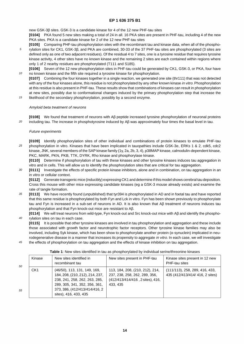
Table 1: New sites identified in tau as phosphorylated by individual serine/�threonine kinases
Kinase New sites identified in recombinant tau
New sites present in PHF-�tau Kinase sites present in 12 new PHF-�tau sites
CK1 (46/50), 113, 131, 149, 169, 184, 208, (210, 212), 214, 237, 238, 241, 258, 262, 263, 285, 289, 305, 341, 352, 356, 361, 373, 386, (412/413/414/416, 2 sites), 416, 433, 435
113, 184, 208, (210, 212), 214, 237, 238, 258, 262, 289, 356, (412/413/414/416 , 2 sites), 416, 433, 435
(111/113), 258, 289, 416, 433, 435 (412/413/414/ 416, 2 sites)
EP 1 636 375 B1
15
5
10
15
20
25
30
35
40
45
50
55
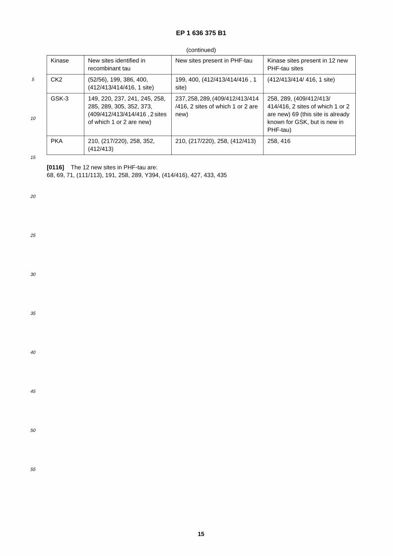
�[0116] The 12 new sites in PHF-�tau are:�68, 69, 71, (111/113), 191, 258, 289, Y394, (414/416), 427, 433, 435
(continued)
Kinase New sites identified in recombinant tau
New sites present in PHF-�tau Kinase sites present in 12 new PHF-�tau sites
CK2 (52/56), 199, 386, 400, (412/413/414/416, 1 site)
199, 400, (412/413/414/416 , 1 site)
(412/413/414/ 416, 1 site)
GSK-�3 149, 220, 237, 241, 245, 258, 285, 289, 305, 352, 373, (409/412/413/414/416 , 2 sites of which 1 or 2 are new)
237, 258, 289, (409/412/413/414 /416, 2 sites of which 1 or 2 are new)
258, 289, (409/412/413/ 414/416, 2 sites of which 1 or 2 are new) 69 (this site is already known for GSK, but is new in PHF-�tau)
PKA 210, (217/220), 258, 352, (412/413)
210, (217/220), 258, (412/413) 258, 416
EP 1 636 375 B1
16
5
10
15
20
25
30
35
40
45
50
55
EP 1 636 375 B1
17
5
10
15
20
25
30
35
40
45
50
55
EP 1 636 375 B1
18
5
10
15
20
25
30
35
40
45
50
55
References
�[0117] The references mentioned herein are all expressly incorporated by reference.
EP 1 636 375 B1
19
5
10
15
20
25
30
35
40
45
50
55
�[0118] Ghoshal N, Smiley JF, DeMaggio AJ, Hoekstra MF, Cochran EJ, Binder LI, Kuret J. (1999) A new molecularlink between the fibrillar and granulovacuolar lesions of Alzheimer’s disease. Am J Pathol. 155, 1163-72.�[0119] Hanger DP, Betts JC, Loviny TL, Blackstock WP, Anderton BH. (1998) New phosphorylation sites identified inhyperphosphorylated tau (paired helical filament-�tau) from Alzheimer’s disease brain using nanoelectrospray massspectrometry. J Neurochem. 71, 2465-76.�[0120] Lambert,�M.P., Barlow, �A.K., Chromy, �B.A., Edwards, �C., Freed, �R., Liosatos,�M., Morgan, �T.E., Rozovsky, �I., Trom-mer, �B., Viola,�K.L., Wals, �P., Zhang, �C., Finch,�C.E., Krafft, �G.A., and Klein,�W.L. (1998). Diffusible, nonfibrillar ligands de-rived from Aβ1-42 are potent central nervous system neurotoxins. Proc. Natl. Acad. Sci. USA 95, 6448-6453.�[0121] Lee VM, Goedert M, Trojanowski JQ. (2001) Neurodegenerative tauopathies. Annu Rev Neurosci. 24, 1121-59.�[0122] Mulot SF, Hughes K, Woodgett JR, Anderton BH, Hanger DP. (1994) PHF-�tau from Alzheimer’s brain comprisesfour species on SDS- �PAGE which can be mimicked by in vitro phosphorylation of human brain tau by glycogen synthasekinase- �3 beta. FEBS Lett. 349, 359-64.�[0123] Scales,�TME, Williamson, R., Anderton, BH, Renolds, CH et al (2002) Tyrosine phosphorylation of specific siteson tau by Src family kinases. Neurobiol Aging 23, S500-501.�[0124] Shirazi,�S.K. and Wood,�J.G. (1993). The protein tyrosine kinase, fyn, in Alzheimer’s disease pathology. Neu-roreport 4, 435-437.�[0125] Singh TJ, Zaidi T, Grundke- �Iqbal I, Iqbal K. (1995a) Modulation of GSK-�3-�catalyzed phosphorylation of micro-tubule-�associated protein tau by non- �proline-�dependent protein kinases. FEBS Lett. 358, 4-8.�[0126] Singh TJ, Haque N, Grundke-�Iqbal I, Iqbal K. (1995b) Rapid Alzheimer-�like phosphorylation of tau by thesynergistic actions of non- �proline- �dependent protein kinases and GSK- �3. FEBS Lett. 358, 267-72.�[0127] Williamson,�R., Scales,�T., Clark,�B.R., Gibb, �G., Reynolds, �C.H., Kellie,�S., Bird, �I.N., Varndell, �I.M., Sheppard,P.W., Everall,�I., and Anderton, �B.H. (2002). Rapid tyrosine phosphorylation of neuronal proteins including tau and focal-adhesion kinase in response to amyloid- �β peptide exposure: involvement of Src family protein kinases. J. Neurosci. 22,10-20.
SEQUENCE LISTING
�[0128]
<110> Proteome Science plcKings College LondonAnderton, BrianHanger, DianeWard, MalcolmByers, Helen<120> Screening Methods<130> SJK/BP6234025<150> GB 0314943.2<151> 2003-06-25<160> 3<170> PatentIn version 3.1<210> 1<211> 428<212> PRT<213> Rattus norvegicus<400> 1
EP 1 636 375 B1
20
5
10
15
20
25
30
35
40
45
50
55
EP 1 636 375 B1
21
5
10
15
20
25
30
35
40
45
50
55
EP 1 636 375 B1
22
5
10
15
20
25
30
35
40
45
50
55
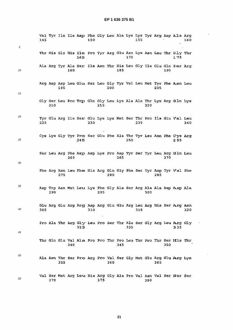
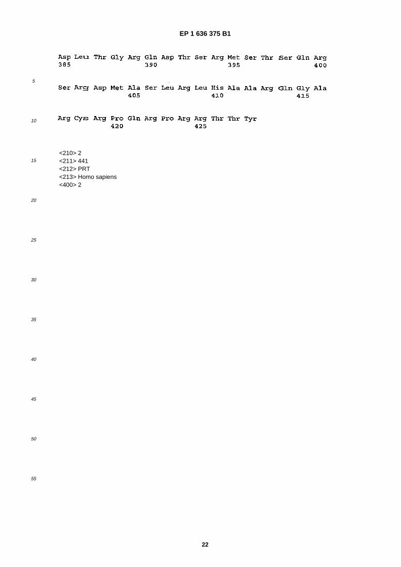
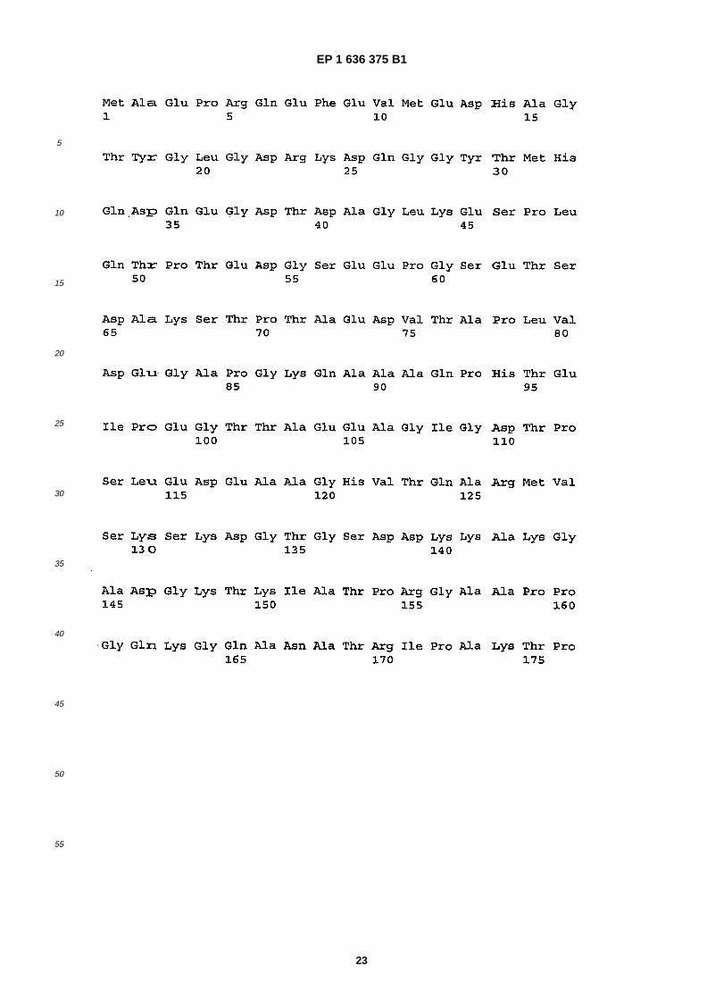
<210> 2<211> 441<212> PRT<213> Homo sapiens<400> 2
EP 1 636 375 B1
23
5
10
15
20
25
30
35
40
45
50
55
EP 1 636 375 B1
24
5
10
15
20
25
30
35
40
45
50
55

EP 1 636 375 B1
25
5
10
15
20
25
30
35
40
45
50
55
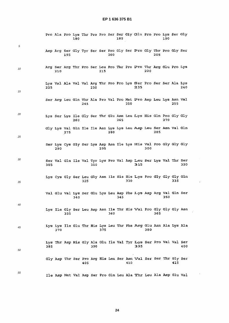
<210> 3<211> 537<212> PRT<213> Homo sapiens<400> 3
EP 1 636 375 B1
26
5
10
15
20
25
30
35
40
45
50
55
EP 1 636 375 B1
27
5
10
15
20
25
30
35
40
45
50
55

EP 1 636 375 B1
28
5
10
15
20
25
30
35
40
45
50
55
Claims
1. A method of screening for candidate substances which are capable of (a) inhibiting the activity of casein kinase 1in phosphorylating a tau protein at one or more sites; or (b) binding to casein kinase 1 to inhibit its interaction witha tau protein at one or more sites, the method comprising the step of determining whether, and optionally the extentto which, the candidate substances have the property of (a) or (b) under conditions in which the casein kinase 1 iscapable of (a) or (b) in the absence of the candidate substance, wherein the one or more sites are selected fromthe group consisting of (S46/T50), S113, S131, T149, T169, S184, S208, (S210/T212), S214, S237, S238, S241,S258, S262, T263, S285, S289, S305, S341, S352, S356, T361, T373, T386, (S412/S412/T414), S416, S431 andS435 of tau protein.
2. The method of claim 1 comprising the steps of:�
(a) contacting at least one candidate substance, the tau protein and casein kinase 1 under conditions in whichthe casein kinase 1 is capable of phosphorylating the site�(s) of the tau protein in the absence of the candidatesubstance;(b) determining whether, and optionally the extent to which, the candidate substance inhibits the phosphorylationof the tau protein at the one or more sites of the tau protein by casein kinase 1; and,(c) selecting the candidate substance which inhibits phosphorylation of the tau protein at the one or more of thesites.
3. The method of claim 1 or claim 2, wherein the casein kinase 1 is a fragment or derivative of full length casein kinase1 having the amino acid sequence set out between amino acids 1 and 428 inclusive in SEQ ID NO: 1.
4. The method of any one of the preceding claims, wherein the casein kinase 1 has greater than 80% sequence identitywith full length casein kinase 1 having the amino acid sequence set out between amino acids 1 and 428 inclusiveof SEQ ID NO: 1.
5. The method of any one of claims 1 to 4, wherein the nucleic acid molecule encoding the casein kinase 1 is capableof hybridising under stringent conditions to a nucleic acid molecule encoding full length casein kinase 1 having the
EP 1 636 375 B1
29
5
10
15
20
25
30
35
40
45
50
55
amino acid sequence set out in SEQ ID NO: 1.
6. The method of any one of the preceding claims, wherein the tau protein is paired helical filament tau.
7. The method of any one of claims 1 to 5, wherein the tau protein is a fragment or derivative of full length tau proteinhaving the amino acid sequence set out between amino acids 1 and 441 inclusive in SEQ ID NO: 2.
8. The method of any one of the preceding claims, wherein the tau protein has greater than 80% sequence identitywith full tau protein having the amino acid sequence set out between amino acids 1 and 441 inclusive in SEQ ID NO: 2.
9. The method of any one of claims 1 to 6, wherein a nucleic acid molecule encoding the tau protein is capable ofhybridising under stringent conditions to a nucleic acid molecule encoding full length tau protein having the aminoacid sequence set out in SEQ ID NO: 3.
10. The method of any one of the preceding claims, wherein the sites of the tau protein are selected from S262 and/orS356.
11. The method of any one of the preceding claims, wherein the sites of the tau protein at one or more sites selectedfrom the group consisting of S113, S258, S289, S416, S433 and S435.
12. The method of any one of the preceding claims, wherein the screening comprises determining the effect of contactingthe candidate substance �(s) with a combination of kinases, simultaneously or sequentially applied to the candidatesubstances and casein kinase 1.
13. The method of claim 12, wherein the combination of kinases comprises casein kinase 1 (CK1) in combination withone or more of casein kinase 2 (CK2), protein kinase A (PKA), glycogen synthase kinase 3α (GSK-�3α) or glycogensynthase kinase 3β (GSK-�3β).
14. The method of claim 12, wherein the combination of kinases comprises casein kinase 1 (CK1) in combination withPKA and GSK-�3β.
15. The method of any one of the preceding claims further comprising determining whether, and optionally the extentto which, the candidate substance inhibits the phosphorylation of an artificial substrate by the casein kinase 1.
16. The method of claim 15, wherein the artificial substrate of casein kinase 1 is not a tau protein fragment.
17. The method of any one of the preceding claims, wherein mass spectroscopy or a site specific recognition agent isused to determine the presence, absence or extent of phosphorylation at one or more sites of the tau protein.
18. The method of claim 17, wherein the site specific recognition agent is a monoclonal antibody.
19. The method of any one of the preceding claims, wherein the screening is carried out in a multiplex assay employinga solid phase on which a plurality of substrates are immobilised.
20. The method of claim 19, wherein the substrates correspond to phosphorylation sites of tau protein.
21. The method of any one of the preceding claims, wherein the screening is carried out in a multiplex assay employinga solid phase on which a plurality of substrates are immobilised.
22. The method of claim 21, wherein the substrates correspond to phosphorylation sites of tau protein.
23. The method of any one of the preceding claims, which comprises having identified a candidate substance as aninhibitor of casein kinase 1, the further step of optimising the structure of the candidate substance.
24. The method of any one of the preceding claims which comprises having identified a candidate substance by themethod of screening, the further step of manufacturing the substance and/or formulating it in pharmaceutical com-position.
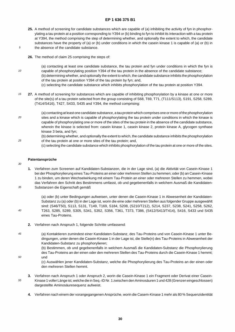
EP 1 636 375 B1
30
5
10
15
20
25
30
35
40
45
50
55
25. A method of screening for candidate substances which are capable of (a) inhibiting the activity of fyn in phosphor-ylating a tau protein at a position corresponding to Y394 or (b) binding to fyn to inhibit its interaction with a tau proteinat Y394, the method comprising the step of determining whether, and optionally the extent to which, the candidatesubstances have the property of (a) or (b) under conditions in which the casein kinase 1 is capable of (a) or (b) inthe absence of the candidate substance.
26. The method of claim 25 comprising the steps of:�
(a) contacting at least one candidate substance, the tau protein and fyn under conditions in which the fyn iscapable of phosphorylating position Y394 of the tau protein in the absence of the candidate substance;(b) determining whether, and optionally the extent to which, the candidate substance inhibits the phosphorylationof the tau protein at position Y394 of the tau protein by fyn; and,(c) selecting the candidate substance which inhibits phosphorylation of the tau protein at position Y394.
27. A method of screening for substances which are capable of inhibiting phosphorylation by a kinase at one or moreof the site �(s) of a tau protein selected from the group consisting of S68, T69, T71, (T111/S113), S191, S258, S289,(T414/S416), T427, S433, S435 and Y394, the method comprising: �
(a) contacting at least one candidate substance, a tau protein which comprises one or more of the phosphorylationsites and a kinase which is capable of phosphorylating the tau protein under conditions in which the kinase iscapable of phosphorylating one or more of the sites of the tau protein in the absence of the candidate substance,wherein the kinase is selected from: casein kinase 1, casein kinase 2, protein kinase A, glycogen synthasekinase 3 beta, and fyn;(b) determining whether, and optionally the extent to which, the candidate substance inhibits the phosphorylationof the tau protein at one or more sites of the tau protein; and,(c) selecting the candidate substance which inhibits phosphorylation of the tau protein at one or more of the sites.
Patentansprüche
1. Verfahren zum Screenen auf Kandidaten-�Substanzen, die in der Lage sind, (a) die Aktivität von Casein- �Kinase 1bei der Phosphorylierung eines Tau-�Proteins an einer oder mehreren Stellen zu hemmen; oder (b) an Casein-�Kinase1 zu binden, um deren Wechselwirkung mit einem Tau-�Protein an einer oder mehreren Stellen zu hemmen, wobeidas Verfahren den Schritt des Bestimmens umfasst, ob und gegebenenfalls in welchem Ausmaß die Kandidaten-Substanzen die Eigenschaft gemäß
(a) oder (b) unter Bedingungen aufweisen, unter denen die Casein-�Kinase 1 in Abwesenheit der Kandidaten-Substanz zu (a) oder (b) in der Lage ist, worin die eine oder mehreren Stellen aus folgender Gruppe ausgewähltsind: (S46/T50), S113, S131, T149, T169, S184, S208, (S210/T212), S214, S237, S238, S241, S258, S262,T263, S285, S289, S305, S341, S352, S356, T361, T373, T386, (S412/S413/T414), S416, S433 und S435eines Tau-�Proteins.
2. Verfahren nach Anspruch 1, folgende Schritte umfassend:�
(a) Kontaktieren zumindest einer Kandidaten-�Substanz, des Tau- �Proteins und von Casein-�Kinase 1 unter Be-dingungen, unter denen die Casein-�Kinase 1 in der Lage ist, die Stelle�(n) des Tau- �Proteins in Abwesenheit derKandidaten-�Substanz zu phosphorylieren;(b) Bestimmen, ob und gegebenenfalls in welchem Ausmaß die Kandidaten-�Substanz die Phosphorylierungdes Tau-�Proteins an der einen oder den mehreren Stellen des Tau-�Proteins durch die Casein-�Kinase 1 hemmt;und(c) Auswählen jener Kandidaten-�Substanz, welche die Phosphorylierung des Tau-�Proteins an der einen oderden mehreren Stellen hemmt.
3. Verfahren nach Anspruch 1 oder Anspruch 2, worin die Casein-�Kinase 1 ein Fragment oder Derivat einer Casein-Kinase 1 voller Länge ist, welche die in Seq.-ID Nr. 1 zwischen den Aminosäuren 1 und 428 (Grenzen eingeschlossen)dargestellte Aminosäuresequenz aufweist.
4. Verfahren nach einem der vorangegangenen Ansprüche, worin die Casem- �Kinase 1 mehr als 80 % Sequenzidentität
EP 1 636 375 B1
31
5
10
15
20
25
30
35
40
45
50
55
mit der Casein- �Kinase 1 voller Länge aufweist, welche die in Seq.-ID Nr. 1 zwischen den Aminosäuren 1 und 428(Grenzen eingeschlossen) dargestellte Aminosäuresequenz aufweist.
5. Verfahren nach einem der Ansprüche 1 bis 4, worin das für die Casein- �Kinase 1 kodierende Nucleinsäuremolekülin der Lage ist, unter stringenten Bedingungen an ein Nucleinsäuremolekül zu hybridisieren, das für eine Casein-Kinase 1 voller Länge kodiert, welche die in Seq.-ID Nr. 1 dargestellte Aminosäuresequenz aufweist.
6. Verfahren nach einem der vorangegangenen Ansprüche, worin das Tau-�Protein Tau eines gepaarten helikalenFilaments ist.
7. Verfahren nach einem der Ansprüche 1 bis 5, worin das Tau-�Protein ein Fragment oder Derivat eines Tau- �Proteinsvoller Länge ist, welches die in Seq.-ID Nr. 2 zwischen den Aminosäuren 1 und 441 (Grenzen eingeschlossen)dargestellte Aminosäuresequenz aufweist.
8. Verfahren nach einem der vorangegangenen Ansprüche, worin das Tau-�Protein mehr als 80 % Sequenzidentitätmit einem Tau-�Protein voller Länge aufweist, welches die in Seq.-ID Nr. 2 zwischen den Aminosäuren 1 und 441(Grenzen eingeschlossen) dargestellte Aminosäuresequenz aufweist.
9. Verfahren nach einem der Ansprüche 1 bis 6, worin ein für das Tau- �Protein kodierendes Nucleinsäuremolekül inder Lage ist, unter stringenten Bedingungen an ein Nucleinsäuremolekül zu hybridisieren, das für ein Tau-�Proteinvoller Länge kodiert, welches die in Seq.-ID Nr. 3 dargestellte Aminosäuresequenz aufweist.
10. Verfahren nach einem der vorangegangenen Ansprüche, worin die Stellen des Tau-�Proteins aus S262 und/�oderS356 ausgewählt sind.
11. Verfahren nach einem der vorangegangenen Ansprüche, worin die Stellen des Tau- �Proteins eine oder mehrereStellen sind, die aus der aus S113, S258, S289, S416, S433 und S435 bestehenden Gruppe ausgewählt sind.
12. Verfahren nach einem der vorangegangenen Ansprüche, worin das Screenen das Bestimmen der Auswirkungendes Kontaktierens der Kandidaten-�Substanz�(en) mit einer Kombination aus Kinasen, die gleichzeitig oder nachein-ander auf die Kandidaten-�Substanzen einwirken, und Casein-�Kinase 1 umfasst.
13. Verfahren nach Anspruch 12, worin die Kombination aus Kinasen Casein- �Kinase 1 (CK1) in Kombination mit eineroder mehreren von Casein-�Kinase 2 (CK2), Protein- �Kinase A (PKA), Glykogen-�Synthase- �Kinase 3α (GSK- �3α) oderGlykogen-�Synthase-�Kinase 3β (GSK-�3β) umfasst.
14. Verfahren nach Anspruch 12, worin die Kombination aus Kinasen Casein-�Kinase 1 (CK1) in Kombination mit PKAund GSK-�3β umfasst.
15. Verfahren nach einem der vorangegangenen Ansprüche, das weiters das Bestimmen umfasst, ob und gegebenen-falls in welchem Ausmaß die Kandidaten-�Substanz die Phosphorylierung eines künstlichen Substrats durch dieCasein- �Kinase 1 hemmt.
16. Verfahren nach Anspruch 15, worin das künstliche Substrat von Casein-�Kinase 1 kein Tau-�Protein- �Fragment ist.
17. Verfahren nach einem der vorangegangenen Ansprüche, worin Massenspektroskopie oder ein ortsspezifischesErkennungsmittel eingesetzt wird, um die Gegenwart, die Abwesenheit oder das Ausmaß von Phosphorylierung aneiner oder mehreren Stellen des Tau- �Proteins zu bestimmen.
18. Verfahren nach Anspruch 17, worin das ortsspezifische Erkennungsmittel ein monoklonaler Antikörper ist.
19. Verfahren nach einem der vorangegangenen Ansprüche, worin das Screenen in einem Multiplex- �Test unter Einsatzeiner festen Phase, auf der mehrere Substrate immobilisiert sind, durchgeführt wird.
20. Verfahren nach Anspruch 19, worin die Substrate Phosphorylierungsstellen eines Tau-�Proteins entsprechen.
21. Verfahren nach einem der vorangegangenen Ansprüche, worin das Screenen in einem Multiplex- �Test unter Einsatzeiner festen Phase, auf der mehrere Substrate immobilisiert sind, durchgeführt wird.
EP 1 636 375 B1
32
5
10
15
20
25
30
35
40
45
50
55
22. Verfahren nach Anspruch 21, worin die Substrate Phosphorylierungsstellen eines Tau-�Proteins entsprechen.
23. Verfahren nach einem der vorangegangenen Ansprüche, das nach dem Identifizieren einer Kandidaten-�Substanzals Inhibitor von Casein-�Kinase 1 den weiteren Schritt des Optimierens der Struktur der Kandidaten-�Substanzumfasst.
24. Verfahren nach einem der vorangegangenen Ansprüche, das nach dem Identifizieren einer Kandidaten-�Substanzdurch das Screening-�Verfahren den weiteren Schritt des Verarbeitens und/�oder Formulierens der Substanz zu einerpharmazeutischen Zusammensetzung umfasst.
25. Verfahren zum Screenen auf Kandidaten-�Substanzen, die in der Lage sind, (a) die Aktivität von Fyn bei der Phos-phorylierung eines Tau-�Proteins an einer Position zu hemmen, die Y394 entspricht, oder (b) an Fyn zu binden, umseine Wechselwirkung mit einem Tau-�Protein an Y394 zu hemmen, wobei das Verfahren den Schritt des Bestimmensumfasst, ob und gegebenenfalls in welchem Ausmaß die Kandidaten-�Substanzen die Eigenschaft gemäß (a) oder(b) unter Bedingungen aufweisen, � unter denen die Casein- �Kinase 1 in Abwesenheit der Kandidaten-�Substanz zu(a) oder (b) in der Lage ist.
26. Verfahren nach Anspruch 25, folgende Schritte umfassend:�
(a) Kontaktieren zumindest einer Kandidaten- �Substanz, des Tau-�Proteins und von Fyn unter Bedingungen,unter denen Fyn in der Lage ist, die Position Y394 des Tau- �Proteins in Abwesenheit der Kandidaten-�Substanzzu phosphorylieren;(b) Bestimmen, ob und gegebenenfalls in welchem Ausmaß die Kandidaten-�Substanz die Phosphorylierungdes Tau-�Proteins an der Position Y394 des Tau-�Proteins durch Fyn hemmt; und(c) Auswählen jener Kandidaten-�Substanz, welche die Phosphorylierung des Tau- �Proteins an der Position Y394hemmt.
27. Verfahren zum Screenen auf Substanzen, die in der Lage sind, Phosphorylierung durch eine Kinase an einer odermehreren Stellen eines Tau- �Proteins zu hemmen, die aus der aus folgenden bestehenden Gruppe ausgewählt sind:S68, T69, T71, (T111/S113), S191, S258, S289, (T414/S416), T427, S433, S435 und Y394, wobei das VerfahrenFolgendes umfasst:�
(a) Kontaktieren zumindest einer Kandidaten- �Substanz, eines Tau- �Proteins, das eine oder mehrere der Phos-phorylierungsstellen umfasst, und einer Kinase, die in der Lage ist, das Tau Protein zu phosphorylieren, unterBedingungen, unter denen die Kinase in der Lage ist, eine oder mehrere Stellen des Tau- �Proteins in Abwesenheitder Kandidaten- �Substanz zu phosphorylieren, worin die Kinase ausgewählt ist aus: Casein- �Kinase 1, Casein-Kinase 2, Protein- �Kinase A, Glykogen- �Synthase- �Kinase 3β und Fyn;(b) Bestimmen, ob und gegebenenfalls in welchem Ausmaß die Kandidaten-�Substanz die Phosphorylierungdes Tau-�Proteins an einer oder mehreren Stellen des Tau-�Proteins hemmt; und(c) Auswählen jener Kandidaten-�Substanz, welche die Phosphorylierung des Tau-�Proteins an einer oder meh-reren der Stellen hemmt.
Revendications
1. Méthode de criblage de substances candidates qui sont capables de (a) inhiber l’activité de la caséine kinase 1dans la phosphorylation d’une protéine tau en un ou plusieurs sites; ou (b) se lier à la caséine kinase 1 pour inhiberson interaction avec une protéine tau en un ou plusieurs sites, la méthode comprenant l’étape de déterminer si, etfacultativement l’étendue à laquelle, les substances candidates ont la propriété de (a) ou (b) dans des conditionsdans lesquelles la caséine kinase 1 est capable de (a) ou (b) en l’absence de la substance candidate, où les un ouplusieurs sites sont sélectionnés dans le groupe consistant en (S46/T50), S113, S131, T149, T169, S184, S208,(S210/T212), S214, S237, S238, S241, S258, S262, T263, S285, S289, S305, S341, S352, S356, T361, T373,T386, (S412/S413.T414), S416, S433 et S435 de la protéine tau.
2. Méthode de la revendication 1 comprenant les étapes de:�
(a) mettre en contact au moins une substance candidate, la protéine tau et la caséine kinase 1 dans desconditions dans lesquelles la caséine kinase 1 est capable de phosphoryler le ou les sites de la protéine tau
EP 1 636 375 B1
33
5
10
15
20
25
30
35
40
45
50
55
en l’absence de la substance candidate;(b) déterminer si, et facultativement l’étendue à laquelle, la substance candidate inhibe la phosphorylation dela protéine tau en le ou les plusieurs sites de la protéine tau par la caséine kinase 1; et(c) sélectionner la substance candidate qui inhibe la phosphorylation de la protéine tau en un ou plusieurs dessites.
3. Méthode de la revendication 1 ou de la revendication 2, où la caséine kinase 1 est un fragment ou dérivé de 1acaséine kinase 1 de pleine longueur ayant la séquence d’acides aminés indiquée entre les acides aminés 1 et 428inclus dans SEQ ID NO:�1.
4. Méthode de l’une quelconque des revendications précédentes, où la caséine kinase 1 a plus de 80% d’identité deséquence avec la caséine kinase 1 de pleine longueur ayant la séquence d’acides aminés indiquée entre les acidesaminés 1 et 428 inclus de SEQ ID NO: �1.
5. Méthode de l’une quelconque des revendications 1 à 4, où la molécule d’acide nucléique codant pour la caséinekinase 1 est capable de s’hybrider en conditions stringentes à une molécule d’acide nucléique codant pour la caséinekinase 1 de pleine longueur ayant la séquence d’acides aminés indiquée à SEQ ID NO:�1.
6. Méthode de l’une quelconque des revendications précédentes, où la protéine tau est tau à filament hélicoïdal apparié.
7. Méthode de l’une quelconque des revendications 1 à 5, où la protéine tau est un fragment ou dérivé de la protéinetau de pleine longueur ayant la séquence d’acides aminés indiquée entre les acides aminés 1 et 441 inclus de SEQID NO: �2.
8. Méthode de l’une quelconque des revendications précédentes, où la protéine tau a plus de 80% d’identité deséquence avec la protéine tau complète ayant la séquence d’acides aminés indiquée entre les acides aminés 1 et441 inclus dans SEQ ID NO: �2.
9. Méthode de l’une quelconque des revendications 1 à 6 où une molécule d’acide nucléique codant pour la protéinetau est capable de s’hybrider en conditions stringentes à une molécule d’acide nucléique codant pour la protéinetau de pleine longueur ayant la séquence d’acides aminés indiquée à SEQ ID NO:�3.
10. Méthode de l’une quelconque des revendications précédentes, où les sites de la protéine tau sont sélectionnésparmi S262 et/ou S356.
11. Méthode de l’une quelconque des revendications précédentes, où les sites de la protéine tau en un ou plusieurssites sélectionnés dans le groupe consistant en S113, S258, S289, 5416, S433 et S435.
12. Méthode de l’une quelconque des revendications précédentes, où le criblage comprend la détermination de l’effetde la mise en contact de la ou des substances candidates avec une combinaison de kinases, simultanément ouséquentiellement appliquées aux substances candidates et la caséine kinase 1.
13. Méthode de la revendication 12, où la combinaison des kinases comprend la caséine kinase 1 (CK1) en combinaisonavec une ou plusieurs de la caséine kinase 2 (CK2), de la protéine kinase A (PKA), de la glycogène synthase kinase3α (GSK-�3α) ou de la glycogène synthase kinase 3β (GSK-�3β).
14. Méthode de la revendication 12, où la combinaison des kinases comprend la caséine kinase 1 (CK1) en combinaisonavec PKA et GSK-�3β.
15. Méthode de l’une quelconque des revendications précédentes consistant de plus à déterminer si, et facultativementl’étendue à laquelle, la substance candidate inhibe la phosphorylation d’un substrat artificiel par la caséine kinase 1.
16. Méthode de la revendication 15, où le substrat artificiel de caséine kinase 1 n’est pas un fragment de protéine tau.
17. Méthode de l’une quelconque des revendications précédentes, où une spectroscopie de masse ou un agent dereconnaissance spécifique du site est utilisé pour déterminer la présence, l’absence ou l’étendue de phosphorylationen un ou plusieurs sites de la protéine tau.
EP 1 636 375 B1
34
5
10
15
20
25
30
35
40
45
50
55
18. Méthode de la revendication 17 où l’agent de reconnaissance spécifique est un anticorps monoclonal.
19. Méthode de l’une quelconque des revendications précédentes, où le criblage est effectué dans un essai multiplexemployant une phase solide sur laquelle sont immobilisés un certain nombre de substrats.
20. Méthode de la revendication 19, où les substrats correspondent à des sites de phosphorylation de la protéine tau.
21. Méthode de l’une quelconque des revendications précédentes, où le criblage est effectué dans un essai multiplexen employant une phase solide sur laquelle sont immobilisés un certain nombre de substrats.
22. Méthode de la revendication 21, où les substrats correspondent à des sites de phosphorylation de la protéine tau.
23. Méthode de l’une quelconque des revendications précédentes qui comprend, ayant identifié une substance candi-date en tant qu’inhibiteur de la caséine kinase 1, l’étape supplémentaire d’optimiser la structure de la substancecandidate.
24. Méthode de l’une quelconque des revendications précédentes qui comprend, ayant identifié une substance candi-date par la méthode de criblage, l’étape supplémentaire de fabriquer la substance et/ou de la formuler en compositionpharmaceutique.
25. Méthode de criblage de substances candidates qui sont capables de (a) inhiber l’activité de fyn dans la phospho-rylation d’une protéine tau en une position correspondant à Y394 ou (b) se lier à fyn pour inhiber son interactionavec une protéine tau à Y394, la méthode comprenant l’étape de déterminer si et facultativement l’étendue à laquelleles substances candidates ont la propriété de (a) ou (b) dans des conditions dans lesquelles la caséine kinase 1est capable de (a) ou (b) en l’absence de la substance candidate.
26. Méthode de la revendication 25 comprenant les étapes de:�
(a) mettre en contact au moins une substance candidate, la protéine tau et fyn dans des conditions danslesquelles fyn est capable de phosphoryler la position Y394 de la protéine tau en l’absence de la substancecandidate;(b) déterminer si, et facultativement l’étendue à laquelle, la substance candidate inhibe la phosphorylation dela protéine tau à la position Y394 de la protéine tau par fyn; et(c) sélectionner la substance candidate qui inhibe la phosphorylation de la protéine tau à la position Y394.
27. Méthode de criblage de substances qui sont capables d’inhiber la phosphorylation par une kinase en un ou plusieursdes sites d’une protéine tau sélectionnée dans le groupe consistant en S68, T69, T71, (T111/S113), S191, S258,S289, (T414/S416), T427, S433, S435 et Y394, la méthode comprenant:�
a) la mise en contact d’au moins une substance candidate, une protéine tau qui comprend un ou plusieurs dessites de phosphorylation et une kinase qui est capable de phosphorylation de la protéine tau dans des conditionsdans lesquelles la kinase est capable de phosphorylation d’un ou plusieurs des sites de la protéine tau enl’absence de la substance candidate, où la kinase est sélectionnée parmi: caséine kinase 1, caséine kinase 2,protéine kinase A, glycogène synthase kinase 3 beta et fyn,(b) déterminer si, et facultativement l’étendue à laquelle la substance candidate inhibe la phosphorylation dela protéine tau en un ou plusieurs sites de la protéine tau, et(c) sélectionner la substance candidate qui inhibe la phosphorylation de la protéine tau en un ou plusieurs dessites.
EP 1 636 375 B1
35
REFERENCES CITED IN THE DESCRIPTION
This list of references cited by the applicant is for the reader’s convenience only. It does not form part of the Europeanpatent document. Even though great care has been taken in compiling the references, errors or omissions cannot beexcluded and the EPO disclaims all liability in this regard.
Patent documents cited in the description
• US 6057117 A [0012]• WO 9519178 A [0014]• WO 9201047 A [0065]• EP 0184187 A [0068]• GB 2188638 A [0068]
• EP 0239400 A [0068]• EP 0120694 A [0068]• EP 0125023 A [0068]• GB 0314943 A [0128]
Non-patent literature cited in the description
• MEIJER. CHEMISTRY AND BIOLOGY, CURRENTBIOLOGY. January 2000, vol. 7, 51-63 [0011]
• LEE G et al. Tyrosine phosphorylation of tau. SOCI-ETY FOR NEUROSCIENCE ABSTRACTS, 2001,vol. 27 (1), 1436 [0013]
• TROJANOWSKI et al. FASEB JOURNAL, 1995, vol.9 (15), 1570-1576 [0015]
• SINGH et al. JOURNAL OF NEUROCHEMISTRY,1995, vol. 64 (3), 1420-1423 [0016]
• MOLECULAR AND CELLULAR BIOCHEMISTRY,1996, vol. 154 (2), 143-151 [0016]
• GOEDERT et al. EMBO J., February 1989, vol. 8 (2),393-9 [0031]
• GOEDERT M ; JAKES R. Expression of separateisoforms of human tau protein: correlation with thetau pattern in brain and effects on tubulin polymeri-zation. EMBO J., 1990, vol. 9, 4225-30 [0031]
• J Biol Chem., 25 March 1993, vol. 268 (9), 6394-401[0038]
• ALTSCHUL et al. J. Mol. Biol., 1990, vol. 215, 403-10[0051]
• AUSUBEL. Short Protocols in Molecular Biology,1992 [0051]
• SAMBROOK et al. Molecular Cloning, A LaboratoryManual. Cold Spring Harbor Laboratory Press, 1989[0051]
• ARMITAGE et al. Nature, 1992, vol. 3 (57), 80-82[0064]
• ROEPSTORFF, P. ; FOHLMAN, J. J. Biomed. MassSpectrom., 1984, vol. 11, 601 [0073]
• BONNER, R. ; SHUSHAN, B. Rapid Commun. MassSpectrom., 1995, vol. 9, 1067-1076 [0073]
• Remington’s Pharmaceutical Sciences. Lippincott,Williams & Wilkins, 2000 [0089]
• GHOSHAL N ; SMILEY JF ; DEMAGGIO AJ ;HOEKSTRA MF ; COCHRAN EJ ; BINDER LI ;KURET J. A new molecular link between the fibrillarand granulovacuolar lesions of Alzheimer’s disease.Am J Pathol., 1999, vol. 155, 1163-72 [0118]
• HANGER DP ; BETTS JC ; LOVINY TL ; BLACK-STOCK WP ; ANDERTON BH. New phosphoryla-tion sites identified in hyperphosphorylated tau(paired helical filament-tau) from Alzheimer’s diseasebrain using nanoelectrospray mass spectrometry. JNeurochem., 1998, vol. 71, 2465-76 [0119]
• LAMBERT,M.P. ; BARLOW,A.K. ;CHROMY,B.A. ; EDWARDS,C. ; FREED,R. ;LIOSATOS,M. ; MORGAN,T.E. ; ROZOVSKY,I. ;TROMMER,B. ; VIOLA,K.L. Diffusible, nonfibrillarligands derived from Aβ1-42 are potent central nerv-ous system neurotoxins. Proc. Natl. Acad. Sci. USA,1998, vol. 95, 6448-6453 [0120]
• LEE VM ; GOEDERT M ; TROJANOWSKI JQ. Neu-rodegenerative tauopathies. Annu Rev Neurosci.,2001, vol. 24, 1121-59 [0121]
• MULOT SF ; HUGHES K ; WOODGETT JR ; AN-DERTON BH ; HANGER DP. PHF-tau from Alzhe-imer’s brain comprises four species on SDS-PAGEwhich can be mimicked by in vitro phosphorylation ofhuman brain tau by glycogen synthase kinase-3 beta.FEBS Lett., 1994, vol. 349, 359-64 [0122]
• SCALES,TME ; WILLIAMSON, R. ; ANDERTON,BH ; RENOLDS, CH et al. Tyrosine phosphorylationof specific sites on tau by Src family kinases. Neuro-biol Aging, 2002, vol. 23, 500-501 [0123]
• SHIRAZI,S.K. ; WOOD,J.G. The protein tyrosine ki-nase, fyn, in Alzheimer’s disease pathology. Neu-roreport, 1993, vol. 4, 435-437 [0124]
• SINGH TJ ; ZAIDI T ; GRUNDKE-IQBAL I ; IQBALK. Modulation of GSK-3-catalyzed phosphorylationof microtubule-associated protein tau by non-pro-line-dependent protein kinases. FEBS Lett., 1995,vol. 358, 4-8 [0125]
• SINGH TJ ; HAQUE N ; GRUNDKE-IQBAL I ;IQBAL K. Rapid Alzheimer-like phosphorylation oftau by the synergistic actions of non-proline-depend-ent protein kinases and GSK-3. FEBS Lett., 1995,vol. 358, 267-72 [0126]

EP 1 636 375 B1
36
• WILLIAMSON,R. ; SCALES,T. ; CLARK,B.R. ;GIBB,G. ; REYNOLDS,C.H. ; KELLIE,S. ;BIRD,I.N. ; VARNDELL,I.M. ; SHEPPARD,P.W. ;EVERALL,I. Rapid tyrosine phosphorylation of neu-ronal proteins including tau and focal-adhesion ki-nase in response to amyloid-β peptide exposure: in-volvement of Src family protein kinases. J. Neurosci.,2002, vol. 22, 10-20 [0127]


