Embed Size (px)
Citation preview

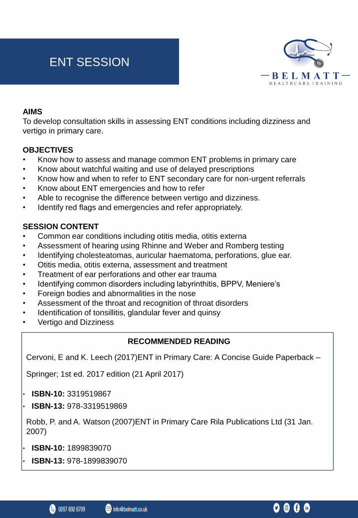
ENT AND DIZZINESS

AIMS
To develop consultation skills in assessing ENT conditions including dizziness and
vertigo in primary care.
OBJECTIVES
• Know how to assess and manage common ENT problems in primary care
• Know about watchful waiting and use of delayed prescriptions
• Know how and when to refer to ENT secondary care for non-urgent referrals
• Know about ENT emergencies and how to refer
• Able to recognise the difference between vertigo and dizziness.
• Identify red flags and emergencies and refer appropriately.
SESSION CONTENT
• Common ear conditions including otitis media, otitis externa
• Assessment of hearing using Rhinne and Weber and Romberg testing
• Identifying cholesteatomas, auricular haematoma, perforations, glue ear.
• Otitis media, otitis externa, assessment and treatment
• Treatment of ear perforations and other ear trauma
• Identifying common disorders including labyrinthitis, BPPV, Meniere’s
• Foreign bodies and abnormalities in the nose
• Assessment of the throat and recognition of throat disorders
• Identification of tonsillitis, glandular fever and quinsy
• Vertigo and Dizziness
RECOMMENDED READING
Cervoni, E and K. Leech (2017)ENT in Primary Care: A Concise Guide Paperback –
Springer; 1st ed. 2017 edition (21 April 2017)
• ISBN-10: 3319519867
• ISBN-13: 978-3319519869
Robb, P. and A. Watson (2007)ENT in Primary Care Rila Publications Ltd (31 Jan.
2007)
• ISBN-10: 1899839070
• ISBN-13: 978-1899839070
ENT SESSION
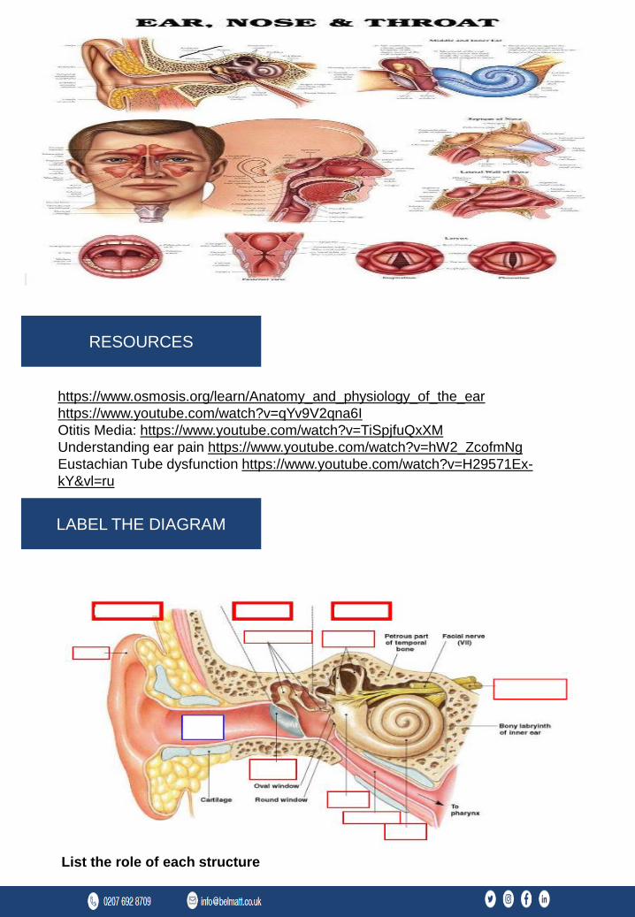
https://www.osmosis.org/learn/Anatomy_and_physiology_of_the_ear
https://www.youtube.com/watch?v=qYv9V2qna6I
Otitis Media: https://www.youtube.com/watch?v=TiSpjfuQxXM
Understanding ear pain https://www.youtube.com/watch?v=hW2_ZcofmNg
Eustachian Tube dysfunction https://www.youtube.com/watch?v=H29571Ex-
kY&vl=ru
RESOURCES
List the role of each structure
LABEL THE DIAGRAM
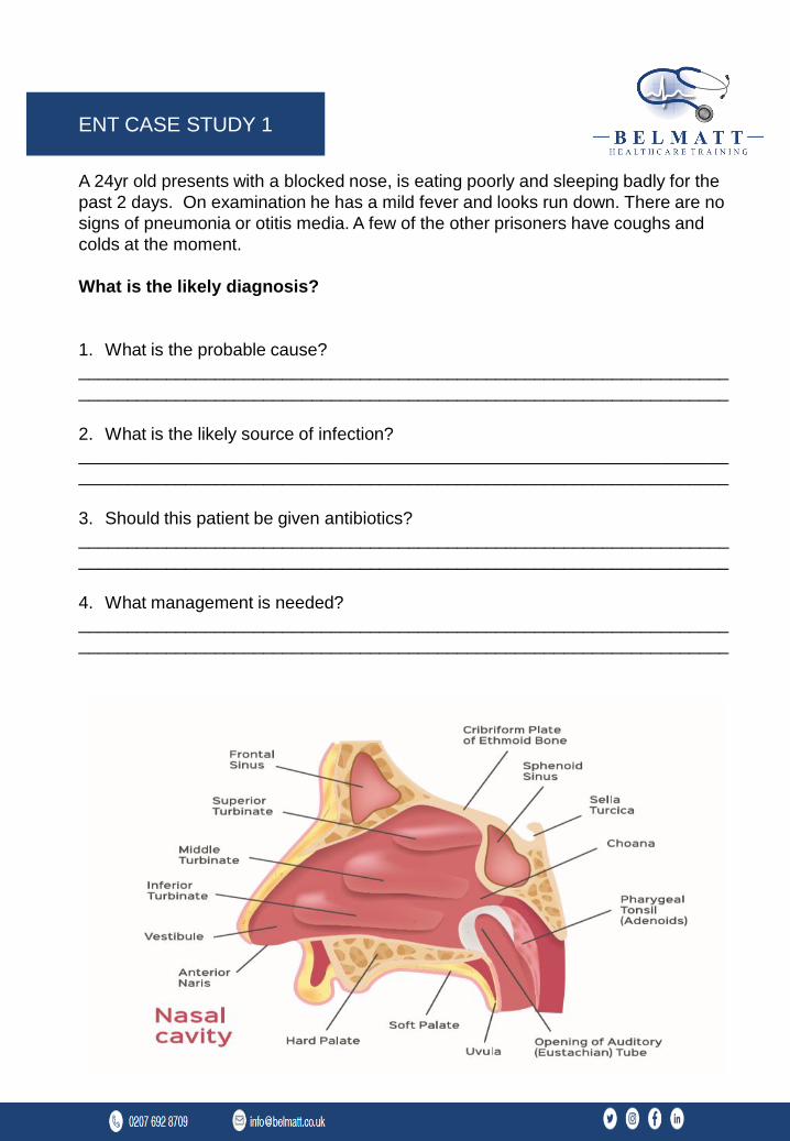
A 24yr old presents with a blocked nose, is eating poorly and sleeping badly for the
past 2 days. On examination he has a mild fever and looks run down. There are no
signs of pneumonia or otitis media. A few of the other prisoners have coughs and
colds at the moment.
What is the likely diagnosis?
1. What is the probable cause?
___________________________________________________________________
___________________________________________________________________
2. What is the likely source of infection?
___________________________________________________________________
___________________________________________________________________
3. Should this patient be given antibiotics?
___________________________________________________________________
___________________________________________________________________
4. What management is needed?
___________________________________________________________________
___________________________________________________________________
ENT CASE STUDY 1
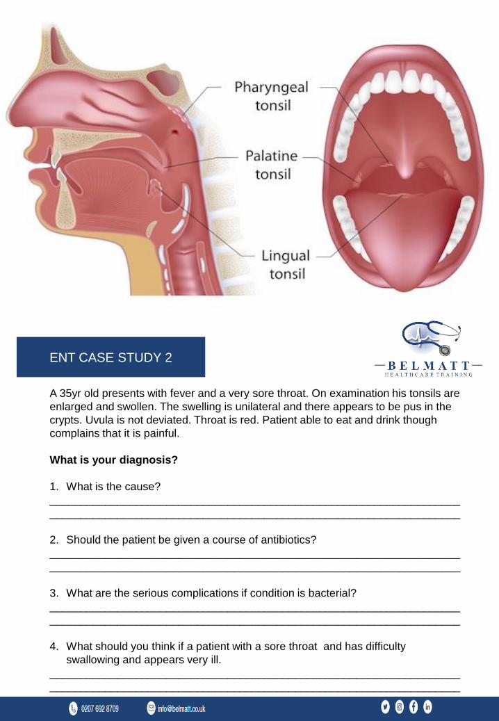
A 35yr old presents with fever and a very sore throat. On examination his tonsils are
enlarged and swollen. The swelling is unilateral and there appears to be pus in the
crypts. Uvula is not deviated. Throat is red. Patient able to eat and drink though
complains that it is painful.
What is your diagnosis?
1. What is the cause?
___________________________________________________________________
___________________________________________________________________
2. Should the patient be given a course of antibiotics?
___________________________________________________________________
___________________________________________________________________
3. What are the serious complications if condition is bacterial?
___________________________________________________________________
___________________________________________________________________
4. What should you think if a patient with a sore throat and has difficulty
swallowing and appears very ill.
___________________________________________________________________
___________________________________________________________________
ENT CASE STUDY 2
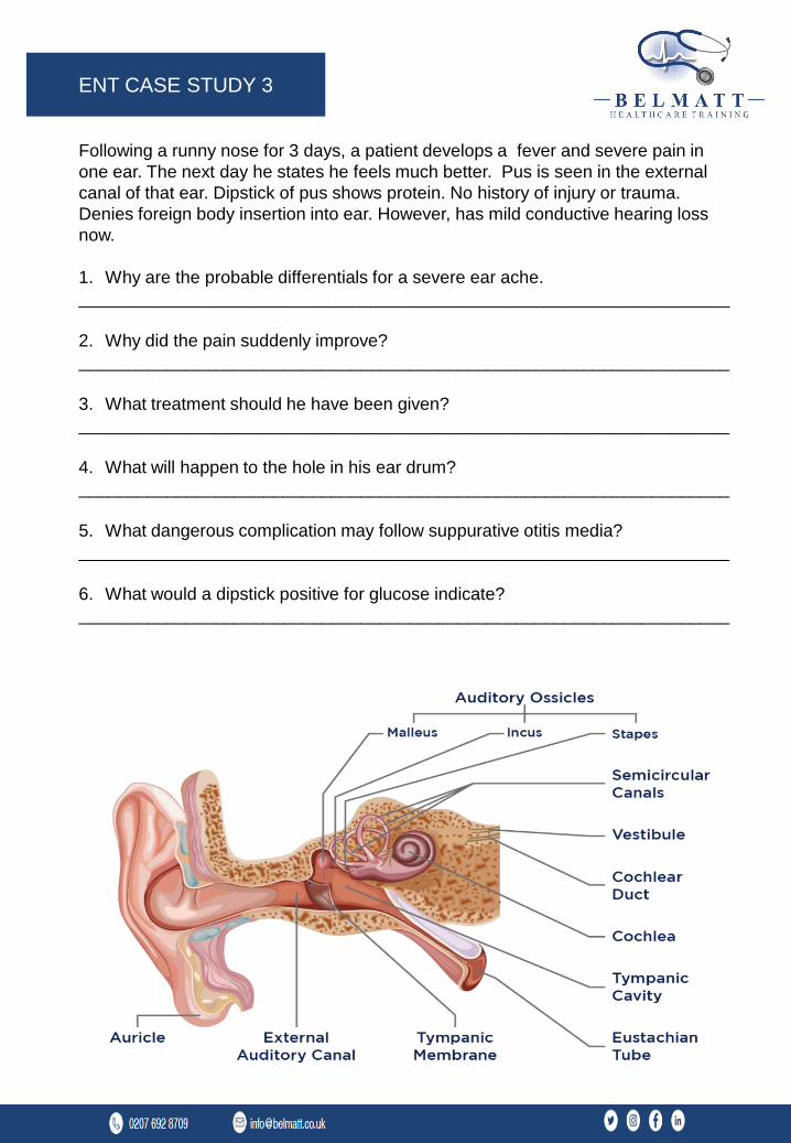
Following a runny nose for 3 days, a patient develops a fever and severe pain in
one ear. The next day he states he feels much better. Pus is seen in the external
canal of that ear. Dipstick of pus shows protein. No history of injury or trauma.
Denies foreign body insertion into ear. However, has mild conductive hearing loss
now.
1. Why are the probable differentials for a severe ear ache.
___________________________________________________________________
2. Why did the pain suddenly improve?
___________________________________________________________________
3. What treatment should he have been given?
___________________________________________________________________
4. What will happen to the hole in his ear drum?
___________________________________________________________________
5. What dangerous complication may follow suppurative otitis media?
___________________________________________________________________
6. What would a dipstick positive for glucose indicate?
___________________________________________________________________
ENT CASE STUDY 3
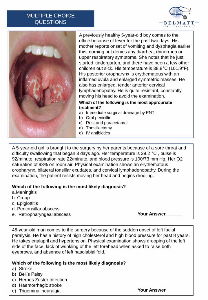
MULTIPLE CHOICE
QUESTIONS
A previously healthy 5-year-old boy comes to the
office because of fever for the past two days. His
mother reports onset of vomiting and dysphagia earlier
this morning but denies any diarrhea, rhinorrhea or
upper respiratory symptoms. She notes that he just
started kindergarten, and there have been a few other
children out sick. His temperature is 38.8°C (101.9°F).
His posterior oropharynx is erythematous with an
inflamed uvula and enlarged symmetric masses. He
also has enlarged, tender anterior cervical
lymphadenopathy. He is quite resistant, constantly
moving his head to avoid the examination.
Which of the following is the most appropriate
treatment?
a) Immediate surgical drainage by ENT
b) Oral penicillin
c) Rest and paracetamol
d) Tonsillectomy
e) IV antibiotics
A 5-year-old girl is brought to the surgery by her parents because of a sore throat and
difficulty swallowing that began 3 days ago. Her temperature is 39.2 °C , pulse is
92/minute, respiration rate 22/minute, and blood pressure is 100/73 mm Hg. Her O2
saturation of 98% on room air. Physical examination shows an erythematous
oropharynx, bilateral tonsillar exudates, and cervical lymphadenopathy. During the
examination, the patient resists moving her head and begins drooling.
Which of the following is the most likely diagnosis?
a.Meningitis
b. Croup
c. Epiglottitis
d. Peritonsillar abscess
e. Retropharyngeal abscess
45-year-old man comes to the surgery because of the sudden onset of left facial
paralysis. He has a history of high cholesterol and high blood pressure for past 8 years.
He takes enalapril and hypertension. Physical examination shows drooping of the left
side of the face, lack of wrinkling of the left forehead when asked to raise both
eyebrows, and absence of left nasolabial fold.
Which of the following is the most likely diagnosis?
a) Stroke
b) Bell’s Palsy
c) Herpes Zoster Infection
d) Haemorrhagic stroke
e) Trigeminal neuralgia
Your Answer ______
Your Answer ______
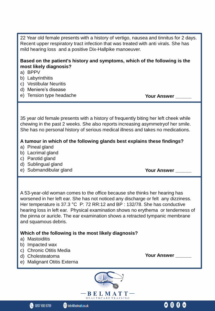
22 Year old female presents with a history of vertigo, nausea and tinnitus for 2 days.
Recent upper respiratory tract infection that was treated with anti virals. She has
mild hearing loss and a positive Dix-Hallpike manoeuver.
Based on the patient's history and symptoms, which of the following is the
most likely diagnosis?
a) BPPV
b) Labyrinthitis
c) Vestibular Neuritis
d) Meniere’s disease
e) Tension type headache
35 year old female presents with a history of frequently biting her left cheek while
chewing in the past 2 weeks. She also reports increasing asymmetryof her smile.
She has no personal history of serious medical illness and takes no medications.
A tumour in which of the following glands best explains these findings?
a) Pineal gland
b) Lacrimal gland
c) Parotid gland
d) Sublingual gland
e) Submandibular gland
A 53-year-old woman comes to the office because she thinks her hearing has
worsened in her left ear. She has not noticed any discharge or felt any dizziness.
Her temperature is 37.3 °C P: 72 RR:12 and BP : 132/78. She has conductive
hearing loss in left ear. Physical examination shows no erythema or tenderness of
the pinna or auricle. The ear examination shows a retracted tympanic membrane
and squamous debris.
Which of the following is the most likely diagnosis?
a) Mastoiditis
b) Impacted wax
c) Chronic Otitis Media
d) Cholesteatoma
e) Malignant Otitis Externa
Your Answer ______
Your Answer ______
Your Answer ______

A 25yr old male comes to the out of hours clinic with an inability to open his mouth.
He has pain and swelling in his neck which has worsened over the past day. He has
bilateral swelling of the floor of the mouth involving his submandibular and
sublingual glands. His molars on the right side are decayed and there is an abscess
around the back molars. The skin of his upper neck is red but intact.
Which of the following is the most likely diagnosis?
a) Periodontitis
b) Sialolithiasis
c) Kawasaki Disease
d) Ludwig’s Angina
e) Steven Johnsons Syndrome
A 65-year-old woman comes to the office because of problems with her hearing and
'ear fullness.' She reports that she has noticed a low pitch noise that others don't
hear that sounds like being by the ocean, and a decrease in hearing in her left ear.
She has also been having intermittent episodes of dizziness when she feels like the
room is spinning.
Which of the following is the most likely diagnosis for this patient?
a) Labyrinthitis
b) Meniere’s Disease
c) BPPV
d) Migraine with aura
e) Multiple Sclerosis
Your Answer ______
Your Answer ______

NON-MEDICAL PRESCRIBER
GUIDELINES
NAME : ………………………………
Aims
To develop skills in assessment and management of common ENT injuries and disorders
presenting to primary acre centers.
Objectives
• To assess and examine the ear
• Develop skills in identifying common ENT complaints
• Improve skills in assessment and examination using otoscope and tuning fork during
assessment of ENT complaints. Identify red flags and emergencies and refer appropriately.
Session content
• Common ear conditions including otitis media, otitisexterna
• Assessment of hearing using Rhine and Weber and Rhombergtesting
• Identifying cholesteatomas, auricular haematoma, perforations, glue ear.
• Otitis media, otitis externa, assessment and treatment
• Treatment of ear perforations and other ear trauma
• Identifying common disorders including labyrinthitis, BPPV, Menieres
• Management of Epistaxis
• Foreign bodies and abnormalities in the nose
• Assessment of the throat and recognition of throat disorders
• Identification of tonsillitis, glandular fever and quinsy
RECOMMENDED READING
Cervoni, E and K. Leech (2017) ENT in Primary Care: A Concise Guide Paperback –
Springer; 1st ed. 2017 edition (21 April 2017)
• ISBN-10:3319519867
• ISBN-13:978-3319519869
Robb, P. and A. Watson (2007)ENT in Primary Care Rila Publications Ltd (31 Jan. 2007)
• ISBN-10:1899839070
• ISBN-13:978-1899839070
ENT Session
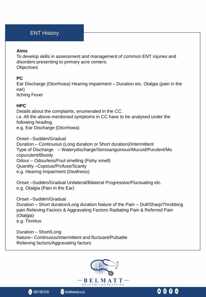
Aims
To develop skills in assessment and management of common ENT injuries and
disorders presenting to primary acre centers.
Objectives
PC
Ear Discharge (Otorrhoea) Hearing impairment – Duration etc. Otalgia (pain in the
ear)
Itching Fever
HPC
Details about the complaints, enumerated in the CC.
i.e. All the above-mentioned symptoms in CC have to be analysed under the
following heading.
e.g. Ear Discharge (Otorrhoea)
Onset –Sudden/Gradual
Duration – Continuous (Long duration or Short duration)/Intermittent
Type of Discharge – Waterydischarge/Serosanguinous/Mucoid/Purulent/Mu
copurulent/Bloody
Odour – Odourless/Foul smelling (Fishy smell)
Quantity –Copious/Profuse/Scanty
e.g. Hearing Impairment (Deafness)
Onset –Sudden/Gradual Unilateral/Bilateral Progressive/Fluctuating etc.
e.g. Otalgia (Pain in the Ear)
Onset –Sudden/Gradual
Duration – Short duration/Long duration Nature of the Pain – Dull/Sharp/Throbbing
pain Relieving Factors & Aggravating Factors Radiating Pain & Referred Pain
(Otalgia)
e.g. Tinnitus
Duration – Short/Long
Nature– Continuous/Intermittent and fluctuant/Pulsatile
Relieving factors/Aggravating factors
ENT History
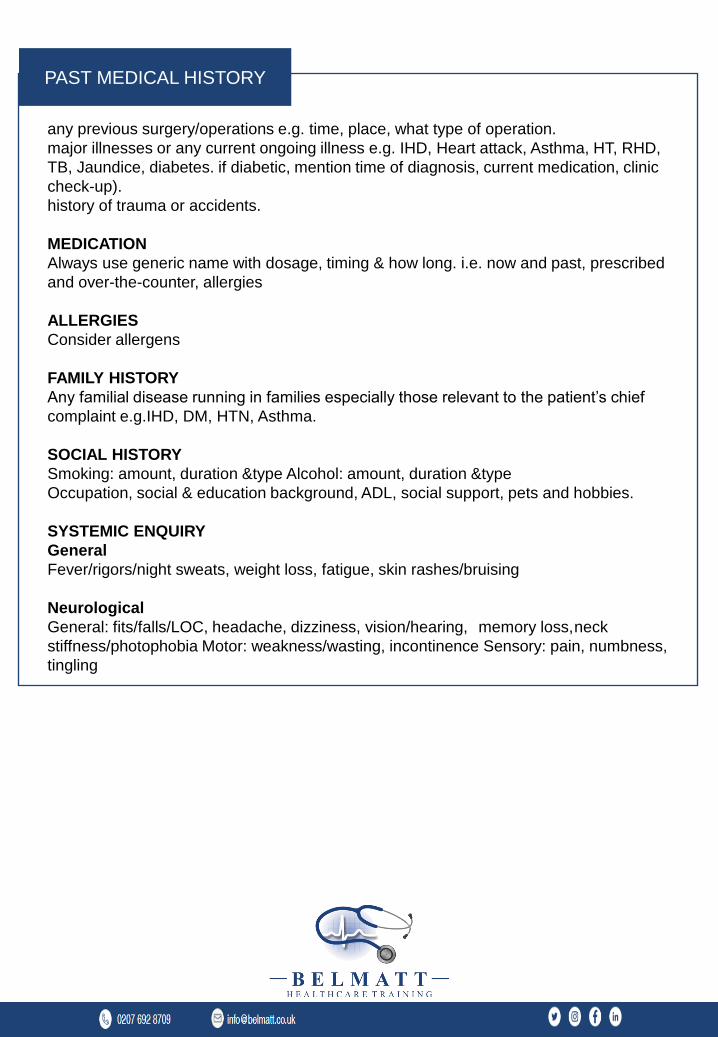
any previous surgery/operations e.g. time, place, what type of operation.
major illnesses or any current ongoing illness e.g. IHD, Heart attack, Asthma, HT, RHD,
TB, Jaundice, diabetes. if diabetic, mention time of diagnosis, current medication, clinic
check-up).
history of trauma or accidents.
MEDICATION
Always use generic name with dosage, timing & how long. i.e. now and past, prescribed
and over-the-counter, allergies
ALLERGIES
Consider allergens
FAMILY HISTORY
Any familial disease running in families especially those relevant to the patient’s chief
complaint e.g.IHD, DM, HTN, Asthma.
SOCIAL HISTORY
Smoking: amount, duration &type Alcohol: amount, duration &type
Occupation, social & education background, ADL, social support, pets and hobbies.
SYSTEMIC ENQUIRY
General
Fever/rigors/night sweats, weight loss, fatigue, skin rashes/bruising
Neurological
General: fits/falls/LOC, headache, dizziness, vision/hearing, memory loss,neck
stiffness/photophobia Motor: weakness/wasting, incontinence Sensory: pain, numbness,
tingling
PAST MEDICAL HISTORY
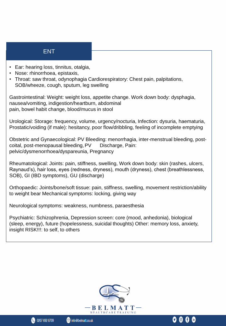
• Ear: hearing loss, tinnitus, otalgia,
• Nose: rhinorrhoea, epistaxis,
• Throat: saw throat, odynophagia Cardiorespiratory: Chest pain, palpitations,
SOB/wheeze, cough, sputum, leg swelling
Gastrointestinal: Weight: weight loss, appetite change. Work down body: dysphagia,
nausea/vomiting, indigestion/heartburn, abdominal
pain, bowel habit change, blood/mucus in stool
Urological: Storage: frequency, volume, urgency/nocturia, Infection: dysuria, haematuria,
Prostatic/voiding (if male): hesitancy, poor flow/dribbling, feeling of incomplete emptying
Obstetric and Gynaecological: PV Bleeding: menorrhagia, inter-menstrual bleeding, post-
coital, post-menopausal bleeding, PV Discharge, Pain:
pelvic/dysmenorrhoea/dyspareunia, Pregnancy
Rheumatological: Joints: pain, stiffness, swelling, Work down body: skin (rashes, ulcers,
Raynaud’s), hair loss, eyes (redness, dryness), mouth (dryness), chest (breathlessness,
SOB), GI (IBD symptoms), GU (discharge)
Orthopaedic: Joints/bone/soft tissue: pain, stiffness, swelling, movement restriction/ability
to weight bear Mechanical symptoms: locking, giving way
Neurological symptoms: weakness, numbness, paraesthesia
Psychiatric: Schizophrenia, Depression screen: core (mood, anhedonia), biological
(sleep, energy), future (hopelessness, suicidal thoughts) Other: memory loss, anxiety,
insight RISK!!!: to self, to others
ENT

Ask the patient to take a sip of water, hold it in their mouth and swallow the water on
command – thyroid masses and thyroglossal cysts will rise
Ask patient to protrude tongue – thyroglossal cyst will rise /and thyroid masses will
not
Look for systemic signs that may relate to neck pathology:
Cachexia – malignancy
Exophthalmos / proptosis – Graves’ disease
If there is a mid-line lump/scar or systemic signs suggestive of thyroid disease, ask
the examiner if a full thyroid status examination should be performed.
Note: Sometimes asking the patient to slightly tilt their head forward can help to relax
the neck muscles.
Lymph nodes can become enlarged for a number of reasons infection/malignancy
Lymph nodes are usually smooth, rubbery, with some mobility.
An enlarged, hard, irregular lymph node would be suggestive of malignancy.
Supraclavicular – left sided enlarged lymph node – Virchow’s node
Anterior cervical chain Posterior cervical chain Sub-mental
Sub-mandibular Occipital
Pre-auricular Post-auricular Parotid
Note: You do not need to follow this specific routine but be clear in your own mind so
that you cover all regions of the neck.
If a mid-line lump is present:
Palpate the lymph nodes:
EAR NOSE THROAT
General inspection Voice – weak/hoarse? Note any dyspnoea or stridorIdentify any scars on the neck – previous surgery (e.g. thyroidectomy) / radiotherapyObserve for any obvious masses in the neck Inspect from the front and both sides

The submandibular glands can be bilaterally palpated inferior and posterior to the body
of the mandible.
Thyroid gland
Place the three middle fingers of each hand along the midline of the neck below the chin
Locate the upper edge of the thyroid cartilage (“Adam’s apple”) Move inferiorly until you
reach the cricoid cartilage/ring
The first two rings of the trachea are located below the cricoid cartilage and the thyroid
isthmus overlies this area
Palpate the thyroid isthmus using the pads of your fingers
Palpate each lobe of the thyroid in turn by moving your fingers out laterally from the
isthmus
Ask the patient to swallow some water, whilst you feel for symmetrical elevation of the
thyroid lobes (asymmetrical elevation may suggest a unilateral thyroid mass) Ask the
patient to protrude their tongue once more (if a mass is a thyroglossal cyst, it will rise
during tongue protrusion)
If a thyroid mass is present, feel above and below it. Assess retrosternal extension by
percussion on the sternum and assess vascularity by auscultation.
Note: A normal thyroid will be impalpable
Submandibular gland
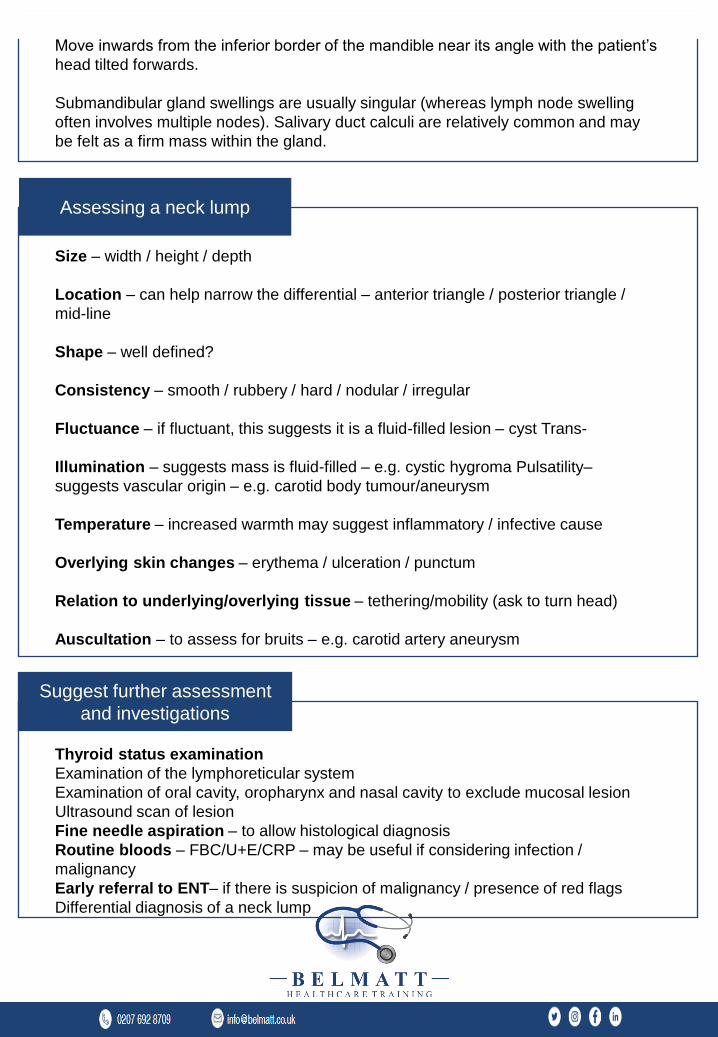
Move inwards from the inferior border of the mandible near its angle with the patient’s
head tilted forwards.
Submandibular gland swellings are usually singular (whereas lymph node swelling
often involves multiple nodes). Salivary duct calculi are relatively common and may
be felt as a firm mass within the gland.
Size – width / height / depth
Location – can help narrow the differential – anterior triangle / posterior triangle /
mid-line
Shape – well defined?
Consistency – smooth / rubbery / hard / nodular / irregular
Fluctuance – if fluctuant, this suggests it is a fluid-filled lesion – cyst Trans-
Illumination – suggests mass is fluid-filled – e.g. cystic hygroma Pulsatility–
suggests vascular origin – e.g. carotid body tumour/aneurysm
Temperature – increased warmth may suggest inflammatory / infective cause
Overlying skin changes – erythema / ulceration / punctum
Relation to underlying/overlying tissue – tethering/mobility (ask to turn head)
Auscultation – to assess for bruits – e.g. carotid artery aneurysm
Thyroid status examination
Examination of the lymphoreticular system
Examination of oral cavity, oropharynx and nasal cavity to exclude mucosal lesion
Ultrasound scan of lesion
Fine needle aspiration – to allow histological diagnosis
Routine bloods – FBC/U+E/CRP – may be useful if considering infection /
malignancy
Early referral to ENT– if there is suspicion of malignancy / presence of red flags
Differential diagnosis of a neck lump
Assessing a neck lump
Suggest further assessment
and investigations

The following features are red flags that should raise your suspicion of malignancy in
the context of a neck lump:
Hard, fixed mass
Patient is over 35 years old
Presence of mucosal lesion in the head or neck A history of persistent hoarseness or
dysphagia Trismus
Ear pain (referred from tongue base)
Differential diagnosis
The location of the lump within the neck can sometimes be useful in narrowing the
differential diagnosis. However, it should be noted that this is not an absolute rule,
with further investigations required to confirm a particular diagnosis.
Lymph nodes – often multiple, may suggest infection or malignancy
Lipoma – painless/smooth mass
Dermoid cyst – cysts formed along the lines of embryological fusion, painless
swellings that do not move with tongue protrusion (more common in children and
young adults).
Sebaceous cyst
Thyroid gland – located below thyroid cartilage
Thyroid nodule – can be single or multiple – adenomas/cysts/malignancy
Thyroglossal cysts – painless/smooth /cystic – rises on tongue protrusion
Laryngocele – reducible tense mass – mass returns on sneezing or nose blowing
Anterior triangle – area of the neck anterior to sternocleidomastoid
Red flags
Examination of the
neck
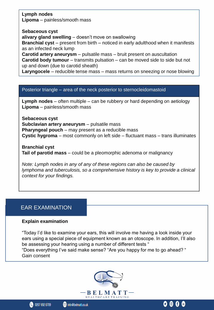
Posterior triangle – area of the neck posterior to sternocleidomastoid
Lymph nodes – often multiple – can be rubbery or hard depending on aetiology
Lipoma – painless/smooth mass
Sebaceous cyst
Subclavian artery aneurysm – pulsatile mass
Pharyngeal pouch – may present as a reducible mass
Cystic hygroma – most commonly on left side – fluctuant mass – trans illuminates
Branchial cyst
Tail of parotid mass – could be a pleomorphic adenoma or malignancy
Note: Lymph nodes in any of any of these regions can also be caused by
lymphoma and tuberculosis, so a comprehensive history is key to provide a clinical
context for your findings.
Explain examination
“Today I’d like to examine your ears, this will involve me having a look inside your
ears using a special piece of equipment known as an otoscope. In addition, I’ll also
be assessing your hearing using a number of different tests “
“Does everything I’ve said make sense? “Are you happy for me to go ahead? “
Gain consent
EAR EXAMINATION
Lymph nodes
Lipoma – painless/smooth mass
Sebaceous cyst
alivary gland swelling – doesn’t move on swallowing
Branchial cyst – present from birth – noticed in early adulthood when it manifests
as an infected neck lump
Carotid artery aneurysm – pulsatile mass – bruit present on auscultation
Carotid body tumour – transmits pulsation – can be moved side to side but not
up and down (due to carotid sheath)
Laryngocele – reducible tense mass – mass returns on sneezing or nose blowing
Pre auricular region: Sinus, swelling, cyst, accessory tragus, lymph nodes
Pinna: Size, shape and position
Post auricular region: Swelling, scar, battle’s sign, Reisinger’s sign, 3-point tenderness
test
External auditory canal: Upwards, backwards, outwards
Tympanic membrane
(Describe & identify normal anatomical landmarks) Colour – Pink/Rising
sun/Red/Bluish(Blood accumulation)
Cone of light,
Four quadrants, umbo,
Handle & lateral process of malleus, Anterior & posterior malleolar folds,
Pars tensa – Retraction, Granulation, Blebs, Sclerotic patches, Perforation (type,
margins, location, size, shape, edge, polyps etc.)
Parsflaccida, Bony annulus, Incudostapedial joint
Fistula test
Mastoid tenderness
Facial nerve
Tuning fork test (Rinne’s, Weber’s, Air bone conduction)
Ask the patient if they have noticed any change in their hearing recently.
Explain that you’re going to say 3 words or 3 numbers and you’d like them to repeat
them back to you (choose two-syllable words or bi-digit numbers).
1. Approximately 60cm from the ear, whisper a number or word.
2. Mask the ear not being tested by rubbing the tragus. Do not place your arm across
the face of the patient when rubbing the tragus, it is far nicer to occlude the ear
from behind the head. If possible, shield the patient’s eyes to prevent any visual
stimulus.
3. Ask the patient to repeat the number or word back to you. If they get 2/3 correct,
then their hearing level is 12db or better. If there is no response use a
conversational voice (48db or worse) or loud voice (76db or worse).
4. If there is no response you can move closer and repeat the test at 15cm. Here the
thresholds are 34db for a whisper and 56db for a conversational voice.
5. Assess the other ear in the same way. Explain to the patient that you are going to
test their hearing using a tuning fork.
External Ear Examination
Gross hearing assessment
Tap a 512Hz tuning fork and place in the midline of the forehead. The tuning fork
should be set in motion by striking it on your knee (not the patient’s knee or a table).
Ask the patient “Where do you hear the sound?“
Normal – sound is heard equally in both ears
Sensorineural deafness – sound is heard louder on the side of the intact ear
Conductive deafness – sound is heard louder on the side of the affected ear
We use 512Hz as this gives the best balance between time of decay and tactile
vibration. Ideally, you want a fork that has a long period of decay and cannot be
detected by vibration sensation.
1. Place a vibrating 512 Hz tuning fork firmly on the mastoid process (apply pressure
to the opposite side of the head to make sure the contact is firm). This tests bone
conduction.
2. Next, move the tuning fork in front of the external auditory meatus (while still
vibrating) to test air conduction. Ask the patient which they heard loudest and take
note of the result.
3. Ask the patient if the sound is louder in front of the ear (external auditory meatus)
or behind it (mastoid process)
Summary of Rinne’s test results:
Normal – Air conduction > Bone conduction (Rinne’s positive)
Sensorineural deafness – Air conduction > Bone conduction (both air and bone
conduction reduced equally)
Conductive deafness – Bone conduction > Air conduction (Rinne’s negative)
Ask the patient if they have any ear discomfort (if so examine the non-painful side
first).
Ask the patient which is their “better” ear. Always examine the better ear first to act as
a marker for comparison.
WEBER TEST
RINNE TEST
OTOSCOPY
Test your otoscope to check that it is working and commence inspection.
PinnaeInspect the pinnae:
Compare symmetry with the other side Deformity Ear piercings
Signs of active infection
ScarsInspect behind the pinnae (mastoid):
Skin changes, Erythema, Scars or previous surgery Ask about any pain in this region
Pre-auricular area (in front of the ear):
Pits, Sinuses, Fistulae
Conchal bowl – look for signs of active infection
Ensure the light is working on the otoscope and apply a sterile speculum (the largest
that will comfortably fit in the external auditory meatus).
Make sure to compare both ears.
Pull the pinna upwards and backwards with your other hand to straighten the external
auditory meatus.
Position otoscope at the external auditory meatus: Otoscope should be held in your
right hand for the patient’s right ear and vice versa
Hold the otoscope like a pencil and rest your hand against the patient’s cheek for
stability. This will also stop damage to the ear if there is any sudden movement.
3. Advance the otoscope under direct vision. Be gentle with the otoscope and ensure
movements are slow and considered otherwise you will cause the patient pain.
4. Look for any wax, swelling, erythema, discharge, foreign bodies or bony swellings.
5. Examine the tympanic membrane (think of it as having 4 quadrants which you
should systematically examine to avoid missing pathology):
Look for any wax, swelling, erythema, discharge, foreign bodies or bony swellings.
External ear exam
External canal and tympanic
membrane
7. Examine the tympanic membrane (think of it as having 4 quadrants which you
should systematically examine to avoid missing pathology):
Colour – pearly grey and translucent (normal) / erythematous (inflammation)
Erythema or bulging of the membrane – inspect for a fluid level e.g. otitis media
Perforation of the membrane – note the size of the perforation
Light reflex – absence/distortion may indicate ↑ inner ear pressure e.g. otitis media
Scarring of the membrane – tympanosclerosis – can result in significant hearing loss
Cholesteatoma – around the superior part of the eardrum
Withdraw the otoscope carefully
Discard the otoscope speculum into a clinical waste in the person that Meniere's
disease is a long-term condition, but vertigo usually significantly improves with
treatment.
Tympanic membrane
MENIERES DISEASEAdvise that an acute attack of vertigo will normally settle within 24 hours in most
people. If there is no improvement after 5–7 days, or there is any deterioration in
symptoms, ask the person to return to exclude an alternative diagnosis.
Advise people experiencing sudden attacks of vertigo to:
Keep medication readily accessible.
Consider the risks before undertaking activities such as operating dangerous
machinery, using ladders or scaffolding, or going swimming.
Advise the person not to drive when they are dizzy, or if they might experience an
episode of vertigo while driving.
The Driver and Vehicle Licensing Agency (DVLA) states that people with 'liability to
sudden and unprovoked or unprecipitated episodes of disabling dizziness' should stop
driving and inform the DVLA. For more information, see the DVLA publication
Assessing fitness to drive: a guide for medical professionals.
Conditions
Advice
Symptomatic treatment for acute attack
If symptoms are severe, hospital admission may be required for intravenous (IV)
labyrinthine sedatives and fluids to maintain hydration, and nutrition.
To rapidly relieve (severe) nausea or vomiting associated with Meniere's disease,
consider administration of buccal prochlorperazine, or a deep intramuscular injection
of prochlorperazine or cyclizine.
To help alleviate nausea, vomiting, and vertigo in other people with Meniere's
disease, consider prescribing a short course (7 days, 14 days if required previously)
of prochlorperazine, or an antihistamine (for example cinnarizine, cyclizine, or
promethazine teoclate).
The person has had previous attacks of Meniere's disease and responded well to
one of these drugs, consider trying that drug as first-line treatment. Always consider
the person's preferences when choosing the drug and delivery route. For more
information, see Prescribing information.
If the person's symptoms deteriorate or do not improve after 5-7 days, reassess to
exclude an alternative diagnosis.
Prescribe promethazine teoclate 25 mg orally at night. The dose may be increased to
100 mg daily.
If symptoms are severe, hospital admission may be required for intravenous (IV)
labyrinthine sedatives and fluids to maintain hydration, and nutrition.
To rapidly relieve (severe) nausea or vomiting associated with Meniere's disease,
consider administration of buccal prochlorperazine, or a deep intramuscular injection
of prochlorperazine or cyclizine.
To help alleviate nausea, vomiting, and vertigo in other people with Meniere's
disease, consider prescribing a short course (7 days, 14 days if required previously)
of prochlorperazine, or an antihistamine (for example cinnarizine, cyclizine, or
promethazine teoclate).
If the person has had previous attacks of Meniere's disease and responded well to
one of these drugs, consider trying that drug as first-line treatment. Always consider
the person's preferences when choosing the drug and delivery route.
If the person's symptoms deteriorate or do not improve after 5-7 days, reassess to
exclude an alternative diagnosis.
TREATMENT

If earwax is totally occluding the ear canal and any of the following are present: Hearing
loss, Earache, Tinnitus, Vertigo, Cough suspected to be due to earwax
If the tympanic membrane is obscured by wax but needs to be viewed to establish a
diagnosis.
If the person wears a hearing aid, wax is present and an impression needs to be taken of
the ear canal for a mould, or if wax is causing the hearing aid to whistle. If there is
hearing loss and the tympanic membrane cannot be seen, then the wax should be
removed as the majority of causes of conductive deafness are diagnosed by examining
the tympanic membrane [Browning, 1994] Once the wax has been removed if hearing
does not improve then alternative causes should be considered [Browning, 1994].
Visualisation of the tympanic membrane may identify causes of conductive deafness.
Wax may need to be removed in order to take an impression of the ear canal, for people
who wear hearing aids. Excess wax may cause the hearing aid to whistle.
Explain that removal of earwax may not necessarily relieve the symptoms (for example
hearing loss may be a sensorineural loss and not due to impacted wax).
Prescribe ear drops for 3–5 days initially, to soften wax and aid removal.
Olive oil, or almond oil drops can be used 3-4 times daily for 3-5 days (do not prescribe
almond oil ear drops to anyone who is allergic to almonds).
Sodium bicarbonate 5%, sodium chloride0.9%,
Sodium chloride 0.9% is not available as a proprietary ear drop product. However,
sodium chloride 0.9% nasal drops can be prescribed for use in the ear (off-label use).
Do not prescribe drops if you suspect the person has a perforated tympanic membrane.
Warn the person that instilling ear drops may cause transient hearing loss, discomfort,
dizziness and irritation of the skin. If symptoms persist, consider ear irrigation, providing
that there are no contraindications. If irrigation is unsuccessful, there are three options:
Advise the person to use ear drops for a further 3–5 days and then return for further
irrigation.
Instil water into the ear. After 15 minutes irrigate the ear again.
Refer to an Ear Nose and Throat specialist for removal of wax.
Advise anyone who has had earwax removed to return if they develop otalgia, or
significant itching of the ear, discharge from the ear (otorrhoea), or swelling of the
external auditory meatus, as this may indicate infection.
EAR WAX
How to remove earwax
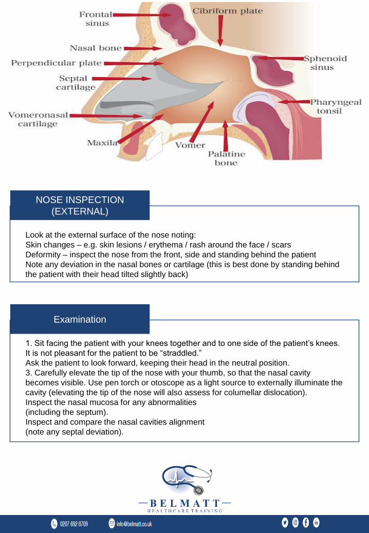
Look at the external surface of the nose noting:
Skin changes – e.g. skin lesions / erythema / rash around the face / scars
Deformity – inspect the nose from the front, side and standing behind the patient
Note any deviation in the nasal bones or cartilage (this is best done by standing behind
the patient with their head tilted slightly back)
1. Sit facing the patient with your knees together and to one side of the patient’s knees.
It is not pleasant for the patient to be “straddled.”
Ask the patient to look forward, keeping their head in the neutral position.
3. Carefully elevate the tip of the nose with your thumb, so that the nasal cavity
becomes visible. Use pen torch or otoscope as a light source to externally illuminate the
cavity (elevating the tip of the nose will also assess for columellar dislocation).
Inspect the nasal mucosa for any abnormalities
(including the septum).
Inspect and compare the nasal cavities alignment
(note any septal deviation).
NOSE INSPECTION
(EXTERNAL)
Examination
Further inspection can be done using an otoscope with a large speculum attached
(inserting only the very tip into the nose) or using Thulium’s speculum which essentially
just widens the nasal cavity to allow you to peer in using a Lightsource.
The correct method for using the Thudicum’s speculum is slightly counter-intuitive,
however, it does allow the best visualisation of the nasal mucosa. Insert your index
finger into the bend of the speculum and support it above with the thumb. The middle
and ring fingers are used to manipulate the prongs of the speculum. You will be aiming
to look at the gap between these two fingers.
Whichever method you use, you should inspect the various elements visible:
Nasal vestibule – skin changes (e.g. ulceration) / swelling / asymmetry
Nasal septum – polyps / deviation / perforation / areas of cautery
Inferior turbinate’s – asymmetry / inflammation / polyps
The turbinates are projections of bone, covered in nasal mucosa, that control airflow
through the nose, exposing it to a large surface area of mucosa which both warms and
cleans the air prior to it arriving at the lungs.
Nasal bones and cartilage
Palpate the nasal bones assessing:
Alignment
Tenderness or irregularity (if suspicious of fracture in trauma)
Palpate the nasal cartilage assessing:
Alignment Tenderness
Palpate the infraorbital ridges and assess eye movement if there is a history of trauma
to screen for an orbital blowout fracture. The classical signs are of infraorbital
tenderness, epistaxis and restricted eye movement (usually on vertical gaze).
Further assessment
Nasal Palpation
A cholesteatoma is an abnormal sac of keratinizing squamous epithelium and
accumulation of keratin within the middle ear or mastoid air cell spaces that can
become infected and also erode neighbouring structures.
Cholesteatoma may be asymptomatic in its early stages. Cholesteatoma most
commonly presents with a persistent or recurrent discharge from the ear that is often
foul smelling. Additionally, a conductive hearing loss may occur (although it is
commonly not noticed) as well as ear discomfort (although this is usually mild and not a
prominent feature of the condition).
Rarely with progression of the disease vertigo, sensorineural hearing loss, facial nerve
palsy, meningitis or intracranial abscess may develop.
The diagnosis requires clear visualisation of the tympanic membrane to identify the
characteristic appearance of a cholesteatoma. If the tympanic membrane can be seen,
a cholesteatoma should be suspected if there is:
A deep retraction pocket in the tympanic membrane, with or without granulation tissue
and skin debris.
A crust-like lesion, often yellow or brown in colour, usually in the upper part of the
tympanic membrane, often surrounded by pus, and sometimes associated with a
perforation of the adjacent tympanic membrane.
If the tympanic membrane cannot be clearly seen because the external auditory canal
is occluded by pus, the person should be treated for presumed infection and brought
back for re-examination after treatment has been completed. The person should either
receive treatment for:
Otitis externa, particularly if there are symptoms and signs of inflammation of the
external auditory canal, or
Acute otitis media if there has been an acute onset of pain associated with the purulent
discharge. If the tympanic membrane cannot be clearly seen after treatment, the
person should be referred to an Ear, Nose, and Throat specialist. Referral should not be
delayed for repeated courses of treatment if the discharge persists.
Emergency admission should be arranged for people with a suspected cholesteatoma
associated with a serious complication of the condition including:
A facial nerve palsy or vertigo.
Other neurological symptoms (including pain) or signs that could be associated with the
development of an intracranial abscess or meningitis.
Routine referral to an Ear, Nose, and Throat specialist should be arranged for people
with a suspected cholesteatoma who do not have a serious complication.
CHOLESTE ATOMA
There are two common methods via which to formally assess nasal airflow shown
below.
Method one
Place your thumb over the nostril not being assessed to occlude airflow.
Ask the patient to breath in through their nose and note the degree of airflow.
Repeat assessment on the other nostril, noting any difference in apparent airflow.
Reduced airflow through a particular nostril may indicate the presence of something
blocking that air passage, such as a polyp, deviated nasal septum or foreign body.
The absence of misting or a disparity in the amount of
misting between the nostrils may suggest unequal or absent airflow through a particular
nostril.
Treatment for occasional symptoms, prescribe an antihistamine first line.
For people with allergic conjunctivitis, children aged 2–5 years of age, and people who
prefer oral treatment, prescribe an oral antihistamine (such as cetirizine or loratadine).
For all other people, prescribe intranasal azelastine first line. Explain the importance of
a good technique.
For people who want preventive treatment to control more frequent or persistent
symptoms:
Advise the person to avoid the causative allergen, if possible.
If allergen avoidance is inadequate or not possible, prescribe drug treatment. If nasal
drops or a spray is prescribed, explain the importance of a good technique.
If the predominant symptom is nasal blockage, or nasal polyps are present, prescribe an
intranasal corticosteroid.
Nasal Airflow Assessment
ALLERGIC RHINITIS
If the predominant symptom is sneezing or nasal discharge, prescribe an oral
antihistamine (if oral treatment is preferred or allergic conjunctivitis is present) or an
intranasal corticosteroid(if a more effective treatment is required).
For pregnant or breastfeeding women, prescribe an intranasal corticosteroid.
If this is not tolerated or additional treatment is required, prescribe an oral antihistamine
(loratadine).
Intranasal sodium cromoglicate and nasal douching (with normal saline) can be used as
alternative or add-on treatments.
For people who require rapid relief of symptoms while awaiting preventive treatment to
take effect:
If nasal congestion is a problem, prescribe an intranasal corticosteroid for up to 7days.
If the person is already using an intranasal corticosteroid, add an oral antihistamine.
If symptoms are severe and/or impairing quality of life, prescribe a 5–10- day course of
prednisolone: 20–40 mg a day in adults, 10 mg a day in children.
Advise people to reconsult after 2–4 weeks if symptoms remain inadequately controlled.
Advantages and disadvantages of first-line drug treatments for allergic rhinitis
Table 1 shows some of the advantages and disadvantages of the different first-line drug
treatments for allergic rhinitis.
Relative efficiency Relative efficiency Onset of action
antihistamine allergic rhinitis Allergicconjunctives
++ ++
Within 1hour
Once-daily options available
Table 1
Not suitable for children < 5 years of age. † Maximum efficacy takes days or weeks to
develop [ARIA, 2010]
Advise allergen avoidance for people with: Suspected pollen allergy.
House dust mite allergy — when symptoms are inadequately controlled with maximal
preventive drug treatment and the responsible allergen has been confirmed by allergy
testing.
Suspected animal allergy — after confirming the responsible allergen by allergen testing.
CHOLESTEATOMA
For people with grass pollen allergy, advise: Against walking in grassy, open spaces,
particularly during the early morning, evening, and night, when pollen counts are at their
highest.
Keeping windows shut in cars and buildings.
Changing car pollen filters with each service, if these are fitted.
For people with confirmed house dust mite allergy inadequately controlled by drug
treatment, advise:
Fitting mattresses and pillows with house dust mite impermeable covers.
Using synthetic pillows and acrylic duvets, and keeping furry toys off the bed.
Washing all bedding and furry toys at least once a week at high temperatures.
Choosing wooden or hard floor surfaces instead of carpets, if possible.
Fitting blinds that can be wiped clean instead of curtains. Surfaces should be wiped
regularly with clean, damp cloth.
For people with confirmed animal allergy, advise that ideally the animal should not be
allowed in t house. When this is not acceptable, advise restricting their presence to the
kitchen.
For people with occupational allergy, advise eliminating or reducing exposure to
allergens, for example by using latex free gloves, using a dust mask, and ensuring that
their environment is adequately ventilated.
Occupational Allergies
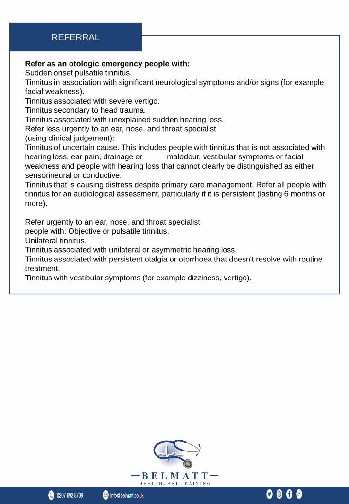
Refer as an otologic emergency people with:
Sudden onset pulsatile tinnitus.
Tinnitus in association with significant neurological symptoms and/or signs (for example
facial weakness).
Tinnitus associated with severe vertigo.
Tinnitus secondary to head trauma.
Tinnitus associated with unexplained sudden hearing loss.
Refer less urgently to an ear, nose, and throat specialist
(using clinical judgement):
Tinnitus of uncertain cause. This includes people with tinnitus that is not associated with
hearing loss, ear pain, drainage or malodour, vestibular symptoms or facial
weakness and people with hearing loss that cannot clearly be distinguished as either
sensorineural or conductive.
Tinnitus that is causing distress despite primary care management. Refer all people with
tinnitus for an audiological assessment, particularly if it is persistent (lasting 6 months or
more).
Refer urgently to an ear, nose, and throat specialist
people with: Objective or pulsatile tinnitus.
Unilateral tinnitus.
Tinnitus associated with unilateral or asymmetric hearing loss.
Tinnitus associated with persistent otalgia or otorrhoea that doesn't resolve with routine
treatment.
Tinnitus with vestibular symptoms (for example dizziness, vertigo).
REFERRAL
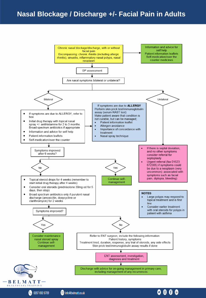
Nasal Blockage / Discharge +/- Facial Pain in Adults
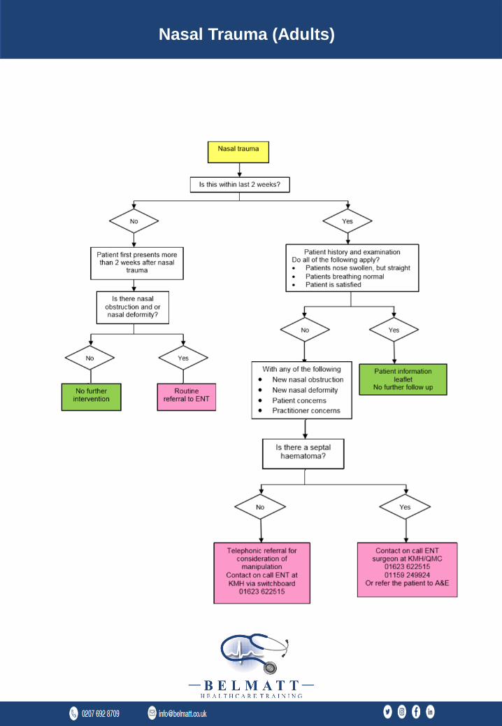
Nasal Trauma (Adults)

Hearing Problems in Children
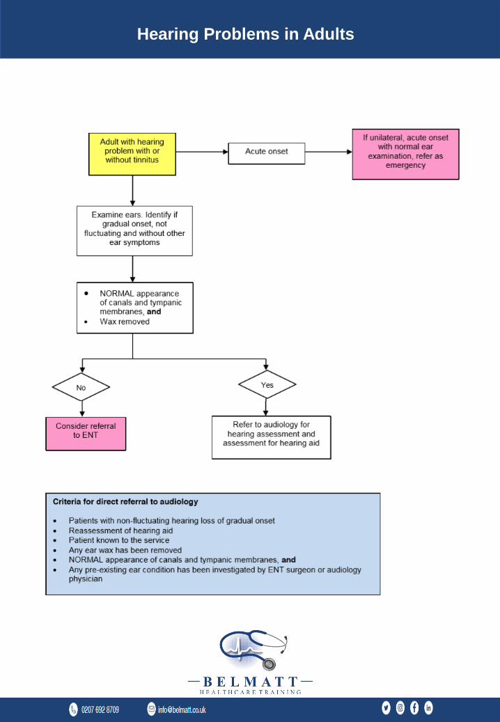
Hearing Problems in Adults

Infectious Sore Throat in Adults
Infectious Sore Throat in Adults

Non-Infectious Sore Throat in Adults
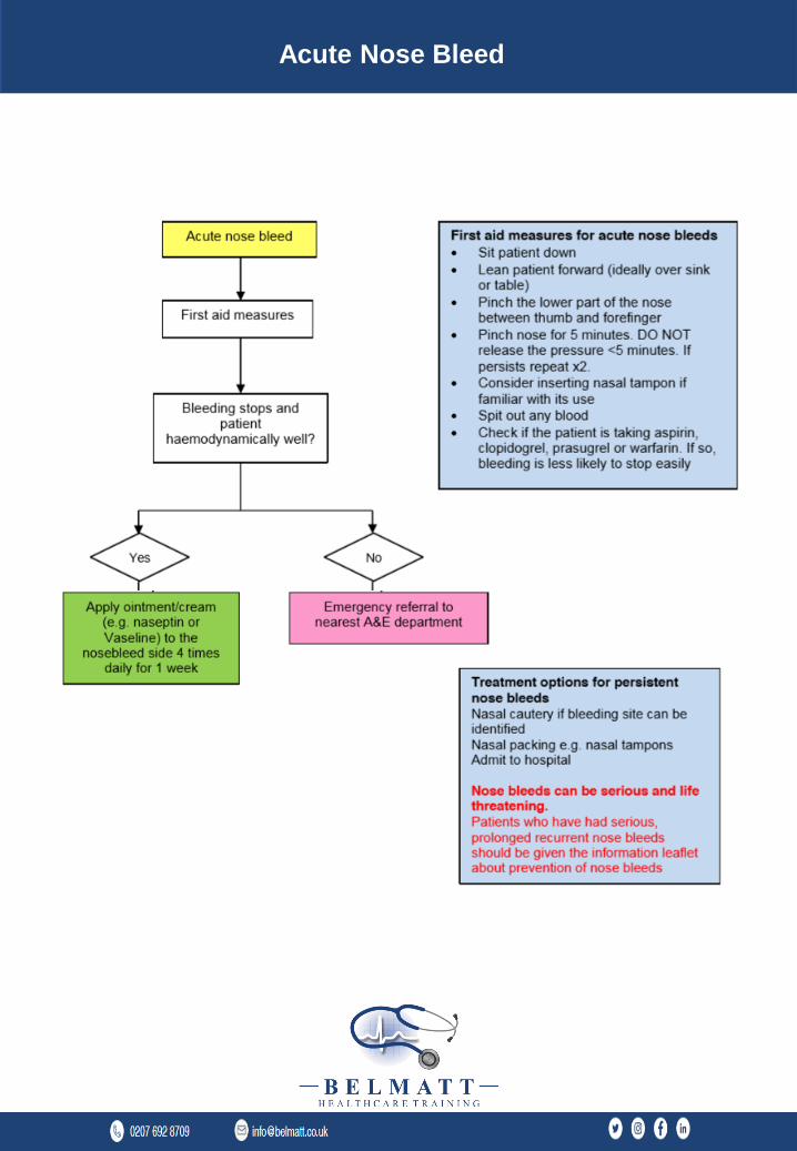
Acute Nose Bleed
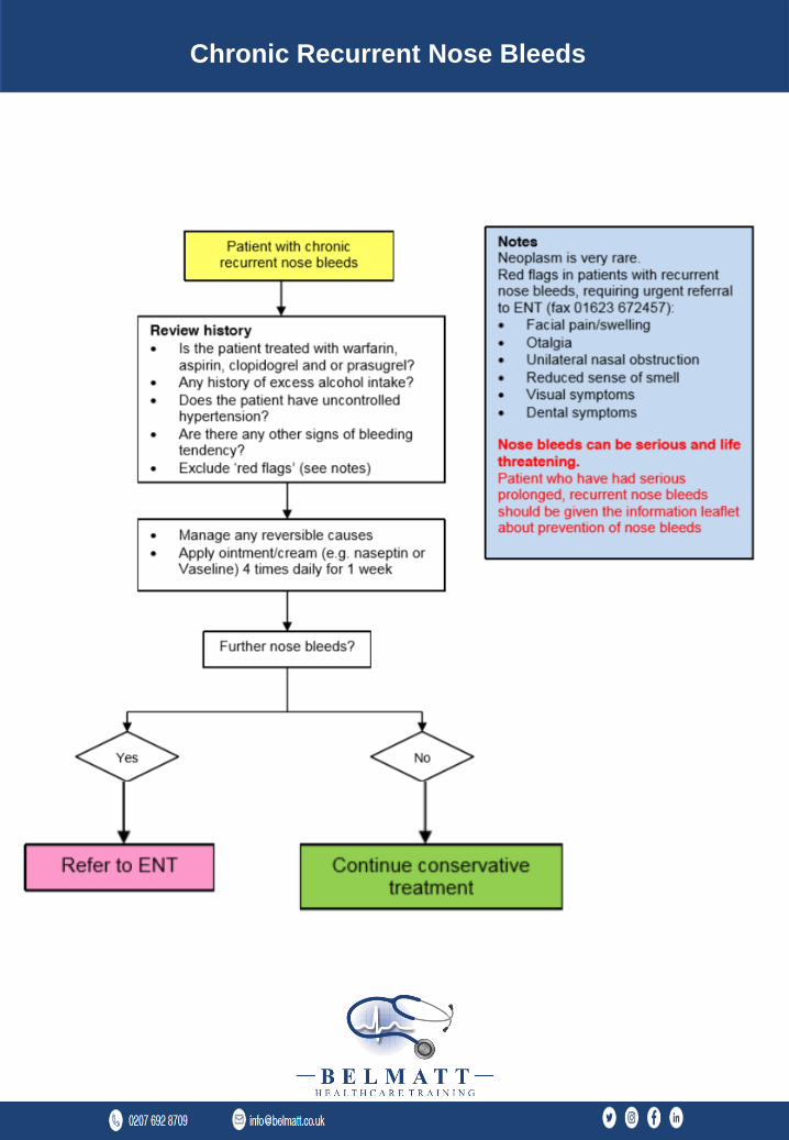
Chronic Recurrent Nose Bleeds
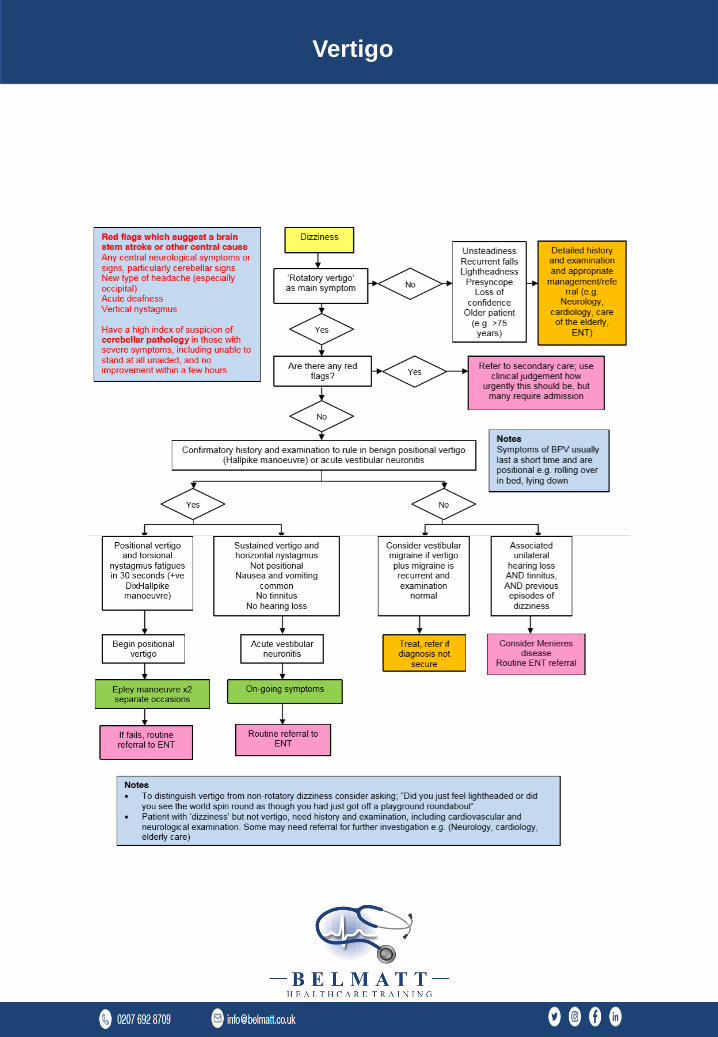
Vertigo
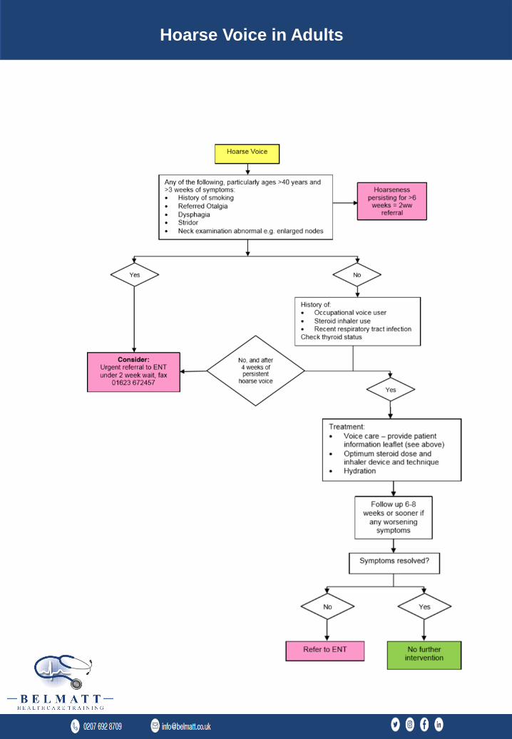
Hoarse Voice in Adults
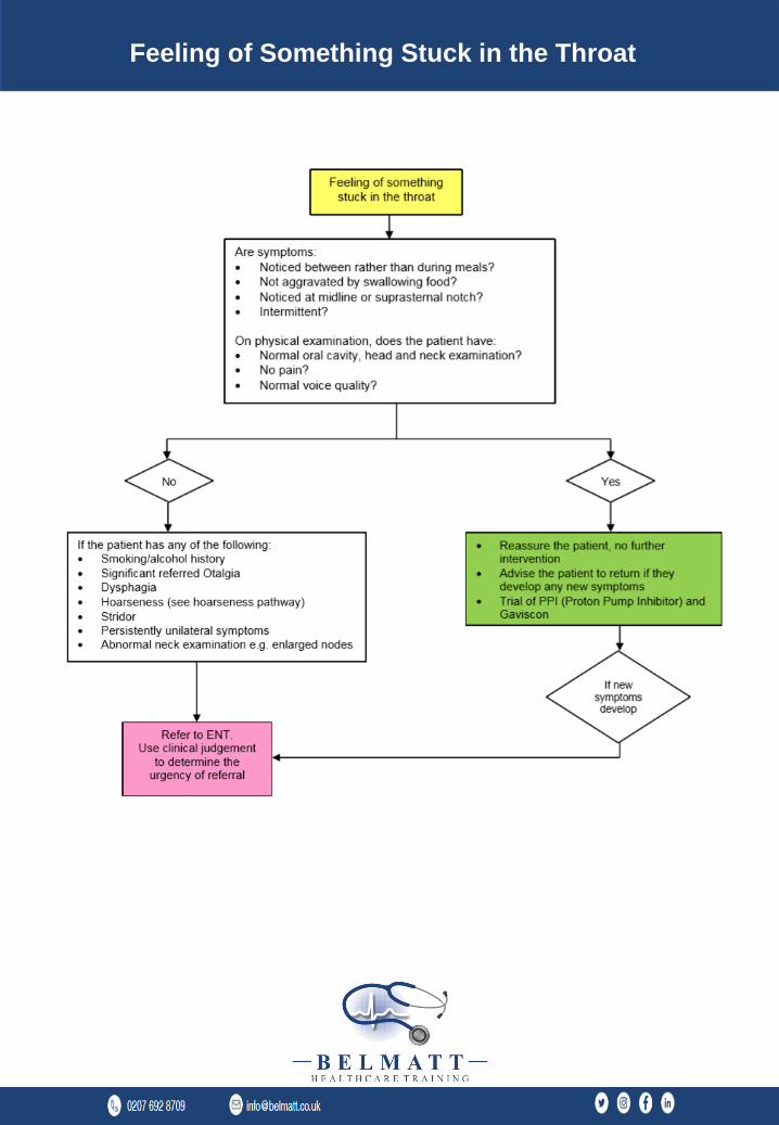
Feeling of Something Stuck in the Throat
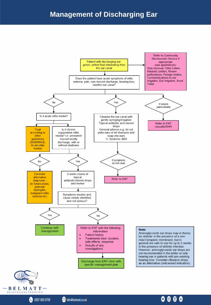
Management of Discharging Ear

Primary Care Management of Snoring in Adults/ Sleep
Apnoea
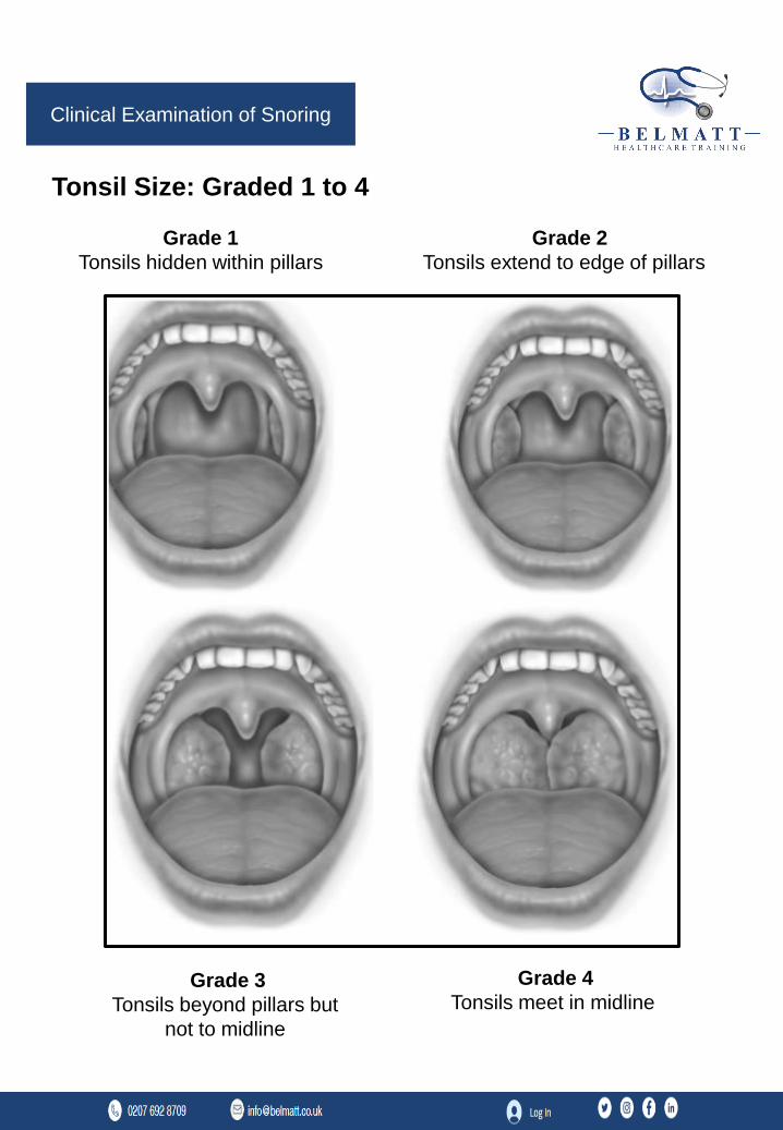
Tonsil Size: Graded 1 to 4
Grade 1
Tonsils hidden within pillars
Grade 2
Tonsils extend to edge of pillars
Grade 3
Tonsils beyond pillars but
not to midline
Grade 4
Tonsils meet in midline
Clinical Examination of Snoring
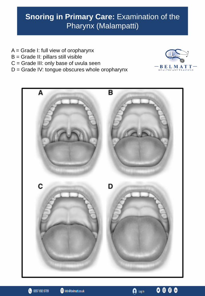
A = Grade I: full view of oropharynx
B = Grade II: pillars still visible
C = Grade III: only base of uvula seen
D = Grade IV: tongue obscures whole oropharynx
Snoring in Primary Care: Examination of the
Pharynx (Malampatti)

C o n t a c t U s
PHONE CALL
0207 692 8709
FOLLOW US
Belmatt Healthcare
Training
EMAIL US
BELMATT HEALTHCARE TRAINING
Belmatt Healthcare Training Limited, provider of postgraduate training.
Registered Office: Suite 570 405 Kings Road, Chelsea, London, England, SW 10 0BB


![ifurd Isreai cll ag] ent to I end r](https://img.dokumen.tips/doc/110x75/63144bedb1e0e0053b0eb3ae/ifurd-isreai-cll-ag-ent-to-i-end-r.jpg)


