Embed Size (px)
Citation preview

The LaryngoscopeLippincott Williams & Wilkins, Inc., Philadelphia© 2003 The American Laryngological,Rhinological and Otological Society, Inc.
Endoscopic Surgery for JuvenileAngiofibroma: When and How
Piero Nicolai, MD; Marco Berlucchi, MD; Davide Tomenzoli, MD; Johnny Cappiello, MD;Matteo Trimarchi, MD; Roberto Maroldi, MD; Giuseppe Battaglia, MD; Antonino R. Antonelli, MD
Objectives/Hypothesis: In recent years, the indi-cations for endoscopic surgery of the sinonasaltract, originally introduced for the treatment ofinflammatory diseases, have been expanded to in-clude selected cases of benign and malignant neo-plastic lesions. The aim of the present study wasto establish the efficacy of endoscopic surgery inthe management of small and intermediate-sizedjuvenile angiofibromas. Study Design: Retrospec-tive study. Methods: We reviewed the clinicalrecords and the preoperative and postoperativeimaging studies of 15 patients with juvenile angio-fibroma who were treated with an endoscopic ap-proach after embolization in the period from Jan-uary 1994 to April 2000. All patients wereprospectively followed by endoscopic and mag-netic resonance imaging evaluations performedat regular intervals (every 4 months during thefirst year and, subsequently, every 6 months). Re-sults: According to a staging system reported in1989, there were two patients with a type I, ninewith a type II, three with a type IIIA, and one witha type IIIB juvenile angiofibroma. Angiographydemonstrated that the vascular supply wasstrictly unilateral in 11 patients and bilateral in 4.Intraoperative blood loss ranged from 80 to 600mL (mean blood loss, 372 mL). During follow-up(range, 24–93 mo; mean follow-up, 50 mo [SD �19.9 mo]), only one patient presented a residuallesion on magnetic resonance imaging, which was16 mm in diameter and was detected 24 monthsafter surgery. Conclusions: The endoscopic ap-proach is a safe and effective technique thatallows removal of small and intermediate-sizedjuvenile angiofibromas (without extensive in-volvement of the infratemporal fossa and cavern-ous sinus) with a low morbidity. Advanced lesionsare more appropriately treated by external ap-
proaches. Key Words: Juvenile nasopharyngealangiofibroma, endoscopic surgery, diode laser, re-currence, imaging.
Laryngoscope, 113:775–782, 2003
INTRODUCTIONJuvenile angiofibroma (JA) is a highly vascularized
benign lesion that typically occurs in adolescent boys com-plaining of nasal obstruction and epistaxis.1 Recent stud-ies seem to suggest that the lesion displays an immuno-histological and electron microscopic profile moreconsistent with a vascular malformation than with a tu-mor2,3; furthermore, the presence of genetic imbalanceshave been demonstrated by comparative genomichybridization.4
Management of JA has always been regarded as aformidable challenge to the otolaryngologist. However,there is general agreement that surgery should be consid-ered the elective therapy. Moreover, the use of radiother-apy and chemotherapy should be better confined to thetreatment of very extensive lesions or recurrent tumorsinvolving critical anatomical areas, where surgical extir-pation can carry a significant morbidity. Several surgicaloptions, including transhyoid, transpalatal, transfacial(through a lateral rhinotomy or a midfacial degloving),and infratemporal approaches, have been advocated. Re-cent reports have focused on more conservative transnasaltreatments, using either purely endoscopic5–20 or com-bined microscopic–endoscopic techniques.21,22
From January 1992 to September 2002, in all, 101benign and malignant lesions of the sinonasal tract ornasopharynx was managed by an endoscopic approach atthe Department of Otolaryngology of the University ofBrescia (Brescia, Italy). The main purpose of the presentreport was to review the results obtained in the group ofpatients who presented with JA, so as to have a betterunderstanding of the possibilities and limitations of theendoscopic approach in this specific disease.
PATIENTS AND METHODSBetween January 1968 and September 2002, 53 patients
with JA were treated at the Department of Otolaryngology, Uni-versity of Brescia; 31 of them had the lesion resected through anexternal approach and 22 underwent a transnasal removal.
Presented at the 105th Annual Meeting of the Triological Society,Boca Raton, FL, May 14, 2002.
From the Departments of Otorhinolaryngology (P.N., M.B., D.T., J.C.,M.T., A.R.A.) and Radiology (G.B., R.M.), University of Brescia, Brescia, Italy.
Editor’s Note: This Manuscript was accepted for publication Decem-ber 9, 2002.
Send Correspondence to Piero Nicolai, MD, Department of Otorhi-nolaryngology, University of Brescia. Piazza Spedali Civili 1, 25123 Bres-cia, Italy. E-mail: [email protected] or [email protected]
Laryngoscope 113: May 2003 Nicolai et al.: Surgery for Juvenile Angiofibroma
775

TAB
LEI.
Pat
ient
Cha
ract
eris
tics.
Pat
ient
No.
Age (y)
Site
sIn
volv
ed(s
ize)
Feed
ing
Ves
sels
(%em
bol
izat
ion)
Sta
ge*
Pre
viou
sS
urge
ryIn
trao
per
ativ
eB
lood
Loss
Rem
oval
ofN
asal
Pac
kage
Hos
pita
lizat
ion
Tim
eFo
llow
-up
118
Nas
opha
rynx
,na
salc
avity
,p
tery
goid
pro
cess
(3.5
�2
�2
cm)
Inte
rnal
max
illar
y,ac
cess
ory
men
inge
al,
and
asce
ndin
gp
hary
ngea
lart
ery
(�90
%)
I–
200
mL
3d
ays
9d
ays
NE
D96
mo
214
Nas
o-or
opha
rynx
,na
salc
avity
,sp
heno
idsi
nus,
pte
rygo
idp
roce
ss(5
.5�
4�
9.5
cm)
Man
dib
ular
and
asce
ndin
gp
hary
ngea
lar
tery
(�90
%)
II–
80m
L2
day
s8
day
sN
ED
84m
o
330
Nas
opha
rynx
,na
salc
avity
,sp
heno
idsi
nus,
pte
rygo
max
illar
yfo
ssa
(3�
2�
3cm
)S
phe
nop
alat
ine
and
vid
ian
arte
ry(9
0%)
IIR
esec
tion
thro
ugh
ala
tera
lrh
inot
omy
25m
ob
efor
e40
0m
L2
day
s4
day
sN
ED
66m
o
414
Nas
opha
rynx
,na
salc
avity
,sp
heno
idsi
nus
(2�
1�
2cm
)S
phe
nop
alat
ine
and
vid
ian
arte
ry(�
95%
)II
–30
0m
L2
day
s5
day
sA
WD
66m
o
514
Nas
opha
rynx
,na
salc
avity
,sp
heno
idsi
nus,
pte
rygo
max
illar
yfo
ssa,
pte
rygo
idp
roce
ss(4
.7�
2.8
�4
cm)
Sp
heno
pal
atin
ean
das
cend
ing
pha
ryng
eal
arte
ry,
infe
rola
tera
ltru
nk(9
0%)
II–
400
mL
3d
ays
12d
ays
NE
D60
mo
614
Nas
opha
rynx
,na
salc
avity
,sp
heno
idsi
nus,
pte
rygo
max
illar
yfo
ssa,
pte
rygo
idp
roce
ss(4
�2.
8�
5cm
)
Asc
end
ing
pha
ryng
eal,
sphe
nop
alat
ine
(bil)
,m
and
ibul
arar
tery
(bil)
(80%
)II
–60
0m
L4
day
s5
day
sN
ED
48m
o
718
Nas
opha
rynx
,na
salc
avity
,sp
heno
idsi
nus,
pte
rygo
max
illar
yfo
ssa
(5�
2.5
�4
cm)
Sp
heno
pal
atin
ear
tery
(�95
%)
II–
500
mL
2d
ays
5d
ays
NE
D48
mo
823
Nas
opha
rynx
,na
salc
avity
,sp
heno
idsi
nus,
pte
rygo
max
illar
yfo
ssa,
infe
rior
orb
itary
fess
ura,
sup
erio
ror
bita
ryfe
ssur
a,p
tery
goid
pro
cess
,p
aras
ella
rre
gion
(ext
rad
ural
)(3
.4�
2�
4.5
cm)
Infe
rola
tera
ltru
nk,
sphe
nop
alat
ine
and
asce
ndin
gp
hary
ngea
lart
ery
(90%
)III
b–
500
mL
2d
ays
7d
ays
NE
D48
mo
915
Pte
rygo
idp
roce
ss,
infr
atem
por
alfo
ssa
(2�
1.5
�1.
5cm
)A
cces
sory
men
inge
alan
dsp
heno
pal
atin
ear
tery
(�90
%)
IIIa
Res
ectio
nth
roug
ha
tran
spal
atal
app
roac
h44
mo
bef
ore
500
mL
2d
ays
7d
ays
NE
D42
mo
1015
Nas
opha
rynx
,na
salc
avity
,sp
heno
idsi
nus,
pte
rygo
max
illar
yfo
ssa
(4.5
�2
�4.
5cm
)S
phe
nop
alat
ine,
acce
ssor
ym
enin
geal
,an
das
cend
ing
pha
ryng
eala
rter
y(�
90%
)II
–20
0m
L2
day
s5
day
sN
ED
42m
o
1113
Nas
opha
rynx
,na
salc
avity
,sp
heno
idsi
nus,
pte
rygo
max
illar
yan
din
frat
emp
oral
foss
a(6
.1�
4.1
�5.
2cm
)
Sp
heno
pal
atin
ear
tery
(bil)
(�95
%)
IIIa
–30
0m
L3
day
s6
day
sN
ED
42m
o
1215
Nas
opha
rynx
,na
salc
avity
,p
tery
goid
pro
cess
(4.5
�3
�4
cm)
Man
dib
ular
,sp
heno
pal
atin
ean
dfa
cial
arte
ry(1
00%
)I
–30
0m
L2
day
s8
day
sN
ED
42m
o
1316
Nas
opha
rynx
,sp
heno
idsi
nus
(3�
2.7
�3
cm)
Man
dib
ular
,vi
dia
n,ac
cess
ory
men
inge
al,
and
asce
ndin
gp
hary
ngea
lart
ery
(60%
)II
Res
ectio
nth
roug
ha
late
ral
rhin
otom
y42
mo
bef
ore
100
mL
2d
ays
5d
ays
NE
D36
1418
Nas
opha
rynx
,na
salc
avity
,sp
heno
idsi
nus,
pte
rygo
max
illar
yan
din
frat
emp
oral
foss
a,p
tery
goid
pro
cess
(7�
4�
6cm
)
Infe
rola
tera
ltru
nk,
sphe
nop
alat
ine
(bil)
(90%
)III
a–
600
mL
2d
ays
5d
ays
NE
D24
mo
1514
Nas
opha
rynx
,na
salc
avity
,sp
heno
idsi
nus,
pte
rygo
max
illar
yfo
ssa
(6.2
�4
�2.
6cm
)S
phe
nop
alat
ine
arte
ry(b
il),
Asc
end
ing
pal
atin
ear
tery
,as
cend
ing
pha
ryng
eal
arte
ry(8
5%)
II–
600
mL
2d
ays
5d
ays
NE
D24
mo
*Acc
ord
ing
toA
ndre
ws
etal
.,19
89.1
NE
D�
noev
iden
ceof
dis
ease
;A
WD
�al
ive
with
dis
ease
.
Laryngoscope 113: May 2003 Nicolai et al.: Surgery for Juvenile Angiofibroma
776
The present report focuses on a cohort of 15 patients (TableI) who were treated by means of an endoscopic approach in theperiod between January 1994 and April 2000 and were followedfor at least 24 months. All were male patients, with ages rangingfrom 13 to 30 years (mean age, 15.8 y). Three of them (cases 3, 9,and 13) had already been treated for their lesions 25, 44, and 42months, respectively, before admission to our department. Pre-operative workup included magnetic resonance imaging (MRI)and angiography in all patients. Magnetic resonance imagingexaminations were performed on axial and coronal planes withcontrast enhancement. The angiographic study, which was per-formed 72 to 24 hours before surgery, encompassed bilateralexamination of the carotid arteries; at the same time, all lesionswere embolized using polyvinyl alcohol particles ranging in sizefrom 150 to 500 �m. Magnetic resonance imaging scans wereretrospectively reviewed by the same radiologist (R.M.) to assessthe volume, extent, and stage of the lesions according to thesystem reported by Andrews et al.1 (Table II). The number offeeding vessels, their compartment of origin (external vs. internalcarotid artery), and the presence of a contralateral supply werealso recorded; correlations among these variables have been eval-uated by means of t test.
The entity of the embolization was expressed by estimatingthe devascularization of the lesion. A value of 100% correspondedto the absence of any blush following embolization. Subtotaldevascularizations were graded by comparing the size of residualblush (in both the anteroposterior and lateral-to-lateral projec-tions) with pretreatment lesion staining.
Every patient had two units of autologous blood bankedduring the 2 weeks before the operation to minimize the intraop-erative or postoperative need for homologous transfusion. Aninformed consent for intraoperative change to an external ap-proach whenever the lesion could not be resected endoscopicallywas obtained from all patients.
All operations were performed by the same surgeon (P.N.).Hypotensive general anesthesia was used in all cases, with thepatient lying supine in a slightly reversed Trendelenburg posi-tion. Both nasal cavities were packed with cottonoid pledgetssoaked in oxymetazoline that were left in place for 10 minutes;further vasoconstriction was achieved by submucosal injection of1% lidocaine with 1:200,000 epinephrine at the level of the root ofthe middle turbinate and of the uncinate process. Even thoughthe surgical technique slightly varied in individual patients ac-cording to the size and extension of the lesion, the dissectionincluded a series of basic surgical steps. Uncinectomy with pres-ervation of the frontal recess area, partial or total resection of themiddle turbinate, and anterior and posterior ethmoidectomy werefirst performed to obtain enough space to adequately work aroundthe lesion with the endoscopic instrumentation. A wide middle
antrostomy was subsequently created to expose the posterior wallof the maxillary sinus, which was removed by angled cuttinginstruments as far laterally as was dictated by the extension ofthe lesion into the pterygomaxillary and infratemporal fossa.After gentle dissection of the mass from the surrounding softtissues, the sphenopalatine or, when required, the internal max-illary artery was isolated and clipped. In patients with sphenoidsinus involvement, the anterior wall of the sinus was entirelyremoved to have a better visualization of the posterior projectionof the lesion. The dissection then proceeded along the subperios-teal plane to free the mass from the nasopharyngeal walls. Whenrequired because of firm adherence, the posterior part of theseptum was resected with the mass. Lesions that were particu-larly large and with deep extension into some critical areas (ie,infratemporal fossa, cavernous sinus) had the nasal-nasopharyngeal portion divided, first, and then removed by usinga diode laser set on pulsed mode (25 W, 0.1 s), vehicled through acontact disposable light guide that was 600 �m in diameter,which was mounted on a malleable handpiece (Dornier Medizin-Laser Gmbh, Germering, Germany). This was performed to speedup the procedure and to gain a bloodless exposure of the deepestextensions of the lesion. Depending on size, the mass was removedtransnasally or transorally. As a final surgical step, patients whoselesion invaded the pterygoid process had the adjacent skull basebone extensively drilled; special attention was paid to disclose anytumor extension along the vidian nerve. After careful review of thesurgical field, both nasal cavities were packed with Lyofoam (SetonHealth Care Group plc, Oldham, UK).
Only one patient (case 4) with a small lesion (2 � 1 � 2 cm)with no lateral extension received a more conservative treatment,which did not include dissection of the pterygomaxillary fossa andspared the middle turbinate. The estimated intraoperative bloodloss was correlated with the presence of afferent vessels from theinternal carotid artery, the number of feeding vessels, and thevolume of the lesion using a t test. All of the patients werefollowed prospectively with periodic evaluations including endos-copy, with storage of the images and MRI scans, scheduled atregular intervals (every 4 months during the first postoperativeyear, every 6 months up to the fifth year, and subsequently oncea year).
RESULTSThe volume of the lesions ranged from 4 to 209 cm3
(mean volume, 61.3 cm3; SD � 60.7 cm3). Nasopharynx,nasal cavity, sphenoid sinus, pterygomaxillary fossa,pterygoid process, and infratemporal fossa were involved,respectively, in 14 (93.3%), 13 (86.6%), 12 (80.0%), 9(60.0%), 8 (60.0%), and 3 (21.4%) patients. One patient(case 8) had a lesion that extended into the inferior andsuperior orbital fissures and into the parasellar region(extradural) (Fig. 1). Two patients had type I lesions, 9had type II lesions, 3 had type IIIA lesions (Fig. 2), and 1patient had a type IIIB lesion.
Angiography demonstrated that all lesions had a vas-cular supply from the distal branches of the internal max-illary artery (sphenopalatine and/or vidian arteries);seven (46.6%) of them received additional vascularizationfrom internal carotid branches (mandibular artery andinferolateral trunk). The lesions were fed by 1 to 5 vessels(mean number of vessels, 2.7) that were distributed asfollows: distal branches of the internal maxillary artery inall cases, ascending pharyngeal artery in nine cases(60.0%), mandibular artery in five cases (33.3%), accessorymeningeal artery in four cases (26.6%), inferolateral trunk
TABLE II.Staging System of Nasopharyngeal Angiofibroma According to
Andrews et al.1
Type I Tumor limited to the nasopharyngeal cavity; bonedestruction negligible or limited to thesphenopalatine foramen
Type II Tumor invading the pterygopalatine fossa or themaxillary, ethmoid, or sphenoid sinus with bonedestruction
Type III Tumor invading the infratemporal fossa or orbitalregion without intracranial involvement (a) or withintracranial extradural (parasellar) involvement (b)
Type IV Intracranial intradural tumor without (a) or with (b)infiltration of the cavernous sinus, pituitary fossa,or optic chiasm
Laryngoscope 113: May 2003 Nicolai et al.: Surgery for Juvenile Angiofibroma
777

in three cases (20.0%), and facial artery in one case (6.6%).In 11 patients (73.4%) the vascular supply to the lesionwas strictly unilateral. Four patients (26.6%) had con-tralateral feeding vessels arising in all cases from thedistal branches of the internal maxillary artery and in one
case (6.6%) from the mandibular artery. Embolization ob-tained a devascularization rate of 60% to 100% (mean,88.9%).
Feeding by internal carotid branch(es) correlatedwith the number of feeders arising from the external
Fig. 1. Patient 8. (A) The coronal plane demonstrates the lesionspreading between V2 (black arrow) and the internal carotid artery(white arrow) to contact the cavernous sinus. The submucosal ex-tension into the nasopharynx is also detectable. (B) Postoperativemagnetic resonance imaging scan (T1-weighted after paragmag-netic contrast agent injection) after 42 months. No enhancing lesionis detectable. The maxillary nerve and the internal carotid artery arenormal. (C) Endoscopic view (0° rigid fiberscope) of the left-sidenasal cavity 4 years after endoscopic treatment.
Laryngoscope 113: May 2003 Nicolai et al.: Surgery for Juvenile Angiofibroma
778

carotid artery (3.42 [SD � 0.78] vs. 2.12 [SD � 1.12][P � .01]). No correlation was found among the otherparameters tested.
Intraoperative blood loss varied from 80 to 600 mL(mean blood loss, 372 mL). No patient required autologousblood transfusion. No intraoperative or perioperative com-plications were observed. Nasal packings were removed 2to 4 days after surgery (mean period, 2.3 d). The meanhospitalization time was 6.4 days.
Definitive pathology reports confirmed the diagnosisof JA. At the control evaluations during the follow-upperiod (range, 24–96 mo; mean period, 51.2 mo; SD � 20.3mo), only the patient who had been treated for a type Itumor with a conservative dissection (case 4) presentedwith a 16-mm lesion involving the floor of the sphenoidsinus that was suspect for residual tumor 24 months aftersurgery at the MRI examination (Fig. 3). Subsequent MRI
evaluations scheduled every 4 months showed an increasein size to 21 mm over a period of 20 months.
One patient each developed a sphenoid mucocele anda synechia between the septum and the inferior turbinate.Both patients were otherwise asymptomatic and were suc-cessfully treated under local anesthesia by endoscopicsurgery.
DISCUSSIONDespite its benign nature, JA displays an aggressive
pattern of growth. From the point of origin, which islocated, according to some authors, in the sphenopalatineforamen23,24 or, according to others, in the posterior partof the pterygopalatine fossa at the anterior aperture of thevidian (or pterygoid) canal,25 the lesion first expands intothe nasopharynx and the nasal cavities. Subsequently, itinvades the sphenoid sinus, the pterygomaxillary fossa,the maxillary sinus, the infratemporal fossa, and the orbitby bone erosion. Intracranial extension, a late event re-ported in up to 36% of the patients,26 usually occurs ex-tradurally at the level of the middle cranial fossa; anteriorfossa invasion is an exceedingly rare finding.27 The in-volvement of most of the aforementioned areas is gener-ally asymptomatic and cannot be detected by endoscopicexamination; however, it must be carefully identified be-fore selecting the surgical approach.
Magnetic resonance imaging is the ideal method for amultiplanar evaluation of the lesion and for obtaining de-tailed information on the relationship between the mass andimportant adjacent structures (ie, orbit, dura, internal ca-rotid artery, cavernous sinus). Although MRI is not com-monly indicated for the assessment of invasion of bony struc-tures, in the present series it proved to be accurate indetecting erosion of the pterygoid process (60%) as well asextension into the pterygomaxillary (60%) and infratempo-ral fossa (21.4%). All these findings were confirmed atsurgery.
Fig. 2. Patient 11. (A) Preoperative magnetic resonance imaging(MRI) scan (T1-weighted after paragmagnetic contrast agent injec-tion). The right-side nasal fossa and sphenoid sinus are occupied bythe juvenile angiofibroma. Remodeling of maxillary sinus medial walland contralateral displacement of the nasal septum are demon-strated. Infratemporal extension of the lesion is present, along withincrease diameter of the internal maxillary artery (white arrows). (B)Three years after surgery, the postoperative MRI scan (T1-weightedafter paragmagnetic contrast agent injection) shows nonenhancingtissue (consistent with scar) replacing the infratemporal part of thejuvenile angiofibroma (arrows). The adjacent pterygoid processdoes not present any relevant abnormality.
Fig. 3. (Case 4). Recurrent juvenile angiofibroma (JA) at 24 monthsafter endonasal surgery. Follow-up magnetic resonance imagingscan (T1-weighted after paragmagnetic contrast agent injection)demonstrates a submucosal enhancing lesion close to the inferiorborder of the basisphenoid (arrows). The different signal betweenthe residual JA and the adjacent adenoid tissue is evident.
Laryngoscope 113: May 2003 Nicolai et al.: Surgery for Juvenile Angiofibroma
779
The pattern of vascularization of the lesion, anotherimportant feature to be considered when planning treat-ment, is assessed by angiography, which is extremelyhelpful, particularly in advanced lesions, in ruling out adirect involvement of the internal carotid artery. Never-theless, the presence of additional (nonembolized) feedersfrom the internal carotid artery did not correlate with theamount of blood loss and did not prevent the endoscopicapproach. The epidemiological profile, endoscopic appear-ance, and imaging findings of the lesion usually allow fora correct diagnosis without performing a biopsy, whichcan expose the patient to the risk of severe hemorrhage.28
Once data on the extent and vascularization of thelesion are available, the surgeon can select the techniquethat allows the best exposure with minimal morbidity. Inthis regard, we think that in the treatment of JA involvingthe nasopharynx, the nasal cavities, the sphenoid sinus, thepterygomaxillary fossa, the ethmoid, and the maxillary si-nus, endoscopic surgery can favorably compete with othertechniques, such as the midfacial degloving and lateral rhi-notomy approach. In fact, since 1994, we have treatedthrough a purely endoscopic approach all the patients withtype I or II lesions (according to the classification of Andrewset al.1) who came to our observation. The first importantadvantage of the endoscopic approach is the possibility ofobtaining a magnified, multi-angled view of the mass and ofits adjacent anatomical structures, which must necessarilybe coupled with a good control of intraoperative bleeding. Inthis respect, preoperative embolization, which in our experi-ence proved to be a safe procedure with only minimal mor-bidity, is a key element. Other tools we found effective inminimizing bleeding included the use of a delicate, atrau-matic dissection technique with the help of bipolar coagula-tion and a diode laser, which is especially helpful for theresection of a part of the lesion or for the incision of the nasalor nasopharyngeal mucosa in a nearly bloodless fashion. Thesecond major advantage of the endoscopic procedure is thatit does not require any skin or mucosal incision, elevation ofsoft tissues from the anterior wall of the maxilla, removal ofbone, facial osteotomies, or plating of the maxillary frame-work. It is well known from experiments in animals,29 aswell as from observations in humans,30,31 that when per-formed in adolescent patients, most of these surgical maneu-vers can interfere with the pattern of midface growth.
Compared with the microscope, one of the major lim-itations of the use of endoscopes for endonasal surgery isthe availability of only one hand for surgical maneuvers.In our experience, in situations such as those which in-volve dissection of the lesion from the basisphenoid orwhen cutting the mucosa with a laser, we found it partic-ularly helpful to have an experienced assistant available.This person may keep the tissue to be dissected undertraction and can also help with suction, using the so-calledtwo-handed technique.32 Resorting to a microscope or toan endoscope holder are additional alternatives that maybe used to overcome such limitations of the standard en-doscopic technique in advanced indications.
Although we have successfully treated three patientswhose lesions extended into the infratemporal fossa andan additional patient with a digitation of the mass thathad grown along the lateral aspect of the cavernous sinus,
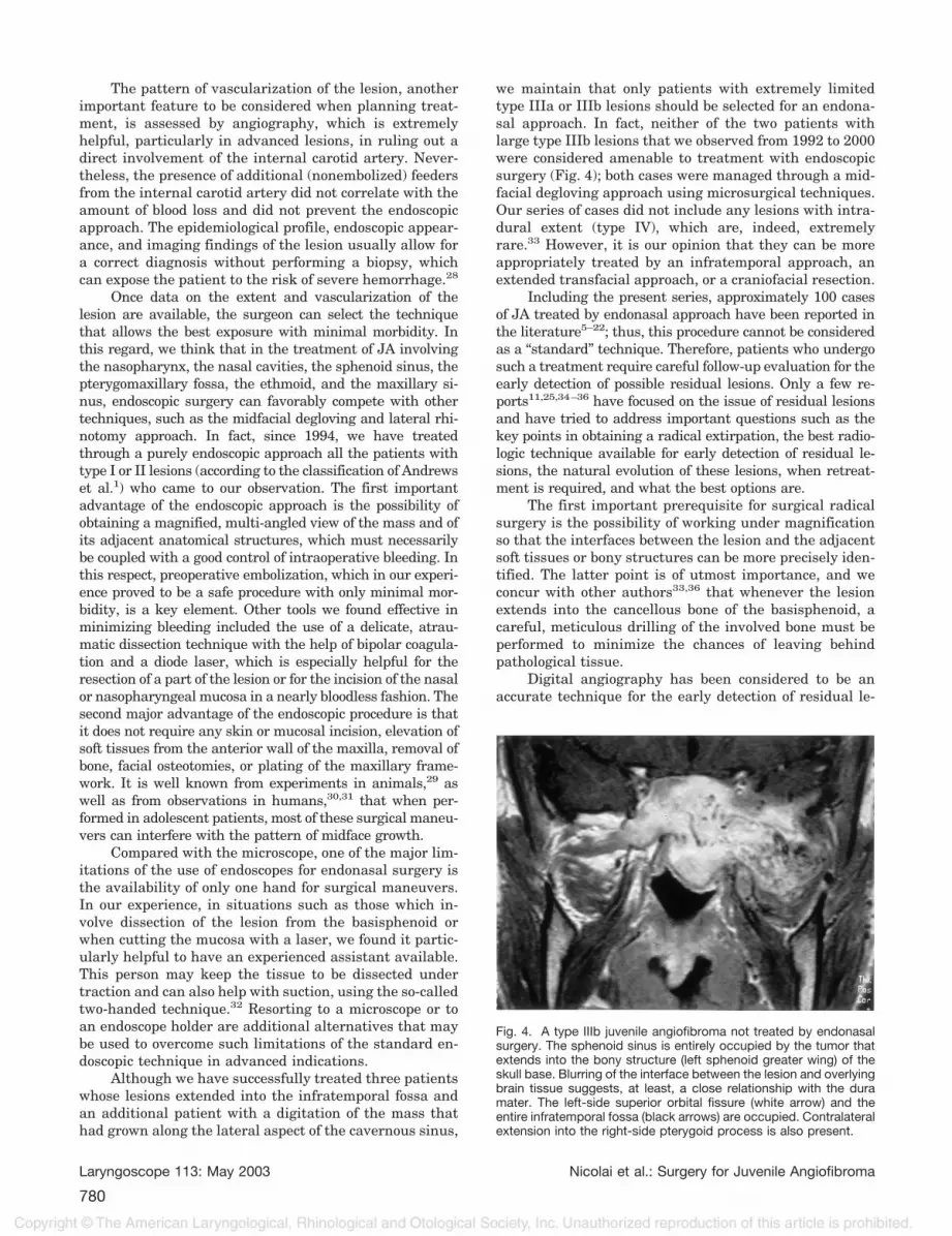
we maintain that only patients with extremely limitedtype IIIa or IIIb lesions should be selected for an endona-sal approach. In fact, neither of the two patients withlarge type IIIb lesions that we observed from 1992 to 2000were considered amenable to treatment with endoscopicsurgery (Fig. 4); both cases were managed through a mid-facial degloving approach using microsurgical techniques.Our series of cases did not include any lesions with intra-dural extent (type IV), which are, indeed, extremelyrare.33 However, it is our opinion that they can be moreappropriately treated by an infratemporal approach, anextended transfacial approach, or a craniofacial resection.
Including the present series, approximately 100 casesof JA treated by endonasal approach have been reported inthe literature5–22; thus, this procedure cannot be consideredas a “standard” technique. Therefore, patients who undergosuch a treatment require careful follow-up evaluation for theearly detection of possible residual lesions. Only a few re-ports11,25,34–36 have focused on the issue of residual lesionsand have tried to address important questions such as thekey points in obtaining a radical extirpation, the best radio-logic technique available for early detection of residual le-sions, the natural evolution of these lesions, when retreat-ment is required, and what the best options are.
The first important prerequisite for surgical radicalsurgery is the possibility of working under magnificationso that the interfaces between the lesion and the adjacentsoft tissues or bony structures can be more precisely iden-tified. The latter point is of utmost importance, and weconcur with other authors33,36 that whenever the lesionextends into the cancellous bone of the basisphenoid, acareful, meticulous drilling of the involved bone must beperformed to minimize the chances of leaving behindpathological tissue.
Digital angiography has been considered to be anaccurate technique for the early detection of residual le-
Fig. 4. A type IIIb juvenile angiofibroma not treated by endonasalsurgery. The sphenoid sinus is entirely occupied by the tumor thatextends into the bony structure (left sphenoid greater wing) of theskull base. Blurring of the interface between the lesion and overlyingbrain tissue suggests, at least, a close relationship with the duramater. The left-side superior orbital fissure (white arrow) and theentire infratemporal fossa (black arrows) are occupied. Contralateralextension into the right-side pterygoid process is also present.
Laryngoscope 113: May 2003 Nicolai et al.: Surgery for Juvenile Angiofibroma
780
sions.37 However, cross-sectional imaging provides a sim-ilar accuracy without the need for invasive procedures. Anadditional advantage of MRI over computed tomography(CT) is the absence of radiation exposure, a relevant as-pect in a population of young patients. In our experience,periodic MRI examinations have proven to be accurate inidentifying one residual tumor and one sphenoid mucocelein patients who were otherwise asymptomatic and pre-sented a normal endoscopic appearance.
The natural history of JA is not fully understood; it iscommonly thought that the lesion displays a constantgrowth during the puberal period, whereas with increasingage it tends to spontaneously involute. However, there is notenough evidence in the literature to substantiate this theoryfor either the primary or the residual lesions. Therefore, anypatient who is found to have a residual lesion at radiologicalcontrol must be carefully evaluated in relation to a series offactors: age, site and size of the lesion, availability for follow-up, and patient compliance. Scheduled endoscopic and radio-logical examinations have the advantage of preventing latediagnosis of residual lesions, which are identified by MRI orCT at an early stage. Small remnants can reasonably befollowed by imaging techniques at regular intervals (4–6mo) to monitor the pattern of growth. Whenever the massclearly and regularly increases in size, surgical resection isgenerally the preferred form of treatment; in small (1–3 cm)residual lesions, radiosurgery can be considered an alterna-tive option today.
The incidence of residual disease in the literaturevaries from 13%16 to 50%.38 This large variability ismainly related to the distribution of patients in the differ-ent published series in terms of stage and the criteria(clinical or radiological) used to establish the presence of aresidual lesion. According to Herman et al.,11 the rates ofremnant identification by CT were 7% and 39.5% in thepatients without and with skull base involvement, respec-tively. In our series, which included patients who werefollowed prospectively by MRI for at least 24 months aftersurgery, the observation of only one residual lesion (6.6%)favorably compares with the results reported in the liter-ature for small and intermediate-size juvenile angiofibro-mas treated by traditional external approaches.
CONCLUSIONThe results of the present study involving a single
institution reveal that, when carefully and properlyplanned according to data provided by imaging, an endo-scopic approach is a promising alternative to externaltechniques in the management of small to intermediate-sized juvenile angiofibromas. During preoperative coun-seling, the need to switch intraoperatively to an externalprocedure should always be discussed with the patient;consequently, informed consent should be obtained. Aschedule for endoscopic and MRI follow-up evaluationsmust be planned, to identify possible residual lesionsearly. Refinement in surgical instrumentation, the use ofCT- or MRI-guided surgical navigation, and the availabil-ity of intraoperative imaging techniques will certainlyallow gradual expansion of the indications for endoscopicsurgery in the management of JA.
BIBLIOGRAPHY1. Andrews JC, Fisch U, Valavanis A, Aeppli U, Makek MS. The
surgical management of extensive nasopharyngeal angio-fibromas with the infratemporal fossa approach. Laryngo-scope 1989;99:429–437.
2. Beham A, Kainz J, Stammberger H, Aubock L, Beham-Schmid C. Immunohistochemical and electron microscopi-cal characterization of stromal cells in nasopharyngeal an-giofibromas. Eur Arch Otorhinolaryngol 1997;254:196–199.
3. Beham A, Beham-Schmid C, Regauer S, Aubock L,Stammberger H. Nasopharyngeal angiofibroma: true neo-plasm or vascular malformation? Adv Anat Pathol 2000;7:36–46.
4. Schick B, Brunner C, Praetorius M, Plinkert PK, Urbschat S.First evidence of genetic imbalances in angiofibromas. La-ryngoscope 2002;112:397–401.
5. Kamel RH. Transnasal endoscopic surgery in juvenile naso-pharyngeal angiofibroma. J Laryngol Otol 1996;110:962–968.
6. Zicot AF, Daele J. Endoscopic surgery for nasal and sinusalvascular tumours: about two cases of nasopharyngeal an-giofibromas and one case of turbinal angioma. Acta Otorhi-nolaryngol Belg 1996;50:177–182.
7. Fagan JJ, Snyderman CH, Carrau RL, Janecka IP. Nasopha-ryngeal angiofibromas: selecting a surgical approach. HeadNeck 1997;19:391–399.
8. Tseng HZ, Chao WY. Transnasal endoscopic approach forjuvenile nasopharyngeal angiofibroma. Am J Otolaryngol1997;18:151–154.
9. Bernal-Sprekelsen M, Algorta Vazquez A, Pueyo J, CarbonellCasasus J. Die endoskopische Resektion juveniler Nasen-Rachen-Fibrome. HNO 1998;2:172–174.
10. Nakamura H, Kawasaki M, Higuchi Y, Seki S, Takahashi S.Transnasal endoscopic resection of juvenile nasopharyn-geal angiofibroma with KTP laser. Eur Arch Otorhinolar-yngol 1999;256:212–214.
11. Herman P, Lot G, Chapot R, Salvan D, Tran Ba Huy P.Long-term follow-up of juvenile nasopharyngeal angiofi-bromas: analysis of recurrences. Laryngoscope 1999;109:140–147.
12. Newlands SD, Weymuller EA Jr. Endoscopic treatment ofjuvenile nasopharyngeal angiofibroma. Am J Rhinol 1999;13:213–219.
13. Stammberger H, Anderhuber W, Walch C, Papaefthymiou G.Possibilities and limitations of endoscopic management ofnasal and paranasal sinus malignancies. Acta Otorhinolar-yngol Belg 1999;53:199–205.
14. Jorissen M, Eloy PH, Rombaux PH, Bachert CL, Daele J.Endoscopic sinus surgery for juvenile nasopharyngeal an-giofibroma. Acta Otorhinolaryngol Belg 2000;54:201–219.
15. Sarria R, Capitan A, Sprekelsen C, Viviente E, Cuervo G,Ferran A. Cirugia endoscopica del angiofibroma nasofarin-geo mediante doble embolizacion. Acta OtorrinolaringolEsp 2000;51:259–262.
16. Carrau RL, Snyderman CH, Kassam AB, Jungreis CA. En-doscopic and endoscopic-assisted surgery for juvenile an-giofibroma. Laryngoscope 2001;111:483–487.
17. Scholtz AW, Appenroth E, Kammen-Jolly K, Scholtz LU,Thumfart WF. Juvenile nasopharyngeal angiofibroma:management and therapy. Laryngoscope 2001;111:681–687.
18. Hazarika P, Nayak DR, Balakrishnan R, Raj G, Pillai S.Endoscopic and KTP laser-assisted surgery for juvenilenasopharyngeal angiofibroma. Am J Otolaryngol 2002;23:282–286.
19. Ochi K, Watanabe S, Miyabe S. Endoscopic transnasal resec-tion of a juvenile angiofibroma using an ultrasonicallyactivated scalped. ORL J Otorhinolaryngol Relat Spec2002;64:290–293.
20. Roger G, Tran Ba Huy P, Froelich P, et al. Exclusively endo-scopic removal of juvenile nasopharyngeal angiofibroma:trends and limits. Arch Otolaryngol Head Neck Surg 2002;128:928–935.
Laryngoscope 113: May 2003 Nicolai et al.: Surgery for Juvenile Angiofibroma
781

21. Schick B, El Tahan ERA, Brors D, Kahle G, Draf W. Experi-ences with endonasal surgery in angiofibroma. Rhinology1999;37:80–85.
22. Stamm AC, Watashi CH, Malheiros PF, Harker LA,Pignatari SSN. Micro-endoscopic surgery of benign si-nonasal tumors. In: Stamm AC, Draf W, eds. Micro-endoscopic Surgery of Paranasal Sinuses and the SkullBase. Berlin: Springer, 2000:489 –514.
23. Neel HB III, Whicker JH, Devine KD, Weiland LH. Juvenileangiofibroma: review of 120 cases. Am J Surg 1973;126:547–556.
24. Bremer JW, Neel HB III, DeSanto LW, Jones GC. Angiofi-broma: treatment trends in 150 patients during 40 years.Laryngoscope 1986;96:1321–1329.
25. Lloyd G, Howard D, Phelps P, Cheesman A. Juvenile angio-fibroma: the lessons of 20 years of modern imaging.J Laryngol Otol 1999;113:127–134.
26. Close LG, Schaefer SD, Mickey BE, Manning SC. Surgicalmanagement of nasopharyngeal angiofibromas involvingthe cavernous sinus. Arch Otolaryngol Head Neck Surg1989;115:1091–1095.
27. Harma RA. Nasopharyngeal angiofibroma: a clinical and his-topathological study. Acta Otolaryngol (Suppl) 1958;146:1–76.
28. Antonelli AR, Cappiello J, Di Lorenzo D, Donajo CA, NicolaiP, Orlandini A. Diagnosis, staging, and treatment of juve-nile nasopharyngeal angiofibroma (JNA). Laryngoscope1987;97:1319–1325.
29. Laurenzo JF, Canady JW, Zimmerman MB, Smith JH.Craniofacial growth in rabbits: effects of midfacial surgicaltrauma and rigid plate fixation. Arch Otolaryngol HeadNeck Surg 1995;121:556–561.
30. Wong L, Dufesne CR, Richtsmeier JT, Manson PN. The effectof rigid fixation on growth of the neurocranium. Plast Re-constr Surg 1991;88:395–403.
31. Lowlicht RA, Jassin B, Kim M, Sasaki CT. Long-term effectsof Le Fort I osteotomy for resection of juvenile nasopha-ryngeal angiofibroma on maxillary growth and dental sen-sation. Arch Otolaryngol Head Neck Surg 2002;128:923–927.
32. May M, Hoffman DF, Sobol SM. Video endoscopic sinus sur-gery: a two-handed technique. Laryngoscope 1990;100:430–432.
33. Danesi G, Panizza B, Mazzoni A, Calabrese V. Anterior ap-proaches in juvenile nasopharyngeal angiofibromas withintracranial extension. Otolaryngol Head Neck Surg 2000;122:277–283.
34. Jones GC, DeSanto LW, Bremer JW, Neel HB III. Juvenileangiofibroma: behaviour and treatment of extensive andresidual tumors. Arch Otolaryngol Head Neck Surg 1986;112:1191–1193.
35. Chagnaud CH, Petit PH, Bartoli JM, et al. Postoperativefollow-up of juvenile nasopharyngeal angiofibromas: as-sessment by CT scan and MR imaging. Eur Radiol 1998;8:756–764.
36. Howard DJ, Lloyd G, Lund V. Recurrence and its avoidancein juvenile angiofibroma. Laryngoscope 2001;111:1509–1511.
37. Bagatella F, Mazzoni A. Microsurgery in juvenile nasopha-ryngeal angiofibroma: a lateronasal approach with naso-maxillary pedicled flap. Skull Base Surg 1995;5:219–226.
38. McCombe A, Lund VJ, Howard DJ. Recurrence in juvenileangiofibroma. Rhinology 1990;28:97–102.
Laryngoscope 113: May 2003 Nicolai et al.: Surgery for Juvenile Angiofibroma
782


![[I Brazilian consensus of endoscopic ultrasonography]](https://img.dokumen.tips/doc/110x75/634ac5bce2b881b8bf0189bc/i-brazilian-consensus-of-endoscopic-ultrasonography.jpg)


