Embed Size (px)
Citation preview

REVIEW TOPIC OF THE WEEK
Endogenous Fibrinolysis
An Important Mediator of Thrombus Formation
and Cardiovascular Risk
Osita N. Okafor, BSC, MBBS,* Diana A. Gorog, MBBS, MD, PHD*y
ABSTRACT
Most acute cardiovascular events are attributable to arterial thrombosis. Plaque rupture or erosion stimulates platelet
activation, aggregation, and thrombosis, whilst simultaneously activating enzymatic processes that mediate endogenous
fibrinolysis to physiologically maintain vessel patency. Interplay between these pathways determines clinical outcome. If
proaggregatory factors predominate, the thrombus may propagate, leading to vessel occlusion. However, if balanced by
a healthy fibrinolytic system, thrombosis may not occur or cause lasting occlusion. Despite abundant evidence for the
fibrinolytic system regulating thrombosis, it has been overlooked compared with platelet reactivity, partly due to a lack
of techniques to measure it. We evaluate evidence for endogenous fibrinolysis in arterial thrombosis and review tech-
niques to assess it, including biomarkers and global assays, such as thromboelastography and the Global Thrombosis
Test. Global assays, simultaneously assessing proaggregatory and fibrinolytic pathways, could play a role in risk strati-
fication and in identifying impaired fibrinolysis as a potential target for pharmacological modulation. (J Am Coll Cardiol
2015;65:1683–99) © 2015 by the American College of Cardiology Foundation.
Cardiovascular disease is the leading cause of
morbidity and mortality in developed coun-
tries. The common pathological process
responsible for the majority of these disorders, in-
cluding acute coronary syndrome (ACS) and ischemic
stroke, is the development of an occlusive arterial
thrombus.
Disruption of an atherosclerotic plaque through
rupture or erosion creates a prothrombotic envi-
ronment to circulating platelets and procoagulant
factors. The major thrombogenic components con-
tained within the atherosclerotic plaque include
tissue factor and collagen (1–3). Exposure to this
thrombotic milieu provides a potent stimulus for
platelet activation, aggregation, and thrombosis
(Figures 1A and 1B). Activation of the coagulation
cascade also leads to direct activation of the enzy-
matic processes that mediate endogenous fibrino-
lysis (Figure 1C). This interaction is important to
ensure that thrombosis is controlled and vessel
patency is maintained.
The interplay between these opposing pathways
is likely to determine the occurrence and clinical
outcome of a resulting thrombus. If proaggregatory
and procoagulant factors predominate, an intra-
luminal thrombus may propagate and lead to com-
plete vessel occlusion, with subsequent lasting
downstream tissue damage (Figure 1B). If, in con-
trast, the prothrombotic factors are balanced by a
healthy fibrinolytic system, then a thrombus may not
develop or may not cause lasting vessel occlusion
(Figure 1C).
From the *East & North Hertfordshire NHS Trust, Hertfordshire, United Kingdom; and yVascular Sciences, National Heart & Lung
Institute, Imperial College, London, United Kingdom. Prof. Gorog is related to a company director of Thromboquest Ltd., who
manufactures the Global Thrombosis Test, but she, her spouse, and her children have no financial involvement or equity interest
in, and have received no financial assistance, support, or grants from Thromboquest Ltd. Thromboquest Ltd. has no involvement
in the design, conduct, or the finance of this review. Dr. Okafor has reported that he has no relationships relevant to the contents
of this paper to disclose.
Manuscript received January 12, 2015; revised manuscript received February 20, 2015, accepted February 23, 2015.
J O U R N A L O F T H E AM E R I C A N C O L L E G E O F C A R D I O L O G Y V O L . 6 5 , N O . 1 6 , 2 0 1 5
ª 2 0 1 5 B Y T H E AM E R I C A N C O L L E G E O F C A R D I O L O G Y F O U N DA T I O N I S S N 0 7 3 5 - 1 0 9 7 / $ 3 6 . 0 0
P U B L I S H E D B Y E L S E V I E R I N C . h t t p : / / d x . d o i . o r g / 1 0 . 1 0 1 6 / j . j a c c . 2 0 1 5 . 0 2 . 0 4 0

IMPORTANCE OF THE ENDOGENOUS
FIBRINOLYTIC SYSTEM IN ACS
An intact endogenous fibrinolytic system
serves to actively prevent the buildup of
formed thrombi through dissolution of
an arterial thrombus (Central Illustration).
Despite a wealth of evidence supporting
its role in preventing lasting arterial occlu-
sion, this pathway has been relatively over-
looked as compared with the understanding,
monitoring, and pharmacological modulation
of platelet reactivity. This may have occurred
due to limitations of earlier methods to
robustly measure the activity of the fibrino-
lytic system. Additionally, besides the use of
plasminogen activators to achieve acute
thrombolysis in the setting of acute myocar-
dial infarction (AMI) and stroke, pharmaco-
logical options available to manipulate the
fibrinolytic state have been very limited.
Evidence from clinical, histopathologic, and au-
topsy studies (4–9), as well as clinical observations,
support the proposal that AMI may represent a
failure of timely, spontaneous endogenous throm-
bolysis. In 585 patients presenting with ST-segment
elevation myocardial infarction (STEMI), sponta-
neous reperfusion (SR), evidenced by electrocardio-
graphic resolution of ST-segment changes, was
observed in 14.9%, and normal coronary flow on
angiography was observed in 14.7% of patients (10).
In 1,667 patients assigned to the primary percuta-
neous coronary intervention arm of the ASSENT 4
(Assessment of the Safety and Efficacy of a New
Treatment Strategy for Acute Myocardial Infarction)
trial (11), SR was associated with a lower composite
of death, heart failure, or shock compared with
those with persistent ST-segment elevation. In 710
STEMI patients undergoing primary percutaneous
coronary intervention, SR was observed in 22%, and
these patients had a lower incidence of death,
congestive heart failure, and recurrent ACS at 30
days than those without SR (12). Furthermore, his-
topathologic studies evaluating aspirated coronary
thrombi from patients with STEMI have demon-
strated significant heterogeneity in the composition
and age of the culprit thrombi (4–7). Among 1,362
STEMI patients, up to 40% demonstrated lytic or
organized thrombi, signifying that thrombus forma-
tion occurred days to weeks before final vessel oc-
clusion (7). This underpins the notion that thrombus
generation is an active and dynamic process, where
constant thrombosis and thrombolysis may occur in
concert.
Autopsy studies of healed plaque disruptions also
provide evidence of thrombus formation as a dynamic
process (8,13). Plaque instability appears to be pre-
sent for some time before an occlusive thrombus is
formed, and may be asymptomatic. Nonocclusive
mural thrombi may form over plaque disruptions,
leading to phasic progression of atherosclerotic le-
sions, but without presenting as ACS (13,14).
Despite the fact that plaque rupture represents a
common unifying event for coronary thrombosis,
there is significant variability in clinical manifesta-
tion and outcome. This variability may be explained,
in part, by the role of endogenous fibrinolysis in
limiting the propagation of formed thrombi and pre-
venting total coronary occlusion (Central Illustration).
In this paper, we review the methods currently
available to assess endogenous fibrinolysis and eval-
uate the evidence for the role of endogenous fibri-
nolysis as a mediator of arterial thrombus formation
in coronary disease.
FACTORS DETERMINING RESISTANCE OF
THROMBUS TO LYSIS
Whole blood clots are more resistant to lysis than
plasma clots, implying that blood cells and fibrin are
responsible for the resistance (15) (Central
Illustration). Platelets play the main role in resis-
tance, but red cell–derived microparticles can also
contribute to thrombin generation, whereas elastase
released from leukocytes trapped or adherent to the
thrombus exerts a plasmin-independent fibrinolytic
effect. Arterial (platelet-rich) thrombi are much more
resistant to lysis than erythrocyte-rich venous
thrombi (16). The mechanisms through which plate-
lets contribute to thrombolysis resistance are 3-fold
(Central Illustration):
1. Platelets contain >90% of the circulating plasmin-
ogen activator inhibitor (PAI)-1. During aggrega-
tion, in response to thrombin, PAI-1 is released
from platelets into the thrombus mass and is the
major determinant of arterial thrombolysis resis-
tance (17).
2. The procoagulant activity or contribution of
platelets to thrombin generation is extremely
important, not only in the generation of, but also in
the lysis of the formed thrombus. A high shear
stress milieu, such as that found in an artery with a
severe stenosis, will trigger microparticle release
from activated platelets, resulting in a burst of
thrombin generation. In addition to PAI-1,
thrombin-activatable fibrinolysis inhibitor (TAFI)
also contributes to thrombolysis resistance.
ABBR EV I A T I ON S
AND ACRONYMS
ACS = acute coronary
syndrome(s)
AMI = acute myocardial
infarction
GTT = Global Thrombosis Test
MACE = major adverse
cardiovascular event(s)
PAI = plasminogen
activator inhibitor
ROTEM = rotational
thromboelastometry
SR = spontaneous reperfusion
STEMI = ST-segment elevation
myocardial infarction
TAFI = thrombin-activatable
fibrinolysis inhibitor
TEG = thromboelastography
t-PA = tissue-type
plasminogen activator
Okafor and Gorog J A C C V O L . 6 5 , N O . 1 6 , 2 0 1 5
Endogenous Thrombolysis in CV Disease A P R I L 2 8 , 2 0 1 5 : 1 6 8 3 – 9 9
1684

FIGURE 1 The Mechanism and Importance of Endogenous Fibrinolysis in Regulation of Occlusive Arterial Thrombus Formation, and its Relevance to
Laboratory Tests Assessing Thrombotic Risk
(A) Under conditions of high shear, such as those that exist in a narrowed coronary artery, stimulation of platelet aggregation by von Willebrand Factor (vWF) results in
the formation of thrombin, the key mediator of thrombus formation. (B) The thrombus achieves structural stability and resistance to dislodgement and to thrombolysis
through fibrin (which crosslinks cells to provide structural stability), plasminogen activator inhibitor (PAI)-1 released from activated platelets, and activation of thrombin-
activatable fibrinolysis inhibitor (TAFI) by thrombin. (C) Endogenous thrombolysis: physiological processes that exist to prevent lasting occlusive thrombus formation,
including the release of tissue plasminogen activator (t-PA) and urokinase plasminogen activator (u-PA) from the vessel wall and plasma, the release of elastase and
cathepsin from adherent neutrophils and monocytes, and the dispersing effect of flow. ADP ¼ adenosine diphosphate; TxA2 ¼ thromboxane A2.
J A C C V O L . 6 5 , N O . 1 6 , 2 0 1 5 Okafor and Gorog
A P R I L 2 8 , 2 0 1 5 : 1 6 8 3 – 9 9 Endogenous Thrombolysis in CV Disease
1685

3. The structure and stability of the resultant fibrin
network is also a determinant of thromboresis-
tance (18). Platelets play an important role in
regulating fibrin network structure (19). Coagula-
tion factor XIII (FXIII), in addition to cross-linking
fibrin, also plays an important role in thrombol-
ysis resistance. Platelets are abundant in FXIII, and
upon activation, FXIII-A exposure on the surface
membrane exerts an antifibrinolytic function by
cross-linking the major plasmin inhibitor a2-
antiplasmin to fibrin, thus inhibiting plasmin-
mediated clot degradation (20,21). FXIII alters the
structure of the fibrin network to reduce pore size
and increase fiber density, thus increasing clot
stability and resistance to lysis (22). Thrombi
formed in FXIII-deficient blood lyse more quickly
than in normal blood, and FXIII concentrate
normalized lysis (23). A common genetic poly-
morphism of FXIII has been shown to increase the
risk of myocardial infarction (24).
MEASUREMENT OF FIBRINOLYTIC STATUS
Interest in the endogenous fibrinolytic system has
fueled the development of techniques to assess and
quantify the activity of this important pathway. Many
CENTRAL ILLUSTRATION Endogenous Thrombolysis in CV Disease: Determinants of Spontaneous Thrombolysis Under
Arterial Flow Conditions
Thrombin converts plasminogen to plasmin, which breaks down the cross-linked fibrin into soluble fibrin degradation products. t-PA is mainly responsible
for the dissolution of fibrin formed in the circulation. Inhibitors of thrombolysis include the release of PAI-1 from platelets, secretion of active PAI-1 from
aggregated platelets, and clot retraction. Potentiators of thrombolysis include: the release of elastase and cathepsin G from white blood cells that become
trapped in the thrombus, which directly break down fibrin; the plasminogen activators uPA and t-PA (released from endothelial cells); and fibrin structural
properties. The thickness and porosity of fibrin fibers will also determine structural stability and susceptibility to thrombolysis. Lp(a), a homologue of
plasminogen, can inhibit t-PA–mediated plasminogen activation. aTAFI ¼ activated thrombin-activatable fibrinolysis inhibitor; CV ¼ cardiovascular; EC ¼
endothelial cell; FXIII ¼ coagulation factor XIII; Lp(a) ¼ lipoprotein (a); PAI-1 ¼ plasminogen activator inhibitor; PAR ¼ protease-activated thrombin re-
ceptor; TAFI ¼ thrombin-activatable fibrinolysis inhibitor; t-PA ¼ tissue plasminogen activator.
Okafor and Gorog J A C C V O L . 6 5 , N O . 1 6 , 2 0 1 5
Endogenous Thrombolysis in CV Disease A P R I L 2 8 , 2 0 1 5 : 1 6 8 3 – 9 9
1686
of the early studies utilizing these techniques pro-
vided compelling evidence of endogenous fibrinolysis
in arterial thrombogenesis, although no current test
has been adopted into widespread clinical use.
Current techniques include: 1) the measurement of
1 or several protein components of the fibrinolytic
cascade; or 2) global assessment of fibrinolytic ca-
pacity utilizing techniques such as euglobulin clot
lysis time, thromboelastography (TEG) (Haemoscope
Corporation, Niles, Illinois) or rotational thromboe-
lastometry (ROTEM) (Tem International GmbH,
Munich, Germany), and the most recent Global
Thrombosis Test (GTT) (Thromboquest Ltd., London,
United Kingdom). These techniques are described in
more detail in the following text.
PLASMA MARKERS OF FIBRINOLYSIS
The fibrinolytic system shares several similarities
with the coagulation cascade, involving a series of
proteolytic enzymatic steps that culminate in the
conversion of plasminogen to plasmin to achieve
fibrin dissolution (Figure 1C, Central Illustration).
Thrombin converts plasminogen to plasmin, which
breaks down the cross-linked fibrin into soluble fibrin
degradation products. Tissue-type plasminogen acti-
vator (t-PA) is mainly responsible for the dissolution
of fibrin formed in the circulation. Fibrinolysis can be
inhibited either by antagonizing plasmin through
alpha 2-antiplasmin or by PAIs. PAI-1, stored in the
alpha granules of platelets, is mainly responsible
for resistance to fibrinolysis. Activation of protease-
activated thrombin receptor 1 by thrombin results
in synthesis and secretion of active PAI-1 from
aggregated platelets and thrombin activation of
TAFI, which inhibits the t-PA–mediated conversion
of plasminogen to plasmin. Plasma lipoprotein (a)
[Lp(a)], a homolog of plasminogen, can inhibit t-PA–
mediated plasminogen activation.
Measurement of individual proteins involved in
fibrinolysis, as a surrogate marker of overall fibrino-
lytic activity, can be undertaken using immunoassays.
Studies have focused on themeasurement of a number
of biomarkers of fibrinolysis, including t-PA (25–30),
PAI-1 (31), alpha-2 antiplasmin, alpha2-antiplasmin-
plasmin complex (31), markers of fibrin degradation
products (D-dimer and soluble fibrin) (25,28,31), and,
more recently, TAFI (31) and Lp(a) (31,32).
Given the potential role of the fibrinolytic system
in the pathogenesis of ACS, studies have attempted
to elucidate the relationship between plasma bio-
markers and incipient cardiovascular risk. The main
limitations of this approach are knowing the relative
importance and contribution of any biomarker to the
overall fibrinolytic system at any given point,
knowing whether to measure levels or activity of
biomarkers, and the confounding association be-
tween fibrinolytic markers and more established car-
diovascular risk factors (33,34). Overall, the outcome
of studies evaluating the role of plasma markers of
fibrinolysis as independent predictors of cardiovas-
cular risk has been disappointing, with much con-
flicting evidence and a number of positive studies
only demonstrating a weak association (31). This,
combined with methodological problems with the
assays, has resulted in greater emphasis being placed
on more global assays of fibrinolytic activity.
EUGLOBULIN CLOT LYSIS TIME
The euglobulin fraction (containing the key activators
of the fibrinolytic cascade, including plasminogen
activators, plasminogen, and plasmin) is precipitated
from citrated plasma and calcium is added to promote
clot formation. The time taken to lyse this clot is
utilized as a measure of fibrinolytic activity (35,36).
This technique from the 1950s has now been super-
seded by more rapid, physiological tests of fibrino-
lytic capacity.
THROMBOELASTOGRAPHY
TEG is a global test of coagulation status, simulta-
neously assessing clot development, stabilization,
and dissolution. Another related and commercially-
available technique is ROTEM (37,38). TEG utilizes a
pin suspended by a torsion wire into a cylinder to
measure the physical properties of a clot. The torsion
wire is connected to a mechanical-electrical trans-
ducer and relays information on the speed and
TABLE 1 Comparison of Thromboelastography (ROTEM) and the GTT, With
Respect to Assessment of Thrombosis
Measurement ROTEM GTT
Thrombus Cross-linked fibrin clot Platelet-rich fibrin clot
Relevance Venous thrombosis Arterial thrombosis
Flow (shear rates/s) Static (0.1/s) High shear (>10,000/s)
Blood sample Citrate-anticoagulated Native blood
Thrombus resistance No Yes
PAI-1 involvement in lysis No Yes
Platelets procoagulant
effect
Little Significant
Activator Tissue factor/kaolin (extrinsic
and intrinsic coagulation
pathways)
High shear stress only
Hyperfibrinolysis (t-PA) Yes Yes
Hypofibrinolytic state No Yes
GTT ¼ global thrombosis test; PAI-1 ¼ plasminogen activator inhibitor-1; ROTEM ¼ rotational
thromboelastometry; t-PA¼ tissue-type plasminogen activator.
J A C C V O L . 6 5 , N O . 1 6 , 2 0 1 5 Okafor and Gorog
A P R I L 2 8 , 2 0 1 5 : 1 6 8 3 – 9 9 Endogenous Thrombolysis in CV Disease
1687

TABLE 2 Studies From 1999 to 2015 That Utilized Multivariate Analysis in Patient Cohorts >1,000 to Assess the Predictive Value of Plasma Fibrinolysis Markers for Adverse CV Events
First Author (Ref. #) Design Patients (n) Population Follow-Up Endpoint Results
Kinlay et al. (28) RCT 2,860 ACS patients enrolled
in MIRACL study
16 weeks CV events (death, nonfatal MI,
cardiac arrest, or worsening
angina)
t-PA antigen associated with increased
risk of CV events (HR: 1.25; p ¼ 0.0014).
This correlation was attenuated following
adjustment for risk factors (HR: 1.14;
p ¼ 0.08)
Zamani et al. (106) RCT 2,925 ACS patients enrolled in
MIRACL study
16 weeks Death and recurrent nonfatal
ACS (MI or unstable angina)
t-PA antigen associated with primary
endpoints, but after adjustment for
risk factors, HR fell from 1.90 to 1.27 for
death (p ¼ NS), and HR 1.20 for
ACS (p ¼ NS).
Wang et al. (66) Prospective 3,209 Subjects in 6th cycle of
Framingham Offspring
Study (1995–1998)
10 yrs Death and major CV events
(MI, unstable angina, heart
failure, and stroke) and
nonmajor CV events
Multiple biomarkers, including D-dimer
and PAI-1. Following multivariate
adjustment, HR for D-dimer associated
with death was 1.24; 95% CI: 1.02–1.50;
p ¼ 0.03; and HR: 1.24 for PAI-1 in
relation to CV events (p ¼ 0.03).
Folsom et al. (34) Prospective 6,391 Subjects without
atherosclerosis
4.6 yrs Cancer death, mortality, and CAD
(MI or coronary death), and CV
disease (cardiac arrest, angina �
revascularization and stroke)
D-dimer, factor VIIIc and PAP not predictive
of CV disease, but independently
associated with cancer death and total
mortality. Following adjustment for risk
factors, mortality increased for each quartile
increment in D-dimer (33% increase;
95% CI: 15–54), factor VIIIc (26% increase;
95% CI: 11–44), and PAP (20% increase;
95% CI: 4–38) (p values not published).
May et al. (25) Prospective 3,582 Women without prior CAD 4.7 yrs Development of CV death, MI, or
coronary revascularization
D-dimer, t-PA antigen, and vWF were not
associated with development of CAD after
adjustment for CV risk factors.
Cushman et al. (108) Nested case-
control study
5,201 (146
selected
cases)
Patients without baseline
vascular disease
2.4 yrs Coronary death, MI, and angina. D-dimer and PAP levels, but not PAI-1,
predicted MI or coronary death, but not
angina. D-dimer values above median
associated with RR: 2.5; 95% CI: 1.1–5.9; and for
PAP with RR: 3.1; 95% CI: 1.3–7.7, independent
of other risk factors.
Nordenhem et al. (107) Case-control
study
1,267 Patients with first
MI identified
Matched to
control group
MI Plasma t-PA/PAI-1 complex associated with
MI, with synergistic interaction in
male smokers (OR: 4.6; 95% CI: 3.3–6.5) or
diabetics (OR: 7.9; 95% CI: 3.9–16.1)
(p values not published).
Smith et al. (64) Prospective 2,398 Men age 49–65 yrs 4 yrs CV events (coronary heart disease
and ischemic stroke combined)
After adjusting for risk factors, fibrinogen
(HR: 1.26; p ¼ 0.005), D-dimer (HR: 1.34;
p ¼ 0.001) and PAI-1 (HR: 1.24; p ¼ 0.013)
were independent risk factors for CV
events. Factor VIIc was inversely related
to CV events (HR: 0.75; p ¼ 0.001).
Continued on the next page
OkaforandGorog
JACC
VOL.65,NO.16,2015
EndogenousThrombolysis
inCVDise
ase
APRIL
28,2015:1
683–99
1688

TABLE 2 Continued
First Author (Ref. #) Design Patients (n) Population Follow-Up Endpoint Results
Morange et al. (109) Prospective 1,057 Patients with CAD
(AtheroGene Study)
6.6 yrs CV death and nonfatal CV events
(MI and stroke)
vWF, fibrinogen, TAT, D-dimers, and PAP were
all associated with CV death but not with
nonfatal CV events. After adjustment for
risk factors and CRP, only fibrinogen and
D-dimer remained associated with CV death
(HR: 1.27; 95% CI: 1.04–1.55; p ¼ 0.019).
Tregouet et al. (110) Prospective 1,668 Patients with CAD
(AtheroGene Study)
2.3 years CV death and nonfatal CV
event (MI)
Activated TAFI independently associated with
risk of CV death (HR: 2.38; 95% CI: 1.56–3.63;
p < 0.0001), even after adjustment for risk
factors (HR: 1.69; 95% CI: 1.07–2.67; p ¼ 0.01).
Total TAFI not associated with CV events.
Gaw et al. (111) Prospective 5,732 Elderly patients with risk
factors for, or established
vascular disease
3.2 yrs CV death, nonfatal MI, fatal or
nonfatal stroke
Lp(a) levels not associated with primary
endpoint (HR: 1.05; 95% CI: 1.00–1.11; p ¼ NS).
Bennet et al. (112) Case-control study 2,047 Patients without CAD or stroke at
baseline who experienced an
MI or coronary death
NA First-ever MI or coronary death Lp(a) values in the top vs. bottom third, after
multivariate adjustment for CV risk factors,
OR: 1.60; 95% CI: 1.38–1.85 (p values not
published). Progressive increase in OR with
higher Lp(a) levels.
Willeit et al. (91) Prospective case-
control study
1,925 Patients without CV disease
who experienced MI or
coronary death
19.4 yrs First-ever MI or coronary death OR for D-dimer was 1.08 (p ¼ 0.019), OR for
t-PA antigen 1.05 (p ¼ 0.167). and OR for
Lp(a) 1.24 (p < 0.001).
Wannamethee et al. (92) Prospective study 3,217 Men age 60–79 yrs without CAD 7 yrs Coronary death, nonfatal MI, and
uncomplicated angina
After adjustment for risk factors, D-dimer
associated with MI/coronary death
(HR: 1.18; p ¼ 0.02), but not with angina
(HR: 0.93; p ¼ NS).
Chien et al. (93) Prospective
cohort study
3,484 Chinese patients without CAD 13.8 yrs All-cause death, stroke, and CAD Lp(a) levels did not correlate with risk of
CV disease (HR: 0.81; p ¼ NS).
Gurdasani et al. (94) Prospective
cohort study
18,720 Healthy subjects, age 39–79 yrs 11.4 yrs Peripheral artery disease, stroke,
and CAD-related events
Lp(a) levels associated with CAD
hospitalization and mortality (HR: 1.13;
p < 0.00001).
Nestel et al. (95) Prospective study 7,863 Patients with prior coronary event 6 yrs Coronary death, nonfatal MI,
ischemic stroke, revascularization,
total CV events, and total
coronary events
Lp(a) levels correlated with CV events
(p < 0.001), total CV events (HR: 1.23;
p ¼ 0.002), and coronary events
(p ¼ 0.03).
Kwon et al. (96) Prospective 1,494 Type 2 diabetic patients with CAD 4.4 yrs MACE (cardiac deaths and
nonfatal MI)
Highest Lp(a) level tertile associated with
MACE (HR: 2.89; p ¼ 0.005).
Kwon et al. (97) Prospective 6,252 Patients with suspected CAD 3.1 yrs MACE (cardiac death and
nonfatal MI)
Elevated Lp(a) associated with MACE
(HR: 1.773; p ¼ 0.005).
Canoui-Poitrine et al. (100) Prospective
cohort study
9,711 Men age 50–59 yrs free of CAD
and stroke
10 yrs CAD events (angina, MI, and
coronary death) and
ischemic stroke
Lp(a) levels associated with CV events
(HR: w1.2; p ¼ 0.001) after adjustment
for risk factors.
Virani et al. (101) Prospective 13,318 (n ¼ 3,467
blacks, n ¼ 9,851
Caucasians)
African-American and Caucasian
adults without CHD or stroke
20 yrs CV events (coronary death, MI,
silent MI, revascularization)
and stroke
Lp(a) levels associated with CV events.
Quintile analysis for the highest
compared with the lowest quintile
demonstrated an HR: 1.35 (p ¼ 0004)
for blacks and HR: 1.27
(p ¼ 0.001) for whites.
O’Donoghue et al. (102) Prospective 6,708 Patients with CAD from 3
studies (PEACE, CARE, and
PROVE-IT-TIMI 22 trial)
MACE (composite of CV death,
MI, or stroke)
No association between Lp(a) levels and MACE
in any of the 3 trials individually or
combined.
Continued on the next page
JACC
VOL.65,NO.16,2015
OkaforandGorog
APRIL
28,2015:1
683–99
EndogenousThrombolysis
inCVDise
ase
1689

dynamics of clot formation. As blood clot formation
occurs around the pin, fibrin strands form between
the cylindrical cup and pin. With additional rotation
of the cylindrical cup, this is transmitted to the pin
and the resulting mechanical-electrical transduction
is depicted in a numerical and graphical representa-
tion. With the ROTEM technique, movement is in-
stead generated from the oscillation of the pin/wire
transduction system, while the cup is held immobile
and an optical detection system is utilized to trans-
duce the signal. TEG can be modified to use a variety
of different activators and inhibitors to provide in-
formation on specific components of the coagulation
system, including platelet function testing and fibri-
nolytic status (38–45). In fibrinolysis assessment, TEG
is typically compared in the presence and absence of
the fibrinolysis inhibitor aprotinin (37,46). Table 1
shows the features of thromboelastography and how
these compare with the GTT.
There are a number of important limitations to the
use of TEG as a clinical tool to assess global throm-
botic status. TEG was originally designed for native,
nonanticoagulated blood (47), but subsequent modi-
fications have included the use of activators of coag-
ulation and additional reagents to evaluate specific
components of hemostasis (38,47). This has helped
standardize the initiation of coagulation, but does not
reflect a patient’s physiological state. Although TEG is
a useful tool for assessing bleeding risk, for example
in cardiac surgery, its practical value in assessing the
(spontaneous) thrombolytic status of patients or the
effect of medications is questionable. The shortcom-
ings of this technique begin with the testing of
citrated and recalcified blood. The effect of extracel-
lular calcium concentration on coagulation indexes
and thromboelastography results is significant (48).
There are significant differences in TEG results be-
tween fresh native whole blood and recalcified citra-
ted whole blood (49,50) and the correlation between
TEG results performed on kaolin- versus nonkaolin-
activated native and citrated blood is poor (51).
In the absence of shear or any other platelet-
activating stimuli, clot formation can be initiated
either by intrinsic (kaolin, ellagic acid) or extrinsic
(tissue factor) activators, and the test results vary
accordingly. However, the major limitation of TEG is
that it fails to assess the procoagulant (thrombin-
generating) and fibrinolysis-inhibiting (PAI-1; TAFI)
properties of platelets. Furthermore, the use of gentle
rotation of a cylindrical cup more closely resembles
the low shear stress environment encountered with
venous stasis and does not reflect the high-shear
situation in stenosed arteries. Furthermore, this
mitigates the contribution of platelet activation and
TABLE
2Continued
First
Auth
or(R
ef.
#)
Design
Patients
(n)
Population
Follow-U
pEndpoint
Resu
lts
SukDanik
etal.(103)
Prosp
ective
27,791
Healthywomenage>45yrs
10yrs
MACE(nonfatalMI,nonfatal
cerebrovascularevent,co
ronary
revascularization,orCVdeath)
Lp(a)levels
inthehighest
vs.
lowest
quartiles
associatedwithadverseevents
(HR:1.35;
p<
0.001fortrendacross
quartiles).
Kamstrupetal.(104)
Prosp
ective
9,330
SubjectswithoutpriorCAD
10yrs
CAD
(includingMI)ordeath
RaisedLp(a)levelassociatedwithHR:1.09
(95%
CI:1.06–1.12;p¼
0.93)forMIand1.06
(95%
CI:1.04–1.08;p¼
0.86)forCAD.
Shilpaketal.(105)
RCT
2,763
Post-m
enopausalwomenage
<80
yrs
withCAD
4.1
yrs
CVevents
(nonfatalMIand
CVdeath)
Lp(a)levels
associatedwithCVevents
(HR:1.54;95%
CI:0.99–2.39;p¼
0.03)
ACS¼
acute
coronary
syndromes;
CAD
¼co
ronaryartery
disease;CARE¼
CholesterolAndRecu
rrentEvents;CHD
¼co
ronaryheart
disease;CI¼
confidence
interval;CRP¼
C-reactiveprotein;CV
¼cardiovascular;
HR¼
hazard
ratio;Lp(a)¼
lipoprotein
(a);
MACE¼
majorad
versecardiovascularevents;MI¼
myocardialinfarction;MIRACL¼
MyocardialIsch
emia
ReductionwithAggressiveCholesterolLowering;NA¼
notapplicable;NS¼
notsignificant;
OR¼
oddsratio;PAP¼
plasm
in-alpha2
-antiplasm
inco
mplex;
PEACE¼
PreventionofEvents
WithAngiotensin-C
onvertingEnzymeInhibitorTherapy;PROVE-IT-TIM
I22¼
PravastatinorAtorvastatinEvaluationandInfectionTherapy-ThrombolysisIn
MyocardialInfarction22;RCT¼
randomizedco
ntrolledtrial;RR¼
relativerisk;
TAFI¼
thrombin-activatable
fibrinolysisinhibitor;TAT¼
thrombin-antithrombin
complex;vWF¼
vonWillebrandfactor;otherabbreviationsasin
Table
1.
Okafor and Gorog J A C C V O L . 6 5 , N O . 1 6 , 2 0 1 5
Endogenous Thrombolysis in CV Disease A P R I L 2 8 , 2 0 1 5 : 1 6 8 3 – 9 9
1690
subsequent thrombin generation, which play key
roles in arterial thrombogenesis.
TEG results have only demonstrated a weak cor-
relation with standard tests of coagulation (52), with
no formal validation or standardization (53) and sig-
nificant interlaboratory variability; coefficients of
variation range between 8% and 40% for TEG and up
to 4% to 84% for ROTEM (54). Due to the problems
with standardization and determination of normal
reference values, TEG is better utilized as a measure
of the change in coagulation status over time, when a
patient’s baseline results are already known (55).
Despite these limitations, TEG benefits from its
availability as a point-of-care test, providing rapid
information on the coagulation profile of patients.
GLOBAL THROMBOSIS TEST
The GTT is a newer point-of-care test that simulta-
neously assesses platelet reactivity, thrombosis,
and thrombolytic activity, from a single, non-
anticoagulated blood sample (56,57). This technique
utilizes native, nonanticoagulated blood that is free
of any external agonists (Table 1). Platelets become
activated by the high shear stress generated by the
passage of blood through a conical tube containing
narrow gaps. The predominant stimulus for platelet
activation in severely-narrowed atherosclerotic cor-
onary arteries is pathologically high shear stress
(>10,000 s�1), which leads to rapid platelet activa-
tion. The GTT mimics this pathological environment
to provide high shear stress as the primary stimulus
for platelet aggregation, platelet microparticle, and
thrombin generation, resulting in occlusive thrombus
formation (58,59). The time taken for an occlusive
thrombus to form in the space downstream, reflecting
platelet aggregation and initiation of coagulation, is
manifested in the arrest of flow as detected by an
optical sensor, and is termed the occlusion time
(OT, s). The restart of blood flow, due to spontaneous
dissolution of the formed thrombus, represents
endogenous thrombolytic activity and is recorded
again by an optical sensor and termed the lysis time
(LT, s).
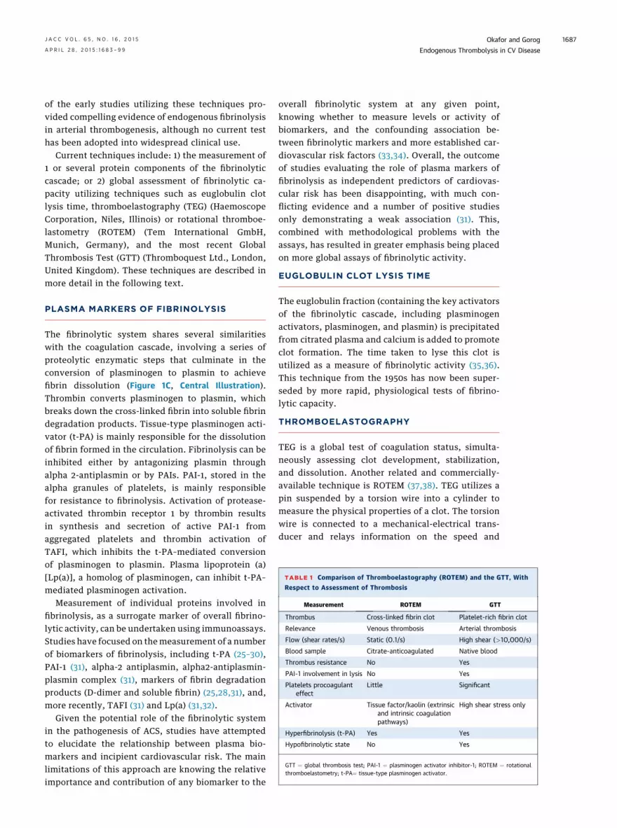
TABLE 3 Clinical Studies Evaluating the GTT in the Prediction of Cardiovascular Risk
First Author (Ref. #) Population Patients (n) Follow-Up Methods Primary Endpoint Results
Sharma et al. (60) End-stage renal disease
patients on hemodialysis
216 276 � 166 days GTT MACE (CV death,
nonfatal MI, CVA,
and peripheral
arterial thrombosis)
Impaired endogenous thrombolysis
(LT >3,000 s) strongly associated
with MACE (HR: 4.25; p ¼ 0.004),
nonfatal MI, and CVA (HR: 14.28;
p ¼ 0.0 ¼ 1) and peripheral
thrombosis (HR: 9.08;
p ¼ 0.003)
Saraf et al. (61) ACS patients receiving dual
antiplatelet therapy
300 12 months GTT MACE (CV death,
nonfatal MI,
or CVA)
LT >3,000 s was an independent
predictor of MACE (HR: 2.52;
p ¼ 0.004) and CV death (HR: 4.2;
p ¼ 0.033).
Saraf et al. (61) ACS vs. healthy
control subjects
300 N/A GTT MACE (CV death,
nonfatal
MI, or CVA)
OT prolonged in ACS (428 s vs. 378 s;
p < 0.001) and LT shorter in ACS
(1,053 s vs. 1,362 s; p < 0.001) than
in control subjects
Suehiro et al. (75) Healthy subjects of smoking
and nonsmoking status
Smokers ¼ 76 vs.
nonsmokers ¼ 63
3 months GTT Effect of smoking on
thrombotic profile
LT was significantly longer in smokers
than in nonsmokers (1,794 s vs.1,530 s;
p ¼ 0.029) with no significant
difference in OT
Ikarugi et al. (76) Healthy young males
and elderly males
Young ¼ 30 vs.
elderly ¼ 34
N/A GTT Effect of age, smoking,
and exercise on
thrombotic profile
LT was significantly longer in elderly vs.
young (p < 0001), and prolonged in
elderly smokers than nonsmokers
(p < 0.001)
Suehiro et al. (77) Males with MetS vs.
control subjects
MetS ¼ 30 vs.
control ¼ 53
N/A GTT Comparison of
thrombotic profile
between groups
LT significantly longer in MetS than in
control subjects (1,494 s vs. 1,246 s).
PAI-1 level correlated with LT
(p < 0.01)
Rosser et al. (98) ACS or stable coronary disease
randomized to vorapaxar vs.
placebo, in addition to
standard of care
57 N/A GTT Thrombotic status,
as shown by OT
and LT of GTT
Vorapaxar treatment prolonged OT
(561 s vs. 372 s; p ¼ 0.003) and
shortened LT (1,158 s vs. 1,733 s;
p ¼ 0.016)
Taomoto et al. (99) Acute cerebrovascular
disease (CVA) vs.
healthy control subjects
CVA ¼ 185
control subjects ¼ 195
N/A GTT Thrombotic status,
as shown by OT
and LT of GTT
In stroke patients, OT was shorter
(p < 0.0001) and LT was longer
(p < 0.0001) than in healthy
control subjects
CVA ¼ cerebrovascular accident (stroke); LT ¼ lysis time; MetS ¼ metabolic syndrome; OT ¼ occlusion time; other abbreviations as in Tables 1 and 2.
J A C C V O L . 6 5 , N O . 1 6 , 2 0 1 5 Okafor and Gorog
A P R I L 2 8 , 2 0 1 5 : 1 6 8 3 – 9 9 Endogenous Thrombolysis in CV Disease
1691

TABLE 4 Studies Utilizing TEG in the Evaluation of Platelet Reactivity and its Correlation to the Risk of Ischemic Events in Nonsurgical Cardiovascular Patients
First Author (Ref. #) Population Patients (n) Follow-Up Method Primary Endpoint Results
Jeong et al. (113) PCI-treated patients receiving
aspirin and clopidogrel
197 24 months MA-thrombin TEG
measurements,
conventional
aggregometry, and
genotyping
Relationship between MA-
thrombin on high on-
treatment platelet reactivity
(HPR) and long-term MACE
HPR and high MA-thrombin
were both independently
associated with MACE (HR:
3.09 and 2.24, respectively).
The combination of both
increased HR for MACE to
5.56; p ¼ 0.0002. High MA-
thrombin also predicted the
risk for HPR (OR: 13.89;
p < 0.001)
Gurbel et al. (114) Patients undergoing PCI and
taking aspirin and clopidogrel
225 36 months ADP-induced (MA-ADP)
and thrombin-induced
(MA-thrombin) TEG
measurement and LTA
Prediction of long-term event
occurrence (ischemic and
bleeding) following stenting
Patients with ischemic events
had higher MA (ADP), MA
(thrombin), and LTA (p <
0.0001 for all), which were
independent predictors of
ischemic events at 3 years
(HR: 10.3, 3.8, and 4.8,
respectively; all p < 0.0001)
Tang et al. (115) Patients undergoing PCI divided
into 3 groups depending on
inhibition rates to aspirin and
clopidogrel (n ¼ 90): control
group (n ¼ 30) and resistance
group (n ¼ 60), who were then
randomized to 2 subgroups
(R þ R and R þ L) to receive
different antiplatelet
combinations
90 12 months TEG Occurrence of CV ischemic events
(including stent thrombosis,
recurrent unstable angina,
and MI)
Patients resistant to antiplatelet
therapy vs. nonresistant
control groups, had an
increased risk of stent
thrombosis (20% vs. 3%),
recurrent unstable angina
(36% vs.10%), and (MI 17%
vs. 1%; p < 0.01).
Randomization to a loading
dose regimen improved
inhibition rates and reduced
the rates of CV events
(p < 0.01)
Gurbel et al. (116) Patients undergoing PCI 84 24 months TEG and conventional
aggregometry.
Biomarker evaluation
with fluorokine
multianalyte profiling
Thrombogenicity and biomarkers
of inflammation and
correlation to the occurrence
of ischemic events
Patients with high MA had an
ischemic event more often
than patients with low MA
(48% vs. 13%; p ¼ 0.02).
Those in the highest MA
group demonstrated higher
levels of CRP, IL-8, and
epidermal and vascular
endothelial growth factors.
Gurbel et al. (117) Patients undergoing
nonemergent PCI
192 6 months ADP-induced LTA and
TEG
Platelet reactivity and clot
strength and the risk of post-
discharge ischemic events
Patients experiencing ischemic
events (n ¼ 38)
demonstrated higher
platelet reactivity by LTA
(63 � 12% vs. 56 � 15%;
p ¼ 0.02), higher clot
strength (MA) (74 � 5 mm
vs. 65 � 4 mm; p ¼ 0.001)
and more rapid fibrin
generation (4.3 � 1.3 min vs.
5.9 � 1.5 min; p ¼ 0.001)
Continued on the next page
OkaforandGorog
JACC
VOL.65,NO.16,2015
EndogenousThrombolysis
inCVDise
ase
APRIL
28,2015:1
683–99
1692

TABLE 4 Continued
First Author (Ref. #) Population Patients (n) Follow-Up Method Primary Endpoint Results
Bliden et al. (118) Patients receiving aspirin (325 mg
qd) and clopidogrel (75 mg qd)
undergoing nonemergent PCI
100 12 months Measurement of platelet
aggregation by
standard LTA and TEG
Correlation between heightened
platelet aggregation and
occurrence of ischemic events
High on-treatment platelet
reactivity, as measured by
aggregometry and TEG,
were significantly related to
ischemic events (p ¼ 0.001
for both assays).
Gurbel et al. (119) African-American and Caucasian
patients undergoing
elective PCI
252 6 months TEG Assess race and sex difference in
thrombogenicity and relate
this to adverse ischemic events
TEG-derived platelet clot
strength measurements (RR:
2.52; p ¼ 0.017) and sex
(RR: 2.56; p ¼ 0.009) as
independent predictors of
ischemic events. African-
American women exhibited
higher thrombogenicity than
the other race and sex
groups (p < 0.05)
Kreutz et al. (24) Patients with coronary artery
disease, treated with aspirin
and clopidogrel
211 3 � 1.9 yrs Platelet aggregometry
assessed by LTA and
clot formation using
TEG. Genotyping of
Val34Leu using
TaqMan assay
Evaluate effects of Val34Leu on
fibrin generation, platelet
aggregation, and long-term
clinical outcomes
Homozygous carriers of 34Leu
variant had the greatest risk
of MI and CV death (p ¼
0.002), associated with
reduced fibrin clot formation
time (TEG K: 1.27 � 0.3 min
vs. 1.68 � 1.1 min;
p ¼ 0.011).
Tang et al. (120) Chinese patients undergoing
PCI for ACS
577 12 months Detection of CYP2C19
G681A and P2Y12C34T polymorphisms
by ligase detection
reaction. Platelet
reactivity assessed by
TEG
Clopidogrel responsiveness and
MACE (CV death, nonfatal MI,
target vessel revascularization,
and stent thrombosis)
118 patients with mutational A
allele of CYP2C19 and
mutational T allele of P2Y12demonstrated lowest ADP
inhibition (49.74 � 32.61%)
and highest prevalence of
clopidogrel low response
(29.7%), which correlated
with the highest CV event
rates (8.5% vs. 1.5%).
Wu et al. (121) NSTEMI patients
undergoing PCI
233 24 h CYP2C19*2 and *3 LOF
alleles were evaluated
using DNA microarray
method. Platelet
reactivity assessed
by TEG
CYP2C19 genotype on HPR and
risk of periprocedural MI
HPR more frequent in patients
with periprocedural MI and
an independent risk factor
following multivariate
analysis (OR: 4.348; p ¼
0.001). HPR also correlated
with 2 CYP2C19 LOF allele
carriage, associated with a
3-fold increased risk
(p ¼ 0.037).
Continued on the next page
JACC
VOL.65,NO.16,2015
OkaforandGorog
APRIL
28,2015:1
683–99
EndogenousThrombolysis
inCVDise
ase
1693

Because the GTT assesses both thrombus formation
and thrombus lysis in native blood, without external
agonists and using high shear, it is arguably the most
physiological assessment of global thrombotic status
currently available.
This test has been studied in patient groups at high
risk of cardiovascular thrombosis, with early results
suggesting that it may be useful in predicting clinical
outcomes (60,61). It has not been compared with
other platelet function tests or TEG. However, the
results would not be expected to correlate because of
the different flows (high vs. low shear stress) and the
use of native versus anticoagulated blood (Table 1).
ENDOGENOUS FIBRINOLYSIS:
EVIDENCE FOR AN IMPORTANT ROLE
IN CARDIOVASCULAR DISEASE
PLASMA MARKERS OF FIBRINOLYSIS IN CARDIO-
VASCULAR DISEASE. A number of studies have
attempted to examine the relationship between
plasma markers of fibrinolysis, signifying impaired
fibrinolysis, and the occurrence of cardiovascular
events.
Genetic polymorphisms in key enzyme regulators
of fibrinolysis may increase susceptibility to throm-
botic events (62). The 4G4G phenotype of the 4G/5G
PAI-1 gene polymorphism was found to be an inde-
pendent predictor of AMI (odds ratio: 2.7, p ¼ 0.002)
(63), and was observed more frequently in patients
with a previous history of AMI than in those with
stable angina. A review of the prospective studies
undertaken between 1999 and 2009, encompassing
some 45 studies and nearly 50,000 patients, demon-
strates the conflicting results regarding the useful-
ness of these markers (31). Most of these were large
epidemiological studies assessing thousands of pa-
tients. Table 2 summarizes publications between 1999
and 2015 that utilized multivariate analysis in patient
cohorts >1,000 to assess the predictive value of
plasma fibrinolysis markers for adverse cardiovascu-
lar events.
In 1 of the larger prospective studies that evaluated
t-PA levels in 3,582 women, there was a weak corre-
lation between t-PA and the development of coronary
artery disease (25). In patients with established cor-
onary disease or AMI, t-PA levels were predictive of
future cardiovascular events. In the Caerphilly Study
of 2,398 men with 13 years of follow-up, baseline PAI-
1 levels were significantly associated with cardiovas-
cular risk (64), but after multivariate analysis, the
correlation became nonsignificant (64). Other studies
evaluating PAI-1 demonstrated a significant associa-
tion with coronary events (65), cardiogenic shock,
TABLE
4Continued
First
Auth
or(R
ef.
#)
Population
Patients
(n)
Follow-U
pMeth
od
Primary
Endpoint
Resu
lts
CaoJetal.(122)
ElderlymenwithCVdisease
receivingdailyasp
irin
therapy(>
75mg)
304
1.8yrs
Plateletaggregation
measuredbyLTAand
TEG
MACE(composite
ofdeath,MI,
unstable
angina,stroke,and
transientisch
emic
attack)
Asp
irin
resistance
(assessedby
TEG)notassociatedwith
vascularevents
(17.7%
vs.
10.9%;p¼
0.452),although
asp
irin-resistance
(defined
byLTA)increasedrisk
of
composite
outcome(18.3%
vs.
9.8%;HR:1.864;
p¼
0.003)
Tangetal.(123)
Chinese
patients
undergoingPCI
670
12months
Antiplateleteffect
assessedbyTEG,
CYP2C19
,ABCB1,
and
PON1genotypes
detectedbyligase
detectionreaction
Relationship
betw
eengenotype
variants
onclopidogrel
resp
onsiveness
andco
rrelation
toMACE(CVdeath,nonfatal
MI,targetvessel
revascularization,andstent
thrombosis)
CYP2C19
LOFallelesfoundin
57.3%
ofpatients
and
associatedwithagenedose-
dependenteffect
ontherisk
oflow
resp
onse
to
clopidogrelandadverse
isch
emic
events
Dridietal.(124)
Patients
withSTEMI
undergoingurgentPCI
233
12months
Plateletactivitymeasured
withTEG-M
A.
Relationship
betw
eenTEGand
myocardialdamage(assessed
withCMR)in
STEMIpatients
TEG-definedhypercoag
ulation
presentin
35.2%
not
correlatedwithinfarctsize,
myocardialsalvageindex,or
adverseevents.
ABCB1¼
ATP-bindingcassette,sub-familyB,member1;
ADP¼
adenosinediphosp
hate;CMR¼
cardiacmag
neticresonance;CYP2C19
¼cytoch
romeP4502C19
;DNA¼
deoxyribonucleic
acid;HPR¼
highplateletreactivity;IL
¼interleukin;LOF¼
loss-of-function;
LTA
¼lighttransm
ittance
aggregometry;MA
¼maxim
um
amplitude;NSTEMI¼
non–ST-segmentelevationmyocardialinfarction;PCI¼
percutaneousco
ronaryintervention;PON1¼
serum
paraoxonase
1;qd¼
daily;STEMI¼
ST-segmentelevationmyocardial
infarction;TEG¼
thromboelastography;otherabbreviationsasin
Tables2and3.
Okafor and Gorog J A C C V O L . 6 5 , N O . 1 6 , 2 0 1 5
Endogenous Thrombolysis in CV Disease A P R I L 2 8 , 2 0 1 5 : 1 6 8 3 – 9 9
1694

death (32), and major adverse cardiovascular events
(MACE) (32). In the Framingham study involving
3,209 participants, PAI-1 levels were not related
to cardiovascular events (66). Other studies have also
failed to demonstrate a prognostic role for base-
line t-PA or PAI-1 levels (27,28). There is also con-
flicting data from studies on the role of other plasma
markers of fibrinolysis, including D-dimer assays
(25,28,31,64), plasmin-alpha2-antiplasmin complex
measurements (31), TAFI (31), and Lp(a) levels (31,32).
Even allowing for publication bias, in that negative
studies are less likely to be published, it is clear that
biomarkers of fibrinolysis may, at best, allow a weak
prediction of increased cardiovascular risk at a pop-
ulation level only. It is difficult to ascertain global
fibrinolytic status on the basis of the plasma level of 1
or even several biomarkers. Furthermore, there is still
controversy regarding the ideal laboratory technique
to use. Determination of the total antigen levels
of plasma markers can be achieved using enzyme-
linked immunosorbent assays; alternatively, mea-
surement of specific biological activity levels of
plasma markers can be undertaken with immuno-
functional chromogenic substrate kinetic assays.
With some plasma markers, such as PAI-1, which has a
relatively long half-life (approximately 1 h), there is
likely to be a good correlation between PAI-1 antigen
and PAI-1 activity levels. However, a poor correlation
has been demonstrated between measurements of
TAFI antigen and TAFI activity (67), which may be a
reflection of its short half-life (approximately 10 min).
These problems with plasma marker measure-
ments are confounded by the additional role of
complementary pathways involved in mediating
endogenous fibrinolysis. Studies have demonstrated
the importance of plasma fibrin architecture in facil-
itating effective endogenous fibrinolysis (68) (Central
Illustration). Additionally, the release of proteolytic
enzymes from thrombus-associated neutrophils,
namely elastase, has been shown to result in direct
digestion of fibrin and inactivation of PAI-1 (69)
(Central Illustration). Moreover, thrombus-adherent
monocytes have been demonstrated to enhance
TAFI activity, reducing fibrinolytic activity and pro-
tecting against clot lysis (70).
Platelets represent an important source of PAI-1,
containing up to 90% of the total PAI-1 content of
blood (71). During thrombus formation, activated
platelets release high local concentrations of PAI-1,
which serve to inhibit thrombolysis and stabilize
clot formation (Central Illustration). The most func-
tionally important source of PAI-1 is, therefore,
platelets, and this pool of PAI-1 varies independently
of plasma PAI-1 levels (72–74).
These studies have highlighted that regulation of
thrombus formation is a dynamic, multifaceted phe-
nomenon, and measurements of individual compo-
nents of the pathway do not give an accurate
reflection of this complex system.
GTT IN CARDIOVASCULAR DISEASE. Because the
balance between prothrombotic factors and endoge-
nous thrombolytic activity determines the propensity
for thrombus formation in ACS, an overall assessment
of thrombotic risk requires a global evaluation of a
patient’s thrombotic profile, including platelet reac-
tivity, activation of the coagulation system (thrombin
generation), and endogenous fibrinolysis.
Clinical studies evaluating the GTT are shown in
Table 3. A study of 300 patients with ACS (61)
revealed that although platelet reactivity was
reduced, endogenous thrombolysis was impaired in
ACS patients compared with healthy volunteers,
despite taking dual antiplatelet medication. There
was no correlation between OT and MACE. Some 23%
of ACS patients had a markedly prolonged LT, a
finding that was not demonstrated in normal sub-
jects. Impaired endogenous thrombolysis was an in-
dependent predictor of MACE. LT >3,000 s was
identified as the optimal cutoff point to predict
MACE; above this level, the hazard ratio for cardio-
vascular events increased as the LT increased. LT
remained a statistically-significant predictor for
MACE, even after adjustments for a number of base-
line cardiovascular risk factors.
The GTT has also been used to assess the thrombotic
profile of patients with established cardiovascular risk
factors. LT was significantly prolonged in smokers
compared with nonsmokers, whereas no significant
difference in OT was observed (75). There was a direct
correlation between LT and daily cigarette consump-
tion. Following 3 months of smoking cessation, LT
values were found to be significantly shorter when
compared with baseline GTT measurements. Another
study demonstrated impaired endogenous thrombo-
lytic activity in elderly male patients and in those who
smoked, but showed no difference in OT (76), sug-
gesting that the increased susceptibility of smokers to
thrombosis may, in part, be related to decreased
fibrinolytic activity. In patients with metabolic syn-
drome, LT was significantly prolonged compared with
normal volunteers, and was associated with signifi-
cantly higher PAI-1 levels, although no difference was
observed in OT (77).
Patients with end-stage renal disease (ESRD) are at
much higher cardiovascular risk than the general
population (60,78), and impairment of endogenous
fibrinolytic activity has also been observed, with
reduced t-PA secretion and elevated levels of
J A C C V O L . 6 5 , N O . 1 6 , 2 0 1 5 Okafor and Gorog
A P R I L 2 8 , 2 0 1 5 : 1 6 8 3 – 9 9 Endogenous Thrombolysis in CV Disease
1695

fibrinogen and PAI-1 (79). Patients with ESRD
demonstrated significantly prolonged OT and LT
compared with normal volunteers (60). Additionally,
42% of patients demonstrated an LT >3,000 s and
34% demonstrated markedly impaired fibrinolytic
status with LT >6,000 s, compared with none of the
control subjects. LT was strongly predictive of the
composite of cardiovascular death, nonfatal myocar-
dial infarction, cerebrovascular events, and periph-
eral thrombotic events, even after adjustment for
baseline variables. No relationship between OT and
MACE was observed.
TEG IN CARDIOVASCULAR DISEASE. TEG has been
applied to guide the use of blood and blood products
during trauma resuscitation (80) and liver and cardiac
surgery (81,82), and, more recently, it has also been
evaluated in obstetric patients (83). It has now been
reliably demonstrated that TEG detects hyper-
fibrinolysis in the perioperative and trauma setting
(84,85), with increasing evidence that TEG-guided
algorithms can help to optimize patient manage-
ment (82,86). Its indications for use in the assessment
of cardiovascular patients are ever expanding,
including the monitoring of patients on aspirin, clo-
pidogrel, and glycoprotein IIb/IIIa antagonists
(87,88). A number of studies have demonstrated that
TEG-derived measurements of platelet responsive-
ness can be utilized as a prognostic marker to predict
the risk of long-term ischemic events (Table 4).
However, TEG has proven to be a less robust mea-
sure of hypofibrinolysis, with unmodified TEG assays
in normal subjects exhibiting only a minor degree of
fibrinolysis. Indeed, in 1 study, the normal range of
ROTEMmaximum lysis at 60min was demonstrated to
be <12% (range 0% to 12%) (89). The current limitation
with existing TEG techniques to evaluate hypofi-
brinolysis has prompted the development of novel
methods to improve its sensitivity. These techniques
have included the use of exogenous urokinase or t-PA
in concentrations that allow for the assessment of clot
formation, whilst simultaneously enhancing clot lysis,
permitting more accurate assessment of hypofi-
brinolysis (90). However, there has been no formal
standardization and very little published data on these
approaches, and further work is required to improve
the sensitivity and standardization of TEG techniques
to evaluate hypofibrinolysis.
A large number of studies have evaluated the
usefulness of TEG in assessing clot strength. In this
regard, TEG has been shown to be very useful in
predicting increased cardiovascular risk in patients
with established coronary disease and in those
undergoing percutaneous coronary intervention
(Table 4).
CONCLUSIONS
Although previously viewed as a secondary phe-
nomenon in response to the formation of thrombi, a
large body of evidence now points to a much
more prominent role for endogenous fibrinolysis in
thrombus formation.
The technical limitations, difficulty in interpreta-
tion, and conflicting data regarding prognostic use-
fulness of plasma markers in patients with coronary
disease limit their adoption into clinical practice.
Recognition of these limitations prompted the de-
velopment of global assays of fibrinolytic status. TEG
is a useful tool for assessing bleeding risk, and has
also been used to assess clot strength, which has
been shown to predict future cardiovascular events.
However, its practical value in assessing the (spon-
taneous) thrombolytic status of patients or the effect
of medications is questionable, due to its inability to
assess the procoagulant and fibrinolysis-inhibiting
properties of platelets and its low shear-stress mi-
lieu, which more closely resembles venous flow. The
GTT provides a physiological assessment of global
thrombotic status by assessing both thrombus for-
mation and thrombus lysis in native blood in a high-
shear setting that is relevant to arterial flow. Early
clinical studies suggest that it may be useful in
identifying patients at risk of future cardiovascular
events. Endogenous fibrinolysis, until recently a
poorly-understood area, represents an expanding
and exciting area for identifying patients at in-
creased cardiovascular risk and as a potential
target for pharmacological modulation to improve
outcomes.
REPRINT REQUESTS AND CORRESPONDENCE: Prof.
Diana A. Gorog, Imperial College, Dovehouse Street,
London SW3 6LY, United Kingdom. E-mail: d.gorog@
imperial.ac.uk.
RE F E RENCE S
1. Fuster V, Moreno PR, Fayad ZA, et al.
Atherothrombosis and high-risk plaque: part I:
evolving concepts. J Am Coll Cardiol 2005;46:
937–54.
2. Penz S, Reininger AJ, Brandi R, et al. Human
atheromatous plaques stimulate thrombus for-
mation by activating platelet glycoprotein VI.
FASEB J 2005;19:898–909.
3. Reininger AJ, Bernlochner I, Penz SM, et al. A 2-
step mechanism of arterial thrombus formation
induced by human atherosclerotic plaques. J Am
Coll Cardiol 2010;55:1147–58.
Okafor and Gorog J A C C V O L . 6 5 , N O . 1 6 , 2 0 1 5
Endogenous Thrombolysis in CV Disease A P R I L 2 8 , 2 0 1 5 : 1 6 8 3 – 9 9
1696

4. Cambruzzi E, Sebben JC, Budzyn R, et al. His-
topathological evaluation of coronary thrombi in
patients with ST-segment elevation myocardial
infarction. Revista Brasileira de Cardiologia Inva-
siva 2012;20:267–73.
5. Rittersma SZ, van der Wal AC, Koch KT, et al.
Plaque instability frequently occurs days or weeks
before occlusive coronary thrombosis: a patho-
logical thrombectomy study in primary percuta-
neous coronary intervention. Circulation 2005;111:
1160–5.
6. Verouden NJ, Kramer MC, Li X, et al. Histopa-
thology of aspirated thrombus and its association
with ST-segment recovery in patients undergoing
primary percutaneous coronary intervention with
routine thrombus aspiration. Catheter Cardiovasc
Interv 2011;77:35–42.
7. Kramer MC, van der Wal AC, Koch KT, et al.
Histopathological features of aspirated thrombi
after percutaneous coronary intervention in pa-
tients with ST-elevation myocardial infarction.
PLoS One 2009;4:e5817.
8. Henriques de GR, van der Wal AC, van der
Loos CM, et al. Sudden unexpected death in young
adults. Discrepancies between initiation of acute
plaque complications and the onset of acute cor-
onary death. Eur Heart J 2002;23:1433–40.
9. Swan HJ. Thrombolysis in acute myocardial
infarction: treatment of the underlying coronary
artery disease. Circulation 1982;66:914–6.
10. Bainey KR, Fu Y, Wagner GS, et al. Sponta-
neous reperfusion in ST-elevation myocardial
infarction: comparison of angiographic and elec-
trocardiographic assessments. Am Heart J 2008;
156:248–55.
11. Assessment of the Safety and Efficacy of a
New Treatment Strategy with Percutaneous Cor-
onary Intervention (ASSENT-4 PCI) investigators.
Primary versus tenecteplase-facilitated percu-
taneous coronary intervention in patients with
ST-segment elevation acute myocardial infarction
(ASSENT-4 PCI): randomised trial. Lancet 2006;
367:569–78.
12. Fefer P, Hod H, Hammerman H, et al., for the
Acute Coronary Syndrome Israeli Survey (ACSIS)
2006 Study Group. Relation of clinically defined
spontaneous reperfusion to outcome in ST-eleva-
tion myocardial infarction. Am J Cardiol 2009;103:
149–53.
13. Mann J, Davies MJ. Mechanisms of progression
in native coronary artery disease: role of healed
plaque disruption. Heart 1999;82:265–8.
14. Burke AP, Kolodgie FD, Farb A, et al. Healed
plaque ruptures and sudden coronary death: evi-
dence that subclinical rupture has a role in plaque
progression. Circulation 2001;103:934–40.
15. Varin R, Mirshahi S, Mirshahi P, et al. Whole
blood clots are more resistant to lysis than plasma
clots—greater efficacy of rivaroxaban. Thromb Res
2013;131:e100–9.
16. Jang IK, Gold HK, Ziskind AA, et al. Differential
sensitivity of erythrocyte-rich and platelet-rich
arterial thrombi to lysis with recombinant tissue-
type plasminogen activator: a possible explanation
for resistance to coronary thrombolysis. Circula-
tion 1989;79:920–8.
17. Zhu Y, Carmeliet P, Fay WP. Plasminogen
activator inhibitor-1 is a major determinant of
arterial thrombolysis resistance. Circulation 1999;
99:3050–5.
18. Undas A. Fibrin clot properties and their
modulation in thrombotic disorders. Thromb
Haemost 2014;112:32–42.
19. Dhall TZ, Shah GA, Ferguson IA, et al. Fibrin
network structure: modification by platelets.
Thromb Haemost 1983;49:42–6.
20. Mitchell JL, Lionikiene AS, Fraser SR, et al.
Functional factor XIII-A is exposed on stimulated
platelet surface. Blood 2014;124:3982–90.
21. Dickneite G, Herwald H, Korte W, et al.
Coagulation factor XIII: a multifunctional trans-
glutaminase with clinical potential in a range of
conditions. Thromb Haemost 2015;113:686–97.
22. Hethershaw EL, Cilia La Corte AL, Duval C,
et al. The effect of blood coagulation factor XIII on
fibrin clot structure and fibrinolysis. J Thromb
Haemost 2014;12:197–205.
23. Mutch NJ, Koikkalainen JS, Fraser SR, et al.
Model thrombi formed under flow reveal the role
of factor-XIII- mediated cross-linking in resistance
to fibrinolysis. J Thromb Haemost 2010;8:
2017–24.
24. Kreutz RP, Bitar A, Owens J, et al. Factor XIII
Val34Leu polymorphism and recurrent myocardial
infarction in patients with coronary artery disease.
J Thromb Thrombolysis 2014;38:380–7.
25. May M, Lawlor DA, Patel R, et al. Associations
of von Willebrand factor, fibrin D-dimer and tissue
plasminogen activator with incident coronary
heart disease: British Women’s Heart and Health
cohort study. Eur J Cardiovasc Prev Rehabil 2007;
14:638–45.
26. Pradhan AD, LaCroix AZ, Langer RD, et al.
Tissue plasminogen activator antigen and D-dimer
as markers for atherothrombotic risk among
healthy postmenopausal women. Circulation
2004;110:292–300.
27. Smith FB, Fowkes FG, Rumley A, et al. Tissue
plasminogen activator and leucocyte elastase as
predictors of cardiovascular events in subjects
with angina pectoris: Edinburgh Artery Study. Eur
Heart J 2000;21:1607–13.
28. Kinlay S, Schwartz GG, Olsson AG, et al.
Endogenous tissue plasminogen activator and risk
of recurrent cardiac events after an acute coronary
syndrome in the MIRACL study. Atherosclerosis
2009;206:551–5.
29. Lee CW, Ahn JM, Park DW, et al. Tissue
plasminogen activator on admission is an impor-
tant predictor of 30-day mortality in patients
with acute myocardial infarction undergoing
primary angioplasty. Atherosclerosis 2008;196:
327–32.
30. Soeki T, Tamura Y, Shinohara H, et al. Plasma
concentrations of fibrinolytic factors in the sub-
acute phase of myocardial infarction predict
recurrent myocardial infarction or sudden cardiac
death. Int J Cardiol 2002;85:277–83.
31. Gorog DA. Prognostic value of plasma fibri-
nolysis activation markers in cardiovascular dis-
ease. J Am Coll Cardiol 2010;55:2701–9.
32. Marcucci R, Brogi D, Sofi F, et al. PAI-1 and
homocysteine, but not lipoprotein (a) and throm-
bophilic polymorphisms, are independently asso-
ciated with the occurrence of major adverse
cardiac events after successful coronary stenting.
Heart 2006;92:377–81.
33. Lowe GD, Danesh J, Lewington S, et al. Tissue
plasminogen activator antigen and coronary heart
disease: prospective study and meta-analysis. Eur
Heart J 2004;25:252–9.
34. Folsom AR, Aleksic N, Park E, et al. Prospec-
tive study of fibrinolytic factors and incident cor-
onary heart disease: the Atherosclerosis Risk in
Communities (ARIC) Study. Arterioscler Thromb
Vasc Biol 2001;21:611–7.
35. Kowalski E, Kope�c M, Niewiarowski S. An
evaluation of the euglobulin method for the
determination of fibrinolysis. J Clin Pathol 1959;
12:215–8.
36. Smith AA, Jacobson LJ, Miller BI, et al. A new
euglobulin clot lysis assay for global fibrinolysis.
Thromb Res 2003;112:329–37.
37. Luddington RJ. Thrombelastography/throm-
boelastometry. Clin Lab Haematol 2005;27:
81–90.
38. Perry DJ, Fitzmaurice DA, Kitchen S, et al.
Point-of-care testing in haemostasis. Br J
Haematol 2010;150:501–14.
39. Bowbrick VA, Mikhailidis DP, Stansby G. Value
of thromboelastography in the assessment of
platelet function. Clin Appl Thromb Hemost 2003;
9:137–42.
40. Kettner SC, Panzer OP, Kozek SA, et al. Use of
abciximab-modified thrombelastography in pa-
tients undergoing cardiac surgery. Anesth Analg
1999;89:580–4.
41. Tuman KJ, McCarthy RJ, Djuric M, et al. Eval-
uation of coagulation during cardiopulmonary
bypass with a heparinase-modified thromboelas-
tographic assay. J Cardiothorac Vasc Anesth 1994;
8:144–9.
42. Levrat A, Gros A, Rugeri L, et al. Evaluation of
rotation thrombelastography for the diagnosis of
hyperfibrinolysis in trauma patients. Br J Anaesth
2008;100:792–7.
43. Genet GF, Ostrowski SR, Sorensen AM, et al.
Detection of tPA-induced hyperfibrinolysis in
whole blood by RapidTEG, KaolinTEG, and func-
tional fibrinogenTEG in healthy individuals. Clin
Appl Thromb Hemost 2012;18:638–44.
44. Kashuk JL, Moore EE, Sawyer M, et al. Primary
fibrinolysis is integral in the pathogenesis of the
acute coagulopathy of trauma. Ann Surg 2010;
252:434–42. discussion 443–4.
45. Dirkmann D, Radu-Berlemann J, Gorlinger K,
et al. Recombinant tissue-type plasminogen acti-
vator-evoked hyperfibrinolysis is enhanced by
acidosis and inhibited by hypothermia but still can
be blocked by tranexamic acid. J Trauma Acute
Care Surg 2013;74:482–8.
46. Avidan MS, Da Fonseca J, Parmar K, et al. The
effects of aprotinin on thromboelastography with
three different activators. Anesthesiology 2001;
95:1169–74.
47. Zambruni A, Thalheimer U, Leandro G,
et al. Thromboelastography with citrated blood:
J A C C V O L . 6 5 , N O . 1 6 , 2 0 1 5 Okafor and Gorog
A P R I L 2 8 , 2 0 1 5 : 1 6 8 3 – 9 9 Endogenous Thrombolysis in CV Disease
1697

comparability with native blood, stability of citrate
storage and effect of repeated sampling. Blood
Coagul Fibrinolysis 2004;15:103–7.
48. Pretorius E, Oberholzer HM, van der Spuy WJ,
et al. Comparing techniques: the use of recalcified
plasma in comparison with citrated plasma alone
and in combination with thrombin in ultrastruc-
tural studies. Hematology 2011;16:337–40.
49. Rajwal S, Richards M, O’Meara M. The use of
recalcified citrated whole blood–a pragmatic
approach for thromboelastography in children.
Paediatr Anaesth 2004;14:656–60.
50. Wasowicz M, Srinivas C, Meineri M, et al.
Technical report: analysis of citrated blood with
thromboelastography: comparison with fresh
blood samples. Can J Anaesth 2008;55:284–9.
51. Thalheimer U, Triantos CK, Samonakis DN,
et al. A comparison of kaolin-activated versus
nonkaolin-activated thromboelastography in
native and citrated blood. Blood Coagul Fibrino-
lysis 2008;19:495–501.
52. Jeger V, Zimmermann H, Exadaktylos AK. Can
RapidTEG accelerate the search for coagulopathies
in the patient with multiple injuries? J Trauma
2009;66:1253–7.
53. Chitlur M, Sorensen B, Rivard GE, et al. Stan-
dardization of thromboelastography: a report
from the TEG-ROTEM working group. Haemophilia
2011;17:532–7.
54. Kitchen DP, Kitchen S, Jennings I, et al. Quality
assurance and quality control of thrombelastog-
raphy and rotational thromboelastometry: the UK
NEQAS for blood coagulation experience. Semin
Thromb Hemost 2010;36:757–63.
55. Dai Y, Lee A, Critchley LA, et al. Does throm-
boelastography predict postoperative thrombo-
embolic events? A systematic review of the
literature. Anesth Analg 2009;108:734–42.
56. Yamamoto J, Yamashita T, Ikarugi H, et al.
Görög Thrombosis Test: a global in-vitro test of
platelet function and thrombolysis. Blood Coagul
Fibrinolysis 2003;14:31–9.
57. Yamamoto J, Inoue N, Otsui K, et al.
Global Thrombosis Test (GTT) can detect major
determinants of haemostasis including platelet
reactivity, endogenous fibrinolytic and thrombin
generating potential. Thromb Res 2014;133:
919–26.
58. Bark DL Jr., Ku DN. Wall shear over high
degree stenoses pertinent to atherothrombosis. J
Biomech 2010;43:2970–7.
59. Maxwell MJ, Westein E, Nesbitt WS, et al.
Identification of a 2-stage platelet aggregation
process mediating shear-dependent thrombus
formation. Blood 2007;109:566–76.
60. Sharma S, Farrington K, Kozarski R, et al.
Impaired thrombolysis: a novel cardiovascular risk
factor in end-stage renal disease. Euro Heart J
2013;34:354–63.
61. Saraf S, Christopoulos C, Salha IB, et al.
Impaired endogenous thrombolysis in acute coro-
nary syndrome patients predicts cardiovascular
death and nonfatal myocardial infarction. J Am
Coll Cardiol 2010;55:2107–15.
62. Isordia-Salas I, Leaños-Miranda A, Sainz IM,
et al. Association of the plasminogen activator
inhibitor-1 gene 4G/5G polymorphism with ST
elevation acute myocardial infarction in young
patients. Rev Esp Cardiol 2009;62:365–72.
63. Onalan O, Balta G, Oto A, et al. Plasminogen
activator inhibitor-1 4G4G genotype is associated
with myocardial infarction but not with stable
coronary artery disease. J Thromb Thrombolysis
2008;26:211–7.
64. Smith A, Patterson C, Yarnell J, et al. Which
hemostatic markers add to the predictive value of
conventional risk factors for coronary heart dis-
ease and ischemic stroke? The Caerphilly Study.
Circulation 2005;112:3080–7.
65. Takazoe K, Ogawa H, Yasue H, et al. Increased
plasminogen activator inhibitor activity and dia-
betes predict subsequent coronary events in pa-
tients with angina pectoris. Ann Med 2001;33:
206–12.
66. Wang TJ, Gona P, Larson MG, et al. Multiple
biomarkers for the prediction of first major car-
diovascular events and death. N Engl J Med 2006;
355:2631–9.
67. Marx PF, Plug T, Havik SR, et al. The activation
peptide of thrombin-activatable fibrinolysis in-
hibitor: a role in activity and stability of the
enzyme? J Thromb Haemost 2009;7:445–52.
68. Collet JP, Allali Y, Lesty C, et al. Altered fibrin
architecture is associated with hypofibrinolysis
and premature coronary atherothrombosis. Arte-
rioscler Thromb Vasc Biol 2006;26:2567–73.
69. Bach-Gansmo ET, Halvorsen S, Godal HC,
et al. D-dimers are degraded by human neutrophil
elastase. Thromb Res 1996;82:177–86.
70. Semeraro F, Ammollo CT, Semeraro N, et al.
Tissue factor-expressing monocytes inhibit fibri-
nolysis through a TAFI-mediated mechanism, and
make clots resistant to heparins. Haematologica
2009;94:819–26.
71. Torr-Brown SR, Sobel BE. Attenuation of
thrombolysis by release of plasminogen activator
inhibitor type-1 from platelets. Thromb Res 1993;
72:413–21.
72. Simpson AJ, Booth NA, Moore NR, et al. The
platelet and plasma pools of plasminogen acti-
vator inhibitor (PAI-1) vary independently in dis-
ease. Br J Haematol 1990;75:543–8.
73. Soeki T, Tamura Y, Fukuda N, et al. Plasma and
platelet plasminogen activator inhibitor-1 in pa-
tients with acute myocardial infarction. Jpn Circ J
2000;64:547–53.
74. Katsaros KM, Kastl SP, Huber K, et al.
Clopidogrel pretreatment abolishes increase of
PAI-1 after coronary stent implantation. Thromb
Res 2008;123:79–84.
75. Suehiro A, Wakabayashi I, Yamashita T, et al.
Attenuation of spontaneous thrombolytic activity
measured by the global thrombosis test in male
habitual smokers. J Thromb Thrombolysis 2014;
37:414–8.
76. Ikarugi H, Yamashita T, Aoki R, et al. Impaired
spontaneous thrombolytic activity in elderly and in
habitual smokers, as measured by a new global
thrombosis test. Blood Coagul Fibrinolysis 2003;
14:781–4.
77. Suehiro A, Wakabayashi I, Uchida K, et al.
Impaired spontaneous thrombolytic activity
measured by global thrombosis test in males with
metabolic syndrome. Thromb Res 2012;129:
499–501.
78. Levey AS, Beto JA, Coranado BE, et al. Con-
trolling the epidemic of cardiovascular disease in
chronic renal disease: what do we know? What do
we need to learn? Where do we go from here?
National Kidney Foundation Task Force on Car-
diovascular Disease. Am J Kidney Dis 1998;32:
853–906.
79. Sagripanti A, Cupisti A, Baicchi U, et al. Plasma
parameters of the prothrombotic state in chronic
uremia. Nephron 1993;63:273–8.
80. Davenport R, Khan S. Management of major
trauma haemorrhage: treatment priorities and
controversies. Br J Haematol 2011;155:537–48.
81. Krenn CG, De Wolf AM. Current approach to
intraoperative monitoring in liver transplantation.
Curr Opin Organ Transplant 2008;13:285–90.
82. Shore-Lesserson L, Manspeizer HE, DePerio M,
et al. Thromboelastography-guided transfusion
algorithm reduces transfusions in complex cardiac
surgery. Anesth Analg 1999;88:312–9.
83. Onwuemene O, Green D, Keith L. Postpartum
hemorrhage management in 2012: predicting the
future. Int J Gynaecol Obstet 2012;119:3–5.
84. MacIvor D, Rebel A, Hassan ZU. How do we
integrate thromboelastography with perioperative
transfusion management? Transfusion 2013;53:
1386–92.
85. Davenport R. Pathogenesis of acute traumatic
coagulopathy. Transfusion 2013;53 Suppl 1:
23S–7S.
86. Royston D, von Kier S. Reduced haemostatic
factor transfusion using heparinase-modified
thromboelastography during cardiopulmonary
bypass. Br J Anaesth 2001;86:575–8.
87. Khurana S, Mattson JC, Westley S, et al.
Monitoring platelet glycoprotein IIb/IIIa-fibrin
interaction with tissue factor-activated throm-
boelastography. J Lab Clin Med 1997;130:401–11.
88. Hobson AR, Petley GW, Dawkins KD, et al. A
novel fifteen minute test for assessment of indi-
vidual time-dependent clotting responses to
aspirin and clopidogrel using modified thrombe-
lastography. Platelets 2007;18:497–505.
89. Lang T, Bauters A, Braun SL, et al. Multi-
centre investigation on reference ranges for
ROTEM thromboelastometry. Blood Coagul Fibri-
nolysis 2005;16:301–10.
90. Kupesiz A, Rajpurkar M, Warrier I, et al.
Tissue plasminogen activator induced fibrinolysis:
standardization of method using thromboelas-
tography. Blood Coagul Fibrinolysis 2010;21:
320–4.
91. Willeit P, Thompson A, Aspelund T, et al. He-
mostatic factors and risk of coronary heart disease
in general populations: new prospective study and
updated meta-analyses. PloS One 2013;8:e55175.
92. Wannamethee SG, Whincup PH, Shaper AG,
et al. Circulating inflammatory and hemostatic
biomarkers are associated with risk of myocardial
infarction and coronary death, but not angina
pectoris, in older men. J Thromb Haemost 2009;7:
1605–11.
Okafor and Gorog J A C C V O L . 6 5 , N O . 1 6 , 2 0 1 5
Endogenous Thrombolysis in CV Disease A P R I L 2 8 , 2 0 1 5 : 1 6 8 3 – 9 9
1698

93. Chien KL, Hsu HC, Su TC, et al. Lipoprotein(a)
and cardiovascular disease in ethnic Chinese: the
Chin-Shan Community Cardiovascular Cohort
Study. Clin Chem 2008;54:285–91.
94. Gurdasani D, Sjouke B, Tsimikas S, et al. Lip-
oprotein(a) and risk of coronary, cerebrovascular,
and peripheral artery disease: the EPIC-Norfolk
prospective population study. Arterioscler Thromb
Vasc Biol 2012;32:3058–65.
95. Nestel PJ, Barnes EH, Tonkin AM, et al. Plasma
lipoprotein(a) concentration predicts future coro-
nary and cardiovascular events in patients with
stable coronary heart disease. Aterioscler Thromb
Vasc Biol 2013;33:2902–8.
96. Kwon SW, Kim JY, Sung JM, et al. Elevated
lipoprotein(a) has incremental prognostic value in
Type 2 Diabetic patients with symptomatic coro-
nary artery disease. J Atheroscler Thrombo 2014
Nov 29 [E-pub ahead of print].
97. Kwon SW, Lee BK, Hong BK, et al. Prognostic
significance of elevated lipoprotein(a) in coronary
artery revascularization patients. Int J Cardiol
2013;167:1990–4.
98. Rosser G, Tricoci P, Morrow D, et al. PAR-1
antagonist vorapaxar favorably improves global
thrombotic status in patients with coronary dis-
ease. J Thromb Thrombolysis 2014;38:423–9.
99. Taomoto K, Ohnishi H, Kuga Y, et al. Platelet
function and spontaneous thrombolytic activity of
patients with cerebral infarction assessed by the
global thrombosis test. Pathophysiol Haemost
Thromb 2010;37:43–8.
100. Canoui-Poitrine F, Luc G, Bard JM, et al.
Relative contribution of lipids and apolipoproteins
to incident coronary heart disease and ischemic
stroke: the PRIME Study. Cerebrovasc Dis 2010;
30:252–9.
101. Virani SS, Brautbar A, Davis BC, et al. Asso-
ciations between lipoprotein (a) levels and car-
diovascular outcomes in black and white subjects:
the Atherosclerosis Risk in Communities (ARIC)
Study. Circulation 2012;125:241–9.
102. O’Donoghue ML, Morrow DA, Tsimikas S,
et al. Lipoprotein(a) for risk assessment in patients
with established coronary artery disease. J Am Coll
Cardiol 2014;63:520–7.
103. Suk Danik J, Rifai N, Buring JE, et al. Lip-
oprotein(a), measured with an assay independent
of apolipoprotein(a) isoform size, and risk of
future cardiovascular events among initially
healthy women. JAMA 2006;296:1363–70.
104. Kamstrup PR, Benn M, Tybjaerg-Hansen A,
et al. Extreme lipoprotein(a) levels and risk of
myocardial infarction in the general population:
the Copenhagen City Heart Study. Circulation
2008;117:176–84.
105. Shilpak MG, Simon JA, Vittinghoff E, et al.
Estrogen and progestin, lipoprotein(a), and the
risk of recurrent coronary heart disease events
after menopause. JAMA 2000;283:1845–52.
106. Zamani P, Schwartz CG, Olsson AG, et al., for
the Myocardial Ischemia Reduction with Aggres-
sive Cholesterol Lowering (MIRACL) Study In-
vestigators. Inflammatory biomarkers, death, and
recurrent nonfatal coronary events after an acute
coronary syndrome in the MIRACL study. J Am
Heart Assoc 2013;2:e003103.
107. Nordenhem A, Leander K, Hallqvist J, et al.
The complex between tPA and PAI-1: risk factor
for myocardial infarction as studied in the SHEEP
project. Thromb Res 2005;116:223–32.
108. Cushman M, Lemaitre RN, Kuller LH, et al.
Fibrinolytic activation markers predict myocardial
infarction in the elderly. The Cardiovascular Health
Study. Arterioscler Thromb Vasc Biol 1999;19:
493–8.
109. Morange PE, Bickel C, Nicaud V, et al. Hae-
mostatic factors and the risk of cardiovascular
death in patients with coronary artery disease: the
AtheroGene study. Arterioscler Thromb Vasc Biol
2006;26:2793–9.
110. Tregouet DA, Schnabel R, Alessi MC, et al., for
the AtheroGene Investigators. Activated thrombin
activatable fibrinolysis inhibitor levels are associ-
atedwith the riskof cardiovascular death inpatients
with coronary artery disease: the AtheroGene
study. J Thromb Haemost 2009;7:49–57.
111. Gaw A, Murray HM, Brown EA, for the PROS-
PER Study Group. Plasma lipoprotein(a) [Lp(a)]
concentrations and cardiovascular events in the
elderly: evidence from the prospective study of
pravastatin in the elderly at risk (PROSPER).
Atherosclerosis 2005;180:381–8.
112. Bennet A, Di Angelantonio E, Ergou S, et al.
Lipoprotein(a) levels and risk of future coronary
heart disease: large-scale prospective data. Arch
Intern Med 2008;168:598–608.
113. Jeong YH, Bliden KP, Shuldiner AR, et al.
Thrombin-induced platelet-fibrin clot strength: rela-
tion to high on-clopidogrel platelet reactivity, geno-
type, and post-percutaneous coronary intervention
outcomes. Thromb Haemost 2014;111:713–24.
114. Gurbel PA, Bliden KP, Navickas IA, et al.
Adenosine diphosphate-induced platelet-fibrin
clot strength: a new thrombelastographic indica-
tor of long-term poststenting ischemic events. Am
Heart J 2010;160:346–54.
115. Tang FK, Lin LJ, Hua N, et al. Earlier applica-
tion of loading doses of aspirin and clopidogrel
decreases rate of recurrent cardiovascular
ischemic events for patients undergoing percuta-
neous coronary intervention. Chin Med J (Engl)
2012;125:631–8.
116. Gurbel PA, Bliden KP, Kreutz RP, et al. The
link between heightened thrombogenicity and
inflammation: pre-procedure characterization of
the patient at high risk for recurrent events after
stenting. Platelets 2009;20:97–104.
117. Gurbel PA, Bliden KP, Guyer K, et al. Platelet
reactivity in patients and recurrent events post-
stenting: results of the PREPARE POST-STENTING
Study. J Am Coll Cardiol 2005;46:1820–6.
118. Bliden KP, DiChiara J, Tantry US, et al.
Increased risk in patients with high platelet ag-
gregation receiving chronic clopidogrel therapy
undergoing percutaneous coronary interventionis
the current antiplatelet therapy adequate? J Am
Coll Cardiol 2007;49:657–66.
119. Gurbel PA, Bliden KP, Cohen E, et al. Race and
sex differences in thrombogenicity: risk of
ischemic events following coronary stenting.
Blood Coagul Fibrinolysis 2008;19:268–75.
120. Tang XF, Zhang JH, Wang J, et al. Effects of
coexisting polymorphisms of CYP2C19 and P2Y12
on clopidogrel responsiveness and clinical
outcome in patients with acute coronary syn-
dromes undergoing stent-based coronary inter-
vention. Chin Med J (Engl) 2013;126:1069–75.
121. Wu H, Qian J, Sun A, et al. Association of
CYP2C19 genotype with periprocedural myocardial
infarction after uneventful stent implantation in
Chinese patients receiving clopidogrel pretreat-
ment. Circ J 2012;76:2773–8.
122. Cao J, Liu L, Fan L, et al. The prevalence, risk
factors and prognosis of aspirin resistance in
elderly male patients with cardiovascular disease.
Aging Male 2012;15:140–7.
123. Tang XF, Wang J, Zhang JH, et al. Effect of
the CYP2C19*2 and *3 genotypes, ABCB1 C3435T
and PON1 Q192R alleles on the pharmacodynamics
and adverse clinical events of clopidogrel in Chi-
nese people after percutaneous coronary inter-
vention. Eur J Clin Pharmacol 2013;69:1103–12.
124. Dridi NP, Lønborg JT, Radu MD, et al.
Hypercoagulation assessed by thromboelastog-
raphy is neither related to infarct size nor to
clinical outcome after primary percutaneous cor-
onary intervention. Clin Appl Thromb Hemost
2013;20:825–31.
KEY WORDS atherosclerosis, platelet
activation, platelet aggregation, platelet
function tests, thrombolysis, thrombosis
J A C C V O L . 6 5 , N O . 1 6 , 2 0 1 5 Okafor and Gorog
A P R I L 2 8 , 2 0 1 5 : 1 6 8 3 – 9 9 Endogenous Thrombolysis in CV Disease
1699