Embed Size (px)
Citation preview

Acc
epte
d A
rtic
le
Concomitant differentiation of a population of mouse
embryonic stem cells into neuron-like cells and Schwann
cell-like cells in a slow-flow microfluidic device
Journal: Developmental Dynamics
Manuscript ID SCDVDY-16-0021.R2
Wiley - Manuscript type: Research Article
Date Submitted by the Author: 16-Sep-2016
Complete List of Authors: Ramamurthy, Poornapriya; University of Michigan College of Engineering,
Biomedical Engineering White, Joshua; Exponent Engineering and Scientific Consulting, Biomedical Engineering Park, J.R.; 4. School of Mechanical Engineering, College of Engineering, Chung-Ang University, Mechanical Engineering Hume, Richard; University of Michigan, Molecular, Cellular and Developmental Biology and Neuroscience Ebisu, Fumi; Osaka University Osaka 565-0871, JAPAN, Division of Protein Chemistry, Mendez, Flor; University of Michigan, Cell and Developmental Biology Takayama, Shuichi; University of Michigan, Biomedical Engineering Barald, Kate; University of Michigan, Cell and Developmental Biology and
Biomedical Engineering
Keywords: stem cells, inner ear neurons, Schwann cells, deafness, microfluidics, myelination, cochlear implant
Developmental Dynamics
This is the author manuscript accepted for publication and has undergone full peer review but has not beenthrough the copyediting, typesetting, pagination and proofreading process, which may lead to differencesbetween this version and the Version record. Please cite this article as doi:10.1002/dvdy.24466.
This article is protected by copyright. All rights reserved.

Acc
epte
d A
rtic
leTitle: Concomitant differentiation of a population of mouse embryonic stem cells into
neuron-like cells and Schwann cell-like cells in a slow-flow microfluidic device
Authors: Poornapriya Ramamurthy1,2,3, Joshua B. White1, Joong Yull Park4, Richard I.
Hume5,6, Fumi Ebisu2,7, Flor Mendez2, Shuichi Takayama1, Kate F Barald1,2,6
1. Department of Biomedical Engineering, College of Engineering, University of Michigan, Ann Arbor, Michigan, 48109, USA
2. Department of Cell and Developmental Biology, University of Michigan Medical School, 3728 BSRB, 109 Zina Pitcher Place, Ann Arbor, Michigan, 48109-2200, USA
3. Present Address: Roche Diagnostics Clinical Operations (Study Manager) 79 TW Alexander Drive 4401 Research Commons Bldg/Suite 300, Durham, NC 27709
4. School of Mechanical Engineering, College of Engineering, Chung-Ang University, Seoul 06974, Republic of Korea.
5. Department of Molecular, Cellular and Developmental Biology, Natural Sciences Building, University of Michigan, 48109, USA.
6. Neuroscience Graduate Program, University of Michigan 7. Present address: Division of Protein Chemistry, Institute for Protein Research
Osaka University Osaka 565-0871, Japan. *Corresponding Author Kate F. Barald Department of Cell and Developmental Biology Department of Biomedical Engineering 3053 BSRB
109 Zina Pitcher Place Ann Arbor, MI 48109-2200 [email protected] Tel 734-647-3376 Fax 734-763-1166
Running Title: Concomitant Neuronal and Schwann cell microfluidic differentiation of embryonic stem cells Keywords: inner ear-like neurons from stem cells, stem cell derived Schwann cells, microfluidics, macrophage migration inhibitory factor, neurotrophic cytokines, hearing Grant Sponsors: NIH (NIH/NIDCD 2RO1DC04184-04, 3R01DC004184-08W12 and
R01 DC006436-04A2 (KFB)), NSF (IOS 0930096 (KFB)); Deafness Research
Page 1 of 72
John Wiley & Sons, Inc.
Developmental Dynamics
This article is protected by copyright. All rights reserved.
Acc
epte
d A
rtic
leFoundation (DRF) (KFB); NIHT32 DE007057 (TEAM) (PR); NIH 5T32EB005582-05
(Microfluidics in Biotechnology) (PR), NSF DGE 0718128 (JBW, ID: 2010101926).
Page 2 of 72
John Wiley & Sons, Inc.
Developmental Dynamics
This article is protected by copyright. All rights reserved.
Acc
epte
d A
rtic
le
Bullet Points:
• We have used a simple “ultra-slow flow” microfluidic device to differentiate a
single population of mouse embryonic stem cells into neuron-like cells and
Schwann cell-like cells, using one of three different neuron-inducing agents and
Neuregulin, a Schwann cell inducing agent.
• The agents used to induce the neuronal phenotype were nerve growth factor
(NGF), ciliary neurotrophic factor (CNTF) or macrophage migration inhibitory
factor (MIF), the neurotrophic cytokine that is critical for inner ear development in
vertebrates. Use of different agents resulted in differences in neuronal properties
and influenced the observed early steps in myelination.
• We tested the influence of MIF on directional neurite outgrowth from neuron-like
cells to the Schwann cell-like cells by inhibiting MIF biochemically or by blocking
its effects with a function-blocking monoclonal antibody. Statistically significant
reductions in directed neurite outgrowth were observed under all conditions, but
especially if MIF was used as the neural inducing agent.
• We then “coated” cochlear implants with alginate hydrogel layers containing the
MIF-producing Schwann cell-like cells. Primary mouse statoacoustic ganglion
neurons, spiral ganglion neurons and MIF-induced neuron-like cells exhibited
directed outgrowth in vitro to two types of cochlear implants: implants meant for
human implantation and implants used in animal (e.g. gerbil) studies. No directed
outgrowth from any of the “neuronal” subtypes was seen toward a “bare” or
hydrogel coated cochlear implant or implants coated with undifferentiated
embryonic stem cells.
Page 3 of 72
John Wiley & Sons, Inc.
Developmental Dynamics
This article is protected by copyright. All rights reserved.
Acc
epte
d A
rtic
le• Macrophage migration inhibitory factor (MIF)-induced neuron-like-cells exhibited
directed neurite outgrowth to a wild type mouse Organ of Corti explant in culture
but not towards a MIF-knock out organ of Corti explant.
Page 4 of 72
John Wiley & Sons, Inc.
Developmental Dynamics
This article is protected by copyright. All rights reserved.
Acc
epte
d A
rtic
leABSTRACT
Background: To send meaningful information to the brain, an inner ear cochlear implant
(CI) must become closely coupled to as large and healthy a population of remaining
Spiral Ganglion Neurons (SGN) as possible. Inner ear gangliogenesis depends on
macrophage migration inhibitory factor (MIF), a directionally attractant neurotrophic
cytokine made by both Schwann and supporting cells (Bank et al., 2012). MIF-induced
mouse embryonic stem cell (mESC)-derived “neurons” could potentially substitute for
lost or damaged SGN. mESC-derived “Schwann cells” produce MIF as do all Schwann
cells (Huang et al., 2002; Roth et al., 2007, 2008) and could attract SGN to “ cell
coated” implant.
Results: Neuron- and Schwann cell-like cells were produced from a common population
of mESC in an ultra-slow flow microfluidic device. As the populations interacted;
“neurons” grew over the “Schwann cell” lawn and early events in myelination were
documented. Blocking MIF on the Schwann cell side greatly reduced directional neurite
outgrowth. MIF-expressing “Schwann cells” were used to “coat” a CI: mouse SGN and
MIF-induced “neurons” grew directionally to the CI and to a wild type but not MIF-knock
out Organ of Corti explant.
Conclusions: Two novel stem cell-based approaches for treating the problem of
sensorineural hearing loss are described.
Page 5 of 72
John Wiley & Sons, Inc.
Developmental Dynamics
This article is protected by copyright. All rights reserved.
Acc
epte
d A
rtic
le
INTRODUCTION
Development of new stem cell-based therapeutic approaches is bringing the era of
regenerative medicine into more immediate focus. For example, a current area of
intense interest to neuroscientists and bioengineers is the potential replacement of lost
or diseased central and peripheral neurons and/or peripheral nervous system Schwann
cells with biologically engineered stem cells. Ideally, stem cells would be capable of
taking on the mature properties of both neuronal and Schwann cell types and could also
participate in the critical crosstalk that leads to myelination of the neurons by Schwann
cells.
The inner ear’s spiral ganglion’s myelinated bipolar neurons (SGN) provide the
conduit for sensory messages from peripheral sensory receptors of mechanosensory
hair cells to the central neurons in auditory brain stem nuclei. When the hair cells in the
Organ of Corti are lost or incapacitated, due to injury, ageing or loss of function resulting
from genetic disorders, spiral ganglion neurons lose their peripheral targets and can
progressively degenerate, so that the remaining neurons are less capable of functional
interaction with either the remaining sensory Organ of Corti hair cells or with a cochlear
implant/prosthesis (Pfingst et al., 2015, review). Although spiral ganglion neuron loss in
humans is less pronounced than in animal models of hair cell loss or Knock out mouse
models of hair cell loss (Bermingham-McDonogh and Rubel, 2003; Bodmer, 2008;
Breuskin et al., 2008), it appears that even if spiral ganglion neurons remain as long as
40 years post onset of deafness, their functionality can be impaired (Sato et al., 2006;
Wise et al., 2010),(Jiang et al., 2006).
In order to interact productively with a cochlear implant, the surviving spiral ganglion
neurons must be healthy and sufficiently well-distributed along the cochlea to cover the
range of frequency distribution required to deliver the necessary information for speech
discrimination (Parkins, 1985). Although conductive hearing loss can be remediated by
using hearing aids, middle ear implants and bone anchored hearing aids, a direct
stimulation of the auditory nerve is required when sensorineural hearing loss is severe.
Such stimulation can be achieved by implanting cochlear prostheses/implants in the
scala tympani of the cochlea. However, in the future, direct auditory brain stem
Page 6 of 72
John Wiley & Sons, Inc.
Developmental Dynamics
This article is protected by copyright. All rights reserved.
Acc
epte
d A
rtic
leimplants may well become the standard therapeutic approach (reviewed in Merkus et al,
2014).
Some very promising studies of hearing restoration have recently been done in
mammalian animal models. Endogenous Supporting cells of the inner ear have been
shown to differentiate and replace hair cell function (e.g. Mellado Lagarde et al., 2014;
Wan et al., 2013; Monzack and Cunningham review, 2014). Human inner ear stem
cells transplanted into deafened animal models also restored hearing function (Chen,
Jongkamonwiwat et al., 2012).
Strategies for prolonging survival or regenerating spiral ganglion neurons, many
focused on earlier-identified inner ear neurotrophins as inducing agents (Roehm and
Hansen, 2005; Ramekers et al., 2012) have been a focus of research for many years.
To restore hearing function and prolong survival of existing spiral ganglion neurons,
some researchers have developed paradigms in which a single neurotrophin or a
cocktail of neurotrophins was administered to deafened animals using mini osmotic
pumps or via cochlear implants coated with various gels/hydrogels that can slowly
release such neurotrophins (Winter et al., 2007; Jun et al., 2008; Winter et al., 2008;
Jhaveri et al., 2009). However, such treatment options have not yet progressed to
clinical or even pre-clinical trials in patients with hearing loss (Miller et al., 2002;
Pettingill et al., 2007a, b; O'Leary et al., 2009b; Pfingst et al., 2015).
To improve the performance of cochlear implants, a variety of different strategies to
improve hearing perception are being tested; among these are: 1. Advanced
engineering of cochlear implant devices, which can communicate well with the brain
stem (for a review see Pfingst et al., 2015), 2. Cell replacement therapies, involving
various types of stem cells to augment or substitute for lost or malfunctioning neurons
(Corrales et al, 2006; Coleman et al., 2007: Reyes et al., 2008; Chen, Jongkamonwiwat
et al., 2012) 3. Re-growing spiral ganglion neuronal processes to improve connections
with the implant and concomitantly to reduce the distance between them (Altschuler et
al., 1999); 4. “Classical” neurotrophin-releasing Schwann cells used to ‘coat’ cochlear
implants have been shown to enhance neurite contacts with the devices (O'Leary et al.,
2009).
Page 7 of 72
John Wiley & Sons, Inc.
Developmental Dynamics
This article is protected by copyright. All rights reserved.
Acc
epte
d A
rtic
leThe research described in this report focuses on two stem cell-based strategies to
address sensorineural hearing loss: Replacement of damaged or lost spiral ganglion
neurons and neurotrophic factor-producing cells that could enhance the attractive
properties of a cochlear implant. We used a very-slow-differential-flow microfluidic
device (Park et al., 2009), to differentiate a common population of embryonic stem
cells into two different types of cells—neuron-like cells and Schwann cell-like cells,
using differential flow to deliver inducing agents for neurons and Schwann cells
simultaneously in two streams of fluid, which, although side by side move at different
flow rates.
When macrophage migration inhibitory factor (MIF)—and not nerve growth factor
(NGF) or ciliary neurotrophic factor (CNTF)-- is the neuron-inducing agent, we show that
the neuron-like cells bear some significant resemblance to statoacoustic ganglion or
spiral ganglion neurons of the inner ear. NGF and CNTF also induce neuronal
phenotypes; we have shown in other studies that NGF produces dorsal root ganglion-
like neurons and CNTF induced motor neuron-like neurons (Roth et al., 2007, 2008;
Bank et al., 2012). We have previously shown that MIF is the inner ear’s first
developmentally important neurotrophin (Holmes et al., 2011; Shen et al., 2011; Shen et
al., 2012; Bank et al., 2012, cited in Faculty of 1000) and that receptors for MIF remain
on spiral ganglion neurons into adulthood (Bank et al, 2012). These earlier studies
were done in conventional tissue culture devices/dishes.
In this study, the MIF-induced neuron-like cells produced on the “neuronal”
differentiation side of the slow-flow microfluidic devices were characterized for
electrophysiological functional maturation by patch clamping and for transporters,
neurotransmitters and appropriate ion channel expression by immunocytochemistry and
RTqPCR. The MIF-induced neuron-like cells’ properties were compared to the neuron-
like cells induced with Nerve Growth Factor (NGF) or Ciliary Neurotrophic Factor
(CNTF) as we had done previously in our conventional tissue culture studies (Roth et
al., 2007, 2008; Bank et al., 2012). The neuron-like cells’ maturation is enhanced by
exposure to docosahexaenoic acid (DHA), which is capable of enhancing both
electrophysiological functional maturation (Uauy et al., 2001; Khedr et al., 2004) and
myelination in the microfluidic device (Fig. 4).
Page 8 of 72
John Wiley & Sons, Inc.
Developmental Dynamics
This article is protected by copyright. All rights reserved.

Acc
epte
d A
rtic
leNeuregulin (Gambarotta et al., 2013) was used to induce Schwann cell-like cells as
in our previous studies (Roth et al., 2007, 2008) in the other fluid stream of the device.
Our laboratory made the first embryonic stem cell-derived Schwann cells almost a
decade ago (Roth et al., 2007). We demonstrated previously that these engineered
Schwann cells have all the properties of myelinating Schwann cells (Roth et al., 2007),
acquire Schwann cell-like properties in the expected order during maturation and that
they produce MIF as do all vertebrate Schwann cells (Huang et al., 2002).
Such stem cell-derived neuron-like and Schwann cell-like cells could be used
independently or together to provide therapeutic approaches to alleviate a variety of
neurological disorders, including multiple sclerosis (Taupin, 2011; Zhang et al., 2011)
spinal cord injury (Paino et al., 1994; Goodman et al., 2007; Zhang et al.,
2007;Boomkamp et al., 2012); Parkinsonism (Mena and Garcia de Yebenes, 2008);
peripheral nerve regeneration (Namqung et al., 2015) and, most importantly for these
studies, sensorineural hearing loss/ deafness (Pettingill et al., 2008). Replacement
of the sensory hair cell population from stem cells endogenous to the inner ear is also
under investigation (Li et al., 2003a,b; Liu et al, 2014) although not in these studies.
It has been difficult to study the stages of neuron-Schwann cell interactions that
result in myelination in conventional tissue cultures of primary peripheral system
neurons and Schwann cells (Gingras et al., 2008; Heinen et al., 2008; Callizot et al.,
2011; Jarjour et al., 2012) for several reasons: 1) the inability to distinguish living
primary neurons from Schwann cells in conventional cell co-cultures during the
differentiation and interaction process (Paivalainen et al., 2008), unless one population
or the other is tagged with a live cell label; 2) the difficulty in obtaining immature cultures
of both primary neurons and Schwann cells in order to observe and document the key
EARLY steps in their interactions that lead to myelination and 3) sufficiently specific
stage-specific markers of myelination progress, as our earlier studies documented (Roth
et al., 2007, 2008).
Tissue engineered co-culture systems have been somewhat successful in identifying
key molecular steps and markers in studies of myelination (Hsu and Ni, 2009; Liazoghli
et al., 2012). A number of such co-culture systems have involved variously derived stem
cells (Yang et al., 2008: Wei et al., 2010; Xiong et al., 2012; Yang et al., 2012) mostly to
Page 9 of 72
John Wiley & Sons, Inc.
Developmental Dynamics
This article is protected by copyright. All rights reserved.
Acc
epte
d A
rtic
leprovide the “neuronal” component. However, none of these engineered stem cell based
systems has been sufficient to observe the continuum of cell-cell interactions that result
in early events in myelination.
The very slow-flow devices used in these studies allows us to study
neuronal/Schwann cell interactions that result in onset of myelination during the course
of the experiment, which can be carried out for up to 6 weeks. An osmotic pump is
essential to achieving sufficiently slow flow with low shear that does not cause cell
detachment and that allows us to culture cells in devices with nutrient replenishment
and eliminate wastes for up to 6 weeks.
The device was designed to generate overlapping gradients of one of the
neurotrophic factors (MIF, NGF or CNTF; Bank et al., 2012) as well as Schwann cell-
inducing factors (Neuregulin; Roth et al., 2007) from two different inlets (see cartoon in
Fig. 1). By stably maintaining flow over a 3-6 week period, we were able to correlate cell
differentiation and neuronal outgrowth to computed concentrations of the different
factors and to document many aspects of the interactions between the two cell types in
the device—particularly the very early events in myelination. The microfluidic device
allows us to study the cell-cell interactions between the “neurons” and the “Schwann
cells” with time-lapse digital cinematography and also permits molecular,
immunohistological and electrophysiological analyses of the cells in the device itself.
The role of MIF produced by the “Schwann cells” in promoting directional outgrowth
of processes from the “neurons” towards the Schwann cell side of the device was tested
in two ways. After the embryonic stem cells had differentiated into “Schwann cells” or
“neurons” we blocked MIF production on the Schwann cell side of the device with the
biochemical MIF inhibitor, 4-iodo-6-phenylpyrimidine (4-IPP; Specs, Delft, Netherlands)
at 0.1 µM 4-IPP for 7 days, as we have done previously (Shen et al., 2012). In another
set of experiments, we blocked MIF downstream pathways with a monoclonal antibody
to MIF using conditions we previously established for studies of both mouse and chick
cultured primary neurons and stem cell derived neurons (Bank et al., 2012). Blocking
Page 10 of 72
John Wiley & Sons, Inc.
Developmental Dynamics
This article is protected by copyright. All rights reserved.
Acc
epte
d A
rtic
leMIF resulted in a significant reduction in directional neurite outgrowth towards the
“Schwann cell” lawn in the microfluidic devices (Fig. 2), just as we previously
demonstrated in conventional tissue culture experiments (Bank et al., 2012).
Once we had convincing data confirming our earlier studies that MIF appears to be
involved in directional neurite outgrowth either through its direct effects (see fig. 1 in
Bank et al., 2012) or through its production by an mESC-derived “Schwann cell” lawn
(see fig. 5B in Roth et al., 2007), we used the MIF-producing mouse embryonic stem
cell-derived Schwann cells, encapsulated in sodium algenate hydrogels, to coat two
types of cochlear implants: the multi-electrode type commonly used in human patients
and the simple ball type used in animal studies.
Directional outgrowth of neurites from primary mouse statoacoustic ganglion (which
is the embryonic precursor of both the spiral ganglion and vestibular ganglion in the
vertebrates that develop both SG and VG) and spiral ganglion explants extending
neurites toward the devices was observed by time-lapse cinematography. Contacts
with the implants were visualized by immunocytochemistry using specific markers.
MIF-induced neuron-like cells were also co-cultured with mouse Organ of Corti
explants and directional outgrowth of neurites towards the target was documented
towards wild type but not the MIF-knock out Organ of Corti explants. We had already
demonstrated that this could be done with primary neurons in conventional tissue
culture (Bank et al., 2012).
This study could eventually benefit two types of patients—1) those with low spiral
ganglion neuron counts who are not good candidates for cochlear implant therapy, for
Page 11 of 72
John Wiley & Sons, Inc.
Developmental Dynamics
This article is protected by copyright. All rights reserved.

Acc
epte
d A
rtic
lewhom a replacement population of stem cell derived neuron-like exogenous cells could
enhance functional contacts with their own native Organ of Corti sensory hair cells and
2) patients for whom cochlear implants coated with MIF-producing Schwann cell-like
cells could markedly improve directional outgrowth of their remaining spiral ganglion
neurites to the cochlear implant.
Page 12 of 72
John Wiley & Sons, Inc.
Developmental Dynamics
This article is protected by copyright. All rights reserved.

Acc
epte
d A
rtic
le
RESULTS
Concomitant differentiation of mouse embryonic stem cells (mESC) into neuron-
like cells and Schwann cell-like cells in a slow flow microfluidic device
We differentiated mouse embryonic stem cells into both neuron-like cells and
Schwann cell-like cells in the same microfluidic device to study cell differentiation and
maturation (neuronal or Schwann cell) as well as any cell-cell interactions between the
Schwann cells and the neurites emerging from the neuron-like cells; e.g. those that
might result in evidence of myelination upon contact with the Schwann cell-like cells.
The microfluidic system was designed to establish a gradient flow to nourish the
stem cells with nutrients and growth factors or neurotrophic cytokines in order to
facilitate the differentiation of two different cell populations from a single undifferentiated
stem cell population seeded on the device. Figure 1c is a bright-field image of
undifferentiated mouse embryonic stem cells (ESC; Doetschman et al., 1985) seeded
over the entire surface area of the device after coating the device with sterile porcine gel
(see Experimental Procedures). A cartoon of the device, with the “neuron” side
depicted in red and the “Schwann cell” side depicted in blue is seen in Fig. 1b. Then,
using differential flow, Schwann cell-like cells were induced to form on the right half of
the device (blue outflow source seen in the cartoon in Figs. 1b and 1d) using
concentrations of Neuregulin that we had previously determined produced myelinating
Schwann cells (Roth, Ramamurthy et al., 2007, 2008).
One of 3 different inducing agents was used to produce the neuron-like cells in
the left (red Fig. 1b) half of the device: NGF, CNTF or MIF at concentrations we had
previously optimized (Roth et al., 2007, 2008; Bank et al., 2012; see Experimental
Methods). The “neuron”-inducing agent was delivered form the reservoir indicated in
red in the cartoon in Figs.1b and 1d. DHA enhancement was also tested in the
microfluidic devices in some cases and it was delivered through the same portal. DHA
enhancement had proven to be very effective in our previous studies, even at the lowest
concentration we tested (Bank et al., 2012). Three to 8 duplicate devices for each
inducing agent or condition were prepared in parallel, incubated, fixed, labelled (e.g.
Page 13 of 72
John Wiley & Sons, Inc.
Developmental Dynamics
This article is protected by copyright. All rights reserved.
Acc
epte
d A
rtic
lewith antibodies to Neurofilament or TUJ1), photographed and analyzed for each set of
conditions using MetaMorph© software.
After one week in the microfluidic devices, we saw both morphological and
molecular evidence of neuronal differentiation on the “neuronal” side of the device under
all neuron-inducing conditions at the concentrations tested (see experimental methods),
including DHA alone. Neurofilament-positive cell bodies with extensive processes were
documented photographically and analyzed by MetaMorph© or NIH ImageJ software.
By contrast, little if any neuronal differentiation was seen at 1 week in the presence of
F12 medium alone (control) (Fig. 2), as we had also previously documented (Roth,
Ramamurthy et al., 2007, 2008; Bank et al., 2012).
After 2-3 weeks, we observed that more than 75% of the mES cells exhibited
neuronal processes in response to NGF, CNTF or MIF as well as to any of these
inducing agents enhanced with DHA. However, because the “neuron-like” cells were
clumped together and the processes were found in fascicular bundles (e.g. Bank et al.,
2012, Fig. 2), it was often difficult to determine the actual numbers of neuron-like cell
bodies in the clumps or how many processes were present in the fascicles.
Fasciculation was enhanced markedly by the addition of DHA.
Directional Neurite Outgrowth is observed from the Neuron-like cells towards the
“Schwann cell” sectors in Microfluidic Devices
To determine if there was preferential neurite outgrowth from the neuron-like
cells toward the Schwann cell-like cells in the microfluidic device (Fig. 1a), we used
time-lapse videomicrography and end-stage photomicrography to identify immuno-
labelled (neurofilament or TUJ1) cells and processes on the “Schwann cell” side of the
devices. Photographs of the immuno-labelled cells were analyzed by MetaMorph© or
NIH ImageJ software. For these analyses, we virtually sectioned the device into 5 rows
(I, II, III, IV, V; Figs. 1a and 1b). Rows II, III and IV were divided into three columns for
taking the photomicrographs used in these analyses (see the cartoon in Fig. 1b). The
mouse embryonic stem cells showed concentration-dependent differentiation from the
channel inlet to the outflow port (depicted in the cartoon in (Fig. 1b) as a round green
Page 14 of 72
John Wiley & Sons, Inc.
Developmental Dynamics
This article is protected by copyright. All rights reserved.
Acc
epte
d A
rtic
learea at the bottom. ES cells took on neuronal morphologies in areas closest to the left
inlet (Fig 1a: I, II). The cells that became neuron-like in sectors I, II and any found in III
extended neurites toward the Schwann-cell like cell lawns (e.g. Fig. 1a Row I right
image), since Schwann cell-like cells produce MIF, which is both a neurite outgrowth
and survival factor for many different neuronal subtypes, including those in the inner ear
(Bank et al., 2012) and eye (Ito et al., 2008). We tested the role of MIF directly in
additional studies (Fig. 2 below). The extension of ramifying processes of the neuron-
like cells over the Schwann cell lawn is also typical of the behaviour of primary neurons
on such engineered Schwann cell lawns (see Roth et al., 2007 fig. 5b). Neurofilament
positive neuron-like cells that differentiated in Row III (Fig. 1 a: III middle) had minimal
numbers of neuronal processes; however, the few processes that were present grew
directionally towards the Schwann cell lawn (blue side of the device Figs.1a,b). Neuron-
like cells in row IV also could be labelled for neurofilament but no processes developed
in this region (Fig. 1a IV). There were cells in row IV that were not stained for
neurofilament (Fig. 1a) but could be stained for myelin basic protein MBP, indicating
that they were “Schwann cells” (see also Fig. 4) (as in our previous work, Roth et al.,
2008). Cells in region V did not express markers for either Schwann cells or neurons;
nor was there evidence of myelination, indicating the undifferentiated ES status of these
cells, many of which still expressed Oct4 (Roth et al., 2007 2008; Bank et al., 2012), a
marker for undifferentiated stem cells (Pan et al., 2002). The gradient profile created in
the device, in which the highest concentrations of neuron-inducing and Schwann cell-
inducing media predominated near the inlets facilitates ES cells’ ’differentiation into
neuron-like cells and Schwann cell-like cells with a much higher expression level of their
cell type specific protein markers (Fig. 1a I, II, III). Neurites extending over the Schwann
cell lawn in row I (Fig. 1a I right) also exhibited the most neurite fasciculation compared
with the other sections of the device. We observed cell differentiation by both time-lapse
cinematography and end-stage photomicrography. We measured the total neurite
outgrowth of the processes using confocal microscopy and light photomicrography,
analyzed by MetaMorph© software or NIH ImageJ software (Experimental Procedures).
Page 15 of 72
John Wiley & Sons, Inc.
Developmental Dynamics
This article is protected by copyright. All rights reserved.
Acc
epte
d A
rtic
leMIF production by Schwann cell-like cells appears to play a role in directional
process outgrowth
When devices were stained with an antibody to Neurofilament and appropriate
secondary antibodies, little or no directional outgrowth towards the “Schwann cell” side
of the device was seen under any of the “neuron-inducing” conditions (Fig. 2) if there
were no cells on the “Schwann cell” side of the device. This was also the case if there
were undifferentiated mESC (not exposed to Neuregulin) on the “Schwann cell” side.
However, if Neuregulin had been used to induce a Schwann cell-like phenotype, there
was a significant directional process outgrowth towards the Schwann cell lawns (Fig.
2). Compare the process outgrowth towards the undifferentiated mouse embryonic
stem cell lawns [mESC] with outgrowth towards mESC induced to become Schwann
cell like with Neuregulin [ESSC] (Fig. 2). mES Schwann cell differentiation resulted in
ramification of “neuron-like” processes over the Schwann cell lawns, no matter which
neuronal inducing agent was used on the “neuron” side (see also Fig. 5B Roth et al.,
2007). However, more extensive process outgrowth and more extensive fasciculation
were seen if the inducing agent was MIF (Fig. 2), and the difference was significant
(compare the statistics for the various neurotrophic inducing agents included in the
caption to Fig. 2).
As we had demonstrated in our previous studies (Roth, Ramamurthy et al., 2007;
Bank et al., 2012), MIF production by the differentiated Schwann cell like cells induced
by Neuregulin appears to provide a major impetus for this process extension, whether
by primary neurons (Fig. 5B, Roth et al., 2007) or stem cell-derived “neurons” (Fig. 2).
We examined this directly by blocking MIF downstream effects in one of two ways: 1)
The biochemical MIF inhibitor, 4-iodo-6-phenylpyrimidine (4-IPP; Specs, Delft,
Netherlands), at a final concentration of 0.1 µM, was used as described previously
(Shen et al., 2012) or 2) a MIF function-blocking monoclonal antibody, also used as
described previously (Bank et al., 2012). These MIF blocking strategies were used
ONLY on the Schwann cell side of the device (seen in Blue in the cartoon in Fig. 1)
once the both cell types (“neuron-like and “Schwann cell-like”) had phenotypically
differentiated. Use of either means of inhibiting MIF downstream effects reduced the
Page 16 of 72
John Wiley & Sons, Inc.
Developmental Dynamics
This article is protected by copyright. All rights reserved.
Acc
epte
d A
rtic
leextent of “neurite” outgrowth over the “Schwann cell” lawns to levels seen when either
no cells or undifferentiated mESC (no Neuregulin) were plated on the “Schwann cell”
side of the device. This difference was particularly significant for the MIF-induced
“neurons” (Fig. 2; significance calculations below in the caption).
In summary, for “neurons” induced by DHA alone and by each neurotrophin
(CNTF, NGF and MIF) the ES SC condition has a significantly higher mean value for
directional neurite outgrowth than the mESC, ES SC+4iPP, and α-MIF conditions. This
is not surprising since all Schwann cells are known to produce MIF (Huang et al.,
2002a,b; Nishio et al., 2002; Bank et al., 2002) as well as another neurotrophic cytokine,
monocyte chemoattractant protein 1 (Bianchi et al., 2005; Roth et al., 2007, 2008; Bank
et al., 2012). Furthermore, vertebrate statoacoustic ganglion neurons, spiral ganglion
neurons and MIF-induced stem cell derived neurons are known to express MIF
receptors (Shen et al., 2012; Bank et al., 2012) and to continue to express these
receptors into adulthood (Bank et al., 2012). We have shown that MIF is a directional
neurite outgrowth factor for many neuronal subtypes, particularly those in the inner ear
(Roth, Ramamurthy et al., 2007). We have also shown that it is a neuronal survival
factor (Bank et al., 2012).
Neuron-like cells in the microfluidic devices develop neuronal characteristics as
assessed by appearance of neuronal markers measured with RTqPCR
Analysis of neuronal differentiation using primer pairs (Table 1) for neuronal
markers and for undifferentiated ES cells (Oct4 expression; Pan et al., 2002) was done
by RTqPCR as in our previous studies (Roth et al., 2007; Roth et al., 2008; Bank et al.,
2012). Oct4 remained at high expression levels under proliferative conditions (no
neuronal differentiation media added) and was down-regulated under differentiation
conditions (Fig.3a). Cells treated with cytokines or neurotrophins alone showed a 2-fold
down-regulation of Oct4 gene expression and cells treated with DHA as well as MIF
and/or neurotrophins showed a four-fold Oct4 down-regulation when compared with the
undifferentiated conditions (Fig. 3a).
Page 17 of 72
John Wiley & Sons, Inc.
Developmental Dynamics
This article is protected by copyright. All rights reserved.

Acc
epte
d A
rtic
leIn general, neurotrophins (Flores-Otero et al. 2007; Bank et al., 2012) are
thought to provide molecular signals that mediate the survival of neurons. In mice,
ganglion neuron precursors and developing cochlear neurons express the proneural
gene Ngn-1. NeuroD and neurofilament (Nfil) are upregulated early in inner ear
development (Flores-Otero et al. 2007; Nayagam et al., 2011; Yang et al., 2011;
Shibata et al., 2011: Reyes et al., 2008). In spiral ganglion neurons, three different
forms of Nfil are expressed sequentially (Nayagam et al., 2011;Yang et al., 2011). In
these studies, we found Ngn-1 expression was upregulated under all neuronal
differentiation conditions when compared with cells grown in F12 basal media
(undifferentiated conditions) (Fig.3b). Expression of NeuroD was upregulated in cells
treated with MIF alone, with CNTF+DHA, NGF+DHA and MIF+DHA (Fig.3b). A rise in
NeuroD expression levels suggests that MIF alone without DHA could support the
differentiation and survival of stem cell-derived-neurons. CNTF, MIF, CNTF+DHA,
NGF+DHA, MIF+DHA significantly increased NeuroD expression when compared with
mES cells grown in the F12 basal medium.
Neurofilament (Nfil) expression was upregulated significantly under all the
differentiation conditions compared with F12 basal medium. Nfil expression was
upregulated in cells treated with CNTF+DHA, NGF+DHA and MIF+DHA when
compared with conditions without DHA (Fig.3b). Proneural gene marker upregulation in
cells treated with MIF alone and MIF+DHA suggests that the cells’ expression profile is
consistent with what can be expected of expression in statoacoustic ganglion neurons
(the spiral ganglion neuronal precursors) (Fig. 3b).
A number of transcription factors (GATA3, Brn3a, Ngn1, NeuroD, islet1) as well
as receptors for neurotrophins (TrkB and TrkC) have been defined as mature markers
for differentiating auditory and vestibular neurons (Nayagam et al., 2011; Shibata et al.,
2011; Yang et al., 2011). Peripherin is also expressed concomitantly with axonal growth
during development in the inner ear, and its synthesis appears necessary for axonal
regeneration in the adult (Nayagam et al., 2011; Shibata et al., 2011; Yang et al., 2011).
TrkB, TrkC and Prphn were significantly upregulated under conditions of DHA
supplementation of either of the two neurotrophins or MIF (Nayagam et al., 2011;
Shibata et al., 2011; Yang et al., 2011). Upregulation of these “mature” neuronal
Page 18 of 72
John Wiley & Sons, Inc.
Developmental Dynamics
This article is protected by copyright. All rights reserved.
Acc
epte
d A
rtic
lemarkers and some selected auditory system neuronal markers are seen in Figs. 3c and
3d.
Differentiated neuron-like cells were stained for VgluT1 (a marker for spiral ganglion
neurons of the inner ear), Neurofilament 200 (a neuronal marker) and myelin basic
protein (MBP), which labels mature, neuron-interactive myelinating Schwann cells (Roth
et al., 2007, 2008) (Fig. 1, Fig. 3). Differentiation conditions under which cells were
treated with NGF, CNTF or MIF, either of the neurotrophins+DHA, or MIF+DHA
exhibited more extensive neurite outgrowth than was seen in the F12 Basal medium.
Differentiation conditions with neurotrophins or with MIF supplemented with DHA have
extensive fasciculated bundles of neurites, which were positive for the glutamate
transporter, VgluT1 and neurofilament (Nfil200kDa). By contrast, few neurites stained
for both VgluT1 and Nfil200 under conditions in which CNTF, NGF, MIF or DHA alone
were used as the inducing agents. All the differentiated neuron-like cells in the mid
region of the device showed healthy neurite outgrowth over the lawn of Schwann cell-
like cells as did cells treated with CNTF+DHA, NGF+DHA and MIF+DHA (see Fig.2 for
total neurite outgrowth measurements).
A substantial increase in glutamatergic properties (e.g. increased VgluT staining of
neurites), robust neurite outgrowth with multiple branches as well as evidence of
myelination in the device were all found in MIF-induced neuron-like cells. Although
these properties do not exclusively define spiral ganglion neurons, development of
these properties indicates that the cells were developing at least some of the properties
of spiral ganglion neurons. Such criteria would be critical if the “neuronal” population
were to prove adequate substitutes for lost or diseased spiral ganglion neurons in vivo.
Evidence of Early Steps in Myelination is seen in the Microfluidic Devices in CNTF
and MIF-derived “neurons”:
If NGF was used as the inducing agent, little evidence of myelination was seen
during the time course of these experiments (up to 30 days), even in the presence of
DHA (NGF+DHA: Fig 4 or the presence of Schwann cell-like cells. However, neuron-like
cells induced by CNTF showed evidence of myelination with and without DHA in the
presence of Schwann cell-like cells. When neuron-like cells were induced with the
Page 19 of 72
John Wiley & Sons, Inc.
Developmental Dynamics
This article is protected by copyright. All rights reserved.

Acc
epte
d A
rtic
leneurotrophic cytokine, MIF, the presence of DHA greatly enhanced evidence of
myelination in the presence of the Schwann cell-like cells (Fig. 4).
The process of myelination is extremely difficult to follow at the cellular level in its
entirely in (approximately) real time in vivo, without sacrificing large numbers of animals.
Even in vitro, using primary cell cultures of neurons and Schwann cells, the process is
difficult to observe (Callizot et al, 2011). However, in the microfluidic device, we have
created a microenvironment in which the cells can be differentiated concomitantly into
the two lineages. We can now begin to study the interaction between the two cell types
by time lapse video and by stage-specific immuno-labelling (Roth et al., 2007) as they
mature and mutually influence the other population’s development, as happens in vivo
(Salzer, 2012, 2015; Glenn and Talbot, 2013; Kidd et al., 2013). Schwann cell myelin
production requires the influence of neurons capable of carrying on the complex
sequence of required cell-cell interactions and molecular cross talk (Bodhireddy et al.,
1994; Roth et al., 2007; Salzer, 2012, 2015). We had previously found evidence of
early steps in myelination in interactions between primary inner ear statoacoustic
ganglion neurons and a “Schwann cell” lawn produced by inducing the Schwann cell
phenotype in embryonic stem cells (Roth et al., 2008).
Importantly, the slow flow feature of our device, which closely approximates
interstitial flow with respect to diffusion strength, is physiological for neuronal cells.
Neurons usually develop in and remain separated from high flow rate environments
(blood flow or even lymphatic flow in the body), which at least for the CNS, where
oligodendrocytes and not Schwann cells myelinate neurons, is maintained by the blood-
brain barrier. It is important to note that even in such flow-free environments, neurons
still require a replenished nutrient supply and waste clearing by minimal convective flow,
which is also achieved in our slow flow device. Stability of the flow in the channel is
guaranteed since the operation of the flow is based simply on osmosis, a very stable
phenomenon. The overall pattern of the two differentiated cell types in the device
suggests that the neuron-like cells grown with the Schwann cell-like cells exhibited
directional outgrowth towards the Schwann cell-like target cells (e.g. Fig. 1a Row 1
right). This observation recapitulates our earlier finding that neurites from primary
ganglionic explants similarly ramify over such a stem cell-derived Schwann cell lawn
Page 20 of 72
John Wiley & Sons, Inc.
Developmental Dynamics
This article is protected by copyright. All rights reserved.
Acc
epte
d A
rtic
le(Roth et al., 2007). There is ample evidence of the early stages of myelination (Fig. 4)
as these differentiating “neurons” and “Schwann cells” interact.
Properties of neuron-like cells differentiated by MIF or MIF+DHA could lend
themselves to replacement strategies for inner ear neurons
We used whole-cell patch clamp electrophysiological recordings to determine
whether the neuron-like cells derived from mES cells induced with MIF had excitable
properties that showed voltage gated inward and outward currents. This would be the
minimum requirement for action potentials and therefore render these cells capable of
encoding frequency information from a cochlear implant electrode and conveying it to
the central nervous system (CNS). After 3 weeks, an excitable population of neuron-like
cells was identified in the 80% of cells that had taken on a neuronal morphology. The
population with measurable excitable properties was approximately 25% of the cells
with a neuronal morphology. By contrast, if the mESC were exposed to the F12 basal
medium without any differentiation factors, no such properties could be documented.
To explore the underlying cause of these electrophysiological changes, we
investigated the voltage-dependent properties of Na+ and K+ currents using whole-cell
voltage-clamp electrophysiology. We detected voltage dependent inward and outward
current with long active response times in the MIF/DHA exposed “neurons” (Fig. 5c)
whereas the cells in F12 media alone (basal medium) showed no neuronal morphology
and no active responses or any inward or outward currents. Electrophysiological
recordings were also made in the NGF+DHA condition. However, no inward or outward
currents were found. We also labelled the MIF and/or DHA-induced neuron-like cells for
ion channel protein expression with antibodies for sodium ion channel SP19 and
potassium ion channel Kv3.1 on the same cell preparation (double labeling). SP19 was
distributed all over the somas and along the neurites, as well intra-cellularly (potentially
nuclear staining); Kv3.1 was also expressed in the cell body and along the processes,
although the staining was less well defined (data not shown). This finding does,
however, agree with our RTqPCR data (see Table 1).
Coating of SC cells on Cochlear implants using hydrogel layering
Page 21 of 72
John Wiley & Sons, Inc.
Developmental Dynamics
This article is protected by copyright. All rights reserved.

Acc
epte
d A
rtic
leWe were sufficiently encouraged by the preliminary analyses of MIF-neuron-like
cells that showed these cells had features that could potentially serve in the place of
spiral ganglion neurons, to proceed to “coating” CIs with MIF-producing Schwann cell-
like cells. We then followed the interaction of the MIF-induced neuron-like cells with
these “coated” implants in vitro. Earlier work by Shepherd’s group (Pettingill et al., 2007)
had demonstrated that CIs could be coated with neurotrophin-expressing Schwann cells
that would attract SGN.
Two different types of platinum iridium electrodes (kindly provided by Dr. Bryan
Pfingst, Dept. Otolaryngology Head and Neck Surgery, University of Michigan) were
“coated” with Schwann cell-like cells or undifferentiated mESC using 1% sodium
alginate hydrogel as a scaffold; both types of coated CIs were grown in culture for two
weeks (Fig. 6). The cultures were supplemented with fresh media every week. Control
electrodes were either uncoated, coated in hydrogel layers without cell inclusions or
coated with undifferentiated mESC in the hydrogel layers. Fig. 6a is a photograph of a
cochlear implant of the type used in human patients, coated with Schwann cell-like cells
in the hydrogel layers (5 layers are readily discerned in this photograph). Figs. 6b
through 6d are photographs of the type of cochlear implant electrode used in animal
studies. All are photographs of the same electrode. Five layers of hydrogel containing
Schwann cell-like cells are seen in Fig. 5b. Fig. 5c, is a close up phase contrast
photograph of the electrode ball head. The encapsulated cells were stained for live and
dead cells with ethidium bromide and calcein (LIVE/DEAD® Cell Viability Assays,
Invitrogen, CA). Large numbers of healthy cells (green) and very few dead cells
(orange) were found in the hydrogel layers coated with the Schwann cell-like cells (Fig.
6c) or undifferentiated ES cells (control; not shown) at 6 weeks in culture.
Extension of primary mouse statoacoustic ganglion or spiral ganglion neurites
towards the Schwann cell-like cell-coated cochlear implant but not a bare
cochlear implant was seen by time lapse digital microscopy:
Cochlear implant electrodes of the ball type seen in Fig. 5b-d were then co-
cultured with a) intact primary embryonic mouse statoacoustic ganglia or intact post-
Page 22 of 72
John Wiley & Sons, Inc.
Developmental Dynamics
This article is protected by copyright. All rights reserved.
Acc
epte
d A
rtic
lenatal spiral ganglia or b) neurons dissociated from these ganglia to determine whether
the neurites would demonstrate directional outgrowth towards the cell coated cochlear
implants. We monitored the entire interaction by time-lapse digital videography on a
specially designed microscope with a gas-supplied (5% CO2) tissue culture incubator on
the stage over 5-14 days. Neurite migration and directional outgrowth trajectories were
also photographed every 24 hrs after addition of the cochlear implants to cultures of the
primary ganglia.
We first tested whether primary statoacoustic ganglion neurons exhibited
directed neurite outgrowth toward cochlear implants coated with hydrogels that
contained Schwann cell-like cells. A cocultured SAG explant and the Schwann cell
coated cochlear implant or a SAG explant and a bare implant were placed 1 cm apart in
the tissue culture dish and a video camera was programmed to capture images at
different locations over the dish at 20 min intervals over 5-14 days. We then analyzed
the images to observe any evidence of neurite extension or directional outgrowth from
the ganglion towards the implant. No directed outgrowth was seen towards bare
cochlear implants under any conditions. By contrast, after 2 days, 87%+ 4% of the
processes took the direction towards the Schwann cell-like cell-coated cochlear implant,
even if during the first 2 days some outgrowth was random and non-directional. We also
observed that the explant itself in some cases split in half and some of the neurons
themselves (with and without underlying glial cells) migrated in the cochlear implant’s
direction.
The long-term co-culture (2 wks) of Schwann cell-like cell-coated cochlear
implant and statoacoustic ganglia showed directional outgrowth of neurites and
extensive contact of neuronal processes on the Schwann cell-like cell-coated cochlear
implant from both statoacoustic ganglion neurons explants and dissociated cells. No
neurite outgrowth whatever was seen towards “bare” cochlear implants, those coated
with hydrogel but not cells or implants coated with hydrogels containing undifferentiated
mESC. Although some processes were seen emerging from the ganglia under the three
“control” conditions (bare implant, hydrogel but not cell coated implant or
undifferentiated mESC in the hydrogels), the neurite processes were short, emerged
Page 23 of 72
John Wiley & Sons, Inc.
Developmental Dynamics
This article is protected by copyright. All rights reserved.
Acc
epte
d A
rtic
lerandomly from all sides of the explants and showed no directionality or appreciable
extension even after 2 weeks.
In marked contrast, from day 3 onwards, neuronal processes emerging from the
statoacoustic ganglia were observed to turn in the direction of the Schwann cell-like cell-
coated cochlear implants and to migrate towards them. The emerging neurites and
some migrating cell bodies could be positively stained for neurofilament marker protein
throughout the experiment.
By two weeks, the statoacoustic ganglion neurites had reached the cochlear
implant (Fig. 7) and had penetrated the hydrogel layers containing Schwann cell-like
cells in the hydrogel layers. Fig. 7 a and b are fluorescence micrograph (Fig. 7a) and
phase contrast micrograph (Fig. 7b) of the same part of the coated electrode surface. In
Fig. 7a Neurofilament 150kDa (Nfil) (red) was used to label the neuronal processes and
a few cell bodies. In this double-labelled photograph, the blue staining (DAPI) is
associated with the Schwann cell-like cell nuclei. These are more clearly visible in Fig.
7c, where the DAPI stained nuclei are heavily concentrated close to the electrode head
and seen to belong to the Schwann cell like cells (compare Fig. 7c with 7a). A few
neuronal cell bodies that stained with DAPI are seen in the area above the concentrated
Schwann cell layers (Fig. 7c).
Directional outgrowth of neurites from SGN was seen towards a variety of
targets:
Spiral ganglia isolated from the 6-day old postnatal mouse cochlea were also
cocultured with bare cochlear implants, or with hydrogel coated cochlear implants
containing Schwann cell-like cells or undifferentiated mESC. We used the same
culturing techniques, cinematographic and photographic techniques to observe neurite
extension and document any directional outgrowth both under the light microscope and
by time-lapse live cell imaging. At the experiments end, the cultures were stained for
neurofilament and DAPI. Again, we found that directional neurite outgrowth was only
seen towards the Schwann cell-like cell coated-cochlear implants. Initially, as with the
primary statoacoustic ganglia, at least some outgrowth of neurites was observed 3600
around the cluster of spiral ganglion neuronal cell bodies. After the 3rd day of co-
Page 24 of 72
John Wiley & Sons, Inc.
Developmental Dynamics
This article is protected by copyright. All rights reserved.

Acc
epte
d A
rtic
le
For Peer Review
culture, the majority (91+6 %) of the neurites showed directional outgrowth towards the
Schwann cell-like cell-coated cochlear implant and processes reached the implant by 8-
9 days in culture. Neurite outgrowth from spiral ganglion neurons in the presence of a
bare cochlear implant or one coated with undifferentiated mES cells showed little
extension and no directional outgrowth at all even after 2 weeks in co-culture (data not
shown).
These migration and directional outgrowth studies demonstrate that a Schwann
cell-like cell-encapsulated cochlear implant could potentially deliver directional
molecular cues to attract neurites of an endogenous adult spiral ganglion. Importantly,
we had previously demonstrated that receptors for MIF remain on spiral ganglion
neurons into adulthood (Bank et al., 2012).
Neuron- like mESC-derived cells’ directional outgrowth towards targets:
The MIF-induced/DHA enhanced neuron-like cells were assessed after a week of
co-culture for directional migration and directional outgrowth towards: a) Organ of Corti
explants derived from 6 day old postnatal mouse cochleae (wild type or derived from a
MIF knock out mouse) as described in our previous studies (Bank et al., 2012); b) A
bare cochlear implant; c) A cochlear implant coated with Schwann cell-like cells; d) A
cochlear implant coated with undifferentiated mouse embryonic stem cells (mESC); e) A
hydrogel coated cochlear implant without cells. The neuron-like cells exhibited few
emerging processes and no directional neurite outgrowth toward the bare cochlear
implant, the hydrogel-coated (no encapsulated cells) cochlear implant or the mESC-
coated cochlear implant. In contrast neuron-like cells co-cultured with the Schwann
cell-like cell-cochlear implant showed long processes, rapid migration and directional
outgrowth towards the cochlear implant (71+11% of the processes were oriented
towards the cochlear implant). However, these processes became so fasciculated that
measurements of total “oriented” processes became difficult over time.
MIF-induced neuron-like cells co-cultured with the 6-day old postnatal wild type
mouse Organ of Corti explants showed clusters of neurites with growth cones and
multiple branches growing towards the wild type Organ of Corti (Fig. 8b). Compare this
outgrowth with the robust neurite outgrowth emerging from a wild type primary spiral
Page 25 of 72
John Wiley & Sons, Inc.
Developmental Dynamics
123456789101112131415161718192021222324252627282930313233343536373839404142434445464748495051525354555657585960
This article is protected by copyright. All rights reserved.
Acc
epte
d A
rtic
leganglion neurons towards the wild-type Organ of Corti explant seen in Fig. 8a. Little
outgrowth, random emergence of processes and no directional outgrowth of neurites
was seen if the Organ of Corti explant came from a MIF knock out mouse. These data
recapitulate our finding with primary neurons and Organ of Corti explants. Neither
statoacoustic ganglion neurites nor spiral ganglion neurites emerged towards a MIF
knock out Organ of Corti explant (Bank et al., 2012). These studies indicate that the
MIF/DHA-induced neuron-like cells derived after 3 weeks exposure to the neurotrophic
cytokine MIF and DHA can migrate towards appropriate targets in vitro. Functionality is
being tested electrophysiologically. However, directional outgrowth and
electrophysiological function remain to be thoroughly tested when such electrodes are
implanted in vivo in animal models.
Page 26 of 72
John Wiley & Sons, Inc.
Developmental Dynamics
This article is protected by copyright. All rights reserved.
Acc
epte
d A
rtic
le
DISCUSSION
Concomitant differentiation of neuron-like cells and Schwann cell-like cells in a
microfluidic device
Two novel stem cell-based therapeutic strategies for producing cell populations that
could be used to alleviate sensorineural hearing loss have been assessed in these
studies. We have differentiated a common population of mouse embryonic stem cells
(mESC) (Doetschman et al., 1985; Roth et al., 2007, 2008, Bank et al., 2012) into
neuron-like and Schwann cell-like cells simultaneously in a slow flow microfluidic
device.
These studies were based on earlier work in which we used conventional tissue
culture for such differentiation studies. Our lab had produced the first embryonic stem
cell based model of both the myelinating and non-myelinating Schwann cell in 2007
(Roth et al., 2007, 2008). We also induced mouse embryonic stem cells to take on
neuronal phenotypes with different characteristics depending on which neurotrophin
was used as the inducing agent (NGF, CNTF: Roth et al., 2007, 2008; MIF: Bank et al.,
2012). In these studies, we also used Neuregulin (as in Roth et al., 2007, 2008) to
induce the mESC to become Schwann cell-like on the “Schwann cell” side of the
microfluidic device.
Although both NGF and CNTF served as the “control” neuron-inducing agents in the
microfluidic studies, we used MIF as the inducing neurotrophic cytokine in most of the
studies detailed here both in the microfluidic model and in additional tissue culture
based experiments. This was because we had previously demonstrated that MIF is the
first neurotrophin expressed in the developing inner ear (Holmes et al, 2011; Shen et
al., 2012; Bank et al., 2012), that it is responsible for the earliest steps in both
neurogenesis and directional neurite outgrowth and that it is a neuronal survival factor
(Bank et al., 2012). We had also previously demonstrated that inner ear spiral ganglion
neurons retain the appropriate receptors for MIF into adulthood (Bank et al., 2012). The
neuron-like cells induced by MIF in either conventional tissue culture dishes (Bank et al.
2012) or, as we show here in microfluidic devices, can bear significant resemblance to
Page 27 of 72
John Wiley & Sons, Inc.
Developmental Dynamics
This article is protected by copyright. All rights reserved.
Acc
epte
d A
rtic
leinner ear spiral ganglion neurons (Bank et al., 2012). The use of DHA enhances the
maturation of such “neurons” in conventional tissue culture and in the microfluidic
devices.
In the microfluidic devices, concomitant neuronal cell-type specific differentiation
was seen and documented in morphological, molecular and electrophysiological assays
in the devices themselves. These devices spare both cells and reagents and are labour-
saving because the reservoirs supply a continuously delivered source of medium and
factors (see the cartoons in Figs. 1c, d). Neuron-like cells extended processes over the
Schwann cell lawns (Fig. 1a) and interactions between the ‘neurons’ and ‘Schwann
cells’ resulted in the early steps of myelination (Fig. 4). We had previously
demonstrated such directional outgrowth and early steps in myelination in conventional
tissue culture experiments (Roth et al., 2008). The proximity of the two differentiating
cell populations in the device is as critical for development of mature properties in both
populations as it is in vivo. Without intimate contact with neurons, Schwann cells do not
develop myelin components; nor, of course do they myelinate any cells in their
environment except neurons. The role of MIF in myelination is under study in a number
of systems. Disrupting pathways known to be downstream of MIF, such as Jab1, are
known to cause dysmyenination (Porrello et al., 2011). Such pathways can be easily
studied in these microfluidic devices and in model organisms such as the zebrafish
(Weber et al., in preparation) as in more comprehensive studies of the events in
myelination (Han et al., 2013; Glenn and Talbot, 2013).
The extremely slow flow maintained in the microfluidic device over 3 weeks allows
for the accumulation of autocrine and paracrine factors that would ordinarily be washed
through the device if the flow rate were accelerated (Park et al., 2009). This slow flow
also allows for interactions between the two differentiating cell populations that emerge
from the common population of ES cells—neuron-like and Schwann cell like cells. The
two cell populations differentiate “side by side” and interactions between them can be
documented.
We addressed the role of MIF in the observed directional outgrowth of neuron-like
cells to the “Schwann cell” side of the device by inhibiting MIF’s down stream effects in
one of two ways: use of the biochemical MIF inhibitor 4-iPP as we had done previously
Page 28 of 72
John Wiley & Sons, Inc.
Developmental Dynamics
This article is protected by copyright. All rights reserved.

Acc
epte
d A
rtic
le(Shen et al., 2012) or by using a function-blocking antibody to MIF, again as we had
done in previous studies (Bank et al., 2012). We found that blocking MIF by either of
these means on the Schwann cell side of the device once the embryonic stem cells had
differentiated into Schwann cell-like cells, we could significantly reduce directional
neurite outgrowth over the Schwann cell lawn, no matter which neurotrophin was used
to induce the neuron-like phenotype (Fig. 2). However, the greatest effect was seen if
MIF was the inducing agent or if MIF were enhanced with DHA. Note that blocking MIF
did not totally eliminate directed neurite outgrowth over the Schwann cell lawn. This is
because Schwann cells are known to produce another neurotrophic cytokine, monocyte
chemoattractant protein 1 (MCP1) which also is produced by sensory hair cells of the
inner ear (Bank et al., 2012) and which we have shown to affect both directional neurite
outgrowth from inner ear neurons and their survival (Bianchi et al., 2005; Bank et al.,
2012). The effect of blocking MCP1 was not tested in these experiments.
Meeting a long-term objective: Producing a stem-cell-derived neuronal population
that phenotypically has some resemblance to that of the inner ear’s spiral
ganglion neuron.
One objective of these studies is to produce a stem cell-derived population of
neuron-like cells that could potentially replace lost or damaged spiral ganglion neurons
in human patients, potentially using a patient’s own induced pluripotent stem cells
(Oshima et al., 2010) as a source of such neurons that could alleviate sensorineural
hearing loss.
Researchers have previously attempted to develop glutamatergic neurons from ES
cells by transient expression of neurogenin 1 and supplementation with BDNF and
GDNF. These neurons were found to have some characteristics of spiral ganglion
neurons, including electrophysiological functionality (Reyes et al., 2008). The ‘neurons’
were also implanted in an animal model (Reyes et al., 2008), although no functional
improvement of a malfunctioning auditory system was demonstrated.
More promising later studies from the Rivolta group demonstrated that some
significant improvement in hearing function could be achieved in a deafened gerbil
Page 29 of 72
John Wiley & Sons, Inc.
Developmental Dynamics
This article is protected by copyright. All rights reserved.

Acc
epte
d A
rtic
lemodel (Chen, Jongkamonwiwat et al., 2012) when human embryonic inner ear stem
cells were implanted into the deafened animals. However, the derivation of neurons
from human inner ear stem cells required a complex multi-step many week in vitro
protocol for neuronal differentiation before implanting the cells. By contrast to these
complex differentiation protocols, the ES to neuron conversion described here and in
our previous studies requires only a single step with recombinant MIF (Bank et al.
2012). It is worth emphasizing that MIF is the inner ear’s earliest expressed
neurotrophin and that this neurotrophic cytokine controls gangliogenesis in the
vertebrate inner ear (Shen et al., 2012; Bank et al., 2012). Its use in eliciting a neuronal
population is preferable to the use of other nerve growth factors (NGF, CNTF) or
cytokines, largely because we have also demonstrated that ADULT SGN retain
receptors for MIF (Bank et al., 2012).
Therapeutic Potential: A neuronal replacement strategy
Neuron-like cells derived from MIF and DHA-induced ES cells could potentially be
used in the future to replace lost or damaged sensory neurons of the spiral ganglion in
adults. However, if this paradigm is to be successful, several important hurdles will have
to be met, some of which would be incurred in optimizing the neuronal cell implant
procedure itself and some of which involve meeting critical criteria for monitoring
functional connections of the “neuronal” population to the Organ of Corti if the implant
were to be successful.
First, the cells must have cellular, molecular and electrophysiological properties
sufficiently similar to the cells they replace to be functionally “competent” in situ. Second
any implanted substitute “neuronal” population would have to remain viable and capable
of extending “neurites” in vivo, towards a source of molecular cues that could provide
directional signals and sustain their survival.
MIF is exactly such a neuronal directional outgrowth and survival factor (Holmes et
al., 2011; Shen et al., 2012; Bank et al., 2012). MIF is produced in the native Organ of
Corti by the supporting cells that underlie each sensory hair cell and by the Schwann
cells of the inner ear (Bank et al., 2012). The neurite outgrowth documented in these
Page 30 of 72
John Wiley & Sons, Inc.
Developmental Dynamics
This article is protected by copyright. All rights reserved.

Acc
epte
d A
rtic
lemicrofluidic studies demonstrates that the neuron-like cells also respond to
chemoattractant-producing (MIF-producing) Schwann cells in the microfluidic devices.
Either a function-blocking antibody to MIF (Bank et al., 2012) or the biochemical MIF
inhibitor, 4-iodo-6-phenylpyrimidine (4-IPP, Shen et al., 2012) can prevent directed
outgrowth of these neuron-like cells towards the Schwann cell target cells,
demonstrating that MIF production by the Schwann cell like cells is a key feature of the
neuron-like cells’ directed outgrowth. A MIF-producing Schwann cell like cell-coated
cochlear implant could also provide such cues and is discussed later in this section.
The number of surviving “neuron-like” cells has to be sufficient to provide functionally
viable “neurons” over however many days are required for their neuronal processes to
reach the target tissue (surviving hair cells in the Organ of Corti presumably via the MIF-
producing supporting cells that cup each sensory hair cell) or the cochlear implant.
If the target tissue is the native Organ of Corti, the implant site would have to be
optimized and two additional critical criteria would have to be met: the implanted cells
cannot form a tumor or dedifferentiate and proliferate, even if the cell bolus that
eventually forms is benign.
In the future, in order to prevent tumor formation in the inner ear, once the spiral
ganglion neuronal processes have contacted the cochlear implant, a cell suicide
cassette could be triggered to eliminate the Schwann cell-like cells (e.g. Li, Zhang et al.,
2012), the cell debris of which would presumably be removed by macrophages. Slow
release gels (e.g. Inaoka et al., 2009) that release recombinant MIF could also be used
to coat the cochlear implants, but even with slow release, the supply of MIF in these
gels would eventually be exhausted. It is not known how long it would be necessary to
supply MIF to ensure that a maximum number of spiral ganglion neuronal processes
reach the implant and form functional contacts. Using a stem cell-based source of MIF
could provide the cytokine for a much longer time. The possibility of infection must also
be minimized.
How far have we come towards realizing these criteria? MIF and DHA are capable of
inducing some properties in the mESC-derived neurons that are similar to those of
spiral ganglion neurons under the conditions of the experiment. These include: 1)
Page 31 of 72
John Wiley & Sons, Inc.
Developmental Dynamics
This article is protected by copyright. All rights reserved.

Acc
epte
d A
rtic
leVgluT1 (a marker for spiral ganglion neurons of the inner ear) transporter expression
and protein, which was documented in MIF and DHA induced neuron-like cells.
However, the transporter is also expressed to the same extent in nerve growth
factor- (NGF-) or ciliary neurotrophic factor- (CNTF-) induced mESC-derived neurons,
each of which could also provide a viable source of ‘neurons’. 2) Electrophysiological
excitability and the possibility of action potentials: Ion channel expression was assessed
by RTqPCR and at the protein level by immunocytochemistry. The neuron-like cells’
physiological properties were evaluated by whole cell patch clamping. We were
encouraged by the preliminary electrophysiological experiments showing that MIF+DHA
could induce a neuronal population with promising electrophysiological properties. No
inward or outward currents were found when electrophysiological recordings were made
from the neuron-like cells induced by either NGF or NGF+DHA.
The cells also expressed ion channels (sodium and potassium channels) that are
expected for spiral ganglion neurons (Greenwood et al., 2007; Xie et al., 2007), but
sodium channel protein expression was better defined in the immunohistochemistry
labeling experiments than potassium ion channel expression on the neurites.
These experiments demonstrate that these neuron-like cells express some
functionally mature properties in common with spiral ganglion neurons under in vitro
conditions. We have not yet documented action potentials under any of these
conditions; such recordings will be attempted in the future.
Functionally mature neuron-like mESC-derived cells were experimentally tested for
directional neurite outgrowth when cocultured with a cochlear implant or primary
explanted Organ of Corti targets under in vitro conditions. They exhibited directional
outgrowth towards both the Schwann cell-like coated cochlear implant and wild type but
not the MIF knock out mouse Organ of Corti explants in culture. Therefore, neuron-like
cells derived from mESC using MIF and DHA could potentially be implanted as a stem
cell-based therapy in patients with low spiral ganglion counts who are not presently
candidates for cochlear implant therapy.
Potential of ESC-derived Schwann cell-like cells for alleviating sensorineural
hearing loss
Page 32 of 72
John Wiley & Sons, Inc.
Developmental Dynamics
This article is protected by copyright. All rights reserved.
Acc
epte
d A
rtic
leThe second embryonic stem cell based therapeutic approach described in these
studies is the production of mESC-derived Schwann cell-like cells. These cells are
potentially capable of continuous production of molecular cues, including MIF and
MCP1, that could improve the number of connections between any remaining spiral
ganglion neurons and a Schwann cell-like cell-coated cochlear implant.
In these studies, Schwann cell-like cell-coated cochlear implants in vitro were
studied for their ability to “attract” neurites from both primary embryonic statoacoustic
ganglion and the mature spiral ganglion. Eventually, such a strategy might be employed
to enhance the functionality of a cochlear implant. If successful, this could lead to
improved hearing or better speech perception in patients experiencing hearing loss.
Schwann cells are known to be important for peripheral nerve regeneration
(Bhatheja and Field, 2006; Gambarotta et al., 2013). These properties depend on their
state of differentiation (e.g. quiescent, proliferating or mature Schwann cells) (Baehr and
Bunge, 1989; Kidd et al, 2013; Salzar, 2012, 2015; Grigoryan and Birchmeier, 2015).
In earlier studies by other investigators, the effects of factors secreted by Schwann
cells were examined on a quasi-neuronal model composed of PC12 cells, which are, in
some investigators’ estimations a model for neuronal cells, which were tested for cell
survival and “neurite” outgrowth (Bampton and Taylor, 2005). In these experiments,
Schwann cell-conditioned medium was tested on “neurite” outgrowth from PC12 cells
against a range of isolated factors known to be secreted by Schwann cells. This
conditioned medium showed clear neuritogenic effects and suggested that Schwann
cells are likely candidates for promoting neuronal regeneration (Bampton and Taylor,
2005; see also Wissel et al., 2008).
Other groups have reported enhanced survival of spiral ganglion neurons in animal
models of hearing loss by implanting Schwann cells, which were molecularly
engineered to provide sustained delivery of neurotrophins such as BDNF (Bunge, 1993;
Girard et al., 2005; Golden et al., 2007; Pettingill et al., 2008). Such an engineered
population of Schwann cells was used to coat a cochlear implant, with some success in
attracting spiral ganglion neurons to the implant (Pettingill et al., 2008).
However, the group attributed the attraction of the spiral ganglion neurons to the
Schwann cells on the implant to the engineered neurotrophin expression and release by
Page 33 of 72
John Wiley & Sons, Inc.
Developmental Dynamics
This article is protected by copyright. All rights reserved.

Acc
epte
d A
rtic
lethe Schwann cells. While this could indeed be the reason, it is also possible that since
all Schwann cell express MIF, MIF was either the main source of or contributed to the
attraction.
If this cell coated cochlear implant strategy were to be successful, initially after
cochlear implant was introduced, electrical impedance should be higher due to the
presence of the hydrogel encapsulated cells, but the biocompatible and biodegradable
hydrogel might release the cells in the perilymph or area surrounding the cochlear
implant, which could potentially provide sustained neurotrophin release, which would
also serve to protect the spiral ganglion neurons. Once the hydrogel has completely
degraded after releasing all the cells, the cochlear implant’s own electrical stimulation
would presumably function normally, but this has not been examined either in vitro or in
a living animal.
Combining stem cell based strategies
Various combinations of these approaches could be used. Neuron-like (especially
potentially spiral ganglion-like) embryonic stem cell-derived cells could be implanted into
an inner ear in which hair cell degeneration has begun but not yet progressed to
complete loss. Potential stem cell sources could include a human patient’s own
induced pluripotent stem cells (iPSC) (Oshima et al., 2010). Alternatively, stem cell-
derived Schwann cell-like cells that are capable of sustained delivery of critical
supporting factors like MIF could be used to coat a cochlear implant. Recall that all
Schwann cells, including these engineered Schwann cell-like cells produce MIF (Huang
et al., 2002, Bank et al., 2012). The research studies outlined here are limited to in vitro
conditions; in the future this approach can be applied in animal models in in vivo
studies.
Conclusions
These studies provide first steps towards producing MIF-induced stem cell
derived neurons that could resemble spiral ganglion neurons sufficiently to replace lost
or damaged spiral ganglion neurons. Furthermore, Neuregulin-induced Schwann cell-
Page 34 of 72
John Wiley & Sons, Inc.
Developmental Dynamics
This article is protected by copyright. All rights reserved.

Acc
epte
d A
rtic
lelike cells that produce MIF could be used to “coat” a cochlear implant that provides a
source of MIF to which both immature and mature auditory system neurons respond.
We had earlier demonstrated that both neuronal subtypes retain MIF receptors (Bank et
al., 2012). Thus a common population of “stem cells”, which in this report are mouse
embryonic stem cells, but which in the future could be host derived iPS cells, can be
induced to form two populations of cells that could provide viable approaches for
auditory system repair.
Page 35 of 72
John Wiley & Sons, Inc.
Developmental Dynamics
This article is protected by copyright. All rights reserved.

Acc
epte
d A
rtic
leEXPERIMENTAL PROCEDURES
Culturing mES cells
Cell lines: The cell line used for these studies was the D3 mES cell line (ATCC-CRL-
1934; Doetschman et al., 1985). 10,000 mESC D3 cells were plated onto 0.1% porcine
gel-coated (Sigma) 48 well Primaria® plates (Falcon) and cells were differentiated over
a three-week period, using our previously-reported methods (Roth et al., 2007; 2008).
Undifferentiated mES cells (controls) were fed basal medium daily, while cells in
neuronal differentiation media and Schwann cell differentiation medium were fed every
third day.
Media Preparation: Proliferating/undifferentiated (ES medium): 81% DMEM (Invitrogen,
Carlsbad, CA, USA) without phenol red, but including 1% L-Glut, 1% Pen/Strep, 1%
nonessential amino acids (Invitrogen), 15% FBS (Atlanta Biological, Lawrenceville, GA,
USA), 1% sodium pyruvate (2% stock, Sigma Aldrich, St. Louis, MO, USA), 7 µL/L
mercaptoethanol (Sigma Aldrich), 1,000 U/mL ESGRO (Chemicon, Temecula, CA,
USA).
Differentiation of mES cell into Schwann cell-like cells in vitro
Our previously published methods were used to produce Schwann cell differentiation
(Roth et al., 2007; 2008). The medium consisted of 500 mL alpha modified MEM
(GIBCO 41061-029), 6% Day 11 chick embryo extract (prepared in house), 10% Heat
Inactivated FBS, 58 µL Gentamycin (Invitrogen), 10 µL Neuregulin1 (NRG1) (R&D
Systems).
Differentiation of mES cell into various neuron-like cell types in vitro
Neuronal differentiation: 95% F-12 (Invitrogen), 1% Pen/Strep (Invitrogen), 1% N2
(Invitrogen), 2% B27 (Invitrogen), 2% sodium pyruvate (2% stock), 0.5 µg/L bFGF (R&D
Systems, Minneapolis, MN, USA), 1 µg/L IL-7, 5 µg/L IGF-1 (Sigma), or for specific
neuronal cell types: 1 µg/L ciliary neurotrophic factor (CNTF; R&D Systems) for motor
neuron-like neurons (Roth et al., 2007); 1 µg/L nerve growth factor (NGF; Millipore,
Page 36 of 72
John Wiley & Sons, Inc.
Developmental Dynamics
This article is protected by copyright. All rights reserved.
Acc
epte
d A
rtic
leBillerica MA, CA) (sensory neuron-like cells; Roth et al., 2007); macrophage migration
inhibitory factor (MIF) 1 µg/mL (R&D Systems), + 5 µM docosahexaenoic acid (DHA;
Nu-check Prep Inc., Elysian, MN, USA). DHA supplemented media included: CNTF and
5 µM DHA, NGF and 5 µM DHA, MIF and 5 µM DHA (Bank et al., 2012). To block MIF
release from Schwann cell-like cells, either the biochemical MIF inhibitor, 4-iodo-6-
phenylpyrimidine (4-IPP; Specs, Delft, Netherlands) at 0.1µM (Shen et al., 2012) or the
MIF function-blocking monoclonal antibody under our previously described conditions
(Bank et al., 2012) were added to the Schwann cell reservoir. These MIF blocking
strategies were used ONLY on the Schwann cell side of the device once the cells had
differentiated.
Assessing directed differentiation of D3 cells into neuron-like cells
After three weeks of growth, cells were labeled for expression of neurofilament marker
(NF150) (neuronal differentiation marker). Antibodies (Chemicon, MA) were diluted in
5% normal goat serum (Chemicon) in 0.1% tween20/phosphate buffered saline (PBS)
(Sigma). Cells were fixed in 4% paraformaldehyde (Sigma), and treated with primary
antibody NF150 (R&D systems) at 1:500 dilution followed by secondary antibody Alexa
Fluor 590 at 1:200 dilution (InVitrogen, CA) and DAPI (1:1000 dilution) stain for the cell
nucleus. Photomicrographs were taken with a light microscope or using appropriate
filters on a Leica inverted fluorescence microscope. We repeated each set of
experimental conditions at least three times in separate experiments. The differentiated
cells were labeled by our previously reported ICC techniques (Roth et al., 2007) for ion
channel protein expression with antibodies (Abcam, CA) to sodium channel (SP19) and
potassium channel (Kv3.1).
Isolation and culture of primary mouse Statoacoustic Ganglia Neuron
(statoacoustic ganglion)
Statoacoustic ganglia from Balb/C mouse embryos (E15.5-17.5) were isolated from the
2 ears as previously described (Germiller et al., 2004; Bank et al., 2012) and rinsed in
PBS (Bianchi and Raz, 2004; Bank et al. 2012). The whole ganglia were then plated on
0.1% porcine gel-treated Primaria@ plates or 0.1% porcine gel coated glass cover slips
Page 37 of 72
John Wiley & Sons, Inc.
Developmental Dynamics
This article is protected by copyright. All rights reserved.

Acc
epte
d A
rtic
le(Roth et al., 2008) with the addition of 0.5ml F12 neuronal basal media (InVitrogen) and
incubated overnight for attachment at 370C in a cell culture incubator with 5% CO2.
Under some conditions, the statoacoustic ganglia were dissociated and plated as
individual neurons as previously described (Bianchi et al., 2005; Bank et al., 2012).
Electrophysiological recordings of mESC-derived neuron-like cells; patch clamp
whole cell recording
Whole-cell current- and voltage-clamp recordings were performed on cells selected for
a bipolar neuron-like morphology with long processes. These recordings were made at
room temperature using an Axopatch 200B amplifier with a Digidata 1200 or 1322A
digitizer (Molecular Devices, Sunnyvale, CA). Data were acquired using pCLAMP 9
software (Molecular Devices). Glass electrodes were pulled from borosilicate capillaries
(World Precision Instruments, Sarasota, FL) to achieve an electrical resistance of 3-6 M.
Electrodes were filled with an internal solution containing (in mM): 112 KCl, 2 MgCl2,
0.1 CaCl2, 11 EGTA, 10 HEPES, 5 Na2+ ATP, and buffered to pH 7.2 with KOH. The
external solution contained (in mM): 137 NaCl, 5 KCl, 1.7 CaCl2, 1 MgCl2, 10 HEPES,
16 glucose and buffered to pH 7.2 with NaOH.
Isolation of Organ of Corti and explant culture
A modification of the method of Parker et al., (2010) was used to isolate mouse OC as
previously described (Bank et al., 2012). Day 6 neonatal Balb/C mice were first
decapitated and the outer skin was removed using a scalpel blade. The skull and the
brain were bisected to expose the temporal bone and the tissue and the bones
surrounding the temporal bone were removed. The temporal bone was rinsed in sterile
PBS and transferred to a new plastic 50 mm dish and the cochlea was removed under a
surgical microscope. The bony labyrinth was carefully separated and the apex of the
organ of Corti was exposed to visualize the basal cochlea after the removal of the
labyrinth. Using fine forceps the base was held and the OC carefully removed and
unwound. The isolated OC explants were positioned on 1% porcine gel coated Primaria
(Falcon) plates with 0.5mL F12 basal media and incubated at 5%CO2 in a tissue culture
incubator overnight.
Page 38 of 72
John Wiley & Sons, Inc.
Developmental Dynamics
This article is protected by copyright. All rights reserved.
Acc
epte
d A
rtic
le
Isolation of primary Spiral Ganglia and Spiral Ganglion Neurons
Once the OC explant was isolated, the modiolus of the cochlea was cut in half and the
tissue was minced using a scalpel blade. SGN tissue explants were placed on 0.1%
porcine gel coated Primaria tissue culture plates with 0.5mL F12 media and incubated
overnight in order to promote attachment as previously described (Bank et al., 2012).
Simultaneous differentiation of neuron-like cells and Schwann cell-like cells in a
microfluidic device
Device Preparation and Maintenance: The microfluidic device itself and all tubes,
pipette tips (used as reservoirs) and equipment were sterilized by the same autoclaving
and UV sterilizing procedures used in previous studies (Park et al., 2009). PDMS
microfluidic chambers were autoclaved and subsequently were oxygen-plasma treated,
so that the channel was hydrophilic. Assembly and operation of the microfluidic system
was done in a tissue culture hood under positive pressure to prevent possible
contamination. To prevent air bubble formation initially, a PBS solution-filled 1000 µL
pipette-tip was inserted into the outlet port, and by the force of gravity; PBS was allowed
to flow slowly into the main channel without producing any air bubbles in the channel.
Air bubble formation at all stages of loading, cell seeding and growth was carefully
examined microscopically. None of the many devices used in these experiments
developed air bubbles/locks either initially or during the course of the experiment or the
ensuing analysis.
The microfluidic devices were designed according to previous specifications (Fig. 1)
(Park et al., 2009). Microfluidic chambers and tubing were made hydrophilic and
sterilized by oxygen plasma and UV treatment, respectively. Devices were pre-filled with
PBS by inserting a 1000 µL pipette tip into the outlet and allowing PBS to fill the
chamber by gravity-driven flow. Air bubbles were avoided by maintaining liquid (PBS,
culture medium) on top of the device, thereby preventing bubble formation due to
evaporation or due to connection of pipette tips. Devices were coated with 0.1% porcine
gel for 2 h under UV light before cell seeding. mESchwann cellells were trypsinized and
seeded into devices at a density of 10,000 cells/mL and allowed to adhere overnight at
Page 39 of 72
John Wiley & Sons, Inc.
Developmental Dynamics
This article is protected by copyright. All rights reserved.
Acc
epte
d A
rtic
le37 oC and 5% CO2. When the mESC were fully attached, an osmotic pump was
connected to tubing at the outlet channel and 1 mL pipette tip reservoirs containing
either neuronal differentiation or Schwann cell differentiation media were inserted into
the appropriate inlets (Park et al., 2009). Devices, including osmotic pumps and inlet
and PEG reservoirs, were incubated at 37oC and 5% CO2 over three weeks with the
PEG and inlet reservoirs being replaced once every 7 days.
Assessment of cell type differentiation and myelination in a microfluidic device
At various time points during the 3-week period, cells were fixed with 4% PFA and
labeled with either (a) antibody to neurofilament heavy chain protein (200kDa), a
general marker for neurons, (b) VgluT1, a putative marker for mature “auditory system-
like” neurons, and (c) myelin basic protein, a marker for Schwann cell myelination
(Bodhireddy et al., 1994; Roth et al., 2007, 2008; Salzer, 2012, 2015). Fluorescent
images were taken of 5 sections of the microfluidic device to observe the overall pattern
of neurite outgrowth from the neuron-like cells derived from the mESC and neurite
extension over the mESC-derived Schwann cell-like cell lawns in the other side of the
device as well as any evidence of myelination.
COMSOL simulation
A COMSOL simulation was performed to correlate simulated concentration profiles of
soluble factors to the observed growth patterns in the device. Microfluidic device
geometry was imported into COMSOL Multiphysics 4.2 (COMSOL, Inc.). Fluid density
and viscosity were set based on previous measurements and laminar flow average
velocities were set at the two inlets (1.4 µm/s) and outlet (2.8 µm/s) based on volumetric
flow measurements as described in “Medium viscosity measurement” (Supplemental
Materials). To ensure a fully developed profile, entrance and exit lengths were set to
1000 times greater than 0.06ReD, where Re and D are Reynolds number of flow and
tubing diameter, respectively. A laminar flow solver was used to generate flow profiles,
which, due to the extremely slow flow allowed diffusive mixing of growth factors and
cytokines at the interface of the two separate laminar streams. Concentration profiles of
relevant soluble factors were analyzed by the dilute species transport model. Diffusion
Page 40 of 72
John Wiley & Sons, Inc.
Developmental Dynamics
This article is protected by copyright. All rights reserved.
Acc
epte
d A
rtic
lecoefficients were selected to match previous results for molecules with similar molecular
weights: 1.5x10⁻¹⁰, 4.25x10⁻¹⁰, and 1.27x10⁻¹⁰ m2/s for MIF (Durand et al., 2009), DHA
(Culbertson et al., 2002) and NGF( Lawrence et al., 2009) (or CNTF, since molecular
weights are similar), respectively. A quad mesh system with minimum element size of
3.1x10⁻⁷ and maximum element size of 0.154 mm was generated to fully capture flow
and concentration profiles within the channel for steady laminar flow.
Neurite measurement
Either the MetaMorph© “Neurite Outgrowth Measurement” program or NIH
ImageJ software (downloaded from the internet: https://imagej.nih.gov/ij/) was used to
measure total neurite outgrowth and length of “neurites” under a variety of conditions
(Figs. 1 and 2). Briefly, thresholds were adjusted so that intensity levels for fluorescent
detection were 50% above the background grayscale (MetaMorph® instruction manual
(Universal imaging corporation, documented: T20029). Settings were then optimized for
cell body size before calculating the above parameters.
Even though neurites extended only micrometers in the microfluidic device,
photographs were taken at magnification of 20x in the Leica inverted microscope. Using
MetaMorph® or ImageJ software we were able to analyze the photomicrographs at the
maximum pixel size, representing the neurite measurement in microns in length. The
mean outgrowth could not be normalized to cell numbers because neurite outgrowth
extends from clusters of the mES cells that are individually indistinguishable.
Statistical analysis
For quantification of the MetaMorph and ImageJ data, all the experiments were run at
the minimum in triplicate (e.g. n=8 for all conditions analyzed in Fig. 2). Differences
between treatment groups were analyzed using Mann-Whitney U tests performed in
Matlab. Single tailed tests were performed using the ranksum function and significance
levels were computed exactly using the ‘method’ ‘exact’ option.
Coating a cochlear implant with hydrogel-encapsulated Schwann cell-Like cells
Page 41 of 72
John Wiley & Sons, Inc.
Developmental Dynamics
This article is protected by copyright. All rights reserved.

Acc
epte
d A
rtic
leThe Schwann cell-like cells were differentiated from mESC following our previously
published procedures (Roth et al., 2007) and trypsinized using 0.5% trypsin EDTA. The
cells were counted and diluted to 10,000 cells/mL and centrifuged at 1000RPM for 5
min. The cell pellet was resuspended in 1 mL of Schwann cell differentiation media and
1 mL 2% Sodium alginate (hydrogel) tissue culture grade (Pronovo SLG-100,
Novomatrix) to bring the hydrogel concentration to 1%. To optimize cell growth in
alginate gels we had first tested different concentrations of sodium alginate from 0.5 –
2.0%, which was used for encapsulating the SCs at 10,000 cells/mL. We also tested gel
polymerization achieved with different concentrations of calcium chloride (50mM –
300mM). Optimized conditions for “Schwann cell” viability and gel firmness were
determined to be 1% sodium alginate and 200mM calcium chloride for gel
polymerization.
The cells were thoroughly mixed with alginate and placed in an ice bath. The
cochlear implant was coated with hydrogel in order to support Schwann cell growth by a
modification of the method of Winter et al. (2007) and each implant was monitored daily
under a light microscope to determine if Schwann cell growth was limited to the vicinity
of the electrode. The electrode was initially dipped in an ice-cold 200mM CaCl2 solution,
followed by dipping in the hydrogel – cell mixture solution. This step was repeated to
cover hydrogel-cell mixture on the cochlear implant up to 2-3mms up to 5-6 layers of
coating. Then the whole hydrogel – cell and cochlear implant setup was placed in an
ice-cold 200mM CaCl2 solution for 10 min. Once polymerization was complete, the
electrode was placed on a tissue culture plate (35mm Falcon) and the encapsulated SC
allowed to grow for two days with Schwann cell medium supplementation (Roth et al.,
2007). After we observed that the Schwann cell encapsulation was successful, the
Schwann cell population was tested for survivability. The encapsulated cells were
checked for viability using the Live/Dead stain (Invitrogen, CA) following the
manufacturer’s protocol. The cells were stained for Ethidium homodimer (orange
indicates dead cells) and Calcein (green for the live cells). The Schwann cell coated
cochlear implant was then used for co culture studies with primary statoacoustic
ganglion or spiral ganglion neurons or with mESC-derived “neurons” induced by the
various inducing agents/cytokine.
Page 42 of 72
John Wiley & Sons, Inc.
Developmental Dynamics
This article is protected by copyright. All rights reserved.
Acc
epte
d A
rtic
le
Cocultures of neuron-like cells- with OC explants, with bare cochlear implant or
“Schwann cell”-coated cochlear implants
Neuron-like cells were differentiated from mES cells with MIF on porcine-gel-
coated glass coverslips for 2- 3 weeks; mouse Organ of Corti explants (prepared as
described) were placed in the same 35mm Falcon Primaria TC plate at a distance of 1
cm to study directional neurite outgrowth towards the target (OC). The cells were
cultured and fed with F12 basal media for 5 days and neurite outgrowth was observed
both by time-lapse digital micrography and, at end stage, by ICC staining for TuJ1 for
neuron-specific class III beta-tubulin. Interactions between the neuron-like cells and the
“Schwann cell”-coated cochlear implant were monitored in “real time” by placing the
mESC-derived Schwann cell-like cell coated cochlear implant or bare cochlear implant
with the differentiated neuron-like cells for 5 days. Directional outgrowth was observed
and photomicrographs were taken in time-lapse mode at various locations in the culture
dish every 20 minutes. ICC at “end stage” was performed by fixing the preparation in
4%PFA followed by neurofilament marker as primary mouse antibody followed by Tuj1
primary antibody followed by Alexa Fluor 488-conjugated anti mouse secondary
antibody (Invitrogen, CA). Photomicrographs were taken using a Leica inverted
fluorescence microscope.
Coculture of MIF-induced neuron-like cells or spiral ganglia with bare cochlear
implant, (ES)-cochlear implant and (Schwann cell-coated) cochlear implant
Spiral ganglion (mouse P3) explants were cultured alone for 2 days to promote
strong attachment to the 0.1% porcine gel-coated plate and then a bare cochlear
implant or the “Schwann cell”-encapsulated cochlear implant was placed 1 cm away
from the SG explant. Neurite outgrowth and neurite contact with (Schwann cell)
cochlear implant or bare cochlear implant were monitored for 5 days and
photomicrographs were taken at 24 hr intervals using a light microscope. Schwann cell
(cochlear implant) and spiral ganglion neuron interactions and neurite outgrowth was
also monitored in live time lapse imaging for 2-4 days post positioning of the ganglion
explant using a live cell imaging station -Delta vision microscope in the Microscopy and
Page 43 of 72
John Wiley & Sons, Inc.
Developmental Dynamics
This article is protected by copyright. All rights reserved.
Acc
epte
d A
rtic
leImage Analysis Laboratory of the Cell and Developmental Biology Department. The
camera was assigned (by computer) to visit different locations repeatedly around the
plate. The camera was programmed to revolve around the explant and capture time-
lapse images at 20 min intervals. The actual setup of the experiment is shown in Fig. 6.
RTqPCR Assessment of putative SGN marker expression
The expression of a variety of SGN-“specific” markers was performed by RTqPCR
using our previously published methods (Germiller et al., 2004; Bank et al., 2012 and
modification of methods in Chen et al., 2011), using the primer pairs specified in those
publications.
Total RNA was extracted from: a) whole statoacoustic ganglion; b) whole spiral
ganglia; c) mESC-derived neuronal cultures (+ DHA as described above) at roughly 3
day intervals over a 30 day period; or d) from co-cultures of mESC-derived “neurons”
grown on mESC-derived SC lawns (as in Roth et al., 2008). Total RNA was extracted
using an RNeasy mini kit (Qiagen), reverse-transcribed with Superscript II RNase H-
minus reverse transcriptase (Invitrogen), and amplified using SYBR Green- based PCR
(Applied Biosystems). Controls included DNase I treatment during RNA isolation) and
reverse transcriptase-minus and non-template controls during PCR). The sequences of
the PCR products was confirmed in the University of Michigan’s DNA Sequencing Core.
Acknowledgements
This work was supported by grants from the NIH (NIH/NIDCD 2RO1DC04184-04,
3R01DC004184-08W12 and R01 DC006436-04A2 (KFB)), NSF (IOS 0930096 (KFB));
Deafness Research Foundation (DRF) (KFB); NIHT32 DE007057 (TEAM) (PR); NIH
5T32EB005582-05 (Microfluidics in Biotechnology) (PR), NSF DGE 0718128 (JBW, ID:
2010101926). The authors would like to thank Dr. Robin Davis, Department of
Neuroscience, Rutgers University for extensive discussions and advice on the design of
primer pairs for identification of SGN-specific markers, as well as providing primer pairs
for some of the ion channels analyzed in her studies; Dr. Steven H. Green of the
Page 44 of 72
John Wiley & Sons, Inc.
Developmental Dynamics
This article is protected by copyright. All rights reserved.

Acc
epte
d A
rtic
leUniversity of Iowa for extensive discussions on the development of Spiral Ganglion
neurons and their stage-specific characteristics; Dr. Ethan M. Jewett, UC Berkeley
Departments of Statistics and Computer Science for the statistical study design and
computations, for re-design of some of the figures and for editing the manuscript;
members of the Barald and Takayama labs for comments on the manuscript and
suggestions for additional experimental approaches using microfluidics.
Page 45 of 72
John Wiley & Sons, Inc.
Developmental Dynamics
This article is protected by copyright. All rights reserved.
Acc
epte
d A
rtic
leReferences
• Altschuler RA, O'Shea KS, Miller JM (2008) Stem cell transplantation for auditory
nerve replacement. Hearing research 242:110-116.
• Altschuler RA, Cho Y, Ylikoski J, Pirvola U, Magal E, Miller JM (1999) Rescue
and regrowth of sensory nerves following deafferentation by neurotrophic factors.
Annals of the New York Academy of Sciences 884:305-311.
• Baehr M, Bunge RP (1989) Functional status influences the ability of Schwann
cells to support adult rat retinal ganglion cell survival and axonal regrowth. Exp
Neurol 106:27-40.
• Bampton ET, Taylor JS (2005) Effects of Schwann cell secreted factors on PC12
cell neuritogenesis and survival. Journal of neurobiology 63:29-48.
• Bank, LM, Bianchi, LM, Ebisu, F, Lerman-Sinkoff, D, Smiley, EC, Shen, Y,
Ramamurthy, P, Thompson, DL, Roth, TM, Beck, CR, Flynn, MJ, Teller, RS,
Feng, L, Llewellyn, GN, Holmes, BB, Sharples, C, Coutinho-Budd, J, Linn, SA,
Chervenak, AP, Dolan, DF, Benson, J, Kanicki, A, Martin, C, Altschuler, R,
Jewett, EM, Koch, A, Germiller, JA and Barald, KF (2012). The cytokine
Macrophage Migration Inhibitory Factor (MIF) acts as a neurotrophin for neurons
in the developing mammalian and avian inner ears. Development 139(24):4666-
4674. (Cited in Faculty of 1000).
• Barald KF, Kelley MW. (2004). From placode to polarization: new tunes in inner
ear development. Development, 131(17):4119-30.
• Bermingham-McDonogh O, Rubel EW (2003) Hair cell regeneration: winging our
way towards a sound future. Current opinion in neurobiology 13:119-126.
• Bianchi LM, Daruwalla Z, Roth TM, Attia NP, Lukacs NW, Richards AL, White IO,
Allen SJ, Barald KF. (2005). Immortalized mouse inner ear cell lines demonstrate
a role for chemokines in promoting the growth of developing statoacoustic
ganglion neurons. JARO 6(4):355-67.
• Bhatheja K, Field J (2006) Schwann cells: origins and role in axonal maintenance
and regeneration. The international journal of biochemistry & cell biology
38:1995-1999.
Page 46 of 72
John Wiley & Sons, Inc.
Developmental Dynamics
This article is protected by copyright. All rights reserved.

Acc
epte
d A
rtic
le• Bianchi LM, Raz Y (2004) Methods for providing therapeutic agents to treat
damaged spiral ganglion neurons. Curr Drug Targets CNS Neurol Disord 3:195-
199.
• Bodhireddy SR, WD Lyman, WK Rashbaum and KM Weidenheim. (1994).
Immunohistochemical detection of myelin basic protein is a sensitive marker of
myelination in second trimester human fetal spinal cord. Journal of
neuropathology and experimental neurology 53:144-9.
• Bodmer D (2008) Protection, regeneration and replacement of hair cells in the
cochlea: implications for the future treatment of sensorineural hearing loss. Swiss
Med Wkly 138:708-712.
• Boomkamp SD, MO Riehle, J Wood, MF Olson and SC Barnett. (2012). The
development of a rat in vitro model of spinal cord injury demonstrating the
additive effects of Rho and ROCK inhibitors on neurite outgrowth and
myelination. Glia 60:441-56.
• Breuskin I, Bodson M, Thelen N, Thiry M, Nguyen L, Belachew S, Lefebvre PP,
Malgrange B (2008) Strategies to regenerate hair cells: identification of
progenitors and critical genes. Hear Res 236:1-10.
• Bunge R, Moya F, Bunge M (1981) Observations on the role of Schwann cell
secretion in Schwann cell--axon interactions. Adv Biochem Psychopharmacol
28:229-242.
• Bunge RP (1991) Schwann cells in central regeneration. Ann N Y Acad Schwann
celli 633:229-233.
• Bunge RP (1993) Expanding roles for the Schwann cell: ensheathment,
myelination, trophism and regeneration. Curr Opin Neurobiol 3:805-809.
• Bunge RP (1994) The role of the Schwann cell in trophic support and
regeneration. J Neurol 242:S19-21.
• Burns JC, Corwin JT. (2013). A historical to present-day account of efforts to
answer the question: "what puts the brakes on mammalian hair cell
regeneration?". Hear Res. 297:52-67.
• Callizot N, M Combes, R Steinschneider and P Poindron. (2011). A new long
term in vitro model of myelination. Experimental cell research 317:2374-83.
Page 47 of 72
John Wiley & Sons, Inc.
Developmental Dynamics
This article is protected by copyright. All rights reserved.
Acc
epte
d A
rtic
le• Cao D, Kevala K, Kim J, Moon HS, Jun SB, Lovinger D, Kim HY. (2009).
Docosahexaenoic acid promotes hippocampal neuronal development and
synaptic function. Journal of neurochemistry 111:510-21.
• Chen W, Jongkamonwiwat N, Abbas L, Eshtan SJ, Johnson SL, Kuhn S, Milo M,
Thurlow JK, Andrews PW, Marcotti W, Moore HD, Rivolta MN. (2012).
Restoration of auditory evoked responses by human ES-cell-derived otic
progenitors. Nature. 490(7419):278-82
• Chen WC, Xue HZ, Hsu YL, Liu Q, Patel S, Davis RL. (2011). Complex
distribution patterns of voltage-gated calcium channel α-subunits in the spiral
ganglion. Hearing Research, 278(1-2):52–68.
• Chen WC, Davis RL. (2006). Voltage-gated and two-pore-domain potassium
channels in murine spiral ganglion neurons. Hearing Research, 222(1-2):89–99.
• Chen WC, Xue HZ, Hsu YL, Liu Q, Patel S, Davis RL. (2007). Subunits of
voltage-gated calcium channels in murine spiral ganglion cells. Acta oto-
laryngologica, 127(1):8–12.
• Coleman B, Fallon JB, Pettingill LN, de Silva MG, Shepherd RK. (2007). Auditory
hair cell explant co-cultures promote the differentiation of stem cells into bipolar
neurons. Exp. Cell Res. 313, 232–243.
• Corrales CE, Pan L, Li H, Liberman MC, Heller S, Edge AS. (2006). Engraftment
and differentiation of embryonic stem cell-derived neural progenitor cells in the
cochlear nerve trunk: growth of processes into the organ of Corti. J. Neurobiol.
66, 1489–1500.
• Culbertson CT, Jacobson SC, Ramsey JM. (2002). Diffusion coefficient
measurements in microfluidic devices. Talanta 56:365-73
• Davis RL, Liu Q. (2011). Complex primary afferents: What the distribution of
electrophysiologically-relevant phenotypes within the spiral ganglion tells us
about peripheral neural coding. Hear Res.276:34-43.
• de Felipe MM, Feijoo Redondo AF, Garcia-Sancho J, Schimmang T, Alonso MB.
(2011).Cell- and gene-therapy approaches to inner ear repair. Histology &
Histopathology. 26(7):923-40.
Page 48 of 72
John Wiley & Sons, Inc.
Developmental Dynamics
This article is protected by copyright. All rights reserved.
Acc
epte
d A
rtic
le• Defourny J, Lallemend F, Malgrange B. (2011). Structure and development of
cochlear afferent innervation in mammals. American Journal of Physiology - Cell
Physiology. 301(4):C750-61.
• Doetschman TC, Eistetter H, Katz M, Schmidt W, Kemler R. (1985). The in vitro
development of blastocyst-derived embryonic stem cell lines: Formation of
visceral yolk sac, blood islands and myocardium. J Emb Exp Morph 87:27–45.
• Duran Alonso MB, Feijoo-Redondo A, Conde de Felipe M, Carnicero E, Garcia
AS, Garcia-Sancho J, Rivolta MN, Giraldez F, Schimmang T. (2012). Generation
of inner ear sensory cells from bone marrow-derived human mesenchymal stem
cells. Regenerative Medicine. 7(6):769-83.
• Durand NF, E Saveriades and P Renaud. (2009). Detecting proteins complex
formation using steady-state diffusion in a nanochannel. Analytical and
bioanalytical chemistry 394:421-5.
• Evans AJ, Thompson BC, Wallace GG, Millard R, O'Leary SJ, Clark GM,
Shepherd RK, Richardson RT (2009) Promoting neurite outgrowth from spiral
ganglion neuron explants using polypyrrole/BDNF-coated electrodes. Journal of
biomedical materials research Part A 91:241-250.
• Flores-Otero J, Xue HZ, Davis RL. (2007). Reciprocal regulation of presynaptic
and postsynaptic proteins in bipolar spiral ganglion neurons by neurotrophins. J.
Neurosci. 27: 14023-14034.
• Gambarotta G, Fregnan F, Gnavi S, Perroteau I. (2013). Neuregulin 1 role in
Schwann cell regulation and potential applications to promote peripheral nerve
regeneration. International Review of Neurobiology. 108:223-56.
• Géléoc GS, Holt JR. (2014). Sound strategies for hearing restoration. Science.
2014: 344(6184):1241062
• Germiller JA, Smiley EC, Ellis AD, Hoff JS, Deshmukh I, Allen SJ, Barald KF.
(2004). Molecular characterization of conditionally immortalized cell lines derived
from mouse early embryonic inner ear. Dev Dyn. 231(4):815-27.
• Gingras M, Beaulieu MM, Gagnon V, Durham HD, Berthod F. (2008). In vitro
study of axonal migration and myelination of motor neurons in a three-
dimensional tissue-engineered model. Glia 56:354-64.
Page 49 of 72
John Wiley & Sons, Inc.
Developmental Dynamics
This article is protected by copyright. All rights reserved.

Acc
epte
d A
rtic
le• Girard C, Bemelmans AP, Dufour N, Mallet J, Bachelin C, Nait-Oumesmar B,
Baron-Van Evercooren A, Lachapelle F (2005) Grafts of brain-derived
neurotrophic factor and neurotrophin 3-transduced primate Schwann cells lead to
functional recovery of the demyelinated mouse spinal cord. J Neurosci 25:7924-
7933.
• Glenn TD, Talbot WS. (2013). Signals regulating myelination in peripheral nerves
and the Schwann cell response to injury. Current Opinion in Neurobiology.
23(6):1041-8.
• Golden KL, Pearse DD, Blits B, Garg MS, Oudega M, Wood PM, Bunge MB
(2007) Transduced Schwann cells promote axon growth and myelination after
spinal cord injury. Exp Neurol 207:203-217.
• Goodman I, Golden G, Flitman S, Xie K, McConville M, Levy S, Zimmerman E,
Lebedeva Z, Richter R, Minagar A, Averback P. (2007). A multi-center blinded
prospective study of urine neural thread protein measurements in patients with
suspected Alzheimer's disease. Journal of the American Medical Directors
Association 8:21-30.
• Greenwood D, Jagger DJ, Huang LC, Hoya N, Thorne PR, Wildman SS, King
BF, Pak K, Ryan AF, Housley GD. (2007) P2X receptor signaling inhibits BDNF-
mediated spiral ganglion neuron development in the neonatal rat cochlea.
Development 134:1407-1417.
• Grigoryan T, Birchmeier W. (2015). Molecular signaling mechanisms of axon-glia
communication in the peripheral nervous system. Bioessays. 37(5):502-13.
• Han H, Myllykoski M, Ruskamo S, Wang C, Kursula P. (2013). Myelin-specific
proteins: A structurally diverse group of membrane-interacting molecules.
Biofactors. 39(3):233-241
• Heinen A, Kremer D, Hartung HP, Kury P. (2008). p57 kip2's role beyond
Schwann cell cycle control. Cell Cycle 7:2781-6.
• Holmes KE, Wyatt MJ, Shen YC, Thompson DA, Barald KF. Direct delivery of
MIF morpholinos into the zebrafish otocyst by injection and electroporation
affects inner ear development. J Vis Exp. 2011 Jan 7;(47). pii: 2466. doi:
10.3791/2466.
Page 50 of 72
John Wiley & Sons, Inc.
Developmental Dynamics
This article is protected by copyright. All rights reserved.

Acc
epte
d A
rtic
le• Hsu SH and HC Ni. (2009). Fabrication of the microgrooved/microporous
polylactide substrates as peripheral nerve conduits and in vivo evaluation. Tissue
engineering. Part A 15:1381-90.
• Houle JD, Ye JH (1999) Survival of chronically-injured neurons can be prolonged
by treatment with neurotrophic factors. Neuroscience 94:929-936.
• Hu Z, Ulfendahl M, Olivius NP (2004a) Central migration of neuronal tissue and
embryonic stem cells following transplantation along the adult auditory nerve.
Brain research 1026:68-73.
• Hu Z, Ulfendahl M, Olivius NP (2004b) Survival of neuronal tissue following
xenograft implantation into the adult rat inner ear. Experimental neurology 185:7-
14.
• Hu Z, Ulfendahl M, Olivius NP (2005) NGF stimulates extensive neurite
outgrowth from implanted dorsal root ganglion neurons following transplantation
into the adult rat inner ear. Neurobiology of disease 18:184-192.
• Huang T, J Qin, S Xiong, L Yu, X Huo, H Liao, J Li and D Liu. (2002).
[Expression of macrophage migration inhibitory factor mRNA in Schwann cells].
Zhonghua wai ke za zhi [Chinese journal of surgery] 40:699-701.
• Huang T, Qin JQ, Huo XK, Yu L, Xiong SH, Liu DY, Zhong SZ. (2002)
[Changes in content of macrophage migration inhibitory factor secreted
by Schwann cells after peripheral nerve injury] Zhonghua wai ke za zhi [Chinese
journal of surgery] 40:702-707.
• Hurley PA. Crook JM. Shepherd RK. (2007). Schwann cells revert to non-
myelinating phenotypes in the deafened rat cochlea. European J. Neuroscience.
26(7):1813-1821.
• Inaoka T, Nakagawa T, Kikkawa YS, Tabata Y, Ono K, Yoshida M, Tsubouchi H,
Ido A, Ito J (2009) Local application of hepatocyte growth factor using gelatin
hydrogels attenuates noise-induced hearing loss in guinea pigs. Acta oto-
laryngologica 129:453-457.
• Ito J, Endo T, Nakagawa T, Kita T, Kim TS, Iguchi F (2005) A new method for
drug application to the inner ear. ORL; journal for oto-rhino-laryngology and its
related specialties 67:272-275.
Page 51 of 72
John Wiley & Sons, Inc.
Developmental Dynamics
This article is protected by copyright. All rights reserved.

Acc
epte
d A
rtic
le• Ito K, Yoshiura Y, Ototake M, Nakanishi T. (2008). Macrophage migration
inhibitory factor (MIF) is essential for development of zebrafish, Danio rerio. Dev.
Comp. Immunol. 32, 664-672.
• Jarjour AA, Zhang H, Bauer N, Ffrench-Constant C, Williams A. (2012). In vitro
modeling of central nervous system myelination and remyelination. Glia 60:1-12.
• Jhaveri SJ, Hynd MR, Dowell-Mesfin N, Turner JN, Shain W, Ober CK (2009)
Release of nerve growth factor from HEMA hydrogel-coated substrates and its
effect on the differentiation of neural cells. Biomacromolecules 10:174-183.
• Jiang H, Sha SH, Forge A, Schacht J (2006) Caspase-independent pathways of
hair cell death induced by kanamycin in vivo. Cell Death Differ 13:20-30.
• Jun SB, Hynd MR, Dowell-Mesfin NM, Al-Kofahi Y, Roysam B, Shain W, Kim SJ
(2008) Modulation of cultured neural networks using neurotrophin release from
hydrogel-coated microelectrode arrays. J Neural Eng 5:203-213.
• Khedr EM, Farghaly WM, Amry Sel D, Osman AA. (2004) Neural maturation of
breastfed and formula-fed infants. Acta paediatrica 93:734-738.
• Kidd GJ, Ohno N, Trapp BD. (2013). Biology of Schwann cells. Handbook of
Clinical Neurology. 115:55-79, 2013.
• Kondo T, Johnson SA, Yoder MC, Romand R, Hashino E. (2005). Sonic
hedgehog and retinoic acid synergistically promote sensory fate specification
from bone marrow-derived pluripotent stem cells. Proc. Natl Acad. Schwann celli.
USA 102, 4789–4794.
• Kyritsis N, Kizil C, Zocher S, Kroehne V, Kaslin J, Freudenreich D, Iltzsche A,
Brand M. (2012). Acute inflammation initiates the regenerative response in the
adult zebrafish brain. Science: 38(6112):1353-6.
• Landry TG, Wise AK, Fallon JB, Shepherd RK. (2011). Spiral ganglion neuron
survival and function in the deafened cochlea following chronic neurotrophic
treatment. Hearing Research. 282(1):303-313.
• Lawrence BJ, M Devarapalli and SV Madihally. (2009). Flow dynamics in
bioreactors containing tissue engineering scaffolds. Biotechnology and
bioengineering 102:935-47.
Page 52 of 72
John Wiley & Sons, Inc.
Developmental Dynamics
This article is protected by copyright. All rights reserved.

Acc
epte
d A
rtic
le• Liazoghli D, Roth AD, Thostrup P, Colman DR. (2012). Substrate Micropatterning
as a New in Vitro Cell Culture System to Study Myelination. ACS chemical
neuroscience 3:90-95.
• Li H, Liu H, Heller S. (2003). Pluripotent stem cells from the adult mouse inner
ear. Nature Med. 9, 1293–1299
• Li H, Roblin G, Liu H, Heller S. (2003) Generation of hair cells by stepwise
differentiation of embryonic stem cells. Proc. Natl Acad. Schwann celli. USA 100,
13495–13500.
• Li J, Zhang G, Liu T, Gu H, Yan L, Chen B. (2012) Construction of a novel vector
expressing the fusion suicide gene yCDglyTK and hTERT-shRNA and its
antitumor effects. Exp Ther Med. 4(3):442-448.
• Liu Q, Chen P, Wang J. (2014). Molecular mechanisms and potentials for
differentiating inner ear stem cells into sensory hair cells. Dev Biol. 390(2):93-
101.
• Mellado Lagarde MM, Wan G, Zhang L, Gigliello AR, McInnis JJ, Zhang Y,
Bergles D, Zuo J, Corfas G. (2014).Spontaneous regeneration of cochlear
supporting cells after neonatal ablation ensures hearing in the adult mouse. Proc.
Natl. Acad. Schwann celli. 111(47):16919-24.
• Mena MA, Garcia de Yebenes J. (2008) Glial cells as players in parkinsonism:
the "good," the "bad," and the "mysterious" glia. The Neuroscientist : a review
journal bringing neurobiology, neurology and psychiatry 14:544-60.
• Merkus P, Di Lella F, Di Trapani G, Pasanisi E, Beltrame MA, Zanetti D, Negri M,
Sanna M. (2014). Indications and contraindications of auditory brainstem
implants: systematic review and illustrative cases. [Review]. European Archives
of Oto-Rhino-Laryngology. 271(1):3-13.
• Miller JM, Miller AL, Yamagata T, Bredberg G, Altschuler RA. (2002) Protection
and regrowth of the auditory nerve after deafness: neurotrophins, antioxidants
and depolarization are effective in vivo. Audiology & neuro-otology 7:175-179.
• Monzack EL, Cunningham LL. (2013). Lead roles for supporting actors: critical
functions of inner ear supporting cells. Hear Res. 303:20-9.
Page 53 of 72
John Wiley & Sons, Inc.
Developmental Dynamics
This article is protected by copyright. All rights reserved.
Acc
epte
d A
rtic
le• Namgung U. (2015). The Role of Schwann Cell-Axon Interaction in Peripheral
Nerve Regeneration. Cells Tissues Organs. 200(1):6-12.
• Nayagam BA, Muniak MA, Ryugo DK. (2011). The spiral ganglion: connecting
the peripheral and central auditory systems. Hearing Research. 278(1-2):2-20.
• Nave KA, Werner HB. (2014). Myelination of the nervous system: mechanisms
and functions. Annual Review of Cell & Developmental Biology. 30:503-33.
• Nishio Y, Nishihira J, Ishibashi T, Kato H, Minami A. (2002). Role of macrophage
migration inhibitory factor (MIF) in peripheral nerve regeneration: anti-MIF
antibody induces delay of nerve regeneration and the apoptosis of Schwann
cells. Mol Med. 2002 Sep;8(9): 509-20.
• O'Leary SJ, Richardson RR, McDermott HJ. (2009a) Principles of design and
biological approaches for improving the selectivity of cochlear implant electrodes.
Journal of neural engineering 6:055002.
• O'Leary SJ, Richardson RR, McDermott HJ. (2009b) Principles of design and
biological approaches for improving the selectivity of cochlear implant electrodes.
J Neural Eng 6:055002.
• Oshima K, Shin K, Diensthuber M, Peng AW, Ricci AJ, Heller S. (2010).
Mechanosensitive hair cell-like cells from embryonic and induced pluripotent
stem cells. Cell 141, 704–716.
• Paino CL, Fernandez-Valle C, Bates ML, Bunge MB. (1994) Regrowth of axons
in lesioned adult rat spinal cord: promotion by implants of cultured Schwann
cells. J Neurocytol 23:433-452.
• Paivalainen S, Nissinen M, Honkanen H, Lahti O, Kangas SM, Peltonen J,
Peltonen S, Heape AM. (2008). Myelination in mouse dorsal root
ganglion/Schwann cell cocultures. Molecular and cellular neurosciences 37:568-
78.
• Pan GJ, Chang ZY, Schöler HR, Pei D. (2002). Stem cell pluripotency and
transcription factor Oct4.Cell Res. 2002 Dec;12(5-6):321-9.
• Park JY, Kim SK, Woo DH, Lee EJ, Kim JH, Lee SH. (2009) Differentiation of
neural progenitor cells in a microfluidic chip-generated cytokine gradient. Stem
Cells 27:2646-54
Page 54 of 72
John Wiley & Sons, Inc.
Developmental Dynamics
This article is protected by copyright. All rights reserved.
Acc
epte
d A
rtic
le• Parker M, Brugeaud A, Edge AS. (2010) Primary culture and plasmid
electroporation of the murine organ of Corti. Journal of visualized experiments :
JoVE.
• Parkins CW. (1985) The bionic ear: principles and current status of cochlear
prostheses. Neurosurgery 16:853-865.
• Pettingill LN, Minter RL, Shepherd RK. (2008) Schwann cells genetically modified
to express neurotrophins promote spiral ganglion neuron survival in vitro.
Neuroscience 152:821-828.
• Pettingill LN, Richardson RT, Wise AK, O'Leary SJ, Shepherd RK. (2007a)
Neurotrophic factors and neural prostheses: potential clinical applications based
upon findings in the auditory system. IEEE Trans Biomed Eng 54:1138-1148.
• Pettingill LN, Wise AK, Geaney MS, Shepherd RK. (2011) Enhanced auditory
neuron survival following cell-based BDNF treatment in the deaf guinea pig.
PLoS One 6:e18733.
• Pfingst BE, Zhou N, Colesa DJ, Watts MM, Strahl SB, Garadat SN, Schvartz-
Leyzac KC, Budenz CL, Raphael Y, Zwolan TA. (2015) Importance of cochlear
health for implant function. Hearing Research. 322:77-88,
• Phan PA, Tadros SF, Kim Y, Birnbaumer L, Housley GD. (2010). Developmental
regulation of TRPC3 ion channel expression in the mouse cochlea.
Histochemistry and cell biology, 133(4):437–48.
• Porrello E, Dina G, Panattoni M, Triolo D, Cerri F, Del Carro U, Comi F, Wrabetz
L, Feltri ML, Pardi R, Quattrini A, Previtali SC. (2011). CSN5/Jab1 inactivation in
Schwann cells causes dysmyelinating peripheral neuropathy. J. Peripheral
Nervous System. 16 Supplement 3:S111-S112.
• Ramekers D, Versnel H, Grolman W, Klis SF. (2012) Neurotrophins and their role
in the cochlea. Hearing Research. 288(1-2):19-33.
• Reyes JH, O'Shea KS, Wys NL, Velkey JM, Prieskorn DM, Wesolowski K, Miller
JM, Altschuler RA. (2008) Glutamatergic neuronal differentiation of mouse
embryonic stem cells after transient expression of neurogenin 1 and treatment
with BDNF and GDNF: in vitro and in vivo studies. J. Neurosci. 28, 12622–
12631.
Page 55 of 72
John Wiley & Sons, Inc.
Developmental Dynamics
This article is protected by copyright. All rights reserved.
Acc
epte
d A
rtic
le• Roehm PC, Hansen MR. (2005) Strategies to preserve or regenerate spiral
ganglion neurons. Current opinion in otolaryngology & head and neck surgery
13:294-300.
• Roehm PC, Xu N, Woodson EA, Green SH, Hansen MR. (2008) Membrane
depolarization inhibits spiral ganglion neurite growth via activation of multiple
types of voltage sensitive calcium channels and calpain. Mol Cell Neurosci.
37(2): 376–387.
• Roth TM, Ramamurthy P, Muir D, Wallace MR, Zhu Y, Chang L, Barald KF.
(2008) Influence of hormones and hormone metabolites on the growth of
Schwann cells derived from embryonic stem cells and on tumor cell lines
expressing variable levels of neurofibromin. Dev Dyn. 237(2):513-24.
• Roth TM, Ramamurthy P, Ebisu F, Lisak RP, Bealmear BM, Barald KF. (2007) A
mouse embryonic stem cell model of Schwann cell differentiation for studies of
the role of neurofibromatosis type 1 in Schwann cell development and tumor
formation. Glia, 55(11):1123-33.
• Salzer JL. (2012). Axonal regulation of Schwann cell ensheathment and
myelination. J. Peripheral Nervous System. 17 Suppl 3:14-9.
• Salzer JL. Schwann cell myelination. (2015). Cold Spring Harbor perspectives
in biology. 7(8):a020529,
• Sato T, Doi K, Taniguchi M, Yamashita T, Kubo T, Tohyama M. (2006)
Progressive hearing loss in mice carrying a mutation in the p75 gene. Brain Res
1091:224-234.
• Shen Y, Thompson DL, Kuah MK, Wong KL, Wu KL, Linn SA, Jewett EM, Shu-
Chien AC, Barald KF. (2012) The cytokine macrophage migration inhibitory factor
(MIF) acts as a neurotrophin in the developing inner ear of the zebrafish, Danio
rerio. Dev. Biol 363(1):84-94.
• Shi F, Edge ASB. (2013) Prospects for replacement of auditory neurons by stem
cells. Hearing Res. 297:106-112.
• Shibata SB, Budenz CL, Bowling SA, Pfingst BE, Raphael Y. (2011) Nerve
maintenance and regeneration in the damaged cochlea. Hearing Research.
281(1-2):56-64.
Page 56 of 72
John Wiley & Sons, Inc.
Developmental Dynamics
This article is protected by copyright. All rights reserved.

Acc
epte
d A
rtic
le• Taupin P. (2011) Neurogenic drugs and compounds to treat CNS diseases and
disorders. Central nervous system agents in medicinal chemistry 11:35-37.
• Torisawa YS, Chueh BH, Huh D, Ramamurthy P, Roth TM, Barald KF,
Takayama S. (2007). Efficient formation of uniform-sized embryoid bodies using
a compartmentalized microchannel device. Lab Chip. 7(6):770-6
• Uauy R, Hoffman DR, Peirano P, Birch DG, Birch EE. (2001) Essential fatty acids
in visual and brain development. Lipids 36:885-895.
• Wan G, Corfas G, Stone JS. (2013). Inner ear supporting cells: rethinking the
silent majority. Semin Cell Dev Biol. 2013 May;24(5):448-59.
• Wei Y, Gong K, Zheng Z, Liu L, Wang A, Zhang L, Ao Q, Gong Y, Zhang X.
(2010). Schwann -like cell differentiation of rat adipose-derived stem cells by
indirect co-culture with Schwann cells in vitro. Cell proliferation 43:606-16.
• Winter JO, Cogan SF, Rizzo JF. (2007) Neurotrophin-eluting hydrogel coatings
for neural stimulating electrodes. J Biomed Mater Res B Appl Biomater 81:551-
563.
• Winter JO, Gokhale M, Jensen RJ, Cogan SF, Rizzo JF. (2008) Tissue
Engineering Applied to the Retinal Prosthesis: Neurotrophin-Eluting Polymeric
Hydrogel Coatings. Mater Schwann celli Eng C Mater Biol Appl 28:448-453.
• Wise AK, Fallon JB, Neil AJ, Pettingill LN, Geaney MS, Skinner SJ, Shepherd
RK. (2011a) Combining Cell-Based Therapies and Neural Prostheses to Promote
Neural Survival. Neurotherapeutics.
• Wise AK, Fallon JB, Neil AJ, Pettingill LN, Geaney MS, Skinner SJ, Shepherd
RK. (2011b) Combining cell-based therapies and neural prostheses to promote
neural survival. Neurotherapeutics : the journal of the American Society for
Experimental NeuroTherapeutics 8:774-787.
• Wise AK, Hume CR, Flynn BO, Jeelall YS, Suhr CL, Sgro BE, O'Leary SJ,
Shepherd RK, Richardson RT. (2010) Effects of localized neurotrophin gene
expression on spiral ganglion neuron resprouting in the deafened cochlea. Mol
Ther 18:1111-1122.
• Wissel K, Stover T, Hofmann NS, Chernajovsky Y, Daly G, Sasse S, Warnecke
A, Lenarz T, Gross G, Hoffmann A. (2008) Fibroblast-mediated delivery of GDNF
Page 57 of 72
John Wiley & Sons, Inc.
Developmental Dynamics
This article is protected by copyright. All rights reserved.
Acc
epte
d A
rtic
leinduces neuronal-like outgrowth in PC12 cells. Otology & neurotology 29:475-
481.
• Xie D, Hu P, Xiao Z, Wu W, Chen Y, Xia K. (2007). Subunits of voltage-gated
calcium channels in murine spiral ganglion cells. Acta Otolaryngol. 127(1):8-12.
• Yang IH, Gary D, Malone M, Dria S, Houdayer T, Belegu V, McDonald JW,
Hakor N. (2012) Axon myelination and electrical stimulation in a microfluidic,
compartmentalized cell culture platform. Neuromolecular medicine 14:112-8.
• Yang L, Su J, Zhang X, Jiang C. (2008) Hypercapnia modulates synaptic
interaction of cultured brainstem neurons. Respiratory physiology & neurobiology
160:147-59.
• Yang T, Kersigo J, Jahan I, Pan N, Fritzsch B. (2011) The molecular basis of
making spiral ganglion neurons and connecting them to hair cells of the organ of
Corti. Hearing Research. 278(1-2):21-33.
• Zhang H, Yang R, Wang Z, Lin G, Lue TF, Lin CS. (2011) Adipose tissue-derived
stem cells secrete CXCL5 cytokine with neurotrophic effects on cavernous nerve
regeneration. The journal of sexual medicine 8:437-46.
• Zhang X, Zeng Y, Zhang W, Wang J, Wu J, Li J. (2007) Co-transplantation of
neural stem cells and NT-3-overexpressing Schwann cells in transected spinal
cord. Journal of Neurotrauma 24:1863-1877.
• Zhang YQ, Zeng X, He LM, Ding Y, Li Y, Zeng YS. (2010) NT-3 gene modified
Schwann cells promote TrkC gene modified mesenchymal stem cells to
differentiate into neuron-like cells in vitro. Anat Schwann celli Int 85:61-67.
• Zhong SX, Liu ZH. (2004). Immunohistochemical localization of the epithelial
sodium channel in the rat inner ear. Hearing Research 193(1-2):1–8.
Page 58 of 72
John Wiley & Sons, Inc.
Developmental Dynamics
This article is protected by copyright. All rights reserved.
Acc
epte
d A
rtic
leFIGURE LEGENDS
Figure 1. The mouse embryonic stem cells (mESC) seeded on the device differentiated
into both neuron-like and Schwann cell-like cells.. (a) Photomigrographs of the areas of
the device depicted as boxes in the cartoon in (b) were taken after the whole device
was stained for neurofilament 150 kDa – neurites (green); Dapi-nucleus (blue); Myelin
Basic Protein – evidence of Schwann cell myelination (red). After 3 weeks, the top three
rows (I, II, III) show significant neuronal differentiation as well as directional outgrowth of
neurites (green) towards the “Schwann cell” sectors (those on the right). In the bottom
two rows (those with the most media intermixing and least preserved gradients), the
cells apparently did not differentiate into any specific lineage (IV and V). The white
arrows in (a) indicate the directional outgrowth of neurites. (c) Undifferentiated mES
cells in the microfluidic device. (d) A cartoon of the microfluidic device. The gradient flow
illustration (b) and microfluidic chip channel dimensions depicted in (d) are adapted from
(Park et al., 2009).
Figure 2. Neurite measurements of the neuron-like cells differentiated with different
neurotrophic factors with and without DHA in the microfluidic device. The “neurites” of
neuron-like cells cocultured with Schwann cell-like cells in the microfluidic device grew
directionally towards and over the “Schwann cell” lawn. Total process lengths were
measured using MetaMorph® or ImageJ software for this directional outgrowth; 8
experiments using a minimum of 10 photographed fields of view in each were used.
mESC = undifferentiated mouse embryonic (D3) stem cells on the “Schwann cell” side
of the device. ESSC = Embryonic Stem cell-derived Schwann cells; these are
Neuregulin-induced “Schwann cells” derived from the mESC. ESSC +4iPP: under
these conditions, the differentiated “Schwann cell lawns on the Schwann cell side of the
device were treated with the biochemical MIF inhibitor 4iPP (as described in Methods
and in Shen et al., 2012). ESSC+ α-MIF: Downstream MIF effects were blocked by
addition of a function-blocking anti-MIF antibody (α-MIF) as we had done in previous
studies (Bank et al., 2012).
Page 59 of 72
John Wiley & Sons, Inc.
Developmental Dynamics
This article is protected by copyright. All rights reserved.
Acc
epte
d A
rtic
leSignificance Calculations for these data
Differences between treatment groups and statistical significance were analyzed
using Mann-Whitney U tests performed in Matlab. There were statistically significant
differences found amongst these treatment groups with p values as indicated below:
DHA alone: (mESC vs ESSC, p =0.0023; ESSC+4iPP vs ESSC, p = 3.1080 x10⁻⁴;
ESSC+ α-MIF vs ESSC, p = 5.4390 x10⁻⁴)
CNTF alone: (mESC vs ESSC, p =1.5540 x10⁻⁴; ESSC+4iPP vs ESSC, p =7.7700 x10-
5; ESSC+ α-MIF vs ESSC, p = 7.7700 x10⁻⁵)
NGF alone: (mESC vs ESSC, p =0.0050; ESSC+4iPP vs ESSC, p =0.0015; ESSC+ α-
MIF vs ESSC, p = 0.0052)
MIF alone: (mESC vs ESSC, p =5.4390 x10⁻⁴; ESSC+4iPP vs ESSC, p = 7.7700 x10⁻⁵;
ESSC+ α-MIF vs ESSC, p = 7.7700 x10⁻⁵)
Figure 3. RTqPCR quantification of gene marker expression in the differentiated
neuron-like cells (a). OCT4 expression in undifferentiated mES cells and neuron-like
cells differentiated from mES cells. D3: Undifferentiated condition, statoacoustic
ganglion: (SAG) spiral ganglion neuron culture (control). (b). Expression of proneural
marker genes in neuron-like cells cells. Ngn1, NeuroD and Nfil expression is much
higher in neuronally differentiated cells, but not detected in undifferentiated condition
(D3ES). (c). Expression of mature neuronal marker genes: TrkB, TrkC, Prph in the
neuron-like cells are upregulated in all the neuronal differentiated conditions and
expression levels are much higher under conditions with DHA addition. F12: Neuronal
basal media, C: CNTF differentiation condition, N: NGF differentiation condition, M: MIF
differentiation condition, D: DHA differentiation condition, CD: CNTF+DHA differentiation
condition, ND: NGF+DHA differentiation condition, MD: MIF+DHA differentiation
condition. (d). Expression of auditory neuronal markers, GabaAR, VgluT1 and SP19 are
highly upregulated in the neuronal differentiation conditions compared to
undifferentiated conditions and F12 (basal) medium. VgluT1 expression is much higher
Page 60 of 72
John Wiley & Sons, Inc.
Developmental Dynamics
This article is protected by copyright. All rights reserved.

Acc
epte
d A
rtic
lein the MIF+DHA condition than all the other differentiation conditions, which was to be
expected, given that MIF is the inner ear’s first differentiation “neurotrophin” (Holmes et
al., 2011; Bank et al. 2012; Shen et al., 2012).
Figure 4. Observations of myelination onset as neuron-like cells and Schwann cell-like
cells interact in the mid-section of the Microfluidic device Row II. The cultures were
stained for neurofilament Heavy 200 kDa (NF-H) (red), Myelin Basic Protein (blue) and
VgluT1 (green). F12=the basal medium, CNTF=ciliary neurotrophic factor; NGF=nerve
growth factor; MIF=macrophage migration inhibitory factor; DHA=docosahexaenoic
acid (b) Merged images of multi-labelled devices (c) Cartoon showing the location of
Row II, which is also shown Fig. 1.
Figure 5. Patch clamp studies performed on the neuron-like cells induced by MIF. a.
Electrodes were filled with ionic fluorescent rhodamine dye and used to patch clamp the
cell membrane of cells identified as having a neuronal morphology. b. Phase contrast
image of a group of differentiated neuron-like cells; the cells that were identified as
having a bipolar neuronal morphology were patch-clamped using a fire polished glass
pipette electrode. MIF- and DHA-treated ES cells showed voltage dependent inward
and outward current with long active response times, demonstrating voltage-dependent
currents in a neuron-like bipolar cell. Voltage dependent changes were seen in
response to step depolarization from a holding potential of –80 mV. Both transient
inward currents and maintained outward currents were observed. The linear leak
currents and most of the linear capacity currents were removed during data acquisition
with a P/8 protocol (although a small residual capacity transient is visible immediately
after the voltage steps). The ionic conditions were set so that ENa and EK were at
normal levels and neither sodium nor potassium channels were blocked, so the
responses to small voltage steps are dominated by transient inward sodium currents
that got faster as the cell was depolarized, and the response to large steps was
dominated by a maintained outward current.
Page 61 of 72
John Wiley & Sons, Inc.
Developmental Dynamics
This article is protected by copyright. All rights reserved.
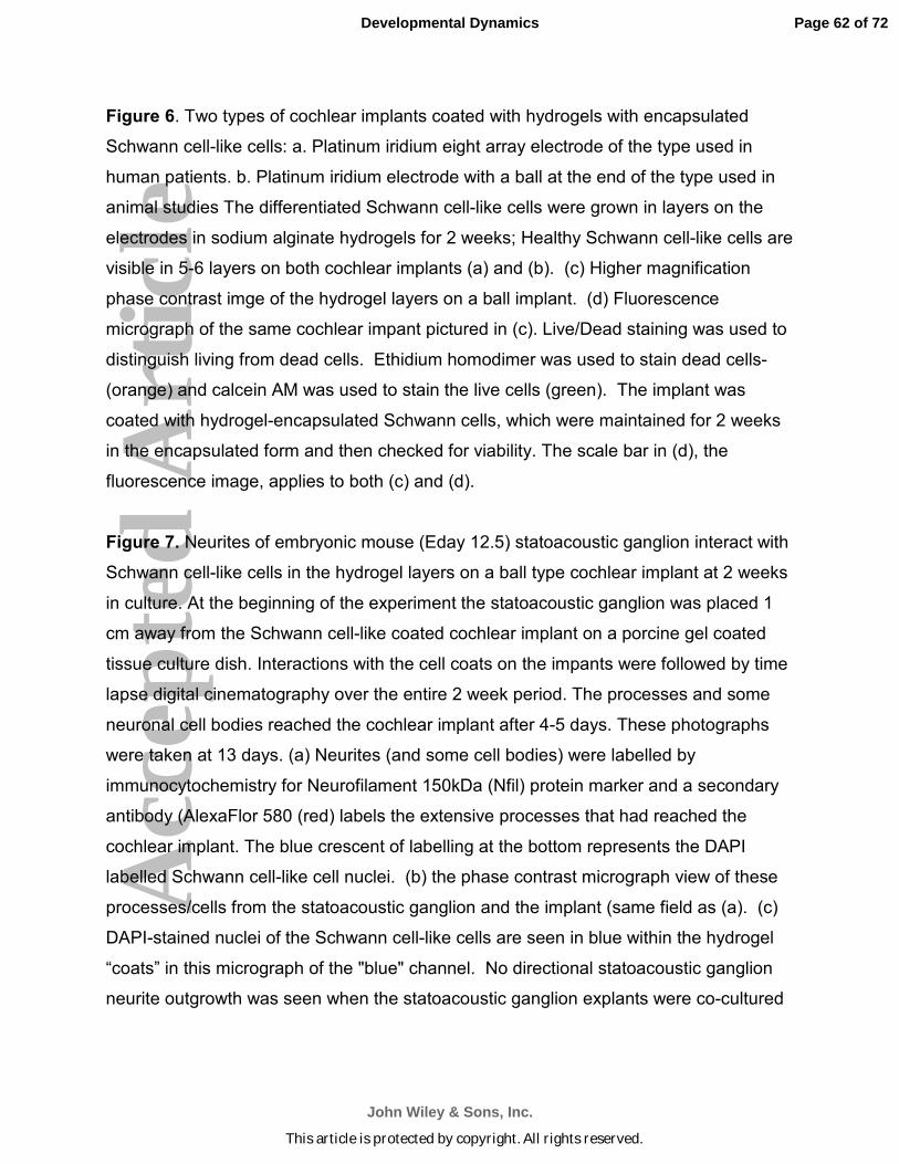
Acc
epte
d A
rtic
leFigure 6. Two types of cochlear implants coated with hydrogels with encapsulated
Schwann cell-like cells: a. Platinum iridium eight array electrode of the type used in
human patients. b. Platinum iridium electrode with a ball at the end of the type used in
animal studies The differentiated Schwann cell-like cells were grown in layers on the
electrodes in sodium alginate hydrogels for 2 weeks; Healthy Schwann cell-like cells are
visible in 5-6 layers on both cochlear implants (a) and (b). (c) Higher magnification
phase contrast imge of the hydrogel layers on a ball implant. (d) Fluorescence
micrograph of the same cochlear impant pictured in (c). Live/Dead staining was used to
distinguish living from dead cells. Ethidium homodimer was used to stain dead cells-
(orange) and calcein AM was used to stain the live cells (green). The implant was
coated with hydrogel-encapsulated Schwann cells, which were maintained for 2 weeks
in the encapsulated form and then checked for viability. The scale bar in (d), the
fluorescence image, applies to both (c) and (d).
Figure 7. Neurites of embryonic mouse (Eday 12.5) statoacoustic ganglion interact with
Schwann cell-like cells in the hydrogel layers on a ball type cochlear implant at 2 weeks
in culture. At the beginning of the experiment the statoacoustic ganglion was placed 1
cm away from the Schwann cell-like coated cochlear implant on a porcine gel coated
tissue culture dish. Interactions with the cell coats on the impants were followed by time
lapse digital cinematography over the entire 2 week period. The processes and some
neuronal cell bodies reached the cochlear implant after 4-5 days. These photographs
were taken at 13 days. (a) Neurites (and some cell bodies) were labelled by
immunocytochemistry for Neurofilament 150kDa (Nfil) protein marker and a secondary
antibody (AlexaFlor 580 (red) labels the extensive processes that had reached the
cochlear implant. The blue crescent of labelling at the bottom represents the DAPI
labelled Schwann cell-like cell nuclei. (b) the phase contrast micrograph view of these
processes/cells from the statoacoustic ganglion and the implant (same field as (a). (c)
DAPI-stained nuclei of the Schwann cell-like cells are seen in blue within the hydrogel
“coats” in this micrograph of the "blue" channel. No directional statoacoustic ganglion
neurite outgrowth was seen when the statoacoustic ganglion explants were co-cultured
Page 62 of 72
John Wiley & Sons, Inc.
Developmental Dynamics
This article is protected by copyright. All rights reserved.

Acc
epte
d A
rtic
lewith a bare cochlear implant or with a cochlear implant coated with hydrogel layers
containing undifferentiated stem cells.
Figure 8. Interaction of primary spiral ganglion neurites (mouse post natal day 3 (a) and
processes from MIF-induced neuron-like cell neurites (b) with wild type Organ of Corti
explants in culture. (a) Polarized image of a primary mouse spiral ganglion (right)
extending extensive neurites directionally toward a wild type Organ of Corti explant (at
left). No directional outgrwoth was seen towards a MIF knock out mouse Organ of Corti
(not shown). This was also demonstrated in separate experiments detailed in Bank et
al., 2012, (Fig. 6 in that paper). (b) MIF-induced neuron-like cells stained with acetylated
tubulin (green) cultured with a wild type Organ of Corti explant for a week showed
fasciculated neurites (approaching form the right) growing towards the wild-type Organ
of Corti (at lower left) No such outgrowth was seen towards OC explants from MIF-KO
mice (as was also demonstrated for primary spiral ganglion neuronal processes in Bank
et al., 2012 Fig. 6).
Page 63 of 72
John Wiley & Sons, Inc.
Developmental Dynamics
This article is protected by copyright. All rights reserved.
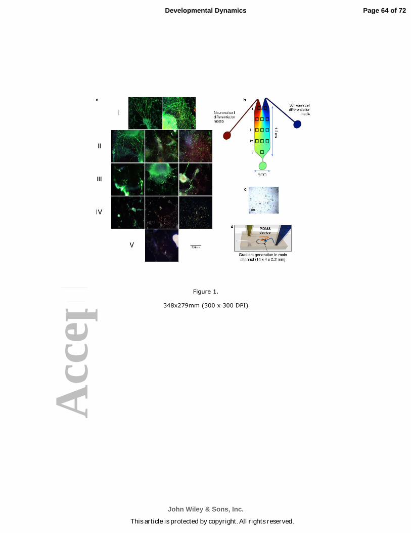
Acc
epte
d A
rtic
le
Figure 1.
348x279mm (300 x 300 DPI)
Page 64 of 72
John Wiley & Sons, Inc.
Developmental Dynamics
This article is protected by copyright. All rights reserved.
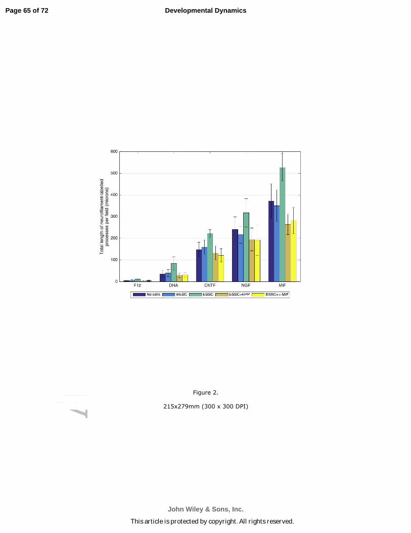
Acc
epte
d A
rtic
le
Figure 2.
215x279mm (300 x 300 DPI)
Page 65 of 72
John Wiley & Sons, Inc.
Developmental Dynamics
This article is protected by copyright. All rights reserved.
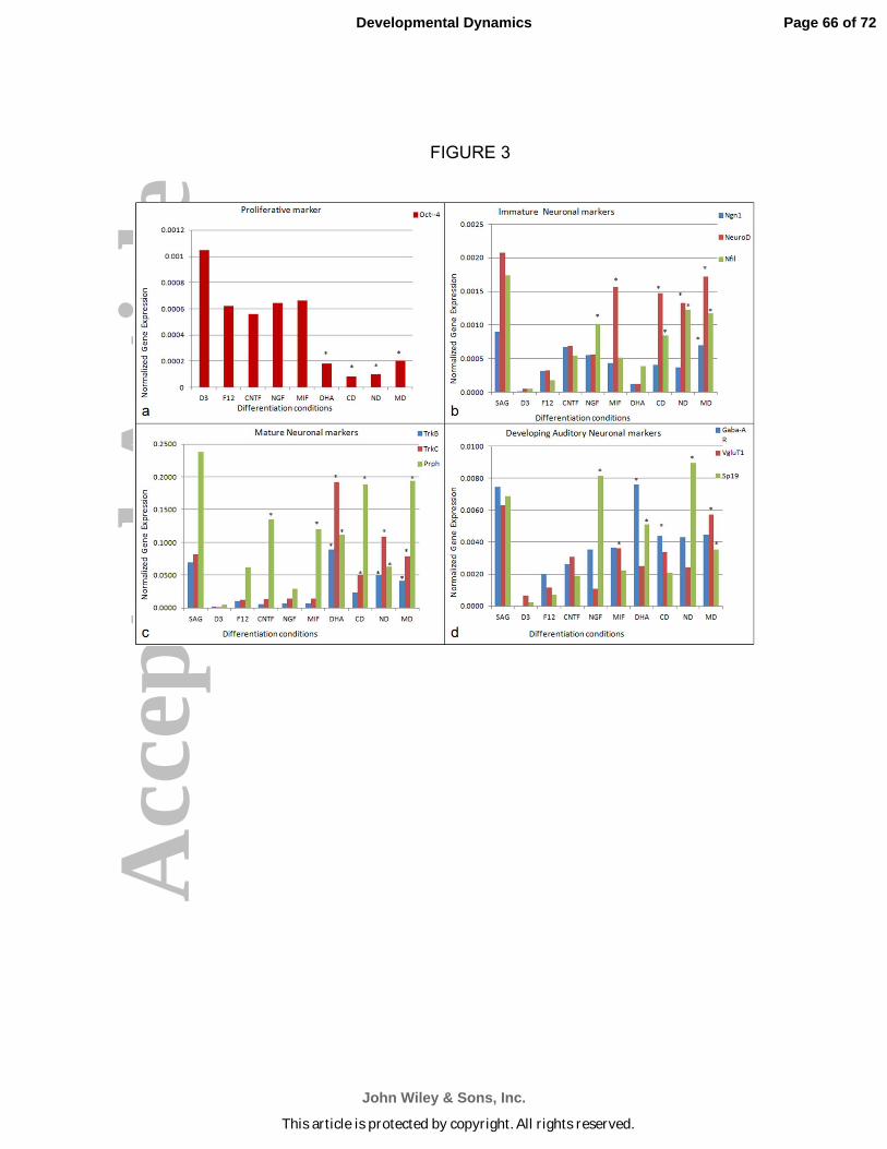
Acc
epte
d A
rtic
le
FIGURE 3
Page 66 of 72
John Wiley & Sons, Inc.
Developmental Dynamics
This article is protected by copyright. All rights reserved.

Acc
epte
d A
rtic
lePage 67 of 72
John Wiley & Sons, Inc.
Developmental Dynamics
This article is protected by copyright. All rights reserved.
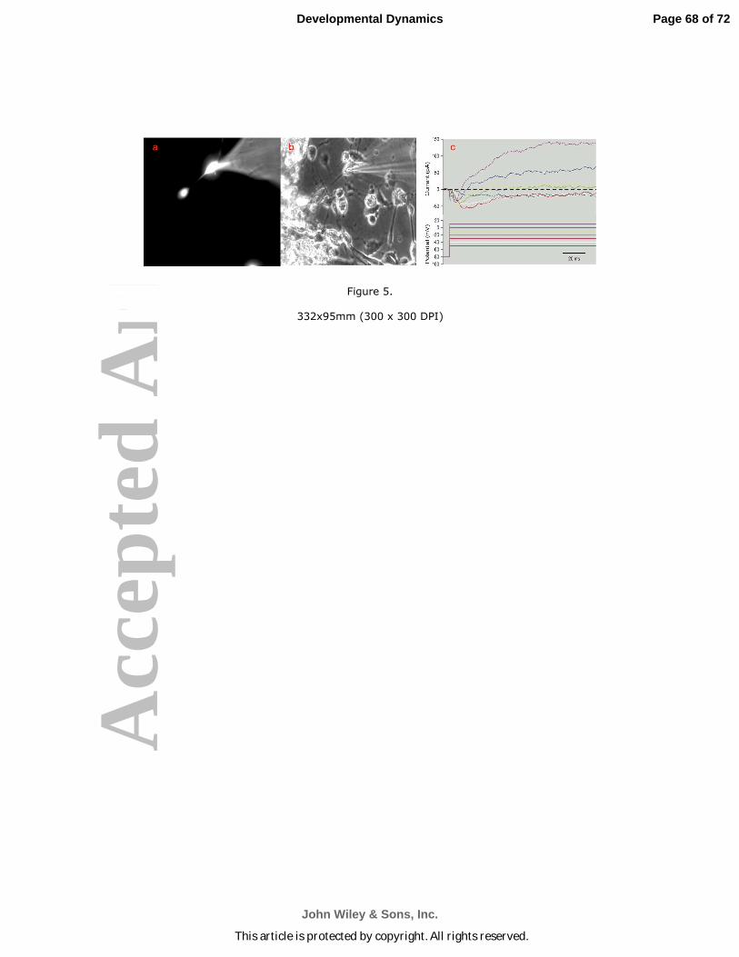
Acc
epte
d A
rtic
le
Figure 5.
332x95mm (300 x 300 DPI)
Page 68 of 72
John Wiley & Sons, Inc.
Developmental Dynamics
This article is protected by copyright. All rights reserved.
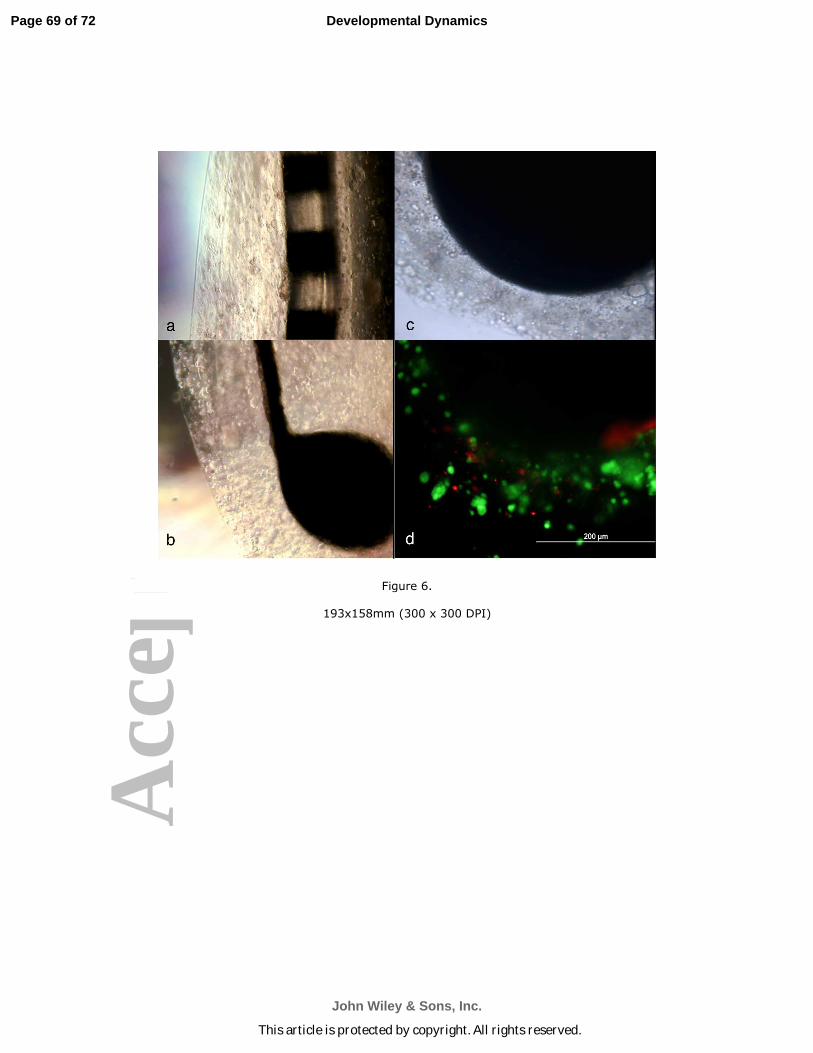
Acc
epte
d A
rtic
le
Figure 6.
193x158mm (300 x 300 DPI)
Page 69 of 72
John Wiley & Sons, Inc.
Developmental Dynamics
This article is protected by copyright. All rights reserved.

Acc
epte
d A
rtic
le
Figure 7.
204x97mm (300 x 300 DPI)
Page 70 of 72
John Wiley & Sons, Inc.
Developmental Dynamics
This article is protected by copyright. All rights reserved.
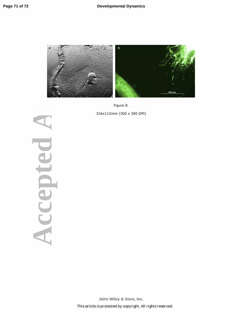
Acc
epte
d A
rtic
le
Figure 8.
316x112mm (300 x 300 DPI)
Page 71 of 72
John Wiley & Sons, Inc.
Developmental Dynamics
This article is protected by copyright. All rights reserved.
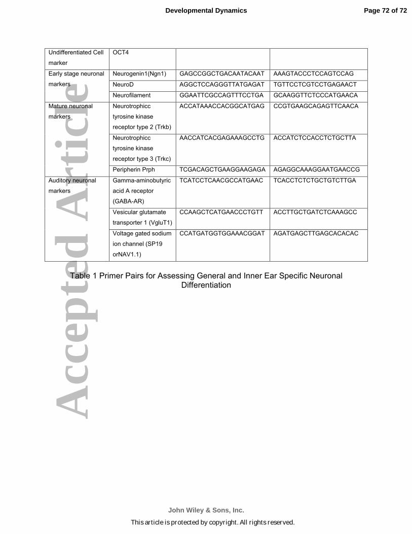
Acc
epte
d A
rtic
leUndifferentiated Cell
marker
OCT4
Early stage neuronal
markers
Neurogenin1(Ngn1) GAGCCGGCTGACAATACAAT AAAGTACCCTCCAGTCCAG
NeuroD AGGCTCCAGGGTTATGAGAT TGTTCCTCGTCCTGAGAACT
Neurofilament GGAATTCGCCAGTTTCCTGA GCAAGGTTCTCCCATGAACA
Mature neuronal
markers
Neurotrophicc
tyrosine kinase
receptor type 2 (Trkb)
ACCATAAACCACGGCATGAG CCGTGAAGCAGAGTTCAACA
Neurotrophicc
tyrosine kinase
receptor type 3 (Trkc)
AACCATCACGAGAAAGCCTG ACCATCTCCACCTCTGCTTA
Peripherin Prph TCGACAGCTGAAGGAAGAGA AGAGGCAAAGGAATGAACCG
Auditory neuronal
markers
Gamma-aminobutyric
acid A receptor
(GABA-AR)
TCATCCTCAACGCCATGAAC TCACCTCTCTGCTGTCTTGA
Vesicular glutamate
transporter 1 (VgluT1)
CCAAGCTCATGAACCCTGTT ACCTTGCTGATCTCAAAGCC
Voltage gated sodium
ion channel (SP19
orNAV1.1)
CCATGATGGTGGAAACGGAT AGATGAGCTTGAGCACACAC
Table 1 Primer Pairs for Assessing General and Inner Ear Specific Neuronal Differentiation
Page 72 of 72
John Wiley & Sons, Inc.
Developmental Dynamics
This article is protected by copyright. All rights reserved.


