Embed Size (px)
Citation preview

Dual Role of CD38 in Microglial Activation and Activation-
induced Cell Death
Running title: The CD38/cADPR pathway in microglial activation and death
Lior Mayo, * Jasmine Jacob-Hirsch,† Ninette Amariglio,† Gideon Rechavi,†
Marie-Jo Moutin,§ Frances E. Lund, ‡ and Reuven Stein*2
*Department of Neurobiochemistry, George S. Wise Faculty of Life Sciences, Tel Aviv
University, 69978 Ramat Aviv, †Department of Pediatric Hematology-Oncology, Safra
Children's Hospital, Sheba Medical Center, Tel Hashomer and Sackler Faculty of Medicine,
Tel Aviv University, 52621 Tel Aviv, Israel, ‡Trudeau Institute, 154 Algonquin Avenue,
Saranac Lake, New York 12983 and §Grenoble Institut des Neurosciences équipe 1, Centre
de Recherche INSERM(n°836)-UJF-CEA-CHU, 38042 Grenoble, France.
1This work was supported by the Israel Science Foundation (643/02), the Adams
Super-Center for Brain Research, and National Institutes of Health (R01-AI-057996
to F.E.L.).
2Address correspondence and reprint requests to Dr. Reuven Stein, Department of
Neurobiochemistry, George S. Wise Faculty of Life Sciences, Tel Aviv University,
69978 Ramat Aviv, Israel. E-mail: [email protected]
3Abbreviations used in this paper: AICD, activation-induced cell death; cADPR, cylic
ADP-ribose; AMD, actinomycin D; N(8-Br-A)D+, nicotinamide 8-bromoadenine
dinucleotide; iNOS, inducible nitric oxide synthetase; WT, wild-type; CHX,
cycloheximide; MTT, 3-(4,5-dimethylthiazol-2-yl)-2,5-diphenyl tetrazolium bromide;
LDH, lactate dehydrogenase; NGD, nicotinamide–guanine dinucleotide; cGDPR,
cyclic GDP-ribose; SNAP, S-nitroso-N-acetylpenicillamine; MFI, mean fluorescence
intensity.

2
Keywords: Cell activation; CD38, microglia, cell surface molecule; apoptosis; nitric
oxide; neuroimmunology
3
Abstract
Microglia, the resident immune cells of the central nervous system, are normally
quiescent but become activated after infection or injury. Their properties then change,
and they promote both repair and damage processes. The extent of microglial
activation is regulated, in part by activation-induced cell death (AICD). Although
many apoptotic aspects of the microglial AICD mechanism have been elucidated,
little is known about the connection between the activation step and the death process.
Using mouse primary microglial cultures, we show that the ectoenzyme CD38, via its
calcium-mobilizing metabolite cyclic-ADP-ribose (cADPR), helps promote
microglial activation and AICD induced by LPS plus IFNγ (LPS/IFNγ), suggesting
that CD38 links the two processes. Accordingly, CD38 expression and activity, as
well as intracellular calcium concentration ([Ca2+] i) in the primary microglia were
increased by LPS/IFNγ treatment. Moreover, CD38 deficiency or treatment with
cADPR antagonists conferred partial resistance to LPS/IFNγ-induced AICD and also
reduced [Ca2+] i. Microglial activation, indicated by induced expression of nitric-
oxide-synthase-2 mRNA and production of NO, secretion and mRNA expression of
TNFα and IL-12 p40, and expression of IL-6 mRNA, was attenuated by CD38
deficiency or cADPR-antagonist treatment. The observed effects of CD38 on
microglial activation is probably mediated via a cADPR-dependent increase in
[Ca2+] i, and the effect on AICD by regulation of NO production. Our results thus
suggest that CD38 significantly affects regulation of the amount and function of
activated microglia, with important consequences for injury and repair processes in
the brain.
4
Introduction
Microglia, the resident immune cells of the CNS, are quiescent in the mature brain
under physiological conditions. Following acute brain injury microglia migrate to the
damaged sites, where they proliferate and become activated (1, 2). Activation is
accompanied by morphological changes, up-regulated expression of cell-surface
proteins (e.g. MHC class-II molecules, CD11b and scavenger receptors), cytokines
(e.g. TNFα, IL-6 and IL-12), chemokines (e.g. CCL12), and NO production (3, 4).
Activated microglia can promote both protective and harmful effects.
Beneficial effects include removal of cell debris and myelin fragments, buffering of
toxic compounds, and secretion of neurotrophins and cytokines capable of supporting
survival of injured neurons (5, 6). Microglia can also be beneficial in
neurodegenerative diseases, for example by clearing β-amyloid (7). However, they
can also produce and secrete toxic factors, including TNFα and IL-1β and free
radicals such as NO (8-10); accordingly, some authors have postulated that microglial
activation participates in the pathogenesis of several neurodegenerative diseases,
including Alzheimer's and Parkinson's (11-13). Activated microglia might also be at
least partially responsible for secondary damage after traumatic brain injury (14, 15);
thus, their numbers and activity need to be strictly regulated. Indeed, following the
initial rapid increase in activated microglia after acute brain injury, their numbers
gradually decrease (16, 17). Among the mechanisms thought to help control the
disappearance of activated microglia after brain damage is their elimination via cell
death, termed microglial activation-induced cell death (AICD)1, and observed for
example in animal models of brain damage and microglial activation (18-22).
Consistently with those observations, treatment with agents known to induce
5
microglial activation, such as LPS and IFNγ, induces apoptosis in microglial cell lines
and primary cultures (23-26).
Our group and others have characterized the apoptotic pathways of AICD
induced by LPS and IFNγ (LPS/IFNγ) and demonstrated their mediation via the NO-
promoted (24) Bak/Bax-activated mitochondrial gateway (26) and caspase 11 (23).
Although these studies have helped to elucidate this AICD mechanism, little is known
about the relationship between the activation step and the death process. Here we
show that CD38, an ectoenzyme that uses β-NAD to generate multiple Ca2+-
mobilizing metabolites (27-29), participates in both the activation and the AICD
processes of LPS/IFNγ-treated microglia. The CD38 effects were mediated, at least in
part, via cyclic ADP-ribose (cADPR).
6
Materials and Methods
Animals, drugs and reagents
CD38-deficient mice (Cd38−/−) (30), backcrossed 12 generations to BALB/cBy mice
(31), were obtained from the Trudeau Institute Breeding Facility (Saranac Lake, NY)
and were maintained in accordance with all applicable rules and guidelines of the
Animal Care and Use Committee of Tel Aviv University.
Reagents used were purchased from Sigma (St. Louis, MO) unless indicated
otherwise. Mouse IFNγ was purchased from R&D Systems, (Minneapolis, MN);
ryanodine was from Calbiochem (La Jolla, CA); actinomycin D (AMD) was from
ICN Biomedicals (Aurora, OH); Hanks’s buffered salt solution (HBSS) was from
Biological Industries (Kibbutz Beit Ha-Emek, Israel); DMEM, fura-2 AM, and
Pluronic F-127 were from Invitrogen Life Technologies (Paisley, UK). Nicotinamide
8-bromoadenine dinucleotide N(8-Br-A)D+ was prepared as described (32).
Antibodies
Rabbit anti-inducible nitric oxide synthetase (iNOS) polyclonal Abs (#AB1631) were
from Chemicon (Temecula, CA). Rat anti-caspase-11 (17D9, #C1354) and mouse
anti-β-tubulin monoclonal Abs (#T4026) were purchased from Sigma (St. Louis,
MO). Phycoerythrin (PE)-conjugated rat anti-CD38 (NIMR-5, #1635-09), low
endotoxin/azide free rat anti-CD38 (90, #1640-14) monoclonal Abs and low
endotoxin/azide free Rat IgG2a isotype control Ab (#0117-14) were from Southern
Biotechnology Associates (Birmingham, AL), and PE-conjugated rat anti-CD11b
monoclonal Ab (M1/70, 101207) was from Biolegend (San Diego, CA). Purified rat
IgG2a anti-mouse monoclonal CD38 antibody (clone NIM-R5) was produced by the
Trudeau monoclonal antibody facility. Secondary Ab F(ab')2 fragment mouse anti-rat
7
IgG (H+L) (#212-006-168) was purchased from Jackson ImmunoResearch, West
Grove, PA.
Cell culture
N9 mouse microglial cell line (33) was maintained in DMEM supplemented with 10%
FCS) (Invitrogen). CD38 expressing (CD38exp) and non-expressing [empty vector
transfected Ba/F3 cells (CD38neg)] Ba/F3 cells (34) were cultured in RPMI 1640
supplemented with 10% WEHI-3 supernatant (containing IL-3), 10% FCS, non-
essential amino acids, sodium pyruvate, L-glutamine, HEPES, β- mercaptoethanol
and antibiotics. Mouse primary microglia were prepared as previously described (26),
with minor modifications. Cerebral cortices from 1- to 3-day-old neonatal BALB/c
[(wild-type (WT)] and Cd38−/− mice were dissected in ice-cold HBSS, carefully
stripped of their meninges, and digested with 0.25% trypsin for 10 min at 37°C.
Trypsinization was stopped by addition of an equal volume of culture medium
[DMEM, 10% low endotoxin (<10 EU/ml) FCS (Hyclone, Logan, UT), penicillin 100
U/ml and streptomycin 100 µg/ml] to which we added 0.02% deoxyribonuclease I.
The cells were dispersed into a single cell level by repeated pipetting, then pelleted
and re-suspended in culture medium. The cell suspension was then passed through a
100-µm pore mesh. Cells were seeded at a density of 300,000 cells/ml (equivalent to
62,500 cells/cm2) in 6-well, 12-well or 96-well plates and cultured at 37°C in
humidified 5% CO2−95% air. Medium was replaced every 4–5 days. These cultures
('mixed glia') reached confluence after 7–10 days and were used between 15 and 20
days after preparation. Microglia were isolated from the mixed glia cultures by a mild
trypsinization procedure as previously described (35). Briefly, treatment of the
confluent mixed glial cultures with trypsin (0.06%) resulted in detachment of an intact
layer of cells containing almost all the astrocytes and leaving behind a highly enriched

8
population of microglia [>98%, as determined by staining with fluorescein-
conjugated Griffonia simplicifolia isolectin B4 (Vector Laboratories, Peterborough,
UK) and by flow cytometric analysis using the PE conjugated CD11b Ab (data not
shown)]. The attached microglia were allowed to recover for 24 h and then were
subjected to the different treatments.
Cell viability assays
Primary microglial cultures prepared in 96-well plates as described above were
treated with LPS (100 ng/ml) and IFNγ (100 units/ml) (hereafter termed LPS/IFNγ
treatment) for the indicated time periods. In some experiments the cells were
preincubated with 8-Br-cADPR (100 µM), ryanodine (30 µM), N(8-Br-A)D+ (100
µM), cycloheximide [CHX (50−100 ng/ml)], or AMD (5−25 ng/ml) for 1−2 h prior,
to the addition of LPS/IFNγ. In the cross-linking experiments primary microglia were
cultured in 24-well plates in the absence or presence of anti-CD38 Abs (clone 90 or
NIM-R5) or a rat IgG2a isotype control Ab with or without 5µg/ml secondary Ab
F(ab')2. On termination of the experiments, the medium from each well was carefully
collected and replaced with an equal volume of fresh medium. Cell viability was then
determined by the 3-(4,5-dimethylthiazol-2-yl)-2,5-diphenyl tetrazolium bromide
(MTT) assay. MTT was added to the new medium at a final concentration of 0.5
mg/ml, followed by incubation at 37°C in a CO2 incubator. After 2 h the medium was
aspirated and dimethyl sulfate (100 µl) was added to the wells. Insoluble crystals were
dissolved by mixing and the plates were read on a SpectraMax micro-ELISA plate
reader, using a test wavelength of 570 nm and a reference wavelength of 690 nm. Cell
death was assessed by monitoring the release of lactate dehydrogenase (LDH) to the
culture medium (which was collected at the end of the experiments, see above) using
the CytoTox-One Homogeneous Membrane Integrity Assay kit (Promega, Madison,

9
WI) according to the manufacturer’s instructions. The LDH signal was read using a
test wavelength of 560 nm and a reference wavelength of 590 nm in a Synergy HT
Multi-Detection Microplate Reader. The viability of the Ba/F3 cells was assessed as
previously described (26). Briefly, cells were plated in 96-well plates (2 × 10⁴ cells
per well). After 18 h primary Abs were added to the culture medium and the cells
were grown in the presence or absence of the Abs for additional 24 h. Then MTT was
added to the culture medium at a final concentration of 0.5 mg/ml, followed by
incubation at 37°C in a CO2 incubator for 2 h. Acid-isopropanol (100 µl of 0.04 M
HCl in isopropanol) was then added to the wells and mixed. The plates were read on a
micro-ELISA reader (SpectraMax microplate reader) as described above.
Nitrite quantification
Nitrite, measured as a reflection of NO production in culture supernatants, was
assayed using modified Griess reagent. In brief, upon termination of the experiment
the culture medium was replaced with an equal volume of new medium, and 50 µl of
the removed medium was mixed with an equal volume of Griess reagent in 96-well
plates and incubated at room temperature for 15 min. Absorbance at 540 nm was then
measured using a micro-ELISA reader (SpectraMax). To normalize the amount of
nitrite measured in each well to the amount of cells present in that well, MTT assay
was performed after the new medium was added to the cultured cells, as described
above.
cADPR measurements
WT and Cd38−/− primary microglia were washed twice with ice cold PBS and
resuspended on ice with 25 mM sodium phosphate buffer at pH 7.4, homogenized by
passing through a 23g needle (using the 500 µl Bee Stinger Gastight Syringe) and
aliquot was removed from each sample to determine the protein content. Perchloric

10
acid (HClO4) was then added to a final concentration of 0.6 M and homogenates were
incubated on ice for 10 min followed by centrifugation (16,000 × g, 10 min, 4°C).
Supernatants were transferred to new tubes and the pH adjusted to pH 7 by K2CO3
titration. Homogenates were left on ice for 30 min to enable gas evaporation after
which the samples were then centrifuged (2,000 × g, 5 min, 4°C) to eliminate salts.
Supernatants were collected into new tubes and kept frozen until used.
The measurement of cADPR from samples was then performed essentially as already
described for determining cADPR content of synaptosomes (36). The contaminating
nucleotides were eliminated by incubation overnight in NaH2PO4 at pH 8.0 with a mix
of three nucleotide hydrolytic enzymes (nucleotide pyrophosphatase, alkaline
phosphatase and NAD+-glycohydrolase). Then, the conversion of cADPR into NAD+
in the presence of Aplysia ADP-ribosyl cyclase, and the cycling reaction were
allowed to proceed. Results were compared to standard solutions of cADPR analyzed
in the same experimental conditions.
Immunoblot analysis
Total extracts (30 µg protein) from each treatment were separated by 12.5%
SDS−PAGE and electroblotted onto supported nitrocellulose. Each blot was blocked
for 1 h in Tris-buffered saline-Tween-20 (10 mM Tris base, 150 mM NaCl and 0.05
% Tween-20) containing 5% fat-free milk, and then incubated with a primary
antibody overnight at 4oC. Washing of membranes three times (10 min each) with
Tris-buffered saline-Tween-20 was followed by incubation for 1 h at room
temperature with the appropriate second antibody (goat anti-mouse, donkey anti-rat,
or goat anti-rabbit IgG peroxidase conjugate; Jackson ImmunoResearch Laboratories,
West Grove, PA). The blots were developed using the Supersignal West Pico
11
chemiluminescence kit (Pierce Biotechnology, Rockford, IL). Each blot was reprobed
with β-tubulin to verify that protein was uniformly loaded across the gel.
Assays of GDP-ribosyl cyclase activity
The ectocellular cyclase activity of CD38 was measured using nicotinamide–guanine
dinucleotide (NGD+) instead of NAD+ as a surrogate substrate because the product,
cyclic GDP-ribose (cGDPR), is fluorescent and is not susceptible to enzymatic
hydrolysis (37). GDP-ribosyl cyclase activity was assayed by incubating NGD+ with
intact microglial primary cultures or N9 microglial cells. Microglia grown in 96-well
plates, untreated or treated with LPS/IFNγ for 24 h (primary cultures) or 15 h (N9
cells), were washed once with HBSS and then incubated with HBSS (100 µl/well) at
37°C. After 20 min, NGD+ (40 µM) was added to each well and the enzymatic
conversion of NGD+ to cGDPR was measured fluorometrically by monitoring the
increase in fluorescence at 420 nm upon excitation at 300 nm for 10 min, as described
previously (32).
Flow cytometry
Microglia were cultured in 6-well plates and treated as indicated. After incubation the
microglia were gently removed from the tissue culture plates by pipetting, then
centrifuged (700 × g, 10 min, 4°C) and incubated (30 min, 4°C) in flow cytometry
buffer (PBS containing 1% BSA and 0.05% sodium azide) containing PE-conjugated
anti-CD38 (NIM-R5) monoclonal Ab, PE-conjugated anti CD11b monoclonal Ab, or
isotype-matched control Abs. After washing, antigen expression on 5,000 live cells
was determined by FACS analysis (FACSort; Becton Dickinson, Mountain View,
CA) and CellQuest software (Becton Dickinson).
12
Gene Expression Profiling
RNA extraction, sample preparation, hybridization, and array processing. N9 cells
(106 per plate) were washed once with 10 ml of ice cold PBS, then lysed with 1
ml/plate of TRIzol reagent (Invitrogen) and RNA was prepared according to the
manufacturer's instructions. Total RNA from each sample was used to prepare
biotinylated target RNA, with minor modifications from the manufacturer's
recommendations. Briefly, 10 µg of total RNA was used to generate first-strand
cDNA by using a T7-linked oligo (dT) primer. After second-strand synthesis, in vitro
transcription was performed with biotinylated UTP and CTP (Enzo Diagnostics),
resulting in approximately 100-fold amplification of RNA, which was then processed
as per manufacturer's recommendation using an Affymetrix GeneChip Instrument
System. Briefly, spike controls were added to 10 µg fragmented cRNA before
overnight hybridization. Arrays were then washed and stained with streptavidin-
phycoerythrin, before being scanned on an Affymetrix GeneChip scanner. Quality and
amount of starting RNA were confirmed using an agarose gel. Following scanning,
array images were assessed by eye to confirm scanner alignment and the absence of
significant bubbles or scratches. 3′/5′ ratios for GAPDH and β-actin were confirmed
to be within acceptable limits range (0.80−0.85 and 1.14−1.2 respectively), and BioB
spike controls were found to be present on 100%, with BioC, BioD and CreX also
present in increasing intensity. When scaled to a target intensity of 150 (using
Affymetrix MAS 5.0 array analysis software), scaling factors for all arrays were
within acceptable limits (0.85−1.06) as were background, Q values and mean
intensities. All experiments were performed using Affymetrix Mouse 430A 2.0
oligonucleotide arrays, as described at
http://www.affymetrix.Com/products/arrays/specific/mgu74.affx. Genes were filtered
13
using Mas 5 algorithm results. A list of 12,827 probe sets of “valid genes”,
representing probe sets with signals higher than 20 and detected as “present” in at
least one sample was obtained.
Analysis of gene expression data. DNA-microarray data were analyzed using the
Expander 3.2 program (38). Data were normalized using Quantile normalization,
filtered to include only genes that changed at least 2 fold form control (untreated
sample), and standardized (mean 0, variation 1). Analysis yielded a list of 1,153
'active genes'.
Total RNA isolation, cDNA synthesis and semi-quantitative RT−PCR analysis.
Primary microglia were untreated or treated with LPS/IFNγ for 10 h. In some
experiments the cells were preincubated for 2 h with 8-Br-cADPR (100 µM). Total
RNA was isolated using the RNeasy Plus Mini Kit (Qiagen, Hilden, Germany)
according to the manufacturer's instructions. Total RNA (1 µg) was used as template
for DNA synthesis, employing random decamer primers in a 20-µl reaction volume
using the Reverse-iT 1st Strand Synthesis Kit (ABgene, Epsom, UK) according to
the manufacturer's instructions. The cDNA mixture was diluted 1:10 with PCR-grade
water. mRNA expression of the indicated genes was measured using the semi-
quantitative RT−PCR approach. The following primers were used according to the
real-time PCR primer and probe database (39): TNF-α, sense 5′-GGC TTT CCG
AAT TCA CTG GAG-3′, antisense 5′-CCC CGG CCT TCC AAA TAA A-3′ (target
size, 264 bp); IL 12 p40, sense 5′-GGA AGC ACG GCA GCA GAA TA-3′, antisense
5′-AAC TTG AGG GAG AAG TAG GAA TGG-3′ (target size, 180 bp); IL-6, sense
5′-GAG GAT ACC ACT CCC AAC AGA CC-3′, antisense 5′-AAG TGC ATC ATC
GTT GTT CAT ACA-3′ (target size, 141 bp); nitric oxide synthase (NOS)2, sense 5′-
CAG CTG GGC TGT ACA AAC CTT-3′, antisense 5′-CAT TGG AAG TGA AGC
14
GTT TCG-3′ (target size, 95 bp); and GAPDH, sense 5′-CAT GGC CTT CCG TGT
TCC TA-3′, antisense 5′-ATG CCT GCT TCA CCA CCT TCT-3′ (target size, 107
bp). PCR reactions were carried out using 5 µl of cDNA. The resulting PCR products,
obtained after 24−26 cycles (in the logarithmic phase of the PCR reaction) were
resolved on E-Gel Pre-cast Agarose 2% (Invitrogen). The individual PCR products
were scanned and their intensity determined (ImageMaster). The value of each of the
IL-6, IL-12 p40, TNFα and NOS2 PCR products was then normalized to the intensity
of its corresponding housekeeping gene, the GAPDH PCR product.
Measurement of TNFα and IL-12 p40 secretion
TNFα and IL-12 p40 secreted into the culture medium were assayed using a DuoSet
ELISA kit (R&D Systems) according to the manufacturer’s instructions. Primary
microglia grown in 96-well plates (for TNFα) or 24-well plates (for IL-12 p40) were
treated with LPS/IFNγ for 24 h. In some experiments, the cells were preincubated for
2 h with 8-Br-cADPR (100 µM) or N(8-Br-A)D+ (100 µM) prior to LPS/IFNγ
treatment. The culture medium was carefully removed from each well, centrifuged at
1,000 × g (5 min, 4°C), and then kept frozen at −80°C until used. Cytokine quantity in
each well was normalized to either the MTT value (TNFα) or the protein
concentration (IL-12 p40) measured in the same well.
Calcium measurements
Intracellular calcium concentrations [Ca2+] i were monitored using the Ca2+-sensitive
fluorescent indicator fura-2 AM, as described (40). Briefly, primary microglial cells
were seeded on 18-mm poly-L-lysine-coated circular coverslips in a 12-well plate.
Prior to treatment with LPS/IFNγ, cells were left untreated or were treated with
LPS/IFNγ, in some experiments after preincubation for 2 h with 8-Br-cADPR (100
15
µM). After 24 h the cells were loaded with 2.5 µM fura-2 AM for 60 min at 37°C (in
a 5% CO2 incubator). The fura-2 AM was loaded as a mixture (1:1 in culture medium)
with the mild detergent pluronic F-127 (0.05%; used to facilitate loading of the AM
ester indicator). Measurements were performed in an external solution (140 mM
NaCl, 3 mM KCl, 2 mM CaCl2, 10 mM HEPES, and 2 mg/ml glucose, pH 7.2–7.4,
300–320 mOsm) at room temperature. For excitation of fura-2, cells were illuminated
with two alternating wavelengths, 340 and 380 nm. Excitation was performed and
[Ca2+] i values calculated from the ratio (R) of fluorescence recorded at excitation
wavelengths of 340 and at 380 nm. Calibrations (by conversion of R340/380 values
into molar calcium concentrations) were performed as described (41).
Statistical analysis
Data was expressed as mean values ± SEM. Statistical evaluation was determined by
two-tailed Student’s t test. A value of p < 0.05 was considered to be statistically
significant.
16
Results
Macromolecule synthesis as an essential requirement for microglial AICD
In some apoptotic systems the death process depends on synthesis of macromolecules.
We were therefore interested in examining whether AICD in microglia is dependent
on RNA or protein synthesis. AICD was induced by combined treatment with LPS
(100 ng/ml) and IFNγ (100 units/ml) (LPS/IFNγ). N9 cells were treated with
LPS/IFNγ in the absence or presence of the RNA-synthesis inhibitor AMD or the
protein synthesis inhibitor CHX. Determination of cell viability by the MTT assay
disclosed that death of the LPS/IFNγ-treated N9 cells was dose-dependently
diminished by both inhibitors (Fig. 1A-B). CHX also inhibited the death of LPS/IFNγ-
treated primary mouse microglia (data not shown). These results suggested that in
order for microglial AICD to occur, some essential gene products need to be
synthesized.
DNA-microarray analysis for identification of genes that are preferentially induced
by LPS/IFNγ
In an attempt to identify the genes whose upregulated expression is needed for
microglial AICD, we employed the DNA-microarray technology. In a previous study
we showed that treatment of N9 cells with LPS (100 ng/ml) alone or IFNγ (100
units/ml) alone did not substantially affect their viability while the combination of
LPS/IFNγ induced AICD (26). We therefore prepared total RNA from N9 cells that
had been untreated (control) or treated with LPS/IFNγ, LPS, or IFNγ for 6 h, a time
point by which the death gene mRNAs had already been synthesized. This was the
earliest time after LPS/IFNγ treatment by which the AMD could not rescue the cells
17
from LPS/IFNγ-induced death and the cell morphology still appeared healthy (data
not shown).
The global gene-expression profiles in the above-mentioned treatments were
recorded by Affymetrix oligonucleotide microarrays. Comparative analysis of the
DNA-microarray results from the four different samples identified 1,153 probe sets
that were changed by at least twofold in one or more of the samples. Of all of these
probe sets only 89 genes were preferentially induced by the LPS/IFNγ treatment and
only 28 of these genes were induced synergistically by the combination of LPS/IFNγ.
Among the genes identified by this analysis were Nos2 (which encodes the iNOS
enzyme that catalyzes NO production) and caspase-11(Fig. 2A). As NO and caspase-
11 are known to be upregulated and to promote microglial AICD by different
pathways (23, 24, 26), it suggested to us that other genes identified in this analysis
might also be AICD-promoting candidate genes.
CD38 plays an important role in LPS/IFNγ-induced AICD
One of the 28 genes identified from the DNA microarray analysis as being
synergistically induced by LPS/IFNγ treatment was Cd38 (Fig. 2B) that encodes the
ecto-enzyme CD38. CD38 catabolizes its substrate, NAD+, to produce metabolites
(e.g. cADPR) that increase cytosolic Ca2+ concentration. CD38 can also act as a cell-
surface receptor capable of initiating a signal transduction cascade and cell death in
immune cells (34, 42-47). Therefore, we suspected that it might play an important role
also in microglial AICD. To examine this possibility, we first verified the DNA
microarray results at the protein expression level. Membrane CD38 expression was
determined by FACS analysis. As shown in Fig. 2B, and in agreement with the DNA-
microarray results, CD38-protein expression in N9 cells was preferentially induced by

18
the LPS/IFNγ treatment relative to treatment with LPS or IFNγ. Fig. 3 shows that the
LPS/IFNγ treatment also induced an increase in membrane CD38 expression in the
primary microglia (1.63-fold induction after 24 h) and that as expected, CD38-
deficient primary microglia obtained from mice that lack CD38 did not express
plasma membrane CD38.
Having verified the DNA microarray results we next examined the need for
CD38 in microglial AICD. WT and Cd38−/− primary microglia were treated with
LPS/IFNγ for 24, 48, or 72 h, and the amount of cell death in each case was
determined by measuring the activity of LDH released into the culture medium. Fig.
4A shows that LPS/IFNγ treatment induced a time-course-dependent release of LDH
in both WT and Cd38−/− primary microglial cultures, but that the amount of LDH
released from the CD38-deficient microglia was substantially and significantly lower
(approximately 50% reduction 72 h after treatment) than observed in the WT cultures.
The results were similar when cell death/viability was assessed by the MTT assay
(Fig.4B). Taken together, these results suggest that CD38−/− primary microglia are
significantly more resistant than the WT microglia to LPS/IFNγ-induced AICD, and
therefore that CD38 plays an important role in promoting this death process.
CD38 was reported to regulate apoptosis in hematopoietic cells independently
of its enzymatic activity (34). We therefore examined the effect of cross-linking of
CD38, using anti-CD38 agonist Abs (clone 90 and NIM-R5) (1, 5 and 25 µg/ml), on
primary microglia treated for 72 h with or without LPS/IFNγ. None of these
treatments, however, had any apparent effect on the viability of the cells (Fig. 5A). To
assess the effectiveness of the cross-linking Abs, their ability to induce cell death of
CD38neg and CD38exp Ba/F3 cells was examined. As shown in Abs Fig. 5B both clone
19
90 and NIM-R5 anti-CD38 Abs induced cell death in Ba/F3 CD38exp but not in
CD38neg Ba/F3 cells (Fig. 5B) as previously reported (34). Taken together these
results suggest that the effect of CD38 on AICD is not mediated via its receptor
signaling activity but by a different mechanism. In view of the important role of Ca2+
signaling in apoptosis (48), and the role of CD38/cADPR in intracellular Ca2+ levels
regulation, we hypothesized that microglial AICD is mediated via CD38-dependent
cADPR. However, it was previously reported that CD38 is not responsible for the
production of most, if not all, of cADPR content in brain tissues (32, 36).
Therefore, we first tested whether CD38 regulates cADPR content in primary
microglia. WT and Cd38−/− primary microglial cultures were treated without or with
LPS/IFNγ for 24 h and then the endogenous cADPR content in the WT and Cd38−/−
primary microglial homogenates were determined by the sensitive cycling assay (36).
As shown in Fig. 5C, the cADPR content of Cd38−/− primary microglial homogenates,
either untreated or treated with LPS/IFNγ, were markedly lower than in WT primary
microglial homogenates (3 and 7 fold reduction respectively). In addition, we
observed that the cADPR content of the microglial cells was increased in the
LPS/IFNγ-treated cells. These results thus suggest the CD38 is most likely
responsible for the generation of most cADPR in microglia and thus that CD38-
dependent cADPR might play a role in microglial responses. We therefore next
examined whether LPS/IFNγ induces an increase in CD38 cyclase activity in intact
primary microglia. Cyclase activity was monitored spectrofluorimetrically, using
NGD+ as a substrate (NGD+-ribosyl cyclase activity). Fig. 5D shows that treatment of
WT primary microglia with LPS/IFNγ for 24 h induced an increase of approximately
5 fold in ectocellular GDP-ribosyl cyclase activity relative to untreated cells. No
20
GDP-ribosyl cyclase activity was detected in CD38-deficient primary microglia,
indicating that the induced increase is dependent on CD38.
To further examine the role of CD38 enzymatic activity in microglial AICD,
we examined the role of the CD38 metabolite cADPR in this process. WT primary
microglia were treated with LPS/IFNγ for 48 h in the absence or presence of the
cADPR inhibitors 8-Br-cADPR (a potent cADPR antagonist) and N(8-Br-A)D+ [an
NAD+ analog that can be cyclized by CD38 into the cADPR antagonist 8-Br-cADPR
(34)] as well as ryanodine, a cADPR antagonist (49, 50). Fig. 6 shows that all three
compounds inhibited the LPS/IFNγ-induced AICD to the same extent as CD38
deficiency.
Taken together, these results suggest that CD38 regulates the LPS/IFNγ-
induced AICD in primary microglia, and that this effect is due, at least in part, to the
enzymatic activity of CD38 via its production of cADPR.
CD38 is an important player in LPS/IFNγ-induced NO production
NO and caspase 11 are essential different mediators of LPS/IFNγ-induced microglial
AICD (23, 24, 26). We were therefore interested in examining whether the effect of
CD38 on LPS/IFNγ-induced AICD in primary microglia is mediated via CD38-
dependent regulation of NO production, via caspase-11 expression, or both.
Examination of the expression of iNOS and caspase-11 in LPS/IFNγ-treated microglia
disclosed that iNOS expression induced by LPS/IFNγ in Cd38−/− primary microglia
was markedly lower (by 2.5-fold) than in WT primary microglia. The induction of
caspase-11, however, was not affected by CD38 expression (Fig. 7A). These results
thus suggest that CD38 controls microglial AICD by promoting NO production (via
21
its effect on iNOS) but not on caspase-11 activation. To further assess if and how
CD38 regulates NO production we examined the effect of CD38 deficiency and
cADPR antagonists as well as agonist anti-CD38 Abs on LPS/IFNγ-induced NO
production. Fig. 7B shows that NO production was significantly lower in LPS/IFNγ-
treated Cd38−/− primary microglia than in WT primary microglia. Fig. 7C shows that
the cADPR antagonists 8-Br-cADPR and ryanodine and the CD38 substrate analog
N(8-Br-A)D+ decreased LPS/IFNγ-induced NO production in WT primary microglia
to similar extents (~40% inhibition). Importantly, the reduction in NO production
caused by 8-Br-cADPR, ryanodine or by N(8-Br-A)D+ was similar to that caused by
CD38 deficiency. In contrast to the effect of the cADPR antagonists on NO
production, the crosslinking of CD38 did not have any effect on NO production in
either LPS/IFNγ-untreated or treated WT primary microglia (Fig 7D). These results
suggest that in primary microglia CD38 modulates LPS/IFNγ-induced NO production,
and that this effect is mediated, at least in part, by the Ca2+-mobilizing cADPR
metabolite. Taken together these results suggest that CD38 plays an important role in
the regulation of LPS/IFNγ-induced NO production and that this effect is mediated
via regulation of iNOS induction. In view of the essential role of NO in microglial
AICD, these results further support the suggestion that CD38 regulates AICD by
promoting NO production. To further assess the relationship between NO production
and the effect of CD38 on AICD, we examined the susceptibility of WT and Cd38−/−
primary microglia to external NO by treating them with the NO donor S-nitroso-N-
acetylpenicillamine (SNAP). Fig. 7E shows that WT and Cd38−/− primary microglia
exhibited similar susceptibilities to SNAP at all concentrations. This result suggested
that the mechanism underlying the enhanced resistance of Cd38−/− cells to AICD is
mediated not by an inhibition of downstream events in the NO-induced apoptotic

22
pathway but rather by attenuation of NO production. Thus, once NO is generated, the
sensitivity of Cd38−/− primary microglia to NO is similar to that of the WT cells.
CD38 is an important mediator of LPS/IFNγ-induced microglial activation
Apart from its role in microglial AICD, NO production is an important feature of
microglia activation. In view of our results that CD38/cADPR regulates NO
production and the notion that elevation of intracellular Ca2+ plays a central role in
microglial activation (51-53) we hypothesize that CD38 might act also as a mediator
of microglial activation via a cADPR-dependent increase in [Ca2+] i. To test this
hypothesis we examined whether LPS/IFNγ increases [Ca2+] i and assessed the effects
of CD38 deficiency and the presence of the cADPR antagonist 8-Br-cADPR on any
such LPS/IFNγ-induced increase in [Ca2+] i. Fig. 8 shows that the basal level of [Ca2+] i
in Cd38−/− primary microglia was lower than in WT microglia (75.12 ± 2.9 nM and
85.25 ± 2.19 nM, respectively). Treatment with LPS/IFNγ for 24 h increased [Ca2+] i
in the WT but not in the Cd38−/− primary microglia. Furthermore, 8-Br-cADPR
completely blocked the LPS/IFNγ-induced increase in [Ca2+] i.
Taken together, our results show that treatment with LPS/IFNγ induced an increase in
[Ca2+] i in primary microglia and that this increase was mediated by the CD38/cADPR
pathway. They therefore suggest that, in addition to its effect on AICD, CD38 might
also participate in the Ca2+-dependent microglial activation events induced by
LPS/IFNγ. In the next experiments, therefore, we examined the role of CD38 in
LPS/IFNγ-induced microglial activation by assessing the effects of CD38 expression
and cADPR antagonists on such activation. Microglial activation was assessed in
terms of the induced expression of TNFα, IL-12 p40, IL-6, and NOS2 mRNAs, as
determined by semi-quantitative RT−PCR analysis. WT and Cd38−/− primary
23
microglia were treated with or without LPS/IFNγ for 10 h, and total RNA was then
prepared. Fig. 9A shows that the expression levels of TNFα, IL-12 p40, IL-6, and
NOS2 mRNAs were lower in Cd38−/− primary microglia than in the WT, and that the
cADPR inhibitor 8-Br-cADPR reduced the amounts of IL-6, NOS2, TNFα, and IL-12
p40 (IL-12) mRNAs in LPS/IFNγ-treated WT microglia to approximately the levels
detected in LPS/IFNγ-treated Cd38−/− primary microglia. Quantitative analysis (Fig.
9B) of the intensities of each of the different RT−PCR products normalized to the
intensity of GAPDH in the same treatment from 3−5 different independent
experiments confirmed the results of the representative experiments shown in Fig. 9A.
To further verify the results of the semi-quantitative RT−PCR, we also examined the
effects of CD38 deficiency and the cADPR antagonist on the LPS/IFNγ-induced
secretion of TNFα and IL-12 p40 protein. Fig. 9C shows that the amounts of TNFα
and IL-12 p40 proteins (measured by ELISA) secreted after 24 h from LPS/IFNγ-
treated Cd38−/− primary microglia were significantly lower (by 20% and 50%,
respectively) than those secreted from WT LPS/IFNγ-treated primary microglia.
Furthermore, treatment with 8-Br-cADPR, ryanodine or N(8-Br-A)D+ reduced the
amounts of LPS/IFNγ-induced TNFα and IL-12 p40 secretion in WT primary
microglia to about the same levels as those secreted by Cd38−/− primary microglia. In
contrast, treatment with the cross-liking Abs did not affect TNFα or IL-12 p40
secretion in LPS/IFNγ-untreated or treated primary microglia (Fig. 9D). Taken
together these results show that CD38 deficiency, and cADPR antagonists all
attenuate the induction of key players in microglial activation, and thus suggest that
CD38, via its metabolite cADPR, regulates the LPS/IFNγ signaling that triggers
microglial activation.
24
Discussion
Microglial activation plays an important role both in the beneficial and in the harmful
actions of microglia in the CNS. Microglial AICD is one of the processes suggested to
regulate the numbers of microglia that become activated after brain derangement. In
the present study we sought to identify the mechanism connecting microglial
activation to AICD. Our experiments with the inhibitors of macromolecule synthesis
disclosed that newly synthesized mRNA and proteins are needed for microglial
AICD. Analysis of a DNA microarray enabled us to identify CD38 as a potential
candidate for mediating microglial AICD. CD38 is an ectoenzyme that catabolizes
NAD(P)+ to generate multiple Ca2+-mobilizing metabolites, including ADPR, cADPR,
and nicotinic acid adenine dinucleotide phosphate (NAADP+) (27, 54, 55). CD38 can
also act as a cell-surface receptor capable of initiating a signal transduction. Our
results show that CD38, via its metabolite cADPR, but not its receptor activity, plays
an important role in mediating LPS/IFNγ-induced microglial AICD and activation.
Evidence for the participation of CD38 and cADPR in microglial activation
and AICD came from the following observations: (i) CD38 expression and enzymatic
activity were induced in microglia by treatment with LPS/IFNγ. (ii) CD38 is the main
contributor of cADPR content in both LPS/IFNγ treated and untreated primary
microglia. (iii) CD38 expression in primary microglia was found to be needed for
LPS/IFNγ-induced increase in [Ca2+] i and, at least in part, for induction of various
features of microglial activation, including NOS2 induction and NO production, the
mRNA induction and protein secretion of TNFα and IL-12 p40, and IL-6 mRNA
induction. (iv) CD38-deficient primary microglia were more resistant than WT
microglia to LPS/IFNγ-induced AICD. (v) cADPR antagonists inhibited LPS/IFNγ-
25
induced microglial activation and AICD in WT primary microglia and the extent of
this inhibition was similar to that obtained by CD38 deficiency.
The finding that CD38 plays an important role in microglial activation is in
line with published studies in which CD38 was assigned as a key regulator of diverse
biological processes, particularly in hematopoietic cells. Such processes include
response to inflammatory challenges (32, 56), activation of B, T, and NK cells (57-
60), and Ig isotype switching (61). In addition, CD38 was shown to mediate
chemotaxis in response to formyl peptide receptor ligands such as fMLF in primary
human monocytes and N9 cells (56, 62).
The results showing that CD38 is the main contributor of primary microglia
cADPR content are in line with previous studies which showed that CD38 is the main
ADP-ribosyl-cyclase in bone marrow myeloid cells (32). However, it was reported
that the cADPR content in Cd38−/− in adult (32, 36) and newborn (36) brain tissues is
nearly equivalent to that of the corresponding WT brain tissues and that CD38-
deficient neural cells exhibit an ADP-ribosyl cyclase activity (36, 63). These results
lead to the suggestion that CD38 may not be the major cADPR producing enzyme in
brain (36). We believe that the likely reason for the discrepancy between our results
and the published findings is that the published experiments examined the cADPR
content in total brain tissue, whereas microglial cells only represent a small proportion
(5-12% in different areas of the brain) (64) of the total cells in the brain. Thus, some
of the cell types in the brain, such as the neural cells, express the still unknown
cyclase, while other cell types, like microglial cells express CD38.
Mechanism whereby CD38 regulates LPS/IFNγ-induced microglial activation and
AICD

26
CD38, via its metabolite cADPR, regulates Ca2+-mediated signal transduction by
increasing the amounts of intracellular free Ca2+ in cells [by promoting Ca2+ release
from ryanodine receptor-gated stores and by regulating the influx of extracellular Ca2+
(32, 65)]. It thus seems feasible that CD38 mediates cADPR-associated LPS/IFNγ
responses by increasing [Ca2+] i. Our results showed that LPS/IFNγ indeed induces an
increase in [Ca2+] i and that this increase was mediated by CD38 and its metabolite
cADPR. Ca2+ is also an important mediator of the activation of LPS signaling in
primary microglia (51). There is an interesting similarity between the presently
observed effects of CD38 deficiency and cADPR antagonists on LPS/IFNγ-induced
primary microglial activation and the previously reported effect of the Ca2+ chelator
BAPTA (51) on primary microglia activation induced by LPS. According to that
report, BAPTA inhibited LPS-induced production of NO and secretion of TNFα and
IL-12 in WT primary microglial cultures.
It should be noted that CD38 produces, in addition to cADPR, two other
important Ca2+-mobilizing metabolites, NAADP+ and ADPR (27). NAADP+ induces
Ca2+ release from intracellular lysosomal stores whereas ADPR, the major metabolite
produced by CD38, induces Ca2+ influx via the transient receptor potential (TRPM2)
cation channels (66-68). Our data does not exclude the possibility that these additional
metabolites generated by CD38 may also play a role in regulating microglial cell
activation or AICD. In addition, we cannot exclude the possibility that the loss of
CD38 alters NAD+ homeostasis (69, 70), which may lead in turn to “ disregulated”
activity of other NAD+-utilizing enzymes such as ecto-ADP-ribosyltransferases
(ART) (31) and poly(ADP-ribose) polymerases (PARP). While NAD dysregulation or
production of the other metabolites produced by CD38 might also participate in
LPS/IFNγ-mediated microglial activation and AICD, our data clearly indicate that
27
cADPR is important in these processes. Furthermore, our experiments using the CD38
substrate analog N(8-Br-A)D+ also strongly suggested that CD38 mediates effects
through an enzyme-dependent mechanism and not by functioning as a cell-surface
signaling receptor as reported in hematopoietic cells (34, 43). This conclusion is
further supported by our results which show that in primary microglia the cross-
linking of CD38 using two different agonist anti-CD38 Abs did not affect either the
viability of NO production or cytokine secretion by LPS/IFNγ-treated or untreated
WT microglia.
Our results, point to a regulatory role for CD38 in LPS/IFNγ-induced
microglial activation, and are in line with studies that suggested that cADPR has an
important function in mediating the effects of LPS (71) and of fMLF (56) in the N9
cell line. In our study, however, the role of CD38 in LPS/IFNγ-induced microglial
activation was demonstrated directly, and this CD38 effect was shown to occur in
primary microglial cultures. The former finding is of great interest because it is
becoming increasingly evident that microglia do not constitute a single, uniform cell
population but rather comprise a family of cells (72). It is therefore of the utmost
importance to study the characteristics of microglial activation in primary microglial
cultures.
It should be noted that although microglial activation and AICD were
attenuated by CD38 deficiency and cADPR antagonists, the inhibition was not
complete. With regard to microglial AICD, it is known that this process is mediated
by several pathways and molecules (23, 26). CD38 is thus one of several molecules
whose induced expression and combined action culminates in microglial cell death.
Similarly, additional signaling systems beside CD38/cADPR are likely to participate
in the induction of microglial activation by LPS/IFNγ. Indeed, we found that
28
CD38/cADPR does not regulate caspase 11 expression, an important mediator of
AICD pathway. Furthermore, data from others have shown that enhancement of [Ca
2+] i alone did not induce NO production or cytokine secretion in primary microglia
(51). Thus, it is clear that the CD38/cADPR-dependent signaling pathway is only one
of perhaps many pathways that contribute to microglial activation and AICD.
Linking microglial activation to AICD
Studies by our group (26) and by other (23, 24) have shown that NO is an essential
mediator of LPS/IFNγ-induced AICD. Our present finding that CD38 promotes the
LPS/IFNγ-induced production of NO in primary microglia suggests that the effect of
CD38 on microglial AICD is mediated via this CD38-mediated regulation of iNOS
induction and NO production. This likelihood is further supported by the observation
that CD38 does not affect caspase-11 expression, also an essential step in LPS/IFNγ-
induced AICD (23). Moreover, we did not observe any substantial differences in the
expression levels of key apoptotic proteins (Bcl-xL, MCl-1, Bid, Bim caspase-1, and
caspase-7) between CD38-expressing and CD38-deficient primary microglia (data not
shown). Furthermore, in contrast to the observed resistance of CD38-deficient
primary microglia to LPS/IFNγ, these cells were as sensitive as CD38-expressing
microglia to NO donor toxicity and to the well established apoptotic trigger
staurosporine (data not shown). Taken together, these results suggest that the lack of
CD38 does not endow the cells with a general resistance to apoptosis but rather
confers a specific resistance to LPS/IFNγ-induced AICD, probably by reducing the
amount of NO. Our results further suggest that CD38 links activation to AICD by
controlling the amount of NO that the cells produce.
Activated microglia are present in almost all neurological diseases, where
they may promote both beneficial and harmful effects (72). Our demonstration of the
29
important role of CD38 in microglial activation and AICD suggests that CD38, via its
regulatory effect on microglial functions and fate, might play an important role in
acute and chronic brain derangements, and hence that manipulation of CD38
expression and/or activity might represent a useful future approach for treating
neurological diseases. Towards that aim it will be necessary to extend the present in-
vitro study to systems in vivo, and to study microglial activation and brain
susceptibility to injury and inflammation in CD38-deficient mice after various brain
insults. Such studies are currently in progress in our laboratory.
30
Acknowledgements
We are grateful to Ms. Shirley Smith for excellent editorial assistance. We thank Dr.
Uri Ashery (Tel Aviv University, Israel) for help with calcium measurements and Dr.
Norman Oppenheimer (Univ. California, San Francisco) for providing N(8-Br-A)D+.
Disclosure
The authors have no financial conflict of interest.
31
Figure legends
FIGURE 1. Microglial AICD depends on synthesis of macromolecules. N9 cells
were treated without or with LPS/IFNγ in the absence or presence of the indicated
concentrations of the RNA synthesis inhibitor AMD (A) or the protein synthesis
inhibitor CHX (B). Cell viability was determined by the MTT assay as described in
Materials and Methods. Survival was assessed in terms of MTT values after
treatment, expressed as a percentage of the MTT values without treatment. Values for
each treatment are expressed as percentages of the corresponding control (cells
untreated or treated with the inhibitor). Values for a given treatment relative to
inhibitor untreated cells are defined as significant if * P < 0.05 (Student's t-test; n = 3).
FIGURE 2. Nos2, caspase-11 or CD38 expression is preferentially induced by the
LPS/IFNγ treatment; DNA-microarray and protein expression analysis. N9 cells were
treated without (control) or with LPS (100 ng/ml) or IFNγ (100 units/ml) or LPS/IFNγ
(LPS (100 ng/ml) and IFNγ (100 units/ml). RNA samples and protein extracts were
prepared after 6 and 15 h respectively. The RNA was subjected to gene expression
profiling and the results obtained were analyzed as described in Materials and
Methods. The protein extracts were analyzed by immunoblotting (A) for the
expression of Nos2, and caspase-11, as described in Materials and Methods. The
immunoblotting results shown are from a representative experiment for each protein
(one of 3−5 independent experiments). Immunoblots obtained in at least three
independent experiments (including the one shown here) were scanned and the
intensities of the different proteins were assessed by densitometric analysis. Data are
presented as mean values of the intensities obtained for each protein after its
32
normalization to β-tubulin. The data presented are expressed as the ratios of the mean
intensities of caspase-11 and Nos2 in LPS-treated, IFNγ-treated, or control cells to
those of LPS/IFNγ-treated cells (defined as 1 A.U). Values shown in the histograms
are means ± SEM (bars) (n = 3−5). The gene-expression results (obtained from the
DNA-microarray analysis) are shown (in parentheses) above each of the
corresponding immunoblot results and are expressed for each gene in the same
manner as described for the corresponding proteins. For determination of CD38
expression (B), N9 cells were treated as described above for 15 h and then were
harvested, stained with PE-conjugated anti-CD38 Abs (PE-CD38), and analyzed by
flow cytometry as described in Materials and Methods. The results of a representative
experiment for each treatment (one of at least three independent experiments) are
shown in the right panels. The empty histogram represents the background staining
using an isotype-matched control Ab; the filled area represents CD38 staining. The
mean fluorescence intensities (MFIs) for the different treatments of the (at least) three
experiments were calculated and their mean values determined. The left panels
present the quantification of the results of three independent experiments (including
the one shown here), expressed as CD38 expression values (as determined by the
MFIs) in the LPS-, IFNγ-, or LPS/IFNγ-treated cells relative to CD38 expression
values in the control (defined as 1 A.U.). Values shown are means ± SEM (bars) (n =
3−4). The gene-expression results (obtained from the DNA-microarray analysis) are
shown (in brackets) above each of the corresponding flow cytometry results.
FIGURE 3. LPS/IFNγ induces CD38 expression in primary microglia. LPS/IFNγ
induces CD38 expression in the plasma membrane. WT and Cd38−/− primary
microglial cultures were grown in the absence or presence of LPS/IFNγ for 24 h and
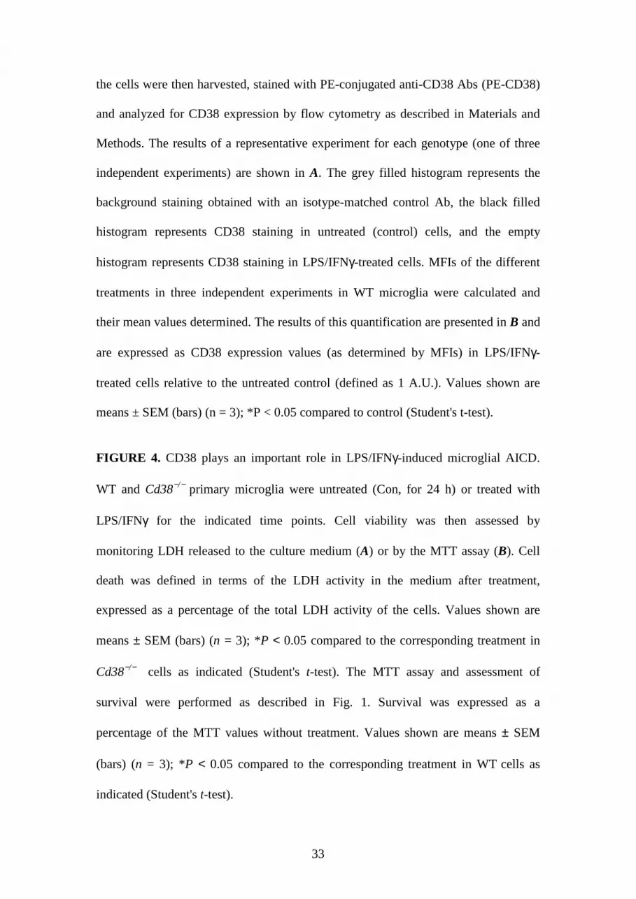
33
the cells were then harvested, stained with PE-conjugated anti-CD38 Abs (PE-CD38)
and analyzed for CD38 expression by flow cytometry as described in Materials and
Methods. The results of a representative experiment for each genotype (one of three
independent experiments) are shown in A. The grey filled histogram represents the
background staining obtained with an isotype-matched control Ab, the black filled
histogram represents CD38 staining in untreated (control) cells, and the empty
histogram represents CD38 staining in LPS/IFNγ-treated cells. MFIs of the different
treatments in three independent experiments in WT microglia were calculated and
their mean values determined. The results of this quantification are presented in B and
are expressed as CD38 expression values (as determined by MFIs) in LPS/IFNγ-
treated cells relative to the untreated control (defined as 1 A.U.). Values shown are
means ± SEM (bars) (n = 3); *P < 0.05 compared to control (Student's t-test).
FIGURE 4. CD38 plays an important role in LPS/IFNγ-induced microglial AICD.
WT and Cd38−/− primary microglia were untreated (Con, for 24 h) or treated with
LPS/IFNγ for the indicated time points. Cell viability was then assessed by
monitoring LDH released to the culture medium (A) or by the MTT assay (B). Cell
death was defined in terms of the LDH activity in the medium after treatment,
expressed as a percentage of the total LDH activity of the cells. Values shown are
means ± SEM (bars) (n = 3); *P < 0.05 compared to the corresponding treatment in
Cd38−/− cells as indicated (Student's t-test). The MTT assay and assessment of
survival were performed as described in Fig. 1. Survival was expressed as a
percentage of the MTT values without treatment. Values shown are means ± SEM
(bars) (n = 3); *P < 0.05 compared to the corresponding treatment in WT cells as
indicated (Student's t-test).

34
FIGURE 5. Assessment of CD38 receptor and enzymatic activities in primary
microglia. A, CD38 cross-linking does not affect the viability of primary microglia.
WT primary microglia were untreated (Control) or treated with LPS/IFNγ for 72 h in
absence or presence of the indicated concentrations of the primary monoclonal Abs:
clone 90 (90) or NIM-R5 anti-CD38 Ab or with isotype control Ab [(rat IgG2a
(IgG)], without (−) or with (+) the secondary anti-rat IgG Ab F(ab')2 (3 µg/ml). Cell
death was assessed by monitoring LDH released into the culture medium, and was
defined and expressed as described in the legend of Fig. 4. Values shown are means ±
SEM (bars) (n = 3). B, CD38 cross-linking induces cell death in CD38exp but not
CD38neg Ba/F3 cells. CD38neg and CD38exp Ba/F3 cells were stimulated with the
CD38 agonist Abs or isotype control Ab for 24 h. Cell viability was assessed by the
MTT assay as described in Materials and Methods. Survival was defined and
expressed (left panel) as described in the legend of Fig. 4. Values shown are means ±
SEM (bars) (n = 3); *P < 0.05 compared to isotype control treated CD38exp Ba/F3
cells (Student's t-test). Expression of CD38 in the CD38neg and CD38exp Ba/F3 cells
(right panel). Ba/F3 cells were stained and analyzed for CD38 expression by flow
cytometry as described in Fig. 3. The results of a representative experiment for each
genotype (one of three independent experiments) are shown. The empty histogram
represents the background staining (Mock) obtained with an isotype control Ab, the
black filled histogram represents CD38 staining in the CD38exp cells, and the grey
filled histogram represents CD38 staining in CD38neg Ba/F3 cells. C, cADPR content
is lower in Cd38−/− primary microglia than in WT primary microglia. WT and Cd38−/−
primary microglia were untreated (Control) or treated with LPS/IFNγ for 24 h and
then extracts were prepared from the cells and their cADPR content was determined
35
using the cycling assay (as described in the Material and Methods). Values shown are
means ± SEM (bars) (n = 4 – 5, for untreated and LPS/IFNγ-treated respectively); *P
< 0.05 compared to the corresponding treatment in Cd38−/− cells (Student's t-test).
D, LPS/IFNγ treatment induces GDP-ribosyl cyclase activity in WT primary
microglia. WT and Cd38−/− primary microglia were grown in the absence (control) or
presence of LPS/IFNγ for 24 h, and were then subjected to the GDP-ribosyl cyclase
activity assay as described in Materials and Methods. Accumulation of the product,
cGDPR, was measured fluorometrically. Data presented for WT primary microglia are
the fluorescence values obtained after 10 min (no specific fluorescence was detected
for Cd38−/− primary microglia). Values shown are means ± SEM (bars) (n = 3)
FIGURE 6. The role of cADPR in LPS/IFNγ responses. cADPR plays an important
role in LPS/IFNγ-induced microglial AICD. WT and Cd38−/− primary microglia were
untreated or treated or for 48 h in the absence (−) or presence (+) of the cADPR
antagonists 8-Br-cADPR (100 µM) and ryanodine (30 µM) or the CD38 substrate
analog N(8-Br-A)D+ (100 µM). Cell death was assessed by monitoring LDH released
to the culture medium, and was defined and expressed as described in the legend of
Fig. 4. Values shown are means ± SEM (bars) (n = 3); *P < 0.05 compared to WT
microglia treated with LPS/IFNγ (Student's t-test).
FIGURE 7. Participation of NO but not caspase 11 in the effect of CD38 on
microglial AICD. A, iNOS and caspase-11 expression in WT and Cd38−/− primary
microglia. Cells were treated without (−) or with LPS/IFNγ (+) for 24 h. Protein
extracts were then prepared, and iNOS and caspase-11 expression were examined by
immunoblot analysis (left panel). The results shown are from a representative
36
experiment (one of three independent experiments). Immunoblots obtained from three
independent experiments (including the one shown here) were scanned and the
intensities of the different proteins were assessed by densitometric analysis. Data
(right panel) are expressed as mean values of the intensities obtained for each protein
after its normalization to β-tubulin, and are shown as the values obtained for each
protein in LPS/IFNγ-treated Cd38−/− microglia relative to LPS/IFNγ-treated WT
microglia (defined as 1 A.U.). B, C, the effect of CD38 and cADPR on NO
production. WT and Cd38−/− primary microglia were untreated (control) or treated
with LPS/IFNγ for the indicated times (B) or for 24 h in the absence (−) or presence
(+) of 8-Br-cADPR (100 µM), ryanodine (30 µM) or N(8-Br-A)D+ (100 µM) (C). NO
production (Griess reaction) and MTT values were then measured as described in
Material and Methods. Nitrite levels in the medium of each treatment were
normalized to the number of live cells in that treatment, as determined by MTT assay,
and the results shown in B and C are expressed as the normalized nitrite values in
each LPS/IFNγ treatment relative to the normalized nitrite values of its corresponding
control [(24 h, untreated or treated with 8-Br-cADPR, ryanodine or N(8-Br-A)D+
alone) (fold increase)]. Values shown are means ± SEM (bars) (n = 3). *P < 0.05
compared to the corresponding LPS/IFNγ-treated Cd38−/− microglia (B) or to
LPS/IFNγ-treated WT microglia (C) (Student's t-test). D, CD38 cross-linking does not
affect NO production in LPS/IFNγ-untreated or treated primary microglia. WT
primary microglia were untreated (Control) or treated with LPS/IFNγ for 24 h in
absence or presence of primary or secondary Ab as described in the legend of Fig 5A.
Nitrite levels in the medium of each treatment were normalized as described in B and
C and expressed as the normalized nitrite values in each treatment relative to the
normalized nitrite values in primary microglia treated only with LPS/IFNγ. Values
37
shown are means ± SEM (bars) (n = 3). E, WT and Cd38−/− primary microglia exhibit
similar susceptibilities to SNAP toxicity. WT and Cd38−/− primary microglia were
treated with the indicated concentrations of the NO donor SNAP for 24 h, and cell
death was then determined by the LDH assay, as described in the legend of Fig. 4A.
Values shown are means ± SEM (bars) (n = 3).
FIGURE 8. LPS/IFNγ induces enhancement in [Ca+2] i via the CD38/cADPR
pathway. WT and Cd38−/− primary microglia were untreated (−) or treated with
LPS/IFNγ (+) for 24 h in the absence (−) or presence (+) of the cADPR antagonist 8-
Br-cADPR. The cells were loaded with fura-2 AM as described in Material and
Methods. [Ca+2] i levels were determined on the basis of the fluorescence intensity
ratio (F340/380). Values shown are means ± SEM (bars) from 2−3 independent
experiments (n = 100-140 cells); *P < 0.05 relative to untreated WT cells; #P < 0.05
relative to LPS/IFNγ-treated WT cells.
FIGURE 9. CD38 and cADPR are important players in microglial activation. WT
and Cd38−/− primary microglia were grown without (−) or with (+) LPS/IFNγ in the
absence (−) or presence (+) of 8-Br-cADPR (100 µM), ryanodine (30 µM) or N(8-Br-
A)D+ (100 µM), as described in Materials and Methods. Semi-quantitative RT−PCR
analysis was performed as described in Materials and Methods. PCR products were
separated on agarose gel electrophoresis and the results of a representative experiment
(one of 3−5 independent experiments) are shown in A. B, Quantification of the semi-
quantitative RT−PCR results. Intensities of the PCR products in the different
independent experiments (including the one shown) were measured and normalized to
GAPDH, as described in Materials and Methods. Results are recorded as the
normalized PCR product intensity value in each treatment expressed as a percentage
38
of the value obtained for the LPS/IFNγ treatment. Values shown are means ± SEM
(bars) (n = 3−5); *P < 0.05 relative to LPS/IFNγ treatment. For analysis of TNFα and
IL-12 p40 secretion (C), media were collected and the concentrations of TNFα and
IL-12 p40 in the media were determined by ELISA, as described in Materials and
Methods. Results are expressed as the amounts of TNFα and IL-12 p40 released to
the medium, normalized to the number of cells (MTT assay) or protein concentration,
respectively, in the corresponding plates, as described in Materials and Methods. Data
are recorded as the normalized values in the different treatments expressed as
percentages of the value obtained for the LPS/IFNγ treatment in the WT. Values
shown are means ± SEM (bars) (n = 4); *P < 0.05 relative to LPS/IFNγ treatment in
the WT. D, CD38 cross-linking does not affect TNFα and IL-12 p40 secretion in
LPS/IFNγ-untreated or treated primary microglia. WT primary microglia were
untreated (Control) or treated with LPS/IFNγ for 72 h in absence or presence of
primary or secondary Abs as described in the legend of Fig 5A. Analysis of TNFα and
IL-12 p40 secretion and expression of the data are as described in C. Values shown
are means ± SEM (bars) (n = 3);

39
References
1. Raivich, G., M. Bohatschek, C. U. Kloss, A. Werner, L. L. Jones, and G. W.
Kreutzberg. 1999. Neuroglial activation repertoire in the injured brain: graded
response, molecular mechanisms and cues to physiological function. Brain
Research Reviews 30: 77-105.
2. Streit, W. J., J. R. Conde, S. E. Fendrick, B. E. Flanary, and C. L. Mariani.
2005. Role of microglia in the central nervous system's immune response.
Neurol Res 27: 685-691.
3. Roy, A., Y. K. Fung, X. Liu, and K. Pahan. 2006. Up-regulation of microglial
CD11b expression by nitric oxide. J Biol Chem 281: 14971-14980.
4. Weiner, H. L., and D. Frenkel. 2006. Immunology and immunotherapy of
Alzheimer's disease. Nat Rev Immunol 6: 404-416.
5. Batchelor, P. E., G. T. Liberatore, J. Y. Wong, M. J. Porritt, F. Frerichs, G. A.
Donnan, and D. W. Howells. 1999. Activated macrophages and microglia
induce dopaminergic sprouting in the injured striatum and express brain-
derived neurotrophic factor and glial cell line-derived neurotrophic factor. J
Neurosci 19: 1708-1716.
6. Schwartz, M. 2003. Macrophages and microglia in central nervous system
injury: are they helpful or harmful? J Cereb Blood Flow Metab 23: 385-394.
7. Paresce, D. M., R. N. Ghosh, and F. R. Maxfield. 1996. Microglial cells
internalize aggregates of the Alzheimer's disease amyloid beta-protein via a
scavenger receptor. Neuron 17: 553-565.

40
8. Boje, K. M., and P. K. Arora. 1992. Microglial-produced nitric oxide and
reactive nitrogen oxides mediate neuronal cell death. Brain Res 587: 250-256.
9. Chao, C. C., S. Hu, T. W. Molitor, E. G. Shaskan, and P. K. Peterson. 1992.
Activated microglia mediate neuronal cell injury via a nitric oxide mechanism.
J Immunol 149: 2736-2741.
10. Chao, C. C., S. Hu, and P. K. Peterson. 1995. Glia, cytokines, and
neurotoxicity. Crit Rev Neurobiol 9: 189-205.
11. McGeer, E. G., and P. L. McGeer. 1998. The importance of inflammatory
mechanisms in Alzheimer disease. Exp Gerontol 33: 371-378.
12. Halliday, G., S. R. Robinson, C. Shepherd, and J. Kril. 2000. Alzheimer's
disease and inflammation: a review of cellular and therapeutic mechanisms.
Clin Exp Pharmacol Physiol 27: 1-8.
13. Kim, Y. S., and T. H. Joh. 2006. Microglia, major player in the brain
inflammation: their roles in the pathogenesis of Parkinson's disease. Exp Mol
Med 38: 333-347.
14. Beschorner, R., T. D. Nguyen, F. Gozalan, I. Pedal, R. Mattern, H. J.
Schluesener, R. Meyermann, and J. M. Schwab. 2002. CD14 expression by
activated parenchymal microglia/macrophages and infiltrating monocytes
following human traumatic brain injury. Acta Neuropathology 103: 541-549.
15. Koshinaga, M., Y. Katayama, M. Fukushima, H. Oshima, T. Suma, and T.
Takahata. 2000. Rapid and widespread microglial activation induced by
traumatic brain injury in rat brain slices. Journal of Neurotrauma 17: 185-192.
16. Streit, W. J., S. A. Walter, and N. A. Pennell. 1999. Reactive microgliosis.
Prog Neurobiol 57: 563-581.

41
17. Kreutzberg, G. W. 1996. Microglia: a sensor for pathological events in the
CNS. Trends Neurosci 19: 312-318.
18. Dihne, M., F. Block, H. Korr, and R. Topper. 2001. Time course of glial
proliferation and glial apoptosis following excitotoxic CNS injury. Brain Res
902: 178-189.
19. Vela, J. M., A. Yanez, B. Gonzalez, and B. Castellano. 2002. Time course of
proliferation and elimination of microglia/macrophages in different
neurodegenerative conditions. J Neurotrauma 19: 1503-1520.
20. Bonetti, B., J. Pohl, Y. L. Gao, and C. S. Raine. 1997. Cell death during
autoimmune demyelination: effector but not target cells are eliminated by
apoptosis. J Immunol 159: 5733-5741.
21. Gehrmann, J., and R. B. Banati. 1995. Microglial turnover in the injured CNS:
activated microglia undergo delayed DNA fragmentation following peripheral
nerve injury. J Neuropathol Exp Neurol 54: 680-688.
22. White, C. A., P. A. McCombe, and M. P. Pender. 1998. Microglia are more
susceptible than macrophages to apoptosis in the central nervous system in
experimental autoimmune encephalomyelitis through a mechanism not
involving Fas (CD95). Int Immunol 10: 935-941.
23. Lee, J., J. Hur, P. Lee, J. Y. Kim, N. Cho, S. Y. Kim, H. Kim, M. S. Lee, and
K. Suk. 2001. Dual role of inflammatory stimuli in activation-induced cell
death of mouse microglial cells. Initiation of two separate apoptotic pathways
via induction of interferon regulatory factor-1 and caspase-11. J Biol Chem
276: 32956-32965.
24. Lee, P., J. Lee, S. Kim, M. S. Lee, H. Yagita, S. Y. Kim, H. Kim, and K. Suk.
2001. NO as an autocrine mediator in the apoptosis of activated microglial
42
cells: correlation between activation and apoptosis of microglial cells. Brain
Res 892: 380-385.
25. Liu, B., K. Wang, H. M. Gao, B. Mandavilli, J. Y. Wang, and J. S. Hong.
2001. Molecular consequences of activated microglia in the brain:
overactivation induces apoptosis. J Neurochem 77: 182-189.
26. Mayo, L., and R. Stein. 2007. Characterization of LPS and interferon-gamma
triggered activation-induced cell death in N9 and primary microglial cells:
induction of the mitochondrial gateway by nitric oxide. Cell Death Differ 14:
183-186.
27. Schuber, F., and F. E. Lund. 2004. Structure and enzymology of ADP-ribosyl
cyclases: conserved enzymes that produce multiple calcium mobilizing
metabolites. Current Molecular Medicine 4: 249-261.
28. Funaro, A., E. Ferrero, K. Mehta, and F. Malavasi. 2000. Schematic portrait of
human CD38 and related molecules. Chemical Immunology 75: 256-273.
29. Fliegert, R., A. Gasser, and A. H. Guse. 2007. Regulation of calcium
signalling by adenine-based second messengers. Biochem Soc Trans 35: 109-
114.
30. Cockayne, D. A., T. Muchamuel, J. C. Grimaldi, H. Muller-Steffner, T. D.
Randall, F. E. Lund, R. Murray, F. Schuber, and M. C. Howard. 1998. Mice
deficient for the ecto-nicotinamide adenine dinucleotide glycohydrolase CD38
exhibit altered humoral immune responses. Blood 92: 1324-1333.
31. Krebs, C., S. Adriouch, F. Braasch, W. Koestner, E. H. Leiter, M. Seman, F.
E. Lund, N. Oppenheimer, F. Haag, and F. Koch-Nolte. 2005. CD38 controls
ADP-ribosyltransferase-2-catalyzed ADP-ribosylation of T cell surface
proteins. J Immunol 174: 3298-3305.
43
32. Partida-Sanchez, S., D. A. Cockayne, S. Monard, E. L. Jacobson, N.
Oppenheimer, B. Garvy, K. Kusser, S. Goodrich, M. Howard, A. Harmsen, T.
D. Randall, and F. E. Lund. 2001. Cyclic ADP-ribose production by CD38
regulates intracellular calcium release, extracellular calcium influx and
chemotaxis in neutrophils and is required for bacterial clearance in vivo. Nat
Med 7: 1209-1216.
33. Righi, M., L. Mori, G. De Libero, M. Sironi, A. Biondi, A. Mantovani, S. D.
Donini, and P. Ricciardi-Castagnoli. 1989. Monokine production by microglial
cell clones. Eur J Immunol 19: 1443-1448.
34. Lund, F. E., H. Muller-Steffner, H. Romero-Ramirez, M. E. Moreno-Garcia,
S. Partida-Sanchez, M. Makris, N. J. Oppenheimer, L. Santos-Argumedo, and
F. Schuber. 2006. CD38 induces apoptosis of a murine pro-B leukemic cell
line by a tyrosine kinase-dependent but ADP-ribosyl cyclase- and NAD
glycohydrolase-independent mechanism. Int Immunol 18: 1029-1042.
35. Saura, J., J. M. Tusell, and J. Serratosa. 2003. High-yield isolation of murine
microglia by mild trypsinization. Glia 44: 183-189.
36. Ceni, C., N. Pochon, M. Villaz, H. Muller-Steffner, F. Schuber, J. Baratier, M.
De Waard, M. Ronjat, and M. J. Moutin. 2006. The CD38-independent ADP-
ribosyl cyclase from mouse brain synaptosomes: a comparative study of
neonate and adult brain. Biochem J 395: 417-426.
37. Graeff, R. M., T. F. Walseth, K. Fryxell, W. D. Branton, and H. C. Lee. 1994.
Enzymatic synthesis and characterizations of cyclic GDP-ribose. A procedure
for distinguishing enzymes with ADP-ribosyl cyclase activity. J Biol Chem
269: 30260-30267.

44
38. Shamir, R., A. Maron-Katz, A. Tanay, C. Linhart, I. Steinfeld, R. Sharan, Y.
Shiloh, and R. Elkon. 2005. EXPANDER--an integrative program suite for
microarray data analysis. BMC Bioinformatics 6: 232.
39. Pattyn, F., P. Robbrecht, A. De Paepe, F. Speleman, and J. Vandesompele.
2006. RTPrimerDB: the real-time PCR primer and probe database, major
update 2006. Nucleic Acids Res 34: D684-688.
40. Yizhar, O., U. Matti, R. Melamed, Y. Hagalili, D. Bruns, J. Rettig, and U.
Ashery. 2004. Tomosyn inhibits priming of large dense-core vesicles in a
calcium-dependent manner. Proc Natl Acad Sci U S A 101: 2578-2583.
41. Grynkiewicz, G., M. Poenie, and R. Y. Tsien. 1985. A new generation of
Ca2+ indicators with greatly improved fluorescence properties. J Biol Chem
260: 3440-3450.
42. Lee, H. C. 2000. Enzymatic functions and structures of CD38 and homologs.
Chemical Immunology 75: 39-59.
43. Lund, F. E., D. A. Cockayne, T. D. Randall, N. Solvason, F. Schuber, and M.
C. Howard. 1998. CD38: a new paradigm in lymphocyte activation and signal
transduction. Immunological Reviews 161: 79-93.
44. Lund, F. E., H. M. Muller-Steffner, N. Yu, C. D. Stout, F. Schuber, and M. C.
Howard. 1999. CD38 signaling in B lymphocytes is controlled by its
ectodomain but occurs independently of enzymatically generated ADP-ribose
or cyclic ADP-ribose. J Immunol 162: 2693-2702.
45. Gregorini, A., M. Tomasetti, C. Cinti, D. Colomba, and S. Colomba. 2006.
CD38 expression enhances sensitivity of lymphoma T and B cell lines to
biochemical and receptor-mediated apoptosis. Cell Biology International 30:
727-732.

45
46. Kumagai, M., E. Coustan-Smith, D. J. Murray, O. Silvennoinen, K. G. Murti,
W. E. Evans, F. Malavasi, and D. Campana. 1995. Ligation of CD38
suppresses human B lymphopoiesis. J Exp Med 181: 1101-1110.
47. Silvennoinen, O., H. Nishigaki, A. Kitanaka, M. Kumagai, C. Ito, F. Malavasi,
Q. Lin, M. E. Conley, and D. Campana. 1996. CD38 signal transduction in
human B cell precursors. Rapid induction of tyrosine phosphorylation,
activation of syk tyrosine kinase, and phosphorylation of phospholipase C-
gamma and phosphatidylinositol 3-kinase. J Immunol 156: 100-107.
48. Rizzuto, R., P. Pinton, D. Ferrari, M. Chami, G. Szabadkai, P. J. Magalhaes,
F. Di Virgilio, and T. Pozzan. 2003. Calcium and apoptosis: facts and
hypotheses. Oncogene 22: 8619-8627.
49. Guse, A. H. 2005. Second messenger function and the structure-activity
relationship of cyclic adenosine diphosphoribose (cADPR). Febs J 272: 4590-
4597.
50. White, T. A., M. S. Kannan, and T. F. Walseth. 2003. Intracellular calcium
signaling through the cADPR pathway is agonist specific in porcine airway
smooth muscle. Faseb J 17: 482-484.
51. Hoffmann, A., O. Kann, C. Ohlemeyer, U. K. Hanisch, and H. Kettenmann.
2003. Elevation of basal intracellular calcium as a central element in the
activation of brain macrophages (microglia): suppression of receptor-evoked
calcium signaling and control of release function. J Neurosci 23: 4410-4419.
52. Moller, T. 2002. Calcium signaling in microglial cells. Glia 40: 184-194.
53. Farber, K., and H. Kettenmann. 2006. Functional role of calcium signals for
microglial function. Glia 54: 656-665.
46
54. Howard, M., J. C. Grimaldi, J. F. Bazan, F. E. Lund, L. Santos-Argumedo, R.
M. Parkhouse, T. F. Walseth, and H. C. Lee. 1993. Formation and hydrolysis
of cyclic ADP-ribose catalyzed by lymphocyte antigen CD38. Science 262:
1056-1059.
55. Lee, H. C. 2005. Nicotinic acid adenine dinucleotide phosphate (NAADP)-
mediated calcium signaling. J Biol Chem 280: 33693-33696.
56. Partida-Sanchez, S., P. Iribarren, M. E. Moreno-Garcia, J. L. Gao, P. M.
Murphy, N. Oppenheimer, J. M. Wang, and F. E. Lund. 2004. Chemotaxis and
calcium responses of phagocytes to formyl peptide receptor ligands is
differentially regulated by cyclic ADP ribose. J Immunol 172: 1896-1906.
57. Funaro, A., G. C. Spagnoli, C. M. Ausiello, M. Alessio, S. Roggero, D. Delia,
M. Zaccolo, and F. Malavasi. 1990. Involvement of the multilineage CD38
molecule in a unique pathway of cell activation and proliferation. J Immunol
145: 2390-2396.
58. Sconocchia, G., J. A. Titus, A. Mazzoni, A. Visintin, F. Pericle, S. W. Hicks,
F. Malavasi, and D. M. Segal. 1999. CD38 triggers cytotoxic responses in
activated human natural killer cells. Blood 94: 3864-3871.
59. Mallone, R., A. Funaro, M. Zubiaur, G. Baj, C. M. Ausiello, C. Tacchetti, J.
Sancho, C. Grossi, and F. Malavasi. 2001. Signaling through CD38 induces
NK cell activation. Int Immunol 13: 397-409.
60. Lund, F. E., N. W. Solvason, M. P. Cooke, A. W. Health, J. C. Grimaldi, R.
M. Parkhouse, C. C. Goodnow, and M. C. Howard. 1995. Signaling through
murine CD38 is impaired in antigen receptor-unresponsive B cells. Eur J
Immunol 25: 1338-1345.

47
61. Yasue, T., M. Baba, S. Mori, C. Mizoguchi, S. Uehara, and K. Takatsu. 1999.
IgG1 production by sIgD+ splenic B cells and peritoneal B-1 cells in response
to IL-5 and CD38 ligation. Int Immunol 11: 915-923.
62. Pfister, M., A. Ogilvie, C. P. da Silva, A. Grahnert, A. H. Guse, and S.
Hauschildt. 2001. NAD degradation and regulation of CD38 expression by
human monocytes/macrophages. Eur J Biochem 268: 5601-5608.
63. Ceni, C., H. Muller-Steffner, F. Lund, N. Pochon, A. Schweitzer, M. De
Waard, F. Schuber, M. Villaz, and M. J. Moutin. 2003. Evidence for an
intracellular ADP-ribosyl cyclase/NAD+-glycohydrolase in brain from CD38-
deficient mice. J Biol Chem 278: 40670-40678.
64. Lawson, L. J., V. H. Perry, P. Dri, and S. Gordon. 1990. Heterogeneity in the
distribution and morphology of microglia in the normal adult mouse brain.
Neuroscience 39: 151-170.
65. Guse, A. H., C. P. da Silva, I. Berg, A. L. Skapenko, K. Weber, P. Heyer, M.
Hohenegger, G. A. Ashamu, H. Schulze-Koops, B. V. Potter, and G. W. Mayr.
1999. Regulation of calcium signalling in T lymphocytes by the second
messenger cyclic ADP-ribose. Nature 398: 70-73.
66. Sano, Y., K. Inamura, A. Miyake, S. Mochizuki, H. Yokoi, H. Matsushime,
and K. Furuichi. 2001. Immunocyte Ca2+ influx system mediated by
LTRPC2. Science 293: 1327-1330.
67. Perraud, A. L., A. Fleig, C. A. Dunn, L. A. Bagley, P. Launay, C. Schmitz, A.
J. Stokes, Q. Zhu, M. J. Bessman, R. Penner, J. P. Kinet, and A. M.
Scharenberg. 2001. ADP-ribose gating of the calcium-permeable LTRPC2
channel revealed by Nudix motif homology. Nature 411: 595-599.

48
68. Hara, Y., M. Wakamori, M. Ishii, E. Maeno, M. Nishida, T. Yoshida, H.
Yamada, S. Shimizu, E. Mori, J. Kudoh, N. Shimizu, H. Kurose, Y. Okada, K.
Imoto, and Y. Mori. 2002. LTRPC2 Ca2+-permeable channel activated by
changes in redox status confers susceptibility to cell death. Mol Cell 9: 163-
173.
69. Young, G. S., E. Choleris, F. E. Lund, and J. B. Kirkland. 2006. Decreased
cADPR and increased NAD+ in the Cd38-/- mouse. Biochem Biophys Res
Commun 346: 188-192.
70. Aksoy, P., T. A. White, M. Thompson, and E. N. Chini. 2006. Regulation of
intracellular levels of NAD: a novel role for CD38. Biochem Biophys Res
Commun 345: 1386-1392.
71. Franco, L., N. Bodrato, I. Moreschi, C. Usai, S. Bruzzone, S. Scarf i, E.
Zocchi, and A. De Flora. 2006. Cyclic ADP-ribose is a second messenger in
the lipopolysaccharide-stimulated activation of murine N9 microglial cell line.
J Neurochem 99: 165-176.
72. Schwartz, M., O. Butovsky, W. Bruck, and U. K. Hanisch. 2006. Microglial
phenotype: is the commitment reversible? Trends Neurosci 29: 68-74.


![A Common Carcinogen Benzo [a] Pyrene Causes Neuronal Death In Mouse via Microglial Activation](https://img.dokumen.tips/doc/110x75/635009610ca35926a70957ac/a-common-carcinogen-benzo-a-pyrene-causes-neuronal-death-in-mouse-via-microglial.jpg)


