Embed Size (px)
Citation preview
DISEASES OF AQUATIC ORGANISMSDis Aquat Org
Vol. 78: 115–127, 2007doi: 10.3354/dao01859
Published December 13
INTRODUCTION
Adherence to host tissues is an essential requirementfor many bacterial pathogens to establish infectionsin specific hosts and cause disease. Gram-positivebacteria achieve this in part by employing an arrayof surface-displayed protein virulence factors called
MSCRAMs (microbial surface components recogniz-ing adhesive matrix molecules) (Patti et al. 1994).These surface-displayed proteins mediate initiationand propagation of infection through adherence tohost endothelial tissue and immune system evasion(Schneewind et al. 1995) often determining both bacte-rial host range and site of infection (Foster & McDevitt
© Inter-Research 2007 · www.int-res.com*Corresponding author. Email: [email protected]
Sortase inhibitor phenyl vinyl sulfone inhibits Renibacterium salmoninarum adherence and
invasion of host cells
Ponnerassery S. Sudheesh1, 2, Samuel Crane1, 3, Kenneth D. Cain2, Mark S. Strom1,*
1Northwest Fisheries Science Center, NOAA Fisheries Service, 2725 Montlake Boulevard East, Seattle,Washington 98112, USA
2Department of Fish and Wildlife Resources and The Aquaculture Research Institute, University of Idaho,Moscow, Idaho 83844, USA
3Present address: Graduate Center, The City University of New York and the Sackler Institute for Comparative Genomics,American Museum of Natural History, Central Park West at 79th Street, New York, New York 10024, USA
ABSTRACT: Renibacterium salmoninarum, the causative agent of bacterial kidney disease insalmonid fishes, is a Gram-positive diplococcobacillus belonging to the family Micrococcaceae.Analysis of the genome sequence of the bacterium demonstrated the presence of a sortase homolog(srtD), a gene specifying an enzyme found in Gram-positive bacteria and required for covalentanchoring of cell surface proteins. Interference of sortase activity is being examined as a target fortherapeutic prevention of infection by several pathogenic Gram-positive bacterial species. In silicoanalysis identified 8 open reading frames containing sortase recognition motifs, suggesting theseproteins are translocated to the bacterial cell wall. The sortase and potential sortase substrate genesare transcribed in R. salmoninarum, suggesting they encode functional proteins. Treatment of R.salmoninarum with phenyl vinyl sulfone (PVS) significantly reduced bacterial adherence to Chinooksalmon fibronectin. In addition, the ability of the PVS-treated bacteria to adhere to Chinook salmonembryo cells (CHSE-214) in vitro was dramatically reduced compared to that of untreated bacteria.More importantly, PVS-treated bacteria were unable to invade and replicate within CHSE-214 cells(demonstrated by an intracellular growth assay and by light microscopy). When treated with PVS, R.salmoninarum was not cytopathic to CHSE-214 cells, whereas untreated bacteria producedcytopathology within a few days. These findings clearly show that PVS, a small molecule drug and aknown sortase inhibitor, can interfere with the ability of R. salmoninarum to adhere and colonize fishcells, with a corresponding decrease in virulence.
KEY WORDS: Renibacterium salmoninarum · Bacterial kidney disease · Anti-virulence chemotherapy ·Adherence · Invasion · Host cell sortase inhibitor · Phenyl vinyl sulfone
Resale or republication not permitted without written consent of the publisher
Dis Aquat Org 78: 115–127, 2007
1994, Kehoe 1994). MSCRAMs have specific C-termi-nal anchoring signals or cell wall sorting (CWS)-motifsthat are recognized by a membrane transpeptidasecalled sortase. Sortase catalyzes covalent anchoring ofthese proteins to the peptidoglycan surface during cellwall synthesis (reviewed in Navarre & Schneewind1999, Ton-That et al. 2004, Marraffini et al. 2006).Elimination of sortase activity and the consequent dis-ruption of surface protein anchoring correlates with adramatic decrease in pathogenicity of Gram-positivebacteria in animal infections (Mazmanian et al. 2000,Bolken et al. 2001, Bierne et al. 2002, Garandeau et al.2002, Jonsson et al. 2002, Weiss et al. 2004). Sortase isalso involved in the assembly of cell surface pili onGram-positive bacteria, which often aid attachment tohost cells (Ton-That & Schneewind 2003). Collectively,these and other findings suggest that sortases arepromising targets for new antibacterial drugs (Schnee-wind et al. 1993, Frankel et al. 2004, Ton-That et al.2004, Weiss et al. 2004, Zink & Burns 2005). However,there have been few studies focused on the develop-ment of specific inhibitors of sortases for the treatmentof Gram-positive bacterial infections.
Vinyl sulfones are a group of small molecule elec-trophilic inactivators of cysteine proteinases (Hanzlik& Thompson 1984) that inhibit sortase enzyme activity.Significant attenuation of virulence with vinyl sulfoneshas been demonstrated for Gram-positive bacteria(Frankel et al. 2004) with no effects on viability, thusavoiding selective pressure to develop resistance(Aberg & Almqvist 2007). Since there is developmentof resistance against almost any antibiotic available fortreatment today, such an anti-infective strategy mayprove valuable in the treatment of bacterial infections.Moreover, because vinyl sulfones are small moleculedrugs, they may have the added advantage of easyabsorption into host tissues.
Renibacterium salmoninarum is a Gram-positive,slow growing bacterium and the causative agent ofbacterial kidney disease (BKD), a chronic granuloma-tous infection in wild and cultured salmonid fish (Fryer& Sanders 1981). A common practice for managingthe disease in fish supplementation and conservationhatcheries is prophylactic use of a macrolide drug, ery-thromycin under a FDA-investigational new animaldrug (INAD) approval system (FDA = US Food andDrug Adminstration). However, erythromycin treat-ment does not completely eliminate the pathogen, andin fact the bacterium and clinical signs of BKDreemerge soon after treatment is stopped (Wolf & Dun-bar 1959). Persistence of the bacterium in the host hasbeen attributed to its ability to survive intraovum(Bruno & Munro 1986, Evelyn et al. 1984) or intracellu-larly (Bandín et al. 1993, Gutenberger et al. 1997,McIntosh et al. 1997, Young & Chapman 1978). Intra-
cellular survival allows the bacteria to escape bothhumoral and cellular immune responses as well as theaction of antibiotics that normally act in the extracellu-lar milieu (Fryer & Lannan 1993). In order to improvetreatment efficacy, azithromycin (another macrolidedrug with better tissue permeability and persistence) isbeing used to treat BKD in valuable endangeredsalmon stocks reared in captivity (Fairgrieve et al.2005, 2006). However, development of resistance toantibiotics is an increasingly important concern, notonly for human health implications, but also in aquaticorganisms (WHO 1997, Cabello 2006). Recent evi-dence also indicates that long-term prophylactic useof erythromycin can produce toxicity and cause areduction in reproductive success in Chinook salmon(Fairgrieve et al. 2006). Clearly there is a strongneed for alternative, non-antibiotic based drugs fortreating BKD.
Genome sequencing of Renibacterium salmoni-narum was recently completed and offers tremendousopportunities for identifying novel vaccine and drugtargets. Until annotation of the genome, the presenceof sortase and sortase-mediated translocation of pro-teins to the cell surface were unknown in this organ-ism. We present a characterization of the R. salmoni-narum sortase and sortase substrate genes, and theinhibitory effect of a known sortase inhibitor, phenylvinyl sulfone (PVS), on the adherence, invasion andreplication of the bacteria in a Chinook salmon embryocell line. In addition, we investigated the effect of PVSon the expression and localization of cell-wall boundproteins in R. salmoninarum.
MATERIALS AND METHODS
Bacteria and culture conditions. Renibacteriumsalmoninarum ATCC 33209 (Rs-33209, American TypeCulture Collection), which was originally isolated froma Chinook salmon in Oregon (Sanders & Fryer 1980)was used in this study. Bacteria were grown at 15°Cwith constant stirring in selective kidney diseasemedium (SKDM-2) broth (Evelyn 1977) containing0.05% (w/v) cysteine-HCl and 10% (v/v) fetal bovineserum. Glycerol stocks of the bacteria were preparedin SKDM-2 medium containing 15% glycerol, andwere kept at –80°C for long-term storage.
Bioinformatic analysis and database searches. In sil-ico discovery of putative sortase enzymes was con-ducted by scanning all open reading frames (ORFs) inthe Rs-33209 genome with a published sortase profilehidden Markov model (HMM) of 6 sortase subfamilies(Comfort & Clubb 2004) using the program HMMER,version 2.3 (Eddy 1998). To further verify subfamilialassignment, potential sortase proteins were aligned to
116
Sudheesh et al.: Inhibition of Renibacterium salmoninarum adherence
a published set of sortase enzymes (Dramsi et al. 2005).A neighbor-joining tree was built from 54 aligned sor-tase amino acid sequences (bootstrapped 100 times)using the program MAFFT (Katoh et al. 2005).
After sortase gene discovery, sortase substrate pro-teins were identified using a modified version of thesubstrate search of Boekhorst et al. (2005). Amino acidsequences were scanned for one tripartite pattern andone cleavage motif using ScanProsite (de Castro et al.2006). The cleavage motif used L-[PA]-X-[TA]-[GNSD]is the most relaxed query (more matches at the cost ofmore false positives) and is based on the subfamily 5sorting signal. The tripartite pattern L-A-X-[TA]-[GNSD]-X(1,11)-[VIFAGTSMLWCNRK](14,20)-X(6)-[RK](1,5) incorporates the cleavage motif, the trans-membrane region and the anchor site typical of sortasesubstrates. Matches to either pattern were retainedand HMMER was used to build a HMM. The entireRs-33209 genome was scanned for matches usingthis HMM.
Sequences containing the query cell wall sorting sig-nal (cleavage or tripartite) were considered true puta-tive sortase substrates if they had 3 properties: (1) theycontained a transmembrane region in the C-terminus,(2) they had no more than 3 transmembrane helicesand (3) they showed no homology to known intracellu-lar proteins (Boekhorst et al. 2005). Transmembraneregions were predicted by the program TMHMM, ver-sion 2.0 (Krogh et al. 2001, Sonnhammer et al. 1998).The genome was also analyzed using the MEME/MAST software, as outlined by Boekhorst et al. (2005)
PCR and RT-PCR analysis. The gene sequencesspecifying sortase and sortase substrate proteins werePCR amplified from genomic DNA of Rs-33209. Tran-scription of sortase and sortase substrates was assessed
by reverse transcription (RT)-PCR. The primer pairused for the sortase gene were 5’-AGCAATTCTA-GCGGCAAAAC-3’ and 5’-GTTCGGAAGATCCAAC-CGTA-3’ and the primer pairs used for sortase sub-strates are listed in Table 1. Total DNA and RNA wereextracted from Rs-33209 cells by DNeasy genomicDNA extraction kit (Qiagen) and Ribopure bacterialRNA extraction kit (Ambion), respectively, accordingto the manufacturers’ instructions. Purified RNA wastreated with DNase I (0.16 U µl–1) at 37°C for 30 min toremove any residual DNA. First-strand cDNA synthe-sis was done using the SuperscriptTM III first-strandsynthesis system for RT-PCR (Invitrogen). After thefirst strand synthesis, the cDNA was treated withRNase H (0.2 U µl–1) at 37°C for 20 min to remove theRNA template from the cDNA:RNA hybrid. PCR andthe second step of RT-PCR reactions were carried outin 50 µl reaction volumes containing 50 ng genomicDNA for PCR or 0.3 µg of cDNA as template DNA forRT-PCR, 200 µM concentration of each dNTP, 30 pM ofeach primer, 1.5 mM MgCl2 and 2.5 U of Taq DNApolymerase (Promega) in 1× Taq reaction buffer. PCRcycling conditions consisted of an initial denaturationat 95°C for 1 min followed by 30 cycles of denaturationat 95°C for 30 s, primer annealing at 48°C for 20 s, andprimer extension at 72°C for 1 min. This was followedby a final extension at 72°C for 2 min. The size andquality of PCR products were determined by elec-trophoresis on 1.5% agarose gel at a constant currentof 100 V and visualized under UV transillumination.One kb and 100 bp DNA ladder (New England Biolab)were used as the standard for size comparison.
Growth of bacteria in SKDM-2 medium containingvinyl sulfones. Six vinyl sulfone drugs, viz. phenylvinyl sulfone (PVS), methyl vinyl sulfone, ethyl vinyl
117
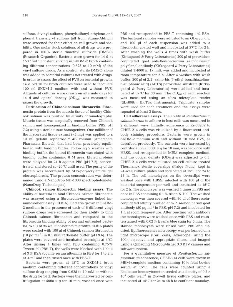
ORF ID Primer pairs used in PCR and RT-PCR Function/Annotation CWS- motif(Accession no.)
RRSA00417 5’-CCTTGTCGGCCTTGTCTTAG-3’ Possible proteinase(EF426712) 5’-GACGACGTCGAACGGTCAG-3’ (Rhodococcus sp. RHA1) LAnTG
RRSA00636 5’-AAGCGCCAGTCAAGAACG-3’ Putative glycosyl hydrolases family 31(EF426714) 5’-ACGACGAAGCAGCAGTAAGC-3’ (Arthrobacter aurescens TC1) LAaTG
RRSA01045 5’-GCCAAGATGGCAGCGTTT-3’ Hypothetical membrane protein(EF426715) 5’-CGAGCGACCACCGTCATAC-3’ (Bifidobacterium longum NCC2705) LAaTG
RRSA01248 5’-GATATGAAATCCCTCTTTTACCG-3’ Putative partial cell-surface adhesin, Cna B-type(EF426716) 5’-CCGAGCGAAGAAAACTGATG-3’ (Chloroflexus aggregans DSM 9485) LAnTG
RRSA01787 5’-ACCAATTCGTGCAGAAAACC-3’ Acid phosphatase (EC 3.1.3.2) LAaTG(EF426717) 5’-TAGTTGAAGCGTTTGCGTTG-3’ (Corynebacterium jeikeium K411)
RRSA02499 5’-TTGGTGGACATCGCGAAC-3’ Hypothetical protein AAur_1465 LAeTG(EF426718) 5’-GAGTGACGGTGCCTTCGTA-3’ (Arthrobacter aurescens TC1)
RRSA00949 5’-CGGGGACGACCAAAACTT-3’ Hypothetical protein Arth_2238 LPvAG(EF426719) 5’-TAAATTTACCGGGTAGCGATAAG-3’ (Arthrobacter sp. FB24)
RRSA02971 5’-GAAAATCGCTGGAACTGTTG-3’ Putative integral membrane protein LAiTG(EF426720) 5’-AGAGCCGCCCTGGCTTAC-3’ (Arthrobacter aurescens TC1)
Table 1. Renibacterium salmoninarum-33209: probable SrtD substrates
Dis Aquat Org 78: 115–127, 2007
sulfone, divinyl sulfone, phenylsulfonyl ethylene andphenyl trans-styryl sulfone (all from Sigma-Aldrich)were screened for their effect on cell growth and via-bility. One molar stock solutions of all drugs were pre-pared in 100% sterile dimethyl sulfoxide (DMSO)(Research Organics). Bacteria were grown for 14 d at15°C with constant stirring in SKDM-2 broth contain-ing different concentrations (0.625 to 10 mM) of thevinyl sulfone drugs. As a control, sterile DMSO alonewas added to bacterial cultures not treated with drugs.In order to assess the effect of PVS on bacterial growth,14 d old 10 ml broth cultures were used to inoculate100 ml SKDM-2 medium with and without PVS.Aliquots of cultures were drawn on alternate days for14 d and optical density (OD525) was measured toassess the growth.
Purification of Chinook salmon fibronectin. Fibro-nectin protein from the muscle tissue of healthy Chin-ook salmon was purified by affinity chromatography.Muscle tissue was aseptically removed from Chinooksalmon and homogenized in binding buffer (PBS, pH7.2) using a sterile tissue homogenizer. One milliliter ofthe macerated tissue extract (~5 mg) was applied to a10 ml gelatin sepharose™ 4B column (AmershamPharmacia Biotech) that had been previously equili-brated with binding buffer. Following 2 washes withbinding buffer, the bound fibronectin was eluted withbinding buffer containing 8 M urea. Eluted proteinswere dialyzed for 24 h against PBS (pH 7.2), concen-trated, and stored at –20°C until used. The purity of theprotein was ascertained by SDS-polyacrylamide gelelectrophoresis. The protein concentration was deter-mined using a NanoDrop ND-1000 spectrophotometer(NanoDrop Technologies).
Chinook salmon fibronectin binding assays. Theability of bacteria to bind Chinook salmon fibronectinwas assayed using a fibronectin-enzyme linked im-munosorbent assay (ELISA). Bacteria grown in SKDM-2 medium in the presence of each of 6 different vinylsulfone drugs were screened for their ability to bindChinook salmon fibronectin and compared to thefibronectin binding ability of normal untreated bacte-ria. Wells of 96 well flat-bottom microtitre ELISA plateswere coated with 100 µl of Chinook salmon fibronectin(10 µg ml–1) in 0.1 mM carbonate buffer (pH 9.6). Theplates were covered and incubated overnight at 4°C.After rinsing 4 times with PBS containing 0.75%Tween-20 (PBS-T), the wells were blocked with 100 µlof 3% BSA (bovine serum albumin) in PBS for 1 to 2 hat 37°C and then rinsed once with PBS-T.
Bacteria were grown at 15°C in SKDM-2 brothmedium containing different concentrations of vinylsulfone drug ranging from 0.625 to 10 mM or withoutthe drug for 14 d. Bacteria were then harvested by cen-trifugation at 5000 × g for 10 min, washed once with
PBS and resuspended in PBS-T containing 1% BSA.The bacterial samples were adjusted to an OD525 of 0.5,and 100 µl of each suspension was added to afibronectin-coated well and incubated at 37°C for 2 h.After washing the wells 4 times with wash buffer(Kirkegaard & Perry Laboratories) 200 µl of peroxidaseconjugated goat anti-Renibacterium salmoninarumpolyclonal antibody (Kirkegaard & Perry Laboratories)diluted 1:4000 in 1× milk was added and incubated atroom temperature for 2 h. After 4 washes with washbuffer, 200 µl of 2,2’-azino-bis (3-ethyl-benzthiazoline-6-sulphonic acid) (ABTS) peroxidase substrate (Kirke-gaard & Perry Laboratories) were added and incu-bated at 37°C for 30 min. The OD405 of each reactionwas measured using an ultra microplate reader(ELX808IU, BioTek Instruments). Triplicate sampleswere used for each treatment and the assays wererepeated at least 3 times.
Cell adherence assays. The ability of Renibacteriumsalmoninarum to adhere to host cells was measured in2 different ways. Initially, adherence of Rs-33209 toCHSE-214 cells was visualized by a fluorescent anti-body staining procedure. Bacteria were grown inSKDM-2 medium with and without 10 mM PVS, asdescribed previously. The bacteria were harvested bycentrifugation at 5000 × g for 10 min, washed once withHBSS, and resuspended in MEM complete medium,and the optical density (OD525) was adjusted to 0.5.CHSE-214 cells were cultured on cell culture-treatedThermanox sterile coverslips (Nalge Nunc) kept in24-well culture plates and incubated at 15°C for 24 to48 h. The cell monolayers on the coverslips werewashed once with HBSS, infected with 100 µl of thebacterial suspension per well and incubated at 15°Cfor 2 h. The monolayer was washed 4 times in PBS andonce in PBS containing 0.1% triton X-100. The washedmonolayer was then covered with 30 µl of fluorescein-conjugated affinity purified anti-R. salmoninarum goatantibody (50 µg ml–1 in PBS, pH 7.2) and incubated for1 h at room temperature. After reacting with antibodythe monolayers were washed once with PBS and coun-terstained with 0.02% Evan’s blue stain for 5 min. Thestained monolayers were rinsed with PBS and air-dried. Epifluorescence microscopy was performed on alight microscope (Carl Zeiss, Axioscope) using the100× objective and appropriate filters, and imagedusing a Qimaging Micropublisher 3.3 RTV camera andsoftware system.
For a quantitative measure of Renibacterium sal-moninarum adherence, CHSE-214 cells were grown inMEM-complete medium containing 10% fetal bovineserum at 15°C. The cells were counted using aNeubauer hemocytometer, seeded at a density of 0.5 ×10⁵ cells well–1 in 24-well tissue culture plates, andincubated at 15°C for 24 to 48 h to confluent monolay-
118
Sudheesh et al.: Inhibition of Renibacterium salmoninarum adherence
ers. Bacteria were grown in SKDM-2 tubes for 14 d at15°C, washed once with PBS and resuspended inMEM-complete medium. For the cell adherence assay,CHSE-214 cells were infected with bacteria at a multi-plicity of infection (m.o.i.) of 10 bacteria per fish cell for2 h at 15°C. The monolayer was then washed 5 timeswith Hank’s balanced salt solution (HBSS). Thewashed cells were disrupted by the addition of 0.5 mlsterile deionized ice-cold water and repeated pipet-ting. Serial dilutions of the lysate were plated ontoSKDM-2 agar for counts of viable bacteria. The percentadherence was calculated as follows:
(CFU on plate/CFU in original inoculum) × 100
where CFU = colony forming units. Assays were per-formed in duplicate and repeated at least 3 times.
Intracellular growth determination. For intracellu-lar growth assay, CHSE-214 cells were infected withbacteria at an m.o.i. of 10 bacteria per fish cell andincubated for 8 h at 15°C. Monolayers were washed 5times with HBSS, followed by the addition of MEMcomplete medium containing 10 µg ml–1 gentamycinand incubation at 15°C for an additional 30 min. Mono-layers were then washed 3 times with HBSS, freshMEM complete medium without gentamycin wasadded and incubated at 15°C. After incubating theplates for different time points, the monolayer waswashed again with HBSS and lysed with sterile deion-ized ice-cold water and repeated pipetting. The cellmonolayers were observed microscopically to assurecomplete lysis. Serial dilutions of the lysate wereplated onto SKDM-2 agar for counts of viable bacteria.The time immediately following the removal of thegentamycin was taken as the zero time point. Assayswere performed in triplicate and repeated at least 3times.
For Giemsa staining, the monolayers were grown oncell culture-treated Thermanox sterile coverslips(Nalge Nunc) kept in 24-well culture plates, and infec-tion was performed as described in the intracellulargrowth assay. The coverslips were removed from thewells at different time points after the gentamycintreatment and incubation, and stained with 0.01%Giemsa stain for 45 min. The stained monolayers onthe coverslips were observed under a light microscope(Carl Zeiss, Axioscope) using a 100× objective, andrepresentative cells from at least 3 repeated experi-ments were photographed using a Qimaging Micro-publisher 3.3 RTV camera and software system.
Cytopathic effect (CPE) of Renibacterium salmoni-narum. The CPE assay was performed by preparingCHSE-214 cell monolayers and bacteria as describedabove. CHSE-214 cells were infected at an m.o.i. of 10bacteria per fish cell and observed for CPE for up to2 wk. Typical CPE of R. salmoninarum on CHSE-214
cells included morphological changes such as forma-tion of rounded cells and cell detachment as the infec-tion progressed. Representative monolayers from atleast 3 repeated experiments were photographedusing a Nikon digital camera mounted on an invertedmicroscope (Carl Zeiss, Axiovert 135). Toxicity of PVSto CHSE-214 cells was assayed by growing the cellmonolayers in 24-well tissue culture plates for 24 to48 h and then exposing them to different doses of PVSranging from 0.001 mM to 100 mM. The monolayerswere incubated at 15°C and observed daily for visibleCPE and morphological changes for 14 d.
Isolation of cell wall-bound proteins. Bacteria weregrown at 15°C for 14 d in SKDM-2 broth medium withor without 10 mM PVS. Bacteria were harvested bycentrifugation at 5000 × g for 10 min. Cell wall-boundproteins of Renibacterium salmoninarum were pre-pared using modified methodology of Stalhammar-Carlemalm et al. (1993). Briefly, the bacteria werewashed twice with Tris-HCl buffer (50 mM, pH 7.3)and resuspended in osmotic digestion buffer (20%sucrose-2.5 µM phenylmethylsulfonyl fluoride in50 mM Tris-HCl, pH 7.3). Cell wall-bound proteinswere enzymatically released from the bacterial cellsby adding mutanolysin (Sigma-Aldrich) in potassiumphosphate buffer (10 mM, pH 6.2) to a final concentra-tion of 350 U ml–1. The digestion reaction was incu-bated for 18 h at 37°C with gentle shaking followed by2 centrifugations at 20 000 × g for 15 min (4°C) in atabletop centrifuge (Eppendorf, 5417R) in order toremove cell debris and remaining protoplasts. Thesupernatant containing the proteins released from thecell wall was frozen at –20°C for subsequent 2-dimen-sional electrophoresis (2-DE). The protein concentra-tion was determined using a NanoDrop ND-1000 spec-trophotometer (NanoDrop Technologies).
Two dimensional polyacrylamide gel electrophore-sis analysis. The cell wall-bound protein samples wereextracted in a sample extraction buffer containing5 M urea, 2 M thiourea, 2 mM tributyl phosphene(TBP), 2% CHAPS, 2% sulfobetaine 3-10, 0.5% biolyteampholyte, 10 mM Tris and 0.001% Orange G dye.Extracted protein samples were applied to 7 cm IPGstrips (pH 4–7) (Bio-Rad) (25 µg total protein strip–1)and rehydrated overnight in a humidified chamber atroom temperature. First dimension isoelectric focusingwas performed on IPG strips using a Mini Protean IEFcell (Bio-Rad). The strips were focused initially for15 min at 250 V, 2 h at 4000 V and then for 20 000 volthours at 4000 V. Focused strips were equilibrated in 5×Tris-HCl glycine gel buffer containing 6 M urea, 2%SDS, 20% glycerol, 5mM TBP and 2.5% acrylamidemonomer. Second dimension separation was carriedout using precast 10–20% polyacrylamide gradientgels with a 2-D miniprep well (Bio-Rad) on a Mini-
119

Dis Aquat Org 78: 115–127, 2007
PROTEAN 3 electrophoresis cell (Bio-Rad). Electro-phoresis was performed using the standard Laemmlibuffer system (Laemmli 1970) at a constant current of5 mA for 30 min and then at a constant current of12 mA for 1.5 h. Prestained BlueRanger® molecularweight marker mix (Pierce) was used to compare themolecular weight of protein spots. Multiple gels wereprepared for the same protein samples from both treat-ments to ensure consistency of protein profiles on 2-Dgels, and isoelectric focusing was carried out simulta-neously to minimize experimental variability. The gelswere silver stained and scanned.
Nucleotide sequence accession numbers. The DNAsequences of the Renibacterium salmoninarum sor-tase and sortase substrate ORFs RRSA01227 (Acc.No. EF426 713), RRSA00417 (Acc. No. EF426712),RRSA00636 (Acc. No. EF426714), RRSA01045 (Acc.No. EF426715), RRSA01248 (Acc. No. EF426716),RRSA01787 (Acc. No. EF426717), RRSA 02499 (Acc.No. EF426718), RRSA00949 (Acc. No. EF426719) andRRSA02971 (Acc. No. EF426720) have been depositedin the GenBank database.
RESULTS
Characterization of Renibacterium salmoninarumsortase and sortase substrates
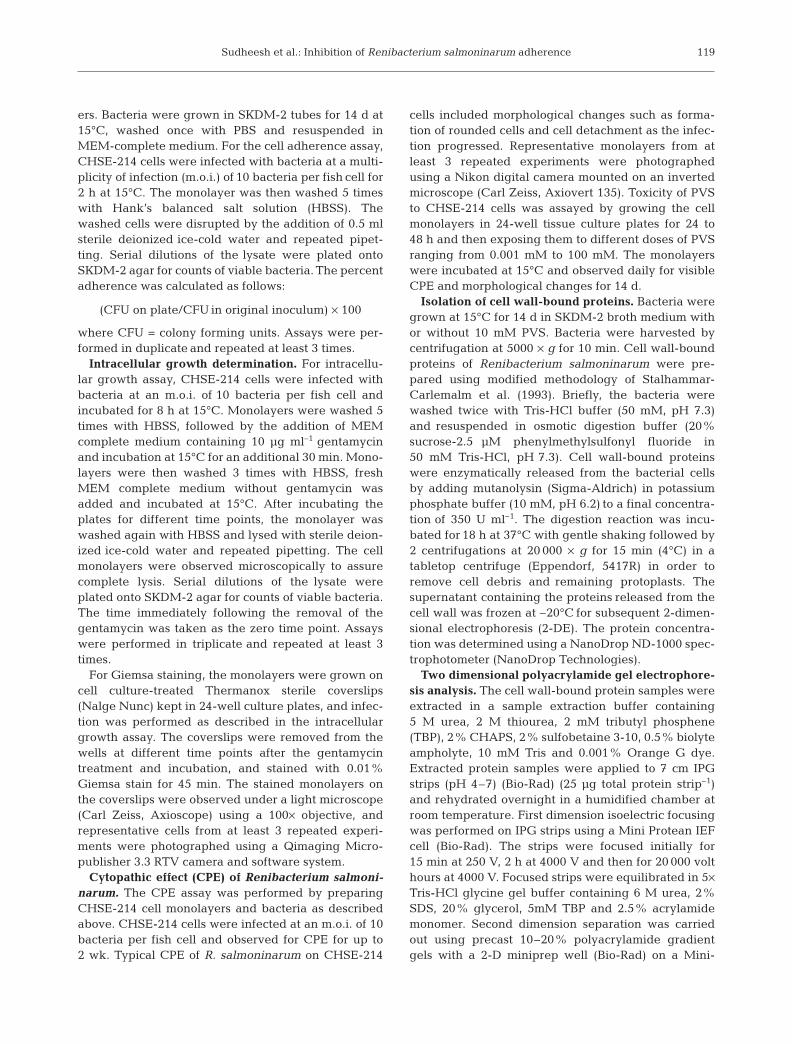
The Rs-33209 genome sequence allowed identificationof an ORF (RRSA01227, Genbank Acc. No. EF426713)specifying a sortase enzyme. The deduced proteinsequence has a calculated molecular weight of30682.58 Da and a pI of 7.87. As calculated fromClustalW pairwise alignments, the amino acid sequenceof the sortase had significant sequence identity to sortaseor sortase-like proteins of Arthrobacter sp. FB24 (61%),Brevibacterium linens (39%), Kineococcus radiotoler-ans (36%), Acidothermus cellulolyticus (32%), Bifido-bacterium longum (32%) and Frankia alni (32%) (Fig. 1).All putative genes in Rs-33209 (3515 total open readingframes) were compared to the 6 sortase subfamilymodels of Comfort & Clubb (2004). The only significantresult was ORF RRSA01227 matching to subfamily 5(score of 220.2 and e-value of 3.1e-66). The amino acidsequence of this sortase protein clustered in a neighbor-joining phylogenetic tree with other Class D sortaseenzymes (result not shown). The sortase subfamily 5described in Comfort & Clubb (2004) is equivalent to theClass D sortase group of Dramsi et al. (2005). Therefore,the Renibacterium salmoninarum sortase gene is de-signated srtD based on the latest classification of sortaseenzymes (Dramsi et al. 2005).
All amino acid sequences were scanned for the tri-partite pattern and cleavage motif of the subfamily 5
sorting signal resulting in 698 matches to the cleavagemotif (LAxTG) and 19 matches to the tripartite pattern(results not shown). According to the criteria used, 8of the putative sortase substrates were true positiveswith both features (Table 1). Searching the Rs-33209genome with an HMM built from these putative sor-tase substrates or by using the MEME/MAST softwareas outlined by Boekhorst et al. (2005) failed to yield anyadditional novel matches (results not shown).
Specific primers were designed to PCR-amplifythese gene sequences from the bacteria. All the geneswere transcribed in the bacteria as observed by RT-PCR (data not shown).
Use of vinyl sulfone sortase inhibitors to inhibit theability of bacteria to bind Chinook salmon fibronectin
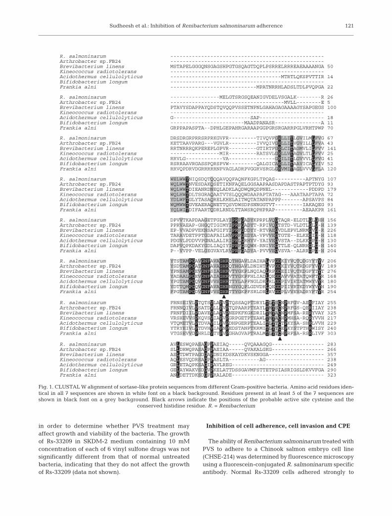
Many of the sortase substrates studied to dateare MSCRAMs. This group of bacterial proteins oftenincludes proteins involved in adherence to eukary-otic matrix proteins such as fibronectin. To inves-tigate the ability of Renibacterium salmoninarumto bind fibronectin (a common ligand of bacterialMSCRAMs), we optimized and used a salmon fibro-nectin ELISA. Fibronectin protein was purified fromChinook salmon muscle tissue by gelatin sepharoseaffinity chromatography; the purity of the proteinwas determined by observing a single high molecu-lar weight protein band on SDS-PAGE. Initial opti-mization of the fibronectin ELISA was performedusing PVS (Frankel et al. 2004). By titrating differentconcentrations of PVS and fibronectin, it was foundthat maximum inhibition of fibronectin binding wasachieved at doses of 10 mM PVS and 10 µg ml–1
fibronectin (Fig. 2A). Fibronectin binding ability wassignificantly reduced (75.8%) in bacteria treated withPVS (Fig. 2B) in comparison with controls. Thefibronectin ELISA was used to screen 5 other vinylsulfone drugs at 10 mM doses for their ability toinhibit fibronectin binding and, thus, to indirectlyassess their ability to inhibit sortase activity. Thereduction in fibronectin binding abilities of methylvinyl sulfone (17.53%), ethyl vinyl sulfone (12.6%),divinyl sulfone (8.14%), phenylsulfonyl ethylene(3.47%) and phenyl trans-styryl sulfone (1.32%) weresignificantly lower than that of PVS. Therefore PVSwas selected for all other experiments.
Growth of bacteria in SKDM-2 medium containingvinyl sulfone drugs
Growth of Rs-33209 in SKDM-2 broth medium wasscreened by measuring the OD525 of the broth culture
120
Sudheesh et al.: Inhibition of Renibacterium salmoninarum adherence
in order to determine whether PVS treatment mayaffect growth and viability of the bacteria. The growthof Rs-33209 in SKDM-2 medium containing 10 mMconcentration of each of 6 vinyl sulfone drugs was notsignificantly different from that of normal untreatedbacteria, indicating that they do not affect the growthof Rs-33209 (data not shown).
Inhibition of cell adherence, cell invasion and CPE
The ability of Renibacterium salmoninarum treated withPVS to adhere to a Chinook salmon embryo cell line(CHSE-214) was determined by fluorescence microscopyusing a fluorescein-conjugated R. salmoninarum specificantibody. Normal Rs-33209 cells adhered strongly to
121
R. salmoninarum -------------------------------------------------- Arthrobacter sp.FB24 -------------------------------------------------- Brevibacterium linens MSTAPELGGGQNSGAGSHPGTGSQAGTDQPLPSRRELRRREAEAAAANGA 50 Kineococcus radiotolerans -------------------------------------------------- Acidothermus cellulolyticus ------------------------------------MTRTLQKSPVTTIR 14 Bifidobacterium longum -------------------------------------------------- Frankia alni ----------------------------MPATNRRHLADSLTDLPVQPGA 22 R. salmoninarum ----------------MELGTSRGSQEANIGVDELVSGALK--------R 26 Arthrobacter sp.FB24 -------------------------------------MVLL--------E 5 Brevibacterium linens PTAVYSDAPPAYQDSTQVQQPVSSETNPNLGAHAGAGAAAAGYSAPGEGS 100 Kineococcus radiotolerans -------------------------------------------------- Acidothermus cellulolyticus G-------------------------SAP--------------------- 18 Bifidobacterium longum ------------------------MAADPANASR---------------A 11 Frankia alni GRPPAPASPTA--DPHLGEPAHRGARAAPGGPGRSRGARRPGLVRHTPWP 70 R. salmoninarum DRSDRGRPRRSRPRKGVFR---------TIVQVFGELLITLGVILMLFVG 67 Arthrobacter sp.FB24 KETTAAVPARG---VGVLR---------IVVQIVGELLITVGVILLLFVA 43 Brevibacterium linens RRTNRRRQKPEREPLGPVR---------GTIRTFGELCITAGMVLILFVV 141 Kineococcus radiotolerans ----------------MIR---------RATSVLGELLVTAGVLTLLFVV 25 Acidothermus cellulolyticus RRVLG------------VA---------------GEILITLGVVVLLFVG 41 Bifidobacterium longum RSRRAAVRGASSPQRSPVW---------QALGICAELLITAAVICALYIV 52 Frankia alni RRVQPDRVDGRRRRRNPVAGLADRPVGGRVSRGLGEVMITAGLVVVLFLA 120 R. salmoninarum WELWWTNIQSDQTQQQAVQQFAQNFKGPLTPQAS----------APTNYG 107 Arthrobacter sp.FB24 WQLWWTNVESDAKQSETIKNFAQELGGSAAPAASDAPDASTPAPTPTDYG 93 Brevibacterium linens WQLWWTDIEANRDNEQLADKLAQDWQNQDPNEL------------PDDPD 179 Kineococcus radiotolerans WQLHWTDLTSGRAQAATVTSLQQQWDAAPAPTATAG---AAATAAPTAPA 72 Acidothermus cellulolyticus YDLWFTGLYTASAQRELKHELAITWQTATANPAPPP-------APSAVPS 84 Bifidobacterium longum WQMWWTGVEAERAQNETTQSVDWSDPSNNGGTVT---------IAKAQEG 93 Frankia alni YQLWITDIFAARTQDRLRNDLTTAWSRQPHPRAP---------AEAAKPR 161 R. salmoninarum DPVVTKAPDAAGETFGLAYIPRFGADYKPRPLVQGTAQR-ELDTLGLGHH 156 Arthrobacter sp.FB24 PPRVAEAP-GHGQTIGIMYIPRFGADYT-RPIVQGTSTD-VLDTLGLG-H 139 Brevibacterium linens EP-VVADPVEKNSAFGIFYIPRFGDDYY-RTVAEGVDLEPVLNRMGVG-R 226 Kineococcus radiotolerans TARAVDETPPTGDAFAILHVPRFGEDYA-VPVVEGTGTE--ELKEGIG-H 118 Acidothermus cellulolyticus PDGVLPDDVVPGNALALIRIPRLGRHYV-YAIVEGVSTA--DLKKGPG-H 130 Bifidobacterium longum DAPVQPKDAKYGDLIAQIYIPRFGSQWH-RNIVEGTTLE-QLNRHGLG-H 140 Frankia alni P--VVPP-VELGEGVAVLRVPRFGADYA-PVVVEGVSVA--ALRRGPG-H 204 R. salmoninarum YTSTAMPGAVGNFAVAGHRQTHGAVLDAIHALVPGDKIYVQTQDGYYTYV 206 Arthrobacter sp.FB24 YSDTAMPGATGNFAVAGHRQTHGAVLDNIHTLVPGDKIYVQTRDGFYVYV 189 Brevibacterium linens YPNSAMPGEVGNFSIAGHRVTYGKPLNQIAQLRPGDEIIVQTKDGFYTYT 276 Kineococcus radiotolerans YADAALPGEVGNFAIAGHRVTYGKPFHLIADLQEGDAVVVATATQWFTYR 168 Acidothermus cellulolyticus YPGTAMPGQVGNFVVSGHRTTYLAPFNGLDKLRLGDPIVIETATMWYVYR 180 Bifidobacterium longum YDTTQMPGQVGNFAVAGHRNGYGQPLGDVDKLQEGDPIIVRTKDYWYVYH 190 Frankia alni FPGTAMPGDVGNFVVSGHRTTYGKPFSRLDELRVGDPLVVEVADRYFTYR 254 R. salmoninarum FRNSEIVLPTQTSVLAPVPTQSSAQPTDRYLTLTSCNPRFGV-AERFIAY 255 Arthrobacter sp.FB24 FRNNQIVLPSATDVLLPVPTQPAARPTEAYLTMTSCNPRFGS-QERIIAY 238 Brevibacterium linens FRNFDIILPDAVEVLAPVPNEPKFKGKDRILTMTACNPMFSA-RERYVAY 325 Kineococcus radiotolerans VRSHEVVSPKQVSVIAPVPGRPGETPTEAWLTMTACHPMHSA-RQRYVVH 217 Acidothermus cellulolyticus VTQMETVLPTDVAVILPVPDHPGERPTEALITLTTCTPKYSA-SHRLVVH 229 Bifidobacterium longum YTRYEIVLPTDVHVIAPNPEDSTANPTKRMITLTTCEPKYSTPTHRWISY 240 Frankia alni VTGSEVVDPHRLDVTYPVPGHAGVAPTRALMTLTTCHPRFSA-RSRLIVF 303 m
m
R. salmoninarum AVLESWQPASAGPPAEIAQ-----QVQAAAGQG----------------- 283 Arthrobacter sp.FB24 SLLDHWQPASAGPPAEIAA-----QVAKALGKG----------------- 266 Brevibacterium linens AELTDWTPAGDGAPDNIKDSKAYDKVSKNGGA------------------ 357 Kineococcus radiotolerans AQLESVQDRSAGPPASLTA----------AG------------------- 238 Acidothermus cellulolyticus GRLETAQPKSAGIPAVLREG------------------------------ 249 Bifidobacterium longum GELAYWAKVSDGVPKELATTDSSGAVMFSTTETPSIASRIGSLDKVVFGA 290 Frankia alni ANLDETTDKSDGPPRALADE------------------------------ 323
Fig. 1. CLUSTAL W alignment of sortase-like protein sequences from different Gram-positive bacteria. Amino acid residues iden-tical in all 7 sequences are shown in white font on a black background. Residues present in at least 5 of the 7 sequences areshown in black font on a grey background. Black arrows indicate the positions of the probable active site cysteine and the
conserved histidine residue. R. = Renibacterium
Dis Aquat Org 78: 115–127, 2007
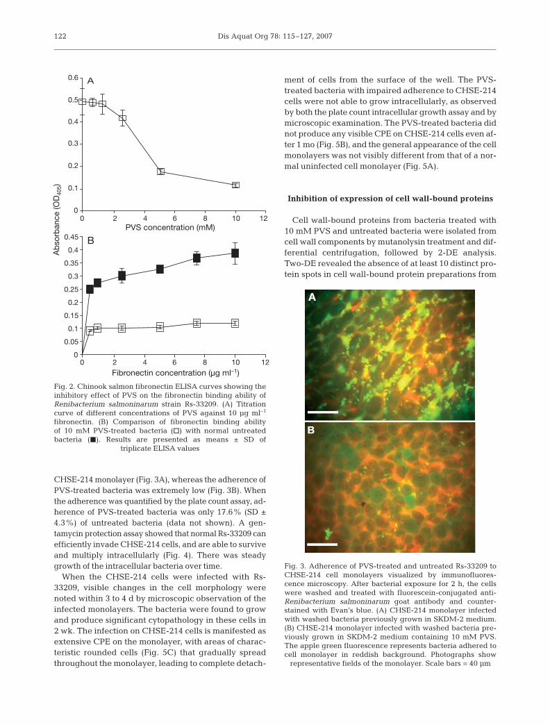
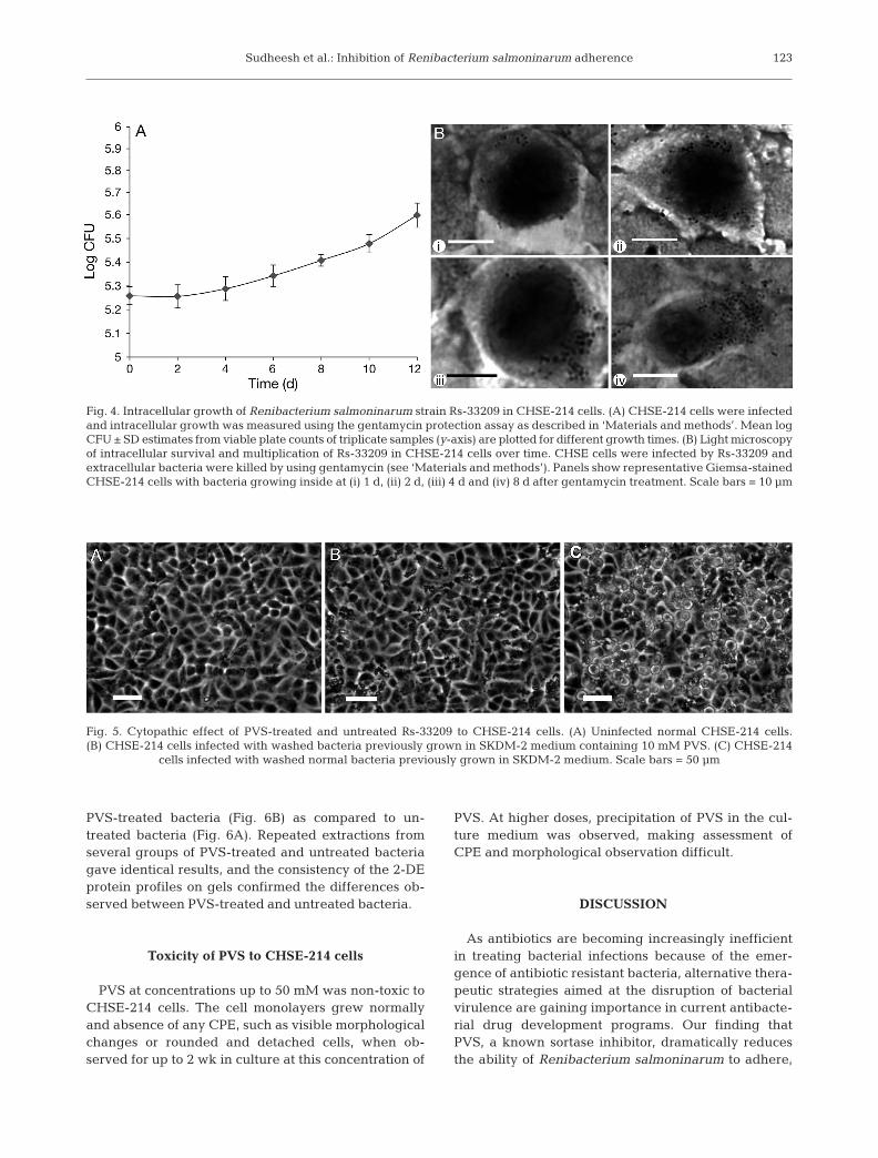
CHSE-214 monolayer (Fig. 3A), whereas the adherence ofPVS-treated bacteria was extremely low (Fig. 3B). Whenthe adherence was quantified by the plate count assay, ad-herence of PVS-treated bacteria was only 17.6% (SD ±4.3%) of untreated bacteria (data not shown). A gen-tamycin protection assay showed that normal Rs-33209 canefficiently invade CHSE-214 cells, and are able to surviveand multiply intracellularly (Fig. 4). There was steadygrowth of the intracellular bacteria over time.
When the CHSE-214 cells were infected with Rs-33209, visible changes in the cell morphology werenoted within 3 to 4 d by microscopic observation of theinfected monolayers. The bacteria were found to growand produce significant cytopathology in these cells in2 wk. The infection on CHSE-214 cells is manifested asextensive CPE on the monolayer, with areas of charac-teristic rounded cells (Fig. 5C) that gradually spreadthroughout the monolayer, leading to complete detach-
ment of cells from the surface of the well. The PVS-treated bacteria with impaired adherence to CHSE-214cells were not able to grow intracellularly, as observedby both the plate count intracellular growth assay and bymicroscopic examination. The PVS-treated bacteria didnot produce any visible CPE on CHSE-214 cells even af-ter 1 mo (Fig. 5B), and the general appearance of the cellmonolayers was not visibly different from that of a nor-mal uninfected cell monolayer (Fig. 5A).
Inhibition of expression of cell wall-bound proteins
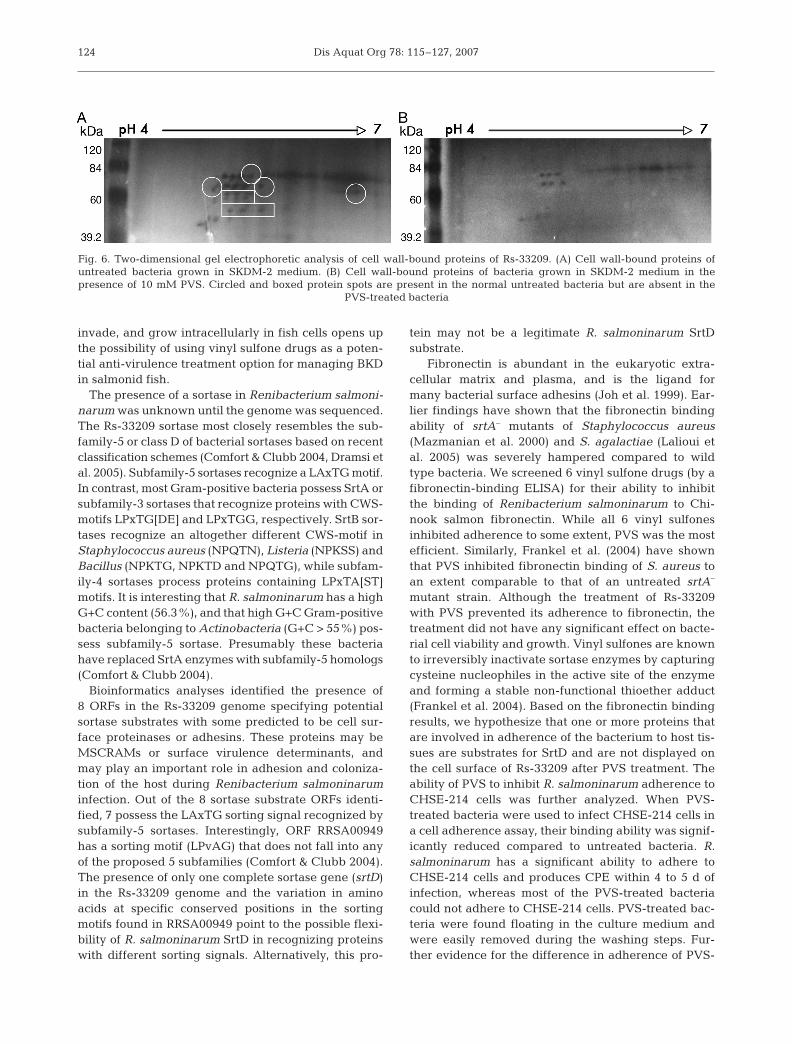
Cell wall-bound proteins from bacteria treated with10 mM PVS and untreated bacteria were isolated fromcell wall components by mutanolysin treatment and dif-ferential centrifugation, followed by 2-DE analysis.Two-DE revealed the absence of at least 10 distinct pro-tein spots in cell wall-bound protein preparations from
122
0
0.1
0.2
0.3
0.4
0.5
0.6
0 2 4 6 8 10 12
0 2 4 6 8 10 12
PVS concentration (mM)
Abs
orba
nce
(OD
405)
0
0.05
0.1
0.15
0.2
0.25
0.3
0.35
0.4
0.45
Fibronectin concentration (µg ml–1)
A
B
Fig. 2. Chinook salmon fibronectin ELISA curves showing theinhibitory effect of PVS on the fibronectin binding ability ofRenibacterium salmoninarum strain Rs-33209. (A) Titrationcurve of different concentrations of PVS against 10 µg ml–1
fibronectin. (B) Comparison of fibronectin binding abilityof 10 mM PVS-treated bacteria (h) with normal untreatedbacteria (j). Results are presented as means ± SD of
triplicate ELISA values
Fig. 3. Adherence of PVS-treated and untreated Rs-33209 toCHSE-214 cell monolayers visualized by immunofluores-cence microscopy. After bacterial exposure for 2 h, the cellswere washed and treated with fluorescein-conjugated anti-Renibacterium salmoninarum goat antibody and counter-stained with Evan’s blue. (A) CHSE-214 monolayer infectedwith washed bacteria previously grown in SKDM-2 medium.(B) CHSE-214 monolayer infected with washed bacteria pre-viously grown in SKDM-2 medium containing 10 mM PVS.The apple green fluorescence represents bacteria adhered tocell monolayer in reddish background. Photographs show
representative fields of the monolayer. Scale bars = 40 µm
Sudheesh et al.: Inhibition of Renibacterium salmoninarum adherence
PVS-treated bacteria (Fig. 6B) as compared to un-treated bacteria (Fig. 6A). Repeated extractions fromseveral groups of PVS-treated and untreated bacteriagave identical results, and the consistency of the 2-DEprotein profiles on gels confirmed the differences ob-served between PVS-treated and untreated bacteria.
Toxicity of PVS to CHSE-214 cells
PVS at concentrations up to 50 mM was non-toxic toCHSE-214 cells. The cell monolayers grew normallyand absence of any CPE, such as visible morphologicalchanges or rounded and detached cells, when ob-served for up to 2 wk in culture at this concentration of
PVS. At higher doses, precipitation of PVS in the cul-ture medium was observed, making assessment ofCPE and morphological observation difficult.
DISCUSSION
As antibiotics are becoming increasingly inefficientin treating bacterial infections because of the emer-gence of antibiotic resistant bacteria, alternative thera-peutic strategies aimed at the disruption of bacterialvirulence are gaining importance in current antibacte-rial drug development programs. Our finding thatPVS, a known sortase inhibitor, dramatically reducesthe ability of Renibacterium salmoninarum to adhere,
123
Fig. 4. Intracellular growth of Renibacterium salmoninarum strain Rs-33209 in CHSE-214 cells. (A) CHSE-214 cells were infectedand intracellular growth was measured using the gentamycin protection assay as described in ‘Materials and methods’. Mean logCFU ± SD estimates from viable plate counts of triplicate samples (y-axis) are plotted for different growth times. (B) Light microscopyof intracellular survival and multiplication of Rs-33209 in CHSE-214 cells over time. CHSE cells were infected by Rs-33209 andextracellular bacteria were killed by using gentamycin (see ‘Materials and methods’). Panels show representative Giemsa-stainedCHSE-214 cells with bacteria growing inside at (i) 1 d, (ii) 2 d, (iii) 4 d and (iv) 8 d after gentamycin treatment. Scale bars = 10 µm
Fig. 5. Cytopathic effect of PVS-treated and untreated Rs-33209 to CHSE-214 cells. (A) Uninfected normal CHSE-214 cells.(B) CHSE-214 cells infected with washed bacteria previously grown in SKDM-2 medium containing 10 mM PVS. (C) CHSE-214
cells infected with washed normal bacteria previously grown in SKDM-2 medium. Scale bars = 50 µm
Dis Aquat Org 78: 115–127, 2007
invade, and grow intracellularly in fish cells opens upthe possibility of using vinyl sulfone drugs as a poten-tial anti-virulence treatment option for managing BKDin salmonid fish.
The presence of a sortase in Renibacterium salmoni-narum was unknown until the genome was sequenced.The Rs-33209 sortase most closely resembles the sub-family-5 or class D of bacterial sortases based on recentclassification schemes (Comfort & Clubb 2004, Dramsi etal. 2005). Subfamily-5 sortases recognize a LAxTG motif.In contrast, most Gram-positive bacteria possess SrtA orsubfamily-3 sortases that recognize proteins with CWS-motifs LPxTG[DE] and LPxTGG, respectively. SrtB sor-tases recognize an altogether different CWS-motif inStaphylococcus aureus (NPQTN), Listeria (NPKSS) andBacillus (NPKTG, NPKTD and NPQTG), while subfam-ily-4 sortases process proteins containing LPxTA[ST]motifs. It is interesting that R. salmoninarum has a highG+C content (56.3%), and that high G+C Gram-positivebacteria belonging to Actinobacteria (G+C > 55%) pos-sess subfamily-5 sortase. Presumably these bacteriahave replaced SrtA enzymes with subfamily-5 homologs(Comfort & Clubb 2004).
Bioinformatics analyses identified the presence of8 ORFs in the Rs-33209 genome specifying potentialsortase substrates with some predicted to be cell sur-face proteinases or adhesins. These proteins may beMSCRAMs or surface virulence determinants, andmay play an important role in adhesion and coloniza-tion of the host during Renibacterium salmoninaruminfection. Out of the 8 sortase substrate ORFs identi-fied, 7 possess the LAxTG sorting signal recognized bysubfamily-5 sortases. Interestingly, ORF RRSA00949has a sorting motif (LPvAG) that does not fall into anyof the proposed 5 subfamilies (Comfort & Clubb 2004).The presence of only one complete sortase gene (srtD)in the Rs-33209 genome and the variation in aminoacids at specific conserved positions in the sortingmotifs found in RRSA00949 point to the possible flexi-bility of R. salmoninarum SrtD in recognizing proteinswith different sorting signals. Alternatively, this pro-
tein may not be a legitimate R. salmoninarum SrtDsubstrate.
Fibronectin is abundant in the eukaryotic extra-cellular matrix and plasma, and is the ligand formany bacterial surface adhesins (Joh et al. 1999). Ear-lier findings have shown that the fibronectin bindingability of srtA− mutants of Staphylococcus aureus(Mazmanian et al. 2000) and S. agalactiae (Lalioui etal. 2005) was severely hampered compared to wildtype bacteria. We screened 6 vinyl sulfone drugs (by afibronectin-binding ELISA) for their ability to inhibitthe binding of Renibacterium salmoninarum to Chi-nook salmon fibronectin. While all 6 vinyl sulfonesinhibited adherence to some extent, PVS was the mostefficient. Similarly, Frankel et al. (2004) have shownthat PVS inhibited fibronectin binding of S. aureus toan extent comparable to that of an untreated srtA−
mutant strain. Although the treatment of Rs-33209with PVS prevented its adherence to fibronectin, thetreatment did not have any significant effect on bacte-rial cell viability and growth. Vinyl sulfones are knownto irreversibly inactivate sortase enzymes by capturingcysteine nucleophiles in the active site of the enzymeand forming a stable non-functional thioether adduct(Frankel et al. 2004). Based on the fibronectin bindingresults, we hypothesize that one or more proteins thatare involved in adherence of the bacterium to host tis-sues are substrates for SrtD and are not displayed onthe cell surface of Rs-33209 after PVS treatment. Theability of PVS to inhibit R. salmoninarum adherence toCHSE-214 cells was further analyzed. When PVS-treated bacteria were used to infect CHSE-214 cells ina cell adherence assay, their binding ability was signif-icantly reduced compared to untreated bacteria. R.salmoninarum has a significant ability to adhere toCHSE-214 cells and produces CPE within 4 to 5 d ofinfection, whereas most of the PVS-treated bacteriacould not adhere to CHSE-214 cells. PVS-treated bac-teria were found floating in the culture medium andwere easily removed during the washing steps. Fur-ther evidence for the difference in adherence of PVS-
124
Fig. 6. Two-dimensional gel electrophoretic analysis of cell wall-bound proteins of Rs-33209. (A) Cell wall-bound proteins ofuntreated bacteria grown in SKDM-2 medium. (B) Cell wall-bound proteins of bacteria grown in SKDM-2 medium in thepresence of 10 mM PVS. Circled and boxed protein spots are present in the normal untreated bacteria but are absent in the
PVS-treated bacteria

Sudheesh et al.: Inhibition of Renibacterium salmoninarum adherence
treated and untreated bacteria came from immunoflu-orescence microscopy of infected monolayers. The flu-orescein-conjugated anti-R.salmoninarum antibodydetected very few bacteria adhering to CHSE-214monolayers infected with Rs-33209 previously grownin the presence of PVS. Taken together, these datastrongly suggests that PVS is inhibitory to the Rs-33209SrtD sortase, which results in the disruption of translo-cation and surface display of many MSCRAMs or othersortase substrates on the bacterial surface. Unequivo-cal proof of this hypothesis will require constructionand characterization of R. salmoninarum mutants lack-ing srtD.
Mere physical contact between the bacteria andCHSE-214 cells did not result in CPE, indicating theneed for specific adherence of the bacteria to CHSE-214 cells to produce CPE. The observation that thePVS-treated bacteria did not produce any CPE evenafter 1 mo of infection suggests that important sor-tase-mediated surface proteins involved in thevirulence of the bacteria were disrupted by the PVStreatment. This finding also supports an earlierstudy demonstrating that extracellular products (ECP)of Renibacterium salmoninarum did not possesshemolytic or cytotoxic activity, and the injection ofECP did not result in toxicity to fish (Bandín et al.1991). Conversely these enzyme activities, possiblylocated on the bacterial cell surface (Bandín et al.1991, Grayson et al. 1995), are expressed only duringintracellular growth (McIntosh et al. 1997). The com-bination of these prior studies and our current find-ings strongly suggests a specific requirement of R.salmoninarum to adhere to fish cells as a prerequisitefor establishing infection. Yet another possibility is thepresence of toxin secretion mechanisms similar to thecontact-dependent type III secretion systems (TTSS)found in Gram-negative bacteria. In fact, genomesequencing has identified a TTSS in at least oneGram-positive bacterium (Symbiobacterium ther-mophilum, Ueda et al. 2004). However, a search of theRs-33209 draft genome did not identify any genesequences involved in TTSS. The fact that the sortasesubstrates identified by the bioinformatics approachin this study include 2 adhesin homologs, a surfaceanchor peptidase, a hydrolase, a sialidase and an acidphosphatase suggests that some of these proteins areinvolved in the adherence of R. salmoninarum toCHSE-214 cells.
It is not surprising that impairment of Renibac-terium salmoninarum adherence results in a failure toinvade and multiply inside CHSE-214 cells. Althoughthe ability of R. salmoninarum to invade phagocytic aswell as epithelial and fibroblast cells of fish has beendocumented (Bandín et al. 1993, Gutenberger et al.1997, McIntosh et al. 1997), this is the first conclusive
demonstration that R. salmoninarum replicates intra-cellularly after invasion. Intracellular survival is anefficient strategy exploited by many pathogenic bac-teria to evade host immune responses and/or to allowthe bacteria to spread to adjacent tissues from a pri-mary site of colonization. The chronic nature of BKD(Fryer & Lannan 1993), along with the ability of R.salmoninarum to survive intracellularly points to asimilar evasion strategy. Intracellular survival likelyprotects R. salmoninarum from exposure to antibioticssuch as erythromycin, a probable contributing factorin the reemergence of R. salmoninarum after cessa-tion of treatment. Microscopic observation of intracel-lular bacteria shows that the multiplication of the bac-teria inside CHSE-214 cells over time results in lysisof host cells and ultimate release of the bacteria intothe extracellular medium (Fig. 4Biv), leading to inva-sion of adjacent cells and further spread of the infec-tion. Hence, it is possible that bacteria freshlyreleased from fish cells several days after ery-thromycin treatment are exposed to sub-clinical con-centrations of the antibiotic leading to inducedmacrolide drug resistance. Therefore, intracellularsurvival combined with the exposure to sub-clinicaldoses of antibiotic and the consequent induced drugresistance results in macrolide resistant R. salmoni-narum, partially explaining the chronic nature of BKDin treated salmon populations.
Results from PCR and RT-PCR analyses confirmedthat the srtD and genes specifying 8 probable sortasesubstrates are transcribed in Rs-33209. This promptedfurther investigation of the cell surface protein translo-cation in Renibacterium salmoninarum. A comparativeproteomic analysis using 2-DE of cell wall-boundproteins from PVS-treated and untreated bacteriarevealed the absence of at least 10 protein spots in thePVS-treated bacteria. This result is additional strongevidence that PVS treatment inhibits R. salmoninarumSrtD activity, preventing the translocation and anchor-ing of these proteins to the cell wall. However, con-firmation that these proteins are the same sortasesubstrates identified by bioinformatics analysis willrequire characterization using mass spectrometricanalysis. Research is underway to characterize each ofthese proteins and determine their roles in host-pathogen interactions.
Our observation that PVS-treated Rs-33209 areincapable of invading and multiplying inside CHSE-214 cells points to the potential use of the drug tospecifically prevent the invasion and intracellulargrowth of Renibacterium salmoninarum in vivo. PVSappears to only affect bacterial surface proteinexpression and presumably virulence, and thereforeis a good example of an anti-infective class of antimi-crobial agents (Aberg & Almqvist 2007, Lee et al.
125

Dis Aquat Org 78: 115–127, 2007
2003). Since inhibition of growth or death of thepathogen is not required for drug effectiveness, it isunlikely that PVS-treated R. salmoninarum willdevelop resistance to the drug. This study alsoshowed PVS is non-toxic to CHSE-214 cells at con-centrations 5 times that needed to show almost com-plete inhibition of adherence of the bacteria to fishcells. Investigations are underway to determine thetoxicity and pharmacokinetics of PVS and its ability toreduce or eliminate R. salmoninarum infection andclinical BKD in experimentally-infected salmonid fish.We anticipate that reduction of virulence of R.salmoninarum using small molecule drugs like vinylsulfones will form an efficient antivirulence strategyand an alternative to antibiotics in controlling BKD.Further experiments are being conducted to generatesrtD deletion mutants to confirm the involvement ofSrtD in R. salmoninarum virulence.
Acknowledgements. We thank S. Miller, Department ofMicrobiology, University of Washington for allowing use ofIEF equipment in his laboratory. We also thank W. Dickhoff,S. Lory, and L. Rhodes for critically reading the manu-script and offering helpful suggestions. This research wassupported by a NSF/USDA-CSREES Microbial GenomeGrant (WNR-2004-00585) (www.nwfsc.noaa.gov/rs-genome)and from the NOAA FCRPS Biological Opinion Implementa-tion Project. Additional funding was provided by the NOAAFisheries Service.
LITERATURE CITED
Aberg V, Almqvist F (2007) Pilicides — small molecules tar-geting bacterial virulence. Org Biomol Chem 5:1827–1834
Bandín I, Santos Y, Bruno DW, Raynard RS, Toranzo AE, BarjaJL (1991) Lack of biological activities in the extracellularproducts of Renibacterium salmoninarum. Can J FishAquat Sci 48:421–425
Bandín I, Ellis AE, Barja JL, Secombes CJ (1993) Interactionbetween rainbow trout macrophages and Renibacteriumsalmoninarum in vitro. Fish Shellfish Immunol 3:25–33
Bierne H, Mazmanian SK, Trost M, Pucciarelli MG and others(2002) Inactivation of the srtA gene in Listeria monocyto-genes inhibits anchoring of surface proteins and affectsvirulence. Mol Microbiol 43:869–881
Boekhorst J, de Been MWHJ, Kleerebezem M, Siezen RJ(2005) Genome-wide detection and analysis of cell wall-bound proteins with LPxTG-like sorting motifs. J Bacteriol187:4928–4934
Bolken TC, Franke CA, Jones KF, Zeller GO, Jones CH, Dut-ton EK, Hruby DE (2001) Inactivation of the srtA gene inStreptococcus gordonii inhibits cell wall anchoring of sur-face proteins and decreases in vitro and in vivo adhesion.Infect Immun 69:75–80
Bruno DW, Munro ALS (1986) Observations on Renibac-terium salmoninarum and the salmonid egg. Dis AquatOrg 1:83–87
Cabello FC (2006) Heavy use of prophylactic antibiotics inaquaculture: a growing problem for human and animalhealth and for the environment. Environ Microbiol 8:1137–1144
Comfort D, Clubb RT (2004) A comparative genome analysisidentifies distinct sorting pathways in Gram-positive bac-teria. Infect Immun 72:2710–2722
de Castro E, Sigrist C, Gattiker A, Bulliard V and others(2006) ScanProsite: detection of PROSITE signaturematches and ProRule-associated functional and structuralresidues in proteins. Nucleic Acids Res 34(Web Serverissue):W362-W365. doi:10.1093/nar/gkl124
Dramsi S, Trieu-Cuot P, Bierne H (2005) Sorting sortases: anomenclature proposal for the various sortases of Gram-positive bacteria. Res Microbiol 156:289–297
Eddy SR (1998) Profile hidden Markov models. Bioinformatics14:755–763
Evelyn TPT (1977) An improved growth medium for the kid-ney disease bacterium and some notes on using themedium. Bull Off Int Epizoot 87:511–513
Evelyn TPT, Prosperi-Porta L, Ketcheson, JE (1984) Furtherevidence for the presence of Renibacterium salmoninarumin salmonid eggs and for the failure of providone-iodineto reduce intra-ovum infection in water-hardened eggs.J Fish Dis 7:173–182
Fairgrieve WT, Masada CL, McAuley WC, Peterson ME,Myers MS, Strom MS (2005) Accumulation and clear-ance of orally administered erythromycin and its deriva-tive, azithromycin, in juvenile fall Chinook salmonOncorhynchus tshawytscha. Dis Aquat Org 64:99–106
Fairgrieve WT, Masada CL, Peterson ME, McAuley WC,McDowell GC, Strom MS (2006) Concentrations of ery-thromycin and azithromycin in mature Chinook salmonOncorhynchus tshawytscha after intraperitoneal injection,and in their progeny. Dis Aquat Org 68:227–234
Foster TJ, McDevitt D (1994) Surface-associated proteins ofStaphylococcus aureus: their possible roles in virulence.FEMS Microbiol Lett 118:199–205
Frankel BA, Bentley M, Kruger RG, McCafferty DG (2004)Vinyl sulfones: inhibitors of SrtA, a transpeptidaserequired for cell wall protein anchoring and virulence inStaphylococcus aureus. J Am Chem Soc 126:3404–3405
Fryer JL, Lannan CN (1993) The history and current status ofRenibacterium salmoninarum, the causative agent of bac-terial kidney disease in Pacific salmon. Fish Res 17:15–33
Fryer JL, Sanders JE (1981) Bacterial kidney disease ofsalmonid fish. Ann Rev Microbiol 35:273–98
Garandeau C, Reglier-Poupet H, Dubail I, Beretti JL, BercheP, Charbit A (2002) The sortase SrtA of Listeria monocyto-genes is involved in processing of internalin and in viru-lence. Infect Immun 70:1382–1390
Grayson TH, Evenden AJ, Gilpin ML, Martin KL, Munn CB(1995) A gene from Renibacterium salmoninarum encod-ing a product, which shows homology to bacterial zinc-metalloproteases. Microbiology 141:1331–41
Gutenberger SK, Duimstra RJ, Rohovec JS, Fryer JL (1997)Intracellular survival of Renibacterium salmoninarum introut mononuclear phagocytes. Dis Aquat Org 28:93–106
Hanzlik RP, Thompson SA (1984) Vinylogous amino acidesters: a new class of inactivators for thiol proteases. JMed Chem 27:711–712
Joh D, Wann ER, Kreikemeyer B, Speziale P, Höök M (1999)Role of fibronectin-binding MSCRAMs in bacterial adher-ence and entry into mammalian cells. Matrix Biol 18:211–223
Jonsson IM, Mazmanian SK, Schneewind O, Verdrengh M,Bremell T, Tarkowski A (2002) On the role of Staphylococ-cus aureus sortase and sortase-catalyzed surface proteinanchoring in murine septic arthritis. J Infect Dis 185:1417–1424
Katoh K, Kuma K, Toh H, Miyata T (2005) MAFFT version 5:
126
Sudheesh et al.: Inhibition of Renibacterium salmoninarum adherence
improvement in accuracy of multiple sequence alignment.Nucleic Acids Res 33:511–518
Kehoe MA (1994) Cell-wall-associated proteins in Gram-positive bacteria. In: Ghuysen JM, Hakenbeck R (eds)Bacterial cell wall, Vol 27. Elsevier Science, Amsterdam,p 217–261
Krogh A, Larsson B, von Heijne G, Sonnhammer ELL (2001)Predicting transmembrane protein topology with a hiddenMarkov model: application to complete genomes. J MolBiol 305:567–580
Laemmli UK (1970) Cleavage of structural proteins during theassembly of the head of bacteriophage T4. Nature 227:680–685
Lalioui L, Pellegrini E, Dramsi S, Baptista M and others (2005)The SrtA sortase of Streptococcus agalactiae is requiredfor cell wall anchoring of proteins containing the LPXTGmotif, for adhesion to epithelial cells, and for colonizationof the mouse intestine. Infect Immun 73:3342–3350
Lee YM, Almqvist F, Hultgren SJ (2003) Targeting virulencefor antimicrobial chemotherapy. Curr Opin Pharm 3:513–519
Marraffini LA, DeDent AC, Schneewind O (2006) Sortase andthe art of anchoring proteins to the envelope of Gram-positive bacteria. Microbiol Mol Biol Rev 70:192–221
Mazmanian SK, Liu G, Jensen ER, Lenoy E, Schneewind O(2000) Staphylococcus aureus sortase mutants defectivein the display of surface proteins and in the pathogenesisof animal infections. Proc Natl Acad Sci USA 97:5510–5515
McIntosh D, Flaño E, Grayson TH, Gilpin ML, Austin B, Vil-lena AJ (1997) Production of putative virulence factors byRenibacterium salmoninarum grown in cell culture.Microbiology 143:3349–3356
Navarre WW, Schneewind O (1999) Surface proteins ofGram-positive bacteria and mechanisms of their targetingto the cell wall envelope. Microbiol Mol Biol Rev 63:174–229
Patti JM, Allen BL, McGavin MJ, Hook M (1994)MSCRAMM-mediated adherence of microorganisms tohost tissues. Annu Rev Microbiol 48:585–617
Sanders JE, Fryer JL (1980) Renibacterium salmoninarumgen. nov., sp. nov., the causative agent of bacterial kidneydisease in salmonid fishes. Int J Syst Bacteriol 30:496–502
Schneewind O, Mihaylova-Petkov D, Model P (1993) Cellwall sorting signals in surface proteins of gram-positive
bacteria. EMBO J 12:4803–4811Schneewind O, Fowler A, Faull KF (1995) Structure of the cell
wall anchor of surface proteins in Staphylococcus aureus.Science 268:103–106
Sonnhammer ELL, von Heijne G, Krogh A (1998) A hiddenMarkov model for predicting transmembrane helices inprotein sequences. In: Glasgow J, Littlejohn T, Major F,Lathrop R, Sankoff D, Sensen C (eds) Proc 6th Int Conf onIntelligent Systems for Molecular Biology (ISMB 6). AAAIPress, Menlo Park, CA, p 175–182
Stalhammar-Carlemalm M, Stenberg L, Lindahl G (1993) Pro-tein Rib: a novel group B streptococcal cell surface proteinthat confers protective immunity and is expressed by moststrains causing invasive infections. J Exp Med 177:1593–1603
Ton-That H, Schneewind O (2003) Assembly of pili on the sur-face of Corynebacterium diphtheriae. Mol Microbiol 50:1429–1438
Ton-That H, Marraffini LA, Schneewind O (2004) Proteinsorting to the cell wall envelope of Gram-positive bacteria.Biochem Biophy Acta 1694:269–278
Ueda K, Yamashita A, Ishikawa J, Shimada M and others(2004) Genome sequence of Symbiobacterium thermo-philum, an uncultivable bacterium that depends on micro-bial commensalism. Nucleic Acids Res 32:4937–4944
Weiss WJ, Lenoy E, Murphy T, Tardio L and others (2004)Effect of srtA and srtB gene expression on the virulence ofStaphylococcus aureus in animal models of infection.Antimicrob Agents Chemother 53:480–486
Wolf K, Dunbar CE (1959) Methods of infecting trout withkidney disease and some effects of temperature onexperimental infections. US Department of the InteriorFish and Wildlife Service Special Scientific ReportFisheries No. 286
WHO (World Health Organization) (1997) The medical impactof the use of antimicrobials in food animals. Report of aWHO Meeting. WHO/EMC/ZOO/97.4. Berlin, Germany,13–17 October 1997. WHO, Geneva
Young CL, Chapman GB (1978) Ultrastructural aspects of thecausative agent and renal histopathology of bacterial kid-ney disease in brook trout (Salvelinus fontinalis). J FishRes Board Can 35:1234–1248
Zink SD, Burns DL (2005) Importance of srtA and srtB forgrowth of Bacillus anthracis in macrophages. InfectImmun 73:5222–5228
127
Editorial responsibility: David Bruno,Aberdeen, UK
Submitted: May 2, 2007; Accepted: August 5, 2007Proofs received from author(s): December 7, 2007