Embed Size (px)
Citation preview
BioMed CentralBMC Plant Biology
ss
Open AcceResearch articleDifferential patterns of reactive oxygen species and antioxidative mechanisms during atrazine injury and sucrose-induced tolerance in Arabidopsis thaliana plantletsFanny Ramel1, Cécile Sulmon1, Matthieu Bogard1,2, Ivan Couée1 and Gwenola Gouesbet*1Address: 1Centre National de la Recherche Scientifique, Université de Rennes I, UMR 6553 ECOBIO, Campus de Beaulieu, bâtiment 14A, F-35042 Rennes Cedex, France and 2INRA, UMR 1095 Génétique, Diversité et Ecophysiologie des Céréales, 234-avenue du Brezet, F-63100 Clermont-Ferrand, France
Email: Fanny Ramel - [email protected]; Cécile Sulmon - [email protected]; Matthieu Bogard - [email protected]; Ivan Couée - [email protected]; Gwenola Gouesbet* - [email protected]
* Corresponding author
AbstractBackground: Besides being essential for plant structure and metabolism, soluble carbohydratesplay important roles in stress responses. Sucrose has been shown to confer to Arabidopsisseedlings a high level of tolerance to the herbicide atrazine, which causes reactive oxygen species(ROS) production and oxidative stress. The effects of atrazine and of exogenous sucrose on ROSpatterns and ROS-scavenging systems were studied. Simultaneous analysis of ROS contents,expression of ROS-related genes and activities of ROS-scavenging enzymes gave an integrative viewof physiological state and detoxifying potential under conditions of sensitivity or tolerance.
Results: Toxicity of atrazine could be related to inefficient activation of singlet oxygen (1O2)quenching pathways leading to 1O2 accumulation. Atrazine treatment also increased hydrogenperoxide (H2O2) content, while reducing gene expressions and enzymatic activities related to twomajor H2O2-detoxification pathways. Conversely, sucrose-protected plantlets in the presence ofatrazine exhibited efficient 1O2 quenching, low 1O2 accumulation and active H2O2-detoxifyingsystems.
Conclusion: In conclusion, sucrose protection was in part due to activation of specific ROSscavenging systems with consequent reduction of oxidative damages. Importance of ROScombination and potential interferences of sucrose, xenobiotic and ROS signalling pathways arediscussed.
BackgroundAlthough molecular oxygen (O2) is used as stable termi-nal electron acceptor in many essential metabolic proc-esses, its partially reduced or activated forms, singlet
oxygen (1O2), superoxide radical (O2.-), hydrogen perox-
ide (H2O2) and hydroxyl radical (HO.), are highly reactive[1]. Overproduction of these reactive oxygen species(ROS) can initiate a variety of autooxidative chain reac-
Published: 13 March 2009
BMC Plant Biology 2009, 9:28 doi:10.1186/1471-2229-9-28
Received: 4 December 2008Accepted: 13 March 2009
This article is available from: http://www.biomedcentral.com/1471-2229/9/28
© 2009 Ramel et al; licensee BioMed Central Ltd. This is an Open Access article distributed under the terms of the Creative Commons Attribution License (http://creativecommons.org/licenses/by/2.0), which permits unrestricted use, distribution, and reproduction in any medium, provided the original work is properly cited.
Page 1 of 18(page number not for citation purposes)
BMC Plant Biology 2009, 9:28 http://www.biomedcentral.com/1471-2229/9/28
tions on membrane unsaturated fatty acids, thus yieldinglipid hydroperoxides and cascades of events ultimatelyleading to destruction of organelles and macromolecules[2].
In plants, ROS are continuously produced as byproductsof various metabolic pathways, principally through elec-tron transport chains in chloroplasts and mitochondria,photorespiration in peroxisomes, oxidases and peroxi-dases [3]. ROS, which also act as signalling molecules,have been shown to affect the expression of multiplegenes [2,4], and to be involved in activation and controlof various genetic stress-response programs [5].
However, numerous environmental factors such as UV-radiation, high light, drought, low or high temperature,mechanical stress and some xenobiotics disturb theprooxidant-antioxidant balance and lead to irreparablemetabolic dysfunctions and cell death [6]. Differentclasses of herbicides are direct or indirect sources of oxida-tive damages in plants. The herbicide atrazine, of the tri-azine family, binds to the D1 protein, which results ininhibition of photosystem II (PSII) by blocking electrontransfer to the plastoquinone pool [7], thus leading toproduction of triplet chlorophyll and 1O2 [8,9].
Because of widespread use, atrazine is a common contam-inant in soils, streams, rivers and lakes [10,11]. The lengthof water residence time associated with high loading ratesresults in prolonged exposure of phytoplankton commu-nities to atrazine. Numerous studies have been carried outon the sensitivity of aquatic photosynthetic communitiestowards atrazine and on effects of this herbicide on reduc-tion of photosynthesis, chlorophyll synthesis, cell growthand nitrogen fixation [12,13]. In the case of wild terres-trial plants, most studies deal with mutations of D1 pro-tein in atrazine-resistant weeds [14], rather than withatrazine-related toxic effects.
Exogenous supply of soluble sugars, particularly sucrose,has been shown to confer to Arabidopsis plantlets a highlevel of atrazine tolerance [15-17]. Transcriptome profil-ing revealed that atrazine sensitivity and sucrose-inducedatrazine tolerance were associated with important modifi-cations of gene expression related to ROS defence mecha-nisms, repair mechanisms, signal transduction andcellular communication [18]. Thus, sucrose-induced atra-zine tolerance was shown to depend on modifications ofgene expression, which to a large extent resulted fromcombined effects of sucrose and atrazine. This stronglysuggested important interactions of sucrose and xenobi-otic signalling or of sucrose and ROS signalling, thusresulting in induction of specific transcription factors andin an integrated response to changing environmental con-ditions [18].
Complex arrays of detoxification mechanisms have beenselected in plants against ROS accumulation and toxicity.Biochemical antioxidants, such as ascorbate, glutathione,tocopherol, flavonoids, anthocyanins and carotenoids[19,20], and ROS-scavenging enzymes, such as superox-ide dismutase (SOD), ascorbate peroxidase (APX), glu-tathione peroxidase (GPX) and catalase (CAT) [21-23],are involved in maintaining the redox balance of cells. Forexample, transgenic plants with enhanced SOD activityexhibit increased tolerance to oxidative stress [22,24,25].Moreover, Ramel et al. [18] have shown that, duringsugar-induced protection against atrazine, expression ofseveral ROS defence-related genes was enhanced.
The present work analyses the relationships between ROSpatterns, expression of genes involved in synthesis of anti-oxidant molecules or antioxidative processes and respec-tive enzyme activities in order to characterize atrazinesensitivity and sucrose-induced tolerance against atrazine-dependent oxidative stress. Atrazine-treated plantlets werefound to exhibit an original pattern of ROS with increasedlevels of 1O2 and H2O2 associated with a decrease of O2
.-
content, whereas the protective sucrose-atrazine combina-tion favored accumulation of O2
.- and H2O2. These ROSpatterns were associated with differences of antioxidantgene expression and enzyme activities, thus suggestingthat atrazine injuries might be due to deficient ROS-detoxification mechanisms. The possible interferences ofsucrose, xenobiotic and ROS signalling are discussed.
ResultsPatterns of accumulation of singlet oxygen, superoxide radical and hydrogen peroxideThe transfer of plantlets after 3 weeks of growth to controland treatment media, as described in Methods, wasdesigned to compare plantlets at the same developmentaland physiological stages. As previously described innumerous studies of sugar effects in plants, mannitoltreatment was used as osmotic control. Moreover, we pre-viously showed that the deleterious effects of atrazine onArabidopsis plantlets followed the same dose-responsecurve and the same time dependence in the absence orpresence of 80 mM mannitol [16,17]. It was also verifiedthat, in accordance with previous studies [26], exogenoussugar treatment resulted in increased levels of endogenoussoluble sugars in Arabidopsis plantlets (data not shown).
At the end of treatments, plantlets were specificallystained for singlet oxygen, superoxide radical, and hydro-gen peroxide. Hideg et al. [27] described some limitationsin the use of vacuum infiltration of ROS probes and rea-gents with excised leaves or leaf segments from pea, spin-ach or tobacco. However, vacuum infiltration has beensuccessfully used on whole Arabidopsis thaliana plantletsunder various experimental conditions [28-30]. Moreo-
Page 2 of 18(page number not for citation purposes)
BMC Plant Biology 2009, 9:28 http://www.biomedcentral.com/1471-2229/9/28
ver, under the conditions of the present work, whateverthe dye used and therefore the ROS detected, the non-stressed plantlets, transferred to 80 mM mannitol or 80mM sucrose media, presented expected responses relatedto ROS production (Fig. 1, 2 and 3; Additional files 1, 2and 3). Plantlets that were transferred for 12 h on manni-tol medium presented the same ROS levels as three-week-
old plantlets prior to transfer (Fig. 1, 2 and 3; Additionalfiles 1, 2 and 3).
Detection and quantification of singlet oxygen (1O2) wereperformed with the specific Singlet Oxygen Sensor Green®
reagent [31]. For atrazine-containing treatments (MA andSA), green fluorescence indicating primary events of 1O2
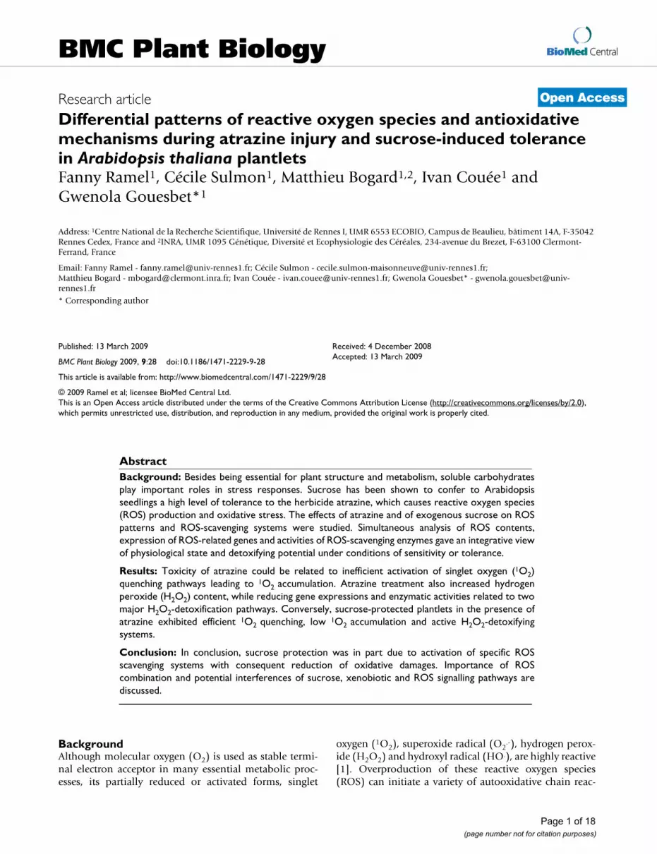
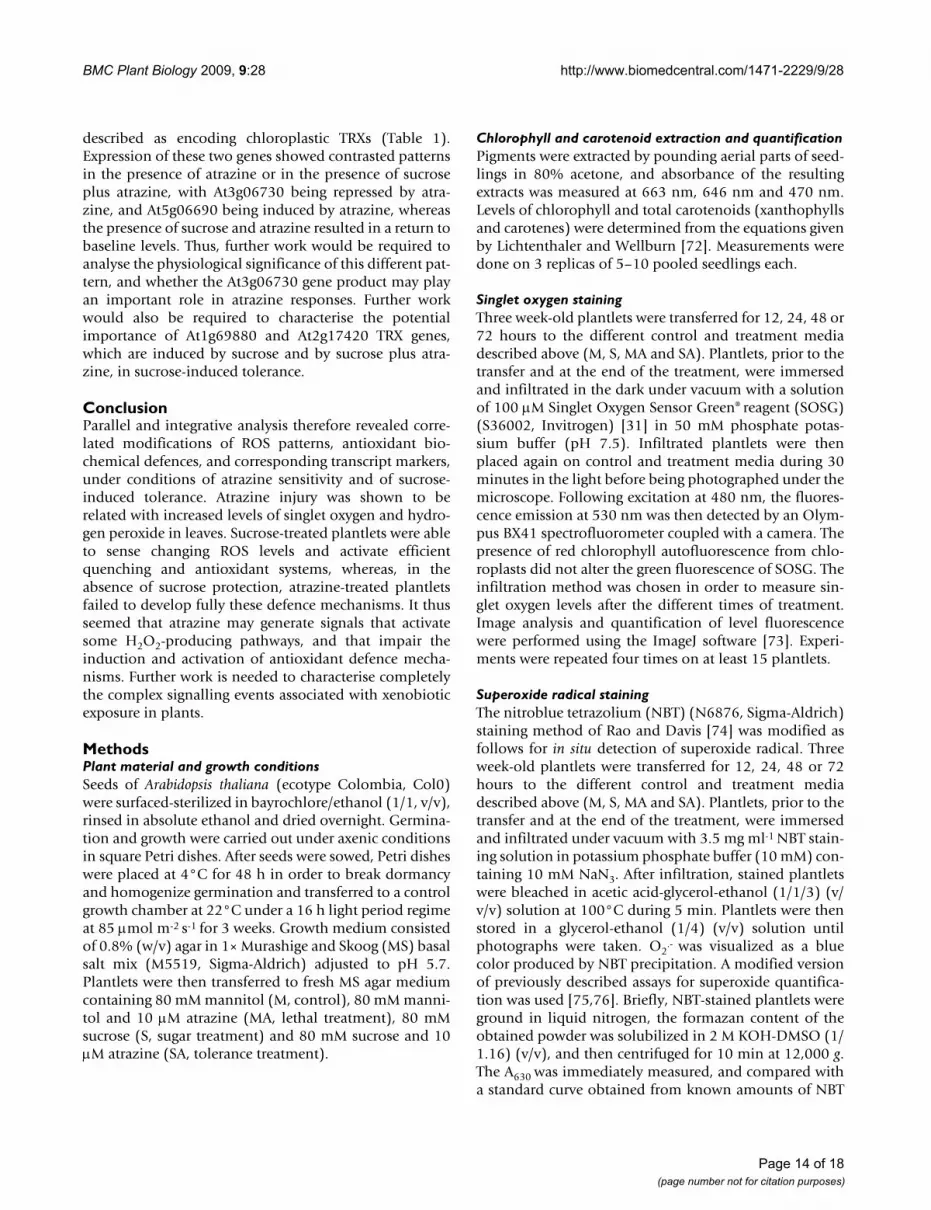
Visualization of singlet oxygen detected with the SOSG fluorescent probeFigure 1Visualization of singlet oxygen detected with the SOSG fluorescent probe. Detections have been done on 3-week-old MS-grown Arabidopsis thaliana plantlets subjected to subsequent treatment (12, 24, 48 or 72 hours) with 80 mM mannitol (M), 80 mM sucrose (S), 80 mM mannitol plus 10 M atrazine (MA) or 80 mM sucrose plus 10 M atrazine (SA). The fluores-cence of SOSG corresponds to the green coloration, while the red color corresponds to chlorophyll autofluorescence. Green fluorescence of roots corresponds to flavonoid and porphyrin autofluorescence. Individual plantlets under the microscope are shown. Quantification of singlet oxygen is presented in Additional file 1.
Page 3 of 18(page number not for citation purposes)

BMC Plant Biology 2009, 9:28 http://www.biomedcentral.com/1471-2229/9/28
Page 4 of 18(page number not for citation purposes)
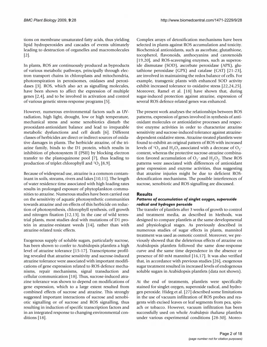
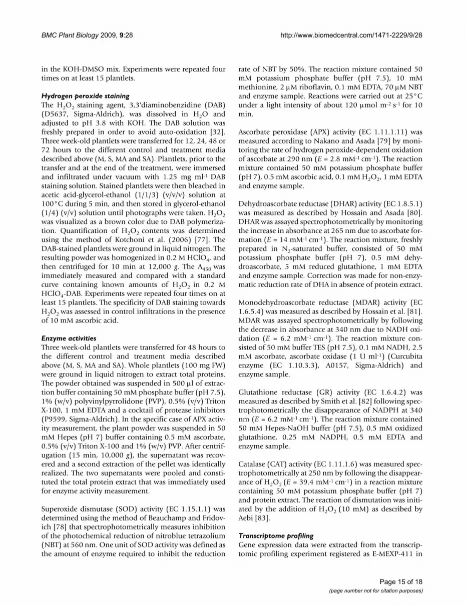
Visualization of superoxide radical detected by NBT stainingFigure 2Visualization of superoxide radical detected by NBT staining. Detections have been done on 3-week-old MS-grown Arabidopsis thaliana plantlets subjected to subsequent treatment (12, 24, 48 or 72 hours) with 80 mM mannitol (M), 80 mM sucrose (S), 80 mM mannitol plus 10 M atrazine (MA) or 80 mM sucrose plus 10 M atrazine (SA). Groups of 15 plantlets are shown. Quantification of superoxide radical is presented in Additional file 2.
BMC Plant Biology 2009, 9:28 http://www.biomedcentral.com/1471-2229/9/28
Page 5 of 18(page number not for citation purposes)
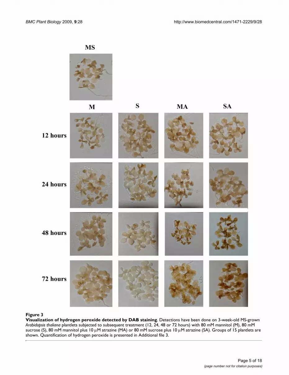
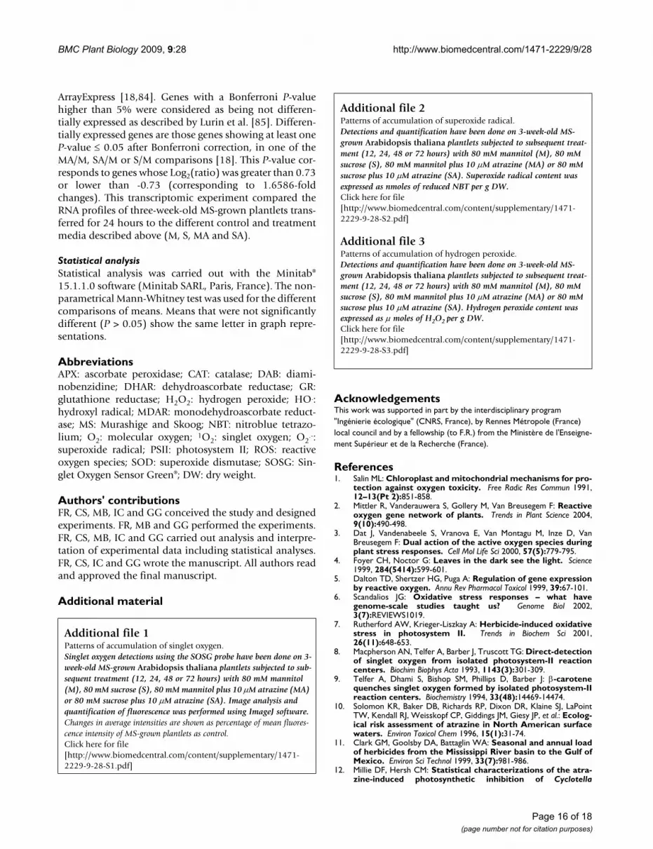
Visualization of hydrogen peroxide detected by DAB stainingFigure 3Visualization of hydrogen peroxide detected by DAB staining. Detections have been done on 3-week-old MS-grown Arabidopsis thaliana plantlets subjected to subsequent treatment (12, 24, 48 or 72 hours) with 80 mM mannitol (M), 80 mM sucrose (S), 80 mM mannitol plus 10 M atrazine (MA) or 80 mM sucrose plus 10 M atrazine (SA). Groups of 15 plantlets are shown. Quantification of hydrogen peroxide is presented in Additional file 3.
BMC Plant Biology 2009, 9:28 http://www.biomedcentral.com/1471-2229/9/28
accumulation was detected in cotyledons as soon as after12 hours of treatment (Fig. 1 and Additional file 1). Tol-erance treatment (SA) maintained a low level of 1O2 incotyledons throughout the treatment, while atrazine treat-ment (MA) strongly increased 1O2 production in cotyle-dons and leaves from 12 to 72 hours of treatment. Thepresence of sucrose in herbicide-containing medium thusappeared to prevent accumulation of 1O2 generated byatrazine.
Superoxide radical (O2.-) detection and quantification
were performed using the nitroblue tetrazolium (NBT)staining method. The levels of superoxide radical stainingafter 12 hours of transfer (Fig. 2 and Additional file 2)were quite similar in the absence (M or S) or presence (MAor SA) of 10 M atrazine. However, the time-courserevealed constant levels of O2
.- in control plantlets (M),while a strong blue coloration appeared in plantletstreated with sucrose (S). This increase was more visible inyoung leaves. Superoxide radical levels in atrazine-treatedplantlets (MA) decreased from 24 hours of treatment. Thecombination of sucrose plus atrazine (SA) led to an inter-mediate state with slight coloration maintained in youngleaves throughout the treatment. Low levels of O2
.-, rela-tively to the mannitol control, were also observed when adrop of 10 M atrazine solution was directly applied toleaf tissue (data not shown).
H2O2 detection and quantification were performed usingthe 3,3'-diaminobenzidine (DAB) staining method [32].Polymerization of DAB, visible as a brown precipitate inthe presence of H2O2, was detected under all conditions.No coloration was observed when infiltration was carriedout in the presence of ascorbic acid, thus confirming theH2O2 specificity of DAB staining, in accordance with pre-vious reports [33-36]. Figure 3 and Additional file 3 sum-marize the time-course of H2O2 accumulation. From 24hours of transfer, control (M) and sucrose-treated (S)plantlets exhibited a much weaker level of H2O2 thanplantlets of atrazine-containing treatments (MA and SA).No variation of H2O2 accumulation was detected in thepresence of mannitol, whereas H2O2 content decreased insucrose-treated plantlets. In contrast, atrazine in theabsence or presence of sucrose tended to increase progres-sively H2O2 levels until 72 hours of treatment. Thisincrease could be detected as early as the fourth hour ofatrazine treatment (data not shown). Likewise, an imme-diate increase of H2O2 levels was also observed when adrop of 10 M atrazine solution was directly applied toleaf tissue (data not shown).
Patterns of singlet oxygen quenching mechanismsTranscriptomic analysis showed that genes linked to thesynthesis of 1O2-quenchers presented contrasted patternsof expression in relation to atrazine sensitivity and toler-
ance (Table 1). Some genes exhibited higher transcriptlevels under tolerance condition (SA) and repressionunder atrazine injury condition (MA), thus suggesting thepossibility of more efficient quenching mechanisms in thepresence of sucrose. Thus, seven genes encoding thiore-doxin family proteins (At2g32920, At2g35010,At2g47470, At3g06730, At4g27080, At5g42980 andAt5g60640) were characterized by significant atrazinerepression of expression, which was lifted by sucrose-atra-zine tolerance treatment (Table 1). Only two genes encod-ing thioredoxin family proteins exhibited higherexpression under atrazine treatment (At5g06690 andAt1g08570) than under sucrose plus atrazine treatment(Table 1). In contrast, two thioredoxin genes (At1g69880and At1g45145) and one thioredoxin reductase gene(At2g17420) were significantly induced under toleranceconditions (SA) compared to atrazine treatment (MA)(Table 1). Thioredoxins have been shown to be involvedin supplying reducing power to reductases detoxifyinglipid hydroperoxides or repairing oxidized proteins [37].Thioredoxins could also act as regulators of scavengingmechanisms [38-40] and as components of signallingpathways of plant antioxidant network. Finally, Das andDas [41] presented evidence that human thioredoxin wasa powerful 1O2 quencher, which could protect cells andtissues against oxidative stress.
Another group of genes exhibited induction of expressionunder atrazine conditions, whereas they were less inducedor not differentially expressed under sucrose-atrazine con-ditions. Activation of these genes might reflect stress sig-nalling due to high 1O2 content in atrazine treated-cells, asrevealed by ROS detection ((Fig. 1 and Additional file 1).Some of these genes belonged to carotenoid biosynthesispathways, such as Zeta-carotene desaturase ZDS1(At3g04870), beta-carotene hydroxylase (At4g25700) or4-hydroxyphenylpyruvate dioxygenase HPD (At1g06570)(Table 1). Carotenoids, which are known to act in chloro-plasts as accessory pigments in light harvesting, candetoxify 1O2 and triplet chlorophyll and dissipate excessexcitation energy [9].
Transcriptome profiling was carried out after 24 hours oftreatment [18]. Measurements of carotenoid levels at dif-ferent times of treatment showed that modifications weremost contrasted after 48 hours of treatment [18]. Thus,given the potential delay between transcription and meta-bolic fluxes, modifications of carotenoid levels after 48hours of treatment were compared with transcript-levelmodifications after 24 hours of treatment. Carotenoid(xanthophylls and carotenes) levels in Arabidopsis thalianaplantlets after 48 hours of treatment are presented inTable 2. Atrazine treatment tended to reduce carotenoidcontents, while addition of sucrose in presence of atrazinemaintained carotenoid levels near control levels. How-
Page 6 of 18(page number not for citation purposes)
BMC Plant Biology 2009, 9:28 http://www.biomedcentral.com/1471-2229/9/28
ever, carotenoid/chlorophyll ratios were not significantlydifferent, thus indicating that the photoprotection role ofcarotenoids was maintained in the presence of atrazine.
Higher induction by atrazine treatment was also found forthe violaxanthin de-epoxidase precursor (At1g08550)gene, which is involved in the xanthophyll cycle (Table 1).Together with carotenoids, zeaxanthin, synthesized fromviolaxanthin via the xanthophyll cycle, protects chloro-plasts by accepting excitation energy from singlet chloro-phyll [42]. Two genes involved in the shikimate
(shikimate kinase, At3g26900) and terpenoid pathways(geranylgeranyl pyrophosphate synthase, At4g36810),which are essential for tocopherol synthesis [43], werealso induced by the herbicide and not differentiallyexpressed by the tolerance treatment (SA) (Table 1). Theantioxidant tocopherol is known to scavenge oxygen freeradicals, lipid peroxy radicals and 1O2 [44]. Finally, thepresence of atrazine alone was found to induce theAt3g55610 gene, which is involved in proline synthesis,with a higher intensity than under conditions of combina-
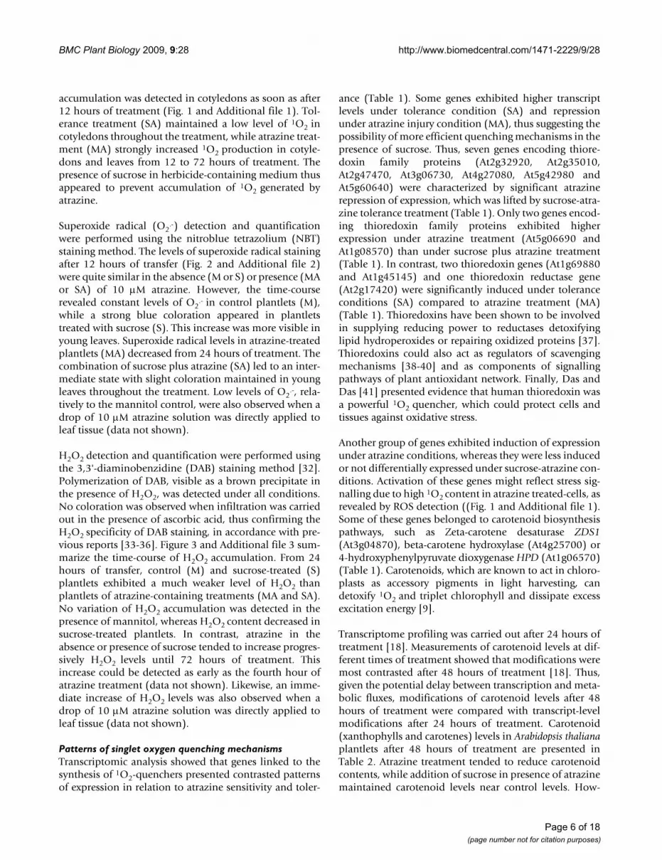
Table 1: Expression of genes involved in singlet oxygen quenching after 24 hours of treatment.
Log2(ratio)
Accession number Gene description Localisation Treatment comparisonS/M MA/M SA/M
At1g08570 Thioredoxin family protein No classification nde 1.04 ndeAt1g45145 Thioredoxin H-type 5 (TRX-H-5) (TOUL) Cytosol nde nde 0.75At1g69880 Thioredoxin, putative No classification 2.05 nde 2.42At2g17420 Thioredoxin reductase 2/NADPH-dependent thioredoxin reductase 2
(NTR2)Cytoplasm 1.22 nde 1.51
At2g32920 Thioredoxin family protein Endomembrane system nde -1.54 ndeAt2g35010 Thioredoxin family protein Mitochondrion nde -1.00 ndeAt2g47470 Thioredoxin family protein Endomembrane system nde -1.74 ndeAt3g06730 Thioredoxin family protein Chloroplast nde -0.74 ndeAt4g27080 Thioredoxin family protein Endoplasmic reticulum nde -0.96 ndeAt5g06690 Thioredoxin family protein Chloroplast -1.15 1.14 ndeAt5g42980 Thioredoxin H-type 3 (TRX-H-3) (GIF1) Cytosol nde -0.94 ndeAt5g60640 Thioredoxin family protein Endomembrane system nde -1.19 nde
At1g06570 4-hydroxyphenylpyruvate dioxygenase (HPD) Chloroplast -0.75 3.18 2.11At3g04870 Zeta-carotene desaturase (ZDS1)/carotene 7.8-desaturase Chloroplast nde 0.94 ndeAt4g25700 Beta-carotene hydroxylase Chloroplast nde 1.07 nde
At1g08550 Violaxanthin de-epoxidase precursor. putative (AVDE1) Photosystem II -1.26 0.91 nde
At3g26900 Shikimate kinase family protein Chloroplast nde 1.69 ndeAt4g36810 Geranylgeranyl pyrophosphate synthase (GGPS1)/GGPP synthetase/
farnesyltranstransferaseChloroplast nde 0.88 nde
At3g55610 Delta 1-pyrroline-5-carboxylate synthetase B/P5CS B (P5CS2) Cytoplasm 0.82 3.63 2.24
Relative expressions of gene are given with their log2(ratio) for sucrose versus mannitol (S/M), mannitol plus atrazine versus mannitol (MA/M) and sucrose plus atrazine versus mannitol (SA/M) comparison. nde: not differentially expressed. Genes with a Bonferroni P-value higher than 5% were considered as being not differentially expressed as described by Lurin et al. [85].
Table 2: Carotenoid content and carotenoid/chlorophyll ratios in leaves of Arabidopsis thaliana plantlets after 48 hours of treatment.
Treatment Carotenoid content(Mean ± SE)g g-1 FW
Carotenoid/Chlorophyll ratios
Mannitol (M) 78.6 ± 0.3 0.172 ± 0.008Sucrose (S) 78.8 ± 0.6 0.168 ± 0.009Mannitol atrazine (MA) 61.2 ± 0.6 0.176 ± 0.012Sucrose atrazine (SA) 72.1± 0.8 0.186 ± 0.010
Page 7 of 18(page number not for citation purposes)
BMC Plant Biology 2009, 9:28 http://www.biomedcentral.com/1471-2229/9/28
tion with sucrose (Table 1). Proline is also known to be an1O2 quencher [45].
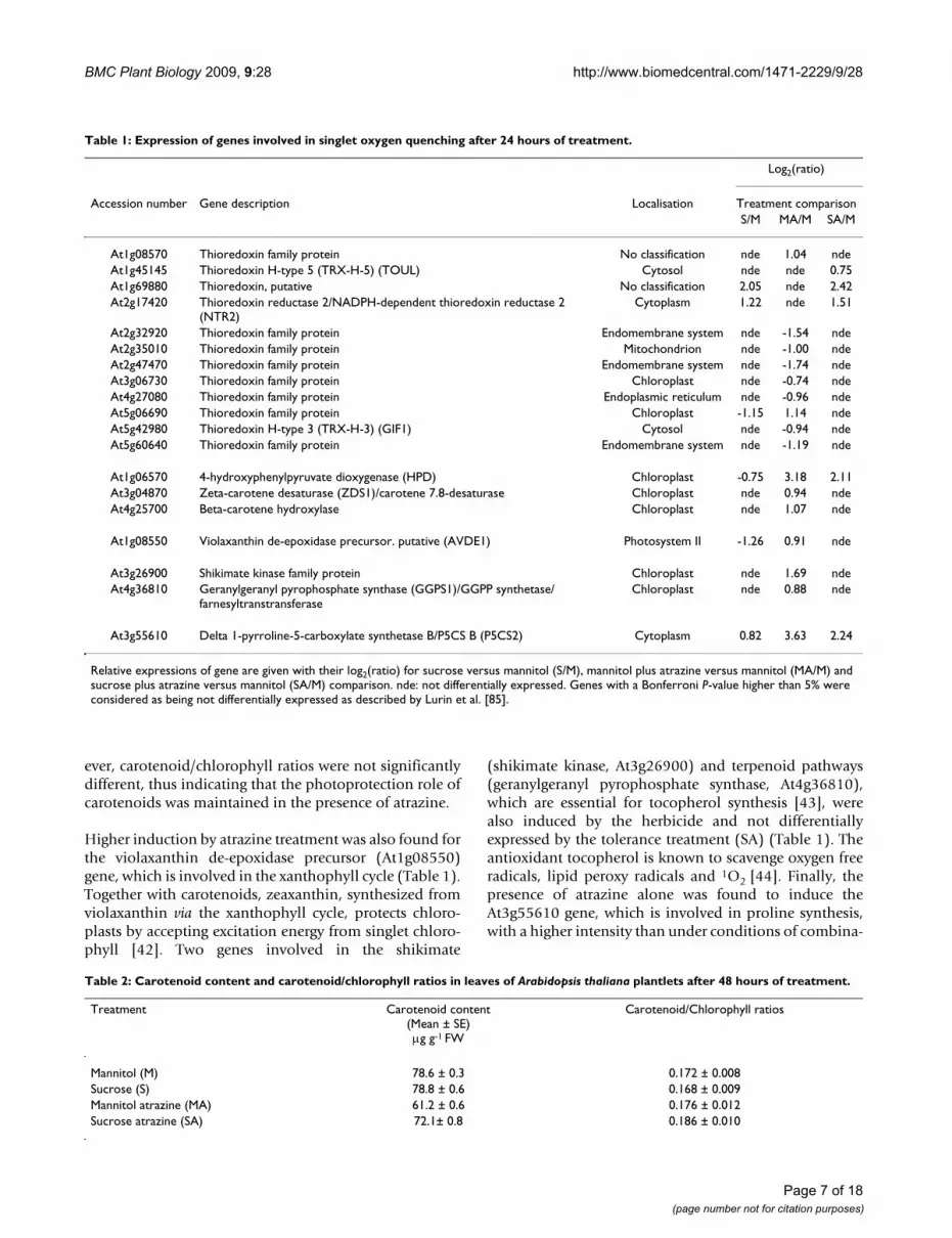
Patterns of superoxide radical scavenging mechanismsExcess of superoxide radical caused by numerous environ-mental stresses is detoxified by superoxide dismutase(SOD) enzymes and converted into H2O2. Seven isoen-zymes have been identified, differing by their metal cofac-tor (Fe, Mn, or Cu and Zn), in Arabidopsis thaliana [46].Transcriptome profiling was carried out after 24 hours oftreatment [18]. Measurements of enzyme activities at dif-ferent times of treatment showed that modifications weremost contrasted after 48 hours of treatment (data notshown). Thus, given the potential delay between tran-scription and protein synthesis, modifications of globalSOD activities after 48 hours of treatment were comparedwith modifications of SOD-encoding transcript levelsafter 24 hours of treatment.
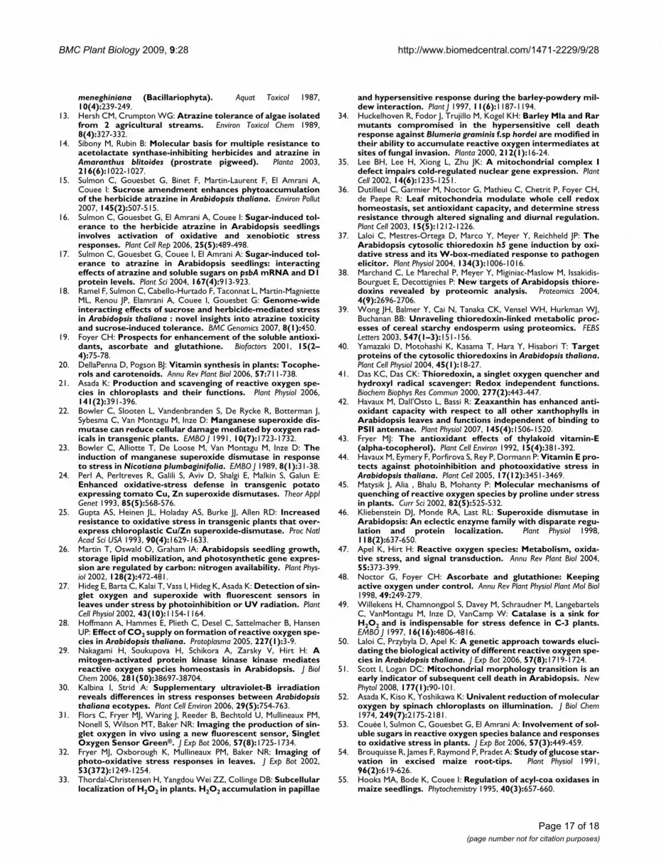
SOD activity (Fig. 4) was decreased by atrazine treatment(MA) in comparison to the mannitol control (M). In con-trast, addition of sucrose in the presence of atrazine (SA)maintained a functional level of SOD activity equivalentto that of the mannitol control. Since sucrose alone wasfound to increase SOD activity, it thus seemed thatsucrose might balance the negative effect of atrazine in thesituation of SA treatment.
Among the six isoenzyme-encoding genes represented inthis microarray analysis (Table 3), three exhibited signifi-cant variations of transcript levels in comparison withcontrol conditions, thus suggesting their potentialinvolvement in O2
.--detoxifying processes in relation toatrazine sensitivity and tolerance. Three genes, encodingCSD1, MSD1, FSD3, were characterized by significantrepression under conditions of atrazine treatment com-pared to control, in accordance with the measurement ofglobal SOD activity (Fig. 4). The CSD1 gene (At1g08830),encoding cytosolic Cu-Zn superoxide dismutase, exhib-ited an induction under tolerance conditions (SA). In con-trast, MSD1 (At3g10920) and FSD3 (At5g23310) genes,which, respectively, encode mitochondrial and chloro-plastic superoxide dismutases, were not differentiallyexpressed in the presence of sucrose. Exogenous sucrose,whether combined or not with atrazine, therefore re-established the basal level of transcripts (Table 3) and ofglobal activity (Fig. 4), thus avoiding the repressive effectsof the herbicide.
Potential origin of hydrogen peroxide accumulation in the presence of atrazineH2O2 contents in atrazine-treated plantlets in the presenceor absence of sucrose seemed to be independent from O2
.-
dismutation. Indeed, O2.- level was low in sucrose plus
atrazine-treated plantlets and null in atrazine-treatedplantlets. Thus, atrazine, in the absence or presence ofsucrose, may promote H2O2-producing pathways inde-pendently from O2
.- and 1O2 accumulation. Transcrip-tomic analysis revealed induction of two genes encodingH2O2-producing enzymes in atrazine-treated plantlets inthe presence or absence of sucrose (SA and MA) (Table 4):amine oxidase (At1g57770) and proline oxidase(At3g30775). Moreover, other potentially H2O2-produc-ing genes were upregulated either under MA condition: aglycolate oxidase putative gene (At3g14420) and a glyoxaloxidase-related gene (At3g53950); or under SA condition:two genes encoding acyl-CoA oxidases (At4g16760,At5g65110) (Table 4).
Patterns of hydrogen peroxide scavenging mechanismsIn order to investigate the efficiency of hydrogen peroxidescavenging mechanisms, global H2O2-scavenging enzymeactivities and transcript levels of related genes were ana-lysed. As explained above, modifications of enzyme activ-ities after 48 hours of treatment were compared withmodifications of transcript levels after 24 hours of treat-ment.
H2O2 can be principally scavenged by two different ways:ascorbate-glutathione cycles and catalases, which playimportant roles in plant defence and senescence. Ascor-bate-glutathione cycles are catalysed by a set of fourenzymes: ascorbate peroxidase (APX), monodehy-
Effects of atrazine and sucrose on SOD enzyme activityFigure 4Effects of atrazine and sucrose on SOD enzyme activity. SOD activity was measured in protein extracts from 3-week-old MS-grown Arabidopsis thaliana plantlets sub-jected to subsequent treatment (48 hours) with 80 mM man-nitol (M), 80 mM sucrose (S), 80 mM mannitol plus 10 M atrazine (MA) or 80 mM sucrose plus 10 M atrazine (SA). SOD activity is expressed in unit/g FW as defined in Meth-ods. Statistical analysis was carried out as described in Meth-ods.
�
����
���
����
���
����
� �
��� ������������������ �
�
�
Page 8 of 18(page number not for citation purposes)
BMC Plant Biology 2009, 9:28 http://www.biomedcentral.com/1471-2229/9/28
droascorbate reductase (MDAR), glutathione-dependentdehydroascorbate reductase (DHAR), and glutathionereductase (GR) [47].
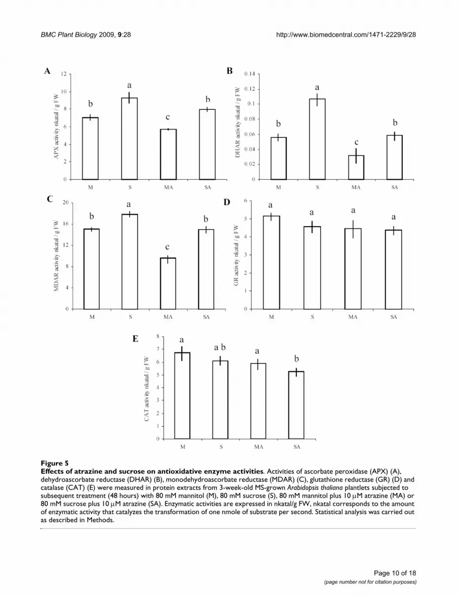
The five enzymes belonging to H2O2-scavenging mecha-nisms presented two different profiles of global activityaccording to the different treatments. The majority ofenzymes involved in ascorbate-glutathione cycles (APX,DHAR and MDAR) were differentially affected by the dif-ferent treatments. Activity of these three enzymes was sig-nificantly reduced by addition of atrazine, while sucrosetreatment had an opposite effect and significantlyincreased these activities (Fig. 5a, b, c). The tolerance con-dition (SA) succeeded to limit repressive effects of the her-bicide and maintained enzyme activities at the controllevel. The fourth enzyme of the ascorbate-glutathionecycles, GR, did not present any significant variation ofactivity between the different treatments (Fig. 5d). Finally,catalase exhibited slightly lower activity under conditions
of sucrose plus atrazine, when compared to control andatrazine-containing medium (Fig. 5e).
The repressive effect of atrazine in the absence of sucrose(MA treatment) on APX global activity was correlated witha general repression of APX genes (Fig. 5a, Table 5).Among the six APX genes present in the microarray, thecytosolic APX1 (At1g07890), the stromal sAPX(At4g08390) and the chloroplastic APX4 (At4g09010)genes exhibited important decrease of transcript levelsunder conditions of atrazine treatment (MA) compared tomannitol control, while the other APX genes were not dif-ferentially expressed in the presence of atrazine. WhereasAPX4 expression remained downregulated in the presenceof sucrose plus atrazine, this tolerant condition balancedthe repressive effects of atrazine for APX1 and sAPX genes,which recovered a level of transcript similar to the control.Finally, and in contrast with global APX activity, the thyl-akoid-bound tAPX (At1g77490) gene was not affected by
Table 3: Expression of genes encoding enzymes involved in O2.- scavenging after 24 hours of treatment.
Log2(ratio)
Accession number Gene description Localisation Treatment comparisonS/M MA/M SA/M
At1g08830 Superoxide dismutase (Cu-Zn) (SODCC)/copper/zinc superoxide dismutase (CSD1)
Cytoplasm 0.80 -0.70 1.22
At2g28190 Superoxide dismutase (Cu-Zn). chloroplast (SODCP)/copper/zinc superoxide dismutase (CSD2)
Chloroplast -0.73 nde -0.76
At3g10920 Superoxide dismutase (Mn). mitochondrial (SODA)/manganese superoxide dismutase (MSD1)
Mitochondrion nde -1.23 nde
At4g25100 Superoxide dismutase (Fe). chloroplast (SODB)/iron superoxide dismutase (FSD1) Chloroplast nde nde ndeAt5g18100 Superoxide dismutase (Cu-Zn)/copper/zinc superoxide dismutase (CSD3) Peroxisome nde nde ndeAt5g23310 Superoxide dismutase (Fe)/iron superoxide dismutase 3 (FSD3) Chloroplast nde -1.34 nde
Relative expressions of gene are given with their log2(ratio) for sucrose versus mannitol (S/M), mannitol plus atrazine versus mannitol (MA/M) and sucrose plus atrazine versus mannitol (SA/M) comparison. nde: not differentially expressed. Genes with a Bonferroni P-value higher than 5% were considered as being not differentially expressed as described by Lurin et al. [85].
Table 4: Expression of genes potentially encoding H2O2-producing enzymes after 24 hours of treatment.
Log2(ratio)
Accession number Gene description Localisation Treatment comparisonS/M MA/M SA/M
At1g57770 Amine oxidase family Chloroplast nde 1.59 0.80At3g14420 (S)-2-hydroxy-acid oxidase, peroxisomal, putative/glycolate
oxidase, putative/short chain alpha-hydroxy acid oxidase, putative Proline oxidase, mitochondrial/osmotic stress-
Peroxisome -1.08 1.33 nde
At3g30775 responsive proline dehydrogenase (POX) (PRO1) (ERD5) Mitochondrion nde 2.51 1.22At3g53950 Glyoxal oxidase-related Endomembrane system nde 1.00 ndeAt4g16760 Acyl-CoA oxidase (ACX1) Peroxisome 0,87 nde 1.48At5g65110 Acyl-CoA oxidase (ACX2) Peroxisome 0.91 nde 1.83
Relative expressions of gene are given with their log2(ratio) for sucrose versus mannitol (S/M), mannitol plus atrazine versus mannitol (MA/M) and sucrose plus atrazine versus mannitol (SA/M) comparison. nde: not differentially expressed. Genes with a Bonferroni P-value higher than 5% were considered as being not differentially expressed as described by Lurin et al. [85].
Page 9 of 18(page number not for citation purposes)
BMC Plant Biology 2009, 9:28 http://www.biomedcentral.com/1471-2229/9/28
Page 10 of 18(page number not for citation purposes)
Effects of atrazine and sucrose on antioxidative enzyme activitiesFigure 5Effects of atrazine and sucrose on antioxidative enzyme activities. Activities of ascorbate peroxidase (APX) (A), dehydroascorbate reductase (DHAR) (B), monodehydroascorbate reductase (MDAR) (C), glutathione reductase (GR) (D) and catalase (CAT) (E) were measured in protein extracts from 3-week-old MS-grown Arabidopsis thaliana plantlets subjected to subsequent treatment (48 hours) with 80 mM mannitol (M), 80 mM sucrose (S), 80 mM mannitol plus 10 M atrazine (MA) or 80 mM sucrose plus 10 M atrazine (SA). Enzymatic activities are expressed in nkatal/g FW, nkatal corresponds to the amount of enzymatic activity that catalyzes the transformation of one nmole of substrate per second. Statistical analysis was carried out as described in Methods.
BMC Plant Biology 2009, 9:28 http://www.biomedcentral.com/1471-2229/9/28
atrazine, while sucrose repressed its expression under Sand SA conditions.
Dehydroascorbate reductase (DHAR) is a key componentof the ascorbate recycling system. DHAR recycles dehy-droascorbate into ascorbate by using reduced glutathioneas a reductant. Two functional DHAR genes, among threethat are encoded in the Arabidopsis thaliana genome, plusa putative gene, were represented in the microarray (Table5). The cytosolic DHAR2 (At1g75270) and putativeDHAR (At5g36270) genes exhibited high induction in thecombined presence of sucrose and atrazine, and, respec-tively, a slight repression or no variation in the presence ofatrazine in comparison to control condition. In contrastto the repressive effects of atrazine, which were associatedwith a decrease of DHAR activity, the increase of DHARtranscript levels in the combined presence of sucrose andatrazine was not associated with an increase of globalDHAR enzyme activity (Fig. 5b).
Reduction of monodehydroascorbate by monodehy-droascorbate reductase (MDAR) is also an important stepin ascorbate recycling. Among the five MDAR genespresent in the microarray, only cytosolic MDAR2(At5g03630) exhibited differential expression patternsaccording to the treatment applied. While atrazinerepressed its expression, the protective combination of
sucrose and atrazine upregulated it (Table 5). Atrazinewas also found to decrease global MDAR activity, whilesucrose plus atrazine treatment resulted in maintenanceof MDAR activity relatively to the mannitol control (Fig.5c).
Glutathione serves as a reductant in oxidation-reductionprocesses, such as recycling of oxidised ascorbate by dehy-droascorbate reductase [48]. Reduction of oxidised glu-tathione is catalysed by glutathione reductase (GR), whichrequires NADPH. Among the two isoenzymes present inthe microarray, only the cytosolic glutathione reductaseGR1 (At3g24170) was found to be induced by sucrose-atrazine and sucrose treatments, while no variation ofexpression was detected in the presence of atrazine (Table5). These variations of expression were not associatedwith changes of global GR activity, since no significant dif-ference of activity was observed between treatments (Fig.5d).
The second way to reduce H2O2 content in cells is activa-tion of catalases (CAT), which catalyse dismutation ofH2O2 into water and oxygen [49]. Little variation of tran-script levels was detected for the three catalase isoenzymes(Table 6). CAT2 (At4g35099) exhibited upregulation byatrazine stress, while CAT3 was slightly downregulated bythe protective sucrose plus atrazine treatment. In relation
Table 5: Expression of genes encoding enzymes involved in ascorbate-glutathione cycles after 24 hours of treatment.
Log2(ratio)
Accession number Gene description Localisation Treatment ComparisonS/M MA/M SA/M
At1g07890 L-ascorbate peroxidase 1. cytosolic (APX1) Cytosol nde -1.92 ndeAt1g77490 L-ascorbate peroxidase. thylakoid-bound (tAPX) Chloroplast -1.03 nde -1.08At3g09640 L-ascorbate peroxidase 2 (APX2) Cytoplasm nde nde ndeAt4g08390 L-ascorbate peroxidase. stromal (sAPX) Chloroplast 1.46 -1.18 ndeAt4g09010 L-ascorbate peroxidase 4 (APX4) Chloroplast -0.79 -1.12 -1.40At4g35000 L-ascorbate peroxidase 3 (APX3) Peroxisome nde nde nde
At1g75270 Dehydroascorbate reductase (DHAR2) Cytoplasm 1.92 -0.91 2.70At5g16710 Dehydroascorbate reductase (DHAR3) Chloroplast nde nde ndeAt5g36270 Dehydroascorbate reductase. putative Cytoplasm 0.80 nde 1.20
At1g63940 Monodehydroascorbate reductase (MDAR5) Chloroplast nde nde ndeAt3g09940 Monodehydroascorbate reductase (MDAR3) Cytoplasm nde nde ndeAt3g27820 Monodehydroascorbate reductase (MDAR4) Cytoplasm nde nde ndeAt3g52880 Monodehydroascorbate reductase (MDAR1) Cytoplasm nde nde ndeAt5g03630 Monodehydroascorbate reductase (MDAR2) Cytoplasm nde -1.35 1.11
At3g24170 Glutathione reductase. putative (GR1) Cytoplasm 1.15 nde 0.92At3g54660 Gluthatione reductase. chloroplast (GR2) Chloroplast nde nde nde
Relative expressions of gene are given with their log2(ratio) for sucrose versus mannitol (S/M), mannitol plus atrazine versus mannitol (MA/M) and sucrose plus atrazine versus mannitol (SA/M) comparison. nde: not differentially expressed. Genes with a Bonferroni P-value higher than 5% were considered as being not differentially expressed as described by Lurin et al. [85].
Page 11 of 18(page number not for citation purposes)
BMC Plant Biology 2009, 9:28 http://www.biomedcentral.com/1471-2229/9/28
with these slight changes of transcript levels (Table 6), glo-bal catalase activities were found to show little variation(Fig. 5e).
DiscussionCharacterisation of the impact of atrazine on ROS patternsROS patterns appear to depend strongly on the nature andintensity of stress conditions applied to plants [50]. It istherefore of great importance to characterise ROS accumu-lation kinetics associated with a particular stress, and notto rely on expected effects. Thus, while, as expected, atra-zine inhibition of photosystem II was associated with 1O2accumulation [7] (Fig. 1 and Additional file 1), decreaseof superoxide radical levels and increase of H2O2 levelswere also observed (Figs. 2, 3 and Additional files 2, 3).This disagreed with the proposed, but experimentallyunproven, accumulation of superoxide radical by triazinetreatment in Arabidopsis leaves [51]. It was howevercoherent with inhibition of photosynthetic activity and ofthe Mehler reaction, whereby superoxide radical is formedby reduction of oxygen at the PSI site [52]. Atrazine bind-ing to D1 protein of PSII and inhibition of electron feed-ing to PSI were indeed likely to decrease superoxideradical production by blocking the Mehler reaction.
The induction of H2O2 accumulation by atrazine was allthe more surprising as it occurred rapidly after transfer toatrazine (Fig. 3 and Additional file 3) and in the absenceor in the presence of sucrose, which by itself had a nega-tive effect on H2O2 accumulation. This is, to our knowl-edge, the first demonstration of rapid in vivo H2O2accumulation under conditions of atrazine treatment. Thenegative effect of sucrose on H2O2 accumulation was con-sistent with the previously-described repression of proteinand lipid catabolism, including a number of oxidase-based processes, by soluble sugars [18,53]. In contrast,atrazine by itself was found to induce a number of genesencoding oxidases, the most highly induced being a geneencoding a proline oxidase (Table 4). Since this inductionoccurred prior to significant impairment of photosystemsand phototrophic growth [18], it could not be ascribed to
a situation of metabolic starvation. Activation of proteinand lipid catabolism and of oxidase-based processes hasbeen reported to occur under conditions of carbohydratelimitation or starvation [54,55]. In this context, it wasextremely interesting that, in the presence of exogenoussucrose, i.e. in a situation of carbohydrate optimum, atra-zine was able to induce a number of oxidase-encodinggenes and other genes typical of carbohydrate-limitationresponse, such as the gene encoding isovaleryl-CoA dehy-drogenase [18,56].
Numerous abiotic stressors, including xenobiotics, areknown to produce oxidative stress in photosyntheticorganisms. This is the case for benzoxazolinone [57], met-ronidazole, dinoterb [58], acetochlor [59], copper [60],wounding [61] and high light [6]. Studies on thesestresses mainly focus on the effects of a single ROS andrarely consider the effects of ROS combination. However,ROS are chemically distinct and selectively perceived forthe fine control of adjusting antioxidants and photosyn-thesis to different environmental stress conditions [62].Indeed, cross-talk between 1O2 and H2O2 has been clearlydemonstrated by Laloi et al. [50], who suggested antago-nistic interactions between 1O2 and H2O2 with a reductionof 1O2-mediated cell death and stress signalling responseby H2O2 content. In contrast, the present condition ofatrazine treatment, which eventually leads to plantletdeath, was characterised by high 1O2, high H2O2 and lowsuperoxide radical levels. Laloi et al. [63], who describedantagonistic effects between 1O2, and H2O2, modulatedH2O2 levels in Arabidopsis transgenic plants at the plastidlevel. It was thus possible that non-antagonistic effects ofH2O2 and 1O2 under conditions of atrazine treatmentwere due to differences of ROS localisation.
Finally, among the set of 29 induced transcription factorsthat have been characterized as 1O2-specific by Gadjev etal. [64], only one was slightly induced (data not shown)during the course of atrazine treatment despite the highaccumulation of singlet oxygen (Fig. 1 and Additional file1). The analysis of 1O2 responses by Gadjev et al. [64] wasbased on studies of the Arabidopsis conditional flu
Table 6: Expression of genes encoding enzymes involved in H2O2 scavenging after 24 hours of treatment.
Log2(ratio)
Accession number Gene description Localisation Treatment comparisonS/M MA/M SA/M
At1g20620 Catalase 3 Peroxisome nde nde -0.74At1g20630 Catalase 1 Peroxisome nde nde ndeAt4g35090 Catalase 2 Peroxisome nde 0.96 nde
Relative expressions of gene are given with their log2(ratio) for sucrose versus mannitol (S/M), mannitol plus atrazine versus mannitol (MA/M) and sucrose plus atrazine versus mannitol (SA/M) comparison. nde: not differentially expressed. Genes with a Bonferroni P-value higher than 5% were considered as being not differentially expressed as described by Lurin et al. [85].
Page 12 of 18(page number not for citation purposes)
BMC Plant Biology 2009, 9:28 http://www.biomedcentral.com/1471-2229/9/28
mutant [65]. It was thus clear that other signals than 1O2were perceived by atrazine-treated plantlets or that atra-zine-induced 1O2 accumulation involved other processesand responses than flu-mutant-dependent 1O2 accumula-tion [50,64,65]. However, full characterisation of the sig-nalling events associated with xenobiotic exposure inplants remains to be carried out.
Impairment of antioxidant defences in the presence of atrazineAtrazine-treated plantlets were characterised by low O2
.-
levels and high H2O2 levels, in contrast with sucrose-treated atrazine-tolerant plants, which showed high O2
.-
and high H2O2 levels. These differences of ROS patternswere associated with striking differences of gene expres-sions and enzyme activities involved in ROS-scavengingpathways.
Thus, atrazine sensitivity was associated with down-regu-lation of key players of H2O2 scavenging. Among the fourenzymes involved in ascorbate-glutathione cycles, whichare essential to remove large amounts of H2O2 generatedby stress [48,66], three enzymes (APX, MDAR and DHAR)exhibited a significant decrease of global activities in atra-zine-treated plantlets. Moreover, this repression was cor-related with a global down-regulation of typicalcorresponding transcripts (APX1, sAPX, DHAR2, andMDAR2), which, conversely, have already been shown toundergo important induction during responses to severalenvironmental abiotic stresses. APX1, a cytosolic enzyme,has previously been described as a central component ofthe reactive oxygen gene network of Arabidopsis [67].Involvement of sAPX in response to oxidative stress hasalso been reported by transcriptional induction in thepresence of H2O2, methylviologen, FeCl3 or UV treat-ments in soybean seedlings [68]. Finally, Yoshida et al.[69] reported the importance of DHAR2 under conditionsof ozone treatment, with higher sensitivity to ozone in aDHAR2-deficient mutant, probably due to insufficientrecycling of ascorbate.
Consequently, repression of these transcripts and decreaseof the corresponding enzyme activities in the presence ofatrazine might accentuate the effects of H2O2 accumula-tion by reduction of ascorbate recycling, thus leading todisruption of antioxidant mechanisms and propagationof atrazine injuries. It was thus clear that the effects of atra-zine at transcript level [18] had actual negative conse-quences on biochemical defences and could be involvedin xenobiotic sensitivity. This is strong evidence that xeno-biotic sensitivity may be linked to gene regulation effectsin plants. Correlatively, the situation of sucrose-inducedtolerance was characterised by the lifting of atrazinerepression, in the case of APX1 and sAPX, or by the induc-tion by sucrose-atrazine combination, in the case of
DHAR2 and MDAR2. These positive effects on transcriptlevels were associated with maintenance of the corre-sponding enzyme activities at control levels. AlthoughROS can mediate induction of protective proteinsinvolved in the stability of specific mRNAs [70], they canalso cause RNA oxidative damages and induce proteininactivation and degradation [71]. Increase of transcriptlevels was therefore likely to be an adaptive response toensure protein synthesis under stress conditions resultingin higher protein turnover.
The decline of O2.- levels in atrazine-treated plantlets,
which, as explained above, could be ascribed to inhibitionof electron transfer through PSI, was associated with ageneral repression of transcripts encoding the differentisoenzymes of SOD and with a decrease of the globalactivity of this O2
.--scavenging enzyme family, thus indi-cating that atrazine-treated cells responded to the lowsuperoxide radical situation. Association of low O2
.- andhigh H2O2 may be a cause for the ill-adapted response ofanti-oxidant defences in atrazine-treated plantlets, thussuggesting that further work should be carried out on theadaptation of organisms to fluctuations of ROS combina-tions.
Mechanisms of sucrose-induced tolerance to singlet oxygenIn contrast with non-induction of H2O2-scavenging sys-tems, atrazine-treated plantlets seemed to be able to sensethe increase of 1O2 levels and induce some genes poten-tially involved in 1O2 quenching (Table 1). Thus, atrazine-treated plantlets, in the absence or presence of sucroseshowed increased expression of 4-Hydroxyphenylpyru-vate dioxygenase (HPD) gene (At1g06570), which couldbe involved in the maintenance of the photoprotectiverole of carotenoids. The At5g06690 gene, encoding a chlo-roplastic thioredoxin, which is a potential 1O2-quencher[41], was also induced in atrazine-treated plantlet. How-ever, generally, the thioredoxin gene family was negativelyaffected by atrazine treatment, with 7 genes among 12 sig-nificantly repressed by atrazine. Correlatively, ten of thesetwelve genes showed lifting of repression or significantinduction in the combined presence of sucrose and atra-zine. Nevertheless, most of these genes encoded extraplas-tidial thioredoxins or thioredoxins of unknownlocalisation (Table 1). The link of thioredoxin gene familydifferential expression with efficient 1O2 quenching in thepresence of atrazine plus sucrose (Fig. 1 and Additionalfile 1) was thus difficult to ascertain. On one hand, severalstudies have shown the efficiency of thioredoxins in main-tenance of cellular reductant environment and in cytopro-tective mechanisms [37-40]. On the other hand, efficient1O2 quenching in the case of PSII inhibition by atrazinewould require the involvement of chloroplastic TRXs. TwoTRX genes, At3g06730 and At5g06690, have been
Page 13 of 18(page number not for citation purposes)
BMC Plant Biology 2009, 9:28 http://www.biomedcentral.com/1471-2229/9/28
described as encoding chloroplastic TRXs (Table 1).Expression of these two genes showed contrasted patternsin the presence of atrazine or in the presence of sucroseplus atrazine, with At3g06730 being repressed by atra-zine, and At5g06690 being induced by atrazine, whereasthe presence of sucrose and atrazine resulted in a return tobaseline levels. Thus, further work would be required toanalyse the physiological significance of this different pat-tern, and whether the At3g06730 gene product may playan important role in atrazine responses. Further workwould also be required to characterise the potentialimportance of At1g69880 and At2g17420 TRX genes,which are induced by sucrose and by sucrose plus atra-zine, in sucrose-induced tolerance.
ConclusionParallel and integrative analysis therefore revealed corre-lated modifications of ROS patterns, antioxidant bio-chemical defences, and corresponding transcript markers,under conditions of atrazine sensitivity and of sucrose-induced tolerance. Atrazine injury was shown to berelated with increased levels of singlet oxygen and hydro-gen peroxide in leaves. Sucrose-treated plantlets were ableto sense changing ROS levels and activate efficientquenching and antioxidant systems, whereas, in theabsence of sucrose protection, atrazine-treated plantletsfailed to develop fully these defence mechanisms. It thusseemed that atrazine may generate signals that activatesome H2O2-producing pathways, and that impair theinduction and activation of antioxidant defence mecha-nisms. Further work is needed to characterise completelythe complex signalling events associated with xenobioticexposure in plants.
MethodsPlant material and growth conditionsSeeds of Arabidopsis thaliana (ecotype Colombia, Col0)were surfaced-sterilized in bayrochlore/ethanol (1/1, v/v),rinsed in absolute ethanol and dried overnight. Germina-tion and growth were carried out under axenic conditionsin square Petri dishes. After seeds were sowed, Petri disheswere placed at 4°C for 48 h in order to break dormancyand homogenize germination and transferred to a controlgrowth chamber at 22°C under a 16 h light period regimeat 85 mol m-2 s-1 for 3 weeks. Growth medium consistedof 0.8% (w/v) agar in 1× Murashige and Skoog (MS) basalsalt mix (M5519, Sigma-Aldrich) adjusted to pH 5.7.Plantlets were then transferred to fresh MS agar mediumcontaining 80 mM mannitol (M, control), 80 mM manni-tol and 10 M atrazine (MA, lethal treatment), 80 mMsucrose (S, sugar treatment) and 80 mM sucrose and 10M atrazine (SA, tolerance treatment).
Chlorophyll and carotenoid extraction and quantificationPigments were extracted by pounding aerial parts of seed-lings in 80% acetone, and absorbance of the resultingextracts was measured at 663 nm, 646 nm and 470 nm.Levels of chlorophyll and total carotenoids (xanthophyllsand carotenes) were determined from the equations givenby Lichtenthaler and Wellburn [72]. Measurements weredone on 3 replicas of 5–10 pooled seedlings each.
Singlet oxygen stainingThree week-old plantlets were transferred for 12, 24, 48 or72 hours to the different control and treatment mediadescribed above (M, S, MA and SA). Plantlets, prior to thetransfer and at the end of the treatment, were immersedand infiltrated in the dark under vacuum with a solutionof 100 M Singlet Oxygen Sensor Green® reagent (SOSG)(S36002, Invitrogen) [31] in 50 mM phosphate potas-sium buffer (pH 7.5). Infiltrated plantlets were thenplaced again on control and treatment media during 30minutes in the light before being photographed under themicroscope. Following excitation at 480 nm, the fluores-cence emission at 530 nm was then detected by an Olym-pus BX41 spectrofluorometer coupled with a camera. Thepresence of red chlorophyll autofluorescence from chlo-roplasts did not alter the green fluorescence of SOSG. Theinfiltration method was chosen in order to measure sin-glet oxygen levels after the different times of treatment.Image analysis and quantification of level fluorescencewere performed using the ImageJ software [73]. Experi-ments were repeated four times on at least 15 plantlets.
Superoxide radical stainingThe nitroblue tetrazolium (NBT) (N6876, Sigma-Aldrich)staining method of Rao and Davis [74] was modified asfollows for in situ detection of superoxide radical. Threeweek-old plantlets were transferred for 12, 24, 48 or 72hours to the different control and treatment mediadescribed above (M, S, MA and SA). Plantlets, prior to thetransfer and at the end of the treatment, were immersedand infiltrated under vacuum with 3.5 mg ml-1 NBT stain-ing solution in potassium phosphate buffer (10 mM) con-taining 10 mM NaN3. After infiltration, stained plantletswere bleached in acetic acid-glycerol-ethanol (1/1/3) (v/v/v) solution at 100°C during 5 min. Plantlets were thenstored in a glycerol-ethanol (1/4) (v/v) solution untilphotographs were taken. O2
.- was visualized as a bluecolor produced by NBT precipitation. A modified versionof previously described assays for superoxide quantifica-tion was used [75,76]. Briefly, NBT-stained plantlets wereground in liquid nitrogen, the formazan content of theobtained powder was solubilized in 2 M KOH-DMSO (1/1.16) (v/v), and then centrifuged for 10 min at 12,000 g.The A630 was immediately measured, and compared witha standard curve obtained from known amounts of NBT
Page 14 of 18(page number not for citation purposes)
BMC Plant Biology 2009, 9:28 http://www.biomedcentral.com/1471-2229/9/28
in the KOH-DMSO mix. Experiments were repeated fourtimes on at least 15 plantlets.
Hydrogen peroxide stainingThe H2O2 staining agent, 3,3'diaminobenzidine (DAB)(D5637, Sigma-Aldrich), was dissolved in H2O andadjusted to pH 3.8 with KOH. The DAB solution wasfreshly prepared in order to avoid auto-oxidation [32].Three week-old plantlets were transferred for 12, 24, 48 or72 hours to the different control and treatment mediadescribed above (M, S, MA and SA). Plantlets, prior to thetransfer and at the end of the treatment, were immersedand infiltrated under vacuum with 1.25 mg ml-1 DABstaining solution. Stained plantlets were then bleached inacetic acid-glycerol-ethanol (1/1/3) (v/v/v) solution at100°C during 5 min, and then stored in glycerol-ethanol(1/4) (v/v) solution until photographs were taken. H2O2was visualized as a brown color due to DAB polymeriza-tion. Quantification of H2O2 contents was determinedusing the method of Kotchoni et al. (2006) [77]. TheDAB-stained plantlets were ground in liquid nitrogen. Theresulting powder was homogenized in 0.2 M HClO4, andthen centrifuged for 10 min at 12,000 g. The A450 wasimmediately measured and compared with a standardcurve containing known amounts of H2O2 in 0.2 MHClO4-DAB. Experiments were repeated four times on atleast 15 plantlets. The specificity of DAB staining towardsH2O2 was assessed in control infiltrations in the presenceof 10 mM ascorbic acid.
Enzyme activitiesThree week-old plantlets were transferred for 48 hours tothe different control and treatment media describedabove (M, S, MA and SA). Whole plantlets (100 mg FW)were ground in liquid nitrogen to extract total proteins.The powder obtained was suspended in 500 l of extrac-tion buffer containing 50 mM phosphate buffer (pH 7.5),1% (w/v) polyvinylpyrrolidone (PVP), 0.5% (v/v) TritonX-100, 1 mM EDTA and a cocktail of protease inhibitors(P9599, Sigma-Aldrich). In the specific case of APX activ-ity measurement, the plant powder was suspended in 50mM Hepes (pH 7) buffer containing 0.5 mM ascorbate,0.5% (v/v) Triton X-100 and 1% (w/v) PVP. After centrif-ugation (15 min, 10,000 g), the supernatant was recov-ered and a second extraction of the pellet was identicallyrealized. The two supernatants were pooled and consti-tuted the total protein extract that was immediately usedfor enzyme activity measurement.
Superoxide dismutase (SOD) activity (EC 1.15.1.1) wasdetermined using the method of Beauchamp and Fridov-ich [78] that spectrophotometrically measures inhibitionof the photochemical reduction of nitroblue tetrazolium(NBT) at 560 nm. One unit of SOD activity was defined asthe amount of enzyme required to inhibit the reduction
rate of NBT by 50%. The reaction mixture contained 50mM potassium phosphate buffer (pH 7.5), 10 mMmethionine, 2 M riboflavin, 0.1 mM EDTA, 70 M NBTand enzyme sample. Reactions were carried out at 25°Cunder a light intensity of about 120 mol m-2 s-1 for 10min.
Ascorbate peroxidase (APX) activity (EC 1.11.1.11) wasmeasured according to Nakano and Asada [79] by moni-toring the rate of hydrogen peroxide-dependent oxidationof ascorbate at 290 nm (E = 2.8 mM-1 cm-1). The reactionmixture contained 50 mM potassium phosphate buffer(pH 7), 0.5 mM ascorbic acid, 0.1 mM H2O2, 1 mM EDTAand enzyme sample.
Dehydroascorbate reductase (DHAR) activity (EC 1.8.5.1)was measured as described by Hossain and Asada [80].DHAR was assayed spectrophotometrically by monitoringthe increase in absorbance at 265 nm due to ascorbate for-mation (E = 14 mM-1 cm-1). The reaction mixture, freshlyprepared in N2-saturated buffer, consisted of 50 mMpotassium phosphate buffer (pH 7), 0.5 mM dehy-droascorbate, 5 mM reduced glutathione, 1 mM EDTAand enzyme sample. Correction was made for non-enzy-matic reduction rate of DHA in absence of protein extract.
Monodehydroascorbate reductase (MDAR) activity (EC1.6.5.4) was measured as described by Hossain et al. [81].MDAR was assayed spectrophotometrically by followingthe decrease in absorbance at 340 nm due to NADH oxi-dation (E = 6.2 mM-1 cm-1). The reaction mixture con-sisted of 50 mM buffer TES (pH 7.5), 0.1 mM NADH, 2.5mM ascorbate, ascorbate oxidase (1 U ml-1) (Curcubitaenzyme (EC 1.10.3.3), A0157, Sigma-Aldrich) andenzyme sample.
Glutathione reductase (GR) activity (EC 1.6.4.2) wasmeasured as described by Smith et al. [82] following spec-trophotometrically the disappearance of NADPH at 340nm (E = 6.2 mM-1 cm-1). The reaction mixture contained50 mM Hepes-NaOH buffer (pH 7.5), 0.5 mM oxidizedglutathione, 0.25 mM NADPH, 0.5 mM EDTA andenzyme sample.
Catalase (CAT) activity (EC 1.11.1.6) was measured spec-trophotometrically at 250 nm by following the disappear-ance of H2O2 (E = 39.4 mM-1 cm-1) in a reaction mixturecontaining 50 mM potassium phosphate buffer (pH 7)and protein extract. The reaction of dismutation was initi-ated by the addition of H2O2 (10 mM) as described byAebi [83].
Transcriptome profilingGene expression data were extracted from the transcrip-tomic profiling experiment registered as E-MEXP-411 in
Page 15 of 18(page number not for citation purposes)
BMC Plant Biology 2009, 9:28 http://www.biomedcentral.com/1471-2229/9/28
ArrayExpress [18,84]. Genes with a Bonferroni P-valuehigher than 5% were considered as being not differen-tially expressed as described by Lurin et al. [85]. Differen-tially expressed genes are those genes showing at least oneP-value 0.05 after Bonferroni correction, in one of theMA/M, SA/M or S/M comparisons [18]. This P-value cor-responds to genes whose Log2(ratio) was greater than 0.73or lower than -0.73 (corresponding to 1.6586-foldchanges). This transcriptomic experiment compared theRNA profiles of three-week-old MS-grown plantlets trans-ferred for 24 hours to the different control and treatmentmedia described above (M, S, MA and SA).
Statistical analysisStatistical analysis was carried out with the Minitab®
15.1.1.0 software (Minitab SARL, Paris, France). The non-parametrical Mann-Whitney test was used for the differentcomparisons of means. Means that were not significantlydifferent (P > 0.05) show the same letter in graph repre-sentations.
AbbreviationsAPX: ascorbate peroxidase; CAT: catalase; DAB: diami-nobenzidine; DHAR: dehydroascorbate reductase; GR:glutathione reductase; H2O2: hydrogen peroxide; HO.:hydroxyl radical; MDAR: monodehydroascorbate reduct-ase; MS: Murashige and Skoog; NBT: nitroblue tetrazo-lium; O2: molecular oxygen; 1O2: singlet oxygen; O2
.-:superoxide radical; PSII: photosystem II; ROS: reactiveoxygen species; SOD: superoxide dismutase; SOSG: Sin-glet Oxygen Sensor Green®; DW: dry weight.
Authors' contributionsFR, CS, MB, IC and GG conceived the study and designedexperiments. FR, MB and GG performed the experiments.FR, CS, MB, IC and GG carried out analysis and interpre-tation of experimental data including statistical analyses.FR, CS, IC and GG wrote the manuscript. All authors readand approved the final manuscript.
Additional material
AcknowledgementsThis work was supported in part by the interdisciplinary program "Ingénierie écologique" (CNRS, France), by Rennes Métropole (France) local council and by a fellowship (to F.R.) from the Ministère de l'Enseigne-ment Supérieur et de la Recherche (France).
References1. Salin ML: Chloroplast and mitochondrial mechanisms for pro-
tection against oxygen toxicity. Free Radic Res Commun 1991,12–13(Pt 2):851-858.
2. Mittler R, Vanderauwera S, Gollery M, Van Breusegem F: Reactiveoxygen gene network of plants. Trends in Plant Science 2004,9(10):490-498.
3. Dat J, Vandenabeele S, Vranova E, Van Montagu M, Inze D, VanBreusegem F: Dual action of the active oxygen species duringplant stress responses. Cell Mol Life Sci 2000, 57(5):779-795.
4. Foyer CH, Noctor G: Leaves in the dark see the light. Science1999, 284(5414):599-601.
5. Dalton TD, Shertzer HG, Puga A: Regulation of gene expressionby reactive oxygen. Annu Rev Pharmacol Toxicol 1999, 39:67-101.
6. Scandalios JG: Oxidative stress responses – what havegenome-scale studies taught us? Genome Biol 2002,3(7):REVIEWS1019.
7. Rutherford AW, Krieger-Liszkay A: Herbicide-induced oxidativestress in photosystem II. Trends in Biochem Sci 2001,26(11):648-653.
8. Macpherson AN, Telfer A, Barber J, Truscott TG: Direct-detectionof singlet oxygen from isolated photosystem-II reactioncenters. Biochim Biophys Acta 1993, 1143(3):301-309.
9. Telfer A, Dhami S, Bishop SM, Phillips D, Barber J: -carotenequenches singlet oxygen formed by isolated photosystem-IIreaction centers. Biochemistry 1994, 33(48):14469-14474.
10. Solomon KR, Baker DB, Richards RP, Dixon DR, Klaine SJ, LaPointTW, Kendall RJ, Weisskopf CP, Giddings JM, Giesy JP, et al.: Ecolog-ical risk assessment of atrazine in North American surfacewaters. Environ Toxicol Chem 1996, 15(1):31-74.
11. Clark GM, Goolsby DA, Battaglin WA: Seasonal and annual loadof herbicides from the Mississippi River basin to the Gulf ofMexico. Environ Sci Technol 1999, 33(7):981-986.
12. Millie DF, Hersh CM: Statistical characterizations of the atra-zine-induced photosynthetic inhibition of Cyclotella
Additional file 1Patterns of accumulation of singlet oxygen.Singlet oxygen detections using the SOSG probe have been done on 3-week-old MS-grown Arabidopsis thaliana plantlets subjected to sub-sequent treatment (12, 24, 48 or 72 hours) with 80 mM mannitol (M), 80 mM sucrose (S), 80 mM mannitol plus 10 M atrazine (MA) or 80 mM sucrose plus 10 M atrazine (SA). Image analysis and quantification of fluorescence was performed using ImageJ software. Changes in average intensities are shown as percentage of mean fluores-cence intensity of MS-grown plantlets as control.Click here for file[http://www.biomedcentral.com/content/supplementary/1471-2229-9-28-S1.pdf]
Additional file 2Patterns of accumulation of superoxide radical. Detections and quantification have been done on 3-week-old MS-grown Arabidopsis thaliana plantlets subjected to subsequent treat-ment (12, 24, 48 or 72 hours) with 80 mM mannitol (M), 80 mM sucrose (S), 80 mM mannitol plus 10 M atrazine (MA) or 80 mM sucrose plus 10 M atrazine (SA). Superoxide radical content was expressed as nmoles of reduced NBT per g DW.Click here for file[http://www.biomedcentral.com/content/supplementary/1471-2229-9-28-S2.pdf]
Additional file 3Patterns of accumulation of hydrogen peroxide. Detections and quantification have been done on 3-week-old MS-grown Arabidopsis thaliana plantlets subjected to subsequent treat-ment (12, 24, 48 or 72 hours) with 80 mM mannitol (M), 80 mM sucrose (S), 80 mM mannitol plus 10 M atrazine (MA) or 80 mM sucrose plus 10 M atrazine (SA). Hydrogen peroxide content was expressed as moles of H2O2 per g DW.Click here for file[http://www.biomedcentral.com/content/supplementary/1471-2229-9-28-S3.pdf]
Page 16 of 18(page number not for citation purposes)
BMC Plant Biology 2009, 9:28 http://www.biomedcentral.com/1471-2229/9/28
meneghiniana (Bacillariophyta). Aquat Toxicol 1987,10(4):239-249.
13. Hersh CM, Crumpton WG: Atrazine tolerance of algae isolatedfrom 2 agricultural streams. Environ Toxicol Chem 1989,8(4):327-332.
14. Sibony M, Rubin B: Molecular basis for multiple resistance toacetolactate synthase-inhibiting herbicides and atrazine inAmaranthus blitoides (prostrate pigweed). Planta 2003,216(6):1022-1027.
15. Sulmon C, Gouesbet G, Binet F, Martin-Laurent F, El Amrani A,Couee I: Sucrose amendment enhances phytoaccumulationof the herbicide atrazine in Arabidopsis thaliana. Environ Pollut2007, 145(2):507-515.
16. Sulmon C, Gouesbet G, El Amrani A, Couee I: Sugar-induced tol-erance to the herbicide atrazine in Arabidopsis seedlingsinvolves activation of oxidative and xenobiotic stressresponses. Plant Cell Rep 2006, 25(5):489-498.
17. Sulmon C, Gouesbet G, Couee I, El Amrani A: Sugar-induced tol-erance to atrazine in Arabidopsis seedlings: interactingeffects of atrazine and soluble sugars on psbA mRNA and D1protein levels. Plant Sci 2004, 167(4):913-923.
18. Ramel F, Sulmon C, Cabello-Hurtado F, Taconnat L, Martin-MagnietteML, Renou JP, Elamrani A, Couee I, Gouesbet G: Genome-wideinteracting effects of sucrose and herbicide-mediated stressin Arabidopsis thaliana : novel insights into atrazine toxicityand sucrose-induced tolerance. BMC Genomics 2007, 8(1):450.
19. Foyer CH: Prospects for enhancement of the soluble antioxi-dants, ascorbate and glutathione. Biofactors 2001, 15(2–4):75-78.
20. DellaPenna D, Pogson BJ: Vitamin synthesis in plants: Tocophe-rols and carotenoids. Annu Rev Plant Biol 2006, 57:711-738.
21. Asada K: Production and scavenging of reactive oxygen spe-cies in chloroplasts and their functions. Plant Physiol 2006,141(2):391-396.
22. Bowler C, Slooten L, Vandenbranden S, De Rycke R, Botterman J,Sybesma C, Van Montagu M, Inze D: Manganese superoxide dis-mutase can reduce cellular damage mediated by oxygen rad-icals in transgenic plants. EMBO J 1991, 10(7):1723-1732.
23. Bowler C, Alliotte T, De Loose M, Van Montagu M, Inze D: Theinduction of manganese superoxide dismutase in responseto stress in Nicotiana plumbaginifolia. EMBO J 1989, 8(1):31-38.
24. Perl A, Perltreves R, Galili S, Aviv D, Shalgi E, Malkin S, Galun E:Enhanced oxidative-stress defense in transgenic potatoexpressing tomato Cu, Zn superoxide dismutases. Theor ApplGenet 1993, 85(5):568-576.
25. Gupta AS, Heinen JL, Holaday AS, Burke JJ, Allen RD: Increasedresistance to oxidative stress in transgenic plants that over-express chloroplastic Cu/Zn superoxide-dismutase. Proc NatlAcad Sci USA 1993, 90(4):1629-1633.
26. Martin T, Oswald O, Graham IA: Arabidopsis seedling growth,storage lipid mobilization, and photosynthetic gene expres-sion are regulated by carbon: nitrogen availability. Plant Phys-iol 2002, 128(2):472-481.
27. Hideg E, Barta C, Kalai T, Vass I, Hideg K, Asada K: Detection of sin-glet oxygen and superoxide with fluorescent sensors inleaves under stress by photoinhibition or UV radiation. PlantCell Physiol 2002, 43(10):1154-1164.
28. Hoffmann A, Hammes E, Plieth C, Desel C, Sattelmacher B, HansenUP: Effect of CO2 supply on formation of reactive oxygen spe-cies in Arabidopsis thaliana. Protoplasma 2005, 227(1):3-9.
29. Nakagami H, Soukupova H, Schikora A, Zarsky V, Hirt H: Amitogen-activated protein kinase kinase kinase mediatesreactive oxygen species homeostasis in Arabidopsis. J BiolChem 2006, 281(50):38697-38704.
30. Kalbina I, Strid A: Supplementary ultraviolet-B irradiationreveals differences in stress responses between Arabidopsisthaliana ecotypes. Plant Cell Environ 2006, 29(5):754-763.
31. Flors C, Fryer MJ, Waring J, Reeder B, Bechtold U, Mullineaux PM,Nonell S, Wilson MT, Baker NR: Imaging the production of sin-glet oxygen in vivo using a new fluorescent sensor, SingletOxygen Sensor Green®. J Exp Bot 2006, 57(8):1725-1734.
32. Fryer MJ, Oxborough K, Mullineaux PM, Baker NR: Imaging ofphoto-oxidative stress responses in leaves. J Exp Bot 2002,53(372):1249-1254.
33. Thordal-Christensen H, Yangdou Wei ZZ, Collinge DB: Subcellularlocalization of H2O2 in plants. H2O2 accumulation in papillae
and hypersensitive response during the barley-powdery mil-dew interaction. Plant J 1997, 11(6):1187-1194.
34. Huckelhoven R, Fodor J, Trujillo M, Kogel KH: Barley Mla and Rarmutants compromised in the hypersensitive cell deathresponse against Blumeria graminis f.sp hordei are modified intheir ability to accumulate reactive oxygen intermediates atsites of fungal invasion. Planta 2000, 212(1):16-24.
35. Lee BH, Lee H, Xiong L, Zhu JK: A mitochondrial complex Idefect impairs cold-regulated nuclear gene expression. PlantCell 2002, 14(6):1235-1251.
36. Dutilleul C, Garmier M, Noctor G, Mathieu C, Chetrit P, Foyer CH,de Paepe R: Leaf mitochondria modulate whole cell redoxhomeostasis, set antioxidant capacity, and determine stressresistance through altered signaling and diurnal regulation.Plant Cell 2003, 15(5):1212-1226.
37. Laloi C, Mestres-Ortega D, Marco Y, Meyer Y, Reichheld JP: TheArabidopsis cytosolic thioredoxin h5 gene induction by oxi-dative stress and its W-box-mediated response to pathogenelicitor. Plant Physiol 2004, 134(3):1006-1016.
38. Marchand C, Le Marechal P, Meyer Y, Miginiac-Maslow M, Issakidis-Bourguet E, Decottignies P: New targets of Arabidopsis thiore-doxins revealed by proteomic analysis. Proteomics 2004,4(9):2696-2706.
39. Wong JH, Balmer Y, Cai N, Tanaka CK, Vensel WH, Hurkman WJ,Buchanan BB: Unraveling thioredoxin-linked metabolic proc-esses of cereal starchy endosperm using proteomics. FEBSLetters 2003, 547(1–3):151-156.
40. Yamazaki D, Motohashi K, Kasama T, Hara Y, Hisabori T: Targetproteins of the cytosolic thioredoxins in Arabidopsis thaliana.Plant Cell Physiol 2004, 45(1):18-27.
41. Das KC, Das CK: Thioredoxin, a singlet oxygen quencher andhydroxyl radical scavenger: Redox independent functions.Biochem Biophys Res Commun 2000, 277(2):443-447.
42. Havaux M, Dall'Osto L, Bassi R: Zeaxanthin has enhanced anti-oxidant capacity with respect to all other xanthophylls inArabidopsis leaves and functions independent of binding toPSII antennae. Plant Physiol 2007, 145(4):1506-1520.
43. Fryer MJ: The antioxidant effects of thylakoid vitamin-E(alpha-tocopherol). Plant Cell Environ 1992, 15(4):381-392.
44. Havaux M, Eymery F, Porfirova S, Rey P, Dormann P: Vitamin E pro-tects against photoinhibition and photooxidative stress inArabidopsis thaliana. Plant Cell 2005, 17(12):3451-3469.
45. Matysik J, Alia , Bhalu B, Mohanty P: Molecular mechanisms ofquenching of reactive oxygen species by proline under stressin plants. Curr Sci 2002, 82(5):525-532.
46. Kliebenstein DJ, Monde RA, Last RL: Superoxide dismutase inArabidopsis: An eclectic enzyme family with disparate regu-lation and protein localization. Plant Physiol 1998,118(2):637-650.
47. Apel K, Hirt H: Reactive oxygen species: Metabolism, oxida-tive stress, and signal transduction. Annu Rev Plant Biol 2004,55:373-399.
48. Noctor G, Foyer CH: Ascorbate and glutathione: Keepingactive oxygen under control. Annu Rev Plant Physiol Plant Mol Biol1998, 49:249-279.
49. Willekens H, Chamnongpol S, Davey M, Schraudner M, LangebartelsC, VanMontagu M, Inze D, VanCamp W: Catalase is a sink forH2O2 and is indispensable for stress defence in C-3 plants.EMBO J 1997, 16(16):4806-4816.
50. Laloi C, Przybyla D, Apel K: A genetic approach towards eluci-dating the biological activity of different reactive oxygen spe-cies in Arabidopsis thaliana. J Exp Bot 2006, 57(8):1719-1724.
51. Scott I, Logan DC: Mitochondrial morphology transition is anearly indicator of subsequent cell death in Arabidopsis. NewPhytol 2008, 177(1):90-101.
52. Asada K, Kiso K, Yoshikawa K: Univalent reduction of molecularoxygen by spinach chloroplasts on illumination. J Biol Chem1974, 249(7):2175-2181.
53. Couée I, Sulmon C, Gouesbet G, El Amrani A: Involvement of sol-uble sugars in reactive oxygen species balance and responsesto oxidative stress in plants. J Exp Bot 2006, 57(3):449-459.
54. Brouquisse R, James F, Raymond P, Pradet A: Study of glucose star-vation in excised maize root-tips. Plant Physiol 1991,96(2):619-626.
55. Hooks MA, Bode K, Couee I: Regulation of acyl-coa oxidases inmaize seedlings. Phytochemistry 1995, 40(3):657-660.
Page 17 of 18(page number not for citation purposes)
BMC Plant Biology 2009, 9:28 http://www.biomedcentral.com/1471-2229/9/28
Publish with BioMed Central and every scientist can read your work free of charge
"BioMed Central will be the most significant development for disseminating the results of biomedical research in our lifetime."
Sir Paul Nurse, Cancer Research UK
Your research papers will be:
available free of charge to the entire biomedical community
peer reviewed and published immediately upon acceptance
cited in PubMed and archived on PubMed Central
yours — you keep the copyright
Submit your manuscript here:http://www.biomedcentral.com/info/publishing_adv.asp
BioMedcentral
56. Däschner K, Couée I, Binder S: The mitochondrial isovaleryl-coenzyme A dehydrogenase of Arabidopsis oxidizes inter-mediates of leucine and valine catabolism. Plant Physiol 2001,126(2):601-612.
57. Batish DR, Singh HP, Setia N, Kaur S, Kohli RK: 2-Benzoxazolinone(BOA) induced oxidative stress, lipid peroxidation andchanges in some antioxidant enzyme activities in mung bean(Phaseolus aureus). Plant Physiol Biochem 2006, 44(11–12):819-827.
58. Shao N, Krieger-Liszkay A, Schroda M, Beck CF: A reporter systemfor the individual detection of hydrogen peroxide and singletoxygen: its use for the assay of reactive oxygen species pro-duced in vivo. Plant J 2007, 50(3):475-487.
59. Chao L, Zhou QX, Chen S, Cui S, Wang ME: Single and joint stressof acetochlor and Pb on three agricultural crops in northeastChina. J Environ Sci (China) 2007, 19(6):719-724.
60. Tewari R, Hahn E-J, Paek K-Y: Modulation of copper toxicity-induced oxidative damage by nitric oxide supply in theadventitious roots of Panax ginseng. Plant Cell Reports 2008,27(1):171-181.
61. Orozco-Cardenas M, Ryan CA: Hydrogen peroxide is generatedsystemically in plant leaves by wounding and systemin via theoctadecanoid pathway. Proc Natl Acad Sci USA 1999,96(11):6553-6557.
62. Laloi C, Apel K, Danon A: Reactive oxygen signalling: the latestnews. Curr Opin Plant Biol 2004, 7(3):323-328.
63. Laloi C, Stachowiak M, Pers-Kamczyc E, Warzych E, Murgia I, Apel K:Cross-talk between singlet oxygen- and hydrogen peroxide-dependent signaling of stress responses in Arabidopsis thal-iana. Proc Natl Acad Sci USA 2007, 104(2):672-677.
64. Gadjev I, Vanderauwera S, Gechev TS, Laloi C, Minkov IN, Shulaev V,Apel K, Inze D, Mittler R, Van Breusegem F: Transcriptomic foot-prints disclose specificity of reactive oxygen species signalingin Arabidopsis. Plant Physiol 2006, 141(2):436-445.
65. op den Camp RGL, Przybyla D, Ochsenbein C, Laloi C, Kim CH,Danon A, Wagner D, Hideg E, Gobel C, Feussner I, et al.: Rapidinduction of distinct stress responses after the release of sin-glet oxygen in Arabidopsis. Plant Cell 2003, 15(10):2320-2332.
66. Jimenez A, Hernandez JA, delRio LA, Sevilla F: Evidence for thepresence of the ascorbate-glutathione cycle in mitochondriaand peroxisomes of pea leaves. Plant Physiol 1997,114(1):275-284.
67. Davletova S, Rizhsky L, Liang HJ, Zhong SQ, Oliver DJ, Coutu J, Shu-laev V, Schlauch K, Mittler R: Cytosolic ascorbate peroxidase 1 isa central component of the reactive oxygen gene network ofArabidopsis. Plant Cell 2005, 17(1):268-281.
68. Moon H, Baek D, Lee B, Prasad DT, Lee SY, Cho MJ, Lim CO, ChoiMS, Bahk J, Kim MO, et al.: Soybean ascorbate peroxidase sup-presses Bax-induced apoptosis in yeast by inhibiting oxygenradical generation. Biochem Biophys Res Commun 2002,290(1):457-462.
69. Yoshida S, Tamaoki M, Shikano T, Nakajima N, Ogawa D, Ioki M,Aono M, Kubo A, Kamada H, Inoue Y, et al.: Cytosolic dehy-droascorbate reductase is important for ozone tolerance inArabidopsis thaliana. Plant Cell Physiol 2006, 47(2):304-308.
70. Chung J-S, Zhu J-K, Bressan RA, Hasegawa PM, Shi H: Reactive oxy-gen species mediate Na+-induced SOS1 mRNA stability inArabidopsis. Plant J 2008, 53(3):554-565.
71. Xiong Y, Contento AL, Bassham DC: Disruption of autophagyresults in constitutive oxidative stress in Arabidopsis.Autophagy 2007, 3(3):257-258.
72. Lichtenthaler HK, Wellburn AR: Determinations of total carote-noids and chlorophylls a and b of leaf extracts in different sol-vents. Biochem Soc Trans 1983, 11:591-592.
73. [http://rsb.info.nih.gov/ij/index.html].74. Rao MV, Davis KR: Ozone-induced cell death occurs via two
distinct mechanisms in Arabidopsis: the role of salicylic acid.Plant J 1999, 17(6):603-614.
75. Rook GA, Steele J, Umar S, Dockrell HM: A simple method for thesolubilisation of reduced NBT, and its use as a colorimetricassay for activation of human macrophages by gamma-inter-feron. J Immunol Methods 1985, 82(1):161-167.
76. Mookerjee A, Basu JM, Majumder S, Chatterjee S, Panda GS, Dutta P,Pal S, Mukherjee P, Efferth T, Roy S, et al.: A novel copper complexinduces ROS generation in doxorubicin resistant Ehrlich asci-tis carcinoma cells and increases activity of antioxidantenzymes in vital organs in vivo. BMC Cancer 2006, 6:267.
77. Kotchoni SO, Kuhns C, Ditzer A, Kirch HH, Bartels D: Over-expression of different aldehyde dehydrogenase genes inArabidopsis thaliana confers tolerance to abiotic stress andprotects plants against lipid peroxidation and oxidativestress. Plant Cell Environ 2006, 29(6):1033-1048.
78. Beauchamp C, Fridovich I: Superoxide dismutase: improvedassays and an assay applicable to acrylamide gels. Anal Bio-chem 1971, 44(1):276-287.
79. Nakano Y, Asada K: Hydrogen-peroxide is scavenged by ascor-bate-specific peroxidase in spinach-chloroplasts. Plant CellPhysiol 1981, 22(5):867-880.
80. Hossain MA, Asada K: Purification of dehydroascorbate reduct-ase from spinach and its characterization as a thiol enzyme.Plant Cell Physiol 1984, 25(1):85-92.
81. Hossain MA, Nakano Y, Asada K: Monodehydroascorbatereductase in spinach-chloroplasts and its participation inregeneration of ascorbate for scavenging hydrogen-perox-ide. Plant Cell Physiol 1984, 25(3):385-395.
82. Smith IK, Vierheller TL, Thorne CA: Assay of glutathione-reduct-ase in crude tissue-homogenates using 5,5'-dithiobis(2-nitrobenzoic acid). Anal Biochem 1988, 175(2):408-413.
83. Aebi H: Catalase in vitro. Methods Enzymol 1984, 105:121-126.84. [http://www.ebi.ac.uk/arrayexpress/].85. Lurin C, Andres C, Aubourg S, Bellaoui M, Bitton F, Bruyere C, Cabo-
che M, Debast C, Gualberto J, Hoffmann B, et al.: Genome-wideanalysis of Arabidopsis pentatricopeptide repeat proteinsreveals their essential role in organelle biogenesis. Plant Cell2004, 16(8):2089-2103.
Page 18 of 18(page number not for citation purposes)


