Embed Size (px)
Citation preview
607
Diagnosis of infections with Leishmania infantum using
PCR–ELISA
J. MARTIN-SANCHEZ"*, M. C. LOPEZ-LOPEZ#, C. ACEDO-SANCHEZ",
J. J. CASTRO-FAJARDO$, J. A. PINEDA% and F. MORILLAS-MARQUEZ"
"Departamento de ParasitologıUa, Facultad de Farmacia, C}Manuel Clavero s}n, Universidad de Granada,
Campus Universitario de Cartuja, 18.071, Granada, Spain# Instituto de ParasitologıUa y Biomedicina ‘LoU pez Neyra ’, C} Ventanilla 11, Consejo Superior de InvestigacionesCientıUficas, 18.001, Granada, Spain$Sociedad Protectora de Animales y Plantas ‘Francisco de AsıUs ’, Avda Badajoz s}n, Granada, Spain%Grupo para el Estudio de Hepatitis VıUrica y Sida, Servicio de Medicina Interna, Hospital Universitario Nuestra Senh orade Valme, Carretera de CaU diz s}n, 41014 Sevilla, Spain
(Received 2 May 2000; revised 3 January 2001; accepted 18 January 2001)
On the basis of partial amplification of a cloned fragment of kDNA of Leishmania infantum which is specific for this
species, we developed a PCR–ELISA technique which avoids the problems associated with classical diagnostic techniques.
This technique was tested on 33 L. infantum strains from 19 different zymodemes, which were recognized equally. It was
also used on human and canine clinical samples. PCR–ELISA has a higher sensitivity than the other techniques used
(IFAT, parasite cultures, optical microscopy of stained samples) and permits detection of a minimum of 0±1 promastigotes
or 1 fg of genomic DNA. PCR–ELISA can be used to diagnose human cutaneous leishmaniasis using material obtained
by scraping the lesion margin, and human visceral leishmaniasis in HIV() individuals and canine leishmaniasis with
peripheral blood samples. The presence of L. infantum in dogs with low antibody titres with IFAT technique (20 and 40)
was demonstrated indicating that seroprevalence data from epidemiological studies underestimate the true rates of
infection.
Key words: Leishmania infantum, kinetoplast DNA, PCR–ELISA, human cutanean leishmaniasis, human visceral
leishmaniasis, canine leishmaniasis.
Leishmania infantum is the aetiological agent of
leishmaniasis in Spain where it is responsible for
both the cutaneous (HCL) and the visceral forms
(HVL). Those include cases of coinfection with
HIV, with important clinical, diagnostic, chemo-
therapeutic, epidemiological and economic compli-
cations (WHO, 1996). It is also responsible for
canine leishmaniasis (CaL) which has been the focus
of a number of studies in Spain to determine
seroprevalence rates for Leishmania. These normally
range from 5 to 10% but rise to above 20% in some
areas (Martı!nez-Cruz et al. 1990; Fisa et al. 1992;
Nieto et al. 1992; Acedo-Sa!nchez et al. 1996;
Morillas et al. 1996). A similar situation has been
reported for other southern European countries such
as Portugal, France and Italy (Lanotte et al. 1978;
Abranches et al. 1983; Bettini & Gradoni, 1986;
Jambou et al. 1986; Semiao Santos et al. 1995). One
very interesting aspect of the epidemiological studies
of CaL is the high percentage of dogs with a titre of
* Corresponding author: Departamento de Parasitologı!a,Facultad de Farmacia, C}Manuel Clavero s}n, Campus
Universitario de Cartuja, 18.071, Granada, Spain.
Tel: 0034 958 243857. Fax: 0034 958 243862.
E-mail : joaquina!platon.ugr.es
anti-Leishmania antibodies lower than the positivity
threshold (titres in indirect fluorescent antibody test,
IFAT, 80 to 160), which could be due to (1) infection
is in the pre-patent period, (2) infection is in the
remission period or (3) cross-reaction (Fisa et al.
1992).
For the diagnosis of HCL, direct detection of the
parasite in skin samples is required (Giemsa’s
staining and}or cultures). The two main drawbacks
of these techniques are the low number of parasites
usually present in these samples and the invasive
nature of a sampling technique such as biopsy. In
immunocompromised subjects the humoral response
to Leishmania infection is often negative (42±2%)
(WHO, 1996) which limits the diagnostic value of
serological techniques. Parasitological diagnosis in
bone-marrow aspirates is the most commonly used
technique and is among the most sensitive tech-
niques (98%) compared to blood, normal skin,
gastrointestinal tract, liver, spleen, pleural liquid and
lymph nodes (WHO, 1996). However, this requires
an invasive sampling method which is not advisable
in individuals generally debilitated by HIV infection.
PCR-based methods for detecting Leishmania
species in clinical samples have been developed
which amplify rRNA and miniexon genes, kineto-
plast DNA (kDNA) and repeated nuclear DNA
Parasitology (2001), 122, 607–615. Printed in the United Kingdom " 2001 Cambridge University Press
J. MartıUn-SaU nchez and others 608
sequences (Quiao, Miles & Wilson, 1995; Delgado et
al. 1996; Ramos et al. 1996; Noyes et al. 1998).
These methods are of variable specificity; some can
detect all Leishmania species while other methods
identify the infecting Leishmania parasite to the
species level. These techniques mostly have a high
sensitivity although some, such as PCR with a
subsequent Southern blot hybridization (Uliana et
al. 1994; Laskay et al. 1995; Andresen et al. 1996),
are laborious and time consuming, and in practice
are not very useful as routine diagnostic techniques.
On the other hand, the diagnostic value of some of
these PCR-based methods has not been confirmed
on a sufficient number of parasite strains and clinical
samples (Piarroux et al. 1993, 1995; Quiao et al.
1995).
In the present paper we describe a PCR–ELISA
technique which can overcome the problems of the
traditional diagnostic techniques, i.e. to (1) diagnose
cases of HCL using less invasive sampling tech-
niques than biopsy, (2) diagnose cases of HVL in
HIV() individuals using peripheral blood samples
instead of bone-marrow and (3) correctly meaning
anti-Leishmania titres lower than the positivity
threshold of CaL in dogs.
L. infantum kDNA target and development of
primers
A 196 bp EcoRI}HaeIII fragment diagnostic for L.
infantum derived from a minicircle region that
exhibits interspecies variation was used (Gramiccia
et al. 1992, GenBank accession no. S49390). Se-
quence data were analysed and sets of primers for
PCR were designed using the GCG Wisconsin
Package software. Primers were synthesized by
Pharmacia Biotech.
Strains and DNA preparation
A total of 33 Spanish strains of L. infantum from 19
different zymodemes that had been previously
characterized by isoenzyme electrophoresis was used
(Martı!n-Sa!nchez et al. 1996, 1999) (Table 1). Other
reference strains used belong to the species L.
donovani (1 strain), L. major (1), L. tropica (1), L.
mexicana (1) and Trypanosoma cruzi (2).
Promastigotes were maintained in EMTM (WHO,
1989) and the parasites were grown to stationary
phase. Genomic DNA was prepared by proteinase K
digestion, phenol}chloroform extraction and ethanol
precipitation (Sambrook, Fritsch & Maniatis, 1989).
Human, dog and sandfly (Phlebotomus perniciosus)
DNA were used as negative controls in the PCR
assay.
PCR amplification
Primers 9 (forward primer: 5«-CAAAAGTCCCC-
ACCAATCCC-3«) and 83 (reverse primer: 5«-AAACCCTGGTCTGGAGGCTTAG-3«) were
used to amplify a 75 bp fragment contained within
the 196 bp fragment. Reactions were carried out in
50 m KCl, 10 m Tris–HCl, pH 8±3, 1±5 m
MgCl#, 200 µ dNTPs, 12±5 p each primer, 5–10
ng DNA or 0±5 µl samples, and 0±6 U Taq polymerase
in a final volume of 25 µl. Each reaction was first
denatured at 94 °C for 3 min and then cycled 40
times in a Progene Techne Thermal Cycler. Cycles
were: 94 °C, 30 sec; 60 °C, 30 sec; 72 °C, 30 sec,
with a final extension at 72 °C during 2 min.
Amplified products were analysed by electrophoresis
in 3±0% agarose gels, stained with ethidium bromide,
and visualized under ultraviolet light.
Southern hybridization and DNA sequencing
Transfer of DNA from agarose gels to nylon
membranes was performed as described by
Sambrook et al. (1989). Labelling of DNA with
digoxigenin hybridizations were carried out as
described by Martı!n-Sa!nchez et al. (1998) using
genomic DNA of one of the L. infantum strains
(Table 1) and the amplified fragment of 121 bp as
probes.
PCR products were purified by agarose gel
fractionation and binding to glass milk. Sequencing
was done using each of the PCR primers and the ABI
PRISMTM BigDye Cycle Sequencing Ready Re-
action kit.
PCR–ELISA
The amplification reaction was carried out using
identical conditions to those used for PCR ampli-
fication except for the use of 2±5 µl of PCR labelling
mix (Boehringer Mannheim) instead of the dNTP
mixture. For ELISA detection of PCR products, we
used the PCR–ELISA kit (DIG Detection) of
Boehringer Mannheim, following the manu-
facturer’s instructions. The oligonucleotide probe
was selected from the 75 bp fragment without the
primers necessary for its amplification. The oligo-
nucleotide 5«-CCA AAC AGG GCA AAA ACC-3«of Tm¯53±7 and %GC¯50 was synthesized,
purified and modified in position 5« with biotin by
Roche Diagnostics, LTD. Hybridization is carried
out at 50 °C for 3 h agitation and washings carried
out 3 times of 5 min duration each time. After
incubation with the substrate (ABTSR), 40 min at
room temperature with gentle shaking, absorbance
values of plates were read at 405 and 492 nm (patent
request number: P9901804).
Clinical samples and DNA preparation
Human samples were obtained from the following
cases. (1) Three leishmaniasis asymptomatic patients
PCR–ELISA for the detection of L. infantum 609
Table 1. Leishmania infantum isolates used in this study and zymodeme data
Strains
Zymodeme Type of lesionInternational code
MHOM}ES}85}DP152 MON183¯GR4 HVL
MHOM}ES}80}DP74 MON24¯GR5 HCL
MCAN}ES}90}DP202 MON105¯GR16 CaL (cutaneous)
MCAN}ES}90}DP204 MON1¯GR1 CaL (visceral)
MHOM}ES}90}DP121 MON28¯GR10 HCL
IPER}ES}90}DP169 MON199 var NP¯GR11 —
IPER}ES}90}DP170 MON199¯GR12 —
IPER}ES}90}DP173 MON183 var NP¯GR13 —
IPER}ES}90}DP175 MON198 var G6PD¯GR14 —
IPER}ES}90}DP191 MON29¯GR6 —
IPER}ES}90}DP192 MON77 var MDH and GPI¯GR8 —
MHOM}ES}90}DP208 MON24¯GR5 HVL-HIVIPER}ES}91}DP275 MON186¯GR7 —
IPER}ES}91}DP277 MON33¯GR3 —
IPER}ES}91}DP279 MON190¯GR15 —
IPER}ES}91}DP296 MON77 var MDH¯GR2 —
MHOM}ES}91}DP289 MON80¯GR20 HCL
MHOM}ES}91}DP302 MON1¯GR1 HVL-HIVMHOM}ES}92}DP418 MON34¯GR18 HVL-HIVMHOM}ES}92}DP419 MON183¯GR4 HVL
MHOM}ES}92}DP421 MON1¯GR1 HCL
MHOM}ES}91}DP422 MON24¯GR5 HVL-HIVMHOM}ES}91}DP423 MON24¯GR5 HVL
MHOM}ES}93}DP424 MON183¯GR4 HVL-HIVMHOM}ES}93}DP435 MON34¯GR18 HVL-HIVMHOM}ES}93}DP444 MON24 var NP¯GR19 HVL-HIVMCAN}ES}96}DP502 MON199 var NP¯GR11 CaL (cutaneous)
MCAN}ES}96}DP503 MON199¯GR12 CaL (visceral)
MCAN}ES}96}DP505 MON199¯GR12 CaL (visceral)
MCAN}ES}96}DP506 MON199 var NP¯GR11 CaL (visceral)
MCAN}ES}96}DP510 MON199¯GR12 CaL (visceral)
MCAN}ES}96}DP511 MON199¯GR12 CaL (visceral)
IPER}ES}98}DP514 MON183¯GR4 —
coinfected with Leishmania-HIV. The presence of
Leishmania in the bone-marrow was confirmed by
optical microscopy of the Giemsa’s-stained smears.
PCR–ELISA was performed on samples of per-
ipheral blood. (2) Seven cases of suspected HCL, 3
children under 6 years old and 4 adults. Skin samples
were obtained by biopsy in 4 of these cases, in the
other 3 cases they were obtained by scraping the
edges of the lesion. (3) Two healthy adults from
whom peripheral blood samples were used.
A total of 70 canine samples were obtained from 31
dogs who had been previously destined for eu-
thanasia by the Society for the Protection of Animals
and Plants of ‘Francisco de Asis ’ in Granada: 21
bone-marrow samples, 28 samples of peripheral
blood, 18 samples of popliteal lymph node aspirate
and 3 skin samples (biopsies of lesion margin).
DNA was prepared according to the technique
described by D’Oliveira et al. (1995) followed by
phenol}chloroform extraction and ethanol precipi-
tation. With peripheral blood samples, buffy coat
was carried out on an initial amount of 1–1±5 ml and
the resulting product was divided into 2 equal parts
for culture and DNA preparation.
Diagnostic techniques used as a reference
Simultaneously to and independent of PCR–ELISA,
the following techniques for diagnosis of leish-
maniasis were applied to the different clinical
samples. (1) Human samples, (a) peripheral blood:
culture; (b) skin, optical microscopy of samples
stained with Geimsa’s stain and cultured. (2) Canine
samples, (a) bone-marrow, lymph node and skin:
culture; (b) peripheral blood, culture and IFAT.
Parasite cultures were grown in EMTM medium
(WHO, 1989) and kept for over 1 month before
negative results were recorded. Positive cultures
were mass cultured and the isolates were charac-
terized by isoenzyme electrophoresis (Martı!n-
Sa!nchez et al. 1994). The IFAT technique was
carried out according to the description by Acedo et
al. (1996).
Analysis of PCR amplification products
Amplification of 5–10 ng of genomic DNA of 3
strains of L. infantum (DP74, DP152 and DP204)
J. MartıUn-SaU nchez and others 610
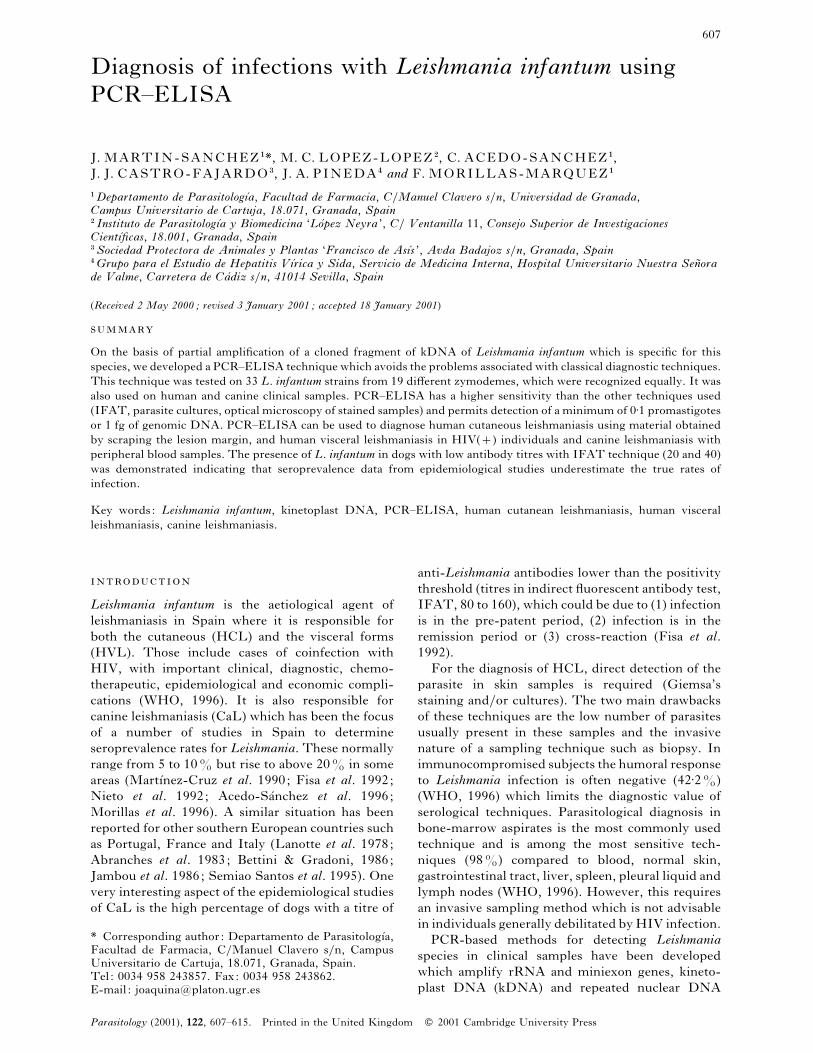
Fig. 1. PCR amplification products generated using
genomic DNA of 3 strains of Leishmania infantum. Lane
1, PCR negative control ; Lane 2, DP74 strain; Lane 3,
DP152 strain; Lane 4, DP204 strain; Lane 5, DNA
molecular weight marker V of Boehringer Mannheim.
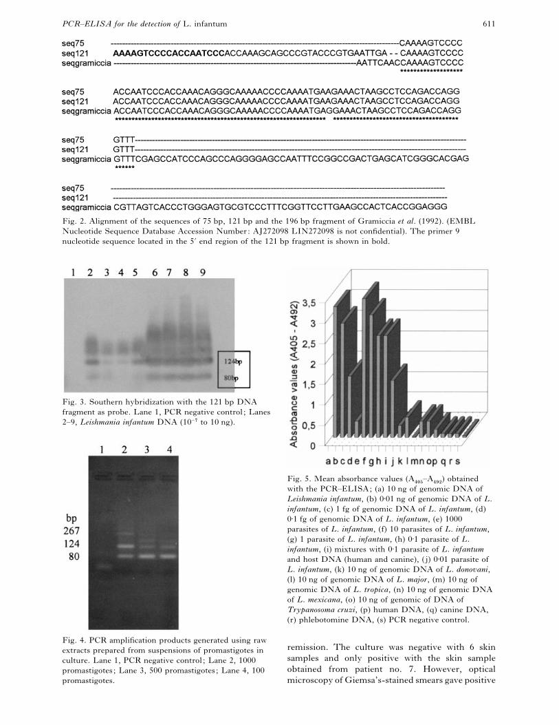
generated 2 main products of 75 and 121 bp (Fig. 1)
determined by DNA sequencing. The 75 bp frag-
ment had 98±7% identity with the fragment theo-
retically designed using the probe of Gramiccia et al.
(1992) (only one of 75 bp was different). The
sequence of the 75 bp fragment was contained within
the 121 bp fragment (Fig. 2). The amplification of
this band of higher size is probably generated by the
presence in the L. infantum kDNA minicircle,
upstream of the primer 9 annealing site, of a region
with high homology to the 3« end of the primer 9.
Since the annealing temperature used in PCR
experiment was higher than Tm of the primer 9 we
suggest that at least a large primer proportion is
present upstream of the 75 bp minicircle fragment.
These same bands were amplified when the re-
maining 30 strains of L. infantum (Table 1) were
used. For some strains, other bands corresponding
to fragments larger than 121 bp were amplified. All
the amplification products of the 33 strains of L.
infantum hybridized with the 2 probes used (genomic
DNA and the 121 bp fragment) (data not shown).
Amplification of 5–50 ng of genomic DNA of the
other Leishmania species, including L. donovani, and
T. cruzi, did not generate any product. Amplification
was also negative when we used host DNA (man,
dog, sandfly) free from L. infantum DNA (data not
shown).
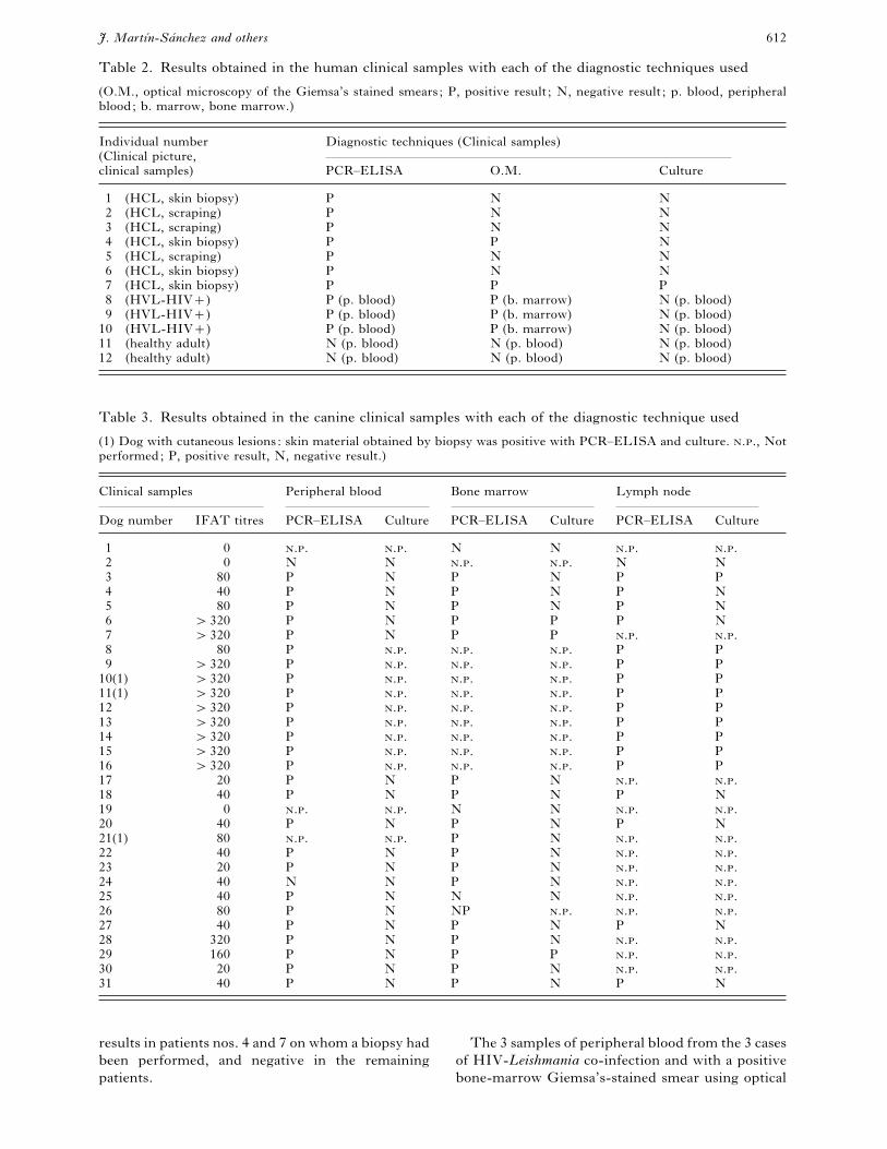
Sensitivity of the PCR assay
Serial dilutions of 10 ng L. infantum genomic DNA
were subjected to PCR and the PCR products were
analysed in agarose gel containing ethidium bromide.
The assay was positive, with amplification of the 75
and 121 bp fragments up to dilution 1}10$, which is
equivalent to the detection of approximately 10 pg of
total DNA. Lower amounts of input DNA gave
negative results. With Southern hybridization it was
possible to detect 1 fg of genomic DNA (dilution
1}10() (Fig. 3). Also, raw extracts prepared from
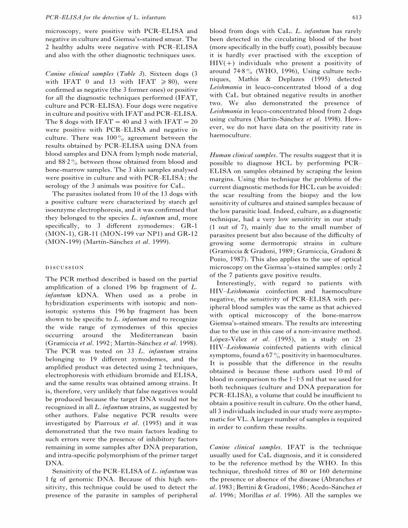
suspensions of promastigotes in culture were used
for PCR, and the detection limit was 50–100
promastigotes (Fig. 4).
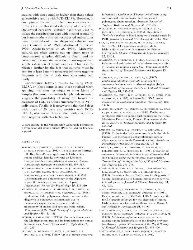
ELISA detection of PCR products
This was designed to solve the two problems posed
by detection by electrophoresis of PCR products i.e.
(1) to increase the sensitivity and (2) amplification
of fragments of higher molecular weight than the two
main products of 75 and 121 bp.
The mean absorbance value (A%!&
–A%*#
) of the
PCR control blank (PCR without DNA) was 0±04,
and PCR with only host DNA (human or canine)
was 0±1. The absorbance values obtained for L.
donovani, L. tropica, L. mexicana and L. major were
lower than 0±5 and for T. cruzi was below 0±2. For the
33 strains of L. infantum studied this was always
greater than 3±0 (Fig. 5).
The detection limit of the PCR–ELISA assay
performed with serial dilutions of 10 ng of L.
infantum genomic DNA (DP74 strain) was 1 fg
(dilution 1}10(), and when this was determined
using raw promastigote extract it corresponded to
0±1 promastigotes. In all cases, the absorbance
readings (A%!&
–A%*#
) were always greater than 1±0.
This corresponds to a rise in the sensitivity of
PCR–ELISA in comparison with PCR detection of
the amplified product by electrophoresis of 100–1000
times that obtained with PCR and ethidium bro-
mide. Amplification of the mixtures with different
proportions of raw extract of 1 promastigote, or L.
infantum genomic DNA, and host DNA (human or
canine) did not bring about changes in the
absorbance levels compared to when L. infantum
DNA was amplified in the absence of host DNA
(Fig. 5: h and i).
Clinical samples: comparison of PCR–ELISA with
other diagnostic techniques applied to HCL, HVL
and CaL
The results obtained with the different samples of
blood, bone-marrow, lymph node material and
human or canine skin, with each of the diagnostic
techniques used, are compiled in Tables 2 and 3.
The reading was considered as positive when
absorbance (A%!&
–A%*#
) was &1.
Human clinical samples (Table 2). The 7 patients with
a clinical picture suggestive of HCL were positive
with PCR–ELISA. Patient no. 1 had been diagnosed
with HCL several years previously and had received
specific treatment for this consisting of infiltration
into the lesion of Glucantime and systemic adminis-
tration of Allopurinol, without achieving complete
PCR–ELISA for the detection of L. infantum 611
Fig. 2. Alignment of the sequences of 75 bp, 121 bp and the 196 bp fragment of Gramiccia et al. (1992). (EMBL
Nucleotide Sequence Database Accession Number: AJ272098 LIN272098 is not confidential). The primer 9
nucleotide sequence located in the 5« end region of the 121 bp fragment is shown in bold.
Fig. 3. Southern hybridization with the 121 bp DNA
fragment as probe. Lane 1, PCR negative control ; Lanes
2–9, Leishmania infantum DNA (10−( to 10 ng).
Fig. 4. PCR amplification products generated using raw
extracts prepared from suspensions of promastigotes in
culture. Lane 1, PCR negative control ; Lane 2, 1000
promastigotes ; Lane 3, 500 promastigotes ; Lane 4, 100
promastigotes.
Fig. 5. Mean absorbance values (A%!&
–A%*#
) obtained
with the PCR–ELISA; (a) 10 ng of genomic DNA of
Leishmania infantum, (b) 0±01 ng of genomic DNA of L.
infantum, (c) 1 fg of genomic DNA of L. infantum, (d)
0±1 fg of genomic DNA of L. infantum, (e) 1000
parasites of L. infantum, (f) 10 parasites of L. infantum,
(g) 1 parasite of L. infantum, (h) 0±1 parasite of L.
infantum, (i) mixtures with 0±1 parasite of L. infantum
and host DNA (human and canine), ( j) 0±01 parasite of
L. infantum, (k) 10 ng of genomic DNA of L. donovani,
(l) 10 ng of genomic DNA of L. major, (m) 10 ng of
genomic DNA of L. tropica, (n) 10 ng of genomic DNA
of L. mexicana, (o) 10 ng of genomic of DNA of
Trypanosoma cruzi, (p) human DNA, (q) canine DNA,
(r) phlebotomine DNA, (s) PCR negative control.
remission. The culture was negative with 6 skin
samples and only positive with the skin sample
obtained from patient no. 7. However, optical
microscopy of Giemsa’s-stained smears gave positive
J. MartıUn-SaU nchez and others 612
Table 2. Results obtained in the human clinical samples with each of the diagnostic techniques used
(O.M., optical microscopy of the Giemsa’s stained smears; P, positive result ; N, negative result ; p. blood, peripheral
blood; b. marrow, bone marrow.)
Individual number
(Clinical picture,
clinical samples)
Diagnostic techniques (Clinical samples)
PCR–ELISA O.M. Culture
1 (HCL, skin biopsy) P N N
2 (HCL, scraping) P N N
3 (HCL, scraping) P N N
4 (HCL, skin biopsy) P P N
5 (HCL, scraping) P N N
6 (HCL, skin biopsy) P N N
7 (HCL, skin biopsy) P P P
8 (HVL-HIV) P (p. blood) P (b. marrow) N (p. blood)
9 (HVL-HIV) P (p. blood) P (b. marrow) N (p. blood)
10 (HVL-HIV) P (p. blood) P (b. marrow) N (p. blood)
11 (healthy adult) N (p. blood) N (p. blood) N (p. blood)
12 (healthy adult) N (p. blood) N (p. blood) N (p. blood)
Table 3. Results obtained in the canine clinical samples with each of the diagnostic technique used
(1) Dog with cutaneous lesions: skin material obtained by biopsy was positive with PCR–ELISA and culture. .., Not
performed; P, positive result, N, negative result.)
Clinical samples Peripheral blood Bone marrow Lymph node
Dog number IFAT titres PCR–ELISA Culture PCR–ELISA Culture PCR–ELISA Culture
1 0 .. .. N N .. ..
2 0 N N .. .. N N
3 80 P N P N P P
4 40 P N P N P N
5 80 P N P N P N
6 "320 P N P P P N
7 "320 P N P P .. ..
8 80 P .. .. .. P P
9 "320 P .. .. .. P P
10(1) "320 P .. .. .. P P
11(1) "320 P .. .. .. P P
12 "320 P .. .. .. P P
13 "320 P .. .. .. P P
14 "320 P .. .. .. P P
15 "320 P .. .. .. P P
16 "320 P .. .. .. P P
17 20 P N P N .. ..
18 40 P N P N P N
19 0 .. .. N N .. ..
20 40 P N P N P N
21(1) 80 .. .. P N .. ..
22 40 P N P N .. ..
23 20 P N P N .. ..
24 40 N N P N .. ..
25 40 P N N N .. ..
26 80 P N NP .. .. ..
27 40 P N P N P N
28 320 P N P N .. ..
29 160 P N P P .. ..
30 20 P N P N .. ..
31 40 P N P N P N
results in patients nos. 4 and 7 on whom a biopsy had
been performed, and negative in the remaining
patients.
The 3 samples of peripheral blood from the 3 cases
of HIV-Leishmania co-infection and with a positive
bone-marrow Giemsa’s-stained smear using optical
PCR–ELISA for the detection of L. infantum 613
microscopy, were positive with PCR–ELISA and
negative in culture and Giemsa’s-stained smear. The
2 healthy adults were negative with PCR–ELISA
and also with the other diagnostic techniques uses.
Canine clinical samples (Table 3). Sixteen dogs (3
with IFAT 0 and 13 with IFAT &80), were
confirmed as negative (the 3 former ones) or positive
for all the diagnostic techniques performed (IFAT,
culture and PCR–ELISA). Four dogs were negative
in culture and positive with IFAT and PCR–ELISA.
The 8 dogs with IFAT¯40 and 3 with IFAT¯20
were positive with PCR–ELISA and negative in
culture. There was 100% agreement between the
results obtained by PCR–ELISA using DNA from
blood samples and DNA from lymph node material,
and 88±2% between those obtained from blood and
bone-marrow samples. The 3 skin samples analysed
were positive in culture and with PCR–ELISA; the
serology of the 3 animals was positive for CaL.
The parasites isolated from 10 of the 13 dogs with
a positive culture were characterized by starch gel
isoenzyme electrophoresis, and it was confirmed that
they belonged to the species L. infantum and, more
specifically, to 3 different zymodemes: GR-1
(MON-1), GR-11 (MON-199 var NP1) and GR-12
(MON-199) (Martı!n-Sa!nchez et al. 1999).
The PCR method described is based on the partial
amplification of a cloned 196 bp fragment of L.
infantum kDNA. When used as a probe in
hybridization experiments with isotopic and non-
isotopic systems this 196 bp fragment has been
shown to be specific to L. infantum and to recognize
the wide range of zymodemes of this species
occurring around the Mediterranean basin
(Gramiccia et al. 1992; Martı!n-Sa!nchez et al. 1998).
The PCR was tested on 33 L. infantum strains
belonging to 19 different zymodemes, and the
amplified product was detected using 2 techniques,
electrophoresis with ethidium bromide and ELISA,
and the same results was obtained among strains. It
is, therefore, very unlikely that false negatives would
be produced because the target DNA would not be
recognized in all L. infantum strains, as suggested by
other authors. False negative PCR results were
investigated by Piarroux et al. (1995) and it was
demonstrated that the two main factors leading to
such errors were the presence of inhibitory factors
remaining in some samples after DNA preparation,
and intra-specific polymorphism of the primer target
DNA.
Sensitivity of the PCR–ELISA of L. infantum was
1 fg of genomic DNA. Because of this high sen-
sitivity, this technique could be used to detect the
presence of the parasite in samples of peripheral
blood from dogs with CaL. L. infantum has rarely
been detected in the circulating blood of the host
(more specifically in the buffy coat), possibly because
it is hardly ever practised with the exception of
HIV() individuals who present a positivity of
around 74±8% (WHO, 1996), Using culture tech-
niques, Mathis & Deplazes (1995) detected
Leishmania in leuco-concentrated blood of a dog
with CaL but obtained negative results in another
two. We also demonstrated the presence of
Leishmania in leuco-concentrated blood from 2 dogs
using cultures (Martı!n-Sa!nchez et al. 1998). How-
ever, we do not have data on the positivity rate in
haemoculture.
Human clinical samples. The results suggest that it is
possible to diagnose HCL by performing PCR–
ELISA on samples obtained by scraping the lesion
margins. Using this technique the problems of the
current diagnostic methods for HCL can be avoided:
the scar resulting from the biopsy and the low
sensitivity of cultures and stained samples because of
the low parasitic load. Indeed, culture, as a diagnostic
technique, had a very low sensitivity in our study
(1 out of 7), mainly due to the small number of
parasites present but also because of the difficulty of
growing some dermotropic strains in culture
(Gramiccia & Gradoni, 1989; Gramiccia, Gradoni &
Pozio, 1987). This also applies to the use of optical
microscopy on the Giemsa’s-stained samples: only 2
of the 7 patients gave positive results.
Interestingly, with regard to patients with
HIV–Leishmania coinfection and haemoculture
negative, the sensitivity of PCR–ELISA with per-
ipheral blood samples was the same as that achieved
with optical microscopy of the bone-marrow
Giemsa’s-stained smears. The results are interesting
due to the use in this case of a non-invasive method.
Lo! pez-Ve! lez et al. (1995), in a study on 25
HIV–Leishmania coinfected patients with clinical
symptoms, found a 67% positivity in haemocultures.
It is possible that the difference in the results
obtained is because these authors used 10 ml of
blood in comparison to the 1–1±5 ml that we used for
both techniques (culture and DNA preparation for
PCR–ELISA), a volume that could be insufficient to
obtain a positive result in culture. On the other hand,
all 3 individuals included in our study were asympto-
matic for VL. A larger number of samples is required
in order to confirm these results.
Canine clinical samples. IFAT is the technique
usually used for CaL diagnosis, and it is considered
to be the reference method by the WHO. In this
technique, threshold titres of 80 or 160 determine
the presence or absence of the disease (Abranches et
al. 1983; Bettini & Gradoni, 1986; Acedo-Sa!nchez et
al. 1996; Morillas et al. 1996). All the samples we
J. MartıUn-SaU nchez and others 614
studied with titres equal or higher than these values
gave positive results with PCR–ELISA. However, in
our opinion the main problem concerns sera with
titres below the threshold values (e.g. of 20 and 40).
On several occasions, cultures have been used to
isolate the parasite from dogs with titres of around 40
but in many others this has not occurred and cultures
have proven to be of limited diagnostic value in these
cases (Lanotte et al. 1978; Martı!nez-Cruz et al.
1990; Acedo-Sa!nchez et al. 1996). Moreover,
cultures are often carried out on lymph node or
bone-marrow samples, difficult to obtain, and in-
volve a more traumatic invasion of host organs than
simple extraction of blood samples. This is com-
plicated further by the fact that cultures must be
kept for at least 1 month to make a definitive negative
diagnosis and this is both time consuming and
costly.
Concordance between results by using PCR–
ELISA on blood samples and those obtained when
applying this same technique to other kinds of
samples (bone-marrow and}or lymph node material)
suggests the possibility of using the former for
diagnosis of CaL, as occurs currently with HIV()
individuals. Finally, it is noteworthy that the 3 dogs
with titres of 20 were also positive with PCR–
ELISA, whereas all those studied with a zero titre
were negative with this technique.
We are grateful to the Subdireccio! n General de Formacio! ny Promocio! n del Conocimiento (PM97-0176) for financial
support.
, ., , . ., , . . ., ,
. . . , . . (1983). Le kala-azar au Portugal.
III. Re! sultats d’une enque# te sur la leishmaniose
canine re! alise! e dans les environs de Lisbonne.
Comparison des zones urbaines et rurales. Annales
Parasitologie Humaine et CompareU e 58, 307–315.
-, ., -, ., -,
. ., -, . ., , .,
, . . -, . (1996).
Leishmaniasis eco-epidemiology in the Alpujarra
region (Granada province, southern Spain).
International Journal for Parasitology 25, 303–310.
, ., , ., -, . ., , .,
, ., , . . , . (1996).
Evaluation of the polymerase chain reaction in the
diagnosis of cutaneous leishmaniasis due to
Leishmania major : a comparison with direct
microscopy of smears and sections from lesions.
Transactions of the Royal Society of Tropical Medicine
and Hygiene 90, 133–135.
, . , . (1986). Canine leishmaniasis in
the Mediterranean area and its implications for human
leishmaniasis. Insect Science and its Applications 7,
241–245.
, ., , ., , ., , .
, . . (1996). Follow-up of a human accidental
infection by Leishmania (Viannia) braziliensis using
conventional immunological techniques and
polymerase chain-reaction. American Journal of
Tropical Medicine and Hygiene 55, 267–272.
’, ., , ., , . .,
, . , . (1995). Detection of
Theileria annulata in blood samples of carrier cattle by
PCR. Journal of Clinical Microbiology 33, 2665–2669.
, ., , ., , ., , . ,
. . (1992). El diagno! stico serolo! gico de la
leishmaniosis canina en la comarca del Priorat
(Tarragona). ClıUnica Veterinaria de pequenh os animales
12, 33–38.
, . , . (1989). Successful in vitro
isolation and cultivation of italian dermotropic strains
of Leishmania infantum sensu lato. Transactions of the
Royal Society of Tropical Medicine and Hygiene 83,
76.
, ., , . , . (1987).
Leishmania infantum sensu lato as an agent of
cutaneous leishmaniasis in Abruzzi region (Italy).
Transactions of the Royal Society of Tropical Medicine
and Hygiene 81, 235–237.
, ., , . ., , . ., , . .
, . (1992). A kinetoplast DNA probe
diagnostic for Leishmania infantum. Parasitology 105,
29–34.
, ., , ., , ., , ., ,
., , . , . (1986). Preliminary
serological study on canine leishmaniasis in the Alpes
Maritimes Department, France. Transactions of the
Royal Society of Tropical Medicine and Hygiene 80,
666–667.
, ., , . ., , . , .
(1978). Ecologie des Leishmanioses dans le Sud de la
France. Les me! thodes d’echantillonnage dans le
de!pistage et l’analyse de l’enzootie canine. Annales de
Parasitologie Humaine et CompareU e 53, 33–45.
, ., , . ., , ., , .,
, . , . (1995). Detection of
cutaneous Leishmania infection in paraffin-embedded
skin biopsies using the polymerase chain reaction.
Transactions of the Royal Society of Tropical Medicine
and Hygiene 89, 273–275.
-, ., , ., , ., -,
. ., , ., , . , .
(1995). Parasitic culture of buffy coat for diagnosis of
visceral leishmaniasis in human immodeficiency virus-
infected patients. Journal of Clinical Microbiology 33,
937–939.
-, ., , ., , . .,
-, . -, . (1998).
Evaluation of the PLiD2-196bp kDNA probe specific
for Leishmania infantum for the diagnosis of canine
leishmaniasis in a focus of southern Spain. Research
and Reviews in Parasitology 58, 91–94.
-, ., , . ., , ., , . .,
, . ., -, . , .
(1999). Leishmania infantum enzymatic variants
causing canine leishmaniasis in the Huelva province
(south-west Spain). Transactions of the Royal Society
of Tropical Medicine and Hygiene 93, 495–496.
-, ., -, ., -
, . . -, . (1994).
PCR–ELISA for the detection of L. infantum 615
Isoenzymatic characterization of the etiologic agent of
canine leishmaniasis in the Granada region of
southern Spain. American Journal of Tropical
Medicine and Hygiene 50, 758–762.
-, ., -, ., -
, . ., -, ., -
, ., -, . ., -
, . -, . (1996).
Leishmania infantum Nicolle, 1908 from southern
Spain: Characterisation of the strains from human
visceral and cutaneous leishmaniasis and from
sandflies; with a numerical analysis of the
isoenzymatic data. Systematic Parasitology 33,
177–182.
-, ., -, ., -
, . ., -! , . -
, . (1990). Epidemiologı!a de la
Leishmaniosis canina en Co! rdoba. Revista IbeU rica de
Parasitologia 50, 1–7.
, . , . (1995). PCR and in vitro
cultivation for detection of Leishmania spp. In
diagnostic samples from humans and dogs. Journal of
Clinical Microbiology 33, 1145–1149.
, ., -, ., 4 , ., -
, ., 4 -, ., , . -
, . . (1996). Leishmaniosis in the focus of the
Axarquı!a region, Malaga province, southern Spain: a
survey of the human, dog, and vector. Parasitology
Research 82, 569–570.
, . ., , ., , . -
, . (1992). Seroprevalence of canine
leishmaniasis around Ca! ceres, Spain. Preventive
Veterinary Medicine 13, 173–178.
, . ., , ., , . . , .
(1998). A nested-PCR-based schizodeme method for
identifying Leishmania kinetoplast minicircle classes
directly from clinical samples and its application to
the study of the epidemiology of Leishmania tropica in
Pakistan. Journal of Clinical Microbiology 36,
2877–2881.
, ., , ., , . ., , .,
, ., , ., , ., ,
. , . (1993). Isolation and characterization
of a repetitive DNA sequence from Leishmania
infantum : development of a visceral leishmaniasis
polymerase chain reaction. American Journal of
Tropical Medicine and Hygiene 49, 364–369.
, ., , ., , ., , .,
, ., , . , . (1995).
Phylogenetic relationships between Old World
Leishmania strains revealed by analysis of a repetitive
DNA sequence. Molecular and Biochemical
Parasitology 32, 746–749.
, ., , . . , . . (1995). Detection
of parasites of the Leishmania donovani complex by a
polymerase chain reaction-solution hybridization
enzyme-linked immunoassay (PCR–SHELA).
Parasitology 110, 269–275.
, ., , . ., , ., , . .
, . (1996). Detection and identification of
human pathogenic Leishmania and Trypanosoma
species by hybridization of PCR-amplified mini-exon
repeats. Experimental Parasitology 82, 242–250.
, . . ., , ., , . ., ,
. . -, . . (1994). Discrimination
amongst Leishmania by polymerase chain reaction
and hybridization with small subunit ribosomal DNA
derived oligonucleotides. Journal of Eukaryotic
Microbiology 41, 324–330.
, ., , . . , . (1989).
Molecular Cloning. A Laboratory Manual, 2nd Edn.
Cold Spring Harbor Laboratory Press, Cold Spring
Harbor, USA.
-, . ., , ., , ., ,
. ., , . , . (1995). Evora district as a
new focus for canine leishmaniasis in Portugal.
Parasitology Research 81, 235–239.
(1989). Handbook on
isolation, characterization and cryopreservation of
Leishmania. UNDP}World Bank}WHO Special
Programme for Research and Training in Tropical
Diseases. Ed. David Evans. WHO, Geneva.
(1996). Epidemiological
analysis of 692 retrospective cases of Leishmania}HIV
co-infection. World Health Organization. Division of
Control of Tropical Diseases. WHO}LEISH}96.39.
WHO, Geneva.


