Embed Size (px)
Citation preview
RESEARCH ARTICLE
Coadministration of the Three AntigenicLeishmania infantum Poly (A) BindingProteins as a DNA Vaccine Induces Protectionagainst Leishmania major Infection in BALB/c MiceManuel Soto1☯*, Laura Corvo1, Esther Garde1, Laura Ramírez1, Virginia Iniesta2,Pedro Bonay1, Carlos Gómez-Nieto2, Víctor M. González3, M. Elena Martín3,Carlos Alonso1, Eduardo A. F. Coelho4,5, Aldina Barral6, Manoel Barral-Netto6,Salvador Iborra7☯*
1 Centro de Biología Molecular Severo Ochoa (CSIC-UAM), Departamento de Biología Molecular,Universidad Autónoma de Madrid, Madrid, Spain, 2 LeishmanCeres Laboratory (GLP Compliance Certified),Parasitology Unit. Veterinary Faculty, University of Extremadura, Cáceres, Spain, 3 Departamento deBioquímica-Investigación, Instituto Ramón y Cajal de Investigación Sanitaria (IRYCIS), Hospital Ramón yCajal, Madrid, Spain, 4 Programa de Pós-Graduação em Ciências da Saúde: Infectologia e MedicinaTropical, Faculdade de Medicina, Universidade Federal de Minas Gerais, Belo Horizonte, Minas Gerais,Brazil, 5 Departamento de Patologia Clínica, COLTEC, Universidade Federal de Minas Gerais, BeloHorizonte, Minas Gerais, Brazil, 6 Centro de Pesquisas Gonçalo Moniz (Fundação Oswaldo Cruz-FIOCRUZ), Salvador, Bahia, Brazil, 7 Immunobiology of Inflammation Laboratory, Department of VascularBiology and Inflammation, Centro Nacional de Investigaciones Cardiovasculares (CNIC), Madrid, Spain
☯ These authors contributed equally to this work.* [email protected] (MS); [email protected] (SI)
Abstract
Background
Highly conserved intracellular proteins from Leishmania have been described as antigens in
natural and experimental infected mammals. The present study aimed to evaluate the anti-
genicity and prophylactic properties of the Leishmania infantum Poly (A) binding proteins
(LiPABPs).
Methodology/Principal Findings
Three different members of the LiPABP family have been described. Recombinant tools
based on these proteins were constructed: recombinant proteins and DNA vaccines. The
three recombinant proteins were employed for coating ELISA plates. Sera from human and
canine patients of visceral leishmaniasis and human patients of mucosal leishmaniasis rec-
ognized the three LiPABPs. In addition, the protective efficacy of a DNA vaccine based on
the combination of the three Leishmania PABPs has been tested in a model of progressive
murine leishmaniasis: BALB/c mice infected with Leishmania major. The induction of a Th1-
like response against the LiPABP family by genetic vaccination was able to down-regulate
PLOS Neglected Tropical Diseases | DOI:10.1371/journal.pntd.0003751 May 8, 2015 1 / 24
OPEN ACCESS
Citation: Soto M, Corvo L, Garde E, Ramírez L,Iniesta V, Bonay P, et al. (2015) Coadministration ofthe Three Antigenic Leishmania infantum Poly (A)Binding Proteins as a DNAVaccine InducesProtection against Leishmania major Infection inBALB/c Mice. PLoS Negl Trop Dis 9(5): e0003751.doi:10.1371/journal.pntd.0003751
Editor: Peter C. Melby, University of Texas MedicalBranch, UNITED STATES
Received: November 27, 2014
Accepted: April 11, 2015
Published: May 8, 2015
Copyright: © 2015 Soto et al. This is an openaccess article distributed under the terms of theCreative Commons Attribution License, which permitsunrestricted use, distribution, and reproduction in anymedium, provided the original author and source arecredited.
Data Availability Statement: All relevant data arewithin the paper and its Supporting Information files.
Funding: The study was supported in Spain bygrants from Ministerio de Ciencia e Innovación FISPI11/00095 and FISPI14/00366 from the Instituto deSalud Carlos III within the Network of TropicalDiseases Research (VI P I+D+I 2008-2011, ISCIII-Subdirección General de Redes y Centros deInvestigación Cooperativa (RD12/0018/0009)). Thiswork was also supported in Brazil by a grant fromCNPq (Ciencia sem Fronteiras-PVE 300174/2014-4).
the IL-10 predominant responses elicited by parasite LiPABPs after infection in this murine
model. This modulation resulted in a partial protection against L.major infection. LiPABPvaccinated mice showed a reduction on the pathology that was accompanied by a decrease
in parasite burdens, in antibody titers against Leishmania antigens and in the IL-4 and IL-10
parasite-specific mediated responses in comparison to control mice groups immunized with
saline or with the non-recombinant plasmid.
Conclusion/Significance
The results presented here demonstrate for the first time the prophylactic properties of a
new family of Leishmania antigenic intracellular proteins, the LiPABPs. The redirection of
the immune response elicited against the LiPABP family (from IL-10 towards IFN-γmediat-
ed responses) by genetic vaccination was able to induce a partial protection against the de-
velopment of the disease in a highly susceptible murine model of leishmaniasis.
Author Summary
Leishmaniases are a wide spectrum of parasite diseases caused by the infection of differentspecies of Leishmania genus in several mammalian hosts, such as humans and dogs,among others. The induction of antibody responses against some parasite intracellularproteins implicated in different structural and functional cellular roles has been associatedwith parasite survival, infection progression, down-regulation of the cellular immune re-sponses against the parasites and the development of pathology. The data presented in thiswork show the antigenic nature of the three members of the Leishmania infantum Poly(A) Binding Protein (LiPABP) family in different forms of natural and experimental leish-maniasis. Using a susceptible model of progressive leishmaniasis (BALB/c mice infectedwith Leishmania major), it was shown that the administration of a LiPABPs based com-bined genetic vaccine was able to induce a partial protection against the disease. Redirec-tion of the IL-10 mediated responses elicited by the parasite LiPABPs after L.majorinfection towards an IFN-γ specific production by vaccination was related to protection.The inclusion of the LiPABPs in a protective vaccine formulation against leishmaniasisis discussed.
IntroductionParasites of the genus Leishmania are protozoan organisms that can infect different mammali-an species including humans and dogs. Leishmania promastigote forms present in the insectvectors (phlebotomine sand flies) are transmitted to the vertebrate hosts during the blood meal[1]. After infection, parasites differentiate in the amastigote forms that multiply inside macro-phages. The infected vertebrates can present a wide spectrum of clinical manifestations, global-ly known as leishmaniasis. Depending on the host and the parasite species, the disease canrange from subclinical forms to severe symptomatic leishmaniasis [2]. In humans, there arevarious parasite species that cause cutaneous leishmaniasis (CL), including Leishmania majorin the Old World and Leishmania braziliensis in the NewWorld, among other species. CL isthe less severe form of the disease and, in some cases, patients heal spontaneously. A percentageof the patients infected with L. braziliensis can develop a form of the disease characterized by
Leishmania Antigenic PABPs Based Genetic Vaccine
PLOS Neglected Tropical Diseases | DOI:10.1371/journal.pntd.0003751 May 8, 2015 2 / 24
A CBMSO institutional grant from Fundación RamónAreces is also acknowledged. EAFC is a grantrecipient of CNPq. The funders had no role in studydesign, data collection and analysis, decision topublish, or preparation of the manuscript.
Competing Interests: The authors have declaredthat no competing interests exist.
the presence of lesions in mucosal areas, known as mucosal leishmaniasis (ML) [3]. Visceralleishmaniasis (VL) is the most severe clinical form of the disease and it is presented in humanand canine patients infected with Leishmania infantum (syn. Leishmania chagasi [4]) in theMediterranean basin and in South America, or in human patients infected with Leishmaniadonovani in Asia and Africa [5]. VL patients usually present hepatosplenomegaly accompaniedby other clinical symptoms like fever, polyclonal hypergammaglobulinemia and anaemia [6].Infected dogs develop similar systemic symptoms accompanied by cutaneous lesions in themucous membranes, skin and eyes [7].
The control of infection in mammalian hosts naturally or experimentally infected with dif-ferent Leishmania species depends mainly on the induction of cellular responses able to acti-vate macrophages to eliminate the intracellular pathogens [8]. The induction of CD4+ Th1cells specific for parasite antigens is critical to control infection due to their ability to secreteIFN-γ, but the activation of other cell subsets to produce this cytokine, like Leishmania antigenspecific CD8+ T cells, is also required for a protective host response to L.major infection inthe C57BL/6 mice strain. On the contrary, the generation of an IL-4 driven Th2 response afterL.major infection in the susceptible BALB/c mice strain has been related to disease (reviewedin [9]). In this model of murine leishmaniasis, T cell or B cell derived IL-10 production has alsobeen implicated in the progression of infection [10,11].
The fact that patients recovered from the disease are resistant to Leishmania reinfection canbe taken as an indication that a vaccine is feasible. Regarding prophylaxis in dogs, there arethree commercial vaccines against canine leishmaniasis: Leishmune, based on the Parasite Fu-cose-Manose-Ligand [12], Leish-Tec, a vaccine constructed by the recombinant version of theamastigote specific A2 protein [13] and CaniLeish, composed of promastigote secreted-excreted factors [14], but none of them are able to completely control the disease [15,16]. In ad-dition, there is no vaccine against this parasite in humans. Some of the candidates for the devel-opment of human vaccines have been tested as second generation products (based on parasiteor insect vector saliva defined antigens) or third generation DNA based vaccines in murine ex-perimental models [17]. The establishment of a protective anti-Leishmania response based onthese recombinant molecules requires the induction of parasite specific long-lasting memory Tcells that will expand as effector T cells for the production of IFN-γ dependent responsesspecific for parasite antigens shortly after infection. Some experimental vaccines based onintracellular proteins that form complexes with nucleic acids, like histones or ribosomal pro-teins have been related with protection against the disease in different experimental models[18,19,20,21,22,23].
Poly(A) binding proteins (PABPs) are nuclear or cytosolic factors that contribute to mRNAmetabolism from nuclear polyadenylation and export to translation initiation and control ofRNA turnover in the cytoplasm [24]. PABPs are able to interact with the Poly(A) tail and otherregions of the mRNAs as well as with other protein factors to exert their functions [25]. Threemembers of the PABP family have been described in L.major. Two of them, namely LmPABP1and LmPABP2, are the cytosolic or nuclear (respectively) orthologues of the PABPs from otherkinetoplastids, whereas the third one (LmPABP3) is a nuclear protein only present in the Leish-mania genus among kinetoplastids [26]. Since the L. infantum PABP2 was recognized by thesera from VL symptomatic dogs [27] this work has extended the analysis of the antigenicity ofall the members of the LiPABP family. The results of this work have demonstrated the antige-nicity of the three LiPABPs in human and canine leishmaniasis. In addition, their immunoge-nicity and protective capacity when administered as a third generation DNA combined vaccinehave been evaluated in a susceptible experimental model of murine leishmaniasis: BALB/cmice infected with L.major.
Leishmania Antigenic PABPs Based Genetic Vaccine
PLOS Neglected Tropical Diseases | DOI:10.1371/journal.pntd.0003751 May 8, 2015 3 / 24
Methods
Mice and parasitesFemale BALB/c mice (6–8 weeks old) were purchased from Harlan (Barcelona, Spain). All pro-cedures were performed according to the Directive 2010/63/UE from the European Union andRD53/2103 from the Spanish Government and approved by the Animal Care and Use Com-mittee at the Centro de Biología Molecular Severo Ochoa (reference CEEA-CBMSO 21/138)and the Bioethical Committee of the Consejo Superior de Investigaciones Científicas (reference100/2014). The project was finally authorized by the Government of the Autonomous Commu-nity of Madrid (reference PROEX121/14).
Promastigotes of L. infantum (MCAN/ES/1996/BCN/150/MON-1) and from L.majorclone V1 (MHOM/IL/80/Friedlin) were cultured at 26°C in Schneider medium (Gibco, NY,U.S.A.) supplemented with 10% Fetal Calf Serum (FCS) (Sigma, MO, U.S.A.), 100 U/ml of pen-icillin and 100 μg/ml of streptomycin. Parasites were kept in a virulent state by passage inBALB/c mice. L.major amastigotes were obtained from infected popliteal lymph nodes. Aftertransformation to the promastigote form, parasites were grown until stationary phase and thenharvested for inoculation in the left hind footpad of mice or for Soluble Leishmania Antigen(SLA) preparation as described in [22].
Cloning of DNA sequences coding for L. infantum PABPs andrecombinant products manufacturingThe L. infantum PABP1 (LinJ35.V3.5360), PABP2 (LinJ.35.4200) and PABP3 (LinJ.25.0080)(namely LiPABP1, LiPABP2 and LiPABP3, respectively) coding regions were obtained fromthe L. infantum genome database (www.genedb.org) using the L.major orthologous protein se-quences as probes (LmjF.35.5040, LmjF.35.4130 and LmjF.35.4130) [26]. Coding regions werePCR amplified using genomic DNA from L. infantum (taking advantage of the lack of intronsequences in this genus) and the next specific primers: LiPABP1 forward, 5’-CGGGATCCATGGACAAGCCGATGCAGATC-3’ and reverse, 5’-CGGATATCACGCCGTCTGATGCGCCTT-3’; LiPABP2 forward, 50- CGGGATCCATGGCCTTTACTGGTCCGAATC-30
and reverse, 5’-CGGGATCCTCAAACGCTCATGTCTGCCAAG-3’; LiPABP3 (forward,5’-CGGGATCCATGGTGGTCCCAGTGCAACG-3; reverse, 5’-CGGATATCAGTTGCCAGTGTGCTGCTGAAG-3’. The restriction sites included for cloning purposes (BamHI forLiPABP2 and BamHI/EcoRV for LiPABP1 and LiPABP3) are underlined. The resultant PCRproducts were inserted into pBluescript II SK (Stratagene. CA, U.S.A). For that purpose thePCR inserts were purified using the High Pure PCR Product Purification Kit (Roche Diagnos-tics GmbH, Mannheim, Germany) and the pBluescript II SK and the PCR inserts were digestedwith the indicated restriction enzymes (New England Biolabs, MA, U.S.A.) and ligated with theT4 DNA ligase (New England Biolabs). Positive clones were selected by a restriction analysis(using the enzymes employed for cloning) and double stranded sequenced. For subcloningpurposes, the three inserts containing the coding regions were obtained by digestion withBamHI for LiPABP2 and BamHI/EcoRV for LiPABP1 and LiPABP3, and inserted in two dif-ferent plasmids. For preparing genetic vaccines, the inserts were subcloned in the correspond-ing sites of the pcDNA3 mammalian expression vector (Invitrogen, CA, U.S.A.). The correctorientation of the LiPABP2 insert was monitored by a restriction analysis made with ApaI, anenzyme that cut the LiPABP2 coding region (positions 1651–1656 of a total length of 1756 nu-cleotides; Gene DB sequence LinJ.35.4200) and it is included in the multiple cloning site of thepcDNA3 plasmids downstream the BamHI cut site. For vaccination assays, the pcDNA3vector and the three recombinant pcDNA3 plasmids were purified by the endotoxin-free
Leishmania Antigenic PABPs Based Genetic Vaccine
PLOS Neglected Tropical Diseases | DOI:10.1371/journal.pntd.0003751 May 8, 2015 4 / 24
Giga-preparation Kit (Qiagen, Hilden, Germany). Plasmids used in vaccination assays weresuspended in sterile phosphate-buffered saline (PBS). For recombinant protein preparations,DNA inserts containing the different coding regions were subcloned into the BamHI(LiPABP2) or the BamHI/SmaI sites (LiPABP1 and LiPABP3) of the pQE30 prokaryotic ex-pression vector (Qiagen). The correct LiPABP2 insert orientation was examined by restrictionwith SmaI enzyme, since the LiPABP2 coding region has a SmaI cut site located at nucleotides1173–1178 of a total length of 1756 (Gene DB sequence LinJ.35.4200) and it is also locateddownstream the BamHI cut sequence in the pQE multiple cloning site. Escherichia coli (M15strain) transfected with the recombinant plasmids were employed for over-expression of thehis-tagged proteins. For that purpose, E. coli cultures were grown at 37°C in an orbital shakerto the mid-growth phase (determined by 0.6 value of Optical Density (O.D.) at 600 nm)and the expression was induced by addition of 1 mM IPTG for 4 h at the same conditions.Since the three LiPABPs formed inclusion bodies, proteins were solubilized under denaturingconditions. For ELISA assays, proteins were purified under denaturant conditions onto Ni-nitrilotriacetic-acid-agarose (Ni-NTA, Qiagen) columns following the manufacturer’s instruc-tions (The QIAespressionistTM; https://www.qiagen.com/es/resources/resourcedetail?id=79ca2f7d-42fe-4d62-8676-4cfa948c9435&lang = en). Briefly, proteins were solubilized in bind-ing buffer (20 mM Tris ClH pH 8.0, 0.5 M NaCl, 8 M Urea, 1 mM β-mercaptoethanol) supple-mented with 5 mM Imidazole, and mounted into a Ni-NTA gravity flow column. Afterwashing in the binding buffer supplemented with 20 mM imidazole, proteins were eluted usingthe binding buffer supplemented with 0.5 M imidazole. After, purified proteins were dialyzedagainst ELISA denaturant coating buffer (20 mM Tris ClH pH 8.0, 0.5 M NaCl, 3 M Urea, 1mM β-mercaptoethanol). For immunization or cells culture experiments, proteins were purifiedunder denaturing conditions and refolded on the affinity column as described in [28] adding20% (p/v) glycerol in those solutions that had not the denaturant agent (urea) to maintain pro-tein solubility [29]. After purification, proteins were dialyzed against PBS plus 20% (p/v) glycer-ol. Polymyxin-agarose columns (Sigma) were employed to remove residual endotoxin content(<10 pg of LPS per μg of recombinant protein, measured by the Quantitative ChromogenicLimulus Amebocyte Assay QCL-1000 (BioWhittaker, MD, U.S.A.)). Protein concentration wasestimated by Bradford method using the Bio-Rad Protein assay (Bio-Rad, CA, U.S.A.).
Sera and ELISA assaysHuman serum samples were obtained from clinical and parasitologically diagnosed patients.For the present study, anonymized samples were randomly selected from a sera bank(LIP-CPqGM-FIOCRUZ) built for previous independent studies conducted in Brazil in en-demic areas for ML [30] or VL [31]. Canine symptomatic VL sera from L. infantum infecteddogs were collected in the Extremadura region of Spain [32]. Control sera were obtained fromhealthy individuals (humans or dogs). For ELISA, MaxiSorp plates (Nunc, Roskilde, Denmark)were coated with 0.2 μg of each one of the recombinant antigens (diluted in ELISA denaturantcoating buffer) for 12 h at 4°C. After four washes with PBS + 0.5% Tween 20 (PBST) wells freebinding sites were blocked with blocking solution (PBST + 5% non-fat milk solution) for 1 h atroom temperature (RT) and incubated with human or canine sera for 2 h at RT (1/200 dilutionin blocking solution). After four washes in PBST, wells were incubated with secondary antibod-ies conjugated to horseradish peroxidase at 1/2,000 dilution in blocking solution for 1 h at RT(anti-dog IgG or anti-human IgG from Nordic Immunological Laboratories, Tilburg, TheNetherlands). After four washes in PBST, the reaction was developed through incubation withorto-phenylenediamine 10 min in the dark, and stopped by addition of 2 N H2SO4. Opticaldensities were read at 450 nm in an ELISA microplate spectrophotometer (Bio-Rad).
Leishmania Antigenic PABPs Based Genetic Vaccine
PLOS Neglected Tropical Diseases | DOI:10.1371/journal.pntd.0003751 May 8, 2015 5 / 24
From the experimental model, sera were obtained at the beginning of the immunization(pre-immune sera), four weeks after the last doses (just before challenge with parasites) as wellas seven weeks after challenge with L.major. The procedure for the ELISAs were the same indi-cated above but the reciprocal end-point titer (defined as the inverse of the highest serum dilu-tion factor giving an absorbance> 0.15) against the recombinant proteins (0.2 μg per well) orSLA (1 μg per well) was determined by serial dilution of the sera. Anti-IgG, anti-IgG1 or anti-IgG2a (all of them at 1/2,000) horseradish peroxidase-conjugated anti-mouse immunoglobu-lins were used as secondary antibodies (purchased by Nordic Immunological Laboratories).
Expression of LiPABPs in mammalian cellsTo confirm that the DNA constructs were functional, COS7 cells were independently trans-fected with 20 μg of pcDNA3-LiPABP1, pcDNA3-LiPABP2 or pcDNA3-LiPABP3 using theFugene reagent (Promega, WI, U.S.A.) according to the manufacturer's protocol. Seventy-twohours post-transfection, the cells were harvested, washed two times with ice-cold PBS, and im-mediately lysed by the addition of Laemmli's buffer. The proteins were resolved by sodiumdodecyl sulfate-polyacrylamide gel electrophoresis (SDS-PAGE) and transferred to nitrocellu-lose membranes (Amersham, Aylesbury, United Kingdom). The blots were probed with mouseantiserum raised against the LiPABPs (1/200) by the independent immunization of the recom-binant LiPABPs (three doses, 12 μg of each one) combined with 25 μg of the next two phos-phorothioate-modified CpG-ODNs: (5’-TCAACGTTGA-3’ and 5’- GCTAGACGTTAGCGT-3’). Anti-IgG (1/2,000) horseradish peroxidase-conjugated anti-mouse immunoglobulins wereemployed as secondary antibodies (Nordic Immunological Laboratories). As control, similarblots were probed with an anti-human PABP antibody (Cell Signaling, Merck Millipore Darm-stadt, Germany) and horseradish peroxidase-conjugated anti-mouse immunoglobulins (1/2,000; Nordic Immunological Laboratories) as secondary reagent. Western blots treated withanti-LiPABPs sera were revealed with the ECLWestern Blotting System (GE HealthcareLife Sciences, PA, U.S.A.) and those probed with anti-human PABP were revealed with4-chloronaphthol (Sigma).
Immunization, parasite challenge and parasite quantificationThree mice groups (n = 16 per group) were made. The LiPABP vaccinated group was inoculat-ed in the right hind footpad with 200 μg of a mixture of the next recombinant plasmids:pcDNA3-LiPABP1, pcDNA3-LiPABP2 or pcDNA3-LiPABP3 (66.6 μg of each plasmid). Ascontrol groups, mice were inoculated with PBS or with 200 μg of the pcDNA3 plasmid. Micewere inoculated three times (two weeks apart). Four weeks after last dose eight mice per groupwere euthanized, in order to obtain their spleens to be used in the analysis of the cellular im-mune responses induced by immunizations. Parasite challenge was carried out at the sametime using the remaining mice by subcutaneous inoculation with 5 × 10⁴ stationary-phase pro-mastigotes of L.major into their left footpads. For the analysis of the clinical signs of infection,footpad swelling was measured with a metric caliper (expressed as thickness of the infected leftfootpad minus thickness of the right footpad). For parasite load determination, the spleen andthe single popliteal lymph node draining the site of infection (left leg) were collected and inde-pendently processed. The complete spleens or lymph nodes were mechanically homogenizedin Schneider's medium supplemented with 20% heat-inactivated FCS, 200 U/ml penicillin and100 μg/ml streptomycin and filtered using a cell strainer (70-μm pore size). Each homogenizedsample tissue was serially diluted (1/3) in a 96-well flat-bottomed microtiter plate containingthe same medium (in triplicates). The number of viable parasites was determined from the
Leishmania Antigenic PABPs Based Genetic Vaccine
PLOS Neglected Tropical Diseases | DOI:10.1371/journal.pntd.0003751 May 8, 2015 6 / 24
highest dilution at which promastigotes could be grown up to 10 days of incubation at 26°C aspreviously described [33].
Cytokine productionSpleen cells obtained from mice inoculated with saline, with the pcDNA3 vector or with theLiPABPs combined genetic vaccine were seeded and independently cultured in RPMI completemedium at 5 × 10⁶ cells per ml (RPMI medium (Sigma)) supplemented with 10% heat-inacti-vated FCS, 20 mM L-glutamine, 200 U/ml penicillin, 100 μg/ml streptomycin and 50 μg/mlgentamicin, during 72 h at 37°C in 5% CO2. Mice (n = 8 per group) were euthanized fourweeks after the last dose or seven weeks after parasite challenge. Cells were cultured alone orwith some of the next stimuli: SLA, the recombinant LiPABP1, LiPABP2, LiPABP3 proteins as-sayed separately (all of them at 12 μg/ml of final concentration), or with a mixture of the threerecombinant LiPABPs at 4 μg/ml each one. The levels of IFN-γ, IL-10 or IL-4 were determinedin the culture supernatants by sandwich ELISA using monoclonal antibodies specific formouse cytokines (capture and detection) provided in commercial kits (Pharmingen, SanDiego, CA, USA), following the manufacturer’s instructions.
Statistical analysisStatistical analysis was performed using the Graph-Pad Prism program. Data were first ana-lyzed by the D'Agostino & Pearson normality test. Parametric data were analyzed by a two-tailed Student's t-test and non-parametric data were analyzed by a MannWhitney test.For both test differences were considered significant when �(+) P< 0.05, ��(++) P< 0.01 or���(+++) P< 0.001.
Results
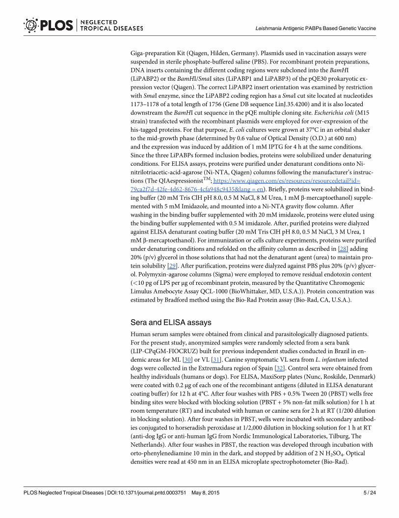
Antigenicity of the Leishmania PABPs during natural infectionsFirst, the antigenicity of the members of the Leishmania PABP was determined. For this pur-pose, the three LiPABPs were expressed as recombinant proteins in E. coli, purified by affinitychromatography and employed for ELISA to investigate the presence of circulating antibodiesagainst them in the sera from human and canine patients affected by VL. The three LiPABPswere recognized by Brazilian human patients suffering VL due to the infection of L. chagasi(syn. L. infantum) (Fig 1A), and by sera samples from Spanish VL symptomatic dogs infectedby L. infantum (Fig 1B). On the basis of the high degree of identity observed between L. infan-tum and L. braziliensis counterparts (S1 Fig) the cross reactivity between LiPABPs and serafrom patients affected by ML due to L. braziliensis was assayed. The reactivity of the sera fromML patients was significantly higher than that from healthy donors for the three LiPABPs (Fig1A). From these data, it was concluded that the three members of the LiPABP family interactwith the host immune system after infection in different vertebrate hosts.
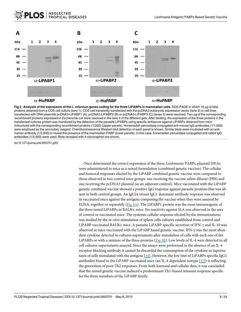
Immunogenicity of LiPABPs administered as a combined DNA-vaccineTo evaluate the immunogenicity of the Leishmania PABPs administered as a combined DNAvaccine, the coding regions for the LiPABPs (LiPABP1, LiPABP2 and LiPABP3) were individu-ally cloned into the eukaryotic expression vector pcDNA3. The correct expression of the DNAvaccines was tested in culture analyzing the expression of the LiPABPs in COS7 cells transfectedwith the recombinant plasmids (Fig 2, upper panels). Using sera frommice immunized with therecombinant versions of the parasite LiPABPs, specific protein bands were observed in the threeimmunoblot panels in the lanes corresponding to cell cultures transfected with the recombinant
Leishmania Antigenic PABPs Based Genetic Vaccine
PLOS Neglected Tropical Diseases | DOI:10.1371/journal.pntd.0003751 May 8, 2015 7 / 24
plasmids (Fig 2, upper panels, lanes 3). No reactivity was observed against proteins from COS7cell samples un-transfected (Fig 2, upper panels, lanes 1) or transfected with the non-recombi-nant plasmid (Fig 2, upper panels, lanes 2). The presence of the mammalian PABP was revealedby probing similar blots with an anti-human PABP antibody (Fig 2, lower panels). The molecu-lar weights of the parasite LiPABPs expressed by mammalian cells were close to the expected(hypothetically calculated from their amino acid sequences) and slightly lower to that observedfor the recombinant LiPABPs expressed in bacteria and possessing a histidine tag in the amino-terminal region (Fig 2, upper panels, lanes 4). The lower bands detected by anti-LiPABPs anti-bodies may result from cleavage of the proteins expressed in the mammalian cells (Fig 2, upperpanels, lanes 3). Similarly, lower bands observed in the lane 4, upper panel of the Fig 2B, may berelated to partially cleaved forms of the recombinant LiPABP2 protein expressed in E. coli, sincethey were absent in protein samples purified under denaturant conditions and appeared whenthe denaturant agent was removed by dialysis against PBS (S2 Fig).
Fig 1. The LiPABPs are antigenic in humans and dogs suffering leishmaniasis. Antibody response ofhumanML (n = 20) or VL patients (n = 20) and healthy individuals (HC, n = 8) against the three recombinantLiPABP proteins (A). Antibody responses of canine VL (VL, n = 38) and healthy animal sera (HC, n = 18)against the recombinant proteins (B). All sera were tested for IgG reactivity by ELISA (1/200). Horseradishperoxidase-conjugated anti-human IgG (1/2,000) or anti-dog IgG (1/2,000) antibodies were used as thesecondary reagents. Results are shown as whisker (min to max) plots. * (P < 0.05) and *** (P < 0.001)statistical differences in the IgG reactivity values between each group of patients and their correspondinghealthy control sera group, evaluated by the Mann-Whitney test.
doi:10.1371/journal.pntd.0003751.g001
Leishmania Antigenic PABPs Based Genetic Vaccine
PLOS Neglected Tropical Diseases | DOI:10.1371/journal.pntd.0003751 May 8, 2015 8 / 24
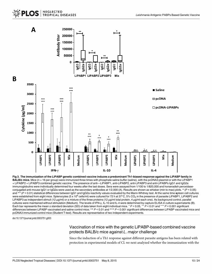
Once determined the correct expression of the three Leishmania PABPs, plasmid DNAswere administered to mice as a mixed formulation (combined genetic vaccine). The cellularand humoral responses elicited by the LiPABP combined genetic vaccine were compared tothose observed in two control mice groups: one receiving the vaccine saline diluent (PBS) andone receiving the pcDNA3 plasmid (as an adjuvant control). Mice vaccinated with the LiPABPgenetic combined vaccine showed a positive IgG response against parasite proteins that was ab-sent in both control groups. An IgG2a versus IgG1 dominant antibody response was observedin vaccinated mice against the antigens composing the vaccine when they were assayed byELISA, together or separately (Fig 3A). The LiPABP1 protein was the most immunogenic ofthe three tested LiPABPs in BALB/c mice. No reactivity against SLA was observed in the seraof control or vaccinated mice. The systemic cellular response elicited by the immunizationswas studied by the in vitro stimulation of spleen cells cultures established from control andLiPABP vaccinated BALB/c mice. A parasite LiPABP-specific secretion of IFN-γ and IL-10 wasobserved in mice vaccinated with the LiPABP based genetic vaccine. IFN-γ was the most abun-dant cytokine detected in cultures supernatants after stimulation of cells with each one of theLiPABPs or with a mixture of the three proteins (Fig 3B). Low levels of IL-4 were detected in allcell cultures supernatants assayed. Since the assays were performed in the absence of an IL-4receptor blocking antibody it cannot be discarded the consumption of the cytokine in superna-tants of cells stimulated with the antigens [34]. However, the low titer of LiPABPs specific IgG1antibodies found in the LiPABP vaccinated mice (an IL-4-dependent isotype [35]) is reflectingthe generation of poor Th2 responses. From both humoral and cellular data, it was concludedthat the mixed genetic vaccine induced a predominant Th1-biased immune response specificfor the three members of the LiPABP family.
Fig 2. Analysis of the expression of the L. infantum genes coding for the three LiPABPs in mammalian cells. SDS-PAGE in which 10 μg of totalproteins obtained from a COS cell culture (lane 1), COS cell transiently transfected with the pcDNA3 eukaryotic expression vector (lane 2) or cell linestransfected with DNA plasmids pcDNA3-LiPABP1 (A), pcDNA3-LiPABP2 (B) or pcDNA3-LiPABP3 (C) (lanes 3) were resolved. Two μg of the correspondingrecombinant proteins expressed in Escherichia coli were resolved in the lane 4 of the different gels. After blotting, the expression of the three proteins in thetransfected cultures protein was monitored by the detection of the parasite LiPABPs using specific antiserum against LiPABPs obtained frommiceimmunized with the corresponding recombinant proteins (1/200) (Upper panels). Horseradish peroxidase-conjugated anti-mouse IgG antibodies (1/1,000)were employed as the secondary reagent. ChemiluminescenceWestern blot detection of each panel is shown. Similar blots were incubated with an anti-human antibody (1/2,000) to reveal the presence of the mammalian PABP (lower panels). In this case, horseradish peroxidase-conjugated anti-rabbit IgGantibodies (1/2,000) were used. Blots revealed with 4-cloronaphtol are shown.
doi:10.1371/journal.pntd.0003751.g002
Leishmania Antigenic PABPs Based Genetic Vaccine
PLOS Neglected Tropical Diseases | DOI:10.1371/journal.pntd.0003751 May 8, 2015 9 / 24
Vaccination of mice with the genetic LiPABP-based combined vaccineprotects BALB/c mice against L.major challengeSince the induction of a Th1 response against different parasite antigens has been related withprotection in experimental models of CL we next analyzed whether the immunization with the
Fig 3. The immunization of the LiPABP genetic combined vaccine induces a predominant Th1-biased response against the LiPABP family inBALB/c mice.Mice (n = 16 per group) were immunized three times with phosphate saline buffer (saline), with the pcDNA3 plasmid or with the LiPABP1+ LiPABP2 + LiPABP3 combined genetic vaccine. The presence of anti—LiPABP1, anti-LiPABP2, anti-LiPABP3 and anti-LiPABPs IgG1 and IgG2aimmunoglobulins were individually determined four weeks after the last doses. Sera were assayed from 1/100 to 1/820,000 and horseradish peroxidase-conjugated anti-mouse IgG1 or IgG2a were used as the secondary antibodies at 1/2,000 (A). Results are shown as whisker (min to max) plots. * (P < 0.05)and ** (P < 0.01) statistical differences between IgG1 and IgG2a reactivity values evaluated by the Mann-Whitney test. At the same time spleen cell cultureswere established from eight mice. Splenocytes (5 x 10⁶ cells/ml) were cultured for 72 h at 37°C, 5% CO2 in the presence of parasite LiPABP1, LiPABP2 andLiPABP3 as independent stimuli (12 μg/ml) or a mixture of the three proteins (12 μg/ml total protein, 4 μg/ml each one). As background control, parallelcultures were maintained without stimulation (Medium). The levels of IFN-γ, IL-10 and IL-4 were determined by capture ELISA in culture supernatants (B).Each bar represents the mean ± standard deviation (SD) of data taken from eight individual mice. + P < 0.05, ++ P < 0.01 and +++ P < 0.001 significantdifferences between LiPABP vaccinated and saline control mice; ** P < 0.01 and *** P < 0.001 significant differences between LiPABP vaccinated mice andpcDNA3 immunized control mice (Student T-test). Results are representative of two independent experiments.
doi:10.1371/journal.pntd.0003751.g003
Leishmania Antigenic PABPs Based Genetic Vaccine
PLOS Neglected Tropical Diseases | DOI:10.1371/journal.pntd.0003751 May 8, 2015 10 / 24
combined vaccine was able to induce protection against L.major infection in the highly suscep-tible BALB/c strain. For that purpose, mice vaccinated with the LiPABP based genetic vaccineas well as mice from control groups (inoculated with the non-recombinant plasmid or the sa-line diluent of the vaccine) were infected with L.major, one month after the last immunization.Mice vaccinated with the LiPABP combined vaccine showed lower footpad swelling than con-trol groups inoculated with the vaccine diluent or the non-recombinant plasmid, from week 3after challenge to the end of the assay (Fig 4A). The time for euthanization, seven weeks post-infection, was determined by the appearance of necrotic lesions in control groups that were ab-sent in the LiPABP vaccinated group. In addition, LiPABP vaccinated mice showed a 1.25-logand 1.21-log reduction in the parasite burdens of the lymph node draining the infected footpad(popliteal lymph node) relative to saline and pcDNA3 immunized control groups, respectively(Fig 4B). Similarly, it was determined the existence of a 1.5-log and 1.4-log reduction in thenumber of viable parasite detected in the spleen of LiPABP vaccinated mice relative to bothcontrol groups (Fig 4B).
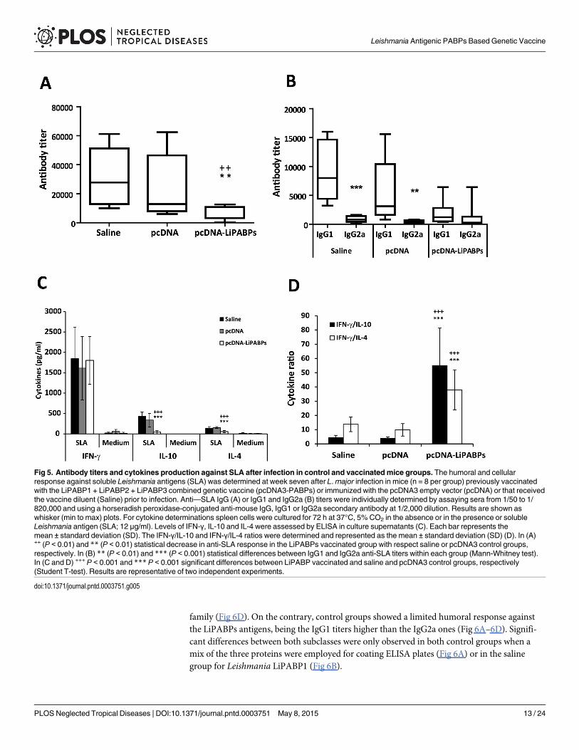
Since the level of the humoral responses elicited against leishmanial antigens in mice in-fected with L.major has been correlated to the severity of the disease [36] the humoral responseagainst SLA was analyzed in the three mice groups assayed. At week seven after infection thetiter of SLA-specific IgG antibodies in the LiPABP vaccinated mice group was lower than thetiter observed in the non-protected control groups (Fig 5A). In addition, the polarization to-wards a Th2 response demonstrated by titers of SLA-specific IgG1 antibodies observed in bothcontrol groups is absent in the LiPABP vaccinated group (Fig 5B). The cellular response againstSLA was then analyzed using spleen cells cultures established from mice from the three testedgroups. A significant decrease in the production of SLA-induced IL-10 and IL-4 was observedin the LiPABP vaccinated and protected mice, relative to both control groups (Fig 5C). Eventhough similar levels of IFN-γ were found in the supernatants of cultures after stimulation withSLA the ratio of IFN-γ/IL-10 and IFN-γ/IL-4 was significantly higher in the protected micegroup than in animals from both control groups (Fig 5D). As control, spleen primary cultureswere grown in the same conditions but in the absence of any other stimuli. The cytokinelevels found in these supernatants were very low (Fig 5C; Medium). Therefore, the parasitescolonizing the spleen of infected mice were not contributing to cytokine release in the in vitrostimulation assay.
It was concluded that immunization with the LiPABPs combined vaccine induced a delay inthe progression of CL due to L.major infection. This partial protection was correlated to thedecrease of parasite specific IL-4 and IL-10 mediated responses, in the absence of changes inthe magnitude of the parasite dependent IFN-γ production.
Administration of the genetic combined vaccine was able to switch theimmune response elicited against the LiPABP family after infectionThe immune response against the LiPABPs after challenge was studied in mice vaccinated withthe LiPABP genetic vaccine as well as in mice without a previous contact with these antigens,namely saline and pcDNA3 to analyze how infection affects the immune response elicitedagainst the proteins composing the LiPABP vaccine in immunized mice, but also in animalswithout a previous contact with these proteins (saline and pcDNA3 groups).
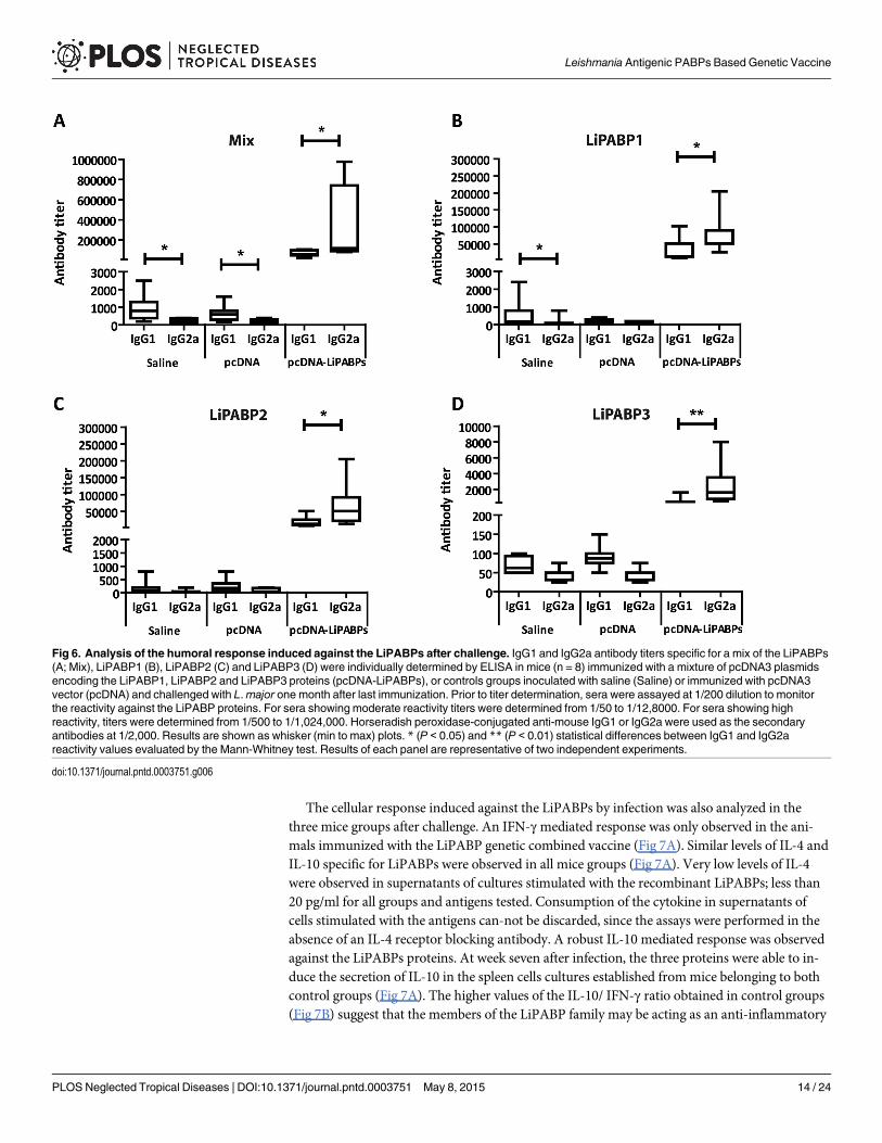
Regarding the humoral response, high IgG1 and IgG2a antibody titers were observed in thesera from LiPABP vaccinated animals after infection (Fig 6A). The predominant IgG2a re-sponse against the LiPABP family induced by vaccination was maintained after parasite chal-lenge in this mice group (Fig 6A). The LiPABP1 (Fig 6B) was the most antigenic member ofthe family followed by LiPABP2 (Fig 6C), being LiPABP3 the lesser antigenic member of the
Leishmania Antigenic PABPs Based Genetic Vaccine
PLOS Neglected Tropical Diseases | DOI:10.1371/journal.pntd.0003751 May 8, 2015 11 / 24

Fig 4. Course of L.major infection in BALB/c mice vaccinated with the LiPABP DNA combinedvaccine.Mice (n = 8 per group) vaccinated with the LiPABP1 + LiPABP2 + LiPABP3 combined geneticvaccine (pcDNA3-PABPs) or inoculated with the pcDNA3 empty vector (pcDNA) or receiving the vaccinediluent (Saline) were challenged in the footpad with 5 × 10⁴ L.major stationary promastigotes one month afterthe last immunization. Footpad swelling is represented as the mean ± SD of the difference of thicknessbetween the infected and the uninfected contra-lateral footpads (A). The numbers of viable parasites in thepopliteal lymph nodes draining the infected legs (DLN) or in spleens were individually determined by limitingdilution at week seven post-challenge. Mean ± SD of the parasite burdens in the complete organ is shown(B). + P < 0.05, ++ P < 0.01 and +++ P < 0.001 significant differences between LiPABP vaccinated and salinecontrol mice; * P < 0.05, ** P < 0.01 and *** P < 0.001 significant differences between LiPABP vaccinatedmice and pcDNA3 immunized control mice (Student T-test). Results in each panel are representative of twoindependent experiments.
doi:10.1371/journal.pntd.0003751.g004
Leishmania Antigenic PABPs Based Genetic Vaccine
PLOS Neglected Tropical Diseases | DOI:10.1371/journal.pntd.0003751 May 8, 2015 12 / 24
family (Fig 6D). On the contrary, control groups showed a limited humoral response againstthe LiPABPs antigens, being the IgG1 titers higher than the IgG2a ones (Fig 6A–6D). Signifi-cant differences between both subclasses were only observed in both control groups when amix of the three proteins were employed for coating ELISA plates (Fig 6A) or in the salinegroup for Leishmania LiPABP1 (Fig 6B).
Fig 5. Antibody titers and cytokines production against SLA after infection in control and vaccinated mice groups. The humoral and cellularresponse against soluble Leishmania antigens (SLA) was determined at week seven after L.major infection in mice (n = 8 per group) previously vaccinatedwith the LiPABP1 + LiPABP2 + LiPABP3 combined genetic vaccine (pcDNA3-PABPs) or immunized with the pcDNA3 empty vector (pcDNA) or that receivedthe vaccine diluent (Saline) prior to infection. Anti—SLA IgG (A) or IgG1 and IgG2a (B) titers were individually determined by assaying sera from 1/50 to 1/820,000 and using a horseradish peroxidase-conjugated anti-mouse IgG, IgG1 or IgG2a secondary antibody at 1/2,000 dilution. Results are shown aswhisker (min to max) plots. For cytokine determinations spleen cells were cultured for 72 h at 37°C, 5% CO2 in the absence or in the presence or solubleLeishmania antigen (SLA; 12 μg/ml). Levels of IFN-γ, IL-10 and IL-4 were assessed by ELISA in culture supernatants (C). Each bar represents themean ± standard deviation (SD). The IFN-γ/IL-10 and IFN-γ/IL-4 ratios were determined and represented as the mean ± standard deviation (SD) (D). In (A)++ (P < 0.01) and ** (P < 0.01) statistical decrease in anti-SLA response in the LiPABPs vaccinated group with respect saline or pcDNA3 control groups,respectively. In (B) ** (P < 0.01) and *** (P < 0.001) statistical differences between IgG1 and IgG2a anti-SLA titers within each group (Mann-Whitney test).In (C and D) +++ P < 0.001 and *** P < 0.001 significant differences between LiPABP vaccinated and saline and pcDNA3 control groups, respectively(Student T-test). Results are representative of two independent experiments.
doi:10.1371/journal.pntd.0003751.g005
Leishmania Antigenic PABPs Based Genetic Vaccine
PLOS Neglected Tropical Diseases | DOI:10.1371/journal.pntd.0003751 May 8, 2015 13 / 24
The cellular response induced against the LiPABPs by infection was also analyzed in thethree mice groups after challenge. An IFN-γmediated response was only observed in the ani-mals immunized with the LiPABP genetic combined vaccine (Fig 7A). Similar levels of IL-4 andIL-10 specific for LiPABPs were observed in all mice groups (Fig 7A). Very low levels of IL-4were observed in supernatants of cultures stimulated with the recombinant LiPABPs; less than20 pg/ml for all groups and antigens tested. Consumption of the cytokine in supernatants ofcells stimulated with the antigens can-not be discarded, since the assays were performed in theabsence of an IL-4 receptor blocking antibody. A robust IL-10 mediated response was observedagainst the LiPABPs proteins. At week seven after infection, the three proteins were able to in-duce the secretion of IL-10 in the spleen cells cultures established frommice belonging to bothcontrol groups (Fig 7A). The higher values of the IL-10/ IFN-γ ratio obtained in control groups(Fig 7B) suggest that the members of the LiPABP family may be acting as an anti-inflammatory
Fig 6. Analysis of the humoral response induced against the LiPABPs after challenge. IgG1 and IgG2a antibody titers specific for a mix of the LiPABPs(A; Mix), LiPABP1 (B), LiPABP2 (C) and LiPABP3 (D) were individually determined by ELISA in mice (n = 8) immunized with a mixture of pcDNA3 plasmidsencoding the LiPABP1, LiPABP2 and LiPABP3 proteins (pcDNA-LiPABPs), or controls groups inoculated with saline (Saline) or immunized with pcDNA3vector (pcDNA) and challenged with L.major one month after last immunization. Prior to titer determination, sera were assayed at 1/200 dilution to monitorthe reactivity against the LiPABP proteins. For sera showing moderate reactivity titers were determined from 1/50 to 1/12,8000. For sera showing highreactivity, titers were determined from 1/500 to 1/1,024,000. Horseradish peroxidase-conjugated anti-mouse IgG1 or IgG2a were used as the secondaryantibodies at 1/2,000. Results are shown as whisker (min to max) plots. * (P < 0.05) and ** (P < 0.01) statistical differences between IgG1 and IgG2areactivity values evaluated by the Mann-Whitney test. Results of each panel are representative of two independent experiments.
doi:10.1371/journal.pntd.0003751.g006
Leishmania Antigenic PABPs Based Genetic Vaccine
PLOS Neglected Tropical Diseases | DOI:10.1371/journal.pntd.0003751 May 8, 2015 14 / 24
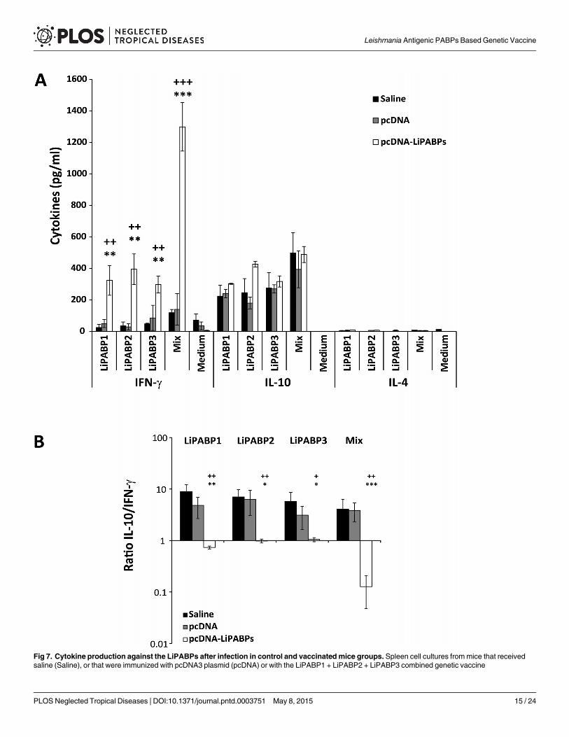
Fig 7. Cytokine production against the LiPABPs after infection in control and vaccinated mice groups. Spleen cell cultures frommice that receivedsaline (Saline), or that were immunized with pcDNA3 plasmid (pcDNA) or with the LiPABP1 + LiPABP2 + LiPABP3 combined genetic vaccine
Leishmania Antigenic PABPs Based Genetic Vaccine
PLOS Neglected Tropical Diseases | DOI:10.1371/journal.pntd.0003751 May 8, 2015 15 / 24
stimulus during infection. The lower value of the IL-10/ IFN-γ ratio observed in the LiPABPvaccinated and protected mice after infection indicates that the Th1 response induced by vacci-nation with the genetic combined vaccine against the three member of the LiPABP family wasmaintained after infection (Fig 7B). Very low levels of the different cytokines were found in cul-tures grown without stimulation (Fig 7A; Medium).
DiscussionHuman and canine VL patients present high titers of antibodies against different parasite anti-gens. Many of them are proteins with an intracellular location (like cytosolic or nuclear factors)having a physiological role inside the cell. These antigenic molecules have been named pananti-gens, because of their conserved nature and their antigenicity in different forms of leishmaniasisas well as in other autoimmune or infectious diseases [37,38]. Alternatively, they are termedpathoantigens [39], because the antibodies produced against them during infection seem to beinvolved in the development of pathology in the visceral forms of the disease [36,40]. Datashown in this work indicate that the LiPABP family can be included within these groups. Theyare antigenic in human and canine VL patients and they have an intracellular location [26]. Asoccur for other pathoantigenic proteins, LiPABPs have essential roles in the parasite, controllingthe RNA processing and translation [26]. Beside the LiPABPs, other RNA binding proteins, likethe Leishmania PUF family of protein factors have been described as antigenic in natural andexperimental VL [41]. Interestingly, other intracellular ribonucleoprotein complexes (like ribo-somes or nucleosome forming histones) or components of protein aggregates (like heat shockproteins) were classified within the panantigenic/pathoantigenic family [37,38,39]. TheLiPABPs are also recognized by the sera from patients with other clinical manifestations, namelyML human patients infected by L. braziliensis (Fig 1), as occur with other intracellular parasiteproteins families. The presence of antibodies against intracellular proteins in sera samples frompatients of CL an ML, as well as in cured VL patients, reflects the strong antigenicity of thesefamilies [30,31,42]. Some variability in the recognition of the individual LiPABPs was observedamong sera samples from different patients. This is a hallmark of the humoral response foundin human or canine patients for most of the characterized Leishmania antigenic proteins[43,44]. This variability should be taken into account in the development of vaccines or diagnos-tics for human and canine leishmaniasis. Regarding the specificity of the humoral response elic-ited against LiPABPs, in a previous report it was shown that anti-LiPABP2 antibodies purifiedfrom VL canine sera did not show cross-reactivity with the human PABP [27]. The molecularbasis of the response specificity might be related to the high degree of divergence betweenLiPABPs and their human orthologues (S3 Fig). In addition, as it is deduced from Fig 2, anti-bodies elicited against the three LiPABPs in mice immunized with the recombinant versions ofthe three LiPABPs were not able to recognize the mammalian PABP.
The induction of humoral responses against these proteins in natural leishmaniasis may de-pend on specific recognition by the B-cell receptor following parasite destruction after lysis ofthe parasites mediated by the complement [45,46] or through the activity of NETs (NeutrophilExtracellular Traps) [47,48]. The identification of many of the pathoantigenic proteins in the
(pcDNA-LiPABPs) were established at week seven after challenge with L.major (n = 8 per group). Splenocytes (5 x 10⁶ cells/ml) were cultured for 72 h at37°C, 5% CO2 in the presence or parasite LiPABP1, LiPABP2 and LiPABP3 as independent stimuli (12 μg/ml), or with a mixture of the three proteins (12 μg/ml total protein, 4 μg/ml each one). As background control, parallel cultures were maintained without stimulation (Medium). Levels of IFN-γ, IL-10 and IL-4were assessed by ELISA in culture supernatants. Each bar represents the mean ± standard deviation (SD) (A). The IL-10/ IFN-γ ratio from data shown inpanel A was calculated and represented as the mean ± standard deviation (SD) (B). + P < 0.05, ++ P < 0.01, +++ P < 0.001 or; * P < 0.05, ** P < 0.01,***P < 0.001 significant differences between LiPABP vaccinated and saline or pcDNA3 control mice groups, respectively (Student T-test). Results arerepresentative of two independent experiments.
doi:10.1371/journal.pntd.0003751.g007
Leishmania Antigenic PABPs Based Genetic Vaccine
PLOS Neglected Tropical Diseases | DOI:10.1371/journal.pntd.0003751 May 8, 2015 16 / 24
secretome/excretome of Leishmania species [49,50] offers an alternative manner of presenta-tion of these antigens to the host immunological system. In this sense, LiPABP2 has beenfound to be part of the secretome of L. donovani [50]. As the infection progresses, the release ofthe pathoantigens will increase due to the increment in the parasite burden with a concomitantinduction of humoral responses that correlate with the development of the immunopathologyassociated with the progressive forms of leishmaniasis [38,39]. An association between the de-velopment of clinical symptoms and the induction of humoral responses was reported in longi-tudinal studies developed in different experimental models of VL in dog and hamster [51,52].Some of these intracellular proteins have immunological properties able to modulate the typeof response against the parasite. As examples, the cytosolic tryparedoxin, a protein that targetsB cells to secrete IL-10 and to produce anti-tryparedoxin specific antibodies [53] or the ribosom-al protein S3a that induces polyclonal expansion of B cells in the host [54]. Additionally, somenuclear factors like the protein termed Le22 or the histone H2B specifically activate IL-10 secre-tion in peripheral blood mononuclear cells (PBMCs) from human VL patients [55,56]. In thissense, our results indicate that BALB/c mice infected with L.major elicited a moderate humoralresponse (IgG1 polarized) against the three Leishmania PABPs (Fig 6). In spite of the low titerfound against the LiPABP in mice from control groups (Fig 6), our data allow to determine thatLiPABP proteins are antigenic in mice infected with L.major. Supporting this conclusion, acomparative analysis of data shown in Fig 3 and data shown in Fig 6 demonstrate that infectionwas able to boost the humoral response elicited against the LiPABP family in mice vaccinatedwith the LiPABP combined genetic vaccine. In addition, the high value of the IL-10/ IFN-γ ratioobserved in mice from control groups after infection when the LiPABPs were employed to stim-ulate spleen cell cultures (Fig 7) reinforces the pathoantigenic characteristics of these proteinfactors, since IL-10 mediated responses promote susceptibility in this mice model [11,57].
It has been proposed that down-modulation of IL-10 mediated responses or the Th2-primed antibody production against pathoantigens can be a promising strategy for the devel-opment of vaccines [21,55,58]. Supporting this hypothesis, some pathoantigenic molecules likehistones H2A and H2B (antigenic nuclear factors recognized by the sera from human VL pa-tients [59] or dogs infected by L. infantum [60]) have also been implicated in the induction ofproliferative responses and IFN-γ production in PBMCs from cured CL patients and fromasymptomatic VL patients [61]. In addition, histone H2B has been described to stimulate IFN-γ producing CD4+ T from donors who developed a protective immune response against Leish-mania [62]. The immunization of vaccines based on the pathoantigenic histones has been asso-ciated with the induction of protection in the BALB/c-L.majormodel when Th1 inducingadjuvants or strategies were employed [22,63]. Other well-known example is the proteintermed LACK; leishmanial homolog of mammalian receptor for activated C kinase. AlthoughLACK was characterized as an antigen recognized by a protective CD4+ T cell clone (Th1) ob-tained from a BALB/c mice immunized with a SLA fraction [64], it was later found to be impli-cated in the induction of the early IL-4 response against L.major occurring in this susceptiblemodel [65]. Remarkably, co-administration of recombinant LACK with Th1 adjuvants inBALB/c mice was able to redirect the naturally induced Th2 responses after L.major infection.Thus, recombinant LACK protein administered with interleukin-12 [66] or as a LACK basedDNA vaccine [67] protected BALB/c mice against L.major infection. Further, it was demon-strated that BALB/c rendered tolerant to LACK, as a result of transgenic expression of this mol-ecule in the thymus, were resistant to infection with L.major and developed a Th1 responseafter infection [68]. Finally, the use of a intranasal LACK DNA vaccine was able to induce aprotective immunity in BALB/c against the infection by L. amazonensis [69] a NewWorld dis-tributed species able to cause a broad spectrum of clinical manifestations, from cutaneous tovisceral leishmaniasis in human patients [70].
Leishmania Antigenic PABPs Based Genetic Vaccine
PLOS Neglected Tropical Diseases | DOI:10.1371/journal.pntd.0003751 May 8, 2015 17 / 24
Our results show that the inoculation of a genetic vaccine based on the three LiPABP pro-teins was able to delay the progression of the disease caused by L.major infection in BALB/cmice by decreasing the footpad swelling and the parasite burdens from LiPABP vaccinatedmice relative to control mice group (Fig 4). This experimental model has been employed be-cause it is highly susceptible to infection showing a progressive form of the disease, as occurs indifferent clinical forms of leishmaniasis in natural hosts [71]. In addition to these clinical orparasitological evidences, LiPABP vaccinated mice showed a limited humoral response againstSLA as well as high SLA-dependent ratios of IFN-γ/IL-4 and IFN-γ/IL-10 (Fig 5) that can beconsidered immunological markers of protection [11,72,73]. The strategy of employingLiPABP eukaryotic expression plasmids for inoculation was selected due to the capacity ofthese third generation vaccines to promote a Th1 response against the heterologous Leishman-ia proteins expressed in the hosts [74]. Due to the heterogenicity of the immune response elic-ited against LiPABPs in natural and experimental hosts, we used a combined genetic vaccine,instead of the immunization of plasmids encoding the individual antigens. It is likely that thethree LiPABPs may be presented to the immune system at the same time after parasite lysis(for B-cell responses) or presented at the same time by professional antigen presenting cells.Thus, induction of a Th1 response against the three proteins may help to reinforce the capacityof the vaccine in controlling disease progression. Using the same mice model employed in thiswork, the protective capacity of a vaccine based on histones was reinforced when it was formu-lated including the four nucleosome histones instead of the use of vaccines based on individualor different pairs of histones [75]. It has been generally demonstrated that experimental vac-cines combining different parasite antigens conferred more solid protection than those con-taining individual parasite determinants (reviewed in [17,74]). Our results demonstrated thatgenetic vaccination of the LiPABPs combined vaccine in BALB/c mice induced a Th1 type re-sponse: anti-LiPABP IgG2a antibodies and IFN-γ secretion upon re-stimulation of spleen cellswith the recombinant LiPABPs (Fig 3). The immune response induced by LiPABP vaccinationwas maintained after infection. The PABPs specific IgG2a/IgG1 and IFN-γ IL-10 ratios werereverted in the LiPABP vaccinated and protected mice, in comparison to mice from both con-trol groups after infection (Figs 6 and 7). Even though SLA is a preparation of total parasite sol-uble proteins, including the LiPABPs, opposite patterns in SLA specific (Fig 5B) and LiPABPsspecific (Fig 6) antibody responses were observed when sera from LiPABP vaccinated and in-fected mice were employed. These differences may be related to the amounts of the LiPABPspresent in the SLA preparation. Thus, ELISA assays performed with the whole antigenic reper-toire of the parasite may minimize the reactivity of the anti-LiPABPs antibodies. In this con-text, the anti-SLA immune response can be considered a marker of the immune humoralresponse against the whole parasite.
Despite the partial protection afforded by the LiPABP combined vaccine in the BALB/c-L.majormodel, the use of these proteins as protective antigens should not be underestimated.Some vaccine candidates inducing partial protection in this mice model have emerged as goodcandidates when tested in other models of infection. As an example BALB/c mice immunizedwith a DNA vaccine based on L. infantum P0 (LiP0) or with the recombinant LiP0 protein com-bined with a Th1 inducing adjuvant, presented a partial protection after challenge with L.major[76,77]. LiP0 vaccinated mice showed an initial significant reduction in lesion size after chal-lenge, but mice ultimately developed non-healing lesion. The delay in the onset of cell growthwas accompanied by a substantial decrease in the parasite load and was correlated to the genera-tion of initial Th1 responses that were changed to a mixed Th1/Th2 response against the LiP0when disease progressed. Interestingly, our data showed that the immune response induced byvaccination against the LiPABP family members was not changed after infection. On the otherhand, the Th1 responses induced by administration of the LiP0 vaccines conferred protection
Leishmania Antigenic PABPs Based Genetic Vaccine
PLOS Neglected Tropical Diseases | DOI:10.1371/journal.pntd.0003751 May 8, 2015 18 / 24
against CL in C57BL/6 mice [77] or against VL in hamster [78]. Similarly, vaccines based on theL.major ribosomal protein LmL3 or LmL5 combined with CpG-ODNs in BALB/c mice inducedpartial protection against CL due to L.major or L. amazonensis but protect mice against VLcaused by L. infantum and against murine CL due to a high virulent challenge of L. braziliensis[19,44]. Remarkably, some other vaccines described as protective in the L.major-BALB/c modelwere unable to induce protection in the later model [79]. Data presented here should be taken asa proof of concept regarding the protective characteristics of Leishmania PABPs and further as-says using alternative forms of administration and different models of experimental infectionshould be performed to determine the real potential of the PABPs for the development of vac-cines. As an example, DNA vaccines based on the LACK antigen were able to induce robustTh1 responses but were unable to protect mice from L.mexicana [80], L. donovani [81] orL. chagasi [82] infection. Notwithstanding, it was demonstrated that the induction of more com-plex immune responses, using prime boost strategy, combining a DNA vaccine and attenuatedviruses (Western reserve virus or vaccinia virus) expressing LACK was correlated to protectionagainst murine VL caused by L. infantum infection [83]. In this sense, dogs experimentally in-fected with L. infantum were protected against VL following an heterologous prime-boost vacci-nation regime with a DNA vaccine encoding LACK and recombinant vaccinia virus (rVACV)expressing LACK [84], or its corresponding non-replicative modified vaccinia (MVA-LACK)[85]. In addition, combination of LiPABPs with other protective antigens should not be ruledout. Of interest, this work demonstrate that Leishmania PABPs are antigenic in different verte-brate hosts infected by distinct parasite species (L. infantum and L. braziliensis in human, L. in-fantum in dogs and L.major in mice) and the high degree of Leishmania PABPs sequenceconservation in different Leishmania species (S1 Fig) makes plausible to test their cross-protec-tive properties. The possibility to induce cross-protection by the use of vaccines based in pro-teins conserved among Leishmania species is one of the advantages of this type of vaccines [86].
As conclusion, this work has established for the first time the antigenicity of the LiPABPs indifferent forms of natural leishmaniasis, as well as in BALB/c mice infected with L.major. In thismodel of murine progressive leishmaniasis, the administration of a LiPABPs based vaccine wasable to dampen the LiPABP-specific humoral and IL-10 mediated responses detected in thenon-vaccinated mice after infection. This control of pathogenic-skewed immune responses cor-related to the induction of partial protection. In accordance with previous reports, data presentedhere indicate that controlling the responses associated with susceptibility and elicited by parasitepathoantigens may be relevant to achieve a protective vaccine against Leishmania infection.
List of accession numbers for genes and proteins mentioned in the textLeishmania infantum PABPs:
LiPABP1 LinJ.35.5360 GeneDB.
LiPABP2 LinJ.35.4200 GeneDB.
LiPABP3 LinJ.25.0080 Gene DB.
Homo sapiens PABP:
GenBank: EAX07265.1
Supporting InformationS1 Fig. Sequence identity comparisons among the three PABPs from Leishmania infantum,Leishmania major and Leishmania braziliensis. The identity values between the three
Leishmania Antigenic PABPs Based Genetic Vaccine
PLOS Neglected Tropical Diseases | DOI:10.1371/journal.pntd.0003751 May 8, 2015 19 / 24
LiPABPs as well as the identity values of the comparison among the three parasite species, isshown (determined by the Smith-Waterman local alignment of sequences (http://emboss.bioinformatics.nl/)). The accession numbers are also included.(TIF)
S2 Fig. Stability of the purified LiPABP2 recombinant protein in the presence or in the ab-sence of urea denaturant agent. Coomassie-staining of a 10% SDS-PAGE showing a Molecu-lar Weight Marker (Mr), a total extract of protein from E. coli cultures expressing the LiPABP2solubilized under denaturant conditions (20 mM Tris ClH pH 8.0, 0.5 M NaCl, 8 M Urea, 1mM β-mercaptoethanol) (1), the Ni-NTA flow-through fraction (2), the recombinant LiPABP2under denaturant conditions (3) and the recombinant LiPABP2 dialyzed against PBS (4).(TIF)
S3 Fig. Amino acid comparison among LiPABPs and human PABP (GenBank accessionnumber EAX07265). The amino acid comparisons, as well as the identity and similarity val-ues, were determined by the Smith-Waterman local alignment of sequences (http://emboss.bioinformatics.nl/). The sequence of the human PABP was rescued as the most identical PABPusing the three LiPABPs as probes in a BLAST analysis (http://www.ncbi.nlm.nih.gov/).(DOCX)
Author ContributionsConceived and designed the experiments: MS CGN EAFCMBN SI. Performed the experi-ments: MS LC EG LR VI VMGMEM SI. Analyzed the data: MS LC EG VMGMEM CA EAFCABMBN SI. Contributed reagents/materials/analysis tools: VI PB CGN VMGMEM ABMBN.Wrote the paper: MS LC VI PB VMG CA EAFCMBN SI.
References1. Dostalova A, Volf P. Leishmania development in sand flies: parasite-vector interactions overview. Para-
sit Vectors. 2012; 5: 276. doi: 10.1186/1756-3305-5-276 PMID: 23206339
2. Murray HW, Berman JD, Davies CR, Saravia NG. Advances in leishmaniasis. Lancet. 2005; 366:1561–1577. PMID: 16257344
3. de Oliveira CI, Brodskyn CI. The immunobiology of Leishmania braziliensis infection. Front Immunol.2012; 3: 145. doi: 10.3389/fimmu.2012.00145 PMID: 22701117
4. Mauricio IL, Stothard JR, Miles MA. The strange case of Leishmania chagasi. Parasitol Today. 2000;16: 188–189. PMID: 10782075
5. Herwaldt BL. Leishmaniasis. Lancet. 1999; 354: 1191–1199. PMID: 10513726
6. Pearson RD, Sousa AQ. Clinical spectrum of Leishmaniasis. Clin Infect Dis. 1996; 22: 1–13. PMID:8824958
7. Baneth G, Koutinas AF, Solano-Gallego L, Bourdeau P, Ferrer L. Canine leishmaniosis—new conceptsand insights on an expanding zoonosis: part one. Trends Parasitol. 2008; 24: 324–330. doi: 10.1016/j.pt.2008.04.001 PMID: 18514028
8. Kaye P, Scott P. Leishmaniasis: complexity at the host-pathogen interface. Nat Rev Microbiol. 2011; 9:604–615. doi: 10.1038/nrmicro2608 PMID: 21747391
9. Kedzierski L, Evans KJ. Immune responses during cutaneous and visceral leishmaniasis. Parasitol.2014; 141:1544–1562. doi: 10.1242/dev.099986 PMID: 24598161
10. Ronet C, Hauyon-La Torre Y, Revaz-Breton M, Mastelic B, Tacchini-Cottier F, Louis J, et al. RegulatoryB cells shape the development of Th2 immune responses in BALB/c mice infected with Leishmaniamajor through IL-10 production. J Immunol. 2010; 184: 886–894. doi: 10.4049/jimmunol.0901114PMID: 19966209
11. Schwarz T, Remer KA, Nahrendorf W, Masic A, Siewe L, Muller W, et al. T cell-derived IL-10 deter-mines leishmaniasis disease outcome and is suppressed by a dendritic cell based vaccine. PLoSPathog. 2013; 9: e1003476. doi: 10.1371/journal.ppat.1003476 PMID: 23825956
Leishmania Antigenic PABPs Based Genetic Vaccine
PLOS Neglected Tropical Diseases | DOI:10.1371/journal.pntd.0003751 May 8, 2015 20 / 24

12. Dantas-Torres F. Leishmune vaccine: the newest tool for prevention and control of canine visceralleishmaniosis and its potential as a transmission-blocking vaccine. Vet Parasitol. 2006; 141: 1–8.PMID: 16750885
13. Fernandes AP, Coelho EA, Machado-Coelho GL, Grimaldi G Jr., Gazzinelli RT. Making an anti-amastigote vaccine for visceral leishmaniasis: rational, update and perspectives. Curr Opin Microbiol.2012; 15: 476–485. PMID: 22698479
14. Bongiorno G, Paparcone R, Manzillo VF, Oliva G, Cuisinier AM, Gradoni L. Vaccination with LiESP/QA-21 (CaniLeish) reduces the intensity of infection in Phlebotomus perniciosus fed on Leishmania in-fantum infected dogs-A preliminary xenodiagnosis study. Vet Parasitol. 2013; 97: 691–695.
15. Fernandes CB, Junior JT, de Jesus C, Souza BM, Larangeira DF, Fraga DB, et al. Comparison of twocommercial vaccines against visceral leishmaniasis in dogs from endemic areas: IgG, and subclasses,parasitism, and parasite transmission by xenodiagnosis. Vaccine. 2014; 32: 1287–1295. doi: 10.1016/j.vaccine.2013.12.046 PMID: 24406392
16. Otranto D, Dantas-Torres F. The prevention of canine leishmaniasis and its impact on public health.Trends Parasitol. 2013; 29: 339–345. doi: 10.1016/j.pt.2013.05.003 PMID: 23746747
17. Beaumier CM, Gillespie PM, Hotez PJ, Bottazzi ME. New vaccines for neglected parasitic diseasesand dengue. Transl Res. 2013; 162: 144–155. doi: 10.1016/j.trsl.2013.03.006 PMID: 23578479
18. Baharia RK, Tandon R, Sahasrabuddhe AA, Sundar S, Dube A. Nucleosomal Histone Proteins ofL. donovani: A Combination of recombinant H2A, H2B, H3 and H4 proteins were highly immunogenicand offered optimum prophylactic efficacy against Leishmania challenge in hamsters. PLoS One. 2014;9: e97911. doi: 10.1371/journal.pone.0097911 PMID: 24926878
19. Ramirez L, Corvo L, Duarte MC, Chavez-Fumagalli MA, Valadares DG, Santos DM, et al. Cross-protec-tive effect of a combined L5 plus L3 Leishmania major ribosomal protein based vaccine combined witha Th1 adjuvant in murine cutaneous and visceral leishmaniasis. Parasit Vectors. 2014; 7: 3. doi: 10.1186/1756-3305-7-3 PMID: 24382098
20. Carneiro MW, Santos DM, Fukutani KF, Clarencio J, Miranda JC, Brodskyn C,et al. Vaccination withL. infantum chagasi nucleosomal histones confers protection against new world cutaneous leishmania-sis caused by Leishmania braziliensis. PLoS One. 2012; 7: e52296. doi: 10.1371/journal.pone.0052296 PMID: 23284976
21. Iborra S, Parody N, Abanades DR, Bonay P, Prates D, Novais FO, et al. Vaccination with the Leishman-ia major ribosomal proteins plus CpG oligodeoxynucleotides induces protection against experimentalcutaneous leishmaniasis in mice. Microbes Infect. 2008; 10: 1133–1141. doi: 10.1016/j.micinf.2008.06.002 PMID: 18603012
22. Iborra S, Soto M, Carrion J, Alonso C, Requena JM. Vaccination with a plasmid DNA cocktail encodingthe nucleosomal histones of Leishmania confers protection against murine cutaneous leishmaniosis.Vaccine. 2004; 22: 3865–3876. PMID: 15364433
23. Melby PC, Ogden GB, Flores HA, ZhaoW, Geldmacher C, Biediger NM, et al. Identification of vaccinecandidates for experimental visceral leishmaniasis by immunization with sequential fractions of a cDNAexpression library. Infect Immun. 2000; 68: 5595–5602. PMID: 10992459
24. Eliseeva IA, Lyabin DN, Ovchinnikov LP. Poly(A)-binding proteins: structure, domain organization, and ac-tivity regulation. Biochemistry. 2013; 78: 1377–1391. doi: 10.1134/S0006297913130014 PMID: 24490729
25. Goss DJ, Kleiman FE. Poly(A) binding proteins: are they all created equal? Wiley Interdiscip Rev RNA.2013; 4: 167–179. doi: 10.1002/wrna.1151 PMID: 23424172
26. da Costa Lima TD, Moura DM, Reis CR, Vasconcelos JR, Ellis L, Carrington M,et al. Functional charac-terization of three Leishmania Poly(A) Binding Protein homologues with distinct binding properties to RNAand protein partners. Eukaryot Cell. 2010; 9: 1484–1494. doi: 10.1128/EC.00148-10 PMID: 20675580
27. Guerra N, Vega-Sendino M, Perez-Morgado MI, Ramos E, Soto M, Gonzalez VM, et al. Identificationand functional characterization of a Poly(A)-Binding Protein from Leishmania infantum (LiPABP). FEBSLett. 2011; 585: 193–198. doi: 10.1016/j.febslet.2010.11.042 PMID: 21115009
28. Shi PY, Maizels N, Weiner AM. Recovery of soluble, active recombinant protein from inclusion bodies.Biotechniques. 1997; 23: 1036–1038. PMID: 9421633
29. Schauer S, Luer C, Moser J. Large scale production of biologically active Escherichia coli glutamyl-tRNA reductase from inclusion bodies. Protein Expr Purif. 2003; 31: 271–275. PMID: 14550647
30. Souza AP, Soto M, Costa JM, Boaventura VS, de Oliveira CI, Cristal JR, et al. Towards a more preciseserological diagnosis of human tegumentary leishmaniasis using Leishmania recombinant proteins.PLoS One. 2013; 8: e66110. doi: 10.1371/journal.pone.0066110 PMID: 23776617
31. Abanades DR, Arruda LV, Arruda ES, Pinto JR, PalmaMS, Caldas AJ, et al. Immunodominant antigensof Leishmania chagasi associated with protection against human visceral leishmaniasis. PLoS NeglTrop Dis. 2012; 6: e1687. doi: 10.1371/journal.pntd.0001687 PMID: 22724032
Leishmania Antigenic PABPs Based Genetic Vaccine
PLOS Neglected Tropical Diseases | DOI:10.1371/journal.pntd.0003751 May 8, 2015 21 / 24
32. Coelho EA, Ramirez L, Costa MA, Coelho VT, Martins VT, Chavez-Fumagalli MA, et al. Specific sero-diagnosis of canine visceral leishmaniasis using Leishmania species ribosomal protein extracts. ClinVaccine Immunol. 2009; 16: 1774–1780. doi: 10.1128/CVI.00295-09 PMID: 19812259
33. Buffet PA, Sulahian A, Garin YJ, Nassar N, Derouin F. Culture microtitration: a sensitive method forquantifying Leishmania infantum in tissues of infected mice. Antimicrob Agents Chemother. 1995; 39:2167–2168. PMID: 8540741
34. Beckmann MP, Schooley KA, Gallis B, Vanden Bos T, Friend D, Alpert AR, et al. Monoclonal antibodiesblock murine IL-4 receptor function. J Immunol. 1990; 144: 4212–4217. PMID: 1692858
35. Coffman RL. Mechanisms of helper T-cell regulation of B-cell activity. Ann N Y Acad Sci. 1993; 681:25–28. PMID: 8102840
36. Miles SA, Conrad SM, Alves RG, Jeronimo SM, Mosser DM. A role for IgG immune complexes duringinfection with the intracellular pathogen Leishmania. J Exp Med. 2005; 201: 747–754. PMID: 15753208
37. Requena JM, Alonso C, Soto M. Evolutionarily conserved proteins as prominent immunogens duringLeishmania infections. Parasitol Today. 2000; 16: 246–250. PMID: 10827430
38. Santarem N, Silvestre R, Tavares J, Silva M, Cabral S, Maciel J, et al. Immune response regulation byLeishmania secreted and nonsecreted antigens. J Biomed Biotechnol. 2007; 2007: 85154. PMID:17710243
39. Chang KP, Reed SG, McGwire BS, Soong L. Leishmaniamodel for microbial virulence: the relevanceof parasite multiplication and pathoantigenicity. Acta Trop. 2003; 85: 375–390. PMID: 12659975
40. Garcia-Alonso M, Blanco A, Reina D, Serrano FJ, Alonso C, Nieto CG. Immunopathology of the uveitisin canine leishmaniasis. Parasite Immunol. 1996; 18: 617–623. PMID: 9226700
41. Folgueira C, Martinez-Bonet M, Requena JM. The Leishmania infantum PUF proteins are targets of thehumoral response during visceral leishmaniasis. BMCRes Notes. 2010; 3: 13. doi: 10.1186/1756-0500-3-13 PMID: 20180988
42. Celeste BJ, Arroyo Sanchez MC, Ramos-Sanchez EM, Castro LG, Lima Costa FA, Goto H. Recombi-nant Leishmania infantum heat shock protein 83 for the serodiagnosis of cutaneous, mucosal, and vis-ceral leishmaniases. Am J Trop Med Hyg. 2014; 90: 860–865. doi: 10.4269/ajtmh.13-0623 PMID:24615136
43. Goto Y, Howard RF, Bhatia A, Trigo J, Nakatani M, Netto EM, et al. Distinct antigen recognition patternduring zoonotic visceral leishmaniasis in humans and dogs. Vet Parasitol. 2009; 160: 215–220. doi:10.1016/j.vetpar.2008.10.097 PMID: 19059724
44. Ramirez L, Santos DM, Souza AP, Coelho EA, Barral A, Alonso C, et al. Evaluation of immune re-sponses and analysis of the effect of vaccination of the Leishmania major recombinant ribosomalproteins L3 or L5 in two different murine models of cutaneous leishmaniasis. Vaccine. 2013; 31:1312–1319. doi: 10.1016/j.vaccine.2012.12.071 PMID: 23313653
45. Mosser DM, Edelson PJ. Activation of the alternative complement pathway by Leishmania promasti-gotes: parasite lysis and attachment to macrophages. J Immunol. 1984; 132: 1501–1505. PMID:6363545
46. Ambrosio AR, De Messias-Reason IJ. Leishmania (Viannia) braziliensis: interaction of mannose-bindinglectin with surface glycoconjugates and complement activation. An antibody-independent defencemechanism. Parasite Immunol. 2005; 27: 333–340. PMID: 16149991
47. Abi Abdallah DS, Denkers EY. Neutrophils cast extracellular traps in response to protozoan parasites.Front Immunol. 2012; 3: 382. doi: 10.3389/fimmu.2012.00382 PMID: 23248631
48. Guimaraes-Costa AB, Nascimento MT, Froment GS, Soares RP, Morgado FN, Conceicao-Silva F,et al. Leishmania amazonensis promastigotes induce and are killed by neutrophil extracellular traps.Proc Natl Acad Sci U. S. A. 2009; 106: 6748–6753. doi: 10.1073/pnas.0900226106 PMID: 19346483
49. Peysselon F, Launay G, Lisacek F, Duclos B, Ricard-Blum S. Comparative analysis of Leishmania exo-proteomes: implication for host-pathogen interactions. Biochim Biophys Acta. 2013; 1834: 2653–2662.doi: 10.1016/j.bbapap.2013.09.015 PMID: 24096101
50. Silverman JM, Chan SK, Robinson DP, Dwyer DM, Nandan D, Foster LJ, et al. Proteomic analysis ofthe secretome of Leishmania donovani. Genome Biol. 2008; 9: R35. doi: 10.1186/gb-2008-9-2-r35PMID: 18282296
51. Fernandez-Cotrina J, Iniesta V, Belinchon-Lorenzo S, Munoz-Madrid R, Serrano F, Parejo JC, et al. Ex-perimental model for reproduction of canine visceral leishmaniosis by Leishmania infantum. Vet Parasi-tol. 2013; 192: 118–128. doi: 10.1016/j.vetpar.2012.10.002 PMID: 23102507
52. Requena JM, Soto M, Doria MD, Alonso C. Immune and clinical parameters associated with Leishman-ia infantum infection in the golden hamster model. Vet Immunol Immunopathol. 2000; 76: 269–281.PMID: 11044559
Leishmania Antigenic PABPs Based Genetic Vaccine
PLOS Neglected Tropical Diseases | DOI:10.1371/journal.pntd.0003751 May 8, 2015 22 / 24
53. Cabral SM, Silvestre RL, Santarem NM, Tavares JC, Silva AF, Cordeiro-da-Silva A. A Leishmania in-fantum cytosolic tryparedoxin activates B cells to secrete interleukin-10 and specific immunoglobulin.Immunology. 2008; 123: 555–565. PMID: 18028371
54. Cordeiro-Da-Silva A, Borges MC, Guilvard E, Ouaissi A. Dual role of the Leishmania major ribosomalprotein S3a homologue in regulation of T- and B-cell activation. Infect Immun. 2001; 69: 6588–6596.PMID: 11598026
55. Suffia I, Ferrua B, Stien X, Mograbi B, Marty P, Rousseau D, et al. A novel Leishmania infantum recom-binant antigen which elicits interleukin 10 production by peripheral blood mononuclear cells of patientswith visceral leishmaniasis. Infect Immun. 2000; 68: 630–636. PMID: 10639426
56. Meddeb-Garnaoui A, Toumi A, Ghelis H, Mahjoub M, Louzir H, Chenik M. Cellular and humoral re-sponses induced by Leishmania histone H2B and its divergent and conserved parts in cutaneous andvisceral leishmaniasis patients, respectively. Vaccine. 2010; 28: 1881–1886. doi: 10.1016/j.vaccine.2009.11.075 PMID: 20005858
57. Noben-Trauth N, Lira R, Nagase H, Paul WE, Sacks DL. The relative contribution of IL-4 receptor sig-naling and IL-10 to susceptibility to Leishmania major. J Immunol. 2003; 170: 5152–5158. PMID:12734362
58. Campos-Neto A. What about Th1/Th2 in cutaneous leishmaniasis vaccine discovery? Braz J Med BiolRes. 2005; 38: 979–984. PMID: 16007269
59. Maalej IA, Chenik M, Louzir H, Ben Salah A, Bahloul C, Amri F, et al. Comparative evaluation of ELISAsbased on ten recombinant or purified Leishmania antigens for the serodiagnosis of Mediterranean vis-ceral leishmaniasis. Am J Trop Med Hyg. 2003; 68: 312–320. PMID: 12685637
60. Soto M, Requena JM, Quijada L, Perez MJ, Nieto CG, Guzman F, et al. Antigenicity of the Leishmaniainfantum histones H2B and H4 during canine viscerocutaneous leishmaniasis. Clin Exp Immunol.1999; 115: 342–349. PMID: 9933463
61. deCarvalho LP, Soto M, Jeronimo S, Dondji B, Bacellar O, Luz V, et al. Characterization of the immune re-sponse to Leishmania infantum recombinant antigens. Microbes Infect. 2003; 5: 7–12. PMID: 12593967
62. Probst P, Stromberg E, Ghalib HW, Mozel M, Badaro R, Reed SG, et al. Identification and characteriza-tion of T cell-stimulating antigens from Leishmania by CD4 T cell expression cloning. J Immunol. 2001;166: 498–505. PMID: 11123329
63. Chenik M, Louzir H, Ksontini H, Dilou A, Abdmouleh I, Dellagi K. Vaccination with the divergent portionof the protein histone H2B of Leishmania protects susceptible BALB/c mice against a virulent challengewith Leishmania major. Vaccine. 2006; 24: 2521–2529. PMID: 16417957
64. Scott P, Caspar P, Sher A. Protection against Leishmania major in BALB/c mice by adoptive transfer ofa T cell clone recognizing a low molecular weight antigen released by promastigotes. J Immunol. 1990;144: 1075–1079. PMID: 2295814
65. Launois P, Maillard I, Pingel S, Swihart KG, Xenarios I, Acha-Orbea H, et al. IL-4 rapidly produced byV beta 4 V alpha 8 CD4+ T cells instructs Th2 development and susceptibility to Leishmania major inBALB/c mice. Immunity. 1997; 6: 541–549. PMID: 9175832
66. Mougneau E, Altare F, Wakil AE, Zheng S, Coppola T, Wang ZE, et al. Expression cloning of a protec-tive Leishmania antigen. Science. 1995; 268: 563–566. PMID: 7725103
67. Gurunathan S, Sacks DL, Brown DR, Reiner SL, Charest H, Glaichenhaus N, et al. Vaccination withDNA encoding the immunodominant LACK parasite antigen confers protective immunity to mice in-fected with Leishmania major. J Exp Med. 1997; 186: 1137–1147. PMID: 9314562
68. Julia V, Rassoulzadegan M, Glaichenhaus N. Resistance to Leishmania major induced by tolerance toa single antigen. Science. 1996; 274: 421–423. PMID: 8832890
69. Pinto EF, Pinheiro RO, Rayol A, Larraga V, Rossi-Bergmann B. Intranasal vaccination against cutane-ous leishmaniasis with a particulated leishmanial antigen or DNA encoding LACK. Infect Immun. 2004;72: 4521–4527. PMID: 15271911
70. Barral A, Pedral-Sampaio D, Grimaldi Junior G, Momen H, McMahon-Pratt D, Ribeiro de Jesus A, et al.Leishmaniasis in Bahia, Brazil: evidence that Leishmania amazonensis produces a wide spectrum ofclinical disease. Am J Trop Med Hyg. 1991; 44: 536–546. PMID: 2063957
71. Handman E Leishmaniasis: current status of vaccine development. Clin Microbiol Rev. 2001; 14:229–243. PMID: 11292637
72. Roberts MT, Stober CB, McKenzie AN, Blackwell JM. Interleukin-4 (IL-4) and IL-10 collude in vaccinefailure for novel exacerbatory antigens in murine Leishmania major infection. Infect Immun. 2005; 73:7620–7628. PMID: 16239566
73. Bhowmick S, Ravindran R, Ali N. IL-4 contributes to failure, and colludes with IL-10 to exacerbateLeishmania donovani infection following administration of a subcutaneous leishmanial antigen vaccine.BMCMicrobiol. 2014; 14: 8. doi: 10.1186/1471-2180-14-8 PMID: 24428931
Leishmania Antigenic PABPs Based Genetic Vaccine
PLOS Neglected Tropical Diseases | DOI:10.1371/journal.pntd.0003751 May 8, 2015 23 / 24
74. Mutiso JM, Macharia JC, Kiio MN, Ichagichu JM, Rikoi H, Gicheru MM. Development of Leishmaniavaccines: predicting the future from past and present experience. J Biomed Res. 2013; 27: 85–102.doi: 10.7555/JBR.27.20120064 PMID: 23554800
75. Carrion J, Folgueira C, Alonso C. Transitory or long-lasting immunity to Leishmania major infection: theresult of immunogenicity and multicomponent properties of histone DNA vaccines. Vaccine. 2008; 26:1155–1165. doi: 10.1016/j.vaccine.2007.12.051 PMID: 18255202
76. Iborra S, Soto M, Carrion J, Nieto A, Fernandez E, Alonso C, et al. The Leishmania infantum acidic ribo-somal protein P0 administered as a DNA vaccine confers protective immunity to Leishmania major in-fection in BALB/c mice. Infect Immun. 2003. 71: 6562–6572. PMID: 14573678
77. Iborra S, Carrion J, Anderson C, Alonso C, Sacks D, Soto M. Vaccination with the Leishmania infantumacidic ribosomal P0 protein plus CpG oligodeoxynucleotides induces protection against cutaneousleishmaniasis in C57BL/6 mice but does not prevent progressive disease in BALB/c mice. InfectImmun. 2005; 73: 5842–5852. PMID: 16113303
78. Pereira L, Abbehusen M, Teixeira C, Cunha J, Nascimento IP, Fukutani K, et al. Vaccination with Leish-mania infantum acidic ribosomal P0 but not with nucleosomal histones proteins controls Leishmania in-fantum Infection in hamsters. PLoS Negl Trop Dis. 2015; 10: e0003490. doi: 10.1371/journal.pntd.0003490 PMID: 25642946
79. Salay G, Dorta ML, Santos NM, Mortara RA, Brodskyn C, Oliveira CI, et al. Testing of four Leishmaniavaccine candidates in a mouse model of infection with Leishmania (Viannia) braziliensis, the maincausative agent of cutaneous leishmaniasis in the NewWorld. Clin Vaccine Immunol. 2007; 14:1173–1181. PMID: 17626159
80. Dumonteil E, Andrade-Narvarez F, Escobedo-Ortegon J, Ramirez-Sierra MJ, Valencia-Pacheco G,Flores-Serrano A, Tavares CA, et al. Comparative study of DNA vaccines encoding various antigensagainst Leishmania mexicana. Dev Biol. 2000; 104: 135–141. PMID: 11713811
81. Melby PC, Yang J, ZhaoW, Perez LE, Cheng J. Leishmania donovani p36 (LACK) DNA vaccine ishighly immunogenic but not protective against experimental visceral leishmaniasis. Infect Immun.2001; 69: 4719–4725. PMID: 11447143
82. Marques-da-Silva EA, Coelho EA, Gomes DC, Vilela MC, Masioli CZ, Tavares CA, et al. Intramuscularimmunization with p36(LACK) DNA vaccine induces IFN-gamma production but does not protectBALB/c mice against Leishmania chagasi intravenous challenge. Parasitol Res. 2005; 98: 67–74.PMID: 16261353
83. Dondji B, Perez-Jimenez E, Goldsmith-Pestana K, EstebanM, McMahon-Pratt D. Heterologous prime-boost vaccination with the LACK antigen protects against murine visceral leishmaniasis. Infect Immun73: 5286–5289. PMID: 16041057
84. Ramiro MJ, Zarate JJ, Hanke T, Rodriguez D, Rodriguez JR, EstebanM, et al. Protection in dogsagainst visceral leishmaniasis caused by Leishmania infantum is achieved by immunization with a het-erologous prime-boost regime using DNA and vaccinia recombinant vectors expressing LACK. Vac-cine. 2003; 21: 2474–2484. PMID: 12744881
85. Ramos I, Alonso A, Marcen JM, Peris A, Castillo JA, Colmenares M, et al. Heterologous prime-boostvaccination with a non-replicative vaccinia recombinant vector expressing LACK confers protectionagainst canine visceral leishmaniasis with a predominant Th1-specific immune response. Vaccine.2008; 26: 333–344. PMID: 18093705
86. de Oliveira CI, Nascimento IP, Barral A, Soto M, Barral-Netto M. Challenges and perspectives in vacci-nation against leishmaniasis. Parasitol Int. 2009; 58: 319–324. doi: 10.1016/j.parint.2009.07.013PMID: 19698801
Leishmania Antigenic PABPs Based Genetic Vaccine
PLOS Neglected Tropical Diseases | DOI:10.1371/journal.pntd.0003751 May 8, 2015 24 / 24


