Embed Size (px)
Citation preview
335
Braz J Med Biol Res 39(3) 2006
Sites of latent infection by BHV-5 in rabbitsBrazilian Journal of Medical and Biological Research (2006) 39: 335-343ISSN 0100-879X
Dexamethasone-induced reactivationof bovine herpesvirus type 5 latentinfection in experimentally infectedrabbits results in a broader distributionof latent viral DNA in the brain
Setor de Virologia, Departamento de Medicina Veterinária Preventiva,Departamento de Microbiologia e Parasitologia,Universidade Federal de Santa Maria, Santa Maria, RS, Brasil
S.V. Mayer, V.L. de Quadros,F.S.F. Vogel,
E.R. Winkelmann,S. Arenhart, R. Weiblen
and E.F. Flores
Abstract
Bovine herpesvirus type 5 (BHV-5) is a major agent of meningoen-cephalitis in cattle and establishes latent infections mainly in sensorynerve ganglia. The distribution of latent BHV-5 DNA in the brain ofrabbits prior to and after virus reactivation was studied using a nestedPCR. Fifteen rabbits inoculated intranasally with BHV-5 wereeuthanized 60 days post-inoculation (group A, N = 8) or submitted todexamethasone treatment (2.6 mg kg-1 day-1, im, for 5 days) andeuthanized 60 days later (group B, N = 7) for tissue examination. Twogroups of BHV-1-infected rabbits (C, N = 3 and D, N = 3) submittedto each treatment were used as controls. Viral DNA of group A rabbitswas consistently detected in trigeminal ganglia (8/8), frequently incerebellum (5/8), anterior cerebral cortex and pons-medulla (3/8) andoccasionally in dorsolateral (2/8), ventrolateral and posterior cerebralcortices, midbrain and thalamus (1/8). Viral DNA of group B rabbitsshowed a broader distribution, being detected at higher frequency inventrolateral (6/7) and posterior cerebral cortices (5/7), pons-medulla(6/7), thalamus (4/7), and midbrain (3/7). In contrast, rabbits inocu-lated with BHV-1 harbored viral DNA almost completely restricted totrigeminal ganglia and the distribution did not change post-reactiva-tion. These results demonstrate that latency by BHV-5 is establishedin several areas of the rabbit’s brain and that virus reactivation leads toa broader distribution of latent viral DNA. Spread of virus fromtrigeminal ganglia and other areas of the brain likely contributes to thisdissemination and may contribute to the recrudescence of neurologi-cal disease frequently observed upon BHV-5 reactivation.
CorrespondenceE.F. Flores
Departamento de Medicina
Veterinária Preventiva
Universidade Federal de Santa Maria
97105-900 Santa Maria, RS
Brasil
Fax: +55-55-3220-8034
E-mail: [email protected]
Research supported by a
MCT/CNPq/CAPES/FINEP grant(PRONEX em Virologia Veterinária215/96). E.F. Flores and R. Weiblen
are recipients of CNPq fellowships
(Nos. 520758/96-0 and 520011/95,
respectively). S.V. Mayer,V.L. de Quadros, E.R. Winkelmann,
and S. Arenhart are recipients
of student fellowships from CNPq.
Received March 15, 2005
Accepted November 17, 2005
Key words• Bovine herpesvirus type 5• BHV-5• BHV-1• Latent infection• Rabbits• Brain
336
Braz J Med Biol Res 39(3) 2006
S.V. Mayer et al.
Introduction
The establishment of latent infection inneurons of sensory and autonomic nerve gan-glia is the hallmark of infection by human andanimal α-herpesviruses and has profound im-plications in the epidemiology and pathogen-esis of these conditions (1). Viral reactivationand shedding may occur under natural or in-duced stimuli, favoring viral transmission andspread (2). Recrudescence of clinical disease(orolabial, nasal and genital lesions, encepha-litis) is a well-documented consequence ofreactivation of human and some animal her-pesviruses (3-5).
Bovine herpesvirus type 5 (BHV-5) is anα-herpesvirus associated with severe, usu-ally fatal meningoencephalitis in cattle (1,6).The disease is characterized by tremors, nys-tagmus, tooth grinding, circling, ataxia, re-cumbency, paddling, and death (5,7). Se-vere outbreaks of neurological disease byBHV-5 have been frequently reported mainlyin Brazil and Argentina (7,8). In animalssurviving acute infection, BHV-5 establishesa lifelong latent infection that can be reacti-vated under certain natural or induced stimuli(4,5). In contrast to many other animal her-pes virus infections, BHV-5 reactivation isfrequently accompanied by recrudescenceof clinical disease, both in the natural hostand in a rabbit model (4,5).
The major sites of latent infection byhuman (i.e., herpes simplex virus 1) andanimal α-herpesviruses (BHV-5, BHV-1,pseudorabies virus) are the sensory nerveganglia innervating the site of primary viralreplication (2,9-11). Therefore, oronasal in-fection results in the establishment of la-tency mainly in trigeminal ganglia (TG) andgenital infection ensues, with colonizationof sacral ganglia with latent viral DNA (2,9-12). However, other possible neural and non-neural sites of latent infection by, or persis-tence of, these viruses have also been de-scribed (13-15). The major sites of latentinfection by BHV-1 are also the sensory
nerve ganglia, where the virus replicateslytically during acute infection (2,14). Inaddition, BHV-1 DNA has been detected intonsils, in CD4+ T lymphocytes and in pe-ripheral blood mononuclear cells of latentlyinfected animals (16,17). After experimen-tal genital infection, latent BHV-1 DNA wasconsistently found in sacral ganglia of heif-ers (18) and in sacral and other nerve gangliaand in regional lymph nodes of bulls (12).The biological significance of latent infec-tion in non-neural sites and neural sites otherthan sensory nerve ganglia remains obscuresince reactivation from these sites has notbeen unequivocally demonstrated.
BHV-5 is very neuroinvasive and neuro-virulent in both the natural host (19-21) andin animal models (4,21-24). The ensuingneurological disease is often fatal, yet casesof mild infection followed by clinical recov-ery or even subclinical neurological infec-tion seem not to be rare (4,5,19,20). Naturaland/or dexamethasone(Dx)-induced BHV-5reactivation in calves and rabbits is followedby virus shedding and frequently by recru-descence of neurological disease (4,5,21).Recently, we demonstrated that, in additionto TG, BHV-5 does establish latent infectionin several areas of the brain of experimen-tally inoculated calves (25). Furthermore,reactivation of latent infection was followedby a wider distribution of the virus in thebrain and establishment of latency in addi-tional sites (25). The dissemination of thevirus to other sites in the brain upon reacti-vation is believed to play a role in humanencephalitis by herpes simplex virus 1 (3,15)and may be determinant in the recrudes-cence of neurological disease frequentlyobserved in calves and rabbits latently in-fected with BHV-5 (4,5,21).
Rabbits have been successfully used tostudy several aspects of acute and latentBHV-5 infections (4,21,26). In the presentstudy, we determined the distribution of la-tent BHV-5 DNA in the brain of experimen-tally infected rabbits before and after Dx-
337
Braz J Med Biol Res 39(3) 2006
Sites of latent infection by BHV-5 in rabbits
induced reactivation, in order to determinewhether reactivation of latent viral DNA canlead to the establishment of latency in otherbrain regions. Our results are consistent withthose observed in cattle (25) and indicate thebiological importance of latent infection inneural sites other than the TG in the patho-genesis of herpesvirus encephalitis upon re-activation.
Material and Methods
Experimental design
Twenty-four rabbits were divided intofour groups (A, B, C, D) and inoculatedintranasally with BHV-5 (groups A and B)or BHV-1 (groups C and D). Animals weremonitored clinically during the acute infec-tion and virus replication was monitored bytesting nasal swabs for infectivity. Sixty daysafter inoculation, rabbits from groups A andC were euthanized for collection of braintissue. The other rabbits (groups B and D)were treated with Dx to reactivate the infec-tion and euthanized 60 days later for tissuecollection. The distribution of latent viralDNA in different sections of the brain ofrabbits of all groups was investigated bynested PCR, using a set of primers for theglycoprotein B (gB) gene.
Cells and viruses
A bovine cell line named CRIB (24),derived from Madin-Darby bovine kidneycells (American Type Culture Collection,CCL-22, Rockville, MD, USA) was used forvirus multiplication, quantitation and isola-tion from nasal swabs and tissues. Cellswere routinely maintained in Eagle’s mini-mal essential medium (Cultilab, Campinas,SP, Brazil) containing 1.6 mg/L penicillin,0.4 mg/L streptomycin, and 5% fetal calfserum (Cultilab). The BHV-1 SV-265 strainwas isolated from a calf with respiratorydisease and the BHV-5 SV-507 strain was
isolated from an outbreak of meningoen-cephalitis in Southern Brazil and has beensubmitted to nucleotide sequencing of theentire DNA genome (27).
Animals, virus inoculation anddexamethasone treatment
Twenty-eight weanling New Zealand rab-bits (30 to 35 days old, 400 to 600 g each), ofboth sexes, seronegative to BHV-5 and BHV-1 by virus neutralizing assay, were used forvirus inoculation. The inoculated groups werekept in separate cages in an animal isolationfacility of the Federal University of SantaMaria (UFSM, building #20, room 4006).The rabbits were inoculated by the intrana-sal route with 0.5 mL of viral suspension ineach nostril (total viral dose: 107 TCID50/animal), with the respective virus as follows:group A: BHV-5, N = 11; group B: BHV-5,N = 7; group C: BHV-1, N = 3; group D:BHV-1, N = 3. Four rabbits were inoculatedwith tissue culture medium and served ascontrols. For virus inoculation, rabbits werepreviously anesthetized by intramuscularadministration of 2 mg Tiletamine/Zolazepan(Zoletil, Virbac, Carros-Cedex, France).Sixty days post-inoculation (pi), animals ofgroups A and C and 2 control rabbits wereeuthanized for tissue collection. Euthanasiawas performed by exsanguination in previ-ously anesthetized animals, according to theprotocol described above. Rabbits of groupsB and D were submitted to daily administra-tions of Dx (2.6 mg kg-1 day-1, im, for 5 days)starting at day 60 pi (4). The Dx-treatedrabbits and 2 control rabbits were euthanizedat day 60 post-Dx (pDx) for tissue collec-tion. All procedures of animal handling andexperimentation were performed accordingto the recommendations of the Brazilian Col-lege on Animal Experimentation (COBEA;law #6.638 of May 8th, 1979 - Ethics Prin-ciples for Animal Experimentation). Theanimal experiments were approved by aninstitutional Ethics and Animal Welfare
338
Braz J Med Biol Res 39(3) 2006
S.V. Mayer et al.
Committee (Comitê de Ética e Bem-EstarAnimal - UFSM, approval #09/2005; pro-cess #23081.013613/2005-72).
Animal monitoring, sample collection andprocessing
After virus inoculation and Dx treatment,rabbits were monitored clinically on a dailybasis. Once the clinical signs of neurologicaldisease appeared, clinical monitoring wasperformed twice a day. Nasal swabs for viralisolation were collected daily up to the dayof Dx administration from animals of groupsA and C, and then up to the day of euthanasiain group B and D rabbits. The swabs wereimmersed in 1 mL Eagle’s minimal essentialmedium containing 5X penicillin and storedat -70ºC. After thawing and centrifugation at3,000 g for 10 min, the supernatants wereinoculated onto monolayers of CRIB cellsgrown in 24-well plates and submitted tothree passages of five days each, with cellsbeing monitored for cytopathic effect. Atautopsy, different sections of the brain and
the TG were collected aseptically and indi-vidually for virus isolation and PCR.
The following structures were collectedwith individual disposable scalpel bladesand plastic forceps: cerebral cortex (ante-rior, posterior, ventrolateral, and dorsolat-eral), olfactory bulb, thalamus, midbrain,pons-medulla oblongata, cerebellum, andTG. Once sectioned, each structure wasplaced in individual plastic bags. The loca-tion of the sections examined for the pres-ence of viral DNA is depicted in Figure 1.Tissue collection was performed bilaterallywhen applicable. Virus isolation was onlyattempted in sections that were positive forviral DNA by PCR. For virus isolation, thetissue samples were processed by preparinga 10% (w/v) homogenized suspension, whichwas inoculated onto CRIB monolayers.Monitoring of virus replication was per-formed as described above.
DNA extraction
Total DNA for PCR was extracted fromapproximately 0.2 g of each section. Tem-plate DNA was prepared by using the Easy-DNA Isolation Kit (Invitrogen, Carlsbad,CA, USA) according to the manufacturerprotocol. The DNA concentration was meas-ured by UV absorbance at 260 nm. For largeareas (dorsolateral, ventrolateral and cer-ebellum cortices, for example), representa-tive fragments were collected bilaterally,pooled and submitted to DNA extraction.
Nested polymerase chain reaction
PCR was performed using two sets ofprimers corresponding to positions 57,338 and57,782 (primers 1 and 2) and 57,143 and57,416 (primers 3 and 4) of the gB gene-coding region of the BHV-5 strain SV-507(27). The target region (273 bp) was initiallyamplified with the external primers: (forward)5'-CCAGTCCAGGCAACCGTCAC-3' (posi-tion 57,338) and (reverse) 5'-CTCGAAAG
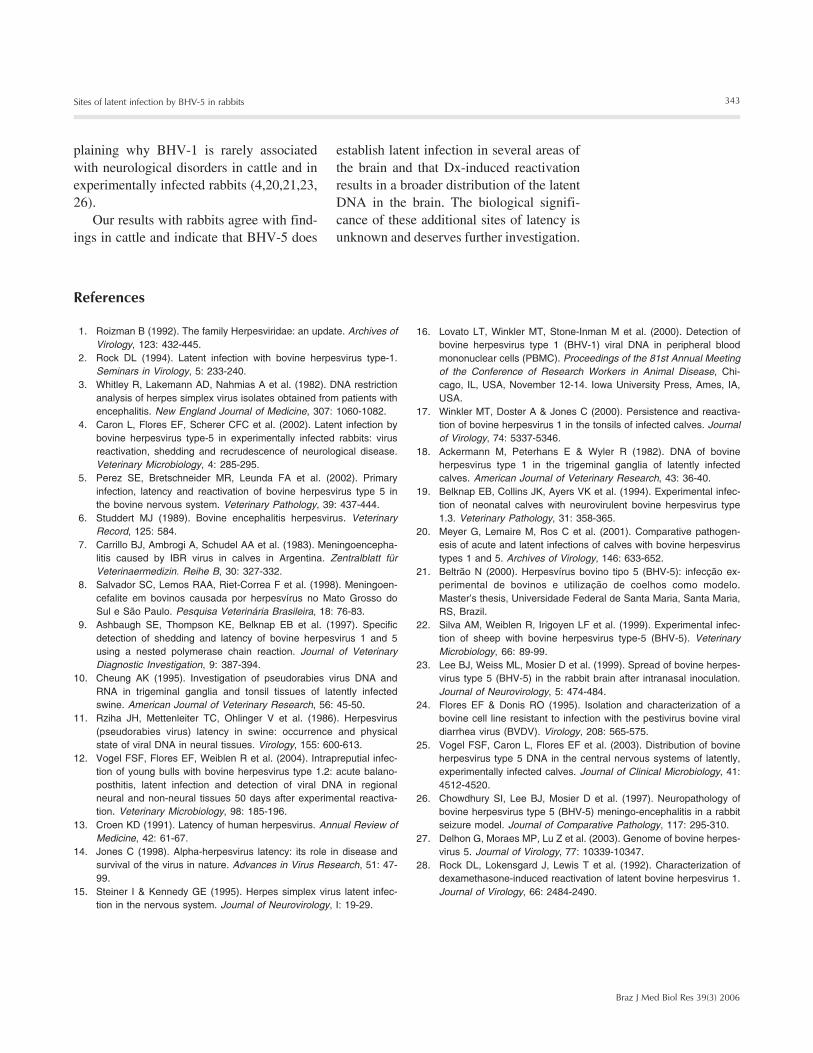
Figure 1. Rabbit brain showingthe sections examined by nestedPCR for the presence of latentbovine herpesvirus type 1 and 5DNA. A, Dorsal view; B, sagittalview. cb = cerebellum; pc = pos-terior cerebral cortex; dlc = dor-solateral cerebral cortex; ac =anterior cerebral cortex; th =thalamus; mb = midbrain; vlc =ventrolateral cerebral cortex; po= pons-medulla; mo = medullaoblongata. Olfactory bulb (ol)and trigeminal ganglia (tg) arenot shown.
dlc
pc
cb ac
pc
cbth mb
pomo
ac
vlc
339
Braz J Med Biol Res 39(3) 2006
Sites of latent infection by BHV-5 in rabbits
CCGAGTACCTGCG-3' (position 57,782) andthen with the internal primers: (forward) 5'-GTGGTGGCCTTTGACCGCGAC-3' (posi-tion 57,143) and (reverse) 5'-GCTCCGGCGAGTAGCTGGTGTG-3' (position 57,416).These two sets of primers recognize the ho-mologous sequence of gB of BHV-1 (E.F.Flores, personal communication). PCR wasperformed in 25 µL using 1 µL template DNA(containing approximately 1 µg total DNA inTE buffer), 100 ng of each primer, 1 mMMgCl2, 10 mM dNTPs, 10% DMSO, 1X reac-tion buffer, and 0.5 units of Taq polymerase(Gibco BRL, Grand Island, NY, USA). ThePCR conditions were: initial denaturation at94ºC for 10 min, followed by 35 cycles of94ºC for 1 min and 56ºC for 40 s for primerannealing and 72ºC for 40 s for primer exten-sion, and a final extension of 7 min at 72ºC.The second PCR was performed using 2 µL ofPCR products from the first PCR. The finalproduct obtained was an amplicon of 273 bp.PCR products were submitted to electropho-resis on 1.5% agarose gel, stained with ethi-dium bromide and visualized under UV light.DNA extracted from the brain of mock-inocu-lated rabbits was used as negative control.DNA extracted from the brain of a calf acutelyinfected with BHV-5 was used as positivecontrol. The specificity of PCR amplificationwas confirmed in a previous report (25).
Results
Acute infection
All rabbits inoculated with BHV-5 (groupsA and B) excreted virus in nasal secretions upto day 7 pi; 4 were still shedding virus at day 9pi and 3 at day 10 pi. Virus shedding stoppedafter day 10 pi. Thereafter, a few rabbits shedvirus for one day sporadically within the inter-val between days 19 and 60 pi. No virusshedding was detected on the days precedingDx treatment, demonstrating that rabbits werelatently infected. Three rabbits (#9, 3 and 5)developed signs of neurological disease and
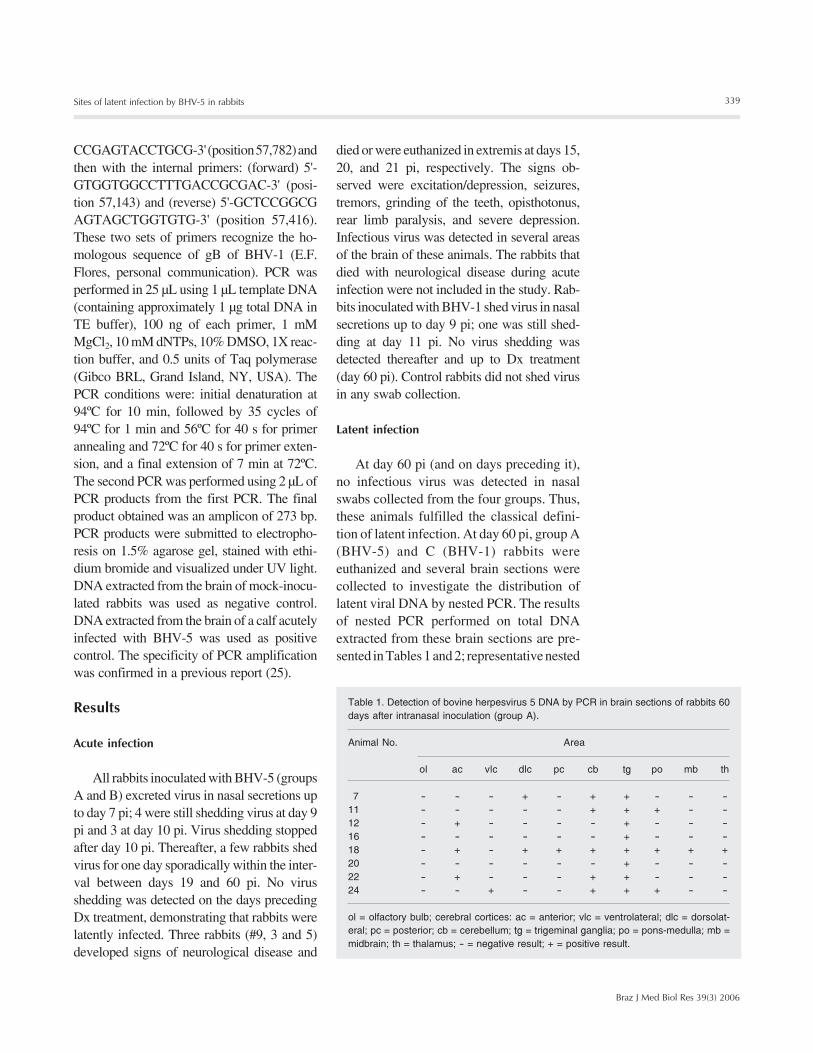
Table 1. Detection of bovine herpesvirus 5 DNA by PCR in brain sections of rabbits 60days after intranasal inoculation (group A).
Animal No. Area
ol ac vlc dlc pc cb tg po mb th
7 - - - + - + + - - -11 - - - - - + + + - -12 - + - - - - + - - -16 - - - - - - + - - -18 - + - + + + + + + +20 - - - - - - + - - -22 - + - - - + + - - -24 - - + - - + + + - -
ol = olfactory bulb; cerebral cortices: ac = anterior; vlc = ventrolateral; dlc = dorsolat-eral; pc = posterior; cb = cerebellum; tg = trigeminal ganglia; po = pons-medulla; mb =midbrain; th = thalamus; - = negative result; + = positive result.
died or were euthanized in extremis at days 15,20, and 21 pi, respectively. The signs ob-served were excitation/depression, seizures,tremors, grinding of the teeth, opisthotonus,rear limb paralysis, and severe depression.Infectious virus was detected in several areasof the brain of these animals. The rabbits thatdied with neurological disease during acuteinfection were not included in the study. Rab-bits inoculated with BHV-1 shed virus in nasalsecretions up to day 9 pi; one was still shed-ding at day 11 pi. No virus shedding wasdetected thereafter and up to Dx treatment(day 60 pi). Control rabbits did not shed virusin any swab collection.
Latent infection
At day 60 pi (and on days preceding it),no infectious virus was detected in nasalswabs collected from the four groups. Thus,these animals fulfilled the classical defini-tion of latent infection. At day 60 pi, group A(BHV-5) and C (BHV-1) rabbits wereeuthanized and several brain sections werecollected to investigate the distribution oflatent viral DNA by nested PCR. The resultsof nested PCR performed on total DNAextracted from these brain sections are pre-sented in Tables 1 and 2; representative nested
340
Braz J Med Biol Res 39(3) 2006
S.V. Mayer et al.
bral cortex (2/8), thalamus, ventrolateral andposterior cerebral cortices, and midbrain (1/8) of group A rabbits (Table 1 and Figure 2).In contrast, in group C rabbits BHV-1 DNAwas detected consistently in TG, and onlyoccasionally found in other sections (Table2, Figure 3). All DNA-positive tissues werenegative for virus isolation, confirming thatthe rabbits were latently infected. Total DNAextracted from the brain of two rabbits usedas controls gave negative results in nestedPCR.
To determine whether the distribution oflatent viral DNA in the brain would changeafter virus reactivation, rabbits from groupsB (BHV-5) and D (BHV-1) were submittedto five daily administrations of Dx begin-ning at day 60 pi. Following Dx treatment,all group B rabbits shed virus in nasal secre-tions, starting at day 3 pDx and lasting 4 to11 days. All group D rabbits also excretedvirus after Dx treatment, with shedding start-ing at day 4 pDx and lasting 7 to 9 days. Atday 60 pDx, rabbits from both groups wereeuthanized for tissue collection. At the timeof tissue collection, no animal was sheddingvirus. The distribution of latent viral DNA inthe brain of BHV-5-infected rabbits, afterDx treatment, is shown in Table 3 and Figure2. Briefly, viral DNA was detected in roughlythe same areas of group A rabbits, yet withhigher frequency in several sections, mainlyin ventrolateral (6/7) and posterior cerebralcortices (5/7), pons-medulla (6/7), thalamus(4/7), and midbrain (3/7). The sections show-ing the highest increase in positivity werethe ventrolateral cerebral cortex (1/8 to 6/7),posterior cerebral cortex (1/8 to 5/7), pons-medulla (3/8 to 6/7), thalamus (1/8 to 4/7),and midbrain (1/8 to 3/7). The other sectionsshowed a roughly similar frequency of latentviral DNA in both groups. Compared togroup A, group B rabbits showed an almosttwo-fold increase in the number of totalDNA-positive sections (38/70 or 54.3% posi-tive sections against 25/80 or 31.2%). Like-wise, rabbits from group B showed a higher
M c- c+ nt tg mo po mb th ac dlc vlc pc cb ob MA
BM c- c+ nt tg mo po mb th ac dlc vlc pc cb ob M
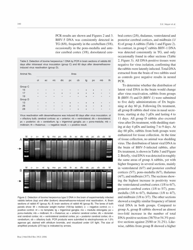
Table 2. Detection of bovine herpesvirus 1 DNA by PCR in brain sections of rabbits 60days after intranasal virus inoculation (group C) and 60 days after dexamethasone-induced virus reactivation (group D).
Animal No. Area
ol ac vlc dlc pc cb tg po mb th
Group C15 - - - - - - + + - -17 - - - + - + + + - -19 - - - - - - + - - -
Group D13 - - - - + - + + - -26 - - - - + - + - + -28 - - - - - + + - - -
Virus reactivation with dexamethasone was induced 60 days after virus inoculation. ol= olfactory bulb; cerebral cortices: ac = anterior; vlc = ventrolateral; dlc = dorsolateral;pc = posterior; cb = cerebellum; tg = trigeminal ganglia; po = pons-medulla; mb =midbrain; th = thalamus; - = negative result; + = positive result.
Figure 2. Detection of bovine herpesvirus type 5 DNA in the brain of experimentally infectedrabbits before (top) and after (bottom) dexamethasone-induced viral reactivation. A, Brainsections of rabbit #7 (group A); B, brain sections of rabbit #6 (group B). The lanes of bothpanels show: M = molecular weight marker (100-bp ladder); c- = negative control; c+ =positive control; nt = no template; tg = trigeminal ganglia; mo = medulla oblongata; po =pons-medulla; mb = midbrain; th = thalamus; ac = anterior cerebral cortex; dlc = dorsolat-eral cerebral cortex; vlc = ventrolateral cerebral cortex; pc = posterior cerebral cortex; cb =cerebellum; ob = olfactory bulb. PCR products were submitted to electrophoresis on 1.5%agarose gel, stained with ethidium bromide, and visualized under UV light. The size ofamplified products (273 bp) is indicated by arrows.
PCR results are shown and Figures 2 and 3.BHV-5 DNA was consistently detected inTG (8/8), frequently in the cerebellum (5/8),occasionally in the pons-medulla and ante-rior cerebral cortex (3/8), dorsolateral cere-
341
Braz J Med Biol Res 39(3) 2006
Sites of latent infection by BHV-5 in rabbits
mean number of PCR-positive sections(mean: 5.6 vs 3.1). Again, in group D rabbits(BHV-1), latent viral DNA was practicallylimited to TG, with only a few other sectionsbeing positive. All PCR-positive sectionswere negative for virus isolation, fulfillingthe classical requirements for defining latentinfection. No DNA-positive section was everfound in the brain of two control, mock-inoculated rabbits.
Discussion
The present results confirm and extendour findings in rabbits in which BHV-5 DNAwas detected in several different areas of thebrain of experimentally infected calves (25).In the present study, BHV-5 DNA was de-tected in several areas of the brain of latentlyinfected rabbits prior to Dx-induced reacti-vation. Furthermore, examination of brainsections of rabbits 60 days after Dx adminis-tration demonstrated a broader distributionof viral DNA in different regions of thebrain. In particular, the ventrolateral, poste-rior cerebral cortices and pons-medullashowed an increased frequency of coloniza-tion with latent viral DNA after virus reacti-vation. The failure to demonstrate infectiousvirus in PCR-positive tissues is consistentwith the biological criteria traditionally usedto define latent infection: presence of viralDNA in the absence of productive viral rep-lication (2,14,15). Taken together, these re-sults demonstrate that further viral dissemi-nation within the brain may occur upon reac-tivation and indicate a potential role of la-tency in central nervous system (CNS) sites,not excluding the TG, in the pathogenesis ofrecrudescent neurological disease that fre-quently accompanies BHV-5 reactivation.Our findings also demonstrate that the biol-ogy of latent infection of BHV-5 in rabbitsresembles that in the natural host, thus vali-dating the use of this species as a laboratorymodel to study different aspects of BHV-5pathogenesis.
Sensory nerve ganglia are the major sitesof latent infection by human and animal α-herpesviruses, but additional sites of latencyhave been identified for herpes simplex vi-rus 1, pseudorabies virus, and BHV-1 (3,9-11,13-17). The biological significance oflatency in non-neural sites and in neural sites
M c- c+ nt tg mo po mb th ac dlc vlc pc cb ob MA
BM c- c+ nt tg mo po mb th ac dlc vlc pc cb ob M
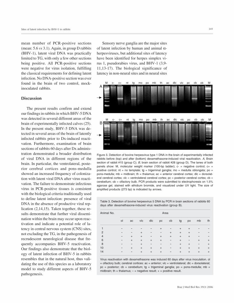
Figure 3. Detection of bovine herpesvirus type 1 DNA in the brain of experimentally infectedrabbits before (top) and after (bottom) dexamethasone-induced viral reactivation. A, Brainsection of rabbit #15 (group C); B, brain section of rabbit #28 (group D). The lanes of bothpanels show: M, molecular weight marker (100-bp ladder); c- = negative control; c+ =positive control; nt = no template; tg = trigeminal ganglia; mo = medulla oblongata; po =pons-medulla; mb = midbrain; th = thalamus; ac = anterior cerebral cortex; dlc = dorsolat-eral cerebral cortex; vlc = ventrolateral cerebral cortex; pc = posterior cerebral cortex; cb =cerebellum; ob = olfactory bulb. PCR products were submitted to electrophoresis on 1.5%agarose gel, stained with ethidium bromide, and visualized under UV light. The size ofamplified products (273 bp) is indicated by arrows.
Table 3. Detection of bovine herpesvirus 5 DNA by PCR in brain sections of rabbits 60days after dexamethasone-induced virus reactivation (group B).
Animal No. Area
ol ac vlc dlc pc cb tg po mb th
1 - - - - + - + + - -2 - - + - - + + + - -4 - + + - + + + + - -6 - + + + + - + - + +8 - - + - - - + + - +
10 - + + + + - + + + +14 - - + + + - + + + +
Virus reactivation with dexamethasone was induced 60 days after virus inoculation. ol= olfactory bulb; cerebral cortices: ac = anterior; vlc = ventrolateral; dlc = dorsolateral;pc = posterior; cb = cerebellum; tg = trigeminal ganglia; po = pons-medulla; mb =midbrain; th = thalamus; - = negative result; + = positive result.
342
Braz J Med Biol Res 39(3) 2006
S.V. Mayer et al.
other than the TG is controversial since at-tempts to reactivate the virus from some ofthese tissues by explant cultures have failed(13-17).
Previous studies have detected latentBHV-5 DNA in some areas of the brain ofexperimentally infected calves (9,20). Fur-thermore, we recently demonstrated thatBHV-5 does establish latency in several ar-eas of the brain of experimentally infectedcalves and that the distribution of latent DNAchanges after virus reactivation, resulting incolonization of additional sites (25). Thebiological significance of the presence ofviral DNA in several areas of the brain dur-ing latent infection, and the possible impli-cations for the pathogenesis of BHV-5 in-fection are unclear at this point. However,colonization of deep areas of the brain withlatent viral DNA may have important impli-cations for the pathogenesis of BHV-5 neu-rological disease that frequently occurs afterreactivation (4,5,21,25). It is conceivablethat virus reactivating from deep areas of thebrain, in addition to virus reactivating fromthe TG, may serve as a source of virus forneurological infection during reactivation.In support of this explanation was the recentdemonstration that the timing, kinetics anddistribution of histological changes in theCNS after BHV-5 reactivation differed fromthose observed during acute infection (4,5,21).
In the present study, latent BHV-5 DNAshowed an irregular distribution even amongrabbits of the same group. However, com-parison between groups A versus B demon-strated a clear tendency of increasing coloni-zation of CNS sites after reactivation. Ingroup A, 2 rabbits (#16 and 20) harboredBHV-5 DNA exclusively in TG, 5 showedpositivity in one (#12), two (#7, 11, 22) orthree sections (#24) besides the TG, and 1rabbit (#18) showed a broad distribution ofBHV-5 DNA in the brain. In comparison,among rabbits submitted to Dx administra-tion prior to PCR examination (group B),none harbored BHV-5 DNA exclusively in
the TG, only 1 (#1) had two additional sites,2 showed DNA in three additional sites (#2,8), and 4 rabbits (#4, 6, 10, and 14) showeda broad distribution of latent DNA in thebrain (four to seven positive areas in addi-tion to TG). These findings unequivocallyshow that the distribution of latent BHV-5DNA changes by becoming more broadlydistributed in regions of the brain followingvirus reactivation. In particular, the ventro-lateral and posterior cortices and the pons-medulla showed the most largest increase inthe frequency of viral DNA detection frompre- to post-reactivation. Thus, reactivatingvirus from TG and also from other areasfound to harbor viral DNA prior to reactiva-tion (i.e., pons-medulla, cerebral cortex, mid-brain, thalamus) possibly contributed to thesecondary invasion of the brain occurringupon reactivation. However, it will be nec-essary to demonstrate that BHV-5 DNA inCNS sites is biologically active, i.e., it can bereactivated as the DNA in the TG, for ex-ample, to support this explanation.
In contrast, the distribution of latent BHV-1 DNA was more restricted, regardless ofwhether the examination was performed inrabbits submitted or not to virus reactiva-tion. These findings are also consistent withresults of previous studies which showedthat TG is a major site of BHV-1 latency inthe natural hosts (2,9,14,18) and in rabbitsused as a model (4,28). Nevertheless, a fewother brain areas also harbored latent DNA,indicating that BHV-1 is capable of invad-ing some brain regions during acute infec-tion and subsequently to establish latency.Indeed, in a parallel experiment, we wereable to detect infectious virus in some areasof the brain of rabbits inoculated with aBHV-1 isolate (data not shown). These dataindicate that BHV-1, although it is not asneuroinvasive as BHV-5, may be able toreach some areas of the brain after intranasalinoculation. The replication and spread withinthe brain, however, would not suffice toproduce clinical meningoencephalitis, ex-
343
Braz J Med Biol Res 39(3) 2006
Sites of latent infection by BHV-5 in rabbits
plaining why BHV-1 is rarely associatedwith neurological disorders in cattle and inexperimentally infected rabbits (4,20,21,23,26).
Our results with rabbits agree with find-ings in cattle and indicate that BHV-5 does
establish latent infection in several areas ofthe brain and that Dx-induced reactivationresults in a broader distribution of the latentDNA in the brain. The biological signifi-cance of these additional sites of latency isunknown and deserves further investigation.
References
1. Roizman B (1992). The family Herpesviridae: an update. Archives ofVirology, 123: 432-445.
2. Rock DL (1994). Latent infection with bovine herpesvirus type-1.Seminars in Virology, 5: 233-240.
3. Whitley R, Lakemann AD, Nahmias A et al. (1982). DNA restrictionanalysis of herpes simplex virus isolates obtained from patients withencephalitis. New England Journal of Medicine, 307: 1060-1082.
4. Caron L, Flores EF, Scherer CFC et al. (2002). Latent infection bybovine herpesvirus type-5 in experimentally infected rabbits: virusreactivation, shedding and recrudescence of neurological disease.Veterinary Microbiology, 4: 285-295.
5. Perez SE, Bretschneider MR, Leunda FA et al. (2002). Primaryinfection, latency and reactivation of bovine herpesvirus type 5 inthe bovine nervous system. Veterinary Pathology, 39: 437-444.
6. Studdert MJ (1989). Bovine encephalitis herpesvirus. VeterinaryRecord, 125: 584.
7. Carrillo BJ, Ambrogi A, Schudel AA et al. (1983). Meningoencepha-litis caused by IBR virus in calves in Argentina. Zentralblatt fürVeterinaermedizin. Reihe B, 30: 327-332.
8. Salvador SC, Lemos RAA, Riet-Correa F et al. (1998). Meningoen-cefalite em bovinos causada por herpesvírus no Mato Grosso doSul e São Paulo. Pesquisa Veterinária Brasileira, 18: 76-83.
9. Ashbaugh SE, Thompson KE, Belknap EB et al. (1997). Specificdetection of shedding and latency of bovine herpesvirus 1 and 5using a nested polymerase chain reaction. Journal of VeterinaryDiagnostic Investigation, 9: 387-394.
10. Cheung AK (1995). Investigation of pseudorabies virus DNA andRNA in trigeminal ganglia and tonsil tissues of latently infectedswine. American Journal of Veterinary Research, 56: 45-50.
11. Rziha JH, Mettenleiter TC, Ohlinger V et al. (1986). Herpesvirus(pseudorabies virus) latency in swine: occurrence and physicalstate of viral DNA in neural tissues. Virology, 155: 600-613.
12. Vogel FSF, Flores EF, Weiblen R et al. (2004). Intrapreputial infec-tion of young bulls with bovine herpesvirus type 1.2: acute balano-posthitis, latent infection and detection of viral DNA in regionalneural and non-neural tissues 50 days after experimental reactiva-tion. Veterinary Microbiology, 98: 185-196.
13. Croen KD (1991). Latency of human herpesvirus. Annual Review ofMedicine, 42: 61-67.
14. Jones C (1998). Alpha-herpesvirus latency: its role in disease andsurvival of the virus in nature. Advances in Virus Research, 51: 47-99.
15. Steiner I & Kennedy GE (1995). Herpes simplex virus latent infec-tion in the nervous system. Journal of Neurovirology, I: 19-29.
16. Lovato LT, Winkler MT, Stone-Inman M et al. (2000). Detection ofbovine herpesvirus type 1 (BHV-1) viral DNA in peripheral bloodmononuclear cells (PBMC). Proceedings of the 81st Annual Meetingof the Conference of Research Workers in Animal Disease, Chi-cago, IL, USA, November 12-14. Iowa University Press, Ames, IA,USA.
17. Winkler MT, Doster A & Jones C (2000). Persistence and reactiva-tion of bovine herpesvirus 1 in the tonsils of infected calves. Journalof Virology, 74: 5337-5346.
18. Ackermann M, Peterhans E & Wyler R (1982). DNA of bovineherpesvirus type 1 in the trigeminal ganglia of latently infectedcalves. American Journal of Veterinary Research, 43: 36-40.
19. Belknap EB, Collins JK, Ayers VK et al. (1994). Experimental infec-tion of neonatal calves with neurovirulent bovine herpesvirus type1.3. Veterinary Pathology, 31: 358-365.
20. Meyer G, Lemaire M, Ros C et al. (2001). Comparative pathogen-esis of acute and latent infections of calves with bovine herpesvirustypes 1 and 5. Archives of Virology, 146: 633-652.
21. Beltrão N (2000). Herpesvírus bovino tipo 5 (BHV-5): infecção ex-perimental de bovinos e utilização de coelhos como modelo.Master’s thesis, Universidade Federal de Santa Maria, Santa Maria,RS, Brazil.
22. Silva AM, Weiblen R, Irigoyen LF et al. (1999). Experimental infec-tion of sheep with bovine herpesvirus type-5 (BHV-5). VeterinaryMicrobiology, 66: 89-99.
23. Lee BJ, Weiss ML, Mosier D et al. (1999). Spread of bovine herpes-virus type 5 (BHV-5) in the rabbit brain after intranasal inoculation.Journal of Neurovirology, 5: 474-484.
24. Flores EF & Donis RO (1995). Isolation and characterization of abovine cell line resistant to infection with the pestivirus bovine viraldiarrhea virus (BVDV). Virology, 208: 565-575.
25. Vogel FSF, Caron L, Flores EF et al. (2003). Distribution of bovineherpesvirus type 5 DNA in the central nervous systems of latently,experimentally infected calves. Journal of Clinical Microbiology, 41:4512-4520.
26. Chowdhury SI, Lee BJ, Mosier D et al. (1997). Neuropathology ofbovine herpesvirus type 5 (BHV-5) meningo-encephalitis in a rabbitseizure model. Journal of Comparative Pathology, 117: 295-310.
27. Delhon G, Moraes MP, Lu Z et al. (2003). Genome of bovine herpes-virus 5. Journal of Virology, 77: 10339-10347.
28. Rock DL, Lokensgard J, Lewis T et al. (1992). Characterization ofdexamethasone-induced reactivation of latent bovine herpesvirus 1.Journal of Virology, 66: 2484-2490.


