Embed Size (px)
Citation preview

HUMAN NEUROSCIENCEORIGINAL RESEARCH ARTICLE
published: 09 September 2014doi: 10.3389/fnhum.2014.00704
Delayed convergence between brain network structure andfunction in rolandic epilepsyRené M. H. Besseling1,2,3, Jacobus F. A. Jansen2,3, Geke M. Overvliet1,4, Sylvie J. M. van der Kruijs1,2,Saskia C. M. Ebus1, Anton J. A. de Louw1,4, Paul A. M. Hofman1,4, Albert P. Aldenkamp1,4 andWalter H. Backes2,3*1 Epilepsy Center Kempenhaeghe, Heeze, Netherlands2 Research School for Mental Health and Neuroscience, Maastricht University, Maastricht, Netherlands3 Department of Radiology, Maastricht University Medical Center, Maastricht, Netherlands4 Department of Neurology, Maastricht University Medical Center, Maastricht, Netherlands
Edited by:Srikantan S. Nagarajan, Universityof California, San Francisco, USA
Reviewed by:Heidi E. Kirsch, University ofCalifornia, San Francisco, USADario J. Englot, University ofCalifornia, San Francisco, USA
*Correspondence:Walter H. Backes, Department ofRadiology, Maastricht UniversityMedical Center, PB 5800, 6202 AZMaastricht, Netherlandse-mail: [email protected]
Introduction: Rolandic epilepsy (RE) manifests during a critical phase of braindevelopment, and has been associated with language impairments. Concordantabnormalities in structural and functional connectivity (SC and FC) have been describedbefore. As SC and FC are under mutual influence, the current study investigatesabnormalities in the SC-FC synergy in RE.
Methods: Twenty-two children with RE (age, mean ± SD: 11.3 ± 2.0 y) and 22healthy controls (age 10.5 ± 1.6 y) underwent structural, diffusion weighted, and resting-state functional magnetic resonance imaging (MRI) at 3T. The probabilistic anatomicallandmarks atlas was used to parcellate the (sub)cortical gray matter. Constrained sphericaldeconvolution tractography and correlation of time series were used to assess SC and FC,respectively. The SC-FC correlation was assessed as a function of age for the non-zerostructural connections over a range of sparsity values (0.01–0.75). A modularity analysiswas performed on the mean SC network of the controls to localize potential global effectsto subnetworks. SC and FC were also assessed separately using graph analysis.
Results: The SC-FC correlation was significantly reduced in children with RE compared tohealthy controls, especially for the youngest participants. This effect was most pronouncedin a left and a right centro-temporal network, as well as in a medial parietal network. Graphanalysis revealed no prominent abnormalities in SC or FC network organization.
Conclusion: Since SC and FC converge during normal maturation, our finding of reducedSC-FC correlation illustrates impaired synergy between brain structure and function.More specifically, since this effect was most pronounced in the youngest participants,RE may represent a developmental disorder of delayed brain network maturation. Theobserved effects seem especially attributable to medial parietal connections, which formsan intermediate between bilateral centro-temporal modules of epileptiform activity, andbear relevance for language function.
Keywords: rolandic epilepsy, structural connectivity, functional connectivity, structure-function correlation, brainmaturation, graph theory
INTRODUCTIONAlthough rolandic epilepsy (RE) is classically considered a benignepilepsy of childhood, over the last decade it has been asso-ciated with inattention, impulsivity, and cognitive complaints(Loiseau and Duché, 1989; Massa et al., 2001). The latter notablyinvolves a range of impaired language abilities, such as oromo-tor deficits, problems in phonological awareness, compromisedwritten language skills, reading disability, and speech sound dis-order (Lundberg et al., 2005; Papavasiliou et al., 2005; Clarkeet al., 2007; Northcott et al., 2007; Overvliet et al., 2011). Theselanguage impairments can clinically be of greater concern than
the seizures, especially since they may persist after spontaneousseizure remission, which in RE is typically seen before the ageof 16 years (Hommet et al., 2001; Panayiotopoulos et al., 2008;Monjauze et al., 2011).
In neuroimaging, there has recently been a renewed interestin the integration of the motor and the language system. Severalstudies have demonstrated that the motor system is not only rel-evant for language in a straightforward way such as coordinationof articulatory movement, but is also involved in purely cognitiveaspects of language, such as speech comprehension (Pulvermülleret al., 2006). For an overview, we refer to the recent work by
Frontiers in Human Neuroscience www.frontiersin.org September 2014 | Volume 8 | Article 704 | 1

Besseling et al. Delayed network convergence in rolandic epilepsy
Cappa and Pulvermuller and the references therein (Cappa andPulvermuller, 2012).
In RE, this moto-lingual integration may be a key con-cept for linking the epileptic focus in the sensorimotor (i.e.,rolandic) cortex to the broad range of associated language impair-ments. Indeed, using functional MRI (fMRI) it has recently beendemonstrated that the integration between the motor and thelanguage system is compromised in RE compared to healthycontrols (Besseling et al., 2013b,c). Concordant abnormalitiesin structural connectivity have also recently been described(Besseling et al., 2013a). However, how these functional andstructural abnormalities relate to each other remains to beinvestigated.
It should be noted that RE manifests during a critical and vul-nerable phase of brain maturation. The networks that are formedduring this period actually are the result of continuous interac-tions between the developing structural and functional circuits,among other factors (Andersen, 2003). Neuropathological cues,such as epileptiform discharges, may offset normal developmentaltrajectories and compromise the final adult network organiza-tion (Andersen, 2003). In other words, abnormalities in eitherstructural or functional connectivity (SC and FC, respectively) ortheir interaction may explain behavioral and cognitive problemsin children with RE, as well as their persistence in those that arein seizure remission (Hommet et al., 2001; Panayiotopoulos et al.,2008; Monjauze et al., 2011).
The study of the association between SC and FC is an emergingfield of research, which is rapidly gaining interest (Sporns, 2013).Several studies have demonstrated that FC is determined (andconstrained) by structural white matter connections (Honey et al.,2007, 2009, 2010; Greicius et al., 2009). Vice versa, simulationstudies have shown that functional dynamics are spontaneouslyself-structuring, and in turn may help to sculpt structural con-nections (Rubinov et al., 2009). It has been suggested that duringnormal brain development, the SC-FC correlation increases aswhite matter connections and network dynamics converge underinfluence of a common optimization process to improve bothnetwork efficiency and efficacy (Hagmann et al., 2010; Supekaret al., 2010).
In epilepsy, abnormalities in either SC or FC have extensivelybeen described, as recently reviewed by van Diessen et al. (2013).However, abnormalities in the SC-FC correlation have hardlybeen investigated, and to our knowledge not yet in the developingchild with epilepsy.
In the current work, the SC-FC correlation is investigatedas a function of age in children with RE compared to healthycontrol children. A modularity analysis is employed to investigatewhether potential abnormalities can be localized to certain sub-networks, and graph-theoretical measures are employed to checkfor underlying aberrant SC and/or FC network organization.
MATERIALS AND METHODSPATIENT SELECTIONTwenty-two children with RE (8 girls; age, mean ± SD: 11.3 ±2.0 years) were selected at our specialized epilepsy referral center.Selection criteria included centro-temporal spikes on EEG andconcordant seizure semiology representing anarthria, hemiclonia
involving the face and/or unilateral extremities, or secondar-ily generalized seizures. In case of poorly observed nocturnalseizures, post-ictal signs of generalized seizure or confirmationof post-ictal hemiparesis was sufficient for inclusion in case ofotherwise typical EEG. Further details were described previously(Overvliet et al., 2013).
In addition, 22 healthy controls were enrolled in the study (11girls, age 10.5 ± 1.6 years). All patients had an IQ >70 and allcontrols attended regular education, and had neither (a historyof) neurological disorders, nor learning problems.
The parents or guardians of all children gave written informedconsent for study participation, and the study was approved bythe ethical review boards of both participating institutions.
MAGNETIC RESONANCE IMAGINGConventional structural magnetic resonance imaging (MRI) wasapplied, as well as diffusion weighted imaging (DWI) and fMRI.Structural imaging included a T1-weigthed sequence. A 3Dfast-spoiled gradient echo sequence was used, employing echotime/repetition time/inversion time (TE/TR/TI) 3.8/8.3/1022 ms,and at a resolution of 1 × 1 × 1 mm3. The acquisition time was8 min.
Also high angular resolution diffusion weighted imaging(HARDI) was performed, employing 66 non-colinear gradientdirections (Jones et al., 1999), and a diffusion-sensitizing b-value of 1200 s/mm2. In addition, a single minimally diffusion-weighted image (b0-scan) was acquired. An echo planar imaging(EPI) sequence was used with TE/TR 72/6600 ms, resolution 2 ×2× 2 mm3, and acquisition time 9 min.
Resting-state functional MRI (rs-fMRI) involved a task-freeT2*-weighted blood oxygen level dependent (BOLD) sequenceof 195 dynamic image volumes at a TR of 2 s, resulting inan acquisition time of 6.5 min. Further settings included: EPIsequence, TE 35 ms, 2 × 2 mm2 in plane resolution, and 4 mmthick axial slices.
CORTICAL PARCELLATIONThe probabilistic anatomical landmark atlas of gyri, sulci, andbasal ganglia was used to parcellate the cortical and subcorticalgray matter (Perrot et al., 2009; Varoquaux et al., 2010). This atlasconsists of N = 137 volumes, each representing a probabilisticmap for a certain brain region. To map this atlas to native T1space, affine registrations were implemented using SPM (version8). In addition, deterministic node labels were constructed byassigning each voxel to its region of maximum probability.
STRUCTURAL CONNECTIVITYThe diffusion-weighted data were preprocessed and tractographywas performed as previously described (Besseling et al., 2012).Briefly, this involved that the diffusion-weighted volumes wereregistered to the b0-scan to correct for head motion and EPIdistortions using affine registrations as implemented in CATNAP(Coregistration, Adjustment, and Tensor-solving: a Nicely Auto-mated Program). CATNAP is based on FSL routines (FMRIBSoftware Library) and includes correction of the gradient direc-tions for rotations (Landman et al., 2007; Leemans and Jones,2009).
Frontiers in Human Neuroscience www.frontiersin.org September 2014 | Volume 8 | Article 704 | 2

Besseling et al. Delayed network convergence in rolandic epilepsy
Next, constrained spherical deconvolution (CSD) was usedto estimate voxel-wise fiber orientation distributions (FODs).CSD FODs can represent multiple fiber orientations per voxel,and thus account for partial volume effects such as within-voxel fiber kissing, crossing and bending (Tournier et al., 2007,2008, 2012). The CSD response function was estimated fromthe data employing high fractional anisotropy voxels (FA > 0.7).A CSD order of lmax = 8 (i.e., 45 spherical harmonics) wasused; for details, see Tournier et al. (2009) and Besseling et al.(2012).
Probabilistic tractography was performed employing MRtrixto extrapolate voxel-wise FODs to global (semi)continuousstreamlines (Tournier et al., 2012). Per subject, 50.000 streamlineswere seeded from the gray matter (FSL based tissue segmentationof the T1 scan), and propagated over the brain to representthe overall topology of the global white matter network. Stan-dard MRtrix tractography settings were used, which includesa streamline propagation step size of 0.2 mm, a minimumradius of curvature of 1 mm, and an FOD amplitude threshold>0.1.
Structural connectivity (SC) was investigated for the deter-ministic node labels. For this, the streamlines were firstmapped to the T1-space based on an affine registration ofthe b0-scan to the T1-scan using FSL (Pannek et al., 2011).Next, for each pair of nodes the interconnecting stream-lines were assessed. As larger nodes typically contain morestreamlines, connection strength was quantified as the num-ber of streamlines normalized for the number of voxels pernode pair (van den Heuvel and Sporns, 2011; Zhang et al.,2011).
FUNCTIONAL CONNECTIVITYPreprocessing of the rs-fMRI data included registration of allfMRI volumes to the first dynamic to correct for head motionemploying SPM8. Subsequently, the mean rs-fMRI image volumewas calculated and used to affinely register the rs-fMRI data to thenative T1-space.
The T1 tissue segmentation was downsampled to the rs-fMRI resolution to calculate averaged time series for the whitematter and the CSF. These time series, combined with the move-ment parameters of the previous step, were used as nuisanceregressors to deconfound the rs-fMRI data employing linearregression. This procedure is assumed to provide a more spe-cific and robust correction for non-neuronal signal fluctuationssuch as scanner drift or physiological noise (cardioballisticsand breathing) than whole-brain signal regression (Smith et al.,2011).
Finally, the rs-fMRI data were smoothed using a Gaussiankernel of full-width-at-half-maximum 10 mm, and band-passfiltered to confine the signal to the range of 0.01–0.1 Hz astypically used in rs-fMRI analyses (Zalesky et al., 2010; Cocchiet al., 2012; Hong et al., 2013).
Functional connectivity (FC) was assessed using correlation ofnode pair time series. The probabilistic atlas maps were used tocalculate these node time series as a weighted average, effectivelyassigning more weight to the core and less weight to the border ofeach node.
CORRELATION BETWEEN STRUCTURAL AND FUNCTIONALCONNECTIVITYTo assess the SC-FC correlation, the FC values were resampled toa normal distribution of mean 0.5 and SD 0.1 following previouswork (Honey et al., 2009; Zhang et al., 2011). Furthermore,negative FC values were set to 0 (Smith et al., 2011).
For each subject, the SC-FC association was calculated byappending the SC and FC values of the non-zero structuralconnections in two separate vectors, which were subsequentlycorrelated. To assess the robustness of the above analysis, it wasapplied over a range of sparsity values. This means that not allstructural connections were included in the correlation analysis,but only a certain top percentage.
The sparsity structure (i.e., which connections were included)was based on the mean SC matrix of the controls, see Figure 1.Since the mean sparsity of the SC of the controls was 0.75, asparsity range of 0.01–0.75 was chosen, corresponding to 93–6987connections. Sparsity values were incremented at a step size of0.01.
MODULARITY ANALYSISTo investigate whether potential abnormalities in SC-FC cor-relation may be contributed to specific brain sub-networks, amodularity analysis was applied to the average SC network ofthe controls using the Brain Connectivity Toolbox (BCT). TheBCT is a well-established collection of mathematical routines forgraph theoretical analysis of complex brain networks (Rubinovand Sporns, 2010). Modularity analysis clusters nodes into mod-ules, based on relatively high within-module and low between-module connectivity (Newman, 2006). Because of heuristics inthe algorithm, the modularity analysis was repeated 100 times,and the most robust modularity structure (of highest occurrence)was retained.
ASSESSING DIFFERENCES IN STRUCTURE-FUNCTION CORRELATIONAbnormalities in SC-FC correlation in patients compared tocontrols were inferred upon using a general linear model (GLM),in which also the effect of age was incorporated. Group differenceswith respect to SC-FC correlation were deemed significant onthe whole-brain level for p < 0.05. If such a significant groupdifference was found, it was investigated whether this could beattributed to one (or multiple) of the sub-networks, employingBonferroni multiple comparisons correction for the number ofsub-networks under investigation.
MEASURES OF NETWORK ORGANIZATIONTo investigate whether SC and/or FC as such were abnormal,graph theoretical measures of network integrity were calculated.For each subject, structural and functional network organizationwere quantified by average path length and clustering coefficient(CC) as assessed using the BCT. Individual SC and FC con-nectivity matrices were scaled with respect to their total con-nectivity weight to normalize the “wiring cost” over subjects(Zhang et al., 2011). Potential differences in graph measuresthus only reflect differences in the distribution of connec-tivity values. This is similar to the SC-FC correlation analy-sis, which also only takes the distribution of the connectivity
Frontiers in Human Neuroscience www.frontiersin.org September 2014 | Volume 8 | Article 704 | 3

Besseling et al. Delayed network convergence in rolandic epilepsy
FIGURE 1 | Assessing the correlation between structural andfunctional connectivity (SC and FC, respectively). The mostprominent structural connections (here at sparsity s = 0.04) areselected. The SC and FC values of these are appended in two vectors,
which are subsequently correlated. For visualization purposes, thevectors are not visualized in full length. SC values were scaled to aGaussian distribution (mean, SD: 0.5, 0.1); negative FC values were setto 0.
values into account and not their amplitude. Furthermore, foreach subject the graph measures were normalized with respectto comparable random graphs (N = 20) (van den Heuveland Sporns, 2011). The comparisons between patients andcontrols were performed using the same GLM as describedabove.
RESULTSThe modularity analysis revealed five clusters: a prefrontal cluster,a medial parietal cluster, an inferior temporo-occipital cluster, andleft and right centro-temporal clusters, see Figure 2.
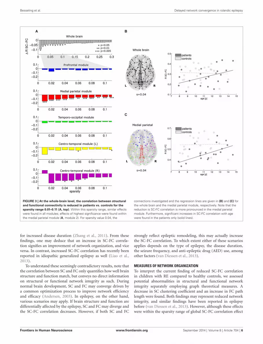
At the whole-brain level, significant reductions in SC-FCcorrelation were found in the patients for the most promi-nent brain connections (sparsity range 0.01–0.11; Figure 3A,top row). Within this sparsity range, the SC-FC correlation wasalso consistently reduced within all five modules. Significanteffects were found in the medial parietal cluster, and also inboth centro-temporal clusters. Within the medial parietal cluster,
the reduction in SC-FC correlation was most robust over thesparsity range, and also most pronounced (Figure 3A; p <
0.005).For a representative sparsity value of 0.04, the connections
under investigation and the regression lines are given for thewhole brain and the medial parietal cluster in Figures 3B,C,respectively. Note the significant progressive increase in SC-FCcorrelation with age in the patients; no such effect was foundin the controls. The group differences were strongest for theyoungest participants and diminished towards the end of the agewindow under investigation (8–14 years).
Concerning graph analysis, no clear differences in either SCor FC network organization were found between patients andcontrols. For single sparsity values, a decrease in SC cluster-ing coefficient and an increase in FC path length were found(sparsity 0.04 and 0.08, respectively), see Figure 4. No con-sistent effects were found over any of the modules underinvestigation.
Frontiers in Human Neuroscience www.frontiersin.org September 2014 | Volume 8 | Article 704 | 4

Besseling et al. Delayed network convergence in rolandic epilepsy
FIGURE 2 | Sub-networks as derived from a modularity analysisof the mean structural connectivity (SC) network of thecontrols. Five sub-networks (i.e., modules) were found; a prefrontalcluster (blue), a medial parietal cluster (red), an inferior
temporo-occipital cluster (green), and a left and a rightcentro-temporal cluster (yellow and magenta, respectively). Forvisualization purposes, only the within-module connections werevisualized, at sparsity level 0.1.
DISCUSSIONIn this study, we investigated the correlation between SC and FCas a function of age in children with RE compared to healthy con-trols. We employed a modularity analysis to investigate whetherpotential effects on the whole-brain level may be attributed tocertain sub-networks. Additionally, graph theoretical measures ofnetwork organization were used to assess SC and FC separately,both at the global level and concerning the identified modules.
MAJOR FINDINGSOur main results are:
1. In children with RE, the SC-FC correlation is reduced com-pared to healthy controls at the whole-brain level;
2. The SC-FC correlation increases with age in the childrenwith RE, implying that the reduction in SC-FC correlation is
less pronounced towards the end of the age window underinvestigation (8–14 years);
3. Similar effects were found at the module level, i.e., for bothcentro-temporal clusters and most notably for the medialparietal cluster, in which the reduction in SC-FC correlationwas stronger than at the whole-brain level;
4. Employing graph analysis, no prominent abnormalities in SCor FC network organization were found.
INTERPRETATION OF DIFFERENCES IN SC-FC CORRELATIONDuring normal brain maturation, network structure and functiongradually adapt to each other, leading to an increase in the SC-FC correlation (Hagmann et al., 2010; Supekar et al., 2010). Inadults with idiopathic generalized epilepsy, reductions in SC-FC correlation have been described which are more pronounced
Frontiers in Human Neuroscience www.frontiersin.org September 2014 | Volume 8 | Article 704 | 5
Besseling et al. Delayed network convergence in rolandic epilepsy
FIGURE 3 | At the whole-brain level, the correlation between structuraland functional connectivity is reduced in patients vs. controls for thesparsity range 0.01–0.11 (A, top). Within this sparsity range, similar effectswere found in all modules; effects of highest significance were found withinthe medial parietal module (A, module 2). For sparsity value 0.04, the
connections investigated and the regression lines are given in (B) and (C) forthe whole brain and the medial parietal module, respectively. Note that thereduction is SC-FC correlation is more pronounced in the medial parietalmodule. Furthermore, significant increases in SC-FC correlation with agewere found in the patients only (solid lines).
for increased disease duration (Zhang et al., 2011). From thesefindings, one may deduce that an increase in SC-FC correla-tion signifies an improvement of network organization, and viceversa. In contrast, increased SC-FC correlation has recently beenreported in idiopathic generalized epilepsy as well (Liao et al.,2013).
To understand these seemingly contradictory results, note thatthe correlation between SC and FC only quantifies how well brainstructure and function match, but conveys no direct informationon structural or functional network integrity as such. Duringnormal brain development, SC and FC may converge driven bya common optimization process to improve network efficiencyand efficacy (Andersen, 2003). In epilepsy, on the other hand,various scenarios may apply. If brain structure and function aredifferentially affected by the epilepsy, SC and FC may diverge andthe SC-FC correlation decreases. However, if both SC and FC
strongly reflect epileptic remodeling, this may actually increasethe SC-FC correlation. To which extent either of these scenariosapplies depends on the type of epilepsy, the disease duration,the seizure frequency, and anti-epileptic drug (AED) use, amongother factors (van Diessen et al., 2013).
MEASURES OF NETWORK ORGANIZATIONTo interpret the current finding of reduced SC-FC correlationin children with RE compared to healthy controls, we assessedpotential abnormalities in structural and functional networkintegrity separately employing graph theoretical measures. Adecrease in SC clustering coefficient and an increase in FC pathlength were found. Both findings may represent reduced networkintegrity, and similar findings have been reported in epilepsybefore (van Diessen et al., 2013). However, although these effectswere within the sparsity range of global SC-FC correlation effect
Frontiers in Human Neuroscience www.frontiersin.org September 2014 | Volume 8 | Article 704 | 6

Besseling et al. Delayed network convergence in rolandic epilepsy
FIGURE 4 | Structural and functional connectivity (SC and FC,respectively) were investigated separately by assessing the normalizedpath length (λ) and clustering coefficient (CC). A decrease in SCclustering coefficient was found for sparsity value 0.04, and an increase inFC path length for sparsity value 0.08.
(0.01–0.11), they were only found for single sparsity values, andwithin this sparsity range, no module effects were found. Thesefindings suggest that in RE, gross abnormalities in structuraland/or functional network organization are not the case. Indeed,RE is a relatively mild disorder compared to other epilepsysyndromes within the same spectrum, such as Landau KleffnerSyndrome (Hughes, 2010; Overvliet et al., 2010). Our finding ofreduced SC-FC correlation should thereby be interpreted in thecontext of aberrant brain maturation, and reflects a reduced con-vergence between network structure and function. In agreement,cortical abnormalities, reflective of impaired development of the
underlying network, have been reported in RE before (Overvlietet al., 2013).
EFFECT OF AGENote that the SC-FC correlation actually progressively increasedwith age in the patients, whereas no age effect was found in thecontrols. This suggests that the maturational convergence of SCand FC is merely delayed in RE. Potentially, for the age rangeunder investigation (8–14 years), network structure and functionare already to a certain extent matched for the controls, whereasin the children with RE, this optimization process is still ongoing.However, since it has been reported before that the cognitivecomplaints may persist, a full recovery of the SC-FC correlationis not expected (Hommet et al., 2001; Andersen, 2003; Monjauzeet al., 2011).
LOCALIZATION TO SUB-NETWORKSConcerning the localization of effects, the overall reduction in SC-FC correlation seems to be particularly attributable to a medialparietal network, where the effect was actually more pronouncedthan at the whole-brain level. Note that the parietal lobe has beenassociated with visuo-spatial skills, which may be impaired in RE(Pinton et al., 2006; Völkl-Kernstock et al., 2006).
More importantly, the medial parietal module provides aninterface between the bilateral centro-temporal modules, fromwhich the epileptiform activity originates (i.e., RE typical centro-temporal spikes). Spread of epileptiform activity to parietalregions has been demonstrated in RE before (Drury and Beydoun,1991; Graf and Lischka, 1998). Furthermore, the SC-FC corre-lation appeared (to a lesser extent) compromised within thesecentro-temporal modules themselves as well. Abnormalities inthe left centro-temporal module may translate to the prominentlanguage impairments in RE, as this module roughly covers clas-sical inferior frontal and supramarginal language areas (Broca’sand Wernicke’s, respectively). Furthermore, since language lat-eralization is limited in children in general (Kadis et al., 2011)and in RE in particular (Besseling et al., 2013c), impairments ofcontralateral (right hemisphere) homotopic cortex may also relateto compromised language skills.
The module effect was more robust for the medial parietalmodule than e.g., for the left centro-temporal module, whichmight be due to variations in the laterality of EEG abnormalities.Defining a homogeneous RE cohort with respect to electrophys-iology is very challenging as the laterality (and extent) of theEEG abnormalities may dramatically change over time (Rivaet al., 2007). Actually this is considered a general confound forepilepsy connectivity studies (van Diessen et al., 2013). Sincein our clinical cohort electrophysiology was assessed during thediagnostic work-up and not at the time of scanning, the patientscan be assumed to be somewhat heterogenous with respect toEEG characteristics. On the group level, an un-lateralized moduleeffect may therefore be the most robust.
METHODOLOGICAL CONSIDERATION AND DIRECTIONS FOR FUTURERESEARCHConcerning the localization of the network impairments, it isunclear whether the modularity structure employed in this study
Frontiers in Human Neuroscience www.frontiersin.org September 2014 | Volume 8 | Article 704 | 7

Besseling et al. Delayed network convergence in rolandic epilepsy
was optimal to detect aberrant sub-networks in RE. The sub-networks we investigated were derived from an unbiased graphtheoretical modularity analysis of the structural data. For theatlas we employed, this yielded a robust set of five compactclusters, two of which were clearly lateralized. These robust clus-ters had a relatively straightforward anatomical description andinterpretation.
Note that specific investigations of e.g., the language networkmay have yielded more illustrative results in the context of theRE-typical neuropsychological profile of language impairment(Overvliet et al., 2010). However, this would be at the cost ofsubjective choices, since at present the language network cannotunambiguously be identified from e.g., functional connectivitypatterns (Beckmann et al., 2005; Besseling et al., 2013c).
In any case, our findings illustrate that investigations into SC-FC correlation can be used to infer where in the brain neuronalnetwork formation is impaired, and as such our study extendsthe work by Zhang et al. on generalized epilepsy to localization-related epilepsies such as RE (Zhang et al., 2011).
In our cross-sectional study, the effect of age could only beassessed by virtue of the variability in age of the subjects included.Longitudinal studies are called for to verify the robustness ofour findings. Follow-up until after seizure remission is proposedfor to investigate to what extent the reported abnormalities mayeventually normalize.
Finally, for practical reasons AED use was not controlled for inthe clinical cohort under investigation. As described previously,about two thirds of the patients were under AED treatment, whichmay have confounded our results (Overvliet et al., 2013). Theeffects described are expected to be more pronounced for a moretightly controlled patient group.
CONCLUSIONIn children with RE, the correlation between SC and FC isreduced compared to healthy controls. As structural nor func-tional network organization is much affected, this is interpretedas reduced synergy of SC and FC due to aberrant brain mat-uration. Since in the patients the discrepancy between SC andFC improves as a function of age, these findings may actu-ally represent maturational delay. Concerning the localizationof brain network abnormalities, the observed effects seem espe-cially attributable to medial parietal connections, which formsan intermediate between bilateral centro-temporal modules ofepileptiform activity.
ACKNOWLEDGMENTSWe would like to acknowledge Esther Peeters for data acquisitionsand Marc Geerlings and Jos Slenter for continuous hardwareand software support. We thank the Dutch Epilepsy Foundationfor research funding, and the participating children and theirfamilies for their time and commitment, and their interest in ourwork.
REFERENCESAndersen, S. L. (2003). Trajectories of brain development: point of vulnerability or
window of opportunity? Neurosci. Biobehav. Rev. 27, 3–18. doi: 10.1016/s0149-7634(03)00005-8
Beckmann, C. F., DeLuca, M., Devlin, J. T., and Smith, S. M. (2005). Investigationsinto resting-state connectivity using independent component analysis. Philos.Trans. R. Soc. Lond. B Biol. Sci. 360, 1001–1013. doi: 10.1098/rstb.2005.1634
Besseling, R. M., Jansen, J. F., Overvliet, G. M., Vaessen, M. J., Braakman, H. M.,Hofman, P. A., et al. (2012). Tract specific reproducibility of tractography basedmorphology and diffusion metrics. PLoS One 7:e34125. doi: 10.1371/journal.pone.0034125
Besseling, R., Jansen, J., Overvliet, G., Van der Kruijs, S., Ebus, S., De Louw,A., et al. (2013a). Reduced structural connectivity between sensorimotor andlanguage areas in rolandic epilepsy. PLoS One 8:e83568. doi: 10.1371/journal.pone.0083568
Besseling, R. M. H., Jansen, J. F., Overvliet, G. M., van der Kruijs, S. J., Vles,J. S., Ebus, S. C. M., et al. (2013b). Reduced functional integration of thesensorimotor and language network in rolandic epilepsy. Neuroimage Clin. 2,239–246. doi: 10.1016/j.nicl.2013.01.004
Besseling, R., Overvliet, G., Jansen, J., Van der Kruijs, S., Vles, J., Ebus, S.,et al. (2013c). Aberrant functional connectivity between motor and languagenetworks in rolandic epilepsy. Epilepsy Res. 8:e65937. doi: 10.1016/j.eplepsyres.2013.10.008
Cappa, S. F., and Pulvermuller, F. (2012). Cortex special issue: language and themotor system. Cortex 48, 785–787. doi: 10.1016/j.cortex.2012.04.010
Clarke, T., Strug, L. J., Murphy, P. L., Bali, B., Carvalho, J., Foster, S., et al. (2007).High risk of reading disability and speech sound disorder in rolandic epilepsyfamilies: case-control study. Epilepsia 48, 2258–2265. doi: 10.1111/j.1528-1167.2007.01276.x
Cocchi, L., Bramati, I. E., Zalesky, A., Furukawa, E., Fontenelle, L. F., Moll, J.,et al. (2012). Altered functional brain connectivity in a non-clinical sampleof young adults with attention-deficit/hyperactivity disorder. J. Neurosci. 32,17753–17761. doi: 10.1523/jneurosci.3272-12.2012
Drury, I., and Beydoun, A. (1991). Benign partial epilepsy of childhood withmonomorphic sharp waves in centrotemporal and other locations. Epilepsia 32,662–667. doi: 10.1111/j.1528-1157.1991.tb04706.x
Graf, M., and Lischka, A. (1998). Topographical EEG analysis of rolandic spikes.Clin. Electroencephalogr. 29, 132–137. doi: 10.1177/155005949802900305
Greicius, M. D., Supekar, K., Menon, V., and Dougherty, R. F. (2009). Resting-state functional connectivity reflects structural connectivity in the default modenetwork. Cereb. Cortex 19, 72–78. doi: 10.1093/cercor/bhn059
Hagmann, P., Sporns, O., Madan, N., Cammoun, L., Pienaar, R., Wedeen, V. J.,et al. (2010). White matter maturation reshapes structural connectivity in thelate developing human brain. Proc. Natl. Acad. Sci. U S A 107, 19067–19072.doi: 10.1073/pnas.1009073107
Hommet, C., Billard, C., Motte, J., Passage, G. D., Perrier, D., Gillet, P., et al. (2001).Cognitive function in adolescents and young adults in complete remission frombenign childhood epilepsy with centro-temporal spikes. Epileptic Disord. 3, 207–216.
Honey, C. J., Kotter, R., Breakspear, M., and Sporns, O. (2007). Network structureof cerebral cortex shapes functional connectivity on multiple time scales. Proc.Natl. Acad. Sci. U S A 104, 10240–10245. doi: 10.1073/pnas.0701519104
Honey, C. J., Sporns, O., Cammoun, L., Gigandet, X., Thiran, J. P., Meuli, R., et al.(2009). Predicting human resting-state functional connectivity from structuralconnectivity. Proc. Natl. Acad. Sci. U S A 106, 2035–2040. doi: 10.1073/pnas.0811168106
Honey, C. J., Thivierge, J. P., and Sporns, O. (2010). Can structure predict functionin the human brain? Neuroimage 52, 766–776. doi: 10.1016/j.neuroimage.2010.01.071
Hong, S. B., Zalesky, A., Cocchi, L., Fornito, A., Choi, E. J., Kim, H. H., et al. (2013).Decreased functional brain connectivity in adolescents with internet addiction.PLoS One 8:e57831. doi: 10.1371/journal.pone.0057831
Hughes, J. R. (2010). Benign epilepsy of childhood with centrotemporal spikes(BECTS): to treat or not to treat, that is the question. Epilepsy Behav. 19, 197–203. doi: 10.1016/j.yebeh.2010.07.018
Jones, D. K., Horsfield, M. A., and Simmons, A. (1999). Optimal strategies for mea-suring diffusion in anisotropic systems by magnetic resonance imaging. Magn.Reson. Med. 42, 515–525. doi: 10.1002/(sici)1522-2594(199909)42:3<515::aid-mrm14=3.3.co;2-h
Kadis, D. S., Pang, E. W., Mills, T., Taylor, M. J., McAndrews, M. P., and Smith,M. L. (2011). Characterizing the normal developmental trajectory of expressivelanguage lateralization using magnetoencephalography. J. Int. Neuropsychol. Soc.17, 896–904. doi: 10.1017/S1355617711000932
Frontiers in Human Neuroscience www.frontiersin.org September 2014 | Volume 8 | Article 704 | 8
Besseling et al. Delayed network convergence in rolandic epilepsy
Landman, B. A., Farrell, J. A., Jones, C. K., Smith, S. A., Prince, J. L., and Mori,S. (2007). Effects of diffusion weighting schemes on the reproducibility ofDTI-derived fractional anisotropy, mean diffusivity and principal eigenvectormeasurements at 1.5T. Neuroimage 36, 1123–1138. doi: 10.1016/j.neuroimage.2007.02.056
Leemans, A., and Jones, D. K. (2009). The B-matrix must be rotated whencorrecting for subject motion in DTI data. Magn. Reson. Med. 61, 1336–1349.doi: 10.1002/mrm.21890
Liao, W., Zhang, Z., Mantini, D., Xu, Q., Wang, Z., Chen, G., et al. (2013). Rela-tionship between large-scale functional and structural covariance networks inidiopathic generalized epilepsy. Brain Connect. 3, 240–254. doi: 10.1089/brain.2012.0132
Loiseau, P., and Duché, B. (1989). Benign childhood epilepsy with centrotemporalspikes. Cleve. Clin. J. Med. 56(Suppl. Pt. 1), S17–S22; discussion S40–S42.
Lundberg, S., Frylmark, A., and Eeg-Olofsson, O. (2005). Children with rolandicepilepsy have abnormalities of oromotor and dichotic listening performance.Dev. Med. Child Neurol. 47, 603–608. doi: 10.1111/j.1469-8749.2005.tb01211.x
Massa, R., de Saint-Martin, A., Carcangiu, R., Rudolf, G., Seegmuller, C., Kleitz,C., et al. (2001). EEG criteria predictive of complicated evolution in idiopathicrolandic epilepsy. Neurology 57, 1071–1079. doi: 10.1212/wnl.57.6.1071
Monjauze, C., Broadbent, H., Boyd, S. G., Neville, B. G., and Baldeweg, T. (2011).Language deficits and altered hemispheric lateralization in young people inremission from BECTS. Epilepsia 52, e79–e83. doi: 10.1111/j.1528-1167.2011.03105.x
Newman, M. E. (2006). Modularity and community structure in networks. Proc.Natl. Acad. Sci. U S A 103, 8577–8582. doi: 10.1073/pnas.0601602103
Northcott, E., Connolly, A. M., Berroya, A., McIntyre, J., Christie, J., Taylor, A., et al.(2007). Memory and phonological awareness in children with Benign, RolandicEpilepsy compared to a matched control group. Epilepsy Res. 75, 57–62. doi: 10.1016/j.eplepsyres.2007.04.004
Overvliet, G. M., Aldenkamp, A. P., Klinkenberg, S., Vles, J. S. H., and Hendriksen,J. (2011). Impaired language performance as a precursor or consequence ofRolandic epilepsy? J. Neurol. Sci. 304, 71–74. doi: 10.1016/j.jns.2011.02.009
Overvliet, G. M., Besseling, R. M. H., Jansen, J. F. A., van der Kruijs, S. J. M.,Vles, J. S. H., Hofman, P. A. M., et al. (2013). Early onset of cortical thinningin children with rolandic epilepsy. Neuroimage Clin. 2, 434–439. doi: 10.1016/j.nicl.2013.03.008
Overvliet, G. M., Besseling, R. M., Vles, J. S., Hofman, P. A., Backes, W. H., van Hall,M. H., et al. (2010). Nocturnal epileptiform EEG discharges, nocturnal epilepticseizures and language impairments in children: review of the literature. EpilepsyBehav. 19, 550–558. doi: 10.1016/j.yebeh.2010.09.015
Panayiotopoulos, C. P., Michael, M., Sanders, S., Valeta, T., and Koutroumanidis,M. (2008). Benign childhood focal epilepsies: assessment of established andnewly recognized syndromes. Brain 131, 2264–2286. doi: 10.1093/brain/awn162
Pannek, K., Mathias, J. L., Bigler, E. D., Brown, G., Taylor, J. D., and Rose, S. E.(2011). The average pathlength map: a diffusion MRI tractography-derivedindex for studying brain pathology. Neuroimage 55, 133–141. doi: 10.1016/j.neuroimage.2010.12.010
Papavasiliou, A., Mattheou, D., Bazigou, H., Kotsalis, C., and Paraskevoulakos,E. (2005). Written language skills in children with benign childhood epilepsywith centrotemporal spikes. Epilepsy Behav. 6, 50–58. doi: 10.1016/j.yebeh.2004.09.008
Perrot, M., Riviere, D., Tucholka, A., and Mangin, J. F. (2009). Joint, Bayesiancortical sulci recognition and spatial normalization. Inf. Process. Med. Imaging21, 176–187. doi: 10.1007/978-3-642-02498-6_15
Pinton, F., Ducot, B., Motte, J., Arbues, A. S., Barondiot, C., Barthez, M. A., et al.(2006). Cognitive functions in children with benign childhood epilepsy withcentrotemporal spikes (BECTS). Epileptic Disord. 8, 11–23.
Pulvermüller, F., Huss, M., Kherif, F., Moscoso del Prado Martin, F., Hauk, O., andShtyrov, Y. (2006). Motor cortex maps articulatory features of speech sounds.Proc. Natl. Acad. Sci. U S A 103, 7865–7870. doi: 10.1073/pnas.0509989103
Riva, D., Vago, C., Franceschetti, S., Pantaleoni, C., D’Arrigo, S., Granata, T., et al.(2007). Intellectual and language findings and their relationship to EEG char-acteristics in benign childhood epilepsy with centrotemporal spikes. EpilepsyBehav. 10, 278–285. doi: 10.1016/j.yebeh.2006.12.003
Rubinov, M., and Sporns, O. (2010). Complex network measures of brain con-nectivity: uses and interpretations. Neuroimage 52, 1059–1069. doi: 10.1016/j.neuroimage.2009.10.003
Rubinov, M., Sporns, O., van Leeuwen, C., and Breakspear, M. (2009). Symbioticrelationship between brain structure and dynamics. BMC Neurosci. 10:55.doi: 10.1186/1471-2202-10-55
Smith, S. M., Miller, K. L., Salimi-Khorshidi, G., Webster, M., Beckmann, C. F.,Nichols, T. E., et al. (2011). Network modelling methods for FMRI. Neuroimage54, 875–891. doi: 10.1016/j.neuroimage.2010.08.063
Sporns, O. (2013). The human connectome: origins and challenges. Neuroimage 80,53–61. doi: 10.1016/j.neuroimage.2013.03.023
Supekar, K., Uddin, L. Q., Prater, K., Amin, H., Greicius, M. D., and Menon,V. (2010). Development of functional and structural connectivity within thedefault mode network in young children. Neuroimage 52, 290–301. doi: 10.1016/j.neuroimage.2010.04.009
Tournier, J. D., Calamante, F., and Connelly, A. (2007). Robust determination ofthe fibre orientation distribution in diffusion MRI: non-negativity constrainedsuper-resolved spherical deconvolution. Neuroimage 35, 1459–1472. doi: 10.1016/j.neuroimage.2007.02.016
Tournier, J. D., Calamante, F., and Connelly, A. (2009). How many diffusiongradient directions are required for HARDI? Proc. Intl. Soc. Mag. Reson.
Tournier, J. D., Calamante, F., and Connelly, A. (2012). MRtrix: diffusion tractog-raphy in crossing fiber regions. Int. J. Imaging Syst. Technol. 22, 53–66. doi: 10.1002/ima.22005
Tournier, J. D., Yeh, C. H., Calamante, F., Cho, K. H., Connelly, A., and Lin, C. P.(2008). Resolving crossing fibres using constrained spherical deconvolution:validation using diffusion-weighted imaging phantom data. Neuroimage 42,617–625. doi: 10.1016/j.neuroimage.2008.05.002
van den Heuvel, M. P., and Sporns, O. (2011). Rich-club organization of the humanconnectome. J. Neurosci. 31, 15775–15786. doi: 10.1523/jneurosci.3539-11.2011
van Diessen, E., Diederen, S. J., Braun, K. P., Jansen, F. E., and Stam, C. J. (2013).Functional and structural brain networks in epilepsy: what have we learned?Epilepsia 54, 1855–1865. doi: 10.1111/epi.12350
Varoquaux, G., Gramfort, A., Poline, J. B., and Thirion, B. (2010). Brain covarianceselection: better individual functional connectivity models using populationprior. arXiv:1008.5071v4.
Völkl-Kernstock, S., Willinger, U., and Feucht, M. (2006). Spacial perceptionand spatial memory in children with benign childhood epilepsy with centro-temporal spikes (BCECTS). Epilepsy Res. 72, 39–48. doi: 10.1016/j.eplepsyres.2006.07.004
Zalesky, A., Fornito, A., and Bullmore, E. T. (2010). Network-based statistic:identifying differences in brain networks. Neuroimage 53, 1197–1207. doi: 10.1016/j.neuroimage.2010.06.041
Zhang, Z., Liao, W., Chen, H., Mantini, D., Ding, J. R., Xu, Q., et al. (2011). Alteredfunctional-structural coupling of large-scale brain networks in idiopathic gen-eralized epilepsy. Brain 134, 2912–2928. doi: 10.1093/brain/awr223
Conflict of Interest Statement: The authors declare that the research was conductedin the absence of any commercial or financial relationships that could be construedas a potential conflict of interest.
Received: 05 February 2014; accepted: 22 August 2014; published online: 09 September2014.Citation: Besseling RMH, Jansen JFA, Overvliet GM, van der Kruijs SJM, EbusSCM, de Louw AJA, Hofman PAM, Aldenkamp AP and Backes WH (2014) Delayedconvergence between brain network structure and function in rolandic epilepsy. Front.Hum. Neurosci. 8:704. doi: 10.3389/fnhum.2014.00704This article was submitted to the journal Frontiers in Human Neuroscience.Copyright © 2014 Besseling, Jansen, Overvliet, van der Kruijs, Ebus, de Louw, Hofman,Aldenkamp and Backes. This is an open-access article distributed under the terms of theCreative Commons Attribution License (CC BY). The use, distribution or reproductionin other forums is permitted, provided the original author(s) or licensor are creditedand that the original publication in this journal is cited, in accordance with acceptedacademic practice. No use, distribution or reproduction is permitted which does notcomply with these terms.
Frontiers in Human Neuroscience www.frontiersin.org September 2014 | Volume 8 | Article 704 | 9


