Embed Size (px)
Citation preview

DegraPol-Foam: A Degradable and Highly PorousPolyesterurethane Foam as a New Substrate for
Bone Formation
*†‡B. Saad, §Y. Kuboki, †M. Welti, †G.K. Uhlschmid, *P. Neuenschwander, and*U.W. Suter
*Department of Materials, Institute of Polymers; †Research Division, Department of Surgery, University Hospital, Zurich,Switzerland; ‡Department of Allied Health Science, Arab American University, Jenin-Palestine; and §Department of
Biochemistry, School of Dentistry, Hokkaido University, Sapporo, Japan
Abstract: Bone morphogenetic protein (BMP) is knownto require a suitable carrier to induce ectopic bone forma-tion in vivo. To evaluate the suitability of DegraPol-foam,a degradable, elastic, and highly porous polyesterurethanefoam as carrier for BMP-induced bone formation, a frac-tion containing all the active BMPs (BMP cocktail) wascombined with DegraPol-foam and implanted subcutane-ously into rats. DegraPol-BMP scaffolds were found toinduce osteogenesis 2 weeks after implantation as evi-denced by morphological and biochemical observations. Inaddition, the osteoblast-compatibility of DegraPol-foamwas examined here. In vitro, primary rat osteoblasts andosteoblasts from the human cell line (HFO1) attached andproliferated preferentially on the surface of the DegraPol-
foam. Both cell types exhibited relatively high attachmentand low doubling time that resulted in a confluent cellmultilayer with spindle-shaped morphology on the surfaceof the foam. Osteoblasts produced high concentrations ofcollagen type I and osteocalcin, and expressed increasinglevels of alkaline phosphatase (ALP) activity. Taken col-lectively, both osteoblasts from rat tibia and from the hu-man cell line HFO1 showed high cell attachment andgrowth, and preserved their phenotype. The geometricalstructure of DegraPol is a suitable carrier for BMP for theinduction of bone formation. Key Words: DegraPol-foam—Bone morphogenetic protein—Biocompatibility—Osteoblasts—Osteogenesis.
To elucidate the biochemical mechanism of osteo-genesis, it is proposed that cells directly involved inbone formation and regulators of general cellularactivities as well as of the calcification process mustbe taken into consideration (1–4). The activity ofbone cells, including the production of extracellularmatrices and calcification, is controlled by circulatingsystemic factors such as parathyroid hormone, calci-tonin, and 1,25-dihydroxy-vitamin D, and by locallyproduced factors or cytokines such as IL-1, IL-6,TNF-a, and bone morphogenetic protein (BMP)(5,6).
One of the most interesting aspects of BMP-
induced chondrogenesis and osteogenesis is that apurified or recombinant BMP needs a certain carrierto induce cartilage or bone formation (1,7–9). Atfirst, the phenomenon was explained by the rapiddiffusion away from the point of administrationwhen it was applied to muscles or skin without acarrier. Thus, the BMP carrier was considered to bea typical drug delivery system. Soon thereafter, itwas shown that the BMP carrier is not merely a de-livery system, but also an important cell supporterfor differentiation since bone formation occurs onlyon the surface of the carrier (10). It was found thatBMP-induced chondrogenesis and osteogenesis arehighly dependent upon the carrier used in the ex-perimental system (10). Vasculature seems to be thecrucial factor that determines osteogenesis or chon-drogenesis (11).
Biodegradable biomaterials designed to degradein vivo in a controlled manner over a predeterminedimplantation period represent a suitable tool as cellcarrier. The performance of adequate biocompatibil-
Received April 2000; revised August 2000.Presented in part at the 12th World Congress of the Interna-
tional Society for Artificial Organs and the 26th Congress of theEuropean Society for Artificial Organs, held August 3–6, 1999, inEdinburgh, Scotland.
Address correspondence and reprint requests to Dr. U.W.Suter, Department of Materials, Institute of Polymers, ETH, CH-8092 Zurich, Switzerland. E-mail: [email protected]
Artificial Organs24(12):939–945, Blackwell Science, Inc.© 2000 International Society for Artificial Organs
939

ity is a fundamental prerequisite for each biomate-rial regardless of its specific material requirements,which differ according to the nature of the applica-tion. The concept of biocompatibility is based on theinteractions between a material and a biological en-vironment, that is, the cellular interactions, whichcharacterize the inflammatory response. Upon im-plantation of polymers, the local tissue reaction con-sists of an inflammatory response, which initiates tis-sue repair, and regeneration processes are observed.
Our interest is the development of three-dimensional biodegradable elastic polymeric systemsfor cell support for tissue regeneration purposes. Werecently developed a novel class of polyesteru-rethanes that shows, both in vitro and in vivo, favor-able cell and tissue compatibility (12). These blockcopolymers collectively termed (DegraPol) containcrystalline domains of short-chain poly[(R)-3-hydroxybutyric acid] (Mn ≈ 2,300) (PHB) blocks andsoft, amorphous domains of poly(e-caprolactone);these amorphous domains can be substituted forothers with different degradation rates. The rangeof degradation rates that can be achieved by theseDegraPol variants suggests that it may be possible tofabricate polymeric scaffolds that degrade at a ratecompatible with osteoblast proliferation and suffi-cient bone matrix secretion. In vitro tests of the bio-compatibility showed that, in addition to the favor-able chondrocyte compatibility, DegraPol was foundto be macrophage and fibroblast compatible (12).Chondrocytes exhibited relatively high cell adhe-sion and growth rates, and maintained their pheno-type for up to 12 days (13). Moreover, we demon-strated that phagocytosis of particles of short-chainpoly[(R)-3-hydroxybutyric acid] (PHB-P) causesdose-dependent cell activation (at PHB-P concentra-tion higher than 200 pg PHB-P/cell) in macrophagesand Kupffer cells, but not in fibroblasts or hepato-cytes (14). Macrophages showed signs of PHB-P bio-degradation (15). Recently, osteoblasts showed theability to take up PHB-P (16).
The present study was designed to evaluate thebiocompatibility of DegraPol-foam (pore size 100–400 mm) to osteoblasts and its usefulness as substratefor the formation of bone tissue. Results obtainedin this study indicate that osteoblasts cultured onDegraPol-foam proliferate and preserve their phe-notype for up to 12 days. Therefore, DegraPol-foamseems to be a suitable substrate for osteoblasts. Inaddition, implantation experiments showed thatDegraPol-foam/BMP composites definitely inducebone formation as evidenced by histological obser-vation, alkaline phosphatase (ALP) activity, and cal-cium content.
MATERIALS AND METHODS
MaterialsBMP was partially purified from guanidine extract
of bovine metatarsus bone followed by a 3 step chro-matographic procedure including hydroxyapatite,heparin-sepharose, and Sephacryl S300 HR columns(10,11). This fraction contains all the bone-inducing-active BMP, but not TGF-b and was designated asS300 BMP cocktail.
DegraPol-foam, a degradable, elastic, and highlyporous polyesterurethane foam, was prepared as de-scribed previously (13,16).
Primary tibia osteoblasts were isolated from 8week old male Sprague-Dawley rats as describedpreviously (17). Osteoblasts were cultured in poly-styrene flasks (Falcon, Inotech Dottikon, Switzer-land) in a humidified atmosphere at 5% CO2, andmaintained in alpha modified eagle medium (a-MEM medium) supplemented with 10% fetal calfserum (FCS), 1 mg D-glucose/ml, 50 mg/ml ascorbicacid, and 50 mg/ml gentamycin. Osteoblasts from thehuman cell line HFO1 were maintained in DMEMmedium supplemented with 10% FCS and 50 mg/mlgentamycin at 34°C. The osteoblast phenotype wasconfirmed by a number of tests assessing the pres-ence of ALP, the synthesis of collagen type I andosteocalcin.
ImplantationA solution (300 mg/ml) of S300 BMP cocktail
was absorbed into each of the DegraPol discs. TheDegraPol/BMP composites were immediately lyoph-ilized. Four week old rats (Wistar, male) were anes-thetized with pentobarbital sodium (4 mg/100 g bodyweight), and the samples were implanted into thebacks of rats subcutaneously. After 2 weeks, sampleswere removed for examination. Histological analy-sis, x-ray examinations, calcium contents, and ALPactivity were carried out as described previously (9–11).
In vitro investigationsThe purity of the isolated cells was confirmed by
measuring the presence of osteocalcin. To that end,culture dishes were washed in phosphate-bufferedsaline (PBS), blocked in culture medium containing10% FCS, and incubated with goat antihuman osteo-calcin antibody for 2 h at room temperature in cul-ture medium containing 10% FCS. After washing inPBS, fluorescein isothiocyanate-conjugated rabbitantibodies to goat IgG were used to visualize theprimary antibodies. The background was measuredin the absence of primary antibodies.
B. SAAD ET AL.940
Artif Organs, Vol. 24, No. 12, 2000

For the determination of cell attachment and cellgrowth, foam discs of 14 mm diameter and 900 mm inthickness were prepared and placed in the bottom ofeach well of a 24 well tissue-culture plate. Primaryisolated rat osteoblasts, osteoblast-like cell lines(MC3T3-E1), and human osteoblast cell line HFO1were trypsinized using 0.05% Trypsin and 0.02%EDTA for 5 min, centrifuged at 200 g for 10 min, andresuspended in the same culture medium. Single cellsuspensions were added to the DegraPol-foams at adensity of 5 × 10⁴ cells per well in 1 ml of theirrespective culture mediums and allowed to attach at37°C. For the measurement of cell adhesion, cellswere washed twice with PBS to remove nonadherentcells, and the number of attached and viable cellswas determined 2 h after cell seeding with the suc-cinate dehydrogenase activity (MTT) assay as de-scribed previously (18). To examine cell growth, theculture medium was replaced after 24 h with newculture medium of the same composition. The celldensity on the foams was determined 1, 2, 4, and 8days after cell seeding using the MTT test. All de-terminations were carried out in duplicates. For eachexperimental value, 3 independent experiments wereconducted. As a positive control, cells were platedonto 24 well tissue-culture plates (NUNC, Roskilde,Denmark).
For the determination of the effect of the cell-substrate interaction on the production of collagentype I and osteocalcin by cultured osteoblasts, cells(2 × 10⁵ cells/DegraPol-foam) were cultured for 4, 8,and 12 days on a polymer scaffold and then solubi-lized for 1 h at 4°C in buffer (pH 7.2) containing 20mM Tris, 1 mM EDTA, 1 mM ethylene glycol-bis(b-aminoethyl ether)tetraacetate (EGTA), 150 mMNaCl, and 0.5% Triton X-100, and sonicated for 30min. Protein concentrations in cell solubilates weredetermined according to Bradford (19) using bovineserum albumin (BSA) as standard. The content ofcollagen type I in cultured cell solubilates was deter-mined in an ELISA as described elsewhere (20).One hundred ml of solutions of 300 mg protein/mlwere incubated in the 96 well microtiter plates for 1h at 37°C or overnight at 4°C. After 3 washing stepsin PBS, nonspecific binding sites were blocked inPBS containing 1% BSA for 1 h at room tempera-ture. After another 3 washing steps with PBS, poly-clonal antibodies against collagen type I (rabbit anti-rat collagen type I, Inotech) and polyclonalantibodies against osteocalcin (goat antihuman os-teocalcin, Milan Analytica, Basel, Switzerland) wereadded (dilution 1:100) in 100 ml PBS containing 2%BSA for 2 h at room temperature. The microtiterplates were then washed, and the second ALP con-
jugated antibody (diluted 1:1000) was added in 100ml PBS containing 2% BSA for 2 h at room tem-perature. After 3 washing steps, 100 ml substrate, 2mg/ml p-nitrophenyl-phosphate (Fluka, Switzerland)in 0.1 M glycine buffer (pH 10.4) containing 1 mMMgCl2 and 1 mM ZnCl2, was added, and the absorp-tion at 405 nm was measured in an ELISA reader.All washing steps were carried out with PBS at roomtemperature. The amount of released osteocalcin inthe culture supernatants also was determined in anELISA test as described previously. Background val-ues measured in the absence of primary antibodieswere subtracted from the experimental values.
ALP was measured according to Puleo et al. (21),with p-nitrophenol as the substrate. Sample volumesof 500 ml of cell solubilates were added to 500 ml of2 mg/ml p-nitrophenyl-phosphate (Fluka, Switzer-land) in 0.1 M glycine buffer (pH 10.4) containing 1mM MgCl2 and 1 mM ZnCl2, and incubated at 37°C.The enzyme activity was quantified by absorbancemeasurements at 405 nm in 5 min intervals (up to 40min) for the amount of p-nitrophenol liberated.Blanks were prepared in an identical manner, andtheir values were subtracted from the values of in-dividual samples.
RESULTS
In vitro investigation of osteoblast biofunctionalityon DegraPol-foam
The synthesis of a collagen matrix, the productionof osteocalcin, and the presence of ALP were mea-sured to evaluate the differentiation status of osteo-blasts from rat tibia and from the cell lines HFO1and MC3T3. Nearly all osteoblasts were found to bepositive for ALP and for osteocalcin. The osteoblastcompatibility of DegraPol was examined by mea-suring the biofunctionality of rat osteoblasts and os-teoblast cell lines HFO1 and MC3T3 cultured onDegraPol-foam, a biodegradable, elastic, and openporous polyesterurethane-foam. Therefore, cell at-tachment, cell morphology, cell growth, the concen-tration of collagen type I and osteocalcin, and ALPactivity were determined.
Figure 1 shows the histological appearances of os-teoblasts from the human cell line HFO1, culturedon DegraPol-foam for 1 week. Both primary isolatedosteoblasts and osteoblasts from the cell lines HFO1and MC3T3 exhibited relatively high proliferationrates that resulted 7 days (rat osteoblasts andMC3T3) and 10 days (HFO1) after cell seeding in aconfluent cell multilayer (up to 10 cell layers) withspindle-like morphology on the surface of the foam.Although the number of osteoblasts in the foam in-creased with increasing culture time, the majority of
DEGRAPOL-FOAM AS NOVEL SUBSTRATE FOR BONE FORMATION 941
Artif Organs, Vol. 24, No. 12, 2000
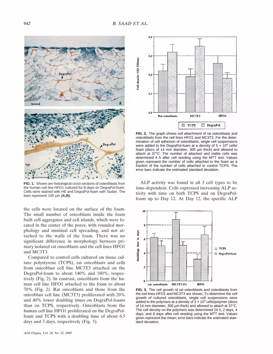
the cells were located on the surface of the foam.The small number of osteoblasts inside the foambuilt cell aggregates and cell islands, which were lo-cated in the center of the pores, with rounded mor-phology and minimal cell spreading, and not at-tached to the walls of the foam. There was nosignificant difference in morphology between pri-mary isolated rat osteoblasts and the cell lines HFO1and MC3T3.
Compared to control cells cultured on tissue cul-ture polystyrene (TCPS), rat osteoblasts and cellsfrom osteoblast cell line MC3T3 attached on theDegraPol-foam to about 140% and 180%, respec-tively (Fig. 2). In contrast, osteoblasts from the hu-man cell line HFO1 attached to the foam to about70% (Fig. 2). Rat osteoblasts and those from theosteoblast cell line (MC3T3) proliferated with 20%and 40% lower doubling times on DegraPol-foamsthan on TCPS, respectively. Osteoblasts from thehuman cell line HFO1 proliferated on the DegraPol-foam and TCPS with a doubling time of about 6.5days and 3 days, respectively (Fig. 3).
ALP activity was found in all 3 cell types to betime-dependent. Cells expressed increasing ALP ac-tivity with time on both TCPS and on DegraPol-foam up to Day 12. At Day 12, the specific ALP
FIG. 1. Shown are histological cross sections of osteoblasts fromthe human cell line HFO1 cultured for 8 days on DegraPol-foam.Cells were stained with HE and DegraPol-foam with Sudan. Thebars represent 100 µm (A,B).
FIG. 2. The graph shows cell attachment of rat osteoblasts andosteoblasts from the cell lines HFO1 and MC3T3. For the deter-mination of cell adhesion of osteoblasts, single cell suspensionswere added to the DegraPol-foam at a density of 5 × 10⁴ cells/foam (discs of 14 mm diameter, 300 µm thick) and allowed toattach at 37°C. The number of attached and viable cells wasdetermined 4 h after cell seeding using the MTT test. Valuesgiven represent the number of cells attached to the foam as afraction of the number of cells attached to control TCPS. Theerror bars indicate the estimated standard deviation.
FIG. 3. The cell growth of rat osteoblasts and osteoblasts fromthe cell lines HFO1 and MC3T3 are shown. To determine the cellgrowth of cultured osteoblasts, single cell suspensions wereadded to the polymers at a density of 2 × 10⁴ cells/polymer (discsof 14 mm diameter, 300 µm thick) and allowed to attach at 37°C.The cell density on the polymers was determined 24 h, 2 days, 4days, and 8 days after cell seeding using the MTT test. Valuesgiven represent the mean; error bars indicate the estimated stan-dard deviation.
B. SAAD ET AL.942
Artif Organs, Vol. 24, No. 12, 2000

activity in human osteoblasts cultured on DegraPol-foam was about 5 fold higher when compared to theactivity measured at Day 4 (10 mm/min/mg). OnTCPS, osteoblasts from the human cell line HFO1expressed 400% to 600% lower ALP activity than onDegraPol-foam. Rat osteoblasts and osteoblastsfrom the cell line MC3T3 cultured on TCPS andDegraPol-foam expressed increasing ALP activitywith time. The specific ALP activity was up to Day 8higher in cells cultured on TCPS compared to cellscultured on DegraPol-foam. In contrast, cells cul-tured on the DegraPol-foams expressed higher ALPactivity compared with TCPS at Day 12 (150% com-pared with TCPS) (Fig. 4). There were no significantdifferences in the amounts of collagen type I andosteocalcin produced throughout the 12 days of theexperiment by all 3 types of osteoblasts cultured onTCPS and DegraPol-foam (data not shown).
DegraPol-foam as carrier of BMP-inducedbone formation
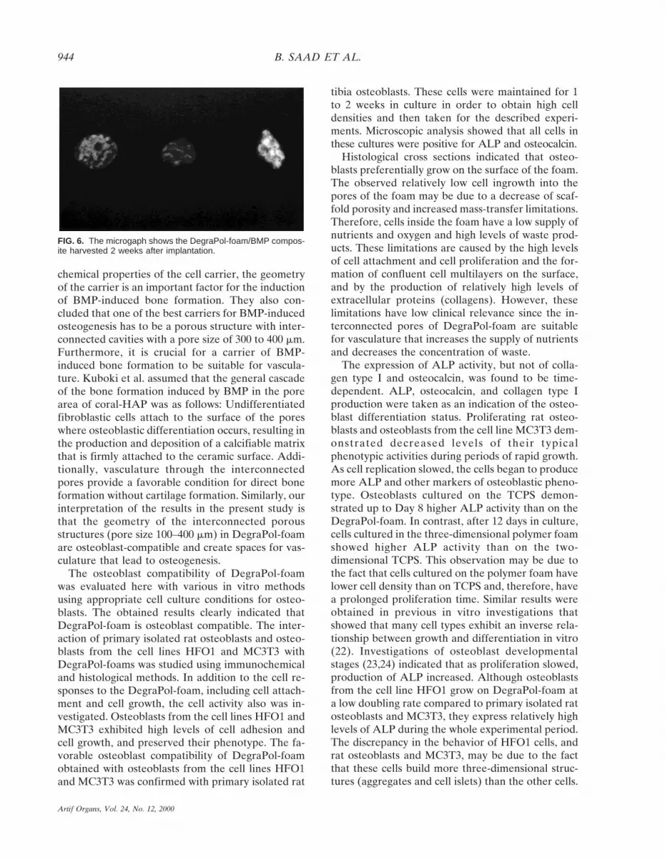
Figure 5 shows a cross section of DegraPol-foam/BMP composite harvested 2 weeks after implan-tation. Early bone formation was found in theDegraPol-foam. Without BMP, DegraPol did not in-duce any bone or cartilage formation after 2 weeksof implantation. Most areas within the pores werefilled with fibrous tissue (data not shown). A clearcalcification was observed in the x-ray analysis ofDegraPol-foam/BMP composite harvested 2 weeksafter implantation (Fig. 6).
The ALP activity reached in DegraPol/BMP com-posite a level of 1.6 mg/pellet. Calcium concentra-tions in DegraPol/BMP composite were 240 mIU/pellet 4 weeks after implantation.
DISCUSSION
The suitability of DegraPol-foam as carrier forBMP-induced bone formation was studied in vivo.Implantation experiments showed that DegraPol-foam/BMP composites definitely induce bone for-mation, as evidenced by histological observation,ALP activity, and calcium concentrations. Kuboki etal. (11) showed that in addition to the physico-
FIG. 4. The graph shows alkaline phosphatase activity in cul-tured osteoblasts. The activity was measured 4, 8, and 12 daysafter cell seeding on DegraPol-foam. Values given represent themean of 3 independent experiments.
FIG. 5. The cross sections are of the DegraPol-foam/BMP com-posite harvested 2 weeks after implantation. Original magnifica-tions were ×25 (A), ×50 (B), and ×100 (C).
DEGRAPOL-FOAM AS NOVEL SUBSTRATE FOR BONE FORMATION 943
Artif Organs, Vol. 24, No. 12, 2000
chemical properties of the cell carrier, the geometryof the carrier is an important factor for the inductionof BMP-induced bone formation. They also con-cluded that one of the best carriers for BMP-inducedosteogenesis has to be a porous structure with inter-connected cavities with a pore size of 300 to 400 mm.Furthermore, it is crucial for a carrier of BMP-induced bone formation to be suitable for vascula-ture. Kuboki et al. assumed that the general cascadeof the bone formation induced by BMP in the porearea of coral-HAP was as follows: Undifferentiatedfibroblastic cells attach to the surface of the poreswhere osteoblastic differentiation occurs, resulting inthe production and deposition of a calcifiable matrixthat is firmly attached to the ceramic surface. Addi-tionally, vasculature through the interconnectedpores provide a favorable condition for direct boneformation without cartilage formation. Similarly, ourinterpretation of the results in the present study isthat the geometry of the interconnected porousstructures (pore size 100–400 mm) in DegraPol-foamare osteoblast-compatible and create spaces for vas-culature that lead to osteogenesis.
The osteoblast compatibility of DegraPol-foamwas evaluated here with various in vitro methodsusing appropriate cell culture conditions for osteo-blasts. The obtained results clearly indicated thatDegraPol-foam is osteoblast compatible. The inter-action of primary isolated rat osteoblasts and osteo-blasts from the cell lines HFO1 and MC3T3 withDegraPol-foams was studied using immunochemicaland histological methods. In addition to the cell re-sponses to the DegraPol-foam, including cell attach-ment and cell growth, the cell activity also was in-vestigated. Osteoblasts from the cell lines HFO1 andMC3T3 exhibited high levels of cell adhesion andcell growth, and preserved their phenotype. The fa-vorable osteoblast compatibility of DegraPol-foamobtained with osteoblasts from the cell lines HFO1and MC3T3 was confirmed with primary isolated rat
tibia osteoblasts. These cells were maintained for 1to 2 weeks in culture in order to obtain high celldensities and then taken for the described experi-ments. Microscopic analysis showed that all cells inthese cultures were positive for ALP and osteocalcin.
Histological cross sections indicated that osteo-blasts preferentially grow on the surface of the foam.The observed relatively low cell ingrowth into thepores of the foam may be due to a decrease of scaf-fold porosity and increased mass-transfer limitations.Therefore, cells inside the foam have a low supply ofnutrients and oxygen and high levels of waste prod-ucts. These limitations are caused by the high levelsof cell attachment and cell proliferation and the for-mation of confluent cell multilayers on the surface,and by the production of relatively high levels ofextracellular proteins (collagens). However, theselimitations have low clinical relevance since the in-terconnected pores of DegraPol-foam are suitablefor vasculature that increases the supply of nutrientsand decreases the concentration of waste.
The expression of ALP activity, but not of colla-gen type I and osteocalcin, was found to be time-dependent. ALP, osteocalcin, and collagen type Iproduction were taken as an indication of the osteo-blast differentiation status. Proliferating rat osteo-blasts and osteoblasts from the cell line MC3T3 dem-onstrated decreased levels of their typicalphenotypic activities during periods of rapid growth.As cell replication slowed, the cells began to producemore ALP and other markers of osteoblastic pheno-type. Osteoblasts cultured on the TCPS demon-strated up to Day 8 higher ALP activity than on theDegraPol-foam. In contrast, after 12 days in culture,cells cultured in the three-dimensional polymer foamshowed higher ALP activity than on the two-dimensional TCPS. This observation may be due tothe fact that cells cultured on the polymer foam havelower cell density than on TCPS and, therefore, havea prolonged proliferation time. Similar results wereobtained in previous in vitro investigations thatshowed that many cell types exhibit an inverse rela-tionship between growth and differentiation in vitro(22). Investigations of osteoblast developmentalstages (23,24) indicated that as proliferation slowed,production of ALP increased. Although osteoblastsfrom the cell line HFO1 grow on DegraPol-foam ata low doubling rate compared to primary isolated ratosteoblasts and MC3T3, they express relatively highlevels of ALP during the whole experimental period.The discrepancy in the behavior of HFO1 cells, andrat osteoblasts and MC3T3, may be due to the factthat these cells build more three-dimensional struc-tures (aggregates and cell islets) than the other cells.
FIG. 6. The microgaph shows the DegraPol-foam/BMP compos-ite harvested 2 weeks after implantation.
B. SAAD ET AL.944
Artif Organs, Vol. 24, No. 12, 2000

Three-dimensional structures enhance the cell–cellinteractions and therefore, the expression of specificphenotype. The amount of collagen type I and os-teocalcin production per cell remained constant withtime for all 3 types of osteoblasts cultured on theDegraPol-foam, indicating that osteoblasts culturedon the polymer were able to maintain their phenotype.
In summary, results obtained in this study indicatethat DegraPol-foam exhibits good osteoblast com-patibility; osteoblasts exhibited relatively high celladhesion, growth rates, and maintained their pheno-type for up to 12 days. In addition, the BMP-inducedbone formation points to the possible use of thisscaffold in the bone healing process, for example, as anosteoblast-carrier for autologous cell transplantation.
Acknowledgments: Financial support from the SwissPriority Program for Materials Research as well as fromthe Swiss National Science Foundation, the Olga Mayen-fisch Stiftung, and the Hartmann-Muller-Stiftung is grate-fully acknowledged.
REFERENCES1. Kuboki Y, Yamaguci H, Yokoyama A, Murata M, Takita H,
Tazaki M, Mizuno M, Hasegawa T, Iida S, Shigenobu K, Fuj-sawa R, Kawamura M, Atsuta T, Matsumoto A, Kato H,Zhou H-Y, Ono I, Takeshita N, Nagai N. Osteogenesis in-duced by BMP-coated biomaterials: biochemical principles ofbone reconstruction in dentistry. In: Davies JE, eds. The BoneBiomaterials Interface. Toronto: University of Toronto Press,1991:127–38.
2. Mizuno M, Kuboki Y. TGF-beta accelerated the osteogenicdifferentiation of bone-marrow cells induced by collagen ma-trix. Biochem Bioph Res Co 1995;211:1091–8.
3. Fujisawa R, Nodasaka Y, Kuboki Y. Further characterizationof interaction between bone sialoprotein (bsp) and collagen.Calcified Tissue Inter 1995;56:140–4.
4. Takita H, Kuboki Y. Conformational-changes of bovine boneosteonectin induced by interaction with calcium. Calcified Tis-sue Inter 1995;56:559–65.
5. Mundy GR. Hormonal factors which regulate bone resorp-tion. In: Mundy GR, Martin TJ, eds. Physiology and Pharma-cology of Bone. New York: Springer-Verlag, 1993.
6. Mundy GR. Cytoknes of bone. In: Mundy GR, Martin TJ,eds. Physiology and Pharmacology of Bone. New York:Springer-Verlag, 1993.
7. Shigenobu K, Kaneda K, Nagai N, Kuboki Y. Localization ofbone morphogenetic protein-induced bone and cartilage for-mation on a new carrier—fibrous collagen membrane. AnnChirurgiae Gynaecologiae 1993;82:85–90.
8. Sasano Y, Ohtani E, Narita K, Kagayama M, Murata M, SaitoT, Shigenobu K, Takita H, Mizuno M, Kuboki Y. BMPs in-duce direct bone-formation in ectopic sites independent of theendochondral ossification in vivo. Anat Record 1993;236:373–80.
9. Kobayashi D, Takita H, Mizuno M, Totsuka Y, Kuboki Y.Time-dependent expression of bone sialoprotein fragments inosteogenesis induced by bone morphogenetic protein. J Bio-chem 1996;19:475–81.
10. Kuboki Y, Saito T, Murata M, Takita H, Mizuno M, Inoue M,Nagai N, Poole AR. Two distinctive BMP-carriers inducezonal chondrogenesis and membranous ossification, respec-tively; geometrical factors of matrices for cell differentiation.Connect Tissue Res 1995;32:219–26.
11. Kuboki Y, Takita H, Kobayashi D, Tsuruga E, Inoue M, Mu-rata M, Nagai N, Dohi Y, Ohgushi H. BMP-induced osteo-
genesis on the surface of hydroxyapatite with geometricallyfeasible and nonfeasible structures: topology of osteogenesis.J Biomed Mat Res 1998;39:190–199.
12. Saad B, Hirt TD, Welti M, Uhlschmid KG, NeuenschwanderP, Suter UW. Development of degradable polyesterurethanesfor medical applications: in vitro and in vivo evaluations. JBiomed Mat Res 1997;36:65–74.
13. Saad B, Moro M, Tun-Kyi A, Welti M, Schmutz P, UhlschmidGK, Neuenschwander P, Suter UW. Chondrocyte biocompat-ibility of highly porous biodegradable DegraPol-foam: in vitroevaluations. J Biomat Sci Polymer Ed 1999;10:1107–19.
14. Saad B, Ciardelli G, Matter S, Welti M, Uhlschmid KG, WeltiM, Neuenschwander P, Suter UW. Cell response of culturedmacrophages, fibroblasts, and co-cultures of Kupffer cells andhepatocytes to particles of short-chain poly[(R)-3-hydroxybutyric acid]. J Mater Sci Mater Med 1996;7:56–61.
15. Ciardelli G, Saad B, Hirt T, Keiser O, Uhlschmid KG, Neuen-schwander P, Suter UW. Phagocytosis of short-chainpoly[(R)-3-hydroxybutyric acid] particles in macrophage cellline. J Mater Sci Mater Med 1995;6:725–30.
16. Saad B, Ciardelli G, Matter S, Welti M, Uhlschmid KG, WeltiM, Neuenschwander P, Suter UW. Degradable and highlyporous polyesterurethane foam as biomaterial: effects andphagocytosis of degradation products in osteoblasts. J BiomedMat 1997;39:594–602.
17. Stringa E, Filanti C, Giuciuglio D, Albini A, Manduca P.Osteoblastic cells from rat long bone. I. Characterization oftheir differentiation in culture. Bone 1995;16:663–70.
18. Saad B, Keiser O, Uhlschmid GK, Marquardt K, Welti M,Neuenschwander P, Suter UW. Multiblock copolyesters asbiomaterials: in vitro biocompatibility testing. J Mat Sci MaterMed 1997;8:497–505.
19. Bradford MM. A rapid and sensitive method for the quanti-tation of microgram quantities of protein utilizing the prin-ciple of protein-dye binding. Anal Biochem 1976;72:248–54.
20. Saad B, Scholl FA, Thomas H, Schawalder HP, Streit V, Wae-chter F, Maier P. Crude liver membrane fractions and extracel-lular matrix components as substrata regulate differentially thepreservation and inducibilty of cytochrome P-450 isoenzymes incultured rat hepatocytes. Eur J Biochem 1993;213:805–14.
21. Puleo DA, Holleran LA, Doremus RH, Larjava H. Osteo-blast responses to orthopedic implant materials in vitro. JBiomed Mat Res 1991;25:711–23.
22. Mooney D, Hansen L, Vacanti J, Langer R, Farmer S, Ing-berg D. Switching from differentiation to growth in hepato-cytes: control by extracellular matrix. J Cell Phys 1992;151:497–505.
23. Arnow MA, Gerstenfeld LC, Owen TA, Tassinari MS, SteinGS, Lian JB. Factors that promote progressive developmentof the osteoblast phenotype in cultured fetal rat calvaria cells.J Cell Phys 1990;143:213–21.
24. Quarles LD, Yohay DA, Lever LW, Caton R, Wenstrup R.Distinct proliferative and differentiated stages of murineMC3T3-E1 cells in culture: an in vitro model of osteoblastdevelopment. J Bone Miner Res 1992;7:683–92.
NOMENCLATURE
ALP alkaline phosphataseBMP bone morphogenetic proteinBSA bovine serum albuminEDTA ethylenediaminetetraacetic acidEGTA ethylene glycol-bis(b-aminoethyl ether)
N,N,N8N8-tetraacetic acidELISA enzyme-linked immunosorbent assayFCS fetal calf serumMTT succinate dehydrogenase activity assayPBS phosphate-buffered salineTCPS tissue culture polystyrene
DEGRAPOL-FOAM AS NOVEL SUBSTRATE FOR BONE FORMATION 945
Artif Organs, Vol. 24, No. 12, 2000


