Embed Size (px)
Citation preview
Cryptorchidism With Short Spermatic Vessels:
Staged Orchiopexy Preserving Spermatic Vessels
A. Dessanti, D. Falchetti, M. Iannuccelli, S. Milianti, C. Altana, A. R. Tanca,M. Ubertazzi, G. P. Strusi and M. FusilloFrom the Department of Pediatric Surgery (AD, MI, MU, MF) and Institute of Radiology (GPS), Azienda Ospedaliero-Universitaria,University of Sassari, Sassari and Department of Pediatric Surgery, Spedali Civili, Brescia (DF, SM, CA, ART), Italy
Purpose: Patients with cryptorchidism can have such short spermatic vesselsthat it is impossible to place the testicle in a satisfactory scrotal position usingconventional orchiopexy. In these cases the most commonly used operation is 1 to2-stage Fowler-Stephens orchiopexy. We present our surgical experience usingstaged inguinal orchiopexy without section of the spermatic vessels in patientswith short spermatic vessels.Materials and Methods: We used 2-stage inguinal orchiopexy in 38 childrenwith intra-abdominal testis or testis peeping through the internal ring andshort spermatic vessels (7 bilateral). Spermatic vessels were not sectioned, butwere lengthened through progressive traction of the spermatic cord wrappedin polytetrafluoroethylene pericardial membrane (Preclude®). In the firststage we mobilized the spermatic cord in the retroperitoneal space and thenwrapped it in the polytetrafluoroethylene membrane. We subsequently at-tached the testis to the invaginated scrotal bottom. At 9 to 12 months weperformed the second stage, which involved removing the polytetrafluoroeth-ylene membrane.Results: From the first to the second stage we observed progressive descent of thetesticle toward the scrotum. At 1 to 8-year followup after the second stage all 45testicles were palpable in a satisfactory scrotal position with stable or increasedtesticular volume.Conclusions: This technique represents an alternative to Fowler-Stephens or-chiopexy, which can be associated with a greater risk of testicular ischemia.
Key Words: cryptorchidism, testis, urogenital abnormalities, urogenital
Abbreviations
and Acronyms
FS � Fowler-Stephens
PTFE � polytetrafluoroethylene
Submitted for publication January 12, 2009.
surgical procedures
CRYPTORCHIDISM cases are usually di-vided into 2 subgroups—those withpalpable and nonpalpable testes. Inpatients with a palpable testis thegonad can usually be fixed easily inthe hemiscrotum using conventionalinguinal orchiopexy.1 Nonpalpabletestis occurs in 20% to 30% of cases.2
Most of these testes are present in
the abdomen or inguinal canal, with0022-5347/09/1823-1163/0THE JOURNAL OF UROLOGY®
Copyright © 2009 by AMERICAN UROLOGICAL ASSOCIATION
20% to 45% being absent or vanish-ing secondary to intrauterine orperinatal torsion.1– 4
Nonpalpable testes and, rarely,palpable testes can have an ex-tremely short spermatic vessel thatdoes not allow them to be placed in asatisfactory scrotal position usingstandard orchiopexy performed via
an inguinal incision or laparoscopy.Vol. 182, 1163-1168, September 2009Printed in U.S.A.
DOI:10.1016/j.juro.2009.05.050www.jurology.com 1163
CRYPTORCHIDISM WITH SHORT SPERMATIC VESSELS1164
For these select cases, which represent approxi-mately 3% of all undescended testes,3,5 the surgi-cal solution proposed is inguinal or laparoscopicFS orchiopexy.6 We present our experience withcryptorchidism involving extremely short sper-matic vessels, where we used staged inguinal or-chiopexy without section of the spermatic vessels.These vessels were lengthened through progres-sive traction after being wrapped in an anti-adhe-sion PTFE pericardial membrane.
MATERIALS AND METHODS
We reviewed the clinical charts of children with cryp-torchidism treated at 2 pediatric surgery units from1997 to 2006. Of 1,698 patients 60 (3.5%) had such shortspermatic vessels that they could not be placed in asatisfactory scrotal position using standard orchiopexy.Of this group only the patients surgically treated by the2 senior authors (AD, DF) were analyzed. Of 38 cases 7were bilateral, for a total of 45 testes. Of the testes 34were intra-abdominal and 11 were peeping through theinternal ring. Patient age was 1 to 5 years (average 35months). In all cases we located the testicle through theopen inguinal procedure only, preceded by preoperativeultrasound in which we were able to evaluate the posi-tion and volume of the testicle. Parents gave informedconsent for the surgery.
Surgical Technique
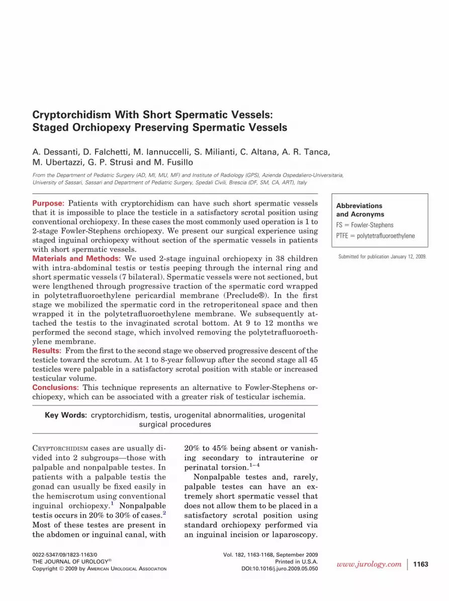
An oblique inguinal incision is made in the skin crease.When the external oblique fascia is opened the proces-sus vaginalis is located (normally open in these cases).It is isolated beyond the internal inguinal ring, asclosely as possible, therefore involving the retroperito-neal space. Once the processus vaginalis is opened thetesticle is located. The testicle normally has a shortspermatic cord that still allows for some mobility in theabdomen. Keeping the processus vaginalis and testiclein light traction, the processus vaginalis is cut in a circleproximal to the testicle so that it is possible to isolateand mobilize the testicle and its spermatic cord in the
Figure 1. Spermatic vessels and vas deferens are dissected dee
wrapped in PTFE membrane and fashioned into conduit.retroperitoneal space. To perform this maneuver, afterdissecting free and ligating the processus vaginalis atthe level of the internal ring, the posterior peritoneumis protected by a long, thin Langenback retractor. Dis-section of the spermatic cord and spermatic vessels iscarried out mainly by dividing the tethering bands oflateral spermatic fascia until their maximum length isobtained.
When it is impossible to place the gonad in a satis-factory scrotal position (when the testicle cannot bebrought past the pubic tubercle) the spermatic cord iswrapped along its entire length in an anti-adhesionPTFE pericardial membrane, which is fashioned into aconduit using a continuous absorbable polydioxanonesuture (Vicryl™, fig. 1).
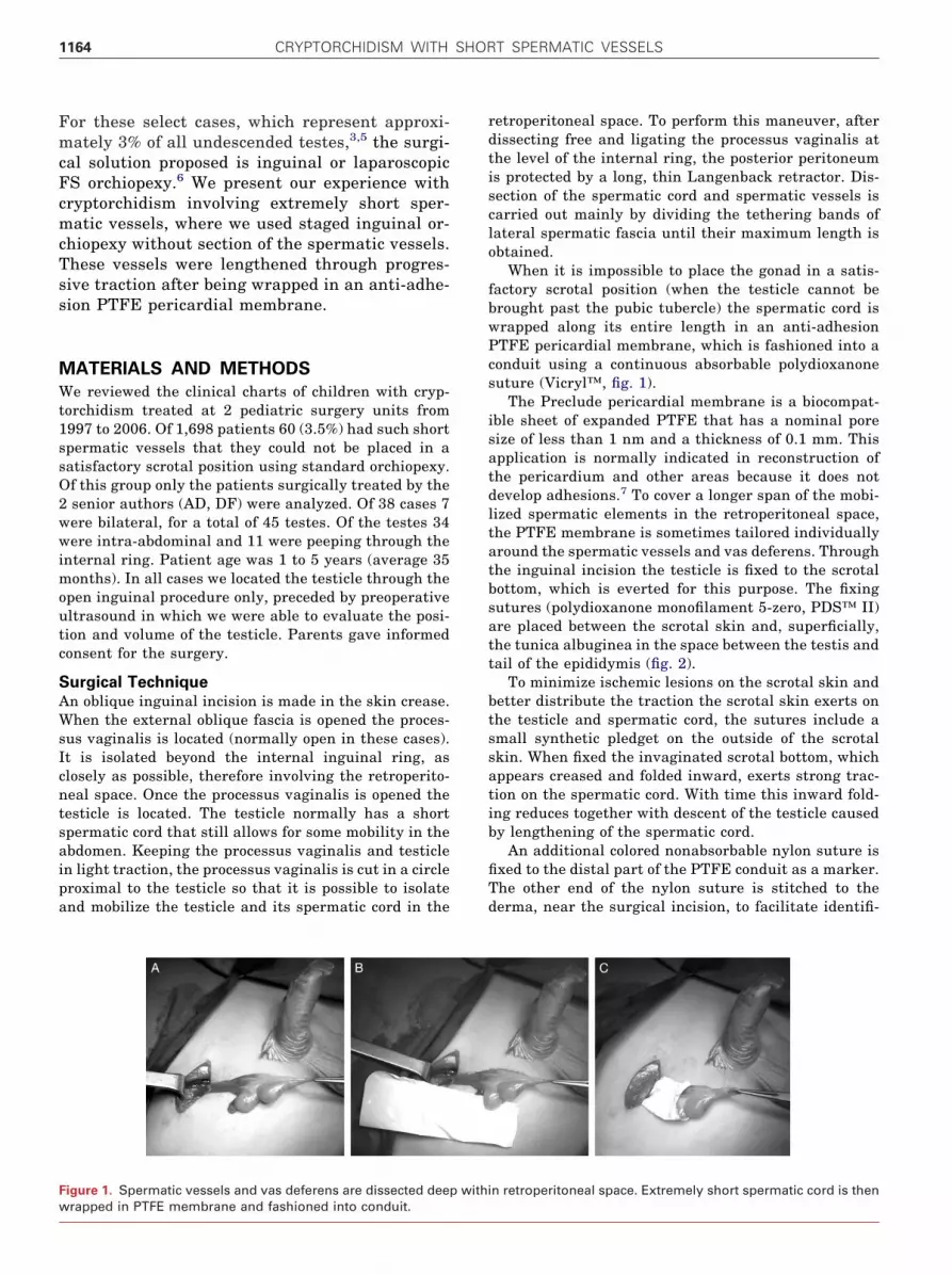
The Preclude pericardial membrane is a biocompat-ible sheet of expanded PTFE that has a nominal poresize of less than 1 nm and a thickness of 0.1 mm. Thisapplication is normally indicated in reconstruction ofthe pericardium and other areas because it does notdevelop adhesions.7 To cover a longer span of the mobi-lized spermatic elements in the retroperitoneal space,the PTFE membrane is sometimes tailored individuallyaround the spermatic vessels and vas deferens. Throughthe inguinal incision the testicle is fixed to the scrotalbottom, which is everted for this purpose. The fixingsutures (polydioxanone monofilament 5-zero, PDS™ II)are placed between the scrotal skin and, superficially,the tunica albuginea in the space between the testis andtail of the epididymis (fig. 2).
To minimize ischemic lesions on the scrotal skin andbetter distribute the traction the scrotal skin exerts onthe testicle and spermatic cord, the sutures include asmall synthetic pledget on the outside of the scrotalskin. When fixed the invaginated scrotal bottom, whichappears creased and folded inward, exerts strong trac-tion on the spermatic cord. With time this inward fold-ing reduces together with descent of the testicle causedby lengthening of the spermatic cord.
An additional colored nonabsorbable nylon suture isfixed to the distal part of the PTFE conduit as a marker.The other end of the nylon suture is stitched to thederma, near the surgical incision, to facilitate identifi-
n retroperitoneal space. Extremely short spermatic cord is then
p withi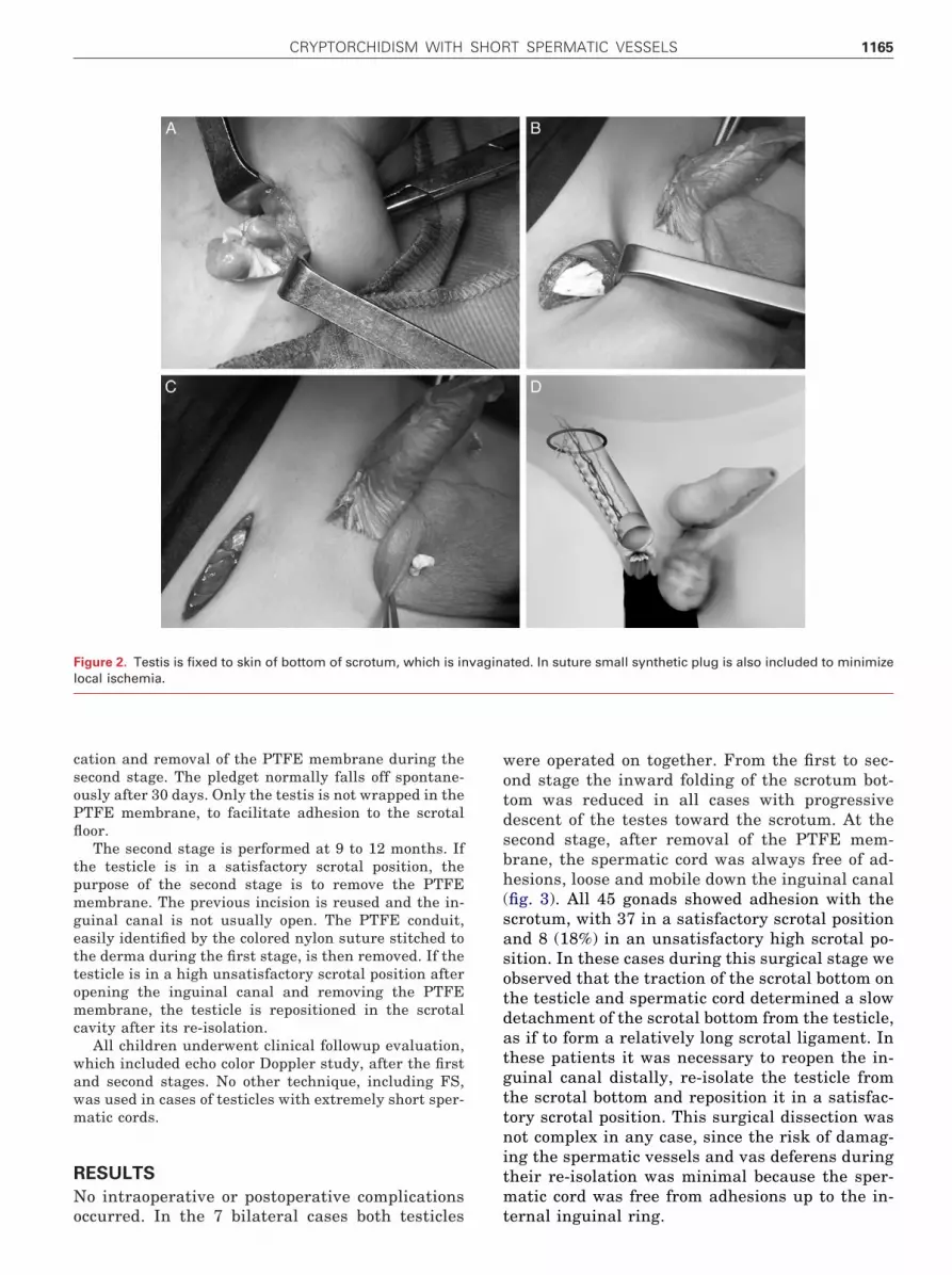
CRYPTORCHIDISM WITH SHORT SPERMATIC VESSELS 1165
cation and removal of the PTFE membrane during thesecond stage. The pledget normally falls off spontane-ously after 30 days. Only the testis is not wrapped in thePTFE membrane, to facilitate adhesion to the scrotalfloor.
The second stage is performed at 9 to 12 months. Ifthe testicle is in a satisfactory scrotal position, thepurpose of the second stage is to remove the PTFEmembrane. The previous incision is reused and the in-guinal canal is not usually open. The PTFE conduit,easily identified by the colored nylon suture stitched tothe derma during the first stage, is then removed. If thetesticle is in a high unsatisfactory scrotal position afteropening the inguinal canal and removing the PTFEmembrane, the testicle is repositioned in the scrotalcavity after its re-isolation.
All children underwent clinical followup evaluation,which included echo color Doppler study, after the firstand second stages. No other technique, including FS,was used in cases of testicles with extremely short sper-matic cords.
RESULTS
No intraoperative or postoperative complications
Figure 2. Testis is fixed to skin of bottom of scrotum, which is inlocal ischemia.
occurred. In the 7 bilateral cases both testicles
were operated on together. From the first to sec-ond stage the inward folding of the scrotum bot-tom was reduced in all cases with progressivedescent of the testes toward the scrotum. At thesecond stage, after removal of the PTFE mem-brane, the spermatic cord was always free of ad-hesions, loose and mobile down the inguinal canal(fig. 3). All 45 gonads showed adhesion with thescrotum, with 37 in a satisfactory scrotal positionand 8 (18%) in an unsatisfactory high scrotal po-sition. In these cases during this surgical stage weobserved that the traction of the scrotal bottom onthe testicle and spermatic cord determined a slowdetachment of the scrotal bottom from the testicle,as if to form a relatively long scrotal ligament. Inthese patients it was necessary to reopen the in-guinal canal distally, re-isolate the testicle fromthe scrotal bottom and reposition it in a satisfac-tory scrotal position. This surgical dissection wasnot complex in any case, since the risk of damag-ing the spermatic vessels and vas deferens duringtheir re-isolation was minimal because the sper-matic cord was free from adhesions up to the in-
ated. In suture small synthetic plug is also included to minimize
vaginternal inguinal ring.

CRYPTORCHIDISM WITH SHORT SPERMATIC VESSELS1166
There was no difference in testicular position atthe second stage in relation to the shape of theproximal part of the conduit (single or doublearound vessels and vas). At 1 to 8 years of followup(mean 3) after the second stage all 45 testicleswere palpable and still firmly in a satisfactoryscrotal position (fig. 4). Echo color Doppler re-vealed good blood supply to all testes and stable orincreased testicular volume compared to preoper-ative and perioperative ultrasound.
DISCUSSION
Surgical treatment of nonpalpable testes withshort spermatic vessels is still a challenge and isfrequently debated by pediatric surgeons. Cork-ery8 and later Steinhardt et al9 reported a 2-stageorchiopexy technique involving wrapping of thespermatic cord in a silastic sheet while the testiswas fixed to the pubic bone. This method was usedwith the sole aim of reducing the risk of iatrogenicinjuries of the spermatic cord during the secondstage of orchiopexy. Orthotopic autotransplanta-tion was proposed to recreate a blood supply to thetesticle and prevent the vanishing complicationbut the technique was rejected due to its unsatis-factory results and complexity.10,11
Currently the most widely used orchiopexytechnique is the 1 or 2-stage FS method.6 Thisapproach requires interruption of the spermaticvessels to bring down the testicle into the correctscrotal position. In this method vascularization ofthe testis is thus supplied only by the vasa defer-entia, with a consequent risk of testicular hypot-rophy or atrophy ranging from 2% to 33%.3,4,12–20
Because it requires division of the spermatic
Figure 3. Second stage. PTFE membrane is removed. Spermaticcord is mobile and extendible because it is free of adhesions.
vessels, the FS procedure represents a manage-
ment dilemma, especially in bilateral cases. Thedecision to use FS vs standard orchiopexy is basedsolely on visual observation during video assistedor open surgery.5,21 Therefore, the decision cannotbe made after extensive surgical isolation of thespermatic cord. In fact, extensive isolation of thespermatic cord would result in interruption of thevasa deferentia, guaranteeing lack of vasculariza-tion of the gonad if FS is chosen. Thus, this pro-cedure can be misused, even being applied in tes-ticles that could benefit from standard orchiopexywithout the risks linked to sectioning of the sper-matic vessels. For the same reasons a positivevisual evaluation regarding potential lengtheningof the spermatic cord after extensive mobilizationcould be incorrect, with the consequent risks of 1)descent of the testicle to an unsatisfactory highinguinoscrotal position, requiring a standard sec-ond stage orchiopexy and 2) extreme mobilizationof the spermatic cord, with the consequent risk ofhypotrophy or atrophy of the testicle due to isch-emia.
The methods and purposes of our surgical tech-nique are based on the following principles. Thetechnique can be performed after extended retro-peritoneal mobilization of the spermatic elements,when lengthening of the spermatic cord achievedis unsatisfactory. The PTFE conduit envelops theentire length of the spermatic cord from its retro-peritoneal tract to prevent adhesions to surround-ing tissues. Progressive elongation of the sper-matic cord and consequent descent of the testisinto the scrotal position between the first andsecond stage are due to the constant and nonisch-emizing traction exerted by the invaginated scro-tal skin on the entire spermatic cord, which is freeof adhesions.
From our experience the PTFE membrane is sothin (0.1 mm) that it does not cause symptomatichernias. Disengagement of the gonad at the sec-ond stage occurred in about 18% of testes, with thegonad appearing stuck in a high scrotal position.
Figure 4. Final scrotal position of testes 3 years after second
stage. A, left unilateral orchiopexy. B, bilateral orchiopexy.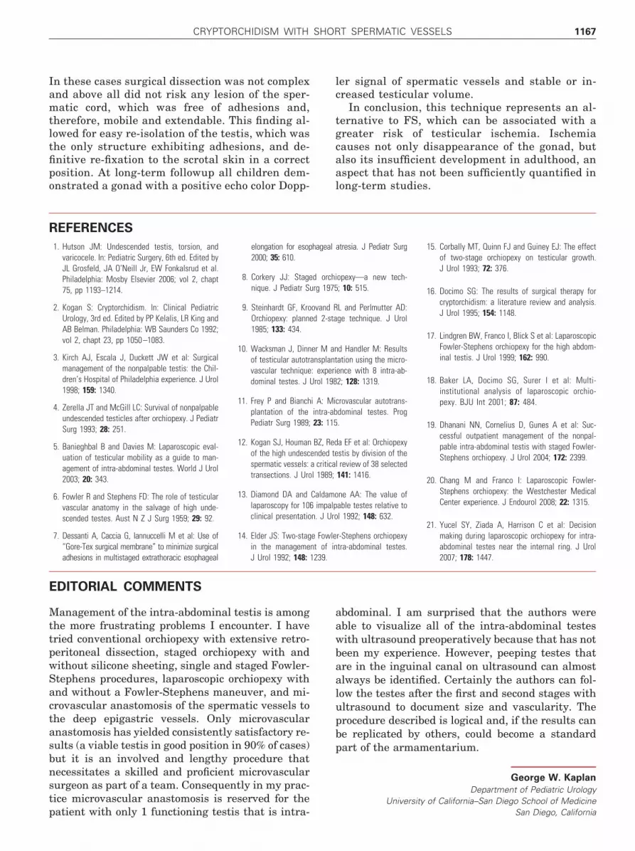
CRYPTORCHIDISM WITH SHORT SPERMATIC VESSELS 1167
In these cases surgical dissection was not complexand above all did not risk any lesion of the sper-matic cord, which was free of adhesions and,therefore, mobile and extendable. This finding al-lowed for easy re-isolation of the testis, which wasthe only structure exhibiting adhesions, and de-finitive re-fixation to the scrotal skin in a correctposition. At long-term followup all children dem-
onstrated a gonad with a positive echo color Dopp-REFERENCES
EDITORIAL COMMENTS
patient with only 1 functioning testis that is intra-
ler signal of spermatic vessels and stable or in-creased testicular volume.
In conclusion, this technique represents an al-ternative to FS, which can be associated with agreater risk of testicular ischemia. Ischemiacauses not only disappearance of the gonad, butalso its insufficient development in adulthood, anaspect that has not been sufficiently quantified in
long-term studies.1. Hutson JM: Undescended testis, torsion, andvaricocele. In: Pediatric Surgery, 6th ed. Edited byJL Grosfeld, JA O’Neill Jr, EW Fonkalsrud et al.Philadelphia: Mosby Elsevier 2006; vol 2, chapt75, pp 1193–1214.
2. Kogan S: Cryptorchidism. In: Clinical PediatricUrology, 3rd ed. Edited by PP Kelalis, LR King andAB Belman. Philadelphia: WB Saunders Co 1992;vol 2, chapt 23, pp 1050 –1083.
3. Kirch AJ, Escala J, Duckett JW et al: Surgicalmanagement of the nonpalpable testis: the Chil-dren’s Hospital of Philadelphia experience. J Urol1998; 159: 1340.
4. Zerella JT and McGill LC: Survival of nonpalpableundescended testicles after orchiopexy. J PediatrSurg 1993; 28: 251.
5. Banieghbal B and Davies M: Laparoscopic eval-uation of testicular mobility as a guide to man-agement of intra-abdominal testes. World J Urol2003; 20: 343.
6. Fowler R and Stephens FD: The role of testicularvascular anatomy in the salvage of high unde-scended testes. Aust N Z J Surg 1959; 29: 92.
7. Dessanti A, Caccia G, Iannuccelli M et al: Use of“Gore-Tex surgical membrane” to minimize surgical
elongation for esophageal atresia. J Pediatr Surg2000; 35: 610.
8. Corkery JJ: Staged orchiopexy—a new tech-nique. J Pediatr Surg 1975; 10: 515.
9. Steinhardt GF, Kroovand RL and Perlmutter AD:Orchiopexy: planned 2-stage technique. J Urol1985; 133: 434.
10. Wacksman J, Dinner M and Handler M: Resultsof testicular autotransplantation using the micro-vascular technique: experience with 8 intra-ab-dominal testes. J Urol 1982; 128: 1319.
11. Frey P and Bianchi A: Microvascular autotrans-plantation of the intra-abdominal testes. ProgPediatr Surg 1989; 23: 115.
12. Kogan SJ, Houman BZ, Reda EF et al: Orchiopexyof the high undescended testis by division of thespermatic vessels: a critical review of 38 selectedtransections. J Urol 1989; 141: 1416.
13. Diamond DA and Caldamone AA: The value oflaparoscopy for 106 impalpable testes relative toclinical presentation. J Urol 1992; 148: 632.
14. Elder JS: Two-stage Fowler-Stephens orchiopexyin the management of intra-abdominal testes.
15. Corbally MT, Quinn FJ and Guiney EJ: The effectof two-stage orchiopexy on testicular growth.J Urol 1993; 72: 376.
16. Docimo SG: The results of surgical therapy forcryptorchidism: a literature review and analysis.J Urol 1995; 154: 1148.
17. Lindgren BW, Franco I, Blick S et al: LaparoscopicFowler-Stephens orchiopexy for the high abdom-inal testis. J Urol 1999; 162: 990.
18. Baker LA, Docimo SG, Surer I et al: Multi-institutional analysis of laparoscopic orchio-pexy. BJU Int 2001; 87: 484.
19. Dhanani NN, Cornelius D, Gunes A et al: Suc-cessful outpatient management of the nonpal-pable intra-abdominal testis with staged Fowler-Stephens orchiopexy. J Urol 2004; 172: 2399.
20. Chang M and Franco I: Laparoscopic Fowler-Stephens orchiopexy: the Westchester MedicalCenter experience. J Endourol 2008; 22: 1315.
21. Yucel SY, Ziada A, Harrison C et al: Decisionmaking during laparoscopic orchiopexy for intra-abdominal testes near the internal ring. J Urol
adhesions in multistaged extrathoracic esophageal J Urol 1992; 148: 1239. 2007; 178: 1447.
Management of the intra-abdominal testis is amongthe more frustrating problems I encounter. I havetried conventional orchiopexy with extensive retro-peritoneal dissection, staged orchiopexy with andwithout silicone sheeting, single and staged Fowler-Stephens procedures, laparoscopic orchiopexy withand without a Fowler-Stephens maneuver, and mi-crovascular anastomosis of the spermatic vessels tothe deep epigastric vessels. Only microvascularanastomosis has yielded consistently satisfactory re-sults (a viable testis in good position in 90% of cases)but it is an involved and lengthy procedure thatnecessitates a skilled and proficient microvascularsurgeon as part of a team. Consequently in my prac-tice microvascular anastomosis is reserved for the
abdominal. I am surprised that the authors wereable to visualize all of the intra-abdominal testeswith ultrasound preoperatively because that has notbeen my experience. However, peeping testes thatare in the inguinal canal on ultrasound can almostalways be identified. Certainly the authors can fol-low the testes after the first and second stages withultrasound to document size and vascularity. Theprocedure described is logical and, if the results canbe replicated by others, could become a standardpart of the armamentarium.
George W. Kaplan
Department of Pediatric UrologyUniversity of California–San Diego School of Medicine
San Diego, California
CRYPTORCHIDISM WITH SHORT SPERMATIC VESSELS1168
The authors present an innovative approach to highundescended testes. They support our long main-tained belief that the spermatic vessels and vas arenot short in cryptorchidism—they are just embed-ded in the endopelvic fascia. Thus, extended mobili-zation by either laparoscopy or open surgery willalmost always move high testes into the scrotum(reference 3 in article). Our success rate is betterthan 90%.1 Careful attention to anatomy is impor-
REFERENCE
Among the cases reported, where the retroper-itoneal attachments of the vessels and vas werestretched into a satisfactory scrotal position, suc-cess was achieved in 100% but a second operationwas needed. The Fowler-Stephens approach, ex-cept in the rarest of cases, appears outmoded.
Howard M. Snyder, III
Division of UrologyUniversity of Pennsylvania School of Medicine
tant. Philadelphia, Pennsylvania
1. Hutcheson JC, Cooper CS and Snyder HM III: The anatomical approach to inguinal orchiopexy. J Urol 2000; 164: 1702.


