Embed Size (px)
Citation preview
COMPARATIVE EVALUATION OF CORONAL SEALING
ABILITY AMONG COMMERCIALLY AVAILABLE LIGHT
CURE TEMPORARY RESTORATIVE MATERIAL WITH
CONVENTIONAL TEMPORARY RESTORATIVE
MATERIAL - AN IN VITRO STUDY
Dissertation submitted to
THE TAMILNADU Dr. M.G.R. MEDICAL UNIVERSITY
In partial fulfilment for the Degree of
MASTER OF DENTAL SURGERY
BRANCH IV
CONSERVATIVE DENTISTRY AND ENDODONTICS
MAY 2019
CERTIFICATE
This is to certify that this dissertation titled “COMPARATIVE
EVALUATION OF CORONAL SEALING ABILITY AMONG
COMMERCIALLY AVAILABLE LIGHT CURE TEMPORARY
RESTORATIVE MATERIAL WITH CONVENTIONAL TEMPORARY
RESTORATIVE MATERIAL - AN IN VITRO STUDY” is a bonafide record of
work done by Dr. Pon Mathini under my guidance and to my satisfaction during her
Post Graduation study period between 2016 – 2019. This dissertation is submitted to
THE TAMILNADU Dr. M.G.R. MEDICAL UNIVERSITY, in partial fulfilment for
the award of the degree of master of dental surgery in Conservative Dentistry and
Endodontics, Branch IV. It has not been submitted (partial or full) for the award of any
other degree or diploma.
__________________________ ____________________________
Dr. V.PRABHAKAR M.D.S., Dr. SRIMAN NARAYANAN. M.D.S.,
__________________________
Dr. V. PRABHAKAR. M.D.S.,
Date:
Place: Coimbatore
Guide, Professor and Head
Department of Conservative Dentistry
and Endodontics,
Sri Ramakrishna Dental College and
Hospital, Coimbatore.
Co Guide and Reader
Department of Conservative Dentistry
and Endodontics,
Sri Ramakrishna Dental College and
Hospital, Coimbatore.
Principal
Sri Ramakrishna Dental College
and Hospital, Coimbatore.
DECLARATION
I hereby declare that no part of the dissertation will be utilized for gaining
financial assistance for research or other promotions without getting prior permission
from the principal, Sri Ramakrishna Dental College and Hospital. In addition, I declare
that no part of this work will be published either in print or in electronics without
permission from the guide who has been actively involved in the dissertation. The
author solely has rights for publishing this work with prior permission from principal,
Sri Ramakrishna Dental College and Hospital, Coimbatore.
____________________ ______________________ _________________________
Signature of H.O.D Signature of the Guide Signature of the Candidate
Name of the candidate
Dr. Pon Mathini
Title of the Study
“COMPARATIVE EVALUATION OF
CORONAL SEALING ABILITY AMONG
COMMERCIALLY AVAILABLE LIGHT CURE
TEMPORARY RESTORATIVE MATERIAL
WITH CONVENTIONAL TEMPORARY
RESTORATIVE MATERIAL - AN IN VITRO
STUDY”
Place of the Study
Sri Ramakrishna Dental College and Hospital
Duration of the course
2016 – 2019
Name of the guide
Dr. V. Prabhakar
Head of the department
Dr. V. Prabhakar
CERTIFICATE II
This is to certify that this dissertation titled “COMPARATIVE
EVALUATION OF CORONAL SEALING ABILITY AMONG
COMMERCIALLY AVAILABLE LIGHT CURE TEMPORARY
RESTORATIVE MATERIAL WITH CONVENTIONAL TEMPORARY
RESTORATIVE MATERIAL - AN IN VITRO STUDY” of the candidate
Dr. Pon Mathini with registration number 241617303 for the award of Master of
Dental Surgery in the branch of Conservative Dentistry and Endodontics. I
personally verified the urkund.com website for the purpose of plagiarism check. I found
that the uploaded thesis file contains from introduction to conclusion pages and result
shows 0 percentage of plagiarism in the dissertation.
_________________________________
Guide & supervisor signature with seal
ACKNOWLEDGEMENT
This thesis is the result of work done with immense support from many people
and it is with immense pleasure that I express my heartfelt gratitude to all of them.
I owe an immense debt of gratitude to my guide Dr. V. Prabhakar, MDS,
Principal & Head of Department, Department of Conservative Dentistry and
Endodontics, Sri Ramakrishna Dental College and Hospital, for his unwavering
guidance, immeasurable encouragement and constant support during my post graduate
tenure both in and out of the department.
I would like to thank and acknowledge Dr. Sriman Narayanan, MDS, Reader,
Department of Conservative Dentistry and Endodontics, Sri Ramakrishna Dental
College and Hospital, my co guide who has always been a source of support and
encouragement at any moment.
I also thank Dr. N. Muthu Krishnan, Professor & Head of Department,
Department of Agricultural Entomology, Tamil Nadu Agricultural University for his
valuable support and guidance while using the stereo microscope.
My list of acknowledgement would become meaningless without dedicating all
my efforts to my parents Dr. S. P. Pon Mudichelvan, MBBS, MD, DDVL and
Mrs. T. Chitra B.E Agri. For all the people I have acknowledged here, above everyone
I bow my head to the Almighty. Without his blessings I would not have such supportive
people around me to have all the good things in my life.
Dr. Pon Mathini
CONTENTS
S.NO. TITLE PAGE NO
1 INTRODUCTION 1
2 AIM AND OBJECTIVE 4
3 REVIEW OF LITERATURE 5
4 MATERIAL AND METHODS 17
5 RESULTS 35
6 DISCUSSION 42
7 SUMMARY AND CONCLUSION 48
8 BIBLIOGRAPHY 51

1
INTRODUCTION
One of the key factors in predicting the failure or success of the root canal
treatment is the use of an appropriate temporary restorative material during the course
of root canal therapy. As these materials provide a temporary seal for the tooth
preventing any percolation of fluids or microorganisms from the oral cavity during the
course of root canal therapy. A temporary material should be easy to manipulate as well
as dimensionally stable with good compressive resistance, abrasive resistance and it
should be compatible with the intracanal medicament used during the root canal
therapy.
Pulpal and periradicular diseases are most commonly caused by bacterial and
its toxins1,2. Chemico mechanical debridement followed by sealing of root canal in all
three dimensions is considered as the major predicting factor for the success of
endodontic therapy.
Sometimes multiple visits are necessary during endodontic treatment as it is not
possible to prepare, shape and obturate the tooth during a single appoinment3.Therefore
selection of an ideal inter appoinment temporary restorative material which provides a
fluid impervious seal to prevent microleakage which can lead to failure of the root canal
treatment is very important during multiple visit endodontic therapy to prevent bacterial
infection.
A seal is defined as something that blocks entry (into or out of container or
other object) hence, it is a difficult term to justify or use clinically since complete
sealing of a tooth is "impossible" with currently available dental materials and due to
the porous nature of the tooth structure itself (especially dentine but also enamel)4.
2
The pathway of fluid through the restorative material into the tooth from oral
cavity is known as microleakage5.
Previous studies have shown that the success of the root canal therapy can be
compromised by coronal leakage6 and that during endodontic therapy the second most
contributing factor for continuing pain following commencement of endodontic
therapy is due to lack of satisfactory temporary filling7.
According to Trope and Ray the quality of root canal filling is equally important
to the quality of coronal seal regarding periapical health which they demonstrated by
correlating between poor periapical status and inappropriate coronal restoration in root
canal filled teeth8.
Duration of usage of a temporary filling material also plays a vital role in
determining the degree of coronal microleakage of the material used. Recent studies
have proposed that immediate restoration should be done in an endodontically treated
tooth as microleakage might occur within few days9 and according to a study done by
Torbinejad et al it was said that bacteria can pass along a root canal filling from coronal
to the apical end within a period of 5 to 73 days10, in another study conducted by Khayat
et al by using fresh human saliva it was said that extensive leakage was seen in an
unfilled cavity and it was completely penetrated by microorganisms within 48 days11.
A number of materials are available for usage as temporary endodontic
restorative materials and can be classified as, non eugenol containing materials such as
zinc oxide-calcium sulphate, zinc oxide-eugenol based materials and light-cured resin
based composite12.
3
The coronal sealing ability of several materials have been studied over a period
of time in search for an ideal temporary filling material to be used during endodontic
therapy.
Hence this following study was contemplated to compare the coronal
microleakage of light cure temporary filling materials (Clip manufactured by Voco and
Systemp Inlay manufactured by Ivoclar Vivadent) with conventional temporary
restorative materials (e – Temp manufactured by Diadent and Tempfil – G
manufactured by Shivam Dental) which are commercially available in the market as it
might provide some knowledge regarding the coronal sealing ability of these materials.
4
AIM AND OBJECTIVE
The aim and objective of this study is to compare the coronal sealing ability
among commercially available light cure temporary restorative material with
conventional temporary restorative material over different periods of time
5
REVIEW OF LITERATURE
A study conducted by Anderson et al(1988)13 to evaluate microleakage among
three different temporary endodontic filling materials used to restore access cavities
namely Cavit, Term and Intermediate restorative material using fluid infiltration
technique and at different time intervals and at the end of the study it was concluded
that Term and Cavit provided a leak proof seal whereas Intermediate restorative
material showed significant microleakage after thermal loading during the 7 day
interval.
A study was conducted to evaluate the sealing ability of two temporary
endodontic filling materials (Term and Cavit) used to seal access cavity margins by
Teplitsky and Meimaris (1988)14 using dye penetration test with methylene blue and
they concluded that Cavit provided a better marginal seal than Term and that it was
unaffected by thermal cycling where as thermal cycling lead to increased microleakage
in Term.
A study conducted by Bobotis et al (1988)15 to evaluate quantitatively the
sealing ability of temporary filling materials (Term, Cavit G, Cavit, Zinc phosphate
cement, Intermediate restorative material, Polycarboxylate cement and Glass Ionomer
Cement) used in endodontic therapy to seal the access preparation and it was concluded
that Term, Cavit, Cavit G and Glass ionomer cement showed better seal when compared
to Zinc phosphate cement during the test period of 8 weeks and Polycarboxylate cement
and Intermediate restorative material are also less effective in preventing microleakage.
Melton, Cobb, and Krell (1990)16 conducted a study to evaluate and compare a
light cure temporary restoration Term with Cavit a self-polymerising temporary
6
restoration by using a carbon black protocol for coronal microleakage and at the end of
the study it was concluded that Cavit produced an effective seal for access opening
restorations whereas TERM did not prevent dye ingress especially in the proximal
region than in the coronal margins.
A study conducted by B.M. Jacquot et al (1996)17 to determine the microleakage
among four temporary restorative materials (Cavit W, Cavit, Intermediate restorative
material and Cavit G) for a period of over 9 days using impedance spectroscopy and
based on the results there was no significant difference between Cavit W and Cavit
whereas Intermediate restorative material showed more significant results than all the
cavit formulations making it a better temporary filling material among the four study
materials.
Barthel et al (1999)18 conducted a study to determine the ability of coronal
temporary restorations in preventing the corono apical leakage of bacteria, the materials
used in this study are Intermediate restorative material, Cavit, Glass ionomer cement,
Cavit in combination with Glass ionomer cememt and Intermediate restorative material
in combination with Glass ionomer cement and the results of the study concluded that
only Glass ionomer cement or Intermediate restorative material in combination with
Glass ionomer cement was capable of preventing bacterial leakage for over a period of
1 month.
A study conducted to evaluate microleakage by Pai et al (1999)19 among
endodontic temporary restorative materials at three areas namely between cavity wall
and access opening filling material, between orginal filling material and an additional
patch of material used to fill a secondary opening in the first filling material placed after
14 days, between second filling material and cavity wall. The primary filling material
7
used is either Intermediate restorative material or amalgam and the secondary filling
materials used were Caviton, Intermediate restorative material or a combination of both
and based on the results it was concluded that there was significantly less micro leakage
between secondary and primary restorative materials which were placed during
different time periods than microleakage between cavity wall and the primary
temporary filling material.
A study Conducted by M.B Uctash and A.C Tinaz (2000)12 to evaluate the
marginal seal using dye penetration test for four temporary restorative materials
(Coltosol, Intermediate restorative material, Algenol, Fermit or Fermit-N) in
endodontic access cavities and concluded that no significant difference was present in
microleakage between low elasticity versus high elasticity light cured composite
materials.
Liberman et al (2001)20 conducted a study to evaluate the microleakage of
cavidentin and Intermediate restorative material which were two widely used temporary
filling materials following repeated vertical loading and based on the results it was
concluded that without vertical loading both the materials provided similar seal quality
whereas under repeated vertical loading of 4 kgs Intermediate restorative material
showed superior seal when compared to Cavidentin thereby making it a suitable
temporary filling materials in areas of occlusal load.
An vitro study conducted by Hanan Balto (2002)21 to evaluate microleakage
among temporary restorative materials used after root canal treatment namely
Intermediate restorative material, Dyract, Cavit by using microbial marker containing
Candida albicans and Streptococcus faecalis and at the end of the 30 day study it was
concluded that Intermediate restorative material showed microleakage as early as 10
8
days when compared to Dyract and cavit which showed microleakage only after 2
weeks.
A study conducted by Lai et al (2007)22 to evaluate the marginal leakage of
temporary fillings namely Intermediate restorative material, Cavit, Copper Bands
Cemented to Zinc phosphate cement and Zinc phosphate cement in complex
endodontic access cavities by using dye penetration test and it was concluded that the
Cavit group showed the least microleakage whereas the rest of the groups showed
severe microleakage right from day 1.
Madarati et al (2007)23 conducted a study to evaluate parametrically the coronal
seal of four different temporary filling materials namely Coltosol, Intermediate
restorative material. Glass Ionomer cement and Zinc Phosphate cement which are
commonly used as filings to seal the access cavity following endodontic therapy over
different time periods namely 1, 2 and 4 weeks and based on the results it was concluded
that Glass Ionomer Cement and Coltosol showed superior seal when compared to
Intermediate restorative material and Zinc Phosphate cements even after 4 weeks time
duration and there was no significant difference between Zinc Phosphate and
Intermediate restorative materials in their sealing ability during different time periods.
A study conducted using an experimental model by Jensen and Abbott (2007)24
to evaluate dye penetration of interim fillings namely Cavit, Intermediate restorative
material, Ketac silver, Ketac fil plus and Z100 composite resin in mesio occluso distal
cavities during endodontic therapy under load with a multiple access chewing
stimulator and the results concluded that Intermediate restorative material showed
significantly more dye penetration than Ketav fil plus, Cavit, Ketac silver and Z100
9
resin composite and that no evidence of dye penetration was seen in Cavit models or
Z100 resin composite models.
A study conducted by Koagel et al (2008)25 to assess and evaluate the coronal
micro leakage of Tempit, Tempit Ultra F, Cavit and Intermediate restorative material
used as access restorations in endodontically treated teeth by using a fluid transport
model woaking at 10 psi and based on the resuts no statistical difference was observed
between Tempit and Tempit Ultra F as well as between Intermediate restorative
material, Cavit and Tempit but comparitively less microleakage was seen in Tempit
Ultra F when compared to Cavit and Intermediate restorative material.
A study conducted to compare sealing abilities among four temporary
endodontic filling material namely Intermediate restorative material, Zinc oxide
Eugenol, Md- Temp, Caviton by Dong-Ho Jung et al (2008)26 using dye penetration
with methylene blue under dynamic loading and based on the results it was concluded
that MD- Temp and Caviton showed lower microleakage when compared to Zinc Oxide
Eugenol and Intermediate restorative material under dynamic loading.
A study conducted by Chailertvanitkul et al (2009)27 to evaluate the association
between bacterial and dye penetration among interim restorations namely Cavit, Ketac
silver reinforced with stainless steel band and Ketac silver used in endodontic therapy
and it was concluded that stainless steel bands maintained the structural integrity in
ketac silver fillings under masticatory load but does not prevent bacterial penetration
and that no association was found between bacterial and dye penetration.
An in vitro study conducted by Shahi et al (2010)28 comparing the dye
penetration among four temporary filling materials namely Coltosol, Zamherir,
Zonalin, and Intermediate restorative material and concluded that Zamherir and
10
Zonalin showed less microleakage when compared to the rest of the two temporary
restorative materials.
A study conducted to assess the coronal sealing ability by Aledrissy et al
(2011)29 among hand mixed temporary materials versus readymade temporary
restorative materials. The materials used were Litrak, Cavisol, Zinconol, Zinc
phosphate cement and at the end of the study it was concluded that Cavisol produced
better seal when compared to the rest of the test materials and thus readymade
temporary filling materials produced a better coronal seal than hand mixed temporary
filling materials.
An in vitro study conducted by Naseri et al (2012)30 to analyse the coronal
sealing ability among three temporary filling materials namely Zonalin, Cavizol,
Coltosol by using dye penetration test with 2% methylene blue dye for a time period
of 1 day to 4th week and based on the results it was concluded that all the experimental
temporary filling materials showed increasing degree of micro leakage from 1st day to
4th week among which Zonalin showed comparatively more micro leakage than Cavizol
and Coltosol during each time interval and no significant difference was obtained in the
microleakage values between Cavizol and Coltosol.
Yun SM et al (2012)31 conducted a study using four temporary materials namely
Caviton, Intermediate restorative material, FujiII, Spacer to evaluate microleakage in
class II access cavity preparation with glucose penetration model and at the end of the
study it was concluded that significantly less microleakage was found in spacer and
Caviton when compared to Intermediate restorative material and Fuji II and according
to SEM observation more intimate tooth restoration adaptation was seen in Caviton and
spacer than the rest of the two study materials and hence Spacer and Caviton are
11
considered superior temporary filling materials in class II access caities among the four
study materials used for this study.
An ex vivo study conducted by G.P.V srikumar et al (2012)32 to evaluate the
marginal seal and coronal microleakage among hydrophilic temporary filling materials
commonly used in endodontic practice following the use of walking bleach material
and concluded that hydrophilic temporary filling material such as Coltosol F and Cavit
G shows minimal dye penetration coronally when exposed to a mixture of walking
bleach paste when compared to other temporary filling materials used during
endodontic therapy.
An in vitro study conducted to compare three different restorative materials by
Sagar et al (2012)33 namely Mineral Trioxide Aggregate, Glass ionomer cement and
flowable composite used as barriers to prevent coronal microleakage in root canal filled
teeth and concluded that flowable composite and Mineral Trioxide Aggregate when
placed at a thickness of 4mm as coronal restoration seals the root canal significantly
when compared to glass ionomer cement and it was also said that placement of Mineral
Trioxide Aggregate or Flowable composite as coronal restorations is advantageous in
case of post preparation or retreatment as its removal is much easier.
Davut Celik et al (2013)34 conducted a study among various temporary fillings
namely Fermin, Cavit G, Coltosol F, Clip, Bms, Ketac Molar Easymix, ProFill,
DuoTemp, or TempBond Clear with Triclosan Tfs for coronal leakage in endodontic
access cavities using dye penetration test with methylene blue in extracted human teeth
and concluded that all the test materials showed some microleakage with Ketac molar
Easymix having the lowest leakage among the tested materials.
12
A study conducted by Natasha Capitani Symanski et al (2013)35 evaluating the
temporary filling material which are recommended by the Brazilian dental school
during and after root canal treatment and said that the remaining tooth structure decides
the choice of temporary filling material and from this study they concluded that pre
mixed hygroscopic materials can be used for simple access namely on the occlusal or
lingual/palatal surfaces for a period of 1 week but Glass Ionomer Cement is preferred
for access cavities involving proximal surfaces and a minimum 3mm thickness of
temporary restorative materials should be used.
Mohammadian M and Jafarzadeh-Kashi TS (2013)36 conducted a study to
compare the coronal sealing ability among 3 different temporary restorative materials
namely Coltosol, Cavizol, Zonalin using dye penetration test and they concluded
according to the results of the study that Cavizol and coltosol showed significantly less
microleakage than Zonalin but there was no significant difference in microleakage
between Coltosol and cavizol and that cavizol and coltosol can be used as temporary
restorative materials for a period of less than a week.
A study conducted by Rajaa T. Sulieman (2013)37 to measure microleakage
among different temporary filling materials namely glass ionomer cement, , amalgam,
zinc phosphate cement, temporary filling "zinc oxide eugenol" and concluded that when
used as a temporary restorative material glass ionomer cement had superior effect and
more ability than other test materials used in this study regarding the ability to reduce
microleakage.
A study conducted to compare the micro leakage by Zalilah Tapsir et al (2013)38
among various restorative materials namely Caviton, Kalzinol, GC Fuji II LC and GC
Fuji IX used as coronal barriers in between endodontic appointments by using dye
13
penetration test with 2% Methylene Blue Dye for a period of 7 days and based on the
results it was concluded that GC Fuji II LC showed the minimum microleakage among
the test materials.
A study conducted by Cardoso et al (2014)39 to evaluate the sealing ability of X
Temp LC a new temporary restorative material with that of Coltosol and Vitro fill using
a dye penetration test with 10% Indian ink for a duration of 14 days and at the end of
the study it was concluded that there was no statistical difference present in the degree
of microleakage between Coltosol and X Temp LC whereas Vitro Fill showed highest
dye penetration which suggests that some degree of microleakage was present in all the
experimental temporary filling materials.
An in vitro study conducted to evaluate the anti microbial activity and sealing
ability among three temporary filling materials by Madhyastha et al (2014)40 namely
Caviton, Intermediate restorative material and Md Temp in which sealing activity was
evaluated with dye penetration test using methylene blue dye and for antimicrobial
activity Candida albicans and streptococcus mutans were used and based on the
statistical analysis it was concluded that best marginal seal was produced by
Intermediate restorative material when compared to Md Temp and Caviton and it was
also said that effective marginal seal is very important for the success of the endodontic
treatment.
An in vitro study conducted by Sadeghi et al (2014)41 to compare coronal
leakage among three different thickness of Angelus Mineral Trioxide Aggregate
namely 2mm, 3mm and 4mm in endodontically treated teeth when used as intra orifice
barrier and concluded that the coronal leakage was the same and there was no
statistically significant difference based on the thickness of the material.
14
Santos et al (2014)42 conducted a study to compare and evaluate coronal
microleakage of some filling materials namely Cimpat Branco, Bioplic, and Maxxion
R Glass ionomer cement used in between endodontic sessions in deciduous teeth and
the study was done by using class V cavities and based on the statistical data it was
concluded that microleakage was lower in Cimpat Branco when compared to Maxxion
R Glass ionomer cement and Bioplic thereby making Cimpat Branco a very suitable
material for restoration during between endodontic appointments in primary teeth than
the rest of the two study materials.
An in vitro study conducted by Machado Cunha et al (2014)43 to evaluate
microleakage among three different temporary sealing materials namely Intermediate
restorative material, Bioplic and Restorative Glass ionomer cement used for endodontic
restorations and based on the results of the study it was concluded that all the study
materials showed microleakage of different behaviour but comparatively bioplic and
RGIC showed similar performances and a better outcome than Intermediate reatorative
material.
A study conducted to assess and compare the sealing ability among three
temporary restorative materials by Bodrumlu et al (2015)44 namely first fill, Cavisol,
Cavit-G when used in non-irradiated and radiated teeth to restore endodontic access
cavities and it was concluded that no statistical difference was present among the study
materials in the non irradiated group similarly sealing ability of Cavisol and Cavit G
were unaltered due to radiotherapy where as the microleaekage of first fill which is a
light cured temporary restorative material increased with radiotherapy as a result of
enamel demineralisation thereby weakening the bond.
15
A study conducted by Udayakumar et al (2016)45 to determine coronal leakage
among different provisional restorations namely Coltosol F, Cavit, Intermadiate
restorative material and Ketac Molar in combination with or without using an intra
canal medicament namely Chlorhexidine and calcium hydroxide following exposure to
human saliva and based on the results it was said that Cavit and coltosol F prevented
bacterial leakage for up to 7 days and that there was less bacterial contamination in intra
canal medicament treated samples for upto 14 days and thus it was concluded from this
study that none of the test samples provided a reliable seal for more than 14 days and
that the inter appointment duration should not exceed beyond 2 weeks when using these
samples during endodontic therapy.
Markose et al (2016)46 conducted a study in order to assess the sealing ability
among various temporary filling materials used to seal the endodontic access cavities
namely Intermediate restorative material, Fermit-N, Cavit-W, Zinc Oxide Eugenol and
concluded that the entire specimen showed dye penetration except the negative control
group and better sealing ability was shown by Fermit-N followed by Cavit- W and Zinc
Oxide Eugenol and maximum dye penetration was shown by Intermediate restorative
material when compared to rest of the study materials.
A study conducted by I. Kriznar et al (2016)47 regarding the microleakage of
bacteria in temporary restorative materials used (Cavit, Voco Clip,Fuji II LC,Excite and
atetric Evoceram, Fuji IX, Adhese and tetric Evoceram) to restore endodontic access
cavity and concluded that all the tested materials showed microleakage but cavit and
adhesively bonded composites offered better sealing when compared to the other test
materials.
16
A study carried out to compare and evaluate solubility, water absorbtion and
sealing ability of GC Caviton, Cavit G and Intermediate restorative material by A R
Prabhakar et al (2017)48 and the results showed a statistical difference in the
microleakage and water absorbtion values in which GC Caviton was superior to
Intermediate restorative material and Cavit G thereby concluding that GC Caviton as
the suitable and best temporary filling materials followed by Cavit G and Intermediate
restorative meterial for interappoinment periods during endodontic therapy.
A study conducted by Maslamani et al (2017)49 to follow up, evaluate and
compare the quality of coronal restoration and its outcome at the completion of
endodontic treatment and at the end of the study it was concluded that adequate coronal
restoration have greater impact in post-operative periapical status when compared to
the quality of root canal filling regarding the outcome of endodontic treatment.
S. Deepak and M. S. Nivedhitha (2017)50 conducted a study to compare coronal
microleakage among three temporary filling materials namely Cavit-G, Intermediate
restorative material and Zinc oxide eugenol cement by using de penetration techniques
and concluded that Zinc Oxide Eugenol showed more microleakage than Intermediate
restorative material and Cavit-G but there was no statistical difference in microleakage
between Intermediate restorative material and Cavit-G.
A study conducted by Pankaj K Srivastava et al (2017)51 to assess the coronal
leakage of temporary fillings in endodontically treated teeth and concluded that none
of the temporary restorations (Coltosol F, Cavit , Ketac Molar , Intermediate restorative
material) resist microleakage after 1 weeks’ time.
17
MATERIALS AND METHODS
MATERIALS USED
❖ 150 Extracted human premolar teeth (Both single and multi rooted)
❖ High speed airotor
❖ No 4 round bur
❖ Diamond fissure bur
❖ Periodontal probe
❖ 3% Sodium hypochlorite solution
❖ Conventional self cureTemporary restorative material
(e –Temp &Tempfil – G)
❖ Light cure Temporary restorative material
(Systemp Inlay & Clip)
❖ Scientec Incubator
❖ Methylene blue stain powder (MERCK pharmaceuticals)
❖ Nail varnish and sticky wax
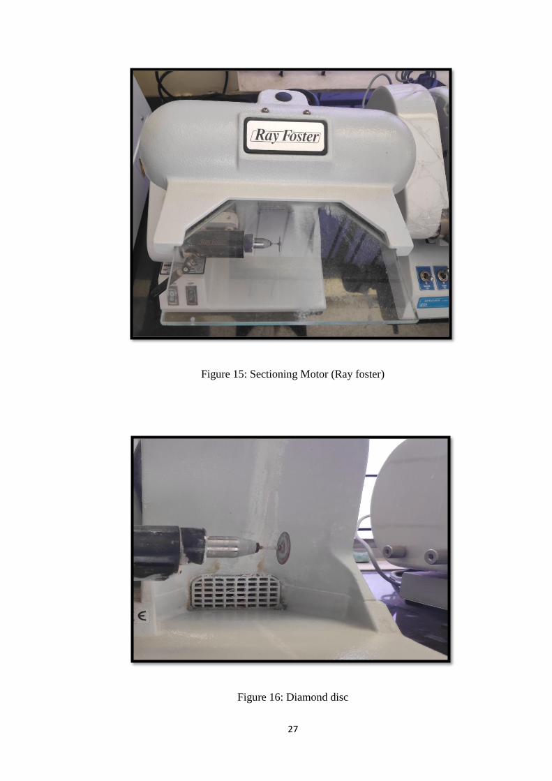
❖ Sectioning motor ( Ray Foster)
❖ Diamond disc
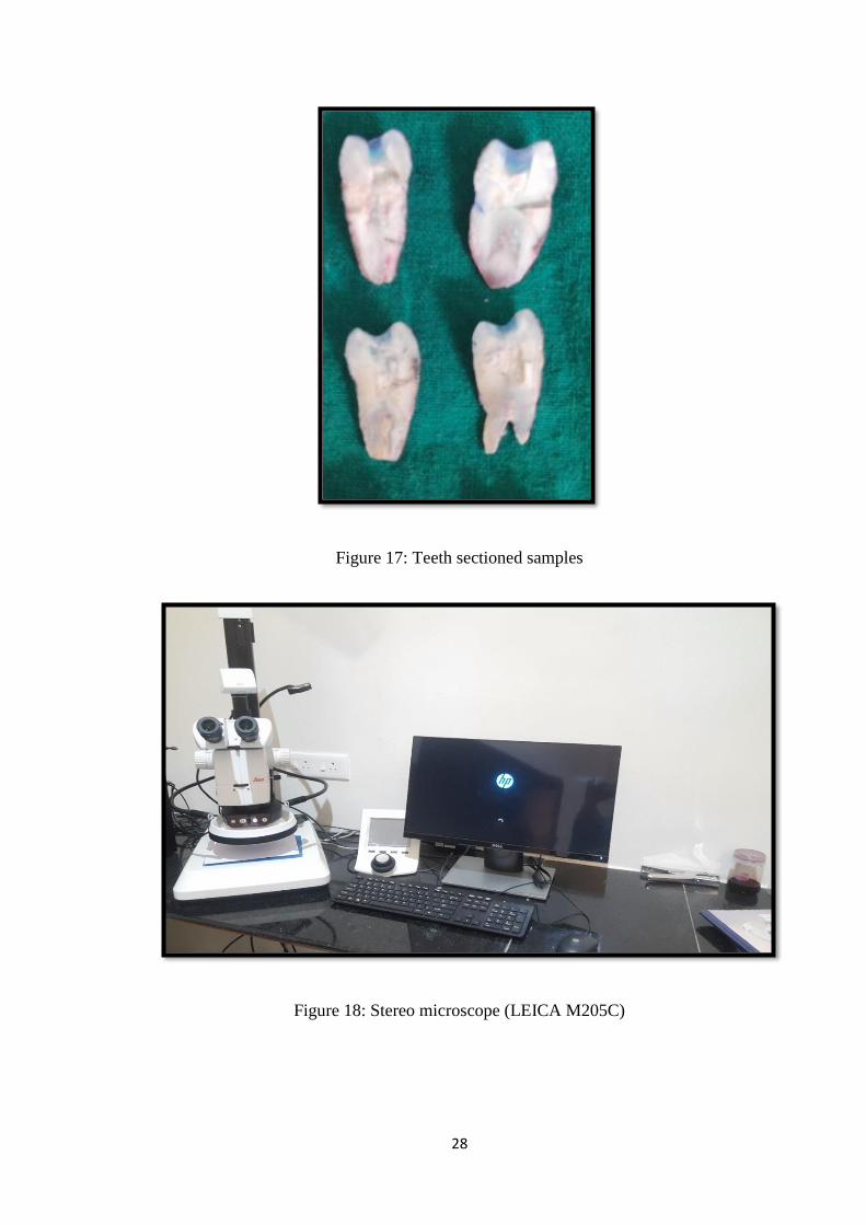
❖ Stereo microscope (LEICA M205C)
EXPERIMENTAL DESIGN
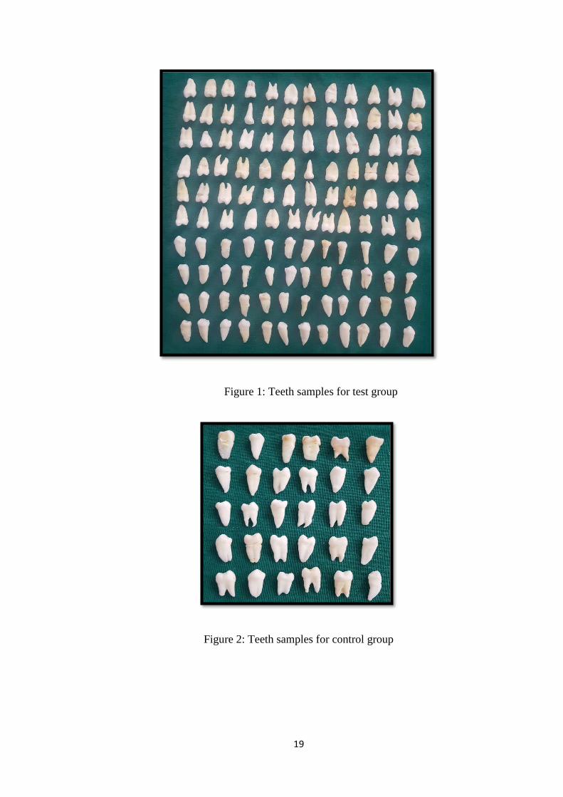
150 extracted human premolar teeth were selected for this study (Figure 1and
2). The teeth were cleaned of soft tissues and debris and were stored in saline to keep it
moist throughout the procedure. Radiographic analysis of all the teeth were done to
confirm the root canal anatomy and to check for the absence of abnormalities like root
resorption and pulp calcification.
18
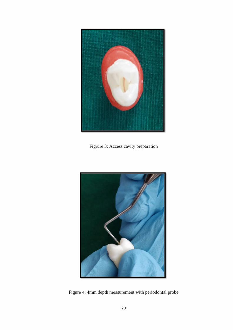
The teeth were divided randomly into 4 experimental groups (n-30 each), a
positive control groups (n-15) and a negative control group (n-15).
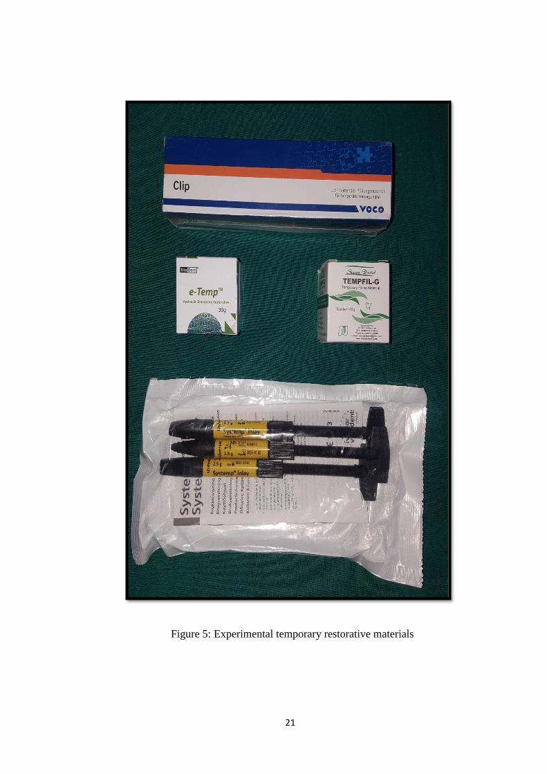
ACCESS CAVITY PREPARATION
Standard endodontic access cavity preparation is done through the occlusal
surface of the teeth with a airotor (high speed) under water coolant, with a round bur
(no 4) for initial preparation and the cavity preparation is extended using a diamond
fissure bur. For ease of preparation the teeth were mounted in custom made wax blocks
made out of modelling wax (Figure 3).
Following removal of pulp tissue and irrigation with 3% sodium hypochlorite
solution, the pulp chamber was dried and a cotton pellet was packed into floor of the
chamber and measured by periodontal probe such that it could accommodate 4mm
thickness of temporary material (Figure 4).
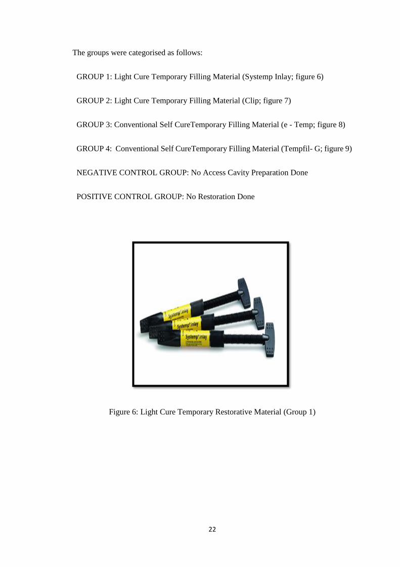
TEMPORARY RESTORATION
Each experimental group was filled with one of the experimental temporary
filling materials which includes two light cure temporary filling materials and two
conventional temporary restorative materials (Figure 5). The positive control group
received no temporary filling except a small cotton pellet in the floor of the pulp
chamber in contrast to the negative control group which received no access cavity
preparation.
19
Figure 1: Teeth samples for test group
Figure 2: Teeth samples for control group
20
Figrure 3: Access cavity preparation
Figure 4: 4mm depth measurement with periodontal probe
21
Figure 5: Experimental temporary restorative materials
22
The groups were categorised as follows:
GROUP 1: Light Cure Temporary Filling Material (Systemp Inlay; figure 6)
GROUP 2: Light Cure Temporary Filling Material (Clip; figure 7)
GROUP 3: Conventional Self CureTemporary Filling Material (e - Temp; figure 8)
GROUP 4: Conventional Self CureTemporary Filling Material (Tempfil- G; figure 9)
NEGATIVE CONTROL GROUP: No Access Cavity Preparation Done
POSITIVE CONTROL GROUP: No Restoration Done
Figure 6: Light Cure Temporary Restorative Material (Group 1)
23
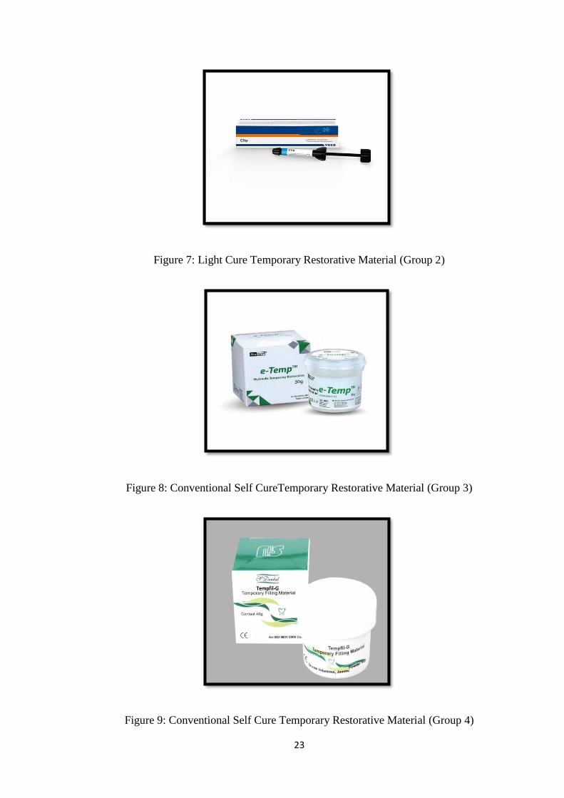
Figure 7: Light Cure Temporary Restorative Material (Group 2)
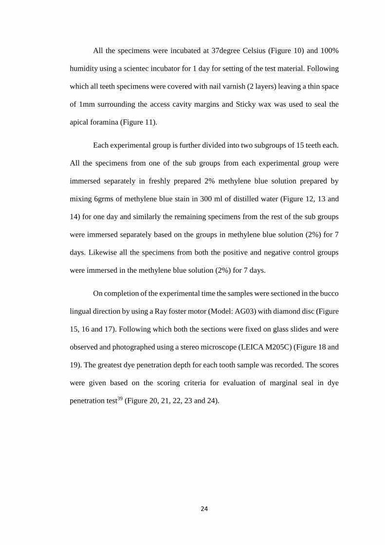
Figure 8: Conventional Self CureTemporary Restorative Material (Group 3)
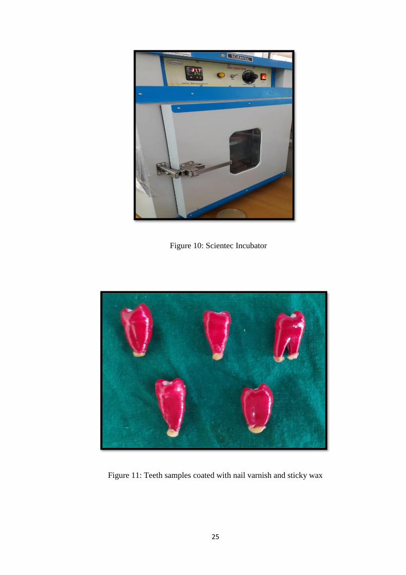
Figure 9: Conventional Self Cure Temporary Restorative Material (Group 4)
24
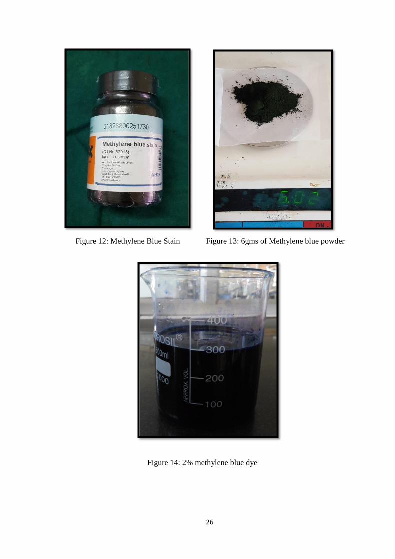
All the specimens were incubated at 37degree Celsius (Figure 10) and 100%
humidity using a scientec incubator for 1 day for setting of the test material. Following
which all teeth specimens were covered with nail varnish (2 layers) leaving a thin space
of 1mm surrounding the access cavity margins and Sticky wax was used to seal the
apical foramina (Figure 11).
Each experimental group is further divided into two subgroups of 15 teeth each.
All the specimens from one of the sub groups from each experimental group were
immersed separately in freshly prepared 2% methylene blue solution prepared by
mixing 6grms of methylene blue stain in 300 ml of distilled water (Figure 12, 13 and
14) for one day and similarly the remaining specimens from the rest of the sub groups
were immersed separately based on the groups in methylene blue solution (2%) for 7
days. Likewise all the specimens from both the positive and negative control groups
were immersed in the methylene blue solution (2%) for 7 days.
On completion of the experimental time the samples were sectioned in the bucco
lingual direction by using a Ray foster motor (Model: AG03) with diamond disc (Figure
15, 16 and 17). Following which both the sections were fixed on glass slides and were
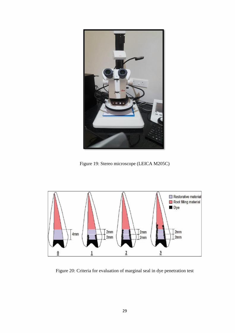
observed and photographed using a stereo microscope (LEICA M205C) (Figure 18 and
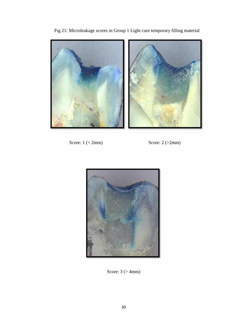
19). The greatest dye penetration depth for each tooth sample was recorded. The scores
were given based on the scoring criteria for evaluation of marginal seal in dye
penetration test39 (Figure 20, 21, 22, 23 and 24).
25
Figure 10: Scientec Incubator
Figure 11: Teeth samples coated with nail varnish and sticky wax
26
Figure 12: Methylene Blue Stain Figure 13: 6gms of Methylene blue powder
Figure 14: 2% methylene blue dye
27
Figure 15: Sectioning Motor (Ray foster)
Figure 16: Diamond disc
28
Figure 17: Teeth sectioned samples
Figure 18: Stereo microscope (LEICA M205C)
29
Figure 19: Stereo microscope (LEICA M205C)
Figure 20: Criteria for evaluation of marginal seal in dye penetration test
30
Fig 21: Microleakage scores in Group 1 Light cure temporary filling material
Score: 1 (< 2mm) Score: 2 (>2mm)
Score: 3 (> 4mm)
31
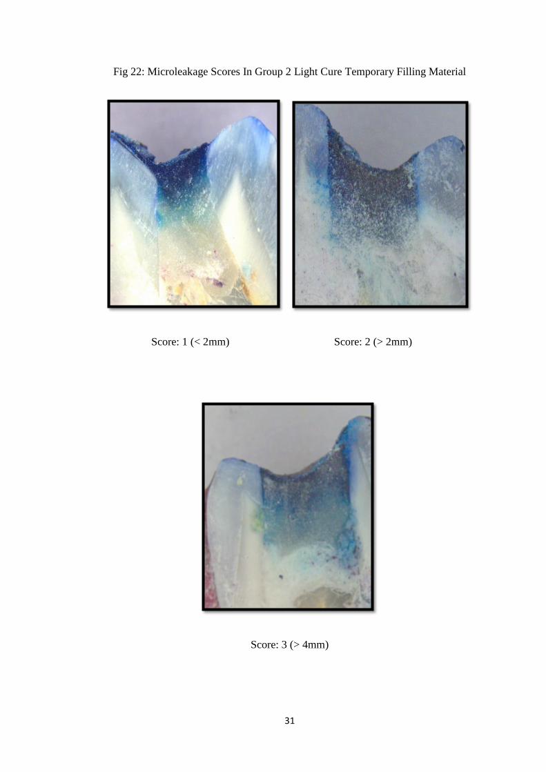
Fig 22: Microleakage Scores In Group 2 Light Cure Temporary Filling Material
Score: 1 (< 2mm) Score: 2 (> 2mm)
Score: 3 (> 4mm)
32
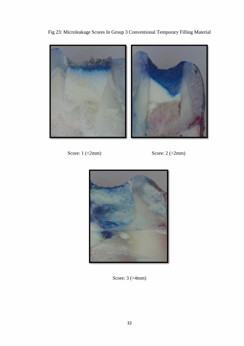
Fig 23: Microleakage Scores In Group 3 Conventional Temporary Filling Material
Score: 1 (<2mm) Score: 2 (>2mm)
Score: 3 (>4mm)
33
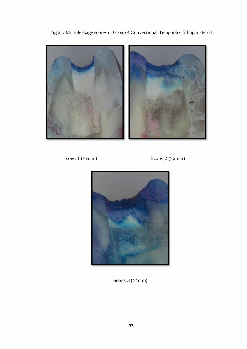
Fig 24: Microleakage scores in Group 4 Conventional Temporary filling material
core: 1 (<2mm) Score: 2 (>2mm)
Score: 3 (>4mm)
34
Statistical analysis was done using 20 SPSS software. Kruskal Wallis Anova
and Median test were done to statistically compare between the groups after one day
and seven days. Post hoc comparison between groups as well as intra group comparison
between one day and 7 days was done using Mann whitney U test. P value of < 0.05
was considered statistically significant.

35
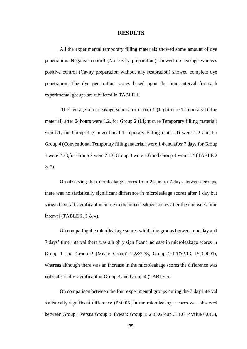
RESULTS
All the experimental temporary filling materials showed some amount of dye
penetration. Negative control (No cavity preparation) showed no leakage whereas
positive control (Cavity preparation without any restoration) showed complete dye
penetration. The dye penetration scores based upon the time interval for each
experimental groups are tabulated in TABLE 1.
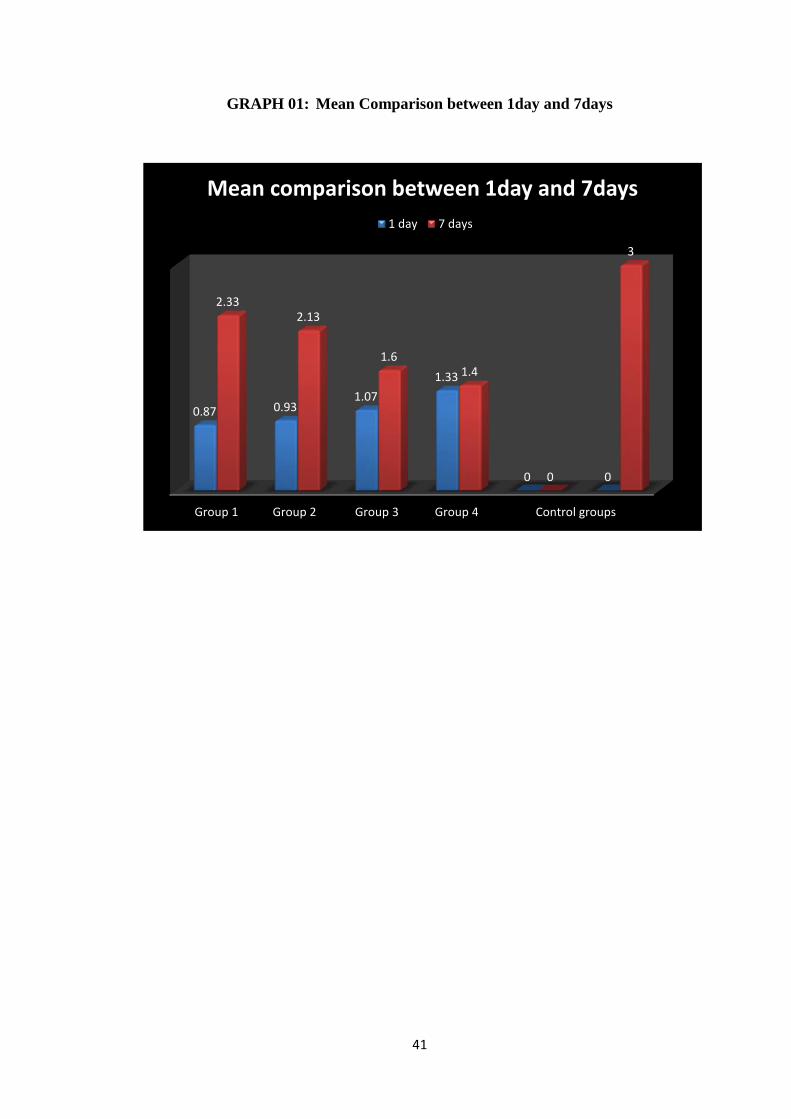
The average microleakage scores for Group 1 (Light cure Temporary filling
material) after 24hours were 1.2, for Group 2 (Light cure Temporary filling material)
were1.1, for Group 3 (Conventional Temporary Filling material) were 1.2 and for
Group 4 (Conventional Temporary filling material) were 1.4 and after 7 days for Group
1 were 2.33,for Group 2 were 2.13, Group 3 were 1.6 and Group 4 were 1.4 (TABLE 2
& 3).
On observing the microleakage scores from 24 hrs to 7 days between groups,
there was no statistically significant difference in microleakage scores after 1 day but
showed overall significant increase in the microleakage scores after the one week time
interval (TABLE 2, 3 & 4).
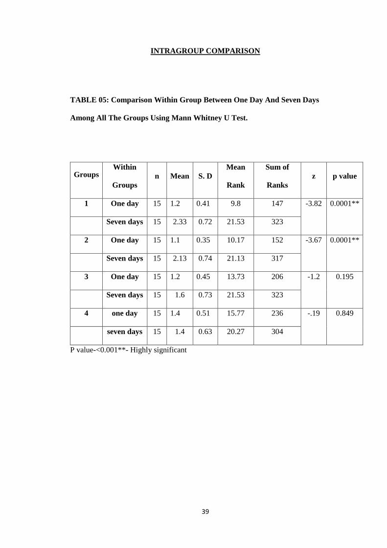
On comparing the microleakage scores within the groups between one day and
7 days’ time interval there was a highly significant increase in microleakage scores in
Group 1 and Group 2 (Mean: Group1-1.2&2.33, Group 2-1.1&2.13, P<0.0001),
whereas although there was an increase in the microleakage scores the difference was
not statistically significant in Group 3 and Group 4 (TABLE 5).
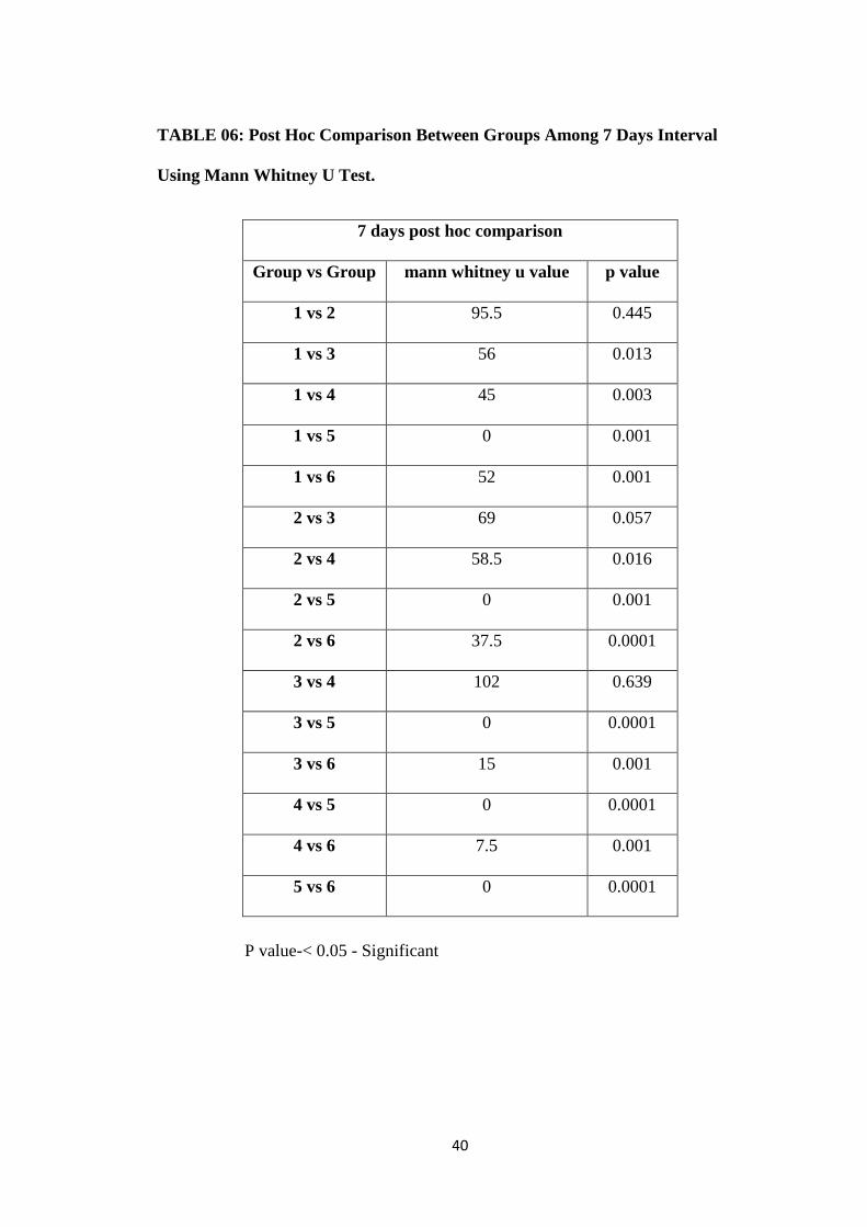
On comparison between the four experimental groups during the 7 day interval
statistically significant difference (P<0.05) in the microleakage scores was observed
between Group 1 versus Group 3 (Mean: Group 1: 2.33,Group 3: 1.6, P value 0.013),
36
Group 1 versus Group 4 (Mean: Group1: 2.33,Group 4: 1.4,P value 0.003) and Group
2 versus Group 4 (Mean: Group 2: 2.13, Group 4: 1.4,P value 0.016), there was no
statistical difference in microleakage scores observed between Group 1 versus Group
2, Group 2 versus Group 3 as well as between Group 3 and Group 4 (TABLE 6).
According to these results all the test materials showed increase in microleakage
from day 1 to day 7 and on comparison at the end of one week Group 4 showed the
least micoleakage scores followed by Group 3 and Group 2 whereas the maximum
microleakage was shown by Group 1 (GRAPH 1).
37
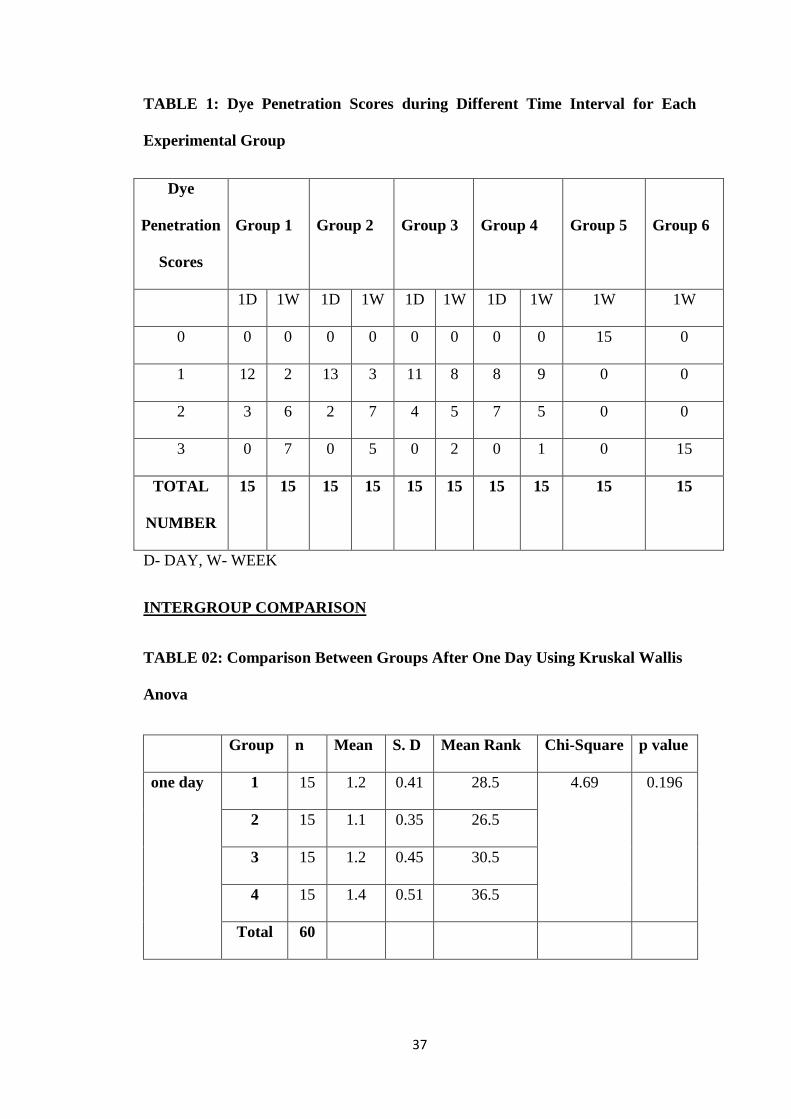
TABLE 1: Dye Penetration Scores during Different Time Interval for Each
Experimental Group
Dye
Penetration
Scores
Group 1
Group 2
Group 3
Group 4
Group 5
Group 6
1D 1W 1D 1W 1D 1W 1D 1W 1W 1W
0 0 0 0 0 0 0 0 0 15 0
1 12 2 13 3 11 8 8 9 0 0
2 3 6 2 7 4 5 7 5 0 0
3 0 7 0 5 0 2 0 1 0 15
TOTAL
NUMBER
15 15 15 15 15 15 15 15 15 15
D- DAY, W- WEEK
INTERGROUP COMPARISON
TABLE 02: Comparison Between Groups After One Day Using Kruskal Wallis
Anova
Group n Mean S. D Mean Rank Chi-Square p value
one day 1 15 1.2 0.41 28.5 4.69 0.196
2 15 1.1 0.35 26.5
3 15 1.2 0.45 30.5
4 15 1.4 0.51 36.5
Total 60
38
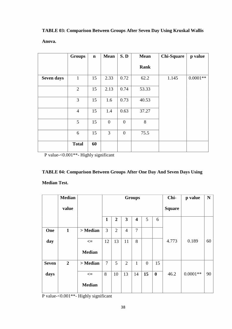
TABLE 03: Comparison Between Groups After Seven Day Using Kruskal Wallis
Anova.
Groups n Mean S. D Mean
Rank
Chi-Square p value
Seven days 1 15 2.33 0.72 62.2 1.145 0.0001**
2 15 2.13 0.74 53.33
3 15 1.6 0.73 40.53
4 15 1.4 0.63 37.27
5 15 0 0 8
6 15 3 0 75.5
Total 60
P value-<0.001**- Highly significant
TABLE 04: Comparison Between Groups After One Day And Seven Days Using
Median Test.
P value-<0.001**- Highly significant
Median
value
Groups Chi-
Square
p value N
1 2 3 4 5 6
4.773
0.189
60
One
day
1 > Median 3 2 4 7
<=
Median
12 13 11 8
Seven
days
2 > Median 7 5 2 1 0 15
46.2
0.0001**
90 <=
Median
8 10 13 14 15 0
39
INTRAGROUP COMPARISON
TABLE 05: Comparison Within Group Between One Day And Seven Days
Among All The Groups Using Mann Whitney U Test.
Groups Within
Groups
n
Mean
S. D
Mean
Rank
Sum of
Ranks
z
p value
1 One day 15 1.2 0.41 9.8 147 -3.82 0.0001**
Seven days 15 2.33 0.72 21.53 323
2 One day 15 1.1 0.35 10.17 152 -3.67 0.0001**
Seven days 15 2.13 0.74 21.13 317
3 One day 15 1.2 0.45 13.73 206 -1.2 0.195
Seven days 15 1.6 0.73 21.53 323
4 one day 15 1.4 0.51 15.77 236 -.19 0.849
seven days 15 1.4 0.63 20.27 304
P value-<0.001**- Highly significant
40
TABLE 06: Post Hoc Comparison Between Groups Among 7 Days Interval
Using Mann Whitney U Test.
7 days post hoc comparison
Group vs Group mann whitney u value p value
1 vs 2 95.5 0.445
1 vs 3 56 0.013
1 vs 4 45 0.003
1 vs 5 0 0.001
1 vs 6 52 0.001
2 vs 3 69 0.057
2 vs 4 58.5 0.016
2 vs 5 0 0.001
2 vs 6 37.5 0.0001
3 vs 4 102 0.639
3 vs 5 0 0.0001
3 vs 6 15 0.001
4 vs 5 0 0.0001
4 vs 6 7.5 0.001
5 vs 6 0 0.0001
P value-< 0.05 - Significant
41
GRAPH 01: Mean Comparison between 1day and 7days
1 2 3 4 5 6
0.87 0.931.07
1.33
0 0
2.332.13
1.61.4
0
3
Mean comparison between 1day and 7days
1 day 7 days
Group 1 Group 2 Group 3 Group 4 Control groups

42
DISCUSSION
Microleakage is one of the most important risk factor associated with
endodontic failure in multi visit endodontics. The prognosis of endodontic therapy is
also predicted on the basis of the provisional restorative material and the main role of a
provisional restorative material is to prevent the seepage of fluids and microorganisms
from the oral cavity into the access cavity51.
Temporary restorative materials in clinical endodontic practice must possess
certain physical properties which allows immediate sealing of the access preparation12.
According to a study conducted by Magura et al based on saliva penetration rate
into the root canals said that if permanent restoration is not done for a duration of more
than 3 months following an endodontic therapy root canal retreatment must be
performed52.
Various factors such as complexity of cases and multiple appointments requires
the temporary material to produce a leak proof seal during the duration of the
endodontic therapy53.
Throughout dental history, temporary fillings have been done using a wide
range of materials but none of the materials seem to fulfil most or all the properties of
an ideal temporary restorative material54.
Various in vivo and in vitro studies have been done to determine the ability of
restorative materials to provide adequate coronal barriers during root canal therapy or
prior to final restoration55.
In the present study we examine and compare the microleakage between four
commercially available temporary filling materials out of which the two are light cured
43
temporary restorative materials (Group 1& Group 2) whereas the remaining two are
conventional self cure temporary restorative materials (Group 3& Group 4) by using
dye penetration test with 2% methylene blue solution.
The most common method used for assessing microleakage is by dye
penetration test using methylene blue dye55. The methylene blue dye has high water
solubility as well as move by simple diffusion and not absorbed by hydroxyl apetite
crystals present in the dentin40 and that it has a molecular size smaller than that of the
bacteria so it may be used as a tool to compare relative leakage30. Hence in the present
study this explains the methodology for using 2% methylene blue dye to verify
microleakage.
According to Webber et al a temporary restorative material should have atleast
3mm thickness to result in superior marginal sealing ability56. Hence the thickness of
all the temporary restorative materials used in this present study was standardised to
4mm to test the microleakage present except for the samples in the positive control
group in which no restoration was placed.
According to M.B Uctash and A.C Tinaz the technique for placing the
experimental temporary filling materials into the access cavity preparation may have
some adverse effects on marginal leakage of the material used12 so in order to overcome
this drawback all the experimental materials in this study were added by using
incremental technique into the access cavities and furthermore the marginal sealing of
all the samples was done by a single operator to reduce variability and as well as all the
test materials were used according to the manufactures instructions to overcome the
chances of manipulative variables.
44
To simulate the clinical condition the teeth samples were incubated at 37 degree
celcius in 100% humidity to ensure complete setting of temporary filling material30.
In this study sealing of the teeth samples was performed by using nail varnish
in order to prevent the leakage of the dye through the tooth structure as observed by
various authors such as Zmener O et al55.
According to a study by Naseri et al, the sealing ability of temporary filling
materials were tested for coronal sealing ability after 1 day, 1 week and 4 weeks as
these are the most frequently used time intervals during dental practice between root
canal therapy appointments or following obturation before the placement of a
permanent restoration30. This justifies that in the present study the time interval
considered to compare the difference in microleakage values between the temporary
restorative materials was between one day and one week.
Two commercially available light cure temporary materials were used in this
study out of which the first light cure temporary material (Group 1) is a single unit
composite based temporary restorative material which has a command set and is based
on Monofunctional Ethyl Triglycol Methacrylate And A Poly Esther Urethane
Dimethacrylate similarly the second light cure temporary material (Group 2) is also a
single component temporary restorative material which consists of Hydro Ethyl
Methacrylate, Acrylate Esters, Butylhydoxytoludene And Polymers.
The remaining two test materials used in this study are conventional self cure
temporary restorative (Group 3& Group 4). Both these materials are self cure temporary
materials which are commercially available in a premixed state which sets under
humidity and composed of Zinc Oxide and calcium sulphate.
45
According to the results of the present study all the experimental temporary
materials devoid of their mechanism of setting showed an increase in the microleakage
values from day 1 to 1 week which coincides with the results of the study conducted by
Mohammadian M and Jafarzadeh-Kashi T S in which the microleakage of the
temporary restorative materials assessed during three different times namely after
1day, one week and one month showed an increase in the amount of microleakage with
time thereby indicating the decrease in the sealing ability of the temporary material over
time36.
According to the results in this study at the end of one week time interval Group
4 and Group 3 (conventional self cure temporary filling material) showed
comparatively lesser microleakage. This can be attributed to the ability of these
materials to set when in contact with moisture and can undergo hygroscopic expansion
thereby maintaining a tight seal at the material and the tooth interface which has been
stated by various authors such as Cruz et al and Lee YC et al57,58.
The conventional temporary materials used in this study are premixed and ready
to use, which can be quickly placed and adjusted into the access cavity. This reduces
the inconsistencies associated with the chair side manipulations57,58. These superior
manipulation properties were considered as supplementary factors responsible for good
coronal sealing ability59,12,60 which coincides with the results of this present study.
Various in vivo and in vitro studies have been conducted regarding the good
sealing property of conventional self cure temporary restorative material (Caviton)57,58
which has similar properties to our Conventional test materials (Group 3 and Group 4)38
thereby coinciding with the results of our present study that the conventional temporary
materials showed superior sealing ability.
46
Group 2 test material used in this study is a composite based temporary
restorative material used without an adhesive system which has polymerisation
shrinkage either due to lack of chemical bonds to tooth structure or due to lack of
micromechanical retention61. Even though the water absorbtion property of resin based
materials increases the volume it cannot fully compensate for the microgaps formed
during polymerisation62,63,64 which might result in microleakage.
Similarly according to S. Erkut et al, our Group 1 test material is a light
polymerised, highly elastic resin based composite temporary filling material made by
adding an antimicrobial agent (Triclosan) to another light cure filling material
(Fermit)65 and previous studies using this temporary filling material has resulted in
considerable microleakage66,12,67. But certain controversies exists such as, according to
M.B Uctash and A.C Tinaz light cure composite temporary filling materials lack
microparticles which results in absorbtion of methylene blue which might be a reason
for increased microleakage values12.
47
This in vitro study has certain limitations such as it does not duplicate the oral
environment such as presence of saliva, the data obtained through dye penetration test
have been questioned by researchers who claim that this technique has large standard
deviations and it is non reproducible68,69.
Clinically in the oral cavity, according to Qvist V micoleakage can be impacted
by the masticatory forces which may vary based on the variables such as Sex, location
of the tooth, age and bruxism70. Hence in the present study occlusal load was not used
because of the above mentioned differences.
According to B.M. Jacquot et al the marginal sealing ability of temporary filling
materials can be adversely affected by temperature fluctuations17 which was in contrast
to Kidd who in his study stated that microleakage was not affected by thermal cycling71.
Hence due to these controversies thermal cycling procedure was not performed in this
study.
Considering these limitations further studies are required to evaluate
micoleakage such as quantitative microbiological tests and quantitative evaluation as
no method is ideal43.
48
SUMMARY AND CONCLUSION
SUMMARY
The key factor for the success of root canal treatment depends on the prevention
of microleakage of fluids and microorganisms from the oral cavity into the endodontic
access through a temporary filling material either during the inter appointment time
period or following obturation before the placement of a permanent restoration which
can be prevented by using an appropriate temporary filling material which has superior
sealing properties.
This study was performed to evaluate and compare the coronal microleakage
between commercially available light cure temporary material and conventional
temporary restorative material over different time periods as it might give an insight
regarding the quality of these temporary restorative materials.
In this study one hundred and fifty extracted premolar teeth were used. The
teeth samples were divided randomly into four experimental group with 30 samples
each and two control groups with 15 samples each. The groups were categorised as
follows:
GROUP 1: Light Cure Temporary Filling Material (Systemp Inlay manufactured by
Ivoclar Vivadent)
GROUP 2: Light Cure Temporary Filling Material (Clip manufactured by Voco)
GROUP 3: Conventional Temporary Filling Material (e- Temp manufactured by
Diadent)
GROUP 4: Conventional Temporary Filling Material (Tempfil - G manufactured by
Shivam dental)
49
NEGATIVE CONTROL GROUP: No Access Cavity Preparation Done
POSITIVE CONTROL GROUP: No Restoration Done
Standard endodontic access cavities were prepared in all the teeth such that it
could accommodate 4mm thickness of the experimental temporary restorative material.
All the test groups were filled with their respective experimental temporary materials
based on their specific manufactures instructions. After the completion of the
experiment duration all the samples were sectioned in the bucco lingual direction and
the sectioned samples where observed and recorded using a stereo microscope.
The mean values were computed and the mean values were calculated and
compared. The difference in the mean scores were used to assess and compare the
sealing ability of the temporary materials used in this study.
CONCLUSION
According to the results of this study all the test materials showed increase in
microleakage from day 1 to day 7 and on comparison at the end of one week
conventional temporary restorarive material (Group 4) showed the least micoleakage
scores whereas the maximum microleakage was shown by light cure temporary filling
material (Group 1).
This study provides an insight to the clinician regarding the quality of various
commercially available temporary restorative materials thereby making it easier to
select the suitable material for specific clinical conditions.
Thereby within the limitations of this study we conclude that all the materials
devoid of the mode of curing showed increase in the microleakage values between day
1 to day 7 and that the conventional self cure temporary filling materials provide less
50
microleakage when compared to light cure temporary materials after one week time
interval.
Though this study is not clinically evident, it suggests that temporary materials
can be used only for a short duration of time and a permanent coronal restoration must
be placed after root canal therapy at the earliest to avoid possible failures.

51
BIBLIOGRAPHY
1. Kakehashi S, Stanley HR, Fitzgerald RJ. The effects of surgical exposures of
dental pulps in germ-free and conventional laboratory rats. Oral Surgery, Oral
Medicine, Oral Pathology. 1965 Sep 1;20(3):340-9.
2. Moller AJ, Fabricius L, Dahlen G, Ohman AE, Heyden GU. Influence on
periapical tissues of indigenous oral bacteria and necrotic pulp tissue in
monkeys. European Journal of Oral Sciences. 1981 Dec;89(6):475-84.
3. Barkhordar RA, Stark MM. Sealing ability of intermediate restorations and
cavity design used in endodontics. Oral surgery, oral medicine, oral pathology.
1990 Jan 1;69(1):99-101.
4. Jensen AL, Abbott PV, Salgado JC. Interim and temporary restoration of teeth
during endodontic treatment. Australian dental journal. 2007 Mar;52:S83-99.
5. Zaia AA, Nakagawa R, De Quadros I, Gomes BP, Ferraz CC, Teixeira FB,
Souza-Filho FJ. An in vitro evaluation of four materials as barriers to coronal
microleakage in root-filled teeth. Int Endod J. 2002;35:729-34.
6. Saunders WP, Saunders EM. Coronal leakage as a cause of failure in root-canal
therapy: a review. Endod Dent Traumatol. 1994;10(3):105–8.
7. Abbott PV. Factors associated with continuing pain in endodontics. Aust Dent
J. 1994;39(3):157–61.
8. Ray HA, Trope M. Periapical status of endodontically treated teeth in relation
to the technical quality of the root filling and the coronal restoration. Int Endod
J. 1995;28(1):12–8.
9. Moreno JO, Alves FR, Goncalves LS, Martinez AM, Rocas IN, Siqueira JF Jr.
Periradicular status and quality of root canal fillings and coronal restorations in
an urban Colombian population. J Endod 2013;39:600-4.
52
10. Torabinejad M, Ung B, Kettering JD. In vitro bacterial penetration of coronally
unsealed endodontically treated teeth. J Endod. 1990;16(12):566–9.
11. Khayat A, Lee SJ, Torabinejad M. Human saliva penetration of coronally
unsealed obturated root canals. J Endod. 1993;19(9):458–61.
12. Uctasli MB, Tinaz AC. Microleakage of different types of temporary restorative
materials used in endodontics. Journal of oral science. 2000;42(2):63-7
13. Anderson RW, Powell BJ, Pashley DH. Microleakage of three temporary
endodontic restorations. Journal of endodontics. 1988 Jan 1;14(10):497-501.
14. Teplitsky PE, Meimaris IT. Sealing ability of Cavit and TERM as intermediate
restorative materials. Journal of endodontics. 1988 Jun 1;14(6):278-82.
15. Bobotis HG, Anderson RW, Pashley DH, Pantera EA. A microleakage study of
temporary restorative materials used in endodontics. Journal of Endodontics.
1989 Dec 1;15(12):569-72.
16. Melton D, Cobb S, Krell KV. A comparison of two temporary restorations:
light-cured resin versus a self-polymerizing temporary restoration. Oral
Surgery, Oral Medicine, Oral Pathology and Oral Radiology. 1990 Aug
1;70(2):221-5
17. Jacquot BM, Panighi MM, Steinmetz P, G'sell C. Microleakage of Cavit,
CavitW, CavitG and IRM by impedance spectroscopy. International Endodontic
Journal. 1996 Jul;29(4):256-61.
18. Barthel CR, Strobach A, Briedigkeit H, Göbel UB, Roulet JF. Leakage in roots
coronally sealed with different temporary fillings. Journal of Endodontics. 1999
Nov 1;25(11):731-4.
53
19. Pai SF, Yang SF, Sue WL, Chueh LH, Rivera EM. Microleakage between
endodontic temporary restorative materials placed at different times. Journal of
endodontics. 1999 Jun 1;25(6):453-6.
20. Liberman R, Ben-Amar A, Frayberg E, Abramovitz I, Metzger Z. Effect of
repeated vertical loads on microleakage of IRM and calcium sulfate-based
temporary fillings. Journal of endodontics. 2001 Dec 1;27(12):724-9.
21. Balto H. An assessment of microbial coronal leakage of temporary filling
materials in endodontically treated teeth. Journal of endodontics. 2002 Nov
1;28(11):762-4.
22. Lai YY, Pai L, Chen CP. Marginal leakage of different temporary restorations
in standardized complex endodontic access preparations. Journal of
endodontics. 2007 Jul 1;33(7):875-8.
23. Madarati A, Rekab MS, Watts DC, Qualtrough A. Time‐dependence of coronal
seal of temporary materials used in endodontics. Australian Endodontic Journal.
2008 Dec;34(3):89-93.
24. Tapsir Z, Ahmed HM, Luddin N, Husein A. Sealing ability of various
restorative materials as coronal barriers between endodontic appointments. The
journal of contemporary dental practice. 2013 Jan 1;14(1):47.
25. Koagel SO, Mines P, Apicella M, Sweet M. In vitro study to compare the
coronal microleakage of Tempit UltraF, Tempit, IRM, and Cavit by using the
fluid transport model. Journal of endodontics. 2008 Apr 1;34(4):442-4.
26. Jung DH, Noh YS, Lee HD, Chang HS, Ryu HW, Min KS. Microleakage of
endodontic temporary restorative materials under dynamic loading. Journal of
Korean Academy of Conservative Dentistry. 2008 May 1;33(3):198-203.
54
27. Chailertvanitkul P, Abbott PV, Riley TV, Sooksuntisakoonchai N. Bacterial and
dye penetration through interim restorations used during endodontic treatment
of molar teeth. Journal of endodontics. 2009 Jul 1;35(7):1017-22.
28. Shahi S, Samiei M, Rahimi S, Nezami H. In vitro comparison of dye penetration
through four temporary restorative materials. Iranian endodontic journal.
2010;5(2):59-63.
29. Aledrissy HI, Abubakr NH, Yahia NA, Ibrahim YE. Coronal microleakage for
readymade and hand mixed temporary filling materials. Iranian endodontic
journal. 2011;6(4):155-159.
30. Naseri M, Ahangari Z, Moghadam MS, Mohammadian M. Coronal sealing
ability of three temporary filling materials. Iranian endodontic journal.
2012;7(1):20-24.
31. Yun SM, Karanxha L, Kim HJ, Jung SH, Park SJ, Min KS. Coronal
microleakage of four temporary restorative materials in Class II-type
endodontic access preparations. Restorative Dentistry & Endodontics. 2012
Mar 1;37(1):29-33.
32. Srikumar GP, Varma KR, Shetty KH, Kumar P. Coronal microleakage with five
different temporary restorative materials following walking bleach technique:
An ex-vivo study. Contemporary clinical dentistry. 2012 Oct;3(4):421-6.
33. Sagar KP, Murthy BS, Kumar M. Comparative Evaluation of Three Different Materials
as Barriers to Coronal Microleakage in Root Filled Teeth: An in Vitro Study. Heal
Talk. 2012;Volume 04:Issue 05.
34. Davut Celik, Erhan Tahan,Tamer Tasdemir,Kursat Er,Kadir Tolga Ceyhanl.
Coronal microleakage of various temporary fillings in standardized endodontic
access cavities. Clinical Dentistry and Research. 2013; 37(2): 23-28.
55
35. Symanski NC, Juber P, Morgental RD, Scarparo RK, Vier-Pelisser FV.
Temporary restorative materials used by Brazilian Dental Schools during and
after endodontic treatment. Revista da Faculdade de Odontologia-UPF. 2013
Dec 10;18(1).
36. Mohammadian M, Jafarzadeh-Kashi TS. In vitro comparison of coronal micro-
leakage of three temporary restorative materials by dye penetration. Zahedan
Journal of Research in Medical Sciences. 2013 Jan 1;15(1):24-7.
37. Sulieman RT. Microleakage of Root Canal Sealed with Temporary Endodontic
Sealing Materials. Tikrit Journal for Dental Sciences. 2014;3(1):24-9.
38. Tapsir Z, Ahmed HM, Luddin N, Husein A. Sealing ability of various
restorative materials as coronal barriers between endodontic appointments. The
journal of contemporary dental practice. 2013 Jan 1;14(1):47-50.
39. Cardoso AS, Silva NC, Silva JM, Herrera DR, Neves AA, Silva EJ. Assessment
of coronal leakage of a new temporary light-curing filling material in
endodontically treated teeth. Indian Journal of Dental Research. 2014 May
1;25(3):321-4.
40. Gidwani K, Madhyastha PS, Srikant N, Suman E, Kotian R. In vitro evaluation
of sealing ability and antimicrobial activity of hydraulic temporary sealing
materials. Journal of Restorative Dentistry. 2014 Jan 1;2(1):13-9.
41. Shiva Sadeghi, Ramin Tabari, Shahroz Almasi. Ex-vivo Sealing Ability of
Different Thicknesses of White and Gray Angelus MTA as an Intra-orifice
Barrier in Endodontically Treated Teeth. 3dj. 2014; 3 (2) :1-5.
42. Dos Santos GL, De Andrada Beltrame AP, Triches TC, Ximenes-Filho M,
Baptista D, Bolan M. Analysis of microleakage of temporary restorative
56
materials in primary teeth. Journal of Indian Society of Pedodontics and
Preventive Dentistry. 2014 Apr 1;32(2):130-4.
43. Cunha CT, De Miranda BF, De Morais JF, Dametto FR, Netoand AF, Chaves
LV. In vitro evaluation of coronal microleakage of some temporary sealing
materials used in endodontic and three different endodontic sealers. JSM Dent.
2014;2(3):1031-4.
44. Bodrumlu EH, Bodrumlu E, Avsar A, Meydan AD. Effect ofradiotherapy on
the sealing ability of temporary filling materials. Eur J Gen Dent 2015;4:8-11.
45. Udayakumar P, Kaushik M, Prashar N, Arya S. Coronal leakage of provisional
restorative materials used in endodontics with and without intracanal
medication after exposure to human saliva. Saudi Endodontic Journal. 2016
May 1;6(2):77-81.
46. Markose A, Krishnan R, Ramesh M, Singh S. A comparison of the sealing
ability of various temporary restorative materials to seal the access cavity: An
in vitro study. Journal of pharmacy & bioallied sciences. 2016 Oct;8(Suppl
1):S42-44.
47. Kriznar I, Seme K, Fidler A. Bacterial microleakage of temporary filling
materials used for endodontic access cavity sealing. Journal of Dental Sciences.
2016 Dec 1;11(4):394-400.
48. Prabhakar AR, Rani NS, Naik SV. Comparative Evaluation of Sealing Ability,
Water Absorption, and Solubility of Three Temporary Restorative Materials:
An in vitro Study. International journal of clinical pediatric dentistry. 2017
Apr;10(2):136-141.
57
49. Maslamani M, Khalaf M, Mitra AK. Association of quality of coronal filling
with the outcome of endodontic treatment: A follow-up study. Dentistry journal.
2017 Jan 11;5(1):5.
50. Deepak S, Nivedhitha MS. Comparison of coronal microleakage of three
temporary restorative material using dye penetration methods. J Adv Pharm Edu
Res. 2017;7(3):232-235.
51. Srivastava PK, Nagpal A, Setya G, Kumar S, Chaudhary A, Dhanker K.
Assessment of Coronal Leakage of Temporary Restorations in Root Canal-
treated Teeth: An in vitro Study. The journal of contemporary dental practice.
2017 Feb;18(2):126-30.
52. Magura ME, Kafrawy AH, Brown CE, Newton CW. Human saliva coronal
microleakage in obturated root canals: an in vitro study. Journal of Endodontics.
1991 Jul 1;17(7):324-31.
53. Bobotis HG, Anderson RW, Pashley DH, Pantera EA. A microleakage study of
temporary restorativematerials used in endodontics. Journal of Endodontics.
1989 Dec 1;15(12):569-72.
54. Devika warrier E, Jayalakshmi. A review on temporary restorative materials.
International Journal Of Pharma sciences and Research. 2016; Jul:Vol 7.
55. Zmener O, Banegas G, Pameijer CH. Coronal microleakage of three temporary
restorative material materials: An in vitro study. J Endod. 2004;30:582-84.
56. Webber RT, del Rio CE, Brady JM, Segall RO. Sealing quality of a temporary
filling material. Oral Surg Oral Med Oral Pathol. 1978;46:123-30.
57. Cruz EV, Shigetani Y, Ishikawa K, Kota K, Iwaku M, Goodis HE. A laboratory
study of coronal microleakage using 4 temporary restorative materials. Int
Endod J. 2002; 35: 315-20.
58
58. Lee YC, Yang SF, Hwang YF, Chueh LH, Chung KH. Microleakage of
endodontic temporary restorative materials. J Endod. 1993;19:516-20.
59. Chohayeb AA, Bassiouny MA. Sealing ability of intermediate restoratives used
in endodontics. J Endod. 1985;11(6):241–4.
60. Aledrissy HII, Abubakr NH, Ahmed N. Coronal microleakage for readymade
and hand mixed temporary filling materials. Iran Endod J. 2011;6(4):155–9.
61. Kriznar I, Seme K, Fidler A. Bacterial microleakage of temporary filling
materials used for endodontic access cavity sealing. Journal of Dental Sciences.
2016 Dec 1;11(4):394-400.
62. Cattani-Lorente MA, Dupuis V, Payan J, Moya F, Meyer JM. Effect of water
on the physical properties of resin-modified glass ionomer cements. Dent Mater.
1999;15:71-8.
63. Ruttermann S, Kruger S, Raab WH, Janda R. Polymerization shrinkage and
hygroscopic expansion of contemporary posterior resin-based filling
materialsea comparative study. J Dent. 2007;35:806-13.
64. Versluis A, Tantbirojn D, Lee MS, Tu LS, DeLong R. Can hygroscopic
expansion compensate polymerization shrinkage? Part I. Deformation of
restored teeth. Dent Mater. 2011;27:126-33.
65. Erkut S, Caglar A, Yilmaz B, Küçükeşmen HC, Ozdemir E. Microleakage of different
provisionalization techniques for class I inlays. Journal of Dental Sciences. 2013 Mar
1;8(1):1-7.
66. Uranga A, Blum JY, Esber S, Parahy E, Prado C. A comparative study of four
coronal obturation materials in endodontic treatment. J Endod. 1999;25:178-80.
59
67. Hahn P, Schaller HG, Mullner U, Hellwig E. Marginal leakage in class II
restorations after use of ceramic-inserts luted with different materials. J Oral
Rehabil. 1998;25:567-74.
68. Wu M-K, Wesselink PR. Endodontic leakage studies reconsidered. Part I.
Methodology, application, and relevance. Int Endod J. 1993;26:37-43.
69. Editorial Board of the Journal of Endodontics. Wanted: a base of evidence. J
Endod. 2007;33:1401-2.
70. Qvist V. The effect of mastication on marginal adaptation of composite
restorations in vivo. Journal of dental research. 1983 Aug;62(8):904-6.
71. Kidd EA. Microleakage : a review. J . dent. 1976; 4 :199-206.


![Coronal Polishing Revised.doc - [CSI] College of Southern Idaho](https://img.dokumen.tips/doc/110x75/631c5d9676d2a4450503822b/coronal-polishing-reviseddoc-csi-college-of-southern-idaho.jpg)