Embed Size (px)
Citation preview
Cc
CRa
b
a
ARRAA
KG�GT
1
pc[cptcmgmfoctrsgbsa
0d
International Journal of Biological Macromolecules 49 (2011) 700– 706
Contents lists available at ScienceDirect
International Journal of Biological Macromolecules
journa l h o me pag e: www.elsev ier .com/ locate / i jb iomac
omparative analysis of gelatin scaffolds crosslinked by genipin and silaneoupling agent
. Tonda-Turoa, P. Gentilea, S. Saracinob, V. Chionoa, V.K. Nandagiri a, G. Muziob,
.A. Canutob, G. Ciardelli a,∗
Department of Mechanics, Politecnico di Torino, Corso Duca degli Abruzzi 24, 10129 Turin, ItalyDepartment of Experimental Medicine and Oncology, Università di Torino, Corso Raffaello 30, 10125 Turin, Italy
r t i c l e i n f o
rticle history:eceived 20 April 2011eceived in revised form 1 June 2011ccepted 1 July 2011vailable online 13 July 2011
a b s t r a c t
Scaffolds based on gelatin (G) are considered promising for tissue engineering, able to mimic the naturalextracellular matrix. G drawback is its poor structural consistency in wet conditions. Therefore, crosslink-ing is necessary to fabricate stable G scaffolds. In this work, a comparative study between the performanceof two different crosslinkers, genipin (GP) and �-glycidoxypropyltrimethoxysilane (GPTMS), is presented.
eywords:enipin-Glycidoxypropyltrimethoxysilaneelatinissue engineering
Flat membranes by solvent casting and porous crosslinked scaffolds by freeze-drying were prepared.Infrared spectroscopy and thermal analysis were applied to confirm G chain crosslinking. Moreover,GP and GPTMS increased the stability of G in aqueous media and improved the mechanical properties.Crosslinking reduced the wettability, especially in the case of G GPTMS samples, due to the introductionof hydrophobic siloxane chains. Both G GP and G GPTMS scaffolds supported MG-63 osteoblast-like celladhesion and proliferation.
. Introduction
In tissue engineering, scaffolds are designed to serve as a tem-orary artificial extracellular matrix (ECM) in order to supportell attachment and guide three-dimensional (3D) tissue formation1,2]. Therefore, an ideal scaffold should mimic the advantageousharacteristics of the natural ECM [3]. The native ECM is com-rised of a complex network of structural and regulatory proteinshat are arrayed into a fibrous matrix. It also provides residentells with specific ligands for cell adhesion and migration, andodulates cell proliferation and function [4,5]. Several investi-
ators, have attempted to develop biomimetic scaffolds able toimic native ECM. Development of three-dimensional (3D) scaf-
olds that can replace the natural ECM should allow the diffusionf nutrient, metabolites and soluble factors until the seeded cellsan produce a new functional matrix and regenerate the desiredissue structures [6,7]. A wide variety of scaffolds based on natu-al and synthetic polymers have been used for the engineering ofoft tissues such as the cartilage [8]; with the addition of an inor-anic component (such as hydroxyapatite, �-tricalcium phosphate,
ioactive glass), these scaffolds have been applied to obtain boneubstitutes for oral and maxillofacial surgical applications in vitrond/or in vivo. Natural polymers are functionally superior to non-∗ Corresponding author. Tel.: +39 011 5646919; fax: +39 011 5646999.E-mail address: [email protected] (G. Ciardelli).
141-8130/$ – see front matter © 2011 Elsevier B.V. All rights reserved.oi:10.1016/j.ijbiomac.2011.07.002
© 2011 Elsevier B.V. All rights reserved.
informational synthetic polymers (e.g., polylactide-co-glycolide),polylactide, polycaprolactone), because they provide structural andchemical signals (such as the Arg-Gly-Asp sequence), which facili-tate cell attachment [9,10].
Among them, gelatin (G) is a non-expensive and commerciallyavailable biomaterial that has gained interest in biomedical engi-neering, mainly because of its biodegradability. G is obtained bythermal denaturation or physical and chemical degradation of col-lagen, the most widespread protein in the body, occurring in mostconnective tissues as skin, tendon and bone [11]. With respect tocollagen, G does not express antigenicity in physiological condi-tions, it is completely resorbable in vivo and its physicochemicalproperties can be suitably modulated [12]. The main limitationof G for the production of tissue substitutes arises from its rapiddissolution in aqueous environments [11].
Chemical and physical crosslinking methods have been usedto increase G stability in aqueous media. Physical crosslinkingmethods for G include microwave irradiation [13], dehydrothermaltreatment (DHT) [14] and UV-treatment [15]. The main advan-tage of physical methods is that they do not cause potential harm,whereas their main drawback arises from the difficulty to obtain thedesired crosslinking degree. Commonly used chemical crosslinkersinclude aldehydes (formaldehyde, glutaraldehyde, glyceraldehyde)
[16], polyepoxy compounds [17] and carbodiimides [18]. The mainlimitation in the use of these products is the possible presence ofsome unreacted crosslinker inside the scaffold with consequent for-mation of toxic products during in vivo biodegradation. For this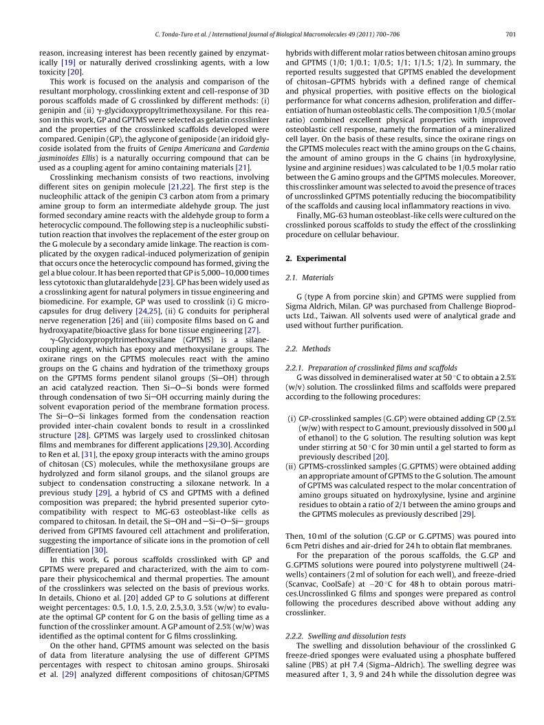
of Biol
rit
rpgsaccju
dnafhttptglabcnh
cogoatsTpsfitohspcccdsd
GpoIwafi
ope
C. Tonda-Turo et al. / International Journal
eason, increasing interest has been recently gained by enzymat-cally [19] or naturally derived crosslinking agents, with a lowoxicity [20].
This work is focused on the analysis and comparison of theesultant morphology, crosslinking extent and cell-response of 3Dorous scaffolds made of G crosslinked by different methods: (i)enipin and (ii) �-glycidoxypropyltrimethoxysilane. For this rea-on in this work, GP and GPTMS were selected as gelatin crosslinkernd the properties of the crosslinked scaffolds developed wereompared. Genipin (GP), the aglycone of geniposide (an iridoid gly-oside isolated from the fruits of Genipa Americana and Gardeniaasminoides Ellis) is a naturally occurring compound that can besed as a coupling agent for amino containing materials [21].
Crosslinking mechanism consists of two reactions, involvingifferent sites on genipin molecule [21,22]. The first step is theucleophilic attack of the genipin C3 carbon atom from a primarymine group to form an intermediate aldehyde group. The justormed secondary amine reacts with the aldehyde group to form aeterocyclic compound. The following step is a nucleophilic substi-ution reaction that involves the replacement of the ester group onhe G molecule by a secondary amide linkage. The reaction is com-licated by the oxygen radical-induced polymerization of genipinhat occurs once the heterocyclic compound has formed, giving theel a blue colour. It has been reported that GP is 5,000–10,000 timesess cytotoxic than glutaraldehyde [23]. GP has been widely used as
crosslinking agent for natural polymers in tissue engineering andiomedicine. For example, GP was used to crosslink (i) G micro-apsules for drug delivery [24,25], (ii) G conduits for peripheralerve regeneration [26] and (iii) composite films based on G andydroxyapatite/bioactive glass for bone tissue engineering [27].
�-Glycidoxypropyltrimethoxysilane (GPTMS) is a silane-oupling agent, which has epoxy and methoxysilane groups. Thexirane rings on the GPTMS molecules react with the aminoroups on the G chains and hydration of the trimethoxy groupsn the GPTMS forms pendent silanol groups (Si OH) throughn acid catalyzed reaction. Then Si O Si bonds were formedhrough condensation of two Si OH occurring mainly during theolvent evaporation period of the membrane formation process.he Si O Si linkages formed from the condensation reactionrovided inter-chain covalent bonds to result in a crosslinkedtructure [28]. GPTMS was largely used to crosslinked chitosanlms and membranes for different applications [29,30]. Accordingo Ren et al. [31], the epoxy group interacts with the amino groupsf chitosan (CS) molecules, while the methoxysilane groups areydrolyzed and form silanol groups, and the silanol groups areubject to condensation constructing a siloxane network. In arevious study [29], a hybrid of CS and GPTMS with a definedomposition was prepared; the hybrid presented superior cyto-ompatibility with respect to MG-63 osteoblast-like cells asompared to chitosan. In detail, the Si OH and Si O Si groupserived from GPTMS favoured cell attachment and proliferation,uggesting the importance of silicate ions in the promotion of cellifferentiation [30].
In this work, G porous scaffolds crosslinked with GP andPTMS were prepared and characterized, with the aim to com-are their physicochemical and thermal properties. The amountf the crosslinkers was selected on the basis of previous works.n details, Chiono et al. [20] added GP to G solutions at different
eight percentages: 0.5, 1.0, 1.5, 2.0, 2.5,3.0, 3.5% (w/w) to evalu-te the optimal GP content for G on the basis of gelling time as aunction of the crosslinker amount. A GP amount of 2.5% (w/w) wasdentified as the optimal content for G films crosslinking.
On the other hand, GPTMS amount was selected on the basisf data from literature analysing the use of different GPTMSercentages with respect to chitosan amino groups. Shirosakit al. [29] analyzed different compositions of chitosan/GPTMS
ogical Macromolecules 49 (2011) 700– 706 701
hybrids with different molar ratios between chitosan amino groupsand GPTMS (1/0; 1/0.1; 1/0.5; 1/1; 1/1.5; 1/2). In summary, thereported results suggested that GPTMS enabled the developmentof chitosan–GPTMS hybrids with a defined range of chemicaland physical properties, with positive effects on the biologicalperformance for what concerns adhesion, proliferation and differ-entiation of human osteoblastic cells. The composition 1/0.5 (molarratio) combined excellent physical properties with improvedosteoblastic cell response, namely the formation of a mineralizedcell layer. On the basis of these results, since the oxirane rings onthe GPTMS molecules react with the amino groups on the G chains,the amount of amino groups in the G chains (in hydroxylysine,lysine and arginine residues) was calculated to be 1/0.5 molar ratiobetween the G amino groups and the GPTMS molecules. Moreover,this crosslinker amount was selected to avoid the presence of tracesof uncrosslinked GPTMS potentially reducing the biocompatibilityof the scaffolds and causing local inflammatory reactions in vivo.
Finally, MG-63 human osteoblast-like cells were cultured on thecrosslinked porous scaffolds to study the effect of the crosslinkingprocedure on cellular behaviour.
2. Experimental
2.1. Materials
G (type A from porcine skin) and GPTMS were supplied fromSigma Aldrich, Milan. GP was purchased from Challenge Bioprod-ucts Ltd., Taiwan. All solvents used were of analytical grade andused without further purification.
2.2. Methods
2.2.1. Preparation of crosslinked films and scaffoldsG was dissolved in demineralised water at 50 ◦C to obtain a 2.5%
(w/v) solution. The crosslinked films and scaffolds were preparedaccording to the following procedures:
(i) GP-crosslinked samples (G GP) were obtained adding GP (2.5%(w/w) with respect to G amount, previously dissolved in 500 �lof ethanol) to the G solution. The resulting solution was keptunder stirring at 50 ◦C for 30 min until a gel started to form aspreviously described [20].
(ii) GPTMS-crosslinked samples (G GPTMS) were obtained addingan appropriate amount of GPTMS to the G solution. The amountof GPTMS was calculated respect to the molar concentration ofamino groups situated on hydroxylysine, lysine and arginineresidues to obtain a ratio of 2/1 between the amino groups andthe GPTMS molecules as previously described [29].
Then, 10 ml of the solution (G GP or G GPTMS) was poured into6 cm Petri dishes and air-dried for 24 h to obtain flat membranes.
For the preparation of the porous scaffolds, the G GP andG GPTMS solutions were poured into polystyrene multiwell (24-wells) containers (2 ml of solution for each well), and freeze-dried(Scanvac, CoolSafe) at −20 ◦C for 48 h to obtain porous matri-ces.Uncrosslinked G films and sponges were prepared as controlfollowing the procedures described above without adding anycrosslinker.
2.2.2. Swelling and dissolution tests
The swelling and dissolution behaviour of the crosslinked Gfreeze-dried sponges were evaluated using a phosphate bufferedsaline (PBS) at pH 7.4 (Sigma–Aldrich). The swelling degree wasmeasured after 1, 3, 9 and 24 h while the dissolution degree was
7 of Biol
ec
�
wr
w
�
wfia
2
sodmates(o�lapv
2
Mwl1ti
lew
2s
fu
2
awtpPtfTwt
02 C. Tonda-Turo et al. / International Journal
valuated after 1, 2, 4, 7 and 14 days. The swelling percentage wasalculated as:
Ws (%) =(
Ws − W0
W0
)× 100 (1)
here W0 and Ws are the sample weights before and after swellingespectively.
After drying at 37 ◦C for 48 h in a vented oven, samples wereeighed again and the dissolution percentage was calculated as:
Wd (%) =(
W0 − Wd
W0
)× 100 (2)
here Wd is the dried sample weight. For each experimental time,ve samples were measured and the result was expressed as anverage value ± standard deviation.
.2.3. Mechanical characterizationCompressive stress–strain curves for the G, G GP and G GPTMS
ponges were measured using MTS QTest/10 device and a load cellf 500 N. Test specimens were cylinder-shaped sponges with 1.2 cmiameter and an average height of around 1–1.2 cm measured byeans of a calibre. The cross-head speed was set at 0.01 mm s−1
nd the load was applied until the specimen was compressedo approximately 80% of its original length. Three specimens forach kind of scaffolds were tested. Young’s modulus (E), collapsetrength and strain (�* and ε*, respectively) and collapse modulusE*) were measured from the stress–strain curves. E is the slopef the linear elastic regime, E* is the slope of the collapse regime,* and ε* are respectively the stress and strain of transition from
inear to collapse regime (determined from the intersection of End E* regression lines) [32]. For each experimental time, five sam-les were measured and the result was expressed as an averagealue ± standard deviation.
.2.4. Differential scanning calorimetry (DSC)Calorimetric measurements were performed using TA INSTRU-
ENTS DSC Q20. G, G GP and G GPTMS (6–8 mg of dried sample)ere hermetically sealed in aluminium pans (to prevent any
oss of liquid during measurements). Heating was carried out at0 ◦C min−1 in the 30–130 ◦C temperature range. Denaturationemperature was determined as the peak value of the correspond-ng endothermic event.
Denaturation temperature (Td) and enthalpy (�Hd) were calcu-ated as the temperature of the maximum value of the denaturationndotherm and the peak area, respectively. Denaturation enthalpyas normalised with respect to G content.
.2.5. Fourier transform infrared-attenuated total reflectancepectroscopy (FTIR-ATR)
Chemical analysis of G, G GP and G GPTMS films were per-ormed by ATR-FTIR spectroscopy over a range of 4000–800 cm−1
sing a Perkin Elmer equipment.
.2.6. Morphological and porosity analysis (SEM-EDS)Morphological analysis (SEM, Philips 525 M) and compositional
nalysis (energy-dispersive spectroscopy, EDS, Philips EDS 9100)ere performed on surfaces and fractured (in liquid nitrogen) sec-
ions of all specimens. The samples were sputter coated with carbonrior to EDS examination and with gold prior to SEM examination.ore dimension was quantified by analyzing the SEM images of frac-ured sections using an image software (ImageJ 1.43). Ten images
or sample type were used for the calculation of average pore sizes.he ImageJ software measured the pore area; the pore diameteras calculated considering the pores as having circular shape withhe same above measured area.
ogical Macromolecules 49 (2011) 700– 706
2.2.7. Surface wettabilityThe static contact angle of G, G GP and G GPTMS was measured
on corresponding cast films. The static contact angle was analyzedat room temperature by a video contact angle system in a KSVinstrument equipped with a CAM 200 software for data acquisi-tion. Sessile drop method was applied, using a 5 �l double distilledwater droplet. For each angle reported, at least five measurementson different surface locations were averaged. Data were reportedas average value of the static contact angle ± standard deviation.
2.2.8. Cell tests2.2.8.1. Cell culture conditions. Human MG-63 osteoblast-likecells (ATCC, Rockville, MD) were grown in minimum essentialmedium (MEM) containing 2 mM l-glutamine, 1% (v/v) antibi-otic/antimycotic solution, 1 mM sodium pyruvate, and 10% (v/v)foetal bovine serum, in an atmosphere of 5% CO2 and 95% air at37 ◦C.
2.2.8.2. Cell growth within scaffolds. G GP and G GPTMS scaffoldswere sterilized 15 minutes by UV radiation and preconditionedfor 12 h in multiwells containing culture medium in absence ofcells. After removing preconditioning medium, MG-63 cells wereseeded on the scaffolds at a concentration of 10,000 cells cm−2.At different experimental times (3, 7, and 14 days after cell seed-ing) the medium was removed and the scaffolds were treated withtrypsin/EDTA (0.25–0.3%) to harvest the cells present within themand evaluate the cell number.
2.2.8.3. Cell count. Cells were counted in a Burker’s hemocytome-ter chamber by using a light microscope (Leitz, Wetzlar, HM-LUX,Germany). For each time interval, five samples were used. Thecell counts obtained from assays were analyzed, averaged andexpressed as number of cells ± standard deviation.
2.2.9. Statistical methodsAll quantitative data were presented as mean ± standard devi-
ation, unless noted otherwise. Statistical analysis was carried outusing Kruskal–Wallis one-way analysis of variance.
3. Results and discussion
3.1. Swelling and dissolution test
One of the main factors affecting biocompatibility of biomateri-als is their water content: a polymer matrix imbibing an adequateamount of water shows properties similar to living tissues in termsof physiological stability, low interfacial tension and permeabilityto biomolecules [33]. In addition, scaffolds showing a high degree ofswelling have a large surface area/volume ratio, which may favourcell infiltration into the porous structure and cell attachment onthe surfaces [33].
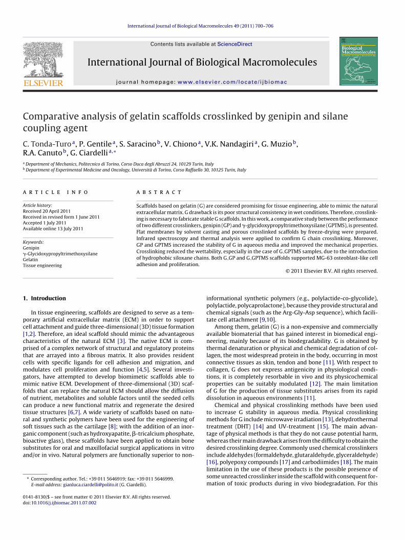
The G GP and G GPTMS freeze-dried scaffolds increased theirweight immediately after the immersion in PBS showing a highdegree of water uptake after only 1 h incubation in PBS (Fig. 1A).The swelling degree was then approximately stable up to 24 h incu-bation time, showing values of 992 ± 52% for G GP and 738 ± 82%for G GPTMS, respectively.
Dissolution tests were performed on crosslinked G samples,with the aim to study their stability in aqueous solution. G GP andG GPTMS sponges lost a weight of 1.7 ± 0.2% and 0.5 ± 0.1% respec-tively after one incubation day in PBS. Weight loss increased withtime reaching a value of 64 ± 2% for G GP and 59 ± 1% for G GPTMS
after two incubation weeks in PBS showing a faster dissolution afterone week compared to the first days (Fig. 1B).Swelling and dissolution tests were not performed on G sam-ples, as uncrosslinked scaffolds in contact with aqueous solution
C. Tonda-Turo et al. / International Journal of Biological Macromolecules 49 (2011) 700– 706 703
F ked p7 ation
lm
atddfib
3
GTtpudttsvotatca
c
pa
Fc
ig. 1. Swelling (A) and dissolution (B) behaviour of the uncrosslinked and crosslin.4 at 37 ◦C. Data are averaged on three measurements. Bars indicate standard devi
ost their integrity in a few hours; therefore it was not possible toeasure their swelling and dissolution degree.Compared to uncrosslinked porous samples, crosslinking by GP
nd GPTMS increased the G stability in aqueous media. Moreover,he G GPTMS samples showed a lower swelling and dissolutionegrees as compared to G GP scaffolds; this behaviour could beue to the decreased hydrophilicity of G GPTMS samples (as con-rmed by contact angle analysis at par. 3.6) reducing the interactionetween the polymeric chains and PBS.
.2. Mechanical characterization
Fig. 2 shows the stress–strain curves measured for G, G GP and GPTMS porous scaffolds by compression tests at 0–80% strain.he G, G GP and G GPTMS compression stress/strain curves showedhe typical trend of soft and porous materials [32]. Samples com-ressed at 0–80% strain did not show final fracture; rather, theynderwent densification. The obtained stress–strain curves can beivided into three zones with different mechanical behaviour: (i)he linear portion of the stress–strain curve at low strain is relativeo the elastic behaviour and its slope is the elastic modulus of thecaffolds (E); (ii) the region at intermediate strain is relative to theiscoelastic behaviour of samples or “collapse regime”; the slopef the curve in the collapse region is the collapse modulus (E*) andhe point of intersection of the regression lines for E and E* is char-cterized by collapse strength (�*) and collapse strain (ε*); (iii) thehird region at high strain is characteristic of the densification pro-ess: scaffolds are highly compressed and only low deformationsre allowed.
The values of E, E*, �*and ε* calculated from the stress–strain
urves are collected in Table 1.The increase of E value from 0.50 ± 0.18 MPa for G sam-les to 1.20 ± 0.29 MPa and 1.94 ± 0.25 MPa respectively for G GPnd G GPTMS scaffolds was a consequence of the material rein-
ig. 2. Stress–strain curves of the uncrosslinked and crosslinked porous scaffoldsompressed at a strain of 0–80%. The cross-head speed was 0.01 mm s−1.
orous scaffolds as a function of time. Measurements were carried out in PBS at pH(n = 5). *p < 0.05 and **p < 0.001 G GP, compared to G GPTMS.
forcement by crosslinking. On the other hand, plastic behaviourof samples was not affected by crosslinking since E* valueswere similar for crosslinked and uncrosslinked samples. The �*
value increased for G GPTMS (0.26 ± 0.02 MPa) as compared to G(0.16 ± 0.03 MPa) and G GP (0.16 ± 0.04 MPa) while ε* decreasedfrom 35.6 ± 2.5% for G to 30.6 ± 2.2% for G GP and 17.3 ± 2.2% forG GPTMS. This behaviour suggests that elastic deformability ofsamples decreased and their stiffness increased with crosslinking,particularly in the case of GPTMS.
The compression mechanical properties of crosslinked sampleswere found to be suitable for applications in the engineering ofsoft tissues such as cartilage [34]. In particular, G GPTMS scaffoldsare characterized by a compression Young’s modulus which is alsocomparable to the one of alveolar bone [35]. The addition of aninorganic component (such as hydroxyapatite, �-tricalcium phos-phate, bioactive glass) could reinforce crosslinked G improving themechanical properties to apply the scaffolds as substitutes for bonetissue engineering.
3.3. Differential scanning calorimetry (DSC)
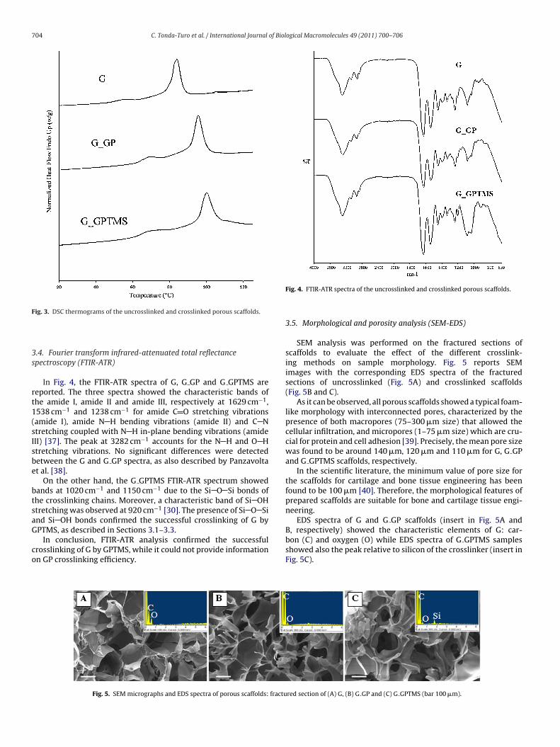
DSC analysis was performed to analyze the thermal behaviour ofmaterials as a function of crosslinking treatments (Fig. 3). Changesin G denaturation temperature (Td) can provide information onthe crosslinking strength: a rise in Td indicates an increase in theprotein network strength and, consequently, an increase in thecrosslinking degree [36].
Uncrosslinked G scaffold samples showed a Td of 84.1 ± 0.5 ◦C,while G GP and G GPTMS samples showed higher Td: 95.3 ± 1.8 ◦Cand 101.5 ± 1.2 ◦C, respectively. This finding suggests that thecrosslinking treatment increased the thermal stability of G helicesas shown by the shift of the Td to higher values, in agreementwith previous reports on crosslinked gelatin [36]. The higher Tdfor G GPTMS as compared to G GP could be attributed to a highercrosslinking degree for G GPTMS samples.
The denaturation enthalpy (�Hd) values measured for G, G GPand G GPTMS were 7.14 J/g, 5.98 J/g and 5.15 J/g, respectively.Crosslinking induced a decrease in the denaturation enthalpy,
caused by a reduction of hydrogen bonds, which break endother-mically, and a simultaneous increase in the extent of covalentcrosslinks, which break exothermically [36].Table 1Elastic modulus (E), collapse modulus (E*), collapse strength (�*) and strain (ε*),calculated from the corresponding stress–strain curves (average values ± standarddeviation).
E (MPa) E* (MPa) �* (MPa) ε* (%)
G 0.50 ± 0.18 0.14 ± 0.04 0.16 ± 0.03 35.60 ± 2.45G GP 1.20 ± 0.29 0.13 ± 0.04 0.16 ± 0.04 30.61 ± 2.24G GPTMS 1.94 ± 0.25 0.14 ± 0.03 0.26 ± 0.02 17.34 ± 2.21
704 C. Tonda-Turo et al. / International Journal of Biological Macromolecules 49 (2011) 700– 706
F
3s
rt1(sIsbe
btsaG
co
bon (C) and oxygen (O) while EDS spectra of G GPTMS samples
ig. 3. DSC thermograms of the uncrosslinked and crosslinked porous scaffolds.
.4. Fourier transform infrared-attenuated total reflectancepectroscopy (FTIR-ATR)
In Fig. 4, the FTIR-ATR spectra of G, G GP and G GPTMS areeported. The three spectra showed the characteristic bands ofhe amide I, amide II and amide III, respectively at 1629 cm−1,538 cm−1 and 1238 cm−1 for amide C O stretching vibrationsamide I), amide N H bending vibrations (amide II) and C Ntretching coupled with N H in-plane bending vibrations (amideII) [37]. The peak at 3282 cm−1 accounts for the N H and O Htretching vibrations. No significant differences were detectedetween the G and G GP spectra, as also described by Panzavoltat al. [38].
On the other hand, the G GPTMS FTIR-ATR spectrum showedands at 1020 cm−1 and 1150 cm−1 due to the Si O Si bonds ofhe crosslinking chains. Moreover, a characteristic band of Si OHtretching was observed at 920 cm−1 [30]. The presence of Si O Sind Si OH bonds confirmed the successful crosslinking of G byPTMS, as described in Sections 3.1–3.3.
In conclusion, FTIR-ATR analysis confirmed the successful
rosslinking of G by GPTMS, while it could not provide informationn GP crosslinking efficiency.Fig. 5. SEM micrographs and EDS spectra of porous scaffolds: fractu
Fig. 4. FTIR-ATR spectra of the uncrosslinked and crosslinked porous scaffolds.
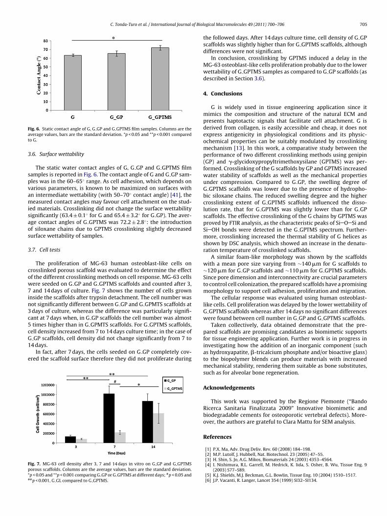
3.5. Morphological and porosity analysis (SEM-EDS)
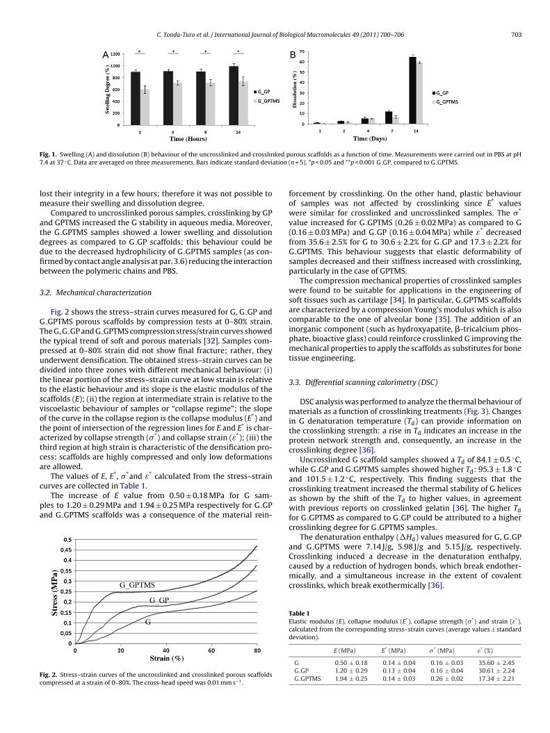
SEM analysis was performed on the fractured sections ofscaffolds to evaluate the effect of the different crosslink-ing methods on sample morphology. Fig. 5 reports SEMimages with the corresponding EDS spectra of the fracturedsections of uncrosslinked (Fig. 5A) and crosslinked scaffolds(Fig. 5B and C).
As it can be observed, all porous scaffolds showed a typical foam-like morphology with interconnected pores, characterized by thepresence of both macropores (75–300 �m size) that allowed thecellular infiltration, and micropores (1–75 �m size) which are cru-cial for protein and cell adhesion [39]. Precisely, the mean pore sizewas found to be around 140 �m, 120 �m and 110 �m for G, G GPand G GPTMS scaffolds, respectively.
In the scientific literature, the minimum value of pore size forthe scaffolds for cartilage and bone tissue engineering has beenfound to be 100 �m [40]. Therefore, the morphological features ofprepared scaffolds are suitable for bone and cartilage tissue engi-neering.
EDS spectra of G and G GP scaffolds (insert in Fig. 5A andB, respectively) showed the characteristic elements of G: car-
showed also the peak relative to silicon of the crosslinker (insert inFig. 5C).
red section of (A) G, (B) G GP and (C) G GPTMS (bar 100 �m).
C. Tonda-Turo et al. / International Journal of Biol
Fat
3
spvamisaos
3
cow7in3c5cG1
e
Fp*#
ig. 6. Static contact angle of G, G GP and G GPTMS film samples. Columns are theverage values, bars are the standard deviation. *p < 0.05 and **p < 0.001 comparedo G.
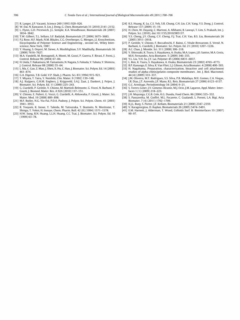
.6. Surface wettability
The static water contact angles of G, G GP and G GPTMS filmamples is reported in Fig. 6. The contact angle of G and G GP sam-les was in the 60–65◦ range. As cell adhesion, which depends onarious parameters, is known to be maximized on surfaces withn intermediate wettability (with 50–70◦ contact angle) [41], theeasured contact angles may favour cell attachment on the stud-
ed materials. Crosslinking did not change the surface wettabilityignificantly (63.4 ± 0.1◦ for G and 65.4 ± 3.2◦ for G GP). The aver-ge contact angles of G GPTMS was 72.2 ± 2.8◦: the introductionf siloxane chains due to GPTMS crosslinking slightly decreasedurface wettability of samples.
.7. Cell tests
The proliferation of MG-63 human osteoblast-like cells onrosslinked porous scaffold was evaluated to determine the effectf the different crosslinking methods on cell response. MG-63 cellsere seeded on G GP and G GPTMS scaffolds and counted after 3,
and 14 days of culture. Fig. 7 shows the number of cells grownnside the scaffolds after trypsin detachment. The cell number wasot significantly different between G GP and G GPMTS scaffolds at
days of culture, whereas the difference was particularly signifi-ant at 7 days when, in G GP scaffolds the cell number was almost
times higher than in G GPMTS scaffolds. For G GPTMS scaffolds,ell density increased from 7 to 14 days culture time; in the case of
GP scaffolds, cell density did not change significantly from 7 to4 days.
In fact, after 7 days, the cells seeded on G GP completely cov-red the scaffold surface therefore they did not proliferate during
ig. 7. MG-63 cell density after 3, 7 and 14 days in vitro on G GP and G GPTMSorous scaffolds. Columns are the average values, bars are the standard deviation.p < 0.05 and **p < 0.001 comparing G GP or G GPTMS at different days; #p < 0.05 and#p < 0.001, G GL compared to G GPTMS.
ogical Macromolecules 49 (2011) 700– 706 705
the followed days. After 14 days culture time, cell density of G GPscaffolds was slightly higher than for G GPTMS scaffolds, althoughdifferences were not significant.
In conclusion, crosslinking by GPTMS induced a delay in theMG-63 osteoblast-like cells proliferation probably due to the lowerwettability of G GPTMS samples as compared to G GP scaffolds (asdescribed in Section 3.6).
4. Conclusions
G is widely used in tissue engineering application since itmimics the composition and structure of the natural ECM andpresents haptotactic signals that facilitate cell attachment. G isderived from collagen, is easily accessible and cheap, it does notexpress antigenicity in physiological conditions and its physic-ochemical properties can be suitably modulated by crosslinkingmechanism [13]. In this work, a comparative study between theperformance of two different crosslinking methods using genipin(GP) and �-glycidoxypropyltrimethoxysilane (GPTMS) was per-formed. Crosslinking of the G scaffolds by GP and GPTMS increasedwater stability of scaffolds as well as the mechanical propertiesunder compression. Compared to G GP, the swelling degree ofG GPTMS scaffolds was lower due to the presence of hydropho-bic siloxane chains. The reduced swelling degree and the highercrosslinking extent of G GPTMS scaffolds influenced the disso-lution rate, that for G GPTMS was slightly lower than for G GPscaffolds. The effective crosslinking of the G chains by GPTMS wasproved by FTIR analysis, as the characteristic peaks of Si O Si andSi OH bonds were detected in the G GPTMS spectrum. Further-more, crosslinking increased the thermal stability of G helices asshown by DSC analysis, which showed an increase in the denatu-ration temperature of crosslinked scaffolds.
A similar foam-like morphology was shown by the scaffoldswith a mean pore size varying from ∼140 �m for G scaffolds to∼120 �m for G GP scaffolds and ∼110 �m for G GPTMS scaffolds.Since pore dimension and interconnectivity are crucial parametersto control cell colonization, the prepared scaffolds have a promisingmorphology to support cell adhesion, proliferation and migration.
The cellular response was evaluated using human osteoblast-like cells. Cell proliferation was delayed by the lower wettability ofG GPTMS scaffolds whereas after 14 days no significant differenceswere found between cell number in G GP and G GPTMS scaffolds.
Taken collectively, data obtained demonstrate that the pre-pared scaffolds are promising candidates as biomimetic supportsfor tissue engineering application. Further work is in progress ininvestigating how the addition of an inorganic component (suchas hydroxyapatite, �-tricalcium phosphate and/or bioactive glass)to the biopolymer blends can produce materials with increasedmechanical stability, rendering them suitable as bone substitutes,such as for alveolar bone regeneration.
Acknowledgements
This work was supported by the Regione Piemonte (“BandoRicerca Sanitaria Finalizzata 2009” Innovative biomimetic andbiodegradable cements for osteoporotic vertebral defects). More-over, the authors are grateful to Clara Mattu for SEM analysis.
References
[1] P.X. Ma, Adv. Drug Deliv. Rev. 60 (2008) 184–198.[2] M.P. Lutolf, J. Hubbell, Nat. Biotechnol. 23 (2005) 47–55.
[3] H. Shin, S. Jo, A.G. Mikos, Biomaterials 24 (2003) 4353–4564.[4] I. Nishimura, R.L. Garrell, M. Hedrick, K. Iida, S. Osher, B. Wu, Tissue Eng. 9(2003) S77–S89.[5] K.J. Shields, M.J. Beckman, G.L. Bowlin, Tissue Eng. 10 (2004) 1510–1517.[6] J.P. Vacanti, R. Langer, Lancet 354 (1999) SI32–SI134.
7 of Biol
[[
[
[
[
[
[[[
[
[
[
[
[
[
[
[
[
[[
[[[[
[
[[
[[
Biomater. 7 (4) (2011) 1702–1709.
06 C. Tonda-Turo et al. / International Journal
[7] R. Langer, J.P. Vacanti, Science 260 (1993) 920–926.[8] W. Dai, N. Kawazoe, X. Lin, J. Dong, G. Chen, Biomaterials 31 (2010) 2141–2152.[9] L. Flynn, G.D. Prestwich, J.L. Semple, K.A. Woodhouse, Biomaterials 28 (2007)
3834–3842.10] T.W. Gilbert, T.L. Sellaro, S.F. Badylak, Biomaterials 27 (2006) 3675–3683.11] P.J. Rose, H.F. Mark, N.M. Bikales, C.G. Overberger, G. Menges, J.I. Kroschwitzm,
Encyclopedia of Polymer Science and Engineering , second ed., Wiley Inter-science, New York, 1987.
12] Y. Huang, S. Onyeri, M. Siewe, A. Moshfeghian, S.V. Madihally, Biomaterials 36(2000) 7616–7627.
13] M.A. Vandelli, M. Romagnoli, A. Monti, M. Gozzi, P. Guerra, F. Rivasi, F. Forni, J.Control. Release 96 (2004) 67–84.
14] H. Ueda, T. Nakamura, M. Yamamoto, N. Nagata, S. Fukuda, Y. Tabata, Y. Shimizu,J. Control. Release 88 (2000) 55–64.
15] L. Ma, C. Gao, Z. Mao, J. Shen, X. Hu, C. Han, J. Biomater. Sci. Polym. Ed. 14 (2003)861–874.
16] G.A. Digenis, T.B. Gold, V.P. Shah, J. Pharm. Sci. 83 (1994) 915–921.17] T. Miyata, T. Taira, Y. Noishiki, Clin. Mater. 9 (1992) 139–148.18] A.J. Kuijpers, G.H.M. Engbers, J. Krijgsveld, S.A.J. Zaat, J. Dankert, J. Feijen, J.
Biomater. Sci. Polym. Ed. 11 (2000) 225–243.19] G. Ciardelli, P. Gentile, V. Chiono, M. Mattioli-Belmonte, G. Vozzi, N. Barbani, P.
Giusti, J. Biomed. Mater. Res. A 92A (2010) 137–151.20] V. Chiono, E. Pulieri, G. Vozzi, G. Ciardelli, A. Ahluwalia, P. Giusti, J. Mater. Sci.
Mater. Med. 19 (2008) 889–898.21] M.F. Butler, N.G. Yiu-Fai, P.D.A. Pudney, J. Polym. Sci. Polym. Chem. 41 (2003)
3941–3953.22] R. Touyam, K. Inoue, Y. Takeda, M. Yatsuzuka, T. Ikumoto, N. Moritome, T.
Shingu, T. Yokoi, H. Inouye, Chem. Pharm. Bull. 42 (8) (1994) 1571–1578.23] H.W. Sung, R.N. Huang, L.L.H. Huang, C.C. Tsai, J. Biomater. Sci. Polym. Ed. 10
(1999) 63–78.
[[[
ogical Macromolecules 49 (2011) 700– 706
24] K.S. Huang, K. Lu, C.S. Yeh, S.R. Chung, C.H. Lin, C.H. Yang, Y.S. Dong, J. Control.Release 137 (2009) 15–19.
25] H. Chen, W. Ouyang, C. Martoni, A. Afkhami, B. Lawuyi, T. Lim, S. Prakash, Int. J.Polym. Sci. (2010), doi:10.1155/2010/985137.
26] Y.S. Cheng, J.Y. Chang, C.Y. Cheng, F.J. Tsai, C.H. Yao, B.S. Liu, Biomaterials 26(2005) 3911–3918.
27] P. Gentile, V. Chiono, F. Boccafoschi, F. Baino, C. Vitale-Brovarone, E. Vernè, N.Barbani, G. Ciardelli, J. Biomater. Sci. Polym. Ed. 21 (2010) 1207–1226.
28] A.C. Chao, J. Membr. Sci. 311 (2008) 306–318.29] Y. Shirosaki, K. Tsuru, S. Hayakawa, A. Osaka, M.A. Lopes, J.D. Santos, M.A. Costa,
M.H. Fernandes, Acta Biomater. 5 (2009) 346–355.30] Y.L. Liu, Y.H. Su, J.Y. Lai, Polymer 45 (2004) 6831–6837.31] L. Ren, K. Tsuru, S. Hayakawa, A. Osaka, Biomaterials 23 (2002) 4765–4773.32] B.P. Kanungo, E. Silva, K. Van Vliet, L.J. Gibson, Acta Biomater. 4 (2008) 490–503.33] H. Nagahama, Preparation, characterization, bioactive and cell attachment
studies of alpha-chitin/gelatin composite membranes , Int. J. Biol. Macromol.44 (4) (2009) 333–337.
34] J.M. Oliveira, M.T. Rodrigues, S.S. Silva, P.B. Malafaya, M.E. Gomes, C.A. Viegas,I.R. Dias, J.T. Azevedo, J.F. Mano, R.L. Reis, Biomaterials 27 (2006) 6123–6137.
35] G.C. Armitage, Periodontology 34 (2004) 9–21.36] S. Torres-Giner, J.V. Gimeno-Alcaniz, M.J. Ocio, J.M. Lagaron, Appl. Mater. Inter-
faces 1 (1) (2009) 218–223.37] J.H. Muyonga, C.G.B. Cole, K.G. Duodu, Food Chem. 86 (2004) 325–332.38] S. Panzavolta, M. Gioffrè, M.L. Focarete, C. Gualandi, L. Foroni, L.A. Bigi, Acta
39] K.J.L. Burg, S. Porter, J.F. Kellam, Biomaterials 21 (2000) 2347–2359.40] V. Karageorgiou, D. Kaplan, Biomaterials 26 (2005) 5474–5491.41] E.M. Harnett, J. Alderman, T. Wood, Colloids Surf. B: Biointerfaces 55 (2007)
90–97.


