Embed Size (px)
Citation preview

J. Physiol. (1986), 371, pp. 219-237 219With 10 text-figures
Printed in Great Britain
CHARACTERIZATION OF THE ACETYLCHOLINE-INDUCEDPOTASSIUM CURRENT IN RABBIT CARDIAC PURKINJE FIBRES
BY E. CARMELIET AND K. MUBAGWAFrom the Laboratory of Physiology, University of Leuven, Campus Gasthuisberg,
B-3000 Leuven, Belgium
(Received 22 March 1985)
SUMMARY
1. Acetylcholine (ACh) induces a K+ current in rabbit cardiac Purkinje fibres. Thequestion was studied whether ACh produces this effect by modifying the propertiesof K+ channels pre-existing in the absence of the neurotransmitter or whether itinduces the formation of a different type of K+ channels.
2. The relaxation properties of the ACh-induced current and its blockade by Cs+and Ba2+ have been investigated using voltage clamp.
3. During hyperpolarizing or depolarizing voltage pulses of moderate amplitude,the ACh-induced current is time independent. For large voltage pulses, time-dependentchanges of the ACh-induced current are observed. These latter changes can beexplained by intercellular K+ accumulation/depletion phenomena or by the effectsof ACh on time-dependent currents (e.g. the late outward current, ix).
4. Cs+ and Ba2+ block the ACh-induced current. The block produced by 20 mM-Cs+is instantaneous and increases with hyperpolarization, i.e. it is voltage dependent.The block produced by Ba2+ at high concentrations (> 1 mM) is also instantaneousbut complete at all potentials studied, and thus voltage independent. At theseconcentrations, either ion also blocks the background inward rectifier (iKi) currentin a similar way.
5. Low [Ba2+] (< 0-1 mM) cause a block of the ACh-induced current which isinstantaneous and little voltage dependent. The block of iK1 in contrast is time andvoltage dependent for the same concentrations. These results indicate that theACh-induced K+ current is different from the background iK1 current.
INTRODUCTION
In the preceding paper (Carmeliet & Mubagwa, 1986 a) it is shown that acetylcholine(ACh) increases a K+ conductance in rabbit cardiac Purkinje fibres. The question thatis immediately raised is whether ACh produces such an effect by modifying theproperties of K+ channels pre-existing in the absence of the neurotransmitter, orwhether it induces the formation of a new type of K+ channel.A major characteristic of the ACh-induced current is inward-going rectification:
more inward current is obtained by a given hyperpolarization than outward currentobtained by a depolarization of the same amplitude. Since the inward rectifier
) by guest on July 10, 2011jp.physoc.orgDownloaded from J Physiol (

E. CARMELIET AND K. MUBAGWA
constitutes an important fraction of the cardiac background current, among pre-existing K+ currents, the most plausible candidate to be modified by ACh is thebackground inward-rectifying component iKi* Results obtained in frog atrialpreparations seem to be in favour of the hypothesis that ACh increases a K+ currentindistinguishable from i1K1: both currents appear time independent (Garnier, Nargeot,Ojeda & Rougier, 1978) and they show similar sensitivities to the blockade producedby Cs+ (Ojeda, Rougier & Tourneur, 1981; Argibay, Dutey, Ildefonse, Ojeda, Rougier& Tourneur, 1983). On the contrary, experiments in sino-atrial and atrioventricularpreparations suggest that the K+ channels activated by ACh are different from iKichannels. In both preparations, ACh produces a current which is time dependentfollowing a voltage pulse, and in sino-atrial tissue ACh gives rise to a Lorentziancomponent with voltage-dependent corner frequency in the membrane noisespectrum (Noma & Trautwein, 1978; Noma, Peper & Trautwein, 1979). Single-channel recordings in the same preparations support the hypothesis thatACh-sensitive channels are different from iK1 channels, since the former channels havea mean open life time much shorter than that of iK1 channels (Sakmann, Noma &Trautwein, 1983).The following experiments were carried out to test whether the ACh-induced
current in rabbit cardiac Purkinje fibres may be distinguished from iK1. For thispurpose, we investigated the relaxation properties and the sensitivity to Cs+ and Ba2+of the ACh-induced current.
METHODS
The experiments were carried out on isolated cardiac Purkinje fibres and the two-micro-electrodevoltage-clamp technique used in the present experiments is the same as the one used previously(Carmeliet & Ramon, 1980; see also Carmeliet & Mubagwa, 1986a).For the experiments in which relaxation of the ACh-induced current was investigated, voltage
pulses of 1-5 s to a given level were given repetitively (every 10-20 s). After a sufficient numberof pulses in normal Tyrode solution, the preparation was exposed to ACh-containing Tyrodesolution. In ACh, only currents recorded during pulses given after 10 min in the presence of theagonist were used. After enough recordings in ACh, a wash-out period was observed. The currentsrecorded after 10 min of ACh wash-out were used as a second control.For the experiments in which the effects of Cs+ or Ba2+ on the ACh-induced current were tested,
membrane currents were first recorded in normal Tyrode solution, before and after application ofACh. The same measurements were repeated in the presence of 20 mM-Cs+ or in the presence ofvarious Ba2+ concentrations, again before and after application of ACh.The current signals to be used for relaxation analysis were recorded on a magnetic tape
(Hewlett-Packard 3964A). They were later sampled at 500 Hz on a computer (PDP-1 1). Averagesof three to ten currents in the same conditions were made. The average current in normal Tyrodesolution was subtracted from the average current in ACh to give the ACh-induced current.
RESULTS
Absence of relaxation of the ACh-induced current
Fig. 1 illustrates the results ofan experiment in which relaxation ofthe ACh-inducedcurrent in rabbit Purkinje fibres was tested. In this experiment tetrodotoxin (TTX,10-5 M) was present in the perfusing solution to reduce Na+ currents. The holdingpotential was also kept negative to the activation potential of other time-dependent
220
) by guest on July 10, 2011jp.physoc.orgDownloaded from J Physiol (

ACETYLCHOLINE AND PURKINJE FIBRES 221
currents. Average currents in the absence and in the presence ofACh are superimposed(Fig. 1, left). For voltage pulses of moderate amplitude, i.e. to potentials between-40 and -95 mV, ACh produces a parallel shift of membrane currents during thepulse. This is what is expected if ACh increases a time-independent current. Thedifference current (Fig. 1, right) changes instantaneously and remains relatively
20 5~~~nA nAio ~ ' 2 n
0x-I- 0f10 ~~~~~~~~~~~~~5
nA [_ jnA
O- 00
-30E 40 5 s
Fig. 1. Effect of ACh (2 x 10⁶ m) on membrane currents. The holding and pulse potentialsare indicated in the lower part of the Figure. Left: superimposed current traces beforeand during ACh exposure (indicated by an open circle). Each current trace is the averageof five original current recordings. Right: ACh-induced current, obtained by subtractingthe current in the absence from the current in the presence ofACh. The current calibrationsare the same in the second and in the lower rows. The horizontal lines give zero currentlevels. Tetrodotoxin (1O-5 m) was present.
constant with time. On return to the holding potential, it also instantaneouslyrecovers to the pre-pulse value. The results suggest that in this range of potentials,the ACh-induced current is time independent.
During depolarizations to potentials beyond -40 mV or during hyperpolarizationsto potentials more negative than -95 mV, the ACh-induced current frequently shows
) by guest on July 10, 2011jp.physoc.orgDownloaded from J Physiol (

222 E. CARMELIET AND K. MUBAGWA
time dependence. For hyperpolarizations beyond -95 mV, the amplitude of theinward ACh-induced current usually decreases with time. Only in exceptional casesdoes the ACh-induced current show an initial rapid increase. Such an increasingACh-induced current is shown in Fig. 2. The ACh-induced current during hyper-polarization to - 130 mV shows an initial increase followed by a slow decrease. Theearly increase in current during hyperpolarization is also obtained at -100 and
[ j
0 0~~~
nA nA-50 _ -, -20
_o
-75 -o0_mV -110-130
Fig. 2. Effect of ACh (2 x 10⁻⁶ M) on membrane currents during large hyperpolarizations.Same preparation and same conventions as in the preceding Figure. Notice time-dependentchanges of the ACh-induced current. The current calibrations are the same in the secondand in the third rows.
-110 mV and resembles the relaxation observed in sino-atrial cells (Sakmann et al.1983). Such an increase in ACh-induced current during hyperpolarization is consistentwith an increase of open probability of the ACh-sensitive channels. The secondarydecrease of ACh-induced current observed at - 130 mV may result either from aninactivation of the ACh-sensitive channels or from a depletion of K+ in theintercellular spaces. Although rabbit Purkinje fibres usually possess wide intercellularspaces, it is possible that with a very large influx of K+ such as the one producedby the hyperpolarizing step to - 130 mV, some depletion occurs. On return to theholding potential, the initial current is usually higher than the control, i.e. an outward
) by guest on July 10, 2011jp.physoc.orgDownloaded from J Physiol (
ACETYLCHOLINE AND PURKINJE FIBRES
tail is obtained. Outward tails probably result from the deactivation of ACh-sensitivechannels, but they may result from intercellular K+ depletion as well.For large depolarizations, ACh-induced outward currents decreasing with time as
well as currents increasing with time could be observed. The time-dependent increase(see Fig. 1) might be related to a K+ accumulation process. Changes in time-dependentcurrents might also contribute to the effect of ACh. A decrease of the late outwardcurrent, ix (see Carmeliet & Mubagwa, 1986a), would tend to superimpose on the effectof ACh on other currents. This will result in a total ACh-induced current decreasingwith time during depolarizations to potentials positive to -50 mV and in an inwardtail on return to the holding potential.From the present results, it can be concluded that, except in rare cases, the
ACh-induced K+ current in rabbit Purkinje fibres does not show relaxation. The timedependence observed during very large voltage pulses can be accounted for by K+accumulation/depletion phenomena or by a change of time-dependent currents byACh.
Blockade of the ACh-induced current by C8+ and Ba2+The permeation of K+ through inward-rectifying channels is blocked by many
alkali or earth-alkali cations in the various tissues where these channels have beenidentified (Hagiwara, Miyazaki & Rosenthal, 1976; Isenberg, 1976; Gay & Stanfield,1977; Standen & Stanfield, 1978, 1980; Carmeliet, 1979; DiFrancesco, 1981; Van derHeyden, Vereecke & Carmeliet, 1983). In rabbit cardiac Purkinje fibres, the magnitudeof the hyperpolarization produced by ACh is reduced by Cs+ or Ba2+ (Mubagwa &Carmeliet, 1983), suggesting that these ions also block the ACh-sensitive channels.In the following results, the pattern of block of the ACh-induced current is comparedto the block produced on iK1. The effects ofCs+ and Ba2+ on the ACh-sensitive channelwere obtained by comparing the ACh-induced currents in the absence and in thepresence of these ions.
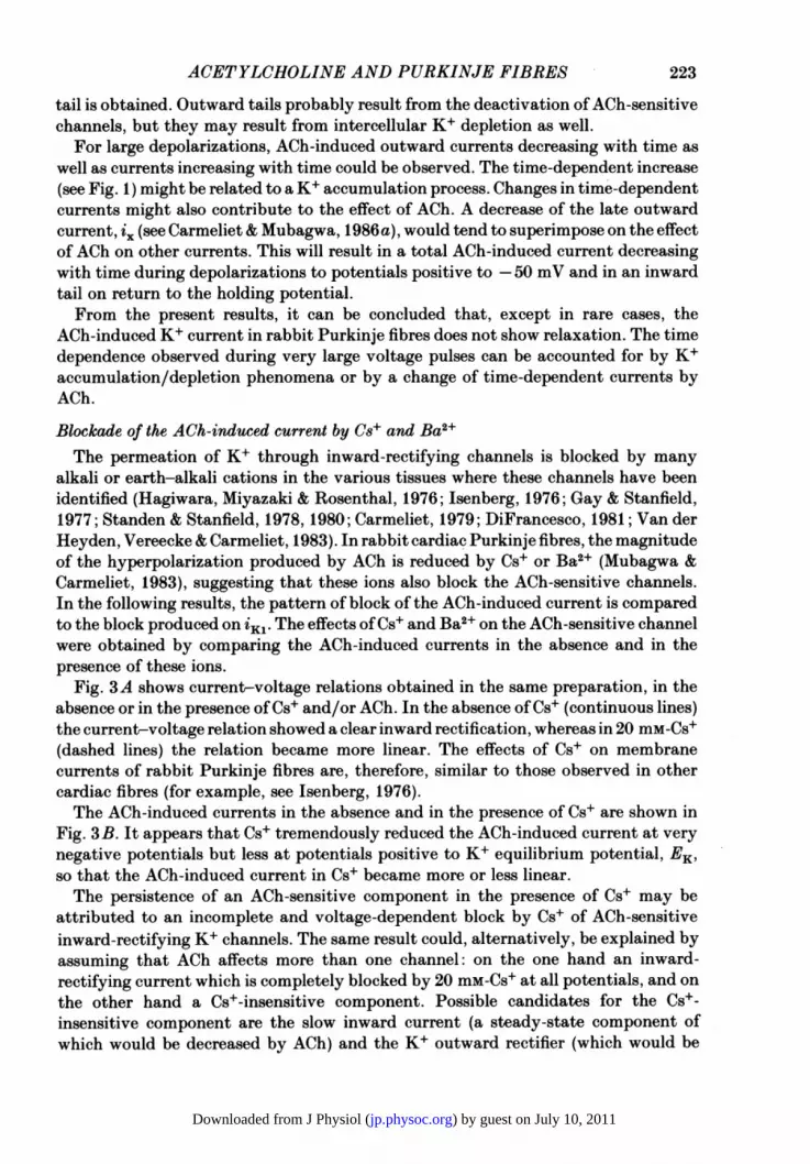
Fig. 3A shows current-voltage relations obtained in the same preparation, in theabsence or in the presence ofCs+ and/or ACh. In the absence of Cs+ (continuous lines)the current-voltage relation showed a clear inward rectification, whereas in 20 mM-Cs+(dashed lines) the relation became more linear. The effects of Cs+ on membranecurrents of rabbit Purkinje fibres are, therefore, similar to those observed in othercardiac fibres (for example, see Isenberg, 1976).The ACh-induced currents in the absence and in the presence of Cs+ are shown in
Fig. 3B. It appears that Cs+ tremendously reduced the ACh-induced current at verynegative potentials but less at potentials positive to K+ equilibrium potential, EK,so that the ACh-induced current in Cs+ became more or less linear.The persistence of an ACh-sensitive component in the presence of Cs+ may be
attributed to an incomplete and voltage-dependent block by Cs+ of ACh-sensitiveinward-rectifying K+ channels. The same result could, alternatively, be explained byassuming that ACh affects more than one channel: on the one hand an inward-rectifying current which is completely blocked by 20 mM-Cs+ at all potentials, and onthe other hand a Cs+-insensitive component. Possible candidates for the Cs+-insensitive component are the slow inward current (a steady-state component ofwhich would be decreased by ACh) and the K+ outward rectifier (which would be
223
) by guest on July 10, 2011jp.physoc.orgDownloaded from J Physiol (
224 E. CARMELIET AND K. MUBAGWA
A
mV-100 -80
x
B
mV-100
0a
0 -.
z 0
-20
0
10
0
-10nA
-20
-30
10
5
0
nA
-5
-10
-15
Fig. 3. Effect of 20 mM-Cs+ on the ACh-induced current. A, the influence ofCs+ was studiedby measuring the change in 'steady-state' membrane current produced by ACh in thepresence (dashed line) and after wash-out (continuous line) of Cs+. (@): normal Tyrodesolution (NTS), before exposure of ACh. ( x ): NTS +ACh. (U): Cs+, before exposure ofACh. (+): Cs++Ach. (s): Cs+, after ACh wash-out. B, ACh-sensitive currents in theabsence (@) and in the presence (EI) of Cs+. Holding potential: -60 mV.
increased by ACh). In Carmeliet & Mubagwa (1986a), it was shown that ACh doesnot change the slow inward current in the absence of catecholamines. It is thereforeunlikely that a steady component of the slow inward current accounts for the Cs+-insensitive ACh-induced component. The possibility that the ACh-sensitive current
) by guest on July 10, 2011jp.physoc.orgDownloaded from J Physiol (

ACETYLCHOLINE AND PURKINJE FIBRES
persisting in Cs+ is a K+ outward rectifier also seems unlikely, since the ACh-inducedcurrent in normal Tyrode solution is usually flat at potentials very positive to EK(see Figs. 2B and 4B in Carmeliet & Mubagwa, 1986a).Due to continuous changes in currents probably by Cs+ loading into the cells, the
ACh-induced current in the presence of Cs+ could not be precisely measured. Thisis especially true at very negative potentials, where the magnitude ofthe ACh-induced
20
X 10
mV-100 -80 -60 -40 / -20
~I 0
x/°x "I" ° nA
X ~~~~~-10
/~/-20
im~~~~~~~~~~~~~~iV"m J LJ I
ACh -30
Fig. 4. Effect of ACh on membrane currents in the presence of 20 mM-Cs+. Currents weremeasured at the end of three successive voltage pulses of equal magnitude. During thefirst (@) and the third (0) pulses, no ACh was applied. During the second pulse ( x ), AChwas applied for 5 s by pressure from an ACh (0-1 M)-filled electrode. Inset: originalrecordings showing the change in membrane current following the application ofACh. Vm,pulse potential. im' membrane clamp current.
current became too small. In order to verify whether a significant Cs+-insensitiveACh-induced component persists at these negative potentials, the effect of ACh wasalso studied by pressure application of the drug on fibres clamped at differentpotentials in the presence of Cs+. In the experiment of Fig. 4, the membrane potentialwas clamped at a given level for 15 s, and the current was measured at the end ofthe voltage pulse. Each voltage pulse was followed by a second pulse during whichACh was administered for 5 s. The application of ACh started 5 s after the beginningofthe second pulse (Fig. 4, inset). For voltage pulses to potentials positive to -60 mV,ACh application resulted in current changes in the outward direction, whereas forpulses to potentials negative to -60 mV, ACh produced no effect. This means thatno reversal for the ACh-sensitive current in Cs+ was found in this experiment. It ismore likely therefore that the effects of ACh which persist in the presence of Cs+ are5
H 7
225
8 PHY 371
) by guest on July 10, 2011jp.physoc.orgDownloaded from J Physiol (
E. CARMELIET AND K. MUBAGWA
due to a less efficient Cs+ block of the ACh-induced inward rectifier than to thepresence of an ACh-sensitive outward rectifier component which is not blocked byCs+. In this last case, a reversal of the ACh-induced current in Cs+ should be present.The results suggest that Cs+ blocks the ACh-sensitive channels and that this blockadeby Cs+ is strongly voltage dependent, being more pronounced with hyperpolarization.
xx 10
mV-100 -80 -60 -40 x
_I L.-..I / / I
AoX\ /8/ U
,t~~~-'i n4 Ao-10
-20
-30
Fig. 5. Effect of5 mM-Ba2+ on theACh-induced current. The influence of Ba2+ was obtainedby studying the effects of ACh (2 x 10⁻⁶ M) on steady-state currents in the absence(continuous line) or in the presence (dashed line) of Ba2+. (0): normal Tyrode solution(NTS), before exposure to ACh. (x ): NTS+ACh. (0): NTS, after ACh wash-out: (U):Ba2+, before exposure to ACh. (+): Ba2+ + ACh. (0I): Ba2+, after ACh wash-out. Holdingpotential: -79 mV.
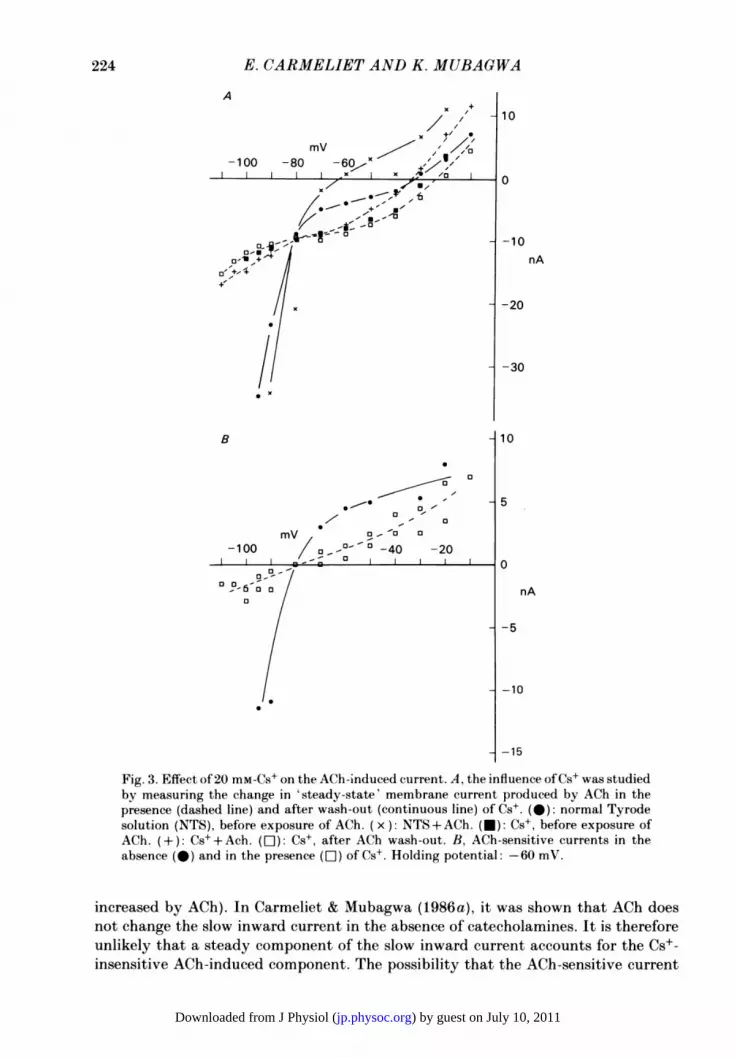
Similar results have been obtained in frog atrium and were taken as evidence in favourofthe hypothesis that ACh modulates the properties of iKl channels (Ojeda et al. 1981;Argibay et al. 1983). Such results can, however, be obtained if ACh increases aninward-rectifying current which is different from iKi but shows a similar sensitivityfor Cs+ (see below).
Other blockers of inward-rectifying channels were tested. In the experimentillustrated in Fig. 5, the effects of ACh in the absence (continuous lines) or in thepresence of 5 mM-Ba2+ (dashed lines) are compared. At the concentration used, Ba2+produced a large reduction in the current flowing during hyperpolarizing pulses.Positive to -60 mV, the current-voltage relation in Ba2+ is markedly shifted in theinward direction, indicating that apart from blocking outward K+ currents, this ionalso increases the slow inward current (Osterrieder, Yang & Trautwein, 1982; Cavalie,Ochi, Pelzer & Trautwein, 1983). Addition ofACh (2 x 10⁻⁶ M; crosses) in the presence
226
) by guest on July 10, 2011jp.physoc.orgDownloaded from J Physiol (

ACETYLCHOLINE AND PURKINJE FIBRES 227
A
EH -80 [Ba21]mV (pM)
0-90
nA
-20[ -95 0-20 _
B _1
-100
-105.tt ^^ 05s
.0~~~~~~
A 0.01
-120 -100 -80 -60
1 S m (mV)
C [Ba2+] (MM) - 20* -* 0-01
mV-1 20 -1 00 -80 -60
^/1/ | _ ~~-20
-60
Fig. 6. Effect of Ba2+ on membrane currents. A, inward currents in the absence (upperpanel) and in the presence (bottom panel) of 10-5 M-Ba2+ during hyperpolarizations from-80 mV. The pulse potentials are indicated near the current traces and are the same incontrol as well as in Ba2+, except for the lowest current trace, obtained at - 105 mV inBa2+. B, time constants (r) of decay in membrane current during hyperpolarization in thepresence of 5 x 10⁻⁶ M-Ba2+. Different symbols correspond to different preparations. Filledsymbols (-, U, *): 54 mM-K+ Tyrode solution. Open symbols (O, A): 108 mM-K+Tyrode solution. C, steady-state current-voltage relations in the absence of Ba2+ (@), in10-5 M-Ba2+ (A) and in 2 x 10⁻³ M-Ba2+. (a). Instantaneous currents (A) in 10-5 M. Thearrows show the direction of change in current during hyperpolarizing pulses in10- M-Ba2+. Holding potential: -80 mV.
8-2
) by guest on July 10, 2011jp.physoc.orgDownloaded from J Physiol (
E. CARMELIET AND K. MUBAGWA
of Ba2+ did not produce any effect on membrane currents in the whole range ofpotentials studied, while there was a clear effect of ACh before Ba2+ application. Thelack of ACh effect even at potentials positive to -50 mV further supports the ideathat ACh does not change i.1 in the absence of catecholamines (see Carmeliet &Mubagwa, 1986a).
In one experiment, Sr2+, another inward rectifier blocker (Ohmori, 1978; Standen& Stanfield, 1978) also decreased the ACh-induced current, although it seemed lesspotent than Cs+ or Ba2+. Its effect was also voltage dependent (not shown).
Separation of the ACh-sensitive current from iK1In the experiments described above, the effects of ACh were examined during
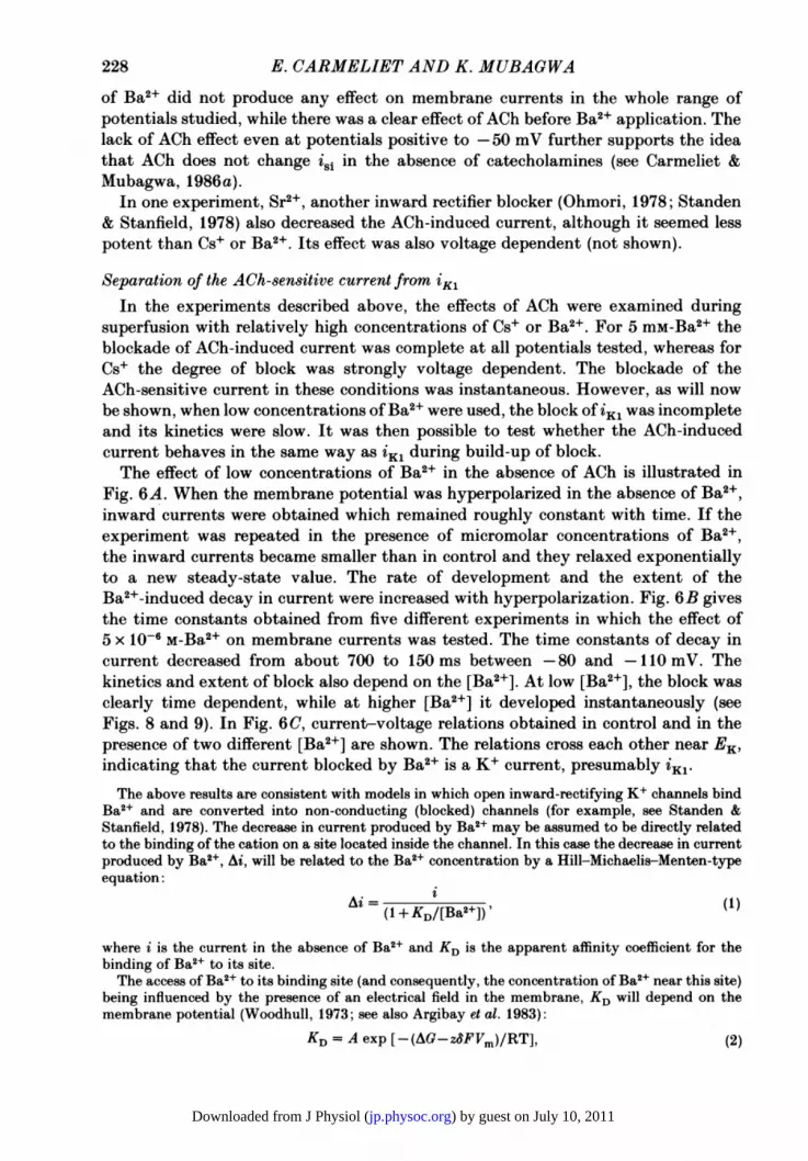
superfusion with relatively high concentrations of Cs+ or Ba2+. For 5 mM-Ba2+ theblockade of ACh-induced current was complete at all potentials tested, whereas forCs+ the degree of block was strongly voltage dependent. The blockade of theACh-sensitive current in these conditions was instantaneous. However, as will nowbe shown, when low concentrations of Ba2+ were used, the block of iK1 was incompleteand its kinetics were slow. It was then possible to test whether the ACh-inducedcurrent behaves in the same way as 'K1 during build-up of block.The effect of low concentrations of Ba2+ in the absence of ACh is illustrated in
Fig. 6A. When the membrane potential was hyperpolarized in the absence of Ba2+,inward currents were obtained which remained roughly constant with time. If theexperiment was repeated in the presence of micromolar concentrations of Ba2+,the inward currents became smaller than in control and they relaxed exponentiallyto a new steady-state value. The rate of development and the extent of theBa2+-induced decay in current were increased with hyperpolarization. Fig. 6B givesthe time constants obtained from five different experiments in which the effect of5 x 10-6 M-Ba2+ on membrane currents was tested. The time constants of decay incurrent decreased from about 700 to 150 ms between -80 and -110 mV. Thekinetics and extent of block also depend on the [Ba2+]. At low [Ba2+], the block wasclearly time dependent, while at higher [Ba2+] it developed instantaneously (seeFigs. 8 and 9). In Fig. 6C, current-voltage relations obtained in control and in thepresence of two different [Ba2+] are shown. The relations cross each other near EK,indicating that the current blocked by Ba2+ is a K+ current, presumably iK1.The above results are consistent with models in which open inward-rectifying K+ channels bind
Ba2+ and are converted into non-conducting (blocked) channels (for example, see Standen &Stanfield, 1978). The decrease in current produced by Ba2+ may be assumed to be directly relatedto the binding of the cation on a site located inside the channel. In this case the decrease in currentproduced by Ba2+, Ai, will be related to the Ba2+ concentration by a Hill-Michaelis-Menten-typeequation:
(1 +KD/[Ba2+])' (1)
where i is the current in the absence of Ba2+ and KD is the apparent affinity coefficient for thebinding of Ba2+ to its site.The access of Ba2+ to its binding site (and consequently, the concentration of Ba2+ near this site)
being influenced by the presence of an electrical field in the membrane, KD will depend on themembrane potential (Woodhull, 1973; see also Argibay et al. 1983):
228
KD = A exp [-(AG-z6FV.)/RT], (2)
) by guest on July 10, 2011jp.physoc.orgDownloaded from J Physiol (
ACETYLCHOLINE AND PURKINJE FIBRES
A Control ACh Difference [Ba2+]
K °
B/leak
KLI -
- -0
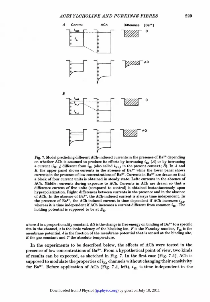
Fig. 7. Model predicting different ACh-induced currents in the presence of Ba2+ dependingon whether ACh is assumed to produce its effects by increasing til (A) or by increasinga current (iK1,2) different from i~l (also called iK1,1 in the present context; B). In A andB, the upper panel shows currents in the absence of Ba2+ while the lower panel showscurrents in the presence of low concentrations of Ba2+. Currents in Ba2+ are drawn so thata block of four current units is obtained in steady state. Left: currents in the absence ofACh. Middle: currents during exposure to ACh. Currents in ACh are drawn so that adifference current of five units (compared to control) is obtained instantaneously uponhyperpolarization. Right: differences between currents in the presence and in the absenceof ACh. In the absence of Ba2+, the ACh-induced current is always time independent. Inthe presence of Ba2+, the ACh-induced current is time dependent if ACh increases iK1'whereas it is time independent if ACh increases a current different from common iK1. Theholding potential is supposed to be at EK.
where A is a proportionality constant, AG is the change in free energy on binding ofBa2+ to a specificsite in the channel, z is the ionic valency of the blocking ion, F is the Faraday number, Vm is themembrane potential, S is the fraction of tihe membrane potential that is sensed at the binding site,R the gas constant and T the absolute temperature.
In the experiments to be described below, the effects of ACh were tested in thepresence of low concentrations of Ba2+. From a hypothetical point of view, two kindsof results can be expected, as sketched in Fig. 7. In the first case (Fig. 7 A), ACh issupposed to modulate the properties of Ki channels without changing their sensitivityfor Ba2+. Before application of ACh (Fig. 7A, left), iK1 is time independent in the
229
) by guest on July 10, 2011jp.physoc.orgDownloaded from J Physiol (

E. CARMELIET AND K. MUBAGWA
absence of Ba2+, but becomes time dependent in the presence of these ions. Afteraddition of ACh (Fig. 7A, middle), the magnitude of iK1 will be scaled by a factorwhich remains constant with time. In particular, the magnitude of the iK1 relaxationinduced by Ba2+ will be increased in the presence of ACh by the same factor as theone with which the total current has been increased. Therefore, the ACh-inducedcurrent (Fig. 7 A, right) will be different in the absence or in the presence of Ba2+,
A B-70mV -98
nA L
-20[1- ~
2 s
Fig. 8. Effect of ACh (2 x 10⁻⁶ M) in the presence of 3 x 10⁻⁶ M-Ba2+. A, current recordingsin the absence (@) and in the presence (0) of ACh are superimposed. Same zero level.B, the same recordings, but with the zero level for the current in the absence ofACh shifteddownward until the relaxing components of the two recordings superimposed. Noticethat neither the amplitude nor the time course of the Ba2+-induced relaxation is changedby ACh.
since ACh will increase the magnitude of a time-independent current in the formercase, and that of a time-dependent current in the later case. The ACh-induced currentwill be time-independent in the absence of Ba2 . In the presence of Ba2 , it will betime dependent. In the second case (Fig. 7B), ACh is supposed to modulate theproperties of a population of channels different from iK1. The current (called iK1,2;see below) carried by these channels is supposed to be time independent. It is alsoassumed that this current is blocked by Ba2+ in a time-independent way, contraryto the standard iK1 (called iK1,1) which is blocked in a time-dependent way. In thiscase, the ACh-induced current will always be time independent in the absence or inthe presence of Ba2+. The magnitude of the Ba2+-induced relaxation will not bechanged, since it is due to iK11 which is assumed to be insensitive to ACh.The results obtained when ACh was added in the presence of low [Ba2+] are
illustrated in Figs. 8 and 9. (1) Fig. 8A shows superimposed currents obtained uponhyperpolarization, before and after application of ACh in Ba2+-containing Tyrodesolution. ACh produced an increase in outward current positive to EK and an increasein inward current negative to EK, i.e. qualitatively the same effects as in normalTyrode solution. Fig. 8B shows that the time-dependent components of the sametwo records in the absence and the presence of ACh can be superimposed. This makesit clear that, despite the fact that the magnitude of total current was doubled inthe presence of ACh, the Ba2+-induced time-dependent component of iKl was notmodified in its magnitude nor in its time course. The ACh-induced change in
230
) by guest on July 10, 2011jp.physoc.orgDownloaded from J Physiol (
ACETYLCHOLINE AND PURKINJE FIBRES
Ba2+0 _ 9 _1 o (M)
nA0-20 - r _
0
10-6
r=10-5
10-4
10'3
~~~~70~~~~~ ~ 0
-70
-100mV 22 s
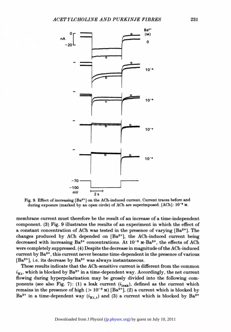
Fig. 9. Effect of increasing [Ba2+] on the ACh-induced current. Current traces before andduring exposure (marked by an open circle) of ACh are superimposed. [ACh]: 10-6 M.
membrane current must therefore be the result of an increase of a time-independentcomponent. (3) Fig. 9 illustrates the results of an experiment in which the effect ofa constant concentration of ACh was tested in the presence of varying [Ba2+]. Thechanges produced by ACh depended on [Ba2+], the ACh-induced current beingdecreased with increasing Ba2+ concentrations. At 1O-3 M-Ba2+, the effects of AChwere completely suppressed. (4) Despite the decrease in magnitude ofthe ACh-inducedcurrent by Ba +, this current never became time-dependent in the presence of various[Ba2+], i.e. its decrease by Ba2+ was always instantaneous.These results indicate that the ACh-sensitive current is different from the common
iK1, which is blocked by Ba2+ in a time-dependent way. Accordingly, the net currentflowing during hyperpolarization may be grossly divided into the following com-ponents (see also Fig. 7): (1) a leak current (iieak), defined as the current whichremains in the presence of high (> 1O-3 M) [Ba2+], (2) a current which is blocked byBa2+ in a time-dependent way (iK11) and (3) a current which is blocked by Ba2+
231
) by guest on July 10, 2011jp.physoc.orgDownloaded from J Physiol (

E. CARMELIET AND K. MUBAGWA
in a time-independent way (K12). The basic name iK1 is also used for this lastcomponent since it is possible that some ACh-sensitive channels may be open in theabsence of agonist (see following paper: Carmeliet & Mubagwa, 1986b). Thesubscript 2 is used to distinguish it from the ACh-insensitive component.A priori, the total change in current following exposure to ACh, Ai, is given by
the sum of ACh-induced changes in the various components:Ai = Aileak+AiKl,l+AiKl,2.
From the experimental results, neither the leak current nor the current giving thetime-dependent component are changed by ACh, i.e. Aileak = 0 and AiKl1, = 0.Thus, only changes of iK12 are responsible for the ACh-induced current:
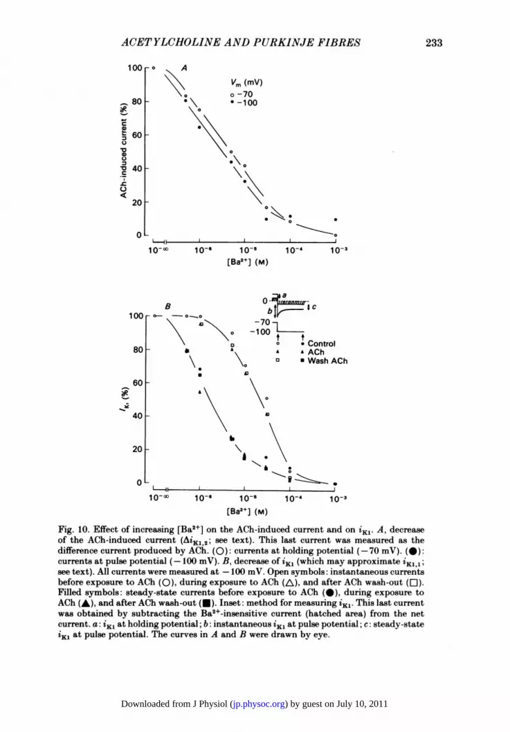
Ai = AiKl,2.Therefore, for obtaining the sensitivity of iKl,2 to Ba2 , the magnitude of theACh-induced current has been measured in function of [Ba2+]. Fig. 1OA comparesthe relative blocks produced by Ba2+ on the ACh-induced current at holding(-70 mV) and pulse potentials (-100 mV). The ACh-induced difference current wasmeasured at both potentials in solutions containing various [Ba2+]. The magnitudeof the ACh-sensitive component decreased with increasing [Ba2+] (see also Fig. 9).The apparent KD for binding of Ba2+ on ACh-sensitive channels (given by theconcentrations of Ba2+ necessary to decrease the ACh-induced current to 50% of themaximal value) are close to each other at both potentials (about 4-2 x 10⁻⁶ M at-70 mV and 2-8 x 10⁻⁶ M at -100 mV), which suggests that the blockade of theACh-sensitive channel by Ba2+ is little voltage dependent in this range of potentials.In another experiment, the apparent KD changed from 1-8 x 10- M at -70 mV to4-8 x 10⁻⁶ M at -100 mV. The average value of 6 for the block of the ACh-sensitivechannel, obtained by plotting log KD as a function of the membrane potential, is 0 4from the two experiments. The weak voltage dependence ofthe block ofACh-sensitivechannels by Ba2+ was confirmed in two other experiments in which the amplitudeof the ACh-induced current was measured at various potentials (between -10 and-100 mV) in the absence and in the presence of 10-5 M-Ba2 . When the ACh-inducedcurrent in Ba2+ was expressed, for each potential, relative to the ACh-induced currentin the absence ofBa2+, the ratio between the two currents changed little (values range:25-55%) for the whole range of potentials (not shown).The lack of significant voltage dependence of the Ba2+-induced blockade of
ACh-sensitive channels is in contrast with the large voltage dependence expected foriK1,1 from its relaxation in Ba2+. In order to obtain the voltage dependence of theblock produced by Ba2+ on iK1, a way for measuring this current had to be found.The Ba2+-sensitive current which exists before the addition of ACh was taken as anapproximate measure of iKl11. By this method, we actually obtain a measure of theACh-insensitive K+ current (i.e. iK1,1) plus the pre-existing part of the ACh-sensitivecomponent (i.e. iKl2 at [ACh] = 0). If this last component is assumed to be small incomparison to the ACh-insensitive component, the behaviour of the sum willapproximate to that of the ACh-insensitive component (iKij). The Ba2+-sensitivecurrent was measured at holding and pulse potentials, in the same two experimentsin which the Ba2+-induced block of iK12 was studied. In normal Tyrode solution, only
232
) by guest on July 10, 2011jp.physoc.orgDownloaded from J Physiol (
ACETYLCHOLINE AND PURKINJE FIBRES
100r
80 -I--
Ca-4-
0
C.)
'O
10
C
U7
AVm (mV)o -70* -100
60 F
40 F
20 F
OL
10- 10- 10
[Ba2+] (M)10-4 10-3
B
\ I A
O .\
b -a
-70n_100 L~
t t0 * ControlA * ACho a Wash ACh
\0
A
a4o
A'
II
10-0° 10-6 lo-5 lo-,, 10-3
[Ba2+] (M)
Fig. 10. Effect of increasing [Ba2+] on the ACh-induced current and on iKI. A, decreaseof the ACh-induced current (AiKl,2; see text). This last current was measured as thedifference current produced by ACh. (0): currents at holding potential (-70 mV). (@):currents at pulse potential (-100 mV). B, decrease of iKi (which may approximate iK1,1;see text). All currents were measured at -100 mV. Open symbols: instantaneous currentsbefore exposure to ACh (0), during exposure to ACh (A), and after ACh wash-out (El).Filled symbols: steady-state currents before exposure to ACh (-), during exposure toACh (A), and after ACh wash-out (U). Inset: method for measuring iKi. This last currentwas obtained by subtracting the Ba2+-insensitive current (hatched area) from the netcurrent. a: iK, at holding potential; b: instantaneous 'Ki at pulse potential; c: steady-stateZK, at pulse potential. The curves in A and B were drawn by eye.
233
100
80
60
40 _
20 F
0
L----4
L-4F
00 0
0
0-
) by guest on July 10, 2011jp.physoc.orgDownloaded from J Physiol (

E. CARMELIET AND K. MUBAGWA
leak was subtracted from the net current (Fig. lOB: inset), but the same results wereobtained in the presence ofACh if both the iieak and the ACh-induced current (AiKl,2)were subtracted. The approximate amplitude of iK1,1 measured at the beginning orat the end of the voltage pulse is plotted as a function of [Ba2+] in Fig. lOB. Thecurrent at the beginning of the voltage pulse is proportional to the open probabilityat the holding potential (-70 mV), whereas the current at the end of the pulse isproportional to the open probability at the pulse potential (-100 mV). In thisexperiment, measurement of instantaneous iK1,1 upon hyperpolarization was used toobtain the steady-state block at holding potential since it was difficult to obtain themagnitude of this current with satisfactory precision, due to the relatively largemagnitude of the leak current compared to the net current at this level. In a secondfibre, in which it was possible to measure iK1,1 at holding potential (-70 mV), the[Ba2+]-response curve at this potential was superimposable to that obtained usinginstantaneous currents following hyperpolarization to -100 mV. In the exampleshown, the amplitude of iK1,1 decreases with increasing Ba2+ concentrations at bothpotentials. Interestingly, the concentration necessary to produce 50% block (apparentKD) is lower by one order of magnitude at the more negative potential 1-3 x 10⁻⁵ Mat -70 mV, 1-3 x 10⁻⁶ M at -100 mV), reflecting a significant increase in affinity forBa2+ by hyperpolarization. Similar results were obtained in another fibre (KD:2-5 x 10⁻⁵ M at -70 mV, 2-1 x 10⁻⁶ at -100 mV). This change in apparent KDcorresponds to a 6 of about 0-9, indicating that the Ba2+-binding site in ACh-insensitive channels senses a major part of the membrane potential, i.e. it is locatedclose to the channel inner mouth.From the assumption that only one Ba2+ is bound per channel (see eqn. (1) above),
a Hill coefficient (nH) of 1 is expected. In the above two preparations, Hill plotsof the data for iK1,1 give a nH of 1.01 + 0 003 (n = number of measurements = 6) andof 1-045 + 0-13 (n = 6) at -70 mV and at -100 mV, respectively. For the ACh-sensi-tive current (iK1 2), H is 0 57 (n = 1) and 0-76 (n = 1) at the same potentials, in theexperiment shown in Fig.1A. Such low Hill coefficients might be due to measurementerrors caused by the small magnitude of ACh-induced currents. In the secondexperiment nH was 0-61 at -70 mV (n = 1), but 1-05+ 0-02 at -100 mV (n = 2).
DISCUSSION
After having shown that ACh increases a K+ conductance (Carmeliet & Mubagwa,1986a), it was interesting to investigate the characteristics of the ACh-sensitive K+current. An important question to answer was whether the properties of thisconductance are superimposable on those of the pre-existing inward rectifier iK1(Garnier et al. 1978; Ojeda et al. 1981 ; Argibay et al. 1983), orwhether the ACh-sensitivecurrent presents characteristics which suggest that it is different from iK1 (Noma &Trautwein, 1978; Sakmann et al. 1983).The ACh-induced current is time independent during voltage pulses of moderate
amplitudes (Fig. 1). For large voltage pulses, the ACh-induced current is frequentlytime dependent. The observed time dependence is, however, not always consistentwith a voltage-dependent gating (i.e. activation or inactivation) of the ACh-sensitivechannels. For depolarizing potentials, increasing as well as decreasing ACh-sensitive
234
) by guest on July 10, 2011jp.physoc.orgDownloaded from J Physiol (

ACETYLCHOLINE AND PURKINJE FIBRES
currents are observed. This can be accounted for by an accumulation process or bysuperimposed changes in time-dependent currents. For large hyperpolarizing pulses,decreasing ACh-sensitive currents are usually obtained, probably following intercel-lular K+ depletion. Exceptionally there may be an initial increase in ACh-inducedcurrent (Fig. 2) which suggests an increase with hyperpolarization of the openprobability of ACh-sensitive channels. Only in this last case do the ACh-sensitivechannels of rabbit cardiac Purkinje fibres behave like those of sino-atrial and ofatrioventricular nodes (Noma & Trautwein, 1978; Sakmann et al. 1983).
Experiments with high concentrations of Cs+ or Ba2+ (Figs. 3-5) do not allow adistinction to be made between common iK1 channels and ACh-sensitive channels.Such a distinction, however, is possible in experiments with low concentrations ofBa2+ (Figs. 6-10). These experiments reveal different sensitivities of the two typesof channels to Ba2+, thus allowing the separation ofthe inward rectifier in the presenceof ACh into two components (iKlii and iKl,2)'When the perfusing solution contains low [Ba2+] a time-dependent decrease in
current is obtained upon hyperpolarization. This time-dependent decrease resemblesthe time-dependent block produced by Ba2+ in skeletal muscle (Standen & Stanfield,1978) and results from a voltage-dependent binding of Ba2+ to a site in iK1i1 channels.The binding of Ba2+ probably causes a decrease in the open-channel probability ofthe channels without changing the single-channel conductance (Bechem, Glitsch &Pott, 1983; Sakmann & Trube, 1984). In the presence of a relaxing iK1 (or K1,1)I theACh-induced current remains time independent, suggesting that ACh-sensitivechannels are different from iK1 (or K1, 1) channels. If ACh were to increase the K+conductance by affecting the same population of channels as the one blocked by Ba2+in a time-dependent way, the following results would have been obtained: (1) aconstant ratio between K+ currents in the absence or in the presence of ACh, whenmeasured at the beginning or at the end of a hyperpolarizing pulse, this ratio beinggiven by the scaling factor of the exponential components measured in the twoexperimental conditions; (2) a time-dependent ACh-induced current during ahyperpolarizing pulse (Fig. 7A).The two expectations are opposite to the experimental findings. The total
magnitude of current is increased in ACh, but the exponential component (which ispart of iKl,1) remains unchanged and the difference current remains constant withtime (Figs. 8-9). These results show that ACh affects a K+ conductance different fromthe one blocked by Ba2+ in a time-dependent way.
It could be imagined that there is only one type of inward-rectifying channels (iK1)and that ACh exerts its effects by modifying the properties of these channels in sucha way that the block produced by Ba2+ on the ACh-activated channels becomesinstantaneous and little voltage dependent. This possibility seems, however, unlikelysince the magnitude of the Ba2+-induced relaxation did not change with ACh. Evenin the presence of increasing ACh concentrations (10-7-10-4 M), the relaxing com-ponent was not changed (not shown). IfACh were to modify iKl properties, increasingACh would have produced a decrease in the relaxing component, since more 'Kichannels would have been transformed into channels with different properties.
Finally, it is tempting to conclude from the difference between common iK1channels and ACh-sensitive channels, that ACh induces the formation of a new type
235
) by guest on July 10, 2011jp.physoc.orgDownloaded from J Physiol (

E. CARMELIET AND K. MUBAGWA
of channels, as suggested for the sino-atrial and the atrioventricular nodes (Noma& Trautwein, 1978; Sakmann et al. 1983). However, the same results can be obtainedif ACh-sensitive channels already exist in the absence of ACh, i.e. if the inwardrectifier K+ current is flowing through two types of channels (1Ki land iK1,2). Bothtypes are sensitive to Ba2 . One type of channel is insensitive to ACh, and its blockby Ba2+ is strongly voltage dependent and of slow kinetics. The other type is sensitiveto ACh, and the block produced by Ba2+ is less voltage dependent and timeindependent. While other experimental techniques (e.g. noise analysis or single-channel recording) are needed to test this hypothesis, there exists some evidencethat the ACh-sensitive channel pre-exists in functional state in the absence of theagonist. With single-channel recording in nodal tissues, Sakmann et al. (1983)observed a channel activity in the absence of ACh, which had the same unit channelconductance as the one in ACh-treated membrane patches. Similar results have beenobtained by Soejima & Noma (1984), who have been able to record the channelactivity of a patch before and during its perfusion with ACh. They observed thatmuscarinic agonists increase the open probability of the basal activity, whereasmuscarinic antagonists produce the opposite effect. In rabbit cardiac Purkinjefibres, the K+ conductance decreases below the normal value on wash-out of ACh,as will be shown in the following paper (Carmeliet & Mubagwa, 1986b). This resultis explained by assuming that desensitized channels are temporarily excluded fromparticipating in the normal membrane conductance.
We thank Mr J. Prenen who performed some of the experiments. We are also grateful to DrJ. Vereecke for critical comments on the original text, and to Mrs L. Heremans and Mr M. Coenenfor technical assistance.
REFERENCES
ARGIBAY, J. A., DUTEY, P., ILDEFONSE, M., OJEDA, C., ROUGIER, O. & TOURNEUR, Y. (1983). Blockby Cs of K current iK1 and of carbachol induced K current iCCh in frog atrium. Pflugers Archiv397, 295-299.
BECHEM, M., GLITSCH, H. G. & PoTT, L. (1983). Properties of an inward rectifying K channel inthe membrane of guinea-pig atrial cardioballs. Pflugers Archiv 399, 186-193.
CARMELIET, E. (1979). Voltage dependent block of inward-going rectification in cardiac Purkinjefibres by external Cs ions. Archives international depharmacodynamie et de therapie 242, 294-295.
CARMELIET, E. & MUBAGWA, K. (1986a). Changes by acetylcholine of membrane currents in rabbitcardiac Purkinje fibres. Journal of Physiology 371, 201-217.
CARMELIET, E. & MUBAGWA, K. (1986b). Desensitization of the acetylcholine-induced increase ofpotassium conductance in rabbit cardiac Purkinje fibres. Journal of Physiology 371, 239-255.
CARMELIET, E. & RAMON, J. (1980). Effects of acetylcholine on time-independent currents in sheepcardiac Purkinje fibers. Pfluigers Archiv 387, 207-216.
CAVALIi, A., OCHI, R., PELZER, D. & TRAUTWEIN, W. (1983). Elementary currents through Ca2+channels in guinea pig myocytes. Pfluigers Archiv 398, 284-297.
DIFRANCESCO, D. (1981). A new interpretation of the pace-maker current in calf Purkinje fibres.Journal of Physiology 314, 359-376.
GARNIER, D., NARGEOT, J., OJEDA, C. & RoUGIER, 0. (1978). The action of acetylcholine onbackground conductance in frog atrial trabeculae. Journal of Physiology 274, 381-396.
GAY, L. A. & STANFIELD, P. R. (1977). Cs causes a voltage-dependent block of inward K currentsin resting skeletal muscle fibres. Nature 267, 169-170.
HAGIWARA, S., MIYAZAKI, S. & ROSENTHAL, N. P. (1976). Potassium current and the effect of cesiumon this current during anomalous rectification of the egg cell membrane of a starfish. Journalof General Physiology 67, 621-638.
236
) by guest on July 10, 2011jp.physoc.orgDownloaded from J Physiol (
ACETYLCHOLINE AND PURKINJE FIBRES
ISENBERG, G. (1976). Cardiac Purkinje fibres: cesium as a tool to block inward rectifying potassiumcurrents. Pflugers Archiv 365, 99-106.
MUBAGWA, K. & CARMELIET, E. (1983). Effects of acetylcholine on electrophysiological propertiesof rabbit cardiac Purkinje fibers. Circulation Research 53, 740-751.
NOMA, A., PEPER, K. & TRAUTWEIN, W. (1979). ACh-induced potassium current fluctuations inthe rabbit sinoatrial node. Pflugers Archiv 381, 255-262.
NOMA, A. & TRAUTWEIN, W. (1978). Relaxation of the ACh-induced potassium current in the rabbitsinoatrial node cell. Pjlitgers Archiv 377, 193-200.
OHMORI, H. (1978). Inactivation kinetics and steady-state current noise in the anomalous rectifierof tunicate egg cell membrane. Journal of Physiology 281, 77-99.
OJEDA, C., ROUGIER, 0. & TOURNEUR, Y. (1981). Effects of Cs on acetylcholine induced current:is iKl increased by acetylcholine in frog atrium? Pfluigers Archiv 391, 57-59.
OSTERRIEDER, W., YANG, Q.-F. & TRAUTWEIN, W. (1982). Effects of barium on the membranecurrents in the S-A node. Pfluigers Archiv 394, 78-84.
SAKMANN, B., NOMA, A. & TRAUTWEIN, W. (1983). Acetylcholine activation of single muscarinicK+ channels in isolated pacemaker cells of the mammalian heart. Nature 303, 250-253.
SAKMANN, B. & TRUBE, G. (1984). Voltage-dependent inactivation of inward-rectifying single-channel currents in the guinea-pig heart cell membrane. Journal of Physiology 347, 659-683.
SOEJIMA, M. & NOMA, A. (1984). Mode of regulation of the ACh-sensitive K channel by themuscarinic receptor in the rabbit atrial cells. Pfliugers Archiv 400, 424-431.
STANDEN, N. B. & STANFIELD, P. R. (1978). A potential- and time-dependent blockade of inwardrectification in frog skeletal muscle fibres by barium and strontium ions. Journal of Physiology280, 169-191.
STANDEN, N. B. & STANFIELD, P. R. (1980). Rubidium block and rubidium permeability of theinward rectifier of frog skeletal muscle fibres. Journal of Physiology 304, 415-435.
VAN DER HEYDEN, G., VEREECKE, J. & CARMELIET, E. (1983). Potential- and time-dependent blockof 'K, by barium in guinea-pig ventricular myocytes. Archives internationales de physiologic et debiochimie 91, P109-1 10.
WOODHULL, A. M. (1973). Ionic blockage ofsodium channels in nerve. Journal of General Physiology61, 687-708.
237
) by guest on July 10, 2011jp.physoc.orgDownloaded from J Physiol (


