Embed Size (px)
Citation preview

Available online at www.sciencedirect.com
a 1778 (2008) 917–930www.elsevier.com/locate/bbamem
Biochimica et Biophysica Act
Ceramides modulate cell-surface acetylcholine receptor levels
C.E. Gallegos, M.F. Pediconi, F.J. Barrantes ⁎
UNESCO Chair of Biophysics and Molecular Neurobiology and Instituto de Investigaciones Bioquímicas de Bahía Blanca,C.C. 857, B8000FWB Bahía Blanca, Argentina
Received 12 July 2007; received in revised form 25 September 2007; accepted 19 October 2007Available online 30 October 2007
Abstract
The effects of ceramides (Cer) on the trafficking of the nicotinic acetylcholine receptor (AChR) to the plasma membrane were studied in CHO-K1/A5 cells, a clonal cell line that heterologously expresses the adult murine form of the receptor. When cells were incubated with short- (C6-Cer)or long- (brain-Cer) chain Cer at low concentrations, an increase in the number of cell-surface AChRs was observed concomitant with a decreasein intracellular receptor levels. The alteration in AChR distribution by low Cer treatment does not appear to be a general mechanism since thesurface expression of the green fluorescent protein derivative of the vesicular stomatitis virus protein (VSVG-GFP) was not affected. High Cerconcentrations caused the opposite effects, decreasing the number of cell-surface AChRs, which exhibited higher affinity for [125I]-α-bungarotoxin, and increasing the intracellular pool, which colocalized with trans-Golgi/TGN specific markers. The generation of endogenous Cerby sphingomyelinase treatment also decreased cell-surface AChR levels. These effects do not involve protein kinase Cζ or protein phosphatase 2Aactivation. Taken together, the results indicate that Cer modulate trafficking of AChRs to and stability at the cell surface.© 2007 Elsevier B.V. All rights reserved.
Keywords: Nicotinic receptor; Ceramide; Sphingosine; Protein traffic; Sphingomyelinase
1. Introduction
Studies from various laboratories, including ours, have dem-onstrated the importance of lipids in the homeostasis and ionchannel function of the nicotinic acetylcholine receptor (AChR)(see reviews in Refs. [1–3]). In particular, sphingolipids (SLs)and cholesterol (Chol) intervene in the regulation of the traf-ficking and cell-surface expression of the AChR [3]. Thus, thedecrease in Chol or SL levels induces changes in the dis-tribution of AChRs, diminishing the number of surface re-ceptors [4,5] and augmenting their retention at intracellularcompartments [6]. In the case of SLs, it has been demonstratedthat impaired biosynthesis results in lower levels of cell-surface
Abbreviations: AChR, nicotinic acetylcholine receptor; Cer, ceramides;CHO, Chinese hamster ovary; Chol, cholesterol; ER, endoplasmic reticulum;Gal-T, galactosyltransferase; GFP, green fluorescence protein; GlcCer, gluco-sylceramide; LDH, lactate dehydrogenase; RFP, red fluorescent protein; SLs,sphingolipids; SM, sphingomyelin; SMase, sphingomyelinase; TGN, trans-Golgi network; VSVG, vesicular stomatitis virus protein; YFP, yellowfluorescent protein; αBTX, α-bungarotoxin⁎ Corresponding author. Tel.: +54 291 4861201; fax: +54 291 4861200.E-mail address: [email protected] (F.J. Barrantes).
0005-2736/$ - see front matter © 2007 Elsevier B.V. All rights reserved.doi:10.1016/j.bbamem.2007.10.019
AChR [7], which can be accounted for by the impairment ofAChR assembly and sequestration of unassembled forms in theendoplasmic reticulum (ER) [8]. The importance of SLs in theregulation of protein traffic is well documented. In yeast,alterations in the synthesis of SL result in defects in the traffickingof the “raft”-resident protein Fus-Mid-GFP from the trans-Golginetwork (TGN) to the plasmamembrane. Newly synthesized cell-surface proteins have been postulated to travel through compart-ments containing qualitatively and quantitatively different SLs,and alteration of their levels could influence protein transport[9,10]. Until the late 1970s, sphingomyelin (SM) was consideredan inert structural lipid, mainly concentrated in the outer layer ofthe plasma membrane. In recent years the emergence of SLs andother lipids as important secondmessengers has been documented[11]. Degradation of SM by activation of sphingomyelinase(SMase) initiates a series of reactions that ends in specific cellularresponses ultimately mediated e.g. by the biosynthetic precursorof SM, ceramide (Cer). Probable targets for Cer action are Cer-activated protein phosphatase 2A (PP2A) [12], a Cer-activatedprotein kinase [13], the guanine-nucleotide exchange factor Vav[14] and the protein kinase Cζ (PKCζ) isoform [15–17]. Otherreported effects of Cer are concerned with the promotion of
918 C.E. Gallegos et al. / Biochimica et Biophysica Acta 1778 (2008) 917–930
changes in the biophysical properties of membranes. SLs andChol have been postulated to occur in laterally segregated lipidmicrodomains termed “rafts” [18], suggested to concentrate sig-naling molecules and receptors in particular regions of the cellsurface [19,20]. Recent studies indicate that Cer are able tocompete with Chol for association with ordered domains in lipidbilayers [21,22].
In this study we analyzed the effects exerted by Cer ondifferent properties of the paradigmatic neurotransmitter recep-tor, AChR, using CHO-K1/A5, a Chinese hamster ovary clonalcell line developed in our laboratory that stably expresses theadult form (α2βδɛ) of the murine muscle AChR [7]. Short-(synthetic C6-Cer) or long- (natural, from bovine brain) chainCer at concentrations of 5 μMwere found to increase AChRs atthe cell surface with a concomitant decrease in intracellularreceptor levels. Higher Cer concentrations (25–37.5 μM) causedthe opposite effects. In addition, we observed an increment in theaffinity of the surface AChRs for the specific competitiveantagonist αBTX. Sphingosine, a lipid structurally and meta-bolically related to Cer, did not produce any significant changesin surface receptors levels, pointing to the specificity of the effectexerted by Cer. The endogenous generation of Cer by SMasetreatment also diminished cell-surface AChRs, albeit through adifferent mechanism. Taken together, our findings indicate thatthe exocytic transport of the AChR can be homeostaticallymodulated by Cer which augment or decrease AChR delivery tothe cell surface depending on the Cer concentration.
2. Materials and methods
2.1. Materials
C6-ceramides (C6-Cer) were purchased from Matreya (Pleasant Gap,Pennsylvania, USA). α-Bungarotoxin (αBTX), D-tubocurarine (d-TC), carba-moylcholine hydrochloride, nystatin, natural ceramides (from bovine brain,brain-Cer), sphingosine, sphingomyelinase (SMase) from Bacillus cereus,okadaic acid, PKCζ pseudo-substrate, mouse monoclonal anti-AChR antibodymAb 210 and bovine albumin (fraction V, cell culture tested), were from SigmaChemical Company (St. Louis, MO, USA). Nutridoma-SP medium withcontrolled lipid content was purchased from Boehringer Ingelheim, Germany.Iodinated α-bungarotoxin ([125I]-αBTX) was purchased from New EnglandNuclear (Boston, Mass., USA). Alexa Fluor488-conjugated αBTX (AlexaFluor488-αBTX), Alexa Fluor594-αBTX, C6-NBD-ceramide and C5-BODIPY-SM were from Molecular Probes, OR, USA. The mouse monoclonal anti-syntaxin 6 antibody was from Stressgen Biotechnologies Corporation, Victoria,BC, Canada. Alexa Fluor488 F (ab′)2 fragment of goat anti-mouse IgG was fromMolecular Probes, OR, USA. The plasmid coding for the green fluorescenceprotein-G-protein of the vesicular stomatitis virus (VSVG-GFP) was a gift fromDr. Alfonso Gonzalez, Catholic University, Santiago, Chile. The plasmid of theenhanced yellow fluorescent protein/human β1,4-galactosyltransferase(pEYFP-Gal-T; Clontech Laboratories Inc., Palo Alto, CA, USA) was a giftfrom Drs. Jennifer Lippincott-Schwartz and George Patterson, NIH. The non-toxic mutant of lysenin conjugated with red fluorescent protein (nt-lysenin-RFP)was a gift from Dr. T. Kobayashi, RIKEN (Institute of Physical and ChemicalResearch), Japan.
2.2. Cell culture
CHO-K1/A5 cells were grown in Ham's F-12 medium supplemented withglutamine, 40 μg/ml gentamicyn sulphate and 10% bovine fetal serum in aNapco (model 6100) incubator maintained at 37 °C with a 5% CO2/95% airmixture. After cells reached 70–80% confluence they were washed and cultured
for an additional period of 30 min and 1, 3 or 5 h under control conditions or inC6-Cer-, brain-Cer- or sphingosine-supplemented medium. Short- and long-chain Cer were added as a complex with albumin as reported by Rosenwald andPagano [23], whereas sphingosine was solubilized in methanol (solventconcentration in the culture medium was kept below 0.2%).
2.3. Sphingomyelinase controlled hydrolysis
This series of experiments were carried out on cells cultured as describedabove and then subjected to SMase (100 mU/ml) hydrolysis for 1 h at 37 °C indelipidated medium. In some cases, 10 min incubation with 0.1 μM okadaic acidor 2.5 μM PKCζ pseudo-substrate preceded the incubation with SMase. Fornystatin treatment, the drug (50 μg/ml) was added in delipidated medium andincubated at 37 °C for 30 min; cells were further incubated for 1 additional hourin the absence or presence of SMase, in the continuous presence of nystatin. Inthe case of long Cer treatments, CHO-K1/A5 cells were incubated with 5 μMC6-Cer or brain-Cer for 24 h in Nutridoma-BO medium devoid of serum.Exogenous SM at a final concentration of 10 μMwas added to cells as describedin a recent work from our laboratory [8] using ethanol:decane (98:2 per vol.) as avehicle. The latter was kept below 0.16%.
2.4. Equilibrium and kinetic studies of [125I]-α-bungarotoxin binding
Surface AChR expression was determined by incubating 70–80% confluentcells with increasing concentrations (5–60 nM) of [125I]-αBTX in cell culturemedium for 1 h at 25 °C. At the end of the incubation period, dishes werewashed twice with Dulbecco's phosphate-buffered saline and the cells wereremoved by scraping and collected with 0.1 N NaOH. The radioactivity wasmeasured in a gamma counter with an efficiency of 80%. Non-specific bindingwas determined from the radioactivity remaining in the dishes when the cellswere first pre-incubated with 10 μM native αBTX or 10 mM carbamoylcholinechloride before addition of [125I]-αBTX. Non-specific binding amounted to 10%in control and treated cells. The metabolic half-life of surface AChR wasdetermined as described previously [24]. Cell cultures were labeled with 10 nM[125I]-αBTX for 1 h at 37 °C, washed twice and incubated for the indicatedperiods. Non-specific binding was determined as described above for theequilibrium experiments. The half-time of degradation was determined fromlinear regression analysis of the decay curve. Total AChR was determined afterlysis of the cells in 10 mM Tris HCl buffer, pH 7.4, containing 150 mM NaCl,5 mM EDTA, 1% Triton X-100, 0.02% NaN3, and 10 μg/ml each chymostatin,leupeptin, pepstatin, tosyl-lysine chloromethyl ketone and tosyl phenylalaninechloromethyl ketone, all added to the buffer immediately before use [25]. Cellextracts were frozen and the binding assay was carried out as describedpreviously [26]. Briefly, the extracts were thawed and 20–40 μl aliquots of thecell lysate were incubated with 10 mM phosphate buffer containing 100 mMNaCl, 1% Triton X-100 and [125I]-αBTX (60 nM) in a final volume of 125 μl. Inthese cases, non-specific binding was determined in samples treated with 10 mMcarbamoylcholine chloride, or boiled for 5 min prior to the binding assay. Non-specific binding obtained by either procedure amounted to 20–30% of the total.The binding reaction was terminated by the addition of 100 μl of the incubationreaction on DE-81 paper dishes (Whatman, Germany). DE-81 papers werewashed three times in 10 mM phosphate buffer containing 0.1% Triton X-100 at10 min intervals. Dried papers were counted in a Beckman gamma counter with80% efficiency. The internal pool of AChR was calculated as the differencebetween the total number of [125I]-αBTX binding sites and cell-surface sites. Inother experiments, total AChR was determined by brief (5 min) treatment of thecells on the Petri dish with 0.5% saponin in phosphate buffer. The detergent wasremoved, the cells washed, and AChR levels determined by the standard [125I]-αBTX binding assay [6,7]. The rate of αBTX association was calculated byfollowing the time-dependent formation of [125I]-αBTX–AChR complexes incells incubated with radioiodinated toxin (10 nM) for different periods.
2.5. Inhibition of initial rates of αBTX binding
Determination of the IC50 for the full agonist carbamoylcholine was carriedout by measuring the initial rate of [125I]-αBTX binding in the presence of theagonist. Cells were incubated in 800 μl of culture medium plus 100 μl of the

919C.E. Gallegos et al. / Biochimica et Biophysica Acta 1778 (2008) 917–930
corresponding carbamoylcholine concentration for 30 min at room temperature.[125I]-αBTX was added to the medium at a final concentration of 10 nM andincubation continued for a further 20 min. The medium was then removed andthe cells were washed with Dulbecco's buffered saline and solubilized by 0.1 NNaOH.
2.6. Protein content
Protein content was determined by the method of Lowry et al. [27] uponsolubilization of cells with 0.1 NNaOH using bovine serum albumin as standard.
2.7. Lactate dehydrogenase activity
In order to evaluate the occurrence of cell damage at high Cerconcentrations, the activity of lactate dehydrogenase (LDH) released in theculture medium was determined [28,29] using a UV kinetic assay (Wiener Lab,Rosario, Argentina) that measures the amount of NADH converted to NAD+.LDH levels from cells disrupted with an ultrasonic homogenizer were used as acontrol of cellular damage.
2.8. Cell transfection
CHO-K1/A5 cells were transiently transfected with the cDNA coding for thefusion proteins pEYFP-Gal-T and VSVG-GFP using the Polifect transfectionreagent (Quiagen Inc. CA, USA). Transfection was performed 24 h before Certreatment.
2.9. Lipid analysis
In some experiments, C6-Cer were added to the culture medium as aprecursor for the synthesis of SM and glucosylceramide (GlcCer). For thispurpose, CHO-K1/A5 cells were cultured in 10 cm culture dishes in the presenceof complete medium for one day. They were subsequently washed with sterilephosphate-buffered saline (PBS; 150 mM NaCl, 10 mM Na2HPO4, 10 mMNaH2PO4, pH 7.4) and Nutridoma-BO medium was added. The cells were thenincubated with 5 μM C6-Cer in the presence of trace concentration (25 nM) ofC6-NBD-Cer at 37 °C for 0.5 and 24 additional hours. Lipid extracts wereprepared as previously described [6]. SLs were separated by thin-layerchromatography using silica gel G plates and CHCl3:CH3OH:H2O, 65:25:4(by vol.) as the mobile phase [30,31]. C6-NBD-Cer and C5-BODIPY-SM wereused as fluorescent standards. Fluorescent lipid spots were detected by UVillumination.
2.10. Fluorescence microscopy
In order to label surface AChRs, cells were incubated with Alexa Fluor488-conjugated αBTX (Alexa Fluor488-αBTX) at a final concentration of 1 μg/ml inPBS, for 1 h, on ice. Excess label was removed by washing with PBS prior tomicroscopy. To label intracellular AChRs, cell-surface AChRs were firstsaturated by incubation of CHO-K1/A5 cells with 1 μg/ml native αBTX in PBSfor 1 h. Cells were then fixed with 4% paraformaldehyde for 40 min,permeabilized with 0.1% Triton X-100 for 20 min, and incubated with AlexaFluor594-αBTX at a final concentration of 1 μg/ml for 1 h at room temperature.
For colocalization studies of intracellular AChRs with the TGN markersyntaxin 6, cells were fixed and permeabilized as above, and incubated withprimary antibody (mouse anti-syntaxin 6, 1:500) for 1 h at room temperature inPBS containing 1% BSA. Cells were then washed three times with PBS andincubated with secondary fluorophore-conjugated antibody (Alexa Fluor488-goat anti-mouse IgG, 1:5000). Alexa Fluor647-αBTX was added with thesecondary antibody to label intracellular AChRs. Incubation was performed atroom temperature for 1 h in PBS containing 1% BSA. Cells were washed threetimes with PBS before coverslips were mounted.
Fluorescence imaging was carried out using a wide-field imaging system(Nikon Eclipse E-600 microscope) accomplished with a SBIG model ST-7digital charge-coupled device camera (765×510 pixels, 9.0×9.0 μm pixel size,Santa Barbara, CA), thermostatically cooled at −10 °C. The ST-7 CCD camerawas driven by the CCDOPS software package (SBIG Astronomical Instruments,
version 5.02, Santa Barbara, CA). For all experiments a 60× (1.4 N.A.) oil-immersion objective was used. Appropriate dichroic and emission filters wereemployed to avoid crossover of fluorescence emission. Confocal images wereacquired with a Leica TCS SP2 microscope (Leica Microsystems HeidelbergGmbH).
2.11. Quantitative analysis of fluorescence microscopy images
Fluorescence images were analyzed with the software Scion Image version4.0.2 (Scion Corp., Frederick,MD). Fluorescence intensities weremeasured in 8-bit images by selecting small membrane areas (cell-surface AChR) or bydelimiting the whole cell (intracellular AChR). Fluorescence intensity valueswere corrected for background fluorescence. The average fluorescence intensityover distinct areas of the cell surface was calculated for randomly chosen cells foreach experimental condition. For illustration purposes, images were processedusing Adobe Photoshop 5.5, scaled with identical parameters, and pseudo-colored according to the corresponding emission wavelength.
2.12. Evaluation of unassembled AChR by fluorescence microscopy
Unassembled AChR was determined using a fluorescence microscopy assaybased on agonist inhibition of αBTX binding recently introduced by ourlaboratory [8]. Briefly, cell-surface AChRswere blockedwith native toxin, fixed,permeabilized and incubated with 10 mM carbamoylcholine for 1 h at roomtemperature, and subsequently incubated with Alexa594-αBTX in the presence ofagonist. As a control for total intracellular AChR, cells were incubated with PBSinstead of agonist for the same period. Quantitative measurements offluorescence intensities of intracellular AChR label in the presence and absenceof agonist provided an estimation of the amount of unassembled AChR.
2.13. AChR internalization studies
Surface AChRs were labeled with Alexa488-αBTX at 4°C as describedabove, followed by incubation at 37 °C for increasing periods (from 0 to 4 h) inthe presence or absence of SMase. The fluorescence intensity at the cell surfaceand the ratio of surface to total AChR was calculated for each incubation time. Insome cases, a double-labeling protocol was carried out as in Borroni et al. [4].Cells were first labeled with Alexa647-αBTX (far red) at 4 °C (to arrest receptorinternalization) and then incubated at 37 °C for 0 and 1 h, respectively, to re-establish endocytic processes. To reveal those AChRs remaining at the cellsurface, cells were then labeled with mAb 210 (which recognizes anextracellular epitope of the α1 AChR subunit) at 4 °C, and subsequentlylabeled with Alexa488-labeled secondary antibody (green).
2.14. Fluorescence microscopy of membrane sheets
For preparation of membrane sheets, CHO-K1/A5 cells were grown onpolylysine-coated glass coverslips and disrupted as described previously [4,5]using a 300 ms ultrasound-pulse in ice-cold KGlu buffer (20 mMHepes, pH 7.2,containing 120 mM potassium glutamate and 20 mM potassium acetate). In thecase of SM hydrolysis, membrane sheets were incubated for 30 min at 37 °C inKGlu buffer containing 100 mU/ml SMase, followed by washing with ice KGlubuffer. Control sheets were incubated with only KGlu buffer. They were thendouble-stained with 1 μg/ml Alexa488-αBTX, and nt-lysenin-RFP for 60 min at4 °C in KGlu buffer, washed 2–3 times with KGlu buffer, fixed for 30 min inPBS containing 4% PFA, and washed with PBS. Finally, membrane sheets werelabeled with 25 μl/ml of a saturated TMA-DPH solution in PBS, in order toidentify membrane sheets avoiding preferential selection according to theirstaining intensity in the Alexa488-αBTX (green) or in the nt-lysenin-RFP (red)channel.
2.15. Lysenin labeling of cell-surface SM
The localization and distribution of cell-surface SM of control and SMase-treated cells were evaluated using lysenin, a fluorescent probe that specificallybinds to SM [32]. We used a non-toxic mutant of lysenin conjugated with redfluorescent protein (nt-lysenin-RFP) [33]. After 1 h incubation in the presence or

920 C.E. Gallegos et al. / Biochimica et Biophysica Acta 1778 (2008) 917–930
absence of SMase, cells were washed and incubated with lysenin (1/50 in PBS)for 1 h on ice. The fluorescence intensity of lysenin-labeled SM at the cellsurface was quantified by fluorescence microscopy.
2.16. Endogenous generation of C5-BODIPY-Cer
Direct observation of endogenously generated Cer was accomplished usingfluorescence microscopy. First cells were incubated with 15 nM C5-BODIPY-SM in Ham's F-12 medium devoid of serum for 30 min on ice to label theplasma membrane of CHO-K1/A5 cells. Upon washing with PBS to remove
Fig. 1. Incubation with low Cer concentrations increases the number of cell-surfaceincubated for 24 h under control conditions (left panel), 5 μMC6-Cer (middle panel) oB) Fluorescent signal of Alexa Fluor488-αBTX arising from surface AChR of cont⁎pb0.05. C) Quantification of intracellular receptors labeled with Alexa Fluor594-αB[125I]-αBTX binding in control CHO-K1/A5 cells (●) and in cells incubated with 5grown either under control conditions (left panel) or in the presence of 5 μMC6-Cer fsignal at the cell surface. ⁎pb0.01. Scale bar: 10 μm. F) Endogenous generation of fluwith C6-NBD-Cer. CHO-K1/A5 cells were incubated with 5 μM C6-Cer+C6-NBD-separated by TLC as described in Materials and methods. C5-BODIPY-SM and C6-Nunder UV light. G) The increment in cell-surface AChR exerted by 5 μM Cer is notbars) and unassembled (empty bars) intracellular AChR in control and CHO-K1/A5 cby preincubation with 10 mM carbamoylcholine before labeling with Alexa Fluor59
unbound C5-BODIPY-SM, cells were incubated for 5 min at 37 °C in theabsence or presence of bacterial SMase [34].
2.17. Data analysis
Data obtained from saturation isotherms, as well as the transformedScatchard plots, association kinetic, displacement experiments, and half-life ofthe cell-surface receptor were analyzed using the Origin 5 software from OriginLab Corporation. Statistically significant differences were determined by theStudent's t-test (two-tailed).
AChRs with concomitant diminution of the intracellular receptor pool. A) Cellsr 5 μM brain-Cer (right panel) were labeled with Alexa488-αBTX for 1 h at 4 °C.rol CHO-K1/A5 cells or those treated with 5 μM Cer for 24 h. ⁎⁎pb0.01 andTX in cells treated with 5 μM C6-Cer. ⁎pb0.05. D) Scatchard plots of specificμM C6-Cer (○). E) Fluorescence images of VSVG-GFP fluorescence in cells
or 24 h (right panel). Bars represent the quantification of VSVG-GFP fluorescentorescent SM and glucosylceramide species upon incubation of CHO-K1/A5 cellsCer for 0.5 h or 24 h. Lipids were extracted and C6-NBD-Cer metabolites wereBD-Cer were used as fluorescent standards. Chromatogram was photographeddue to receptor assembly enhancement. Total (assembled+unassembled=filledells treated with 5 μMC6-Cer for 24 h. Unassembled α subunits were determined4-αBTX. Values are expressed as percentage of total control AChR.

921C.E. Gallegos et al. / Biochimica et Biophysica Acta 1778 (2008) 917–930
3. Results
3.1. Low Cer concentrations increase cell-surface AChR andreduce the number of intracellular receptors
In order to evaluate whether Cer affect the distribution of theAChR, CHO-K1/A5 cells (a clonal CHO cell line expressingthe adult form of the AChR) were incubated for 24 h in lipid-deficient medium (Nutridoma-BO) in the presence of short (C6)Cer at a final concentration of 5 μM and wide-field fluorescencemicroscopy was used to study the distribution of the AChR.Surface AChRs were labeled with the fluorescent derivative ofαBTX, Alexa Fluor488-αBTX (green), and intracellular recep-tors were visualized by Alexa Fluor594-αBTX (red) staining.C6-Cer-treated cells showed a marked (∼50%) increase in cell-surface Alexa Fluor488-αBTX green fluorescence (Fig. 1A–B),whereas the intracellular red fluorescence showed a ∼28%diminution with respect to controls (Fig. 1C).
Fig. 2. Distribution of AChR in CHO cells. A) Percentage of cell-surface (empty barincubated for 5 h under different experimental conditions. Total receptor corresponds(or upon saponin permeabilization); the intracellular pool was calculated as the differHam F-12 medium plus bovine serum (control), in serum-free medium plus albumin,are mean values±SD of five different samples from at least two separate experimentsshown here for the highest concentration of the exogenous lipid. Data shows the perccontrol cells (filled bars). C) Cell-surface fluorescence of Alexa Fluor488-αBTX-labe5 h. ⁎pb0.0001. D) Fluorescence microscopy images of VSVG-GFP expressed in cBars represent the quantification of VSVG-GFP fluorescent signal at the cell surfac
Although short-chain Cer (C2-Cer) are found in nature togetherwith longN-acyl chain Cer [35], the effects exerted by the formermight differ from those induced by long-chain species (see reviewin Ref. [36]).We therefore compared the results obtainedwith C6-Cer with those produced with the more common and naturallyoccurring long-chain Cer, using a complex mixture of long-chainCer from bovine brain. As shown in Fig. 1A–B, brain-Cer alsoincreased (∼42%) surface AChR as assessed by fluorescencemicroscopy.
Experiments using [125I]-αBTX were conducted next todetermine whether Cer treatment affected the equilibrium bindingof this competitive antagonist. The Bmax for the cell-surfaceAChR obtained from Scatchard analysis was 40% higher in C6-Cer-treated cells (from 886 fmol/mg protein in control cellsto 1158 fmol/mg protein in Cer-treated cells; Fig. 1D), infull agreement with the fluorescence microscopy experiments(Fig. 1A–B). The increment in cell-surface AChR induced by C6-Cer was not accompanied by changes in the Kdapp for [125I]-
s) and intracellular [125I]-aBTX binding sites (filled bars) in CHO-K1/A5 cellsto [125I]-αBTX binding sites upon detergent solubilization of CHO-K1/A5 cellsence between the total and cell-surface AChR. Cells were incubated in completeor in the latter medium supplemented with 12.5, 25, or 37.5 μM of C6-Cer. Data. B) Time-dependence of C6-Cer inhibition of cell-surface [
125I]-αBTX binding,entage values obtained after treatment (empty bars) with respect to that found inled receptors in control and CHO-K1/A5 cells treated with 37.5 μM C6-Cer forontrol (left panel) and 37.5 μM C6-Cer-treated (right panel) CHO-K1/A5 cells.e. Scale bar: 10 μm.
922 C.E. Gallegos et al. / Biochimica et Biophysica Acta 1778 (2008) 917–930
αBTX, indicating that the absolute number of receptors, but nottheir affinity, was affected by 24 h Cer treatment (Fig. 1D).
In order to determine whether Cer treatment affected thetrafficking of proteins other than the AChR, CHO-K1/A5 cellswere transfected with the cDNA coding for the vesicularstomatitis virus G protein-green fluorescent protein VSVG-GFP. This fluorescent protein is an efficient probe for followingthe fate of integralmembrane proteins along the exocytic pathway[37]. Transfected CHO-K1/A5 cells were subsequently incubatedwith 5 μMC6-Cer and the surface expression of VSVG-GFP wasquantified. We observed no significant differences between thesurface fluorescent signal of VSVG-GFP of control and Cer-treated cells, indicating that the alteration in the AChR traffic bylow Cer treatment was not a general mechanism (Fig. 1E).
In order to identify the lipid species generated upon 24 hincubation with 5 μM C6-Cer in the presence of trace con-centrations of C6-NBD-Cer, cells were submitted to Folch lipidextraction and subsequent TLC separation with the solventsystem described by Lipsky and Pagano [30]. After 0.5 h in-cubation, C6-NBD-SM was observed in the thin-layer chroma-tograms. However, the fluorescent spot corresponding to C6-NBD-Cer was still present. After 24 h incubation, C6-NBD-SMand C6-NBD-GlcCer fluorescent bands could be visualized,whereas the C6-NBD-Cer were no longer present (Fig. 1F).
As recently reported by Baier and Barrantes [8] the additionof exogenous SM to the culture medium of CHO-K1/A5 cells
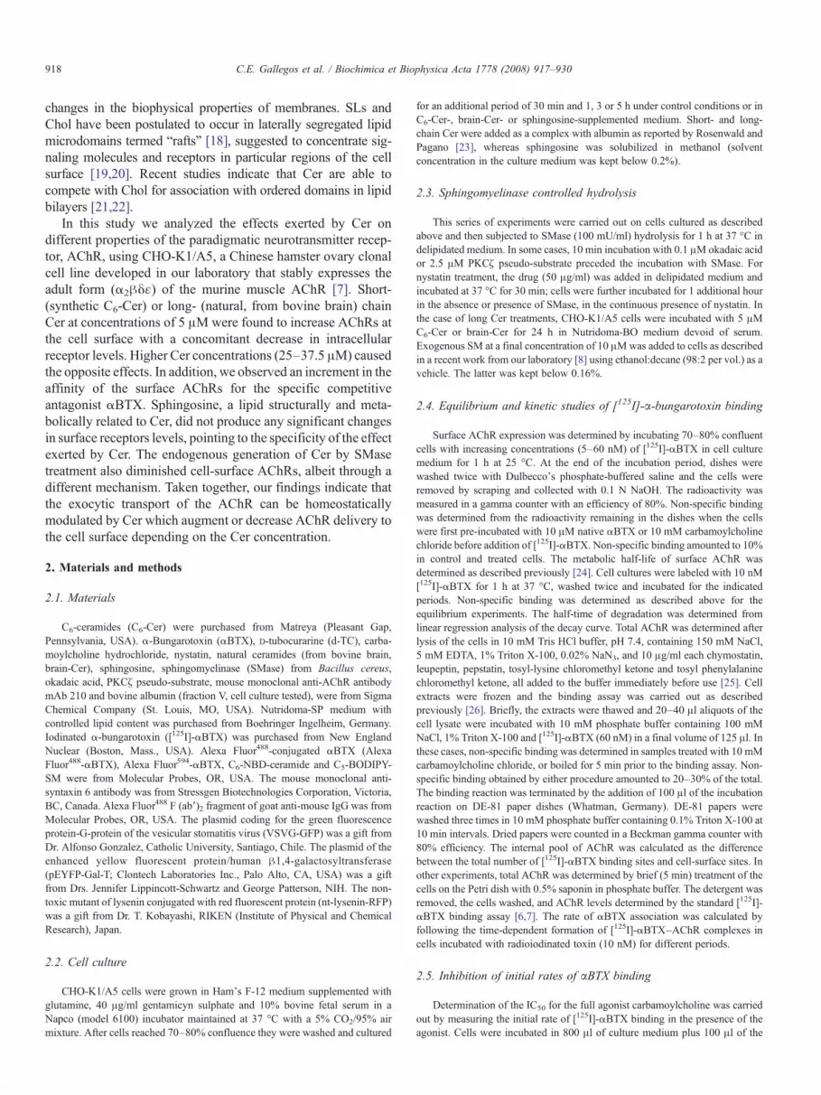
Fig. 3. Pharmacological characterization of AChR from control and C6-Cer-treatedαBTX in control (●) and CHO-K1/A5 cells incubated in the presence of 25 μM oC) Displacement curves of the total radioactive αBTX binding promoted by the preincubated with 37.5 μM C6-Cer. D) Association kinetics of αBTX to AChR for bot
caused a 3-fold increase in the number of AChRs present at theplasma membrane (data not shown).
3.2. The increase in cell-surface AChR by 5 μM Cer does notinvolve enhanced receptor assembly
In order to determine whether the augmented cell-surfaceAChR levels were due to enhanced AChR assembly after a longperiod (24 h) of Cer incubation, unassembled receptors weremeasured next using a fluorescence microscopy assay recentlyintroduced by our laboratory [8]. In control cells the percentageof unassembled α subunits was∼40%, diminishing to∼30% inCer-treated cells (Fig. 1G). Since the total amount of intra-cellular AChR in Cer-treated cells is lower, as judged by thefainter intensity of the fluorescence signal (Fig. 1C) (whichcorresponds to the sum of assembled+unassembled intracel-lular AChRs, Fig. 1G) the ratio of total/unassembled AChR inCer-treated cells did not differ from the control cells.
3.3. Treatment with high short-chain ceramide (C6-Cer)concentrations decreases cell-surface AChR and increases theintracellular receptor pool
We next evaluated the effects of increasing Cer concentra-tions on AChR distribution. Exposure of CHO-K1/A5 cells forperiods of up to 5 h with concentrations of C6-Cer of 12.5 μM
cells. A–B) Scatchard plots obtained with the specific binding of radioiodinatedf C6-Cer (○) (A) and those incubated under control and 37.5 μM C6-Cer (B).sence of the indicated carbamoylcholine concentrations from control and cellsh of the later conditions. Inset: linearization of the association curves in (D).

923C.E. Gallegos et al. / Biochimica et Biophysica Acta 1778 (2008) 917–930
did not affect the binding of the competitive antagonist [125I]-αBTX (Fig. 2A). However, incubations with 25 and 37.5 μMC6-Cer resulted in a decrease in cell-surface AChR with aconcomitant increase in the intracellular AChR pools (Fig. 2A),without significant changes in the total amount of AChR. Thiseffect was time-dependent (Fig. 2B); cell-surface AChR de-creased to 77±7 and 54±11% of control values at 3 and 5 h,respectively. Since short-chain Cer have been postulated toproduce damage in some cell systems [38], a series of ex-periments were carried out to evaluate this possibility using awell known test, the assay of the cytosolic enzyme LDH [28,29].No significant differences were detected in LDH levels betweencontrol cells and cells incubated with 37.5 μM of C6-Cer for 3and 5 h at 37 °C (Supplementary Fig. 1).
Fluorescence microscopy experiments were performed inparallel to study the distribution of AChR in Cer-treated CHO-K1/A5 cells. A 46% reduction in cell-surface Alexa Fluor488-αBTX fluorescence was observed in cells treated with 37.5 μMC6-Cer with respect to controls (Fig. 2C). The surface ex-pression of the fusion protein VSVG-GFP was also significantlyreduced (∼45%) by this treatment (Fig. 2D) indicating that Certreatment at high concentrations affected the traffic of otherproteins besides the AChR.
We next evaluated whether high Cer concentration affectedthe affinity of the AChR–αBTX complex formation. C6-Cerproduced an increase in [125I]-αBTX affinity for the AChR inaddition to a reduction in the density of cell-surface receptor(Fig. 3A–B). The higher affinity of the AChR upon Cer treat-
Fig. 4. High C6-Cer concentrations increase the intracellular pool of AChR and chanwith Alexa Fluor594-αBTX in CHO-K1/A5 cells incubated under control conditionsintracellular Alexa Fluor647-αBTX-labeled-receptors (left panel) with the trans-Go37.5 μM Cer-treated cells. Merged images are given in the right panel. Scale bar: 10(left panel) with syntaxin 6 (middle panel) in cells treated with 5 μM Cer. Right pan
ment was also apparent in competition experiments using thefull agonist carbamoylcholine (Fig. 3C). The apparent equili-brium dissociation constant of those AChRs remaining at thecell surface upon Cer treatment decreased by ∼50% and ∼60%in cells treated with 25 and 37.5 μM C6-Cer, respectively. Thischange in affinity could be accounted for by the correspondingchanges in the kinetics of [125I]-αBTX binding to cell-surfaceAChR, which also showed statistically significant differencesbetween Cer-treated and control cells. As shown in Fig. 3D, therate of association between toxin and AChR increased upon Certreatment. Thus, the decrease in cell-surface AChR levels can-not be accounted for by a decrease in affinity for the competitiveantagonist αBTX.
3.4. C6-Cer treatment arrests AChR at the exiting end of theGolgi complex
Treatment with 37.5 μMC6-Cer for 5 h increased intracellularAlexa Fluor594-αBTX fluorescence by ∼50% (Fig. 4A). Thepattern of intracellular fluorescence also changed: whereas incontrol cells the AChR was quite uniformly distributed over theentire cell, a higher fluorescence intensity was observed at theperinuclear region in cells treated with C6-Cer (Fig. 4B). Sincethis pattern was reminiscent of the trans-Golgi network (TGN),identified in a previous work from our laboratory as the sub-cellular compartment where AChR is arrested upon impairmentof Chol biosynthesis [6], cells were also transiently transfectedwith the pEYFP-Golgi vector, which codifies for the fusion
ge its intracellular distribution. A) Percentage of intracellular receptors labeledor in the presence of 37.5 μM C6-Cer for 5 h. ⁎pb0.001. B) Colocalization oflgi/TGN markers pEYFP-Gal-T and syntaxin 6 (middle panel) in control andμm. C) Colocalization of intracellular Alexa Fluor647-αBTX-labeled-receptorsel: merged images. Scale bar: 10 μm.
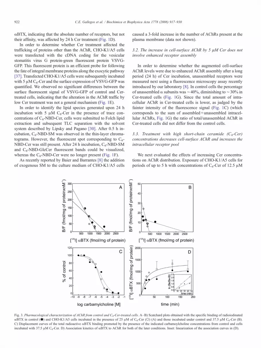
Fig. 5. Brain-Cer affect the AChR distribution in CHO-K1/A5 cells. Cells weregrown either in control medium or in the presence of 37.5 μM brain-Cer for 5 h.A) Fluorescence microscopy images of surface receptors labeled with AlexaFluor488-αBTX in control (left panel) and Cer-treated cells (right panel). Scalebar: 10 μm. B) Percentage of Alexa Fluor488-αBTX fluorescent signal at the cellsurface. ⁎pb0.001. C) Histograms quantifying intracellular receptors labeledwith Alexa Fluor594-αBTX. Fluorescence was quantified as described inMaterials and methods. ⁎pb0.0001.
924 C.E. Gallegos et al. / Biochimica et Biophysica Acta 1778 (2008) 917–930
protein pEYFP-Gal-T. Gal-T is a well knownmarker of the TGN[39,40]. Twenty-four hours after transfection the cells wereincubated with 37.5 μMC6-Cer for 5 h and intracellular AChRswere labeled with Alexa Fluor647-αBTX. Confocal microscopyimages revealed an important degree of colocalization of thefluorescent signals arising from intracellular-labeled AChRs andthe TGNmarker (Fig. 4B). As a specific marker of the TGN [41],the anti-syntaxin 6 antibody yielded similar colocalization withAChRs (Fig. 4B). Thus, treatment with high C6-Cer concentra-tions appears to block trafficking of the AChR to the cell surfaceand result in the accumulation of the AChR at the exiting end ofthe Golgi complex. On the other hand, incubation with low C6-Cer concentrations (5 μM) did not induce any changes in theintracellular distribution of the AChR nor in the structure of theTGN as evaluated by anti-syntaxin 6 labeling (Fig. 4C).
3.5. Long-chain Cer mimic the effect of short-chain Cer onAChR distribution whereas sphingosine does not
Binding experiments with [125I]-αBTX were carried out to testthe effect of long-chain Cer on AChR distribution. After 5 htreatment long-chain brain-Cer (37.5 μM) were also found toreduce surfaceAChRand increase intracellular receptors (Table 1).A similar reduction of cell-surfaceAChR (∼40%)was observed inAlexa Fluor488-αBTX fluorescence microscopy experiments(Fig. 5A–B), with a concomitant increase in intracellular AlexaFluor594-αBTX fluorescence (Fig. 5C).
Since Cer can be rapidly converted to sphingosine [42], itwas necessary to confirm that it was Cer and not sphingosinewhich exerts the cellular effect. For this purpose we evaluatedwhether sphingosine produced changes in AChR distributionanalogous to those detected with Cer. Under conditions similarto those used for Cer experiments, sphingosine did not affectsurface or intracellular AChR (Table 2).
3.6. Endogenously generated Cer also diminish cell-surfaceAChR levels
We next asked the question whether endogenous generationof Cer affects surface expression of the AChR. For this purposeCHO-K1/A5 cells were incubated with 100 mU/ml of bacterialSMase for 1 h at 37 °C. [125I]-αBTX binding assays showedthat cell-surface AChR decreased by ∼35% in SMase-treatedcells (Fig. 6A). Similar results were obtained using the AlexaFluor488-αBTX fluorescence microscopy assay (Fig. 6B).
Table 1Effects of brain-Cer on the surface, intracellular and total AChR in CHO-K1/A5cells
Condition Surface Intracellular Total
Control (6) 527±27 571±140 1098±285Albumin 3 h (5) 524±4 579±104 1103±199Brain-Cer 3 h (5) 369±45⁎⁎ 629±159 998±252Albumin 5 h (7) 532±80 577±115 1109±167Brain-Cer 5 h (7) 285±106⁎⁎ 462±95 747±154⁎
Results are mean values±SD expressed as fmol of [125I]-αBTX specificallybound/mg protein. Numbers between parentheses are the number of individualsamples analyzed. ⁎pb0.025; ⁎⁎pb0.001.
Since Cer have been implicated in the activation of PP2A[12,43], and subunit assembly, insertion and stability of theAChR at the cell membrane have been reported to be affectedby the state of receptor phosphorylation [44–47], we studiednext whether PP2A was involved in the retention of AChR atintracellular compartments. Incubation with 0.1 μM okadaicacid, a well known inhibitor of the Cer-activated PP2A [48], didnot reverse the diminution in AChR surface fluorescence signalproduced by SMase treatment (Supplementary Fig. 2).
The ζ isoform of the enzyme protein kinase C (PKCζ) hasalso been reported to be activated by Cer [49]. To analyze thepossibility that PKCζwas involved in the Cer-induced alterationof AChR traffic, the specific inhibitor PKCζ pseudo-substratewas tested. Similar to what was observed with okadaic acid, thisinhibitor was not able to revert the effects of SMase treatment onsurface AChR levels (data not shown).
An independent assay of SMase action exploited the fluo-rescent dye nt-lysenin-RFP. Lysenin is an earthworm toxin thatspecifically recognizes SM-rich membrane domains and in-duces cytolysis by pore formation [50]. The specific binding of
Table 2Effects of sphingosine on the surface and intracellular AChR expressed in CHO-K1/A5 cells
Condition Surface Intracellular Total
Control (6) 527±27 571±140 1208±121MeOH 5 h (13) 550±33 549±11 1099±22Sphingosine 5 h (12) 511±66 619±92 1130±170
Results are mean values±SD expressed as fmol/mg protein. The effect ofsphingosine was determined after 5 h treatment with 37.5 μM SM. Other detailsare as in Table 1.

Fig. 6. Endogenous generation of Cer also decreases surface AChR. A) Percentage of cell-surface [125I]-αBTX binding sites in CHO-K1/A5 cells incubated for 1 heither under control conditions or in the presence of SMase 100 mU/ml. ⁎pb0.005. B) Cell-surface fluorescence of Alexa488-αBTX-labeled receptors in control (leftpanel) and SMase-treated (right panel) cells. Scale bar: 10 μm. C) Plasma membrane SM labeled with the fluorescent dye nt-lysenin-RFP in control and SMase-treatedcells. ⁎pb0.0001. Scale bar: 10 μm. D) Fluorescence microscopy images of control and SMase-treated membrane sheets obtained as described in Materials andmethods, and labeled with TMA-DPH (left panel), Alexa488-αBTX (middle panel) and nt-lysenin-RFP (right panel). Scale bar: 5 μm. The perimeter of the membranesheets is outlined in the nt-lysenin-RFP panels to facilitate visualization. E) Percentage of Alexa488-αBTX (filled bars) and nt-lysenin-RFP (empty bars) fluorescencein membrane sheets incubated in the absence (control) or in the presence of bacterial SMase. ⁎pb0.001.
925C.E. Gallegos et al. / Biochimica et Biophysica Acta 1778 (2008) 917–930

Fig. 7. Cell-surface AChR diminution in SMase-treated cells is due to an enhancedinternalization. A) Fluorescence microscopy images following the internalizationof plasma membrane AChR labeled with Alexa647-αBTX in control and SMase-treated cells for the indicated times. Scale bar: 10 μm. B) Curves showing thedecrease in Alexa488-αBTX intensity of cell-surface labeled AChRs as a functionof time for control (●) and SMase-treated cells (○). Inset: Internalization kineticsof AChR for the experimental conditions described in (B).
926 C.E. Gallegos et al. / Biochimica et Biophysica Acta 1778 (2008) 917–930
lysenin to SM makes it a useful tool to examine the celldistribution of SM [32]. Here we used the red fluorescent pro-tein adduct of a non-toxic mutant of lysenin, recently developedby Kiyokawa et al. [33]. Only discrete puncta remained at thecell surface after SMase action (Fig. 6C), which reduced thelysenin fluorescence signal by ∼55% (Fig. 6C).
In order to visualize the endogenous generation of Cer, cellswere labeled with a SM probe, C5-BODIPY-SM, and subjectedto SMase treatment [34]. Upon incubation with SMase for 5 minat 37 °C, a significant fraction of the fluorescent adduct, C5-BODIPY-Cer, appeared to have been translocated to the Golgiregion (Supplementary Fig. 3), in agreement with the results ofPagano et al. [51].
To determine whether the decrease in cell-surface AChRmediated by SMase treatment was a result of the mere physicalpresence (or absence) of the lipid in the membrane, another seriesof experiments was conducted on isolated, single-sheets ofAChR-containing plasma membranes, exploiting the formationof single-sheets of plasma membranes by ultrasound treatment asrecently described in work from our laboratory [4,5]. This treat-ment results in the “unroofing” of the cells, leaving behind onlytheir glass-adhered, ventral membrane, and eliminates cytosol,organelles and metabolic integrity of the cell. Upon incubation ofmembrane sheets with SMase for 30 min at 37 °C in KGlu buffer,the specimens were double labeled with Alexa Fluor488-αBTXand nt-lysenin-RFP. TMA-DPH labeling was also performed inorder to identify membrane sheets avoiding preferential selectionaccording to their staining intensity in the Alexa488-αBTX or inthe nt-lysenin-RFP channel. Whereas no significant differenceswere observed in the fluorescence signal arising from AlexaFluor488-αBTX-labeled receptors, a drastic decrease (∼90%) inthe lysenin fluorescence was apparent (Fig. 6D), suggesting thepossibility that the diminution of AChR levels upon SMasetreatment requires the integrity of the cell.
3.7. The mechanism of cell-surface AChR diminution inSMase-treated cells is different from that in Cer-treated cells
We have recently reported that Chol depletion by methyl-β-cyclodextrin causes rapid internalization of the AChR in CHO-K1/A5 cells [4]. Since Chol and SM are found in close as-sociation in biomembranes, forming microdomains [18,52], thedecrease in cell-surface AChR upon SMase treatment observedhere could operate through a similar mechanism. To test thishypothesis we assayed the rate of AChR internalization uponSMase-mediated cell-surface SM depletion using a double-labeling protocol as in Ref. [4]: cells were first labeled withAlexa647-αBTX (far red) at 4 °C and then incubated withSMase at 37 °C for 0 and 1 h, respectively. AChRs remaining atthe cell surface after these two periods were then labeled withmAb 210 (which recognizes an extracellular epitope of the αAChR subunit) at 4 °C, followed by incubation with Alexa488-labeled secondary antibody (green). In control cells, the signalassociated with the AChR was observed almost exclusively atthe cell surface, in the form of patchy, spotted fluorescence(Fig. 7). This is due to the aggregation of AChR moleculeseffected by cross-linking with the bifunctional mAb IgG (see also
Ref. [5]). Comparison of control conditions at t=0 and t=1 hrevealed an increase in the overlap of fluorescence signals from“old” (red) and “newly-labeled” (green) AChRs, indicating that alarge proportion of AChRs remained at the cell surface after 1 hincubation (Fig. 7A). Only few endocytic vesicles (red) could beseen inside cells after 1 h. In contrast, in SMase-treated cells thefluorescence accumulated in abundant single-colored endocyticvesicles (red) concomitantly with a diminution of the overlapbetween old and new receptors at the cell surface (Fig. 7A and seeRef. [4]). In order to determine the kinetics of this process, surfaceAChRs were first labeled with Alexa488-αBTX (green) at 4 °C,followed by incubation with SMase at 37 °C for increasing timeperiods (from 0 to 4 h). The fluorescence intensity at the cell-surface was measured and the ratio of surface/total AChR wascalculated from the time-course of the experiment. The t1/2 wasfound to decrease from 1.7 h in control cells to 1 h in SMase-treated cells (Fig. 7B), indicating that SM depletion by bacterialSMase accelerated AChR internalization kinetics, roughly to the

927C.E. Gallegos et al. / Biochimica et Biophysica Acta 1778 (2008) 917–930
same extent as previously observed in CHO-K1/A5 cell uponChol depletion [4].
3.8. Preincubation with nystatin enhanced the reduction incell-surface AChR levels induced by SMase
In addition to SM hydrolysis, SMase treatment is reportedto produce the rapid internalization of cell-surface Chol[53,54,22]. The possibility that the diminution in plasma mem-brane AChR by SMase was due to Chol internalization rather toSM depletion was therefore tested. For this purpose we in-cubated the cells with the sterol-binding antibiotic nystatin,which specifically interacts with and sequesters the plasmamembrane Chol retained at the membrane [55]. Surface AChRlevels decreased by ∼30% upon nystatin treatment, similar tothe extent observed in SMase-treated cells. When cells wereincubated in the presence of both drugs, a further diminution(50%) in surface AChR levels resulted (Supplementary Fig. 4),indicating that SMase itself was effective in decreasing plasmamembrane AChR even though Chol was not being internalized.
4. Discussion
Cer, intermediate molecules in the metabolism of sphingoli-pids, play an important role as second messengers for manycellular functions [56,36]. Cer associate with lipid “rafts” [57,58]and these Cer-enriched microdomains have been reported tospontaneously fuse into larger Cer-enriched membrane platforms[59–61]. Through these processes Cer appear to “catalyze”various signal transduction pathways recruiting membranereceptors and intracellular signaling molecules.
In the present study we examined the effects of short- andlong-chain Cer on the AChR trafficking to the plasma mem-brane in a mammalian heterologous expression system, theCHO-K1/A5 cells. Most natural Cer have a long (16–24 Catoms) N-acyl chains, but short N-acyl chain Cer (2 C atoms)also occur in nature [35]. We found that low concentrations ofexogenous C6- or brain-Cer augmented the number of AChRs atthe plasma membrane and decreased receptor levels at intra-cellular compartments (Fig. 1A–C). Recently Baier and Bar-rantes [8] showed that SLs are necessary for AChR assemblyand trafficking to the plasma membrane in CHO-K1/A5 cells.Cer are key lipids in the metabolism of SLs. After a 2 hincubation of HL-60 cells with long-chain [14C] C16-Cer, mostof the cell-bound radioactivity was found in free Cer and SMwas the major metabolized SL containing labeled Cer [62].Similar results were reported by Ridgway and Merrian [63] andFuruya et al. [64] using radiolabeled Cer that were convertedinto SM and GlcCer in CHO cells and in cerebellar neurons,respectively. Here, after 30 min incubation of CHO-K1/A5 cellswith 5 μM C6-Cer in the presence of trace concentrations of C6-NBD-Cer, the production of C6-NBD-SM was clearly observed(Fig. 1E). After 24 h incubation, the fluorescent Cer werecompletely converted to complex SL. Although the synthesis ofcomplex SLs was tested only for C6-Cer, long-chain Cer wouldbe expected to behave similarly in the SL biosynthetic pathway[65]. Thus, low Cer treatment could contribute to augmenting
the number of AChRs by an indirect mechanism, i.e. by en-hancing the synthesis of SLs. We have recently demonstratedthat the latter lipids play an important role in the regulation ofAChR exocytic trafficking [8] and that addition of exogenousSMs results in increased levels of AChR at the cell surface [8].
The alteration in AChR distribution after low Cer treatmentdoes not appear to be a general mechanism (Fig. 1D). Further-more, the increment in cell-surface AChR levels produced bylow Cer concentrations without changes in the surface levels ofa predominantly “non-raft” membrane protein like VSVG-GFP[66] reinforces the importance of SLs for the correct traffickingof the AChR to the plasma membrane as recently proposed byBaier and Barrantes [8], and suggests the association of theAChR with lipid domains along the exocytic pathway.
The effect of higher Cer concentrations was also tested in theconcentration range (between 25 and 37.5 μM) usually em-ployed to study Cer effects on protein trafficking [23,67]. In thisrange, Cer markedly and specifically decreased the number ofAChRs present at the plasma membrane with a concomitantincrease in the intracellular receptor pool. Incubation of CHO-K1 cells (the cell line progenitor of the one used in the presentstudy, CHO-K1/A5) with 25 μMC6-Cer is reported to block themovement of VSVG through the medial- and trans-Golgi ap-paratus and its subsequent transport to the cell surface [23].Short-chain Cer were reported to jam the transport of thescavenger receptor CD36 to the plasma membrane of mono-cytes and macrophages [67]. Analogously, high Cer concentra-tions could reduce cell-surface AChRs by blocking the exocytictraffic of newly synthesized receptors, as suggested by thechanges in intracellular receptor distribution and its accumula-tion at the perinuclear region (Fig. 4B). More specifically,pEYFP-Gal-T and syntaxin 6, bona fide markers for the trans-Golgi/TGN [40,41], colocalized with Alexa Fluor647-αBTX-labeled-AChR in large vesicular bodies generated by Certreatment (Fig. 4B), reminiscent of the multivesicular bodiesthat Rosenwald and Pagano [23] characterized by electronmicroscopy. Exogenously added short-chain Cer are rapidlyincorporated into cells and accumulate in the Golgi apparatus[30]. In CHO cells C2-Cer decreased ARF-1 and PKC-αbinding to Golgi-enriched membranes thereby preventingCOP1 transport vesicle formation [68]. It was therefore possiblethat the blockade of AChR transport by exogenous Cer couldaffect in a more general manner the trafficking of proteins otherthan the AChR. This possibility was checked by transfectingCHO-K1/A5 cells with the fluorescent protein VSVG-GFP. Weobserved a significant (∼45%) decrease in the GFP-fluores-cence signal at the plasma membrane upon treatment with highC6-Cer concentrations (Fig. 2D), indicating that a more generalblockage of trafficking is operative at high Cer concentrations.
Besides the reduction in the AChR present at the plasmamembrane, incubation with high C6-Cer concentrations causedan increment in the affinity of the receptor for its canonicalligand, αBTX (Fig. 3A–D). Förster-type resonance energytransfer (FRET) studies from our laboratory showed that SMasedigestion of Torpedo AChR-rich membranes generates Cerspecies with higher affinity for the receptor than the parental SM[69]. The increment in receptor affinity for the competitive

928 C.E. Gallegos et al. / Biochimica et Biophysica Acta 1778 (2008) 917–930
antagonist αBTX after Cer treatment observed here may resultfrom changes in the biophysical properties of the cell membraneupon Cer treatment. Heron et al. [70] found that the increment inthe microviscosity of the membranes resulted in an increment inthe Kd of the serotonin receptor for its ligand. Long-chain Cerhave been shown to increase lipid order in model membranescontaining Chol [71]. In contrast, C2- and C6-Cer cause aconcentration-dependent decrease in lipid order as measured byanisotropy of DPH-PC in plasma membrane vesicles isolatedfrom RBL-2H3 mast cells [72].
Treatment of CHO-K1/A5 cells with SMase, which hydro-lyzes plasma membrane SM with generation of endogenous Cer,also decreased cell-surface AChR levels and accelerated receptorendocytosis. This is in agreement with the reported reduction inCD36 expression resulting from SMase action on monocytes andmacrophages [67]. In macrophages and fibroblasts SMase treat-ment induces formation of numerous vesicles that pinch off fromthe plasma membrane and internalize [34]. Acute Chol depletionfrom CHO-K1/A5 cells by methyl-β-cyclodextrin treatmentdramatically accelerates AChR endocytosis [4]. The accelerationof the internalization kinetics by SMase is very similar to thatresulting fromChol depletion by cyclodextrin [4], suggesting thatboth may result from a common disruption of Chol/SM-rich lipiddomains. In the case of SMase action, this could produce thedisplacement of SM from such lipid domains by virtue of thehigher affinity of the generated Cer for the AChR [69]. Long-chain Cer have the capacity to specifically displace sterol fromlipid domains [22] and have been shown to require an N-linkedacyl chain with at least 8 methylene units to displace Chol fromSM-rich domains [73]. Interestingly, cell-surface Chol is rapidlyinternalized upon SMase action [53,54,22].
The possible synergism between SM and Chol in maintain-ing AChR cell-surface levels was further suggested by thenystatin experiments. Treatment with this sterol-binding anti-biotic (which sequesters membrane-bound Chol but does notremove it from the plasma membrane, Supplementary Fig. 4),decreased AChR levels by ∼30%. Preincubation with nystatinfollowed by SMase treatment in the continuous presence ofthe antibiotic showed a further diminution (∼50%) in surfaceAChR levels, indicating that SMase was effective in decreasingplasma membrane AChR and suggesting that in addition to thekey role played by Chol levels [4,5], SM is also involved in thestability of the plasma membrane-associated AChR. In sum-mary, our results show that Cer were effective in modulating thetrafficking of the AChR expressed in CHO-K1/A5 cells and thatthey exerted these effects in opposite ways and through differentmechanisms depending on the lipid concentration.
Acknowledgments
This work was supported in part by grants from FONCYT,CONICET and UNS, Argentina, to F.J.B., and UNS to M.F.P.
Appendix A. Supplementary data
Supplementary data associated with this article can be found,in the online version, at doi:10.1016/j.bbamem.2007.10.019.
References
[1] F.J. Barrantes, Modulation of nicotinic acetylcholine receptor functionthrough the outer and middle rings of transmembrane domains, Curr. Opin.Drug. Discov. Devel. 6 (2003) 620–632.
[2] F.J. Barrantes, Structural basis for lipid modulation of nicotinic acetylcho-line receptor function, Brain Res. Rev. 47 (2004) 71–95.
[3] F.J. Barrantes, Cholesterol effects on nicotinic acetylcholine receptor, J.Neurochem. 103 (Suppl. 1) (2007) 72–80.
[4] V. Borroni, C.J. Baier, T. Lang, I. Bonini, M.W. White, I. Garbus, F.J.Barrantes, Cholesterol depletion activates rapid internalization of diffrac-tion-limited acetylcholine receptor domains at the cell membrane, Mol.Membr. Biol. 24 (2007) 1–15.
[5] R. Kellner, C.J. Baier, K.I. Willig, S.W. Hell, F.J. Barrantes, Nanoscaleorganization of nicotinic acetylcholine receptors revealed by STED mi-croscopy, Neuroscience 144 (2007) 135–143.
[6] M.F. Pediconi, C.E. Gallegos, E.B. De los Santos, F.J. Barrantes, Meta-bolic cholesterol depletion hinders cell-surface trafficking of the nicotinicacetylcholine receptor, Neuroscience 128 (2004) 239–249.
[7] A.M. Roccamo, M.F. Pediconi, E. Aztiria, L.P. Zanello, A. Wolstenholme,F.J. Barrantes, Cells defective in sphingolipids biosynthesis express lowamount of muscle nicotinic acetylcholine receptor, Eur. J. Neurosci. 11(1999) 1615–1623.
[8] C.J. Baier, F.J. Barrantes, Sphingolipids are necessary for nicotinic ace-tylcholine receptor export in the early secretory pathway, J. Neurochem.101 (2007) 1072–1084.
[9] T.J. Proszinski, R.W. Klemm,M. Gravert, P.P. Hsu, Y. Gloor, J. Wagner, K.Kozak, H. Grabner, K. Walzer, M. Bagnat, K. Simons, C. Walch-Solimena,A genome-wide visual screen reveals a role for sphingolipids and ergosterolin cell surface delivery in yeast, PNAS 102 (2005) 17981–17986.
[10] A.G. Rosenwald, C.E. Machamer, R.E. Pagano, Effects of a sphingolipidsynthesis inhibitor on membrane transport through the secretory pathway,Biochemistry 31 (1992) 3581–3590.
[11] S. Spiegel, D. Foster, R. Kolesnick, Signal transduction through lipidsecond messengers, Curr. Opin. Cell Biol. 8 (1996) 159–167.
[12] R.T. Dobrowsky, C. Kamibayashi, M.C. Mumby, Y.A. Hannun, Ceramideactivates heterotrimeric protein phosphatase 2A, J. Biol. Chem. 268 (1993)15523–15530.
[13] J. Liu, S. Mathias, Z. Yang, R.N. Kolesnick, Renaturation and TNFαstimulation of a 97 kDa ceramide-activated protein phosphatase, J. Biol.Chem. 269 (1994) 3047–3052.
[14] E. Gulbins, K.M. Coggeshall, G. Baier, D. Telford, C. Langlet, G. Baier-Bitterlich, N. Bonnefoy-Berard, P. Burn, A. Wittinghofer, A. Altman,Direct stimulation of Vav-guanine nucleotide exchange activity for ras byphorbol esters and diglycerides, Mol. Cell. Biol. 14 (1994) 4749–4758.
[15] J. Lozano, E. Berra, M.M. Municio, M.T. Díaz-Mecco, I. Domínguez, L.Sanz, J. Moscat, Protein kinase C isoform is critical for kappa B-dependentpromoter activation by sphingomyelinase, J. Biol. Chem. 269 (1994)19200–19202.
[16] G.Muller, M. Ayoub, P. Storz, J. Rannecke, D. Fabbro, K. Pfizenmaier, PKCξ is a molecular switch in signal transduction of TNFα, bifunctionallyregulated by ceramide and arachidonic acid, EMBOJ. 14 (1995) 1961–1969.
[17] P.P. Ruvolo, Intracellular signal transduction pathways activated byceramide and its metabolites, Pharmacol. Res. 47 (2003) 383–392.
[18] K. Simons, E. Ikonen, Functional rafts in cell membranes, Nature 387(1997) 569–572.
[19] K. Simons, D. Toomre, Lipid rafts and signal transduction, Nat. Rev., Mol.Cell Biol. 1 (2000) 31–39.
[20] M. Edidin, The state of lipid rafts: from model membranes to cells, Annu.Rev. Biophys. Biomol. Struct. 32 (2003) 257–283.
[21] Y.J.E. Bjorkqvist, T.K.M. Nyholm, J.P. Slotte, B. Ramsted, Domainformation and stability in complex lipid bilayers as reported by cholesta-trienol, Biophys. J. 88 (2005) 4054–4063.
[22] M. London, E. London, Ceramide selectively displaces cholesterol fromordered domains (rafts): implications for lipid raft structure and function,J. Biol. Chem. 279 (2004) 9997–10004.
[23] A.G. Rosenwald, R.E. Pagano, Inhibition of glycoprotein traffic throughthe secretory pathway by ceramide, J. Biol. Chem. 268 (1993) 4577–4579.

929C.E. Gallegos et al. / Biochimica et Biophysica Acta 1778 (2008) 917–930
[24] J. Patrick, J. McMillan, H. Wolfson, J.C. O'Brien, Acetylcholine receptormetabolism in a nonfusing muscle cell line, J. Biol. Chem. 252 (1977)143–153.
[25] W.N. Green, T. Claudio, Acetylcholine receptor assembly: subunit foldingand oligomerization occur sequentially, Cell 74 (1993) 57–69.
[26] J. Schmidt, M.A. Raftery, A simple assay for the study of solubilizedacetylcholine receptors, Anal. Biochem. 52 (1973) 349–354.
[27] O.H. Lowry, N.J. Rosebrough, A.L. Farr, R.J. Randall, A proteindetermination by the Folin-phenol reagent, J. Biol. Chem. 193 (1951)265–275.
[28] D. Yildiz, N. Ercal, D.W. Armstrong, Nicotine enantiomers and oxidativestress, Toxicology 130 (1998) 155–165.
[29] Y. Fujii, T. Nomura, H. Kanzawa, M. Kameyama, H. Yamanaka, M.Akita, K. Setsu, K. Okamoto, Purification and characterization of entero-toxin produced by Aeromonas sobria, Microbiol. Immunol. 42 (1998)703–714.
[30] N.G. Lipsky, R.E. Pagano, Sphingolipid metabolism in culturedfibroblast: microscopic and biochemical studies employing a fluo-rescent ceramide analogue, Proc. Natl. Acad. Sci. U. S. A. 80 (1983)2608–2612.
[31] A. Loidl, R. Claus, H.P. Deigner, A. Hermetter, High-precision fluo-rescence assay for sphingomyelinase activity of isolated enzymes and celllysates, J. Lipid Res. 43 (2002) 815–823.
[32] Y. Nakai, Y. Sakurai, A. Yamaji, H. Asou, M. Umeda, K. Uyemura, K.Itoh, Lysenin–sphingomyelin binding at the surface of oligodendrocytelineage cells increases during differentiation in vitro, J. Neurosci. Res. 62(2000) 521–529.
[33] E. Kiyokawa, T. Baba, N. Otsuka, A. Makino, S. Ohno, T. Kobayashi,Spatial and functional heterogeneity of sphingolipid-rich membranedomains, J. Biol. Chem. 280 (2005) 24072–24084.
[34] X. Zha, L.M. Pierini, P.L. Leopold, P.J. Skiba, I. Tabas, F.R. Maxfield,Sphingomyelinase treatment induces ATP-independent endocytosis, J. CellBiol. 140 (1998) 39–47.
[35] J. Sot, F.M. Goñi, A. Alonso, Molecular associations and surface-activeproperties of short- and long-N-acyl chain ceramides, Biochim. Biophys.Acta 1711 (2005) 12–19.
[36] W.J. van Blitterswijk, A.H. van der Luit, R.J. Veldman,M. Verheij, J. Borst,Ceramide: second messenger or modulator of membrane structure anddynamics? Biochem. J. 369 (2003) 199–211.
[37] V. Kuhle, G.L. Abrahams, M. Hensel, Intracellular Salmonella entericaredirect exocytic transport processes in a Salmonella pathogenicity island2-dependent manner, Traffic 76 (2006) 716–730.
[38] W. Hu, R. Xu, G. Zhang, J. Jin, Z.M. Szulc, J. Bielawski, Y.A. Hannun, L.M.Obeid, C. Mao, Golgi fragmentation is associated with ceramide-inducedcellular effects, Mol. Biol. Cell. 16 (2005) 1555–1567.
[39] J.M. Mackenzie, M.K. Jones, E.G. Westaway, Markers for trans-Golgimembranes and the intermediate compartment localize to induced mem-branes with distinct replication functions in flavivirus-infected cells,J. Virol. 73 (1999) 9555–9567.
[40] B.E. Schaub, B. Berger, E.G. Berger, J. Rohrer, Transition of galactosyl-transferase 1 from trans-Golgi cisterna to the trans-Golgi network is signalmediated, Mol. Biol. Cell 17 (2006) 5153–5162.
[41] F. Vandenbulcke, D. Nouel, J.P. Vincent, J. Mazella, A. Beaudet, Ligand-induced internalization of neurotensin in transfected COS-7 cells: dif-ferential intracellular trafficking of ligand and receptor, J. Cell Sci. 113(2000) 2963–2975.
[42] T.A. Taha, Y.A. Hannun, L.M. Obeid, Sphingosine kinase: biochemicaland cellular regulation and role in disease, J. Biochem. Mol. Biol. 39(2006) 113–131.
[43] Y.A. Hannun, Functions of ceramide in coordinating cellular responses tostress, Science, 274 (1996) 1855–18599.
[44] A.F. Ross, M. Rapuano, J.H. Schmidt, J.M. Prives, Phosphorylation andassembly of nicotinic acetylcholine receptor subunits in cultured chickmuscle cells, J. Biol. Chem. 262 (1987) 14640–14647.
[45] G. Sadasivam, R. Willmann, S. Lin, S. Erb-Vogtli, X.C. Kong, M.A.Ruegg, C. Fuhrer, Src-family kinases stabilize the neuromuscular synapsein vivo via protein interactions, phosphorylation, and cytoskeletal linkageof acetylcholine receptors, J. Neurosci. 25 (2005) 10479–10493.
[46] E.G. Bruneau, M. Akaaboune, The dynamics of recycled acetylcholinereceptors at the neuromuscular junction in vivo, Development. 133 (2006)4485–4493.
[47] M.A. Lanuza, R. Gizaw, A. Viloria, C.M. Gonzalez, N. Besalduch, V.Dunlap, J. Tomas, P.G. Nelson, Phosphorylation of the nicotinic acetylcho-line receptor in myotube-cholinergic neuron cocultures, J. Neurosci. Res.83 (2006) 1407–1414.
[48] T.A. Haystead, A.T.R. Sim, D. Carling, R.C. Honnor, Y. Tsukitani, P. Cohen,D.G. Hardie, Effects of the tumor promoter okadaic acid on intracellularprotein phosphorylation and metabolism, Nature 337 (1989) 78–81.
[49] N.A. Bourbon, J. Yun, M. Kester, Ceramide directly activates proteinkinase C zeta to regulate a stress-activated protein signaling complex,J. Biol. Chem. 275 (2000) 35617–35623.
[50] A. Yamaji-Hasegawa, A.Makino, T. Baba, Y. Senoh, H. Kimura-Suda, S.B.Sato, N. Terada, S. Ohno, E. Kiyokawa, M. Umeda, T. Kobayashi,Oligomerization and pore formation of a sphingomyelin-specific toxin,lysenin, J. Biol. Chem. 278 (2003) 22762–22770.
[51] R.E. Pagano, O.C. Martin, H.C. Kang, R.P. Haugland, A novel fluorescentceramide analogue for studying membrane traffic in animal cells: ac-cumulation at the Golgi apparatus results in altered spectral properties ofthe sphingolipid precursor, J. Cell Biol. 113 (1991) 1267–1279.
[52] D.A. Brown, E. London, Functions of lipid rafts in biological membranes,Annu. Rev. Cell Dev. Biol. 14 (1998) 111–136.
[53] J.P. Slotte, E.L. Bierman, Depletion of plasma membrane sphingomyelinrapidly alters the distribution of cholesterol between plasma membranesand intracellular cholesterol pools in cultured fibroblast, Biochem. J. 250(1988) 653–658.
[54] N.D. Ridgway, Interactions between metabolism and intracellulardistribution of cholesterol and sphingomyelin, Biochim. Biophys. Acta1484 (2000) 129–141.
[55] P. Tewary, K. Veena, T.J. Pucadyil, A. Chattopadhyay, R. Madhubala, Thesterol-binding antibiotic nystatin inhibits entry of non-opsonized Leish-mania donovani into macrophages, Biochem. Biophys. Res. Commun. 339(2006) 661–666.
[56] Y.A. Hannun, The sphingomyelin cycle and the second messenger functionof ceramide, J. Biol. Chem. 26 (1994) 3125–3128.
[57] X. Xu, R. Bittman, G. Duportall, D. Heissler, C. Vilcheze, E. London, Effectsof the structure of natural sterols and sphingolipids on the formation ofordered sphingolipids/sterol domains (raft), comparison of cholesterol toplant, fungal, and disease-associated sterols and comparison of sphingomye-lin, cerebrosides, and ceramide, J. Biol. Chem. 276 (2001) 33540–33546.
[58] T.Y. Wang, J.R. Silvius, Sphingolipid partitioning into ordered domains incholesterol-free and cholesterol-containing lipid bilayers, Biophys. J. 84(2003) 367–378.
[59] J. Bock, I. Szabo, N. Gamper, C. Adams, E. Gulbins, Ceramide inhibits thepotassium channel Kv1.3 by the formation of membrane platforms,Biochem. Biophys. Res. Commun. 305 (2003) 890–897.
[60] C.R. Bollinger, V. Teichgraber, E. Gulbins, Ceramide-enriched membranedomains, Biochim. Biophys. Acta 1746 (2005) 284–294.
[61] L. Silva, R.F. de Almeida, A. Fedorov, A.P. Matos, M. Prieto, Ceramide-platform formation and -induced biophysical changes in a fluid phos-pholipid membrane, Mol. Membr. Biol. 23 (2006) 137–148.
[62] D. Ardail, I. Popa, J. Bodennec, C. Famy, P. Louisot, J. Portoukalian,Subcellular distribution and metabolic fate of exogenous ceramides takenup by HL-60 cells, Biochim. Biophys. Acta 1583 (2002) 305–310.
[63] N.D. Ridgway, D.L. Merrian, Metabolism of short chain ceramide anddihidroceramide analogues in Chinese hamster ovary (CHO) cells,Biochim. Biophys. Acta 1256 (1995) 57–70.
[64] S. Furuya, J. Mitoma, A. Makino, Y. Hirabayashi, Ceramide and itsinterconvertible metabolite sphingosine function as indispensable lipidfactors involved in survival and dendritic differentiation of cerebellarPurkinje cells, J. Neurochem. 71 (1998) 366–377.
[65] K.Venkataraman,A.H. Futerman,Comparison of themetabolismof L-erythro-and L-threo-sphinganines and ceramides in cultured cells and in subcellularfractions, Biochim. Biophys. Acta 1530 (2001) 219–226.
[66] T. Harder, P. Scheiffele, P. Verkade, K. Simons, Lipid domain structure ofthe plasma membrane revealed by patching of membrane components,J. Cell Biol. 141 (1998) 929–942.

930 C.E. Gallegos et al. / Biochimica et Biophysica Acta 1778 (2008) 917–930
[67] Y. Luan, H.R. Griffiths, Ceramides reduce CD36 cell surface expressionand oxidised LDL uptake by monocytes and macrophages, Arch.Biochem. Biophys. 450 (2006) 89–99.
[68] A. Abousalham, T.C. Hobman, J. Dewald, M. Garbutt, D.N.Brindley, Cell-permeable ceramides preferentially inhibit coated ves-icle formation and exocytosis in Chinese hamster ovary comparedwith Madin–Darby canine kidney cells by preventing the mem-brane association of ADP-ribosylation factor, Biochem. J. 361 (2002)653–661.
[69] I.C. Bonini, S.S. Antollini, C. Gutierrez-Merino, F.J. Barrantes, Sphingo-myelin composition and physical asymmetries in native acetylcholinereceptor-rich membranes, Eur. Biophys. J. 31 (2002) 417–427.
[70] D.S. Heron, M. Shinitzkyt, M. Hershkowitz, D. Samuel, Lipid fluiditymarkedly modulates the binding of serotonin to mouse brain membranes,Proc. Natl. Acad. Sci. U. S. A. 77 (1980) 7463–7467.
[71] T.Y. Wang, J.R. Silvius, Different sphingolipids show differentialpartitioning into sphingolipid/cholesterol-rich domains in lipid bilayers,Biophys. J. 79 (2000) 1478–1489.
[72] A. Gidwani, H.A. Brown, D. Holowka, B. Baird, Disruption of lipid orderby short-chain ceramides correlates with inhibition of phospholipase D anddownstream signaling by FcɛRI, J. Cell Sci. 116 (2003) 3177–3187.
[73] S. Nybond, Y.J. Bjorkqvist, B. Ramstedt, J.P. Slotte, Acyl chain lengthaffects ceramide action on sterol/sphingomyelin-rich domains, Biochim.Biophys. Acta 1718 (2005) 61–66.


