Embed Size (px)
Citation preview
i
Acetylcholine as a Neuromuscular Transmitter
In the Horseshoe Crab, Limulus polyphemus
A Major Qualifying Project Report
Submitted to the Faculty
of the
WORCESTER POLYTECHNIC INSTITUTE
In partial fulfillment of the requirements for the
Degree of Bachelor of Science
By
_____________________________________
Yip Wong
April 28, 2010
Approved by:
___________________________ _________________________ Doctor Daniel Gibson, Co-advisor, Doctor Michael Buckholt, Advisor
ii
Abstract
Previous MQP studies on horseshoe crab muscles provided several lines of
evidence that acetylcholine (ACh) is their neuromuscular junction (NMJ) transmitter.
ACh blockers and acetylcholine receptor (AChR) antibodies reduced responses to
neuronally evoked contractions (Fuller, 2006; Mallozzi, 2005; Vacher, 2007). We used a
histochemical approach to further confirm the presence of ACh and its receptor at NMJs.
Whole mounts and acrylic-embedded sections were treated with antibodies that were
fluorescent, enzymatic, or electron-opaque. Polyclonal anti-ACh confirmed the presence
of ACh in horseshoe crab leg nerves. Monoclonal anti-AChR demonstrated anti-AChR
in some nerves, though less strongly. Immunoelectron microscopy has produced
confirmatory micrographs for ACh.
iii
Acknowledgements
I would like to thank my MQP advisor Dr. Daniel Gibson for his guidance
throughout the project and his teaching of many useful skills that not only helped me out for this project, but would also be very useful for my future career. Moreover, I would like to thank Dr. Michael Buckholt, Dr. JoAnn Whitefleet-Smith, and lab manager Abbie White, for their help in providing necessary material and equipment.
iv
Table of Contents
Abstract………………………………………………………………………………………..….ii
Acknowledgements……………………………………………………………………………..iii
Table of Contents………………………………………………………………….……………iv
List of Figures and Tables…………………………………………………...…………………v
Introduction………………………………………………………………………………………1
Horseshoe Crab…………………………………………………………………………1
Horseshoe Crab Anatomy………………………………………...……………………2
Horseshoe Crab Life Cycle………………………………………………………….…3
Neurotransmitter in Neuromuscular Junctions (NMJs)……………………………...3
Project goals………………………………………………………………..……………6
Materials and Methods………………………………………………………………….………7
Partial Dissection and Specimen Preparation………………………………………..7
Preparation and Storage of Resin……………………………………………………..8
Tissue Embedding ……………………………………………………..……………….9
Serial Sections on the Microtome……………………………………………………10
Treatment of Superfrost Plus Slides…………………………………………………12
Staining for Light and Fluorescent Microscopy. ……………………………………13
Rabbit anti-ACh…………………………………………………..……………13
Mouse anti-Human AChR...…………………………………………………..15
Staining for Transmission Electron Microscopy (TEM)……………...… …………16
Photography for TEM….……………………………………………………………...17
Positive Control and Whole Mount Antibody Staining. …………………………....17
v
Electrical Stimulation of Horseshoe crab Leg Motor Nerve. ……………………...18
Amputation and Glutaraldehyde Fixation Adult Horseshoe Crab Leg.. …………19
Preparation of Glass Micropipets…………………………………………………….20
“Autoimmune Experiment”. …………………………………………………………..20
Results………………………………………………………………….………………………22
Positive control on whole mount tissue.. ……………………………………………22
Immunostaining Experiment.. …………………………………………………..……23
Mouse Anti-AChR. …………………………………………………………….23
Rabbit anti-Ach.. ………………………………………………………………24
“Autoimmune” Experiment……………………………………………………………25
Transmission Electron Microscopy (TEM). ………………………………..……….27
Discussion …………………………………………………………………………….….……28
Acetylcholine is present in the nerves of Larval Horseshoe Crabs.. ….…………28
“Autoimmune Experiment” indicates involvement of AChR in nervous system of
Juvenile Horseshoe Crabs. …………………………………………………….…….29
Future Experiments.. ……………………………………………………….…………29
References……………………………………………………………………………………30
vi
Table of Figures Figure 1: Horseshoe Crabs on the beach…………………………………………….………1
Figure2: Anatomy of Limulus Polyphemus………………………………………….………..2
Figure3: 3a shows a typical neuromuscular junction and 3b shows the chemical
structure of ACh…………………………….…………………………………….……..5
Figure4: 4a shows the larval Horseshoe Crabs and 4b shoes the synaptic vesicles in
horseshoe crabs……………………………...…..……………………………………..5
Figure 5: The red lines show the longitudinal direction along which the crabs were
dissected………………………………………..………………………………………..8
Figure 6: Schematic diagram of exposing Limulus polyphemus to resin mixtures, and
eventually wit curing in pure resin……………………………………..……………9
Figure 7: picture of resin blocked made in different types of containers……………...…10
Figure 8: picture of the boat, diamond knife and sections cut by it………………………11
Figure 9: 9a shows Superfrost Plus Slides and 9b shows Biobond……………………...13
Figure 10: shows Goat anti-rabbit Alexa fluor 488® fluorescence of section stained with
Rabbit anti-ACh……………………………………..……………………………..…..22
Figure 11: ……………………………………………..……………………...………………..23
In 11a counterstained DAPI section shows the location of nerve that fluoresces
with Mouse anti-human AChR. …………………………………..………………….23
11b shows Goat anti-mouse Alexa fluor 555® fluorescence of section stained
with Mouse anti-human AChR. …………………...…………...……………………23
Figure 12: …………………………………………………………….………………………..24
12a shows the experimental section of larval horseshoe crab stained with 1:10
dilution of Rabbit anti-ACh and then 1:10 dilution of Goat anti-Rabbit Alexa Fluor
488®. ……………………………………………..…….………………………………24
vii
12b shows an experimental section of larval horseshoe crab stained with Pierce
ABC peroxidase Kit……………………………………………………………………24
Figure 13: ……………………………………………..………………...……………………..26
13a and b show the control crabs before and after injection of Mouse anti-human
AChR. ……………………………………………..……………………………………26
13c and d show the experimental crabs before and after injection of
antibodies. ……………………………………………………………………………..26
Figure 14: TEM 20,000X picture, GAM-Au 10nm experimental crab leg sections on
nickel grids……………………………………………………………………………..27
Table 1: Taxonomic Classification of the Limulus Polyphemus………………………...….1
1
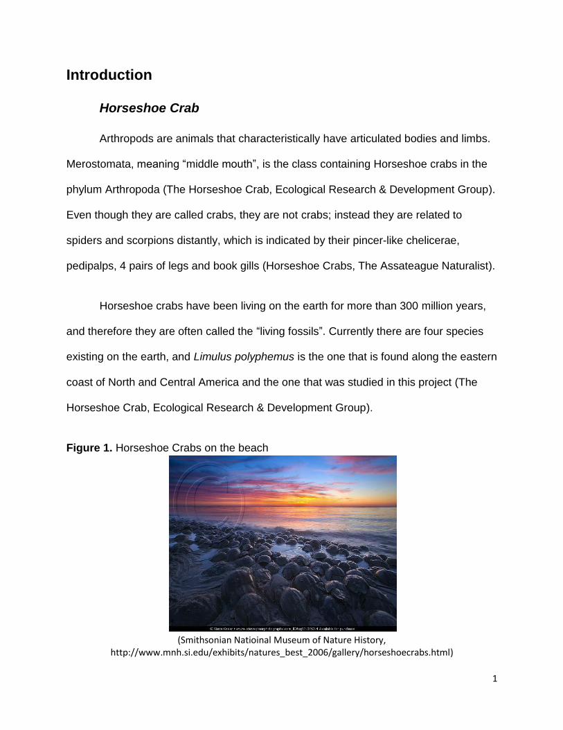
Introduction
Horseshoe Crab
Arthropods are animals that characteristically have articulated bodies and limbs.
Merostomata, meaning “middle mouth”, is the class containing Horseshoe crabs in the
phylum Arthropoda (The Horseshoe Crab, Ecological Research & Development Group).
Even though they are called crabs, they are not crabs; instead they are related to
spiders and scorpions distantly, which is indicated by their pincer-like chelicerae,
pedipalps, 4 pairs of legs and book gills (Horseshoe Crabs, The Assateague Naturalist).
Horseshoe crabs have been living on the earth for more than 300 million years,
and therefore they are often called the “living fossils”. Currently there are four species
existing on the earth, and Limulus polyphemus is the one that is found along the eastern
coast of North and Central America and the one that was studied in this project (The
Horseshoe Crab, Ecological Research & Development Group).
Figure 1. Horseshoe Crabs on the beach
(Smithsonian Natioinal Museum of Nature History,
http://www.mnh.si.edu/exhibits/natures_best_2006/gallery/horseshoecrabs.html)
2
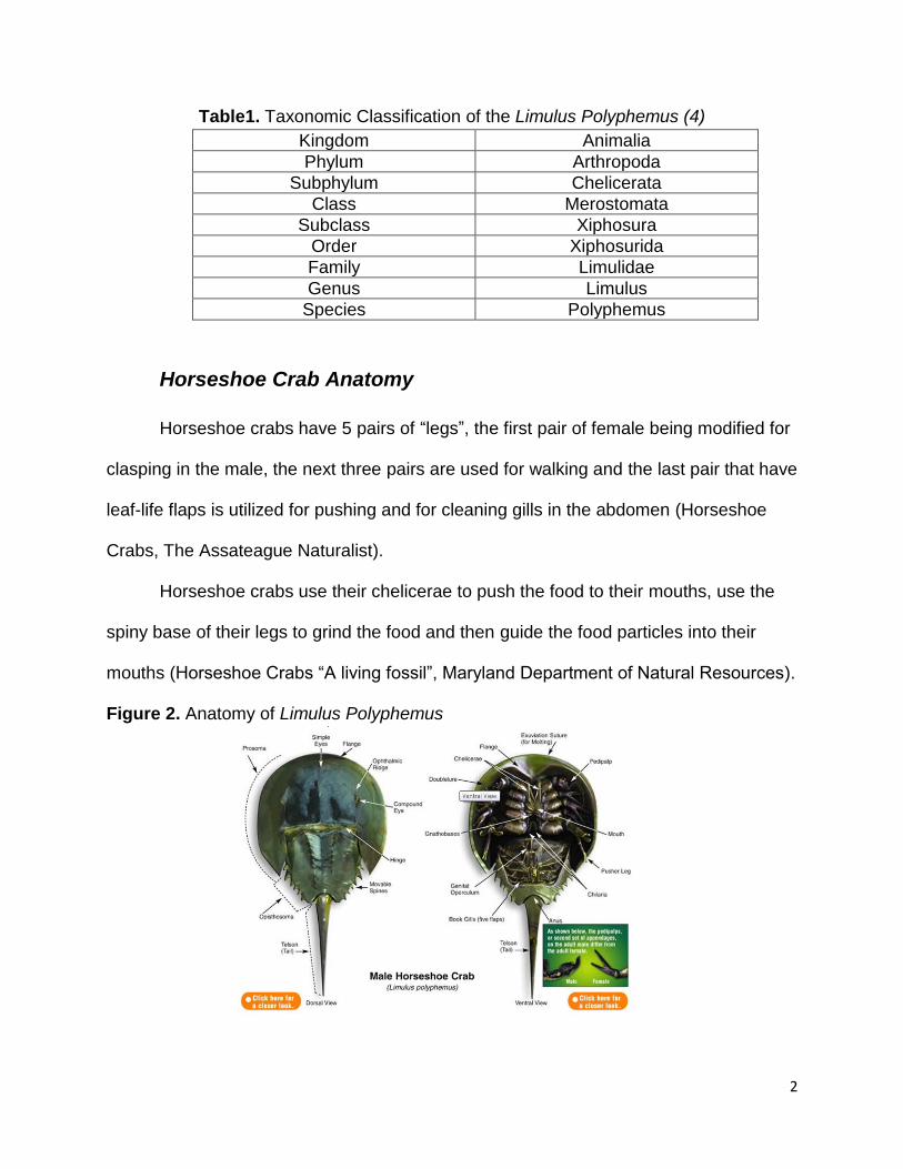
Table1. Taxonomic Classification of the Limulus Polyphemus (4)
Kingdom Animalia
Phylum Arthropoda
Subphylum Chelicerata
Class Merostomata
Subclass Xiphosura
Order Xiphosurida
Family Limulidae
Genus Limulus
Species Polyphemus
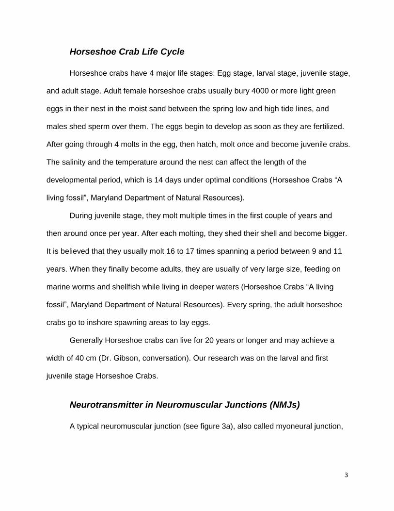
Horseshoe Crab Anatomy
Horseshoe crabs have 5 pairs of “legs”, the first pair of female being modified for
clasping in the male, the next three pairs are used for walking and the last pair that have
leaf-life flaps is utilized for pushing and for cleaning gills in the abdomen (Horseshoe
Crabs, The Assateague Naturalist).
Horseshoe crabs use their chelicerae to push the food to their mouths, use the
spiny base of their legs to grind the food and then guide the food particles into their
mouths (Horseshoe Crabs “A living fossil”, Maryland Department of Natural Resources).
Figure 2. Anatomy of Limulus Polyphemus
3
Horseshoe Crab Life Cycle
Horseshoe crabs have 4 major life stages: Egg stage, larval stage, juvenile stage,
and adult stage. Adult female horseshoe crabs usually bury 4000 or more light green
eggs in their nest in the moist sand between the spring low and high tide lines, and
males shed sperm over them. The eggs begin to develop as soon as they are fertilized.
After going through 4 molts in the egg, then hatch, molt once and become juvenile crabs.
The salinity and the temperature around the nest can affect the length of the
developmental period, which is 14 days under optimal conditions (Horseshoe Crabs “A
living fossil”, Maryland Department of Natural Resources).
During juvenile stage, they molt multiple times in the first couple of years and
then around once per year. After each molting, they shed their shell and become bigger.
It is believed that they usually molt 16 to 17 times spanning a period between 9 and 11
years. When they finally become adults, they are usually of very large size, feeding on
marine worms and shellfish while living in deeper waters (Horseshoe Crabs “A living
fossil”, Maryland Department of Natural Resources). Every spring, the adult horseshoe
crabs go to inshore spawning areas to lay eggs.
Generally Horseshoe crabs can live for 20 years or longer and may achieve a
width of 40 cm (Dr. Gibson, conversation). Our research was on the larval and first
juvenile stage Horseshoe Crabs.
Neurotransmitter in Neuromuscular Junctions (NMJs)
A typical neuromuscular junction (see figure 3a), also called myoneural junction,
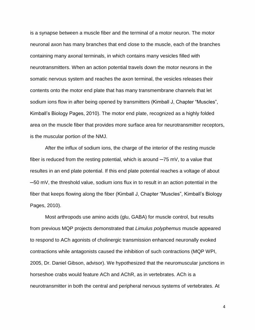
4
is a synapse between a muscle fiber and the terminal of a motor neuron. The motor
neuronal axon has many branches that end close to the muscle, each of the branches
containing many axonal terminals, in which contains many vesicles filled with
neurotransmitters. When an action potential travels down the motor neurons in the
somatic nervous system and reaches the axon terminal, the vesicles releases their
contents onto the motor end plate that has many transmembrane channels that let
sodium ions flow in after being opened by transmitters (Kimball J, Chapter “Muscles”,
Kimball’s Biology Pages, 2010). The motor end plate, recognized as a highly folded
area on the muscle fiber that provides more surface area for neurotransmitter receptors,
is the muscular portion of the NMJ.
After the influx of sodium ions, the charge of the interior of the resting muscle
fiber is reduced from the resting potential, which is around ─75 mV, to a value that
resultes in an end plate potential. If this end plate potential reaches a voltage of about
─50 mV, the threshold value, sodium ions flux in to result in an action potential in the
fiber that keeps flowing along the fiber (Kimball J, Chapter “Muscles”, Kimball’s Biology
Pages, 2010).
Most arthropods use amino acids (glu, GABA) for muscle control, but results
from previous MQP projects demonstrated that Limulus polyphemus muscle appeared
to respond to ACh agonists of cholinergic transmission enhanced neuronally evoked
contractions while antagonists caused the inhibition of such contractions (MQP WPI,
2005, Dr. Daniel Gibson, advisor). We hypothesized that the neuromuscular junctions in
horseshoe crabs would feature ACh and AChR, as in vertebrates. ACh is a
neurotransmitter in both the central and peripheral nervous systems of vertebrates. At
5
NMJs, after stimulation, ACh is released and crosses the synaptic cleft to bind to AChR,
causing sodium channels to open and induce the action potential which in turn causes
the muscle contraction. The diagram below shows a typical vertebrate NMJ (Kimball J,
Chapter “Muscles”, Kimball’s Biology Pages, 2010).
Figure 3. 3a shows a typical neuromuscular junction and 3b shows the
chemical structure of Ach
3a 3b
(3a was originally from Investigating potential treatments for the myasthenias, http://www.muscular-dystrophy.org/research/grants/1589_investigating_potential_treatments_for_the_myasthenias )
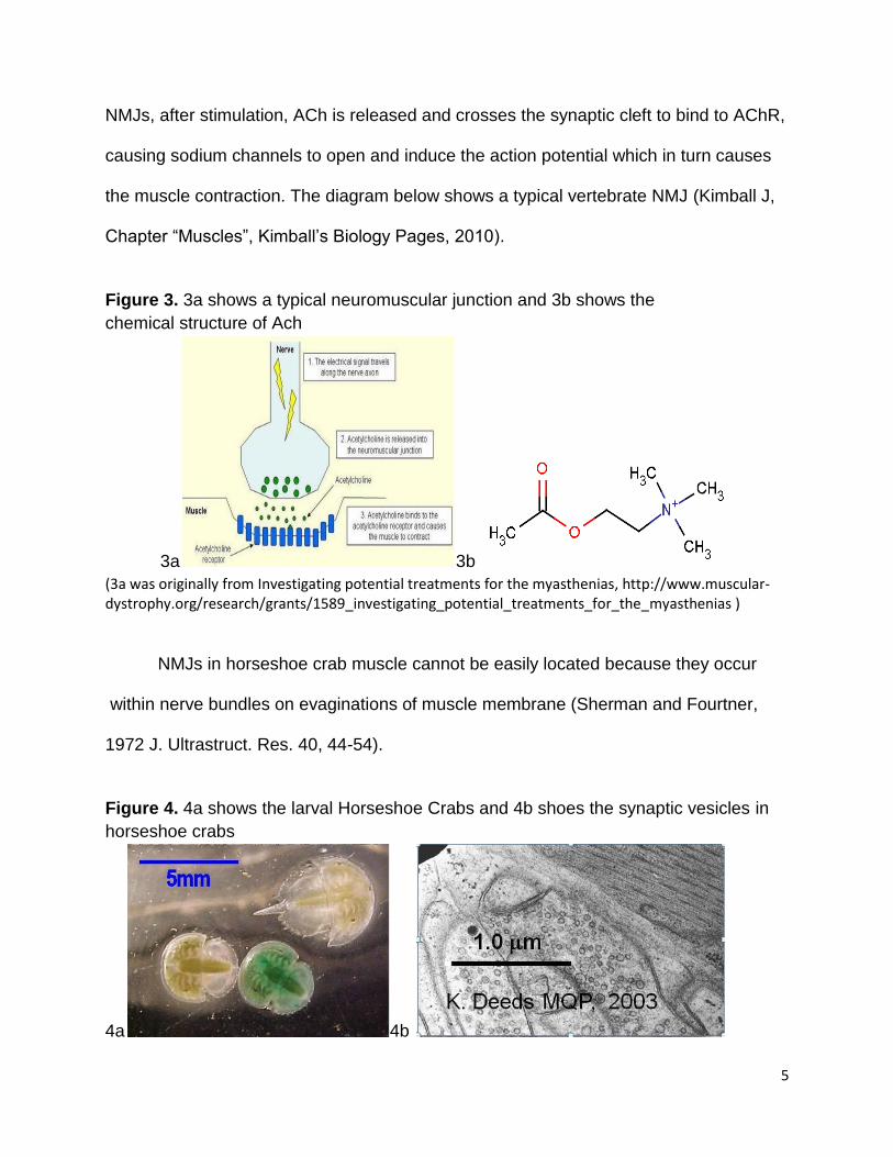
NMJs in horseshoe crab muscle cannot be easily located because they occur
within nerve bundles on evaginations of muscle membrane (Sherman and Fourtner,
1972 J. Ultrastruct. Res. 40, 44-54).
Figure 4. 4a shows the larval Horseshoe Crabs and 4b shoes the synaptic vesicles in
horseshoe crabs
4a 4b
6
Project Goals
The major focus of this project was to detect ACh expression in NMJs in crab
leg muscles. We used Rabbit anti-ACh and Mouse anti- human AChR to determine if
ACh and AChR are present in the NMJs of Limulus polyphemus respectively. The major
focus The presence of ACh in horseshoe crabs would make them a research model of
the study of ACh in the controls the rhythmic clawing movements,neuronal development,
and important therapeutic actions.
7
Materials and Methods
Horseshoe crab eggs were fertilized in vitro. Because it was not spawning
season, the eggs were obtained from female horseshoe crabs directly. Eggs were
placed in a T25 flask with filtered sea water. At the same time, semen was extracted
from the male adult crab directly and was transferred to the sea water in the flask with a
transfer pipette. After then, the eggs were left in the flask at room temperature for about
three to four hours to allow the fertilization to complete and then the seawater was
replaced with clean, fresh, filtered sea water. Following that a period of 6 weeks was
necessary for the embryos to develop into larval crabs. The tissue of larval crabs were
obtained, sectioned and embedded into resin for the use of experiments (MQP WPI,
2008, Dr. Daniel Gibson, advisor ).
Partial Dissection and Specimen Preparation
The crab legs were dissected from the body of larval crabs to both facilitate the
access of experimental solutions to the legs and to allow the easier resin infiltration.
The crabs were placed in Petri dishes filled a shallow level of 4% formaldehyde which
was prepared from 8% formaldehyde and 200 mM phosphate buffer (PB).
During the injection process, tweezers were employed to hold the crabs still and
a 0.5cc Insulin Syringe was employed to inject the fixative into the anterior end through
the hinge of the crabs. The fixative was prepared from 4% formaldehyde and blue food
dye which functioned to show whether the injection process was successful or not.
Because the nervous system is connected with the heart membrane of horseshoe crabs,
this method of injection has the benefit of the quick and direct delivery of formaldehyde.
8
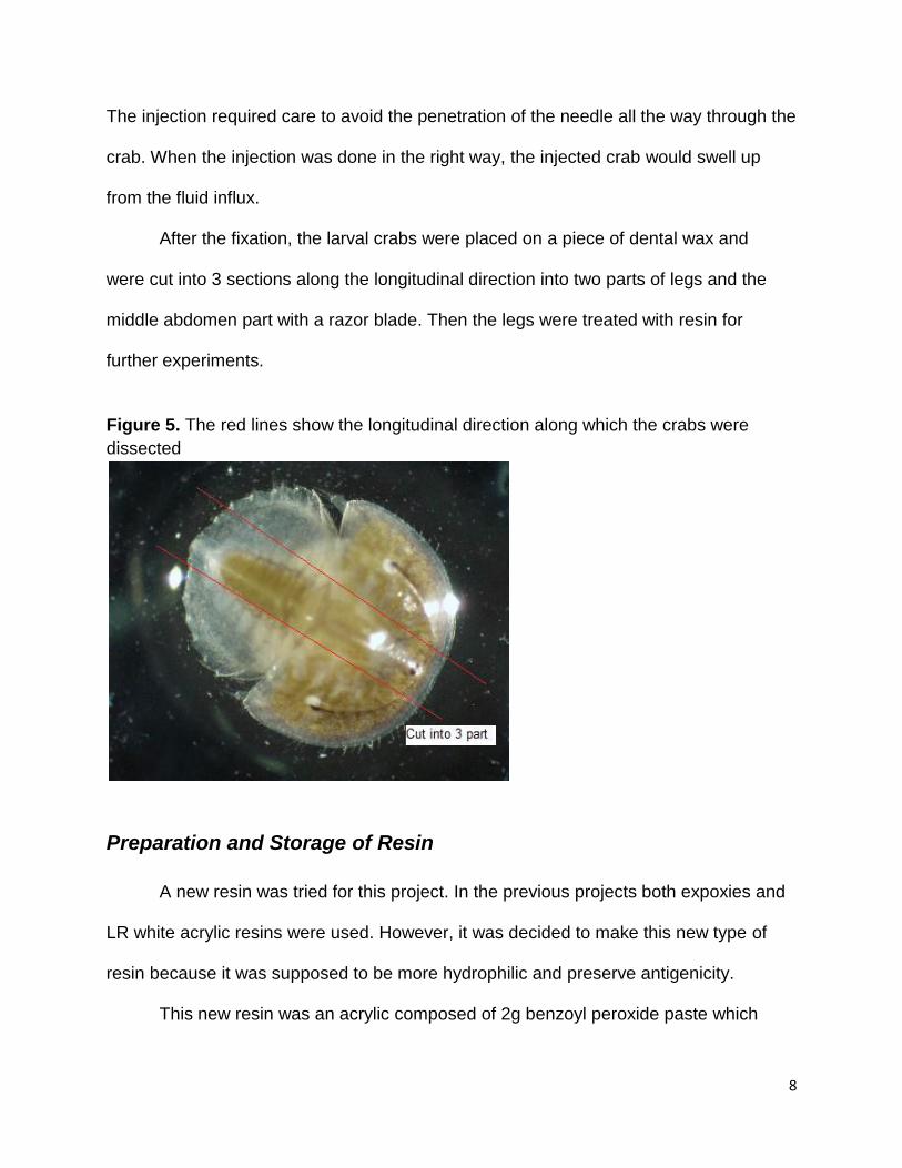
The injection required care to avoid the penetration of the needle all the way through the
crab. When the injection was done in the right way, the injected crab would swell up
from the fluid influx.
After the fixation, the larval crabs were placed on a piece of dental wax and
were cut into 3 sections along the longitudinal direction into two parts of legs and the
middle abdomen part with a razor blade. Then the legs were treated with resin for
further experiments.
Figure 5. The red lines show the longitudinal direction along which the crabs were
dissected
Preparation and Storage of Resin
A new resin was tried for this project. In the previous projects both expoxies and
LR white acrylic resins were used. However, it was decided to make this new type of
resin because it was supposed to be more hydrophilic and preserve antigenicity.
This new resin was an acrylic composed of 2g benzoyl peroxide paste which
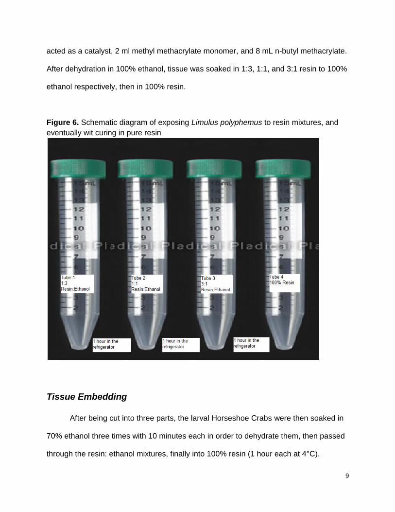
9
acted as a catalyst, 2 ml methyl methacrylate monomer, and 8 mL n-butyl methacrylate.
After dehydration in 100% ethanol, tissue was soaked in 1:3, 1:1, and 3:1 resin to 100%
ethanol respectively, then in 100% resin.
Figure 6. Schematic diagram of exposing Limulus polyphemus to resin mixtures, and
eventually wit curing in pure resin
Tissue Embedding
After being cut into three parts, the larval Horseshoe Crabs were then soaked in
70% ethanol three times with 10 minutes each in order to dehydrate them, then passed
through the resin: ethanol mixtures, finally into 100% resin (1 hour each at 4°C).
10
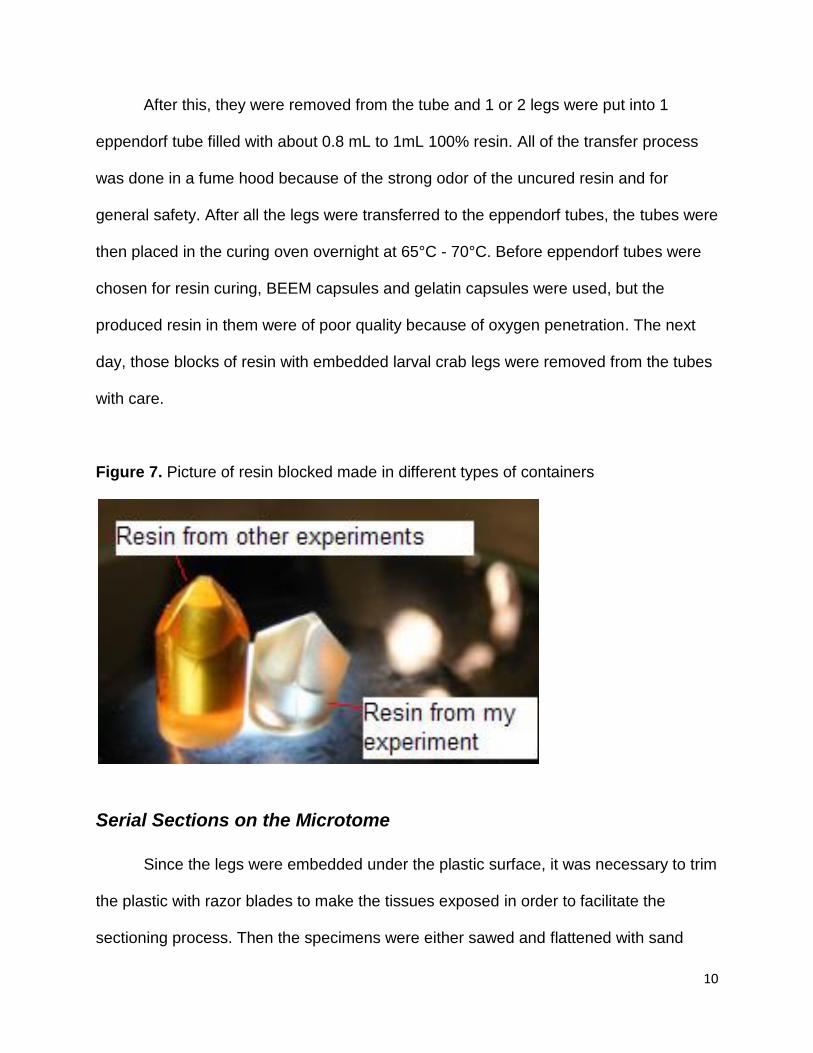
After this, they were removed from the tube and 1 or 2 legs were put into 1
eppendorf tube filled with about 0.8 mL to 1mL 100% resin. All of the transfer process
was done in a fume hood because of the strong odor of the uncured resin and for
general safety. After all the legs were transferred to the eppendorf tubes, the tubes were
then placed in the curing oven overnight at 65°C - 70°C. Before eppendorf tubes were
chosen for resin curing, BEEM capsules and gelatin capsules were used, but the
produced resin in them were of poor quality because of oxygen penetration. The next
day, those blocks of resin with embedded larval crab legs were removed from the tubes
with care.
Figure 7. Picture of resin blocked made in different types of containers
Serial Sections on the Microtome
Since the legs were embedded under the plastic surface, it was necessary to trim
the plastic with razor blades to make the tissues exposed in order to facilitate the
sectioning process. Then the specimens were either sawed and flattened with sand
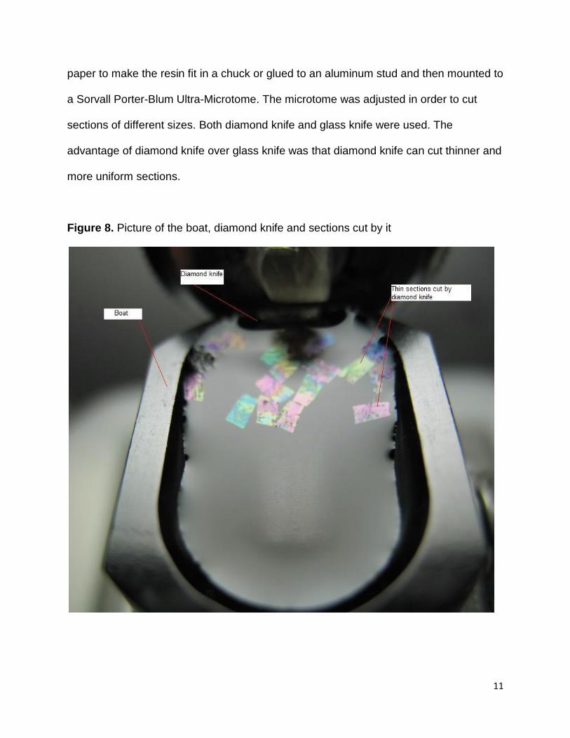
11
paper to make the resin fit in a chuck or glued to an aluminum stud and then mounted to
a Sorvall Porter-Blum Ultra-Microtome. The microtome was adjusted in order to cut
sections of different sizes. Both diamond knife and glass knife were used. The
advantage of diamond knife over glass knife was that diamond knife can cut thinner and
more uniform sections.
Figure 8. Picture of the boat, diamond knife and sections cut by it

12
Cut sections were removed from the boat with the aid of a wire loop and were
placed on Superfrost Plus Slides along the centerline. Then the slides were placed on
the hot plate at 80°C varying from 30 seconds to 1 minute to be dried. As soon as the
slide became dry and the sections were attached to the slides, the slides were removed
from the hot plate immediately to prevent the disturbance of the integrity of the proteins.
Then the sections were stained with 1% Toludine Blue for about 1 minute. Following the
staining, the slide was rinsed with distilled water and then was placed on the hot plate
again to be dried out. After becoming dry, the slide was stained with basic fuchsin
solution for another 1 minute, rinsed with distilled water and then to be dried on the hot
plate again.
Sections were observed under the light microscope to see if the leg was in the
cut sections. If they were present in the sections, further experiment could be continued,
otherwise the same procedure would be used again until sections with leg tissue could
be observed under the microscope.
Treatment of Superfrost Plus Slides
In the first few immunostaining experiments, most of the sections attached to the
regular slides were washed away during the process and therefore the experiment
could not be continued for observation. In order to continue the experiments, more
adhesive Superfrost Plus Slides were purchased from Electron Microscopy Sciences. A
permanent positive charge exists on those slides and this electrostatically attracts tissue
sections to better bind to the slides.
In order to make the slides more adhesive to the tissue sections, some of the
13
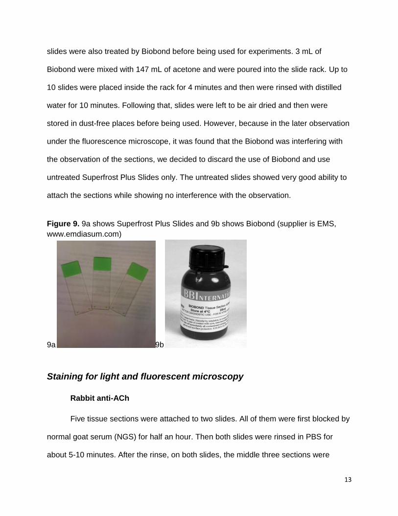
slides were also treated by Biobond before being used for experiments. 3 mL of
Biobond were mixed with 147 mL of acetone and were poured into the slide rack. Up to
10 slides were placed inside the rack for 4 minutes and then were rinsed with distilled
water for 10 minutes. Following that, slides were left to be air dried and then were
stored in dust-free places before being used. However, because in the later observation
under the fluorescence microscope, it was found that the Biobond was interfering with
the observation of the sections, we decided to discard the use of Biobond and use
untreated Superfrost Plus Slides only. The untreated slides showed very good ability to
attach the sections while showing no interference with the observation.
Figure 9. 9a shows Superfrost Plus Slides and 9b shows Biobond (supplier is EMS,
www.emdiasum.com)
9a 9b
Staining for light and fluorescent microscopy
Rabbit anti-ACh
Five tissue sections were attached to two slides. All of them were first blocked by
normal goat serum (NGS) for half an hour. Then both slides were rinsed in PBS for
about 5-10 minutes. After the rinse, on both slides, the middle three sections were
14
soaked with 1:10 dilution of Rabbit anti-ACh while the first and last sections were
soaked the same dilution of normal rabbit serum (NRS). After the application of primary
antibody, the slides were stored in two Petri dishes in order to keep them from being
dried out and then left in the refrigerator overnight.
The next morning, they were removed out of the fridge and soaked in PBS three
times for ten minutes each. The secondary antibodies used were Invitrogen Alexa Goat
anti-Rabbit Fluor 488® and Pierce ABC peroxidase staining kit.
After the rinse, one slide was soaked in 1:10 dilution of goat anti-rabbit
biotinylated secondary antibody from the ABC peroxidase staining kit with PBS for half
an hour, following that it was rinsed with PBS twice for ten minutes each and then was
soaked in the ABC reagent for another half an hour. ABC reagent is avidin bonded to
horseradish peroxidase through the biotin link, and the biotin on the secondary antibody
GAR binds to this complex and facilitates the linkage of horseradish peroxidase. ABC
reagent is composed of 45 µL of regent A and B each and 135 µL of PBS. After the
application of ABC reagent, this slide was rinsed with PBS twice for 10 minutes each
and followed by a 7 minute incubation in peroxide buffer and metal-enhanced Diamino-
benzidine. Oxidized DAB turns black or brown, indicating all reagents are bound and
ACh is present. Then the slide was processed with two more rinses with PBS for 10
minutes each and then a 3 minute rinse with distilled water. Finally, it was ready to be
observed under the microscope.
The other slide was treated with 1:10 dilution Invitrogen Goat anti-Rabbit Alexa
Fluor 488® in phosphate butter saline (PBS) for half an hour. Then it was rinsed with
PBS twice with 10 minutes each and was then counterstained with 1:10 dilution of DAPI

15
with distilled water for a few minutes and covered with a 60mm cover slip before being
observed under the fluorescence microscope. DAPI, alternatively named 4’,6-diamidino-
2-phenylindole, is one type fluorescent stain which binds to DNA strongly, bringing
about a more clear observation of the background structures of the crab on the sections.
DAPI fluorescence in near UV wavelength does not interfere with observation of the
other fluors (modified from MQP WPI, 2008, Dr. Daniel Gibson, advisor).
Mouse anti-Human AChR
Five tissue sections were attached to the two slides. All of them were first
blocked by normal goat serum (NGS) for half an hour. Then both slides were rinsed by
PBS for about 5-10 minutes. After the rinse, on both slides, the middle three sections
were soaked with 1:10 dilution of Mouse monoclonal anti-Human AChR while the first
and last sections were soaked the same dilution of normal mouse serum (NMS). After
the application of primary antibody, the slides were stored in two Petri dishes in order to
keep them from being dried out and then left in the refrigerator overnight.
The next morning, they were removed out of the fridge and soaked in PBS three
times for ten minutes each. During meanwhile, they were put inside the Petri dishes to
prevent being dried out. The secondary antibodies used were Invitrogen GAM Alexa
Fluor 555® or GAM Poly-HRP. After the rinse, one slide was soaked in 1:10 dilution of
Goat anti-Mouse Fluor 555®, following that it was rinsed with PBS twice for 10 minutes
each and then was counterstained with 1:10 dilution of DAPI for a few minutes. The
slide was then covered with a 60 mm cover slip and was observed under the
fluorescence microscope.
The other slide was treated with 1:10 dilution of poly-HRP with PBS for half hour.
16
Following that it was rinsed with PBS twice for 10 minutes each. And then it was treated
with a 7 minute incubation in peroxide- Diamino-benzidine in order to develop the brown
spots which facilitate the observation and indicate the presence of the antigens of
interest.
Staining for Transmission Electron Microscopy (TEM)
The sections needed to be blocked by 0.1M PBS-Tween 80 (0.05%)-NGS
(0.25%), pH 7.2 for 30 minutes. PBS–NGS-Tween 80 contained 50 µL Tween 80, 100
mL PBS, and 250 µL NGS. Following the blocking the sections were soaked for 2 hours
in a 1:100 dilution of primary antibody mixture with PBS –NGS-Tween 80. Primary
antibody mixture contained 5µL Rabbit anti-ACh, 5µL mouse anti-human AChR and 490
µL PBS –NGS-Tween 80. Experimental grids received primary antibody mixture
solution while control grids received 1:10 dilution of NRS solution. After two hours, the
sections were rinsed in PBS –NGS-Tween 80 three times with 10 minutes each and
allowed to soak in the last rinse for 5 minutes.
After rinsing the grids were soaked in the solution of secondary antibody
mixture for overnight incubation and were placed in the fridge. Secondary antibody
mixture contained 5 µL of GAM - Au 10nm, 5 µL GAR-Au 25nm, and 190 µL of PBS –
NGS-Tween 80. The next day, the grids were taken out of the fridge and were rinsed
with PBS –NGS-Tween 80, PBS, and distilled water once respectively for 10 minutes
each. The gold nanoparticles on secondary antibodies appear as round black speckles
when present.
17
Photography for TEM
Nickel grids (variable or 300 mesh) were inserted into a JEOL 100 CX electron
microscope for viewing at 10-7 Torr and 80 kV accelerating voltage. Images were
recorded on Kodak 4889 estar-base film, 3 ½ by 4 ¼ inches. Films were developed in
the Kodak D19 developer, fixed in Kodak rapid fix, rinsed and dried. Negatives were
scanned on a Micro-Tek Backlit Scanner and converted to positives with graphic
software.
Positive Control and Whole Mount Antibody Staining
PB Tx NGS was prepared in advance with a combination of 10mL 0.1 M PB,
30 μL Triton-X (0.3%), and 250 μL Normal Goat Serum (2.5%). Frog leg tissue was
sectioned and was placed into eppendorf tubes. Following that the sectioned tissues
were rinsed in PB Tx NGS 6 times in 3 hours. The first two rinses were 5 minutes apart
and the rest were about 45 minutes apart. Triton-X is a detergent used to permeabilize
membranes.
A mixture of Rabbit anti-ACh, Mouse anti-human AChR and 0.1 M PB Tx NGS
was prepared during the period of rinsing. Normally 1:40 to 1:80 ratio is applicable.
During the experiment, the ratio chosen was 1:40. Therefore the mixture contained 5μl
Rabbit anti-ACh, 5 μL Mouse anti-human AChR and 190 μL 0.1 M PB Tx NGS.
Following the rinse, the tissues were soaked into the mixture and placed on a Nutator in
the refrigerator at 4°C overnight. The nutator helps the tissues absorb the solution of
interest better.
The next day, the eppendorf tubes were removed out of the refrigerator and
18
rinsed with PB 6 times in 3 hours. The first two rinses were 5 minutes apart and the rest
were about 45 minutes apart.
During this period of rinse, the secondary antibody mixture was prepared. The
mixture contained 5 μL Invitrogen Alexa Goat Anti-Mouse Fluor 555® (0.5%), 5 μL
Invitrogen Alexa Goat Anti-Rabbit Fluor 488® (0.5%), and 990 μL 0.1M PB Tx NGS.
Following the rinse, the tissues were soaked into the secondary antibody mixture and
were placed on the nutator in the refrigerator at 4°C overnight.
The third day, the eppendorf tubes were removed out of the refrigerator and were
rinsed in PB using the same protocol as the one used in the previous day. After the
rinse, the tissue was removed out of the tubes and was mounted on slides covered
Fluoro-Gel. A rest period of about 20 minutes was needed for the Fluoro-gel to solidify
slightly before the sections were observed under the microscope. During this rest period,
the slides were placed in the fridge and were covered by foil (Rosahl TW, Spillane D,
Missier M, Herz J, Selig D K, Wolff JR, Hammer RE, Malenka RC, Sudhof TC. 1995).
Electrical Stimulation of Horseshoe crab Leg Motor Nerve
Even though the main focus of this project was on Immunostaining, light
microscopy, fluorescence microscopy and TEM, the technique of using electrical signals
to stimulate the motor nerve of an amputated walking leg of Limulus polyphemus was
also employed as an accessory experiment to prove the presence and expression of
ACh in the NMJs of Horseshoe Crab.
A walking leg was amputated from the Adult Horseshoe Crab. After the
amputation, the leg stopped the rhythmic clawing and was pinned on a Sylgard dish.
Then 2 micro-electrodes that were wired to a stimulator were inserted into the cut end of
19
the leg, forming an electrical circuit on the nerve of the amputated leg. On one end of
two pieces of thin wire the insulation was taken off and then was bent into a hook. The
connectors were united with the insulated ends which were connected to an amplifier
that was also connected to the oscilloscope.
The micro-electrodes leading the electrical current functioned to stimulate the
motor nerve in the leg, causing the rhythmic contractions whose speed depended on
the amount of voltage used. After everything being set up and the leg started clawing
under the electrical stimulation, α-Bungarotoxin was injected with the intention to cease
the clawing activity. At the neuromuscular junction, α-Bungarotoxin binds competitively
and irreversibly to AChR, resulting in paralysis. If the experiment were done
successfully, the leg would stop clawing gradually with an increased amount of injection
of α-Bungarotoxin.
Amputation and Glutaraldehyde Fixation Adult Horseshoe Crab Leg
Because it was desired to apply drugs on amputated Adult Horseshoe Crab Leg
to test their influences on leg contraction, it was necessary to cut the leg off the crab.
Scissors were used to cut off the leg. After the amputation, the cutting site needed to be
clamped for a while to stop the bleeding on the wound. Following that the leg was fixed
with glutaraldehyde. 0.4 mL glutaraldehyde and 10 mL seawater were mixed to make
the fixative which was later used to soak and inject the amputated leg to fix the muscle
(MQP WPI, 2005, Dr. Daniel Gibson, advisor).
20
Preparation of Glass Micropipets
Glass micropipets were made by using a World Precision Instruments PUL-1
micropipette puller. The micropipets were examined to make sure the tips were broken
to an appropriate diameter. When injection is needed, a syringe was combined with the
made micropipets via tubing that was filled with the solution of interest. The advantage
of using such micropipets is they were able insert very tiny needle into the muscle of
horseshoe crabs.
“Autoimmune Experiment”
Autoantibodies to AChR cause a muscle weakness in humans called Myasthenia
Gravis (MG). Mice will develop “Experimental Autoimmune MG” if primed with the
injection of AChR (Jon Lindstrom, Jie Luo, and Alexander Kuryatov, 10/2009). We
injected anti-AChR into living and intact first stage Larval Stage Horseshoe Crabs to
determine if an MG-like state could be induced.
Ten first stage Larval Horseshoe Crabs were chosen and were separated
into two 35mm Petri dishes filled with a shallow level of seawater.
Petri dish 1 was the experimental one and the other was the control one. For the
experimental crabs, they was injected with the antibody solution which was made from
1:10 dilution of mouse anti-Human AChR with seawater plus 200μL red food dye. The
control crabs were injected with the control solution that was composed of 1:10 dilution
of Normal Goat Serum with seawater and blue food dye. Seawater was used because
its electrolytes and concentration are similar to Horseshoe crab blood. The use of food
dye with experimental crabs was because the color facilitates the observation of the
21
injection of the crabs as well as the differentiation between control and experimental
crabs.
During the injection process, 3 mL 23 G BD Integra Syringe was used to inject
the solution of interest into the body of crabs. After the injection was done, the whole
process was recorded with a video recorder to record the activities of the crabs.
22
Results
Positive control on whole mount tissue
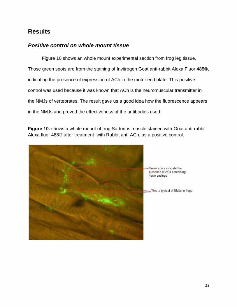
Figure 10 shows an whole mount experimental section from frog leg tissue.
Those green spots are from the staining of Invitrogen Goat anti-rabbit Alexa Fluor 488®,
indicating the presence of expression of ACh in the motor end plate. This positive
control was used because it was known that ACh is the neuromuscular transmitter in
the NMJs of vertebrates. The result gave us a good idea how the fluorescence appears
in the NMJs and proved the effectiveness of the antibodies used.
Figure 10. shows a whole mount of frog Sartorius muscle stained with Goat anti-rabbit
Alexa fluor 488® after treatment with Rabbit anti-ACh, as a positive control.
23
Immunostaining Experiment
Mouse Anti-AChR
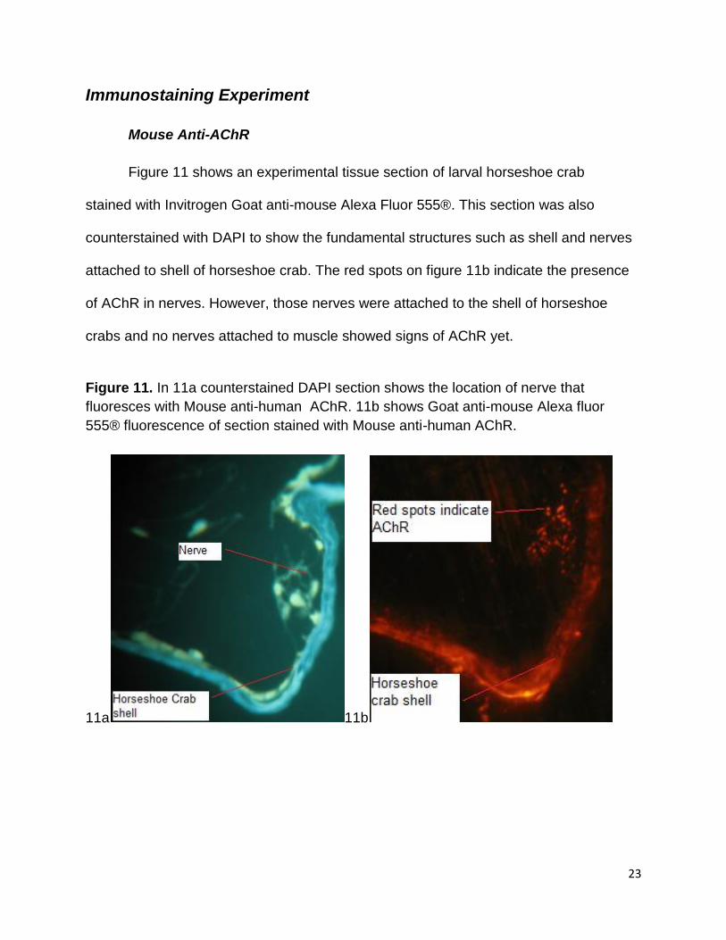
Figure 11 shows an experimental tissue section of larval horseshoe crab
stained with Invitrogen Goat anti-mouse Alexa Fluor 555®. This section was also
counterstained with DAPI to show the fundamental structures such as shell and nerves
attached to shell of horseshoe crab. The red spots on figure 11b indicate the presence
of AChR in nerves. However, those nerves were attached to the shell of horseshoe
crabs and no nerves attached to muscle showed signs of AChR yet.
Figure 11. In 11a counterstained DAPI section shows the location of nerve that
fluoresces with Mouse anti-human AChR. 11b shows Goat anti-mouse Alexa fluor
555® fluorescence of section stained with Mouse anti-human AChR.
11a 11b
24
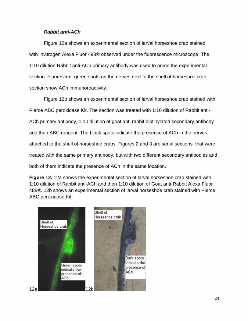
Rabbit anti-ACh
Figure 12a shows an experimental section of larval horseshoe crab stained
with Invitrogen Alexa Fluor 488® observed under the fluorescence microscope. The
1:10 dilution Rabbit anti-ACh primary antibody was used to prime the experimental
section. Fluorescent green spots on the nerves next to the shell of horseshoe crab
section show ACh immunoreactivity.
Figure 12b shows an experimental section of larval horseshoe crab stained with
Pierce ABC peroxidase Kit. The section was treated with 1:10 dilution of Rabbit anti-
ACh primary antibody, 1:10 dilution of goat anti-rabbit biotinylated secondary antibody
and then ABC reagent. The black spots indicate the presence of ACh in the nerves
attached to the shell of horseshoe crabs. Figures 2 and 3 are serial sections that were
treated with the same primary antibody, but with two different secondary antibodies and
both of them indicate the presence of ACh in the same location.
Figure 12. 12a shows the experimental section of larval horseshoe crab stained with
1:10 dilution of Rabbit anti-ACh and then 1:10 dilution of Goat anti-Rabbit Alexa Fluor
488®. 12b shows an experimental section of larval horseshoe crab stained with Pierce
ABC peroxidase Kit
12a 12b
25
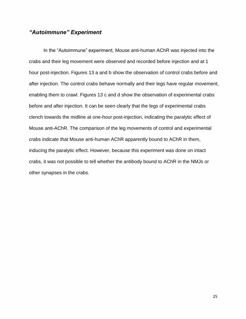
“Autoimmune” Experiment
In the “Autoimmune” experiment, Mouse anti-human AChR was injected into the
crabs and their leg movement were observed and recorded before injection and at 1
hour post-injection. Figures 13 a and b show the observation of control crabs before and
after injection. The control crabs behave normally and their legs have regular movement,
enabling them to crawl. Figures 13 c and d show the observation of experimental crabs
before and after injection. It can be seen clearly that the legs of experimental crabs
clench towards the midline at one-hour post-injection, indicating the paralytic effect of
Mouse anti-AChR. The comparison of the leg movements of control and experimental
crabs indicate that Mouse anti-human AChR apparently bound to AChR in them,
inducing the paralytic effect. However, because this experiment was done on intact
crabs, it was not possible to tell whether the antibody bound to AChR in the NMJs or
other synapses in the crabs.
26
Figure 13. 13a and b show the control crabs before and after injection of Mouse anti-
human AChR. 13c and d show the experimental crabs before and after injection of
antibodies. Normal posture is with spread, waving legs. Controls still exhibit normal
posture one hour post-injection.
13a 13b
13c 13d
27
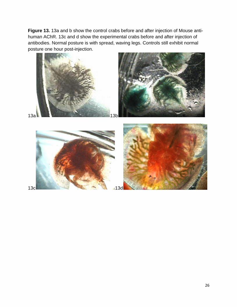
Transmission Electron Microscopy (TEM)
As previously described, the ultra-thin sections on the grids were double stained
with antibodies; Mouse anti-human AChR and Rabbit anti-Ach. Appropriate secondary
antibodies tagged with nanogold particles of 2 different sizes were used to differentiate
the two. Figure 14 shows a transmission electron micrograph of an experimental
horseshoe crab leg section. The section was stained a primary antibody mixture of
Mouse anti-human AChR and Rabbit anti-ACh and then was stained with a secondary
antibody mixture composed of GAM-Au 10nm and GAR-Au 25nm. On the micrograph,
there are many tiny round black balls (25nm) which resulted from the binding of GAR-
Au 25nm to ACh, indicating the presence of expression of ACh in the nerves. Although
ACh appears only present in the nerves in the nerves on this picture, there is a
possibility that the muscle was nearby this nerve area, but it could not be seen because
it was not involved in this section.
Figure 14. TEM 20,000X picture, GAR-Au 25nm experimental crab leg sections on nickel grids
28
Discussion:
The purpose of this project was to demonstrate the presence of ACh and AChR
in neuromuscular junctions of Limulus polyphemus, functioning as the neuromuscular
transmitter. Our approach was immunostaining against these two antigens. The two
primary antibodies used were Rabbit anti-ACh antiserum and Mouse monoclonal anti-
human AChR. Secondary antibodies were fluorescent, enzyme-linked, or linked to
nanogold. We had success at the light microscope level, demonstrating the presence of
both antigens. Immunogold TEM is thus far more equivocal.
Acetylcholine is present in the nerves of Larval Horseshoe Crabs
Figure 3 shows green fluorescent spots that were stained with the Invitrogen
Alexa Fluor 488® in the nervous systems of larval horseshoe crabs observed under the
fluorescent microscope. Those green spots indicate the expression of ACh in the nerves.
Figure 4 shows dark spots that were from the staining of Pierce ABC peroxidase
staining kit. The dark staining appears in the nerves, the same position where the green
fluorescent spots appeared in figure 3, and further demonstrate the presence of ACh in
PNS in larval horseshoe crabs. Both figures provide evidence that ACh is present in the
peripheral nervous system of horseshoe crabs. However, since the presence of Ach
was not demonstrated in neuromuscular junctions where nerves attached to muscle, our
proof is still indirect.
29
“Autoimmune Experiment” indicates involvement of AChR in nervous
system of Juvenile Horseshoe Crabs
“Autoimmune Experiment” showed dramatic paralysis with the injection of Mouse
anti-human AChR. Because the injection was made in intact first stage juvenile crabs,
we cannot tell whether the antibodies were binding to NMJs specifically or to other
synapse in the nervous system.
Future Experiments
Figure 5 shows the green spots from the staining of Invitrogen Alexa Fluor 488®
in the nervous systems of larval horseshoe crabs observed under the fluorescent
microscope in the positive control experiment. Frog leg tissues were used for the whole
mount positive control experiment because it was sure that ACh were present in the
neuromuscular junctions of frog tissue. A mixture of primary antibodies Rabbit anti-ACh
and Mouse anti-human AChR and a mixture of secondary antibodies Goat-anti-Mouse
and Goat-anti-Rabbit were employed for this experiment. Under the wavelength for
GAR, the fluorescent spots along the nerve represent the presence of ACh and its
expression. However, under the wavelength for GAM, evidence demonstrated the
presence of AChR was in the nerves, but not in found any in NMJs yet.
Therefore in the future projects, a new whole mount positive control experiment
should done on frog leg tissue to test all antibodies.
30
References:
MQP WPI, 2008, Dr. Daniel Gibson, advisor
MQP WPI, 2008, Dr. Daniel Gibson, advisor
The Horseshoe Crab, Ecological Research & Development Group
http://horseshoecrab.org/
Horseshoe Crabs, The Assateague Naturalist http://www.assateague.com/horsesho.html
Horseshoe Crabs “A living fossil”, Maryland Department of Natural Resources http://www.dnr.state.md.us/education/horseshoecrab Investigating potential treatments for the myasthenias, http://www.muscular-dystrophy.org/research/grants/1589_investigating_potential_treatments_for_the_myasthenias Kimball J, Chapter “Muscles”, Kimball’s Biology Pages, 2010 http://users.rcn.com/jkimball.ma.ultranet/BiologyPages/M/Muscles.html Lindstrom J, Luo J, and Kuryatov A, 10/2009, Myasthenia Gravis and the Tops and
Bottoms of AChRs Antigenic Structure of the MIR and Specific Immunosuppression of EAMG Using AChR Cytoplasmic Domains, Ann N Y Acad Sci. 2008;1132:29-41.
Rosahl TW, Spillane D, Missier M, Herz J, Selig D K, Wolff JR, Hammer RE, Malenka RC, Sudhof TC. 1995. Essential functions of synapsins I and II in synaptic vesicle regulation. Nature 375:488-493
Sherman, R.G. and C.R. Fourtner. 1972. Ultrastructural Features of Synaptic Regions in Walking Leg Muscles of the Horseshoe Crab, Limulus polyphemus (L.). Journal of Ultrastructure Research 40: 44-54.
Smithsonian Natioinal Museum of Nature History, http://www.mnh.si.edu/exhibits/natures_best_2006/gallery/horseshoecrabs.html


