Embed Size (px)
Citation preview

May 2011, Vol. 40 No. 5
213
Biomimetic Nanostructured Materials — Potential Regulators for Osteogenesis?Michelle Ngiam,1PhD, Luong TH Nguyen,1BSc, Susan Liao,2PhD, Casey K Chan,3,4MD, Seeram Ramakrishna,4,5,6FREng, FNAE, FAAAS
1National University of Singapore (NUS) Graduate School (NGS) for Integrative Sciences and Engineering, Centre for Life Sciences (CeLS)2School of Materials Science and Engineering, Nanyang Technological University3Department of Orthopaedic Surgery, National University Hospital (NUH)4National University of Singapore, Singapore5King Saud University, Riyadh, Saudi Arabia6Institute of Materials Research and Engineering (IMRE), SingaporeAddress for Correspondence: Prof S Ramakrishna, National University of Singapore, University Hall, Lee Kong Chian Wing Level 5, 21 Lower Kent Ridge Road, Singapore 117576.Email: [email protected]
Abstract Nanostructured materials are gaining new impetus owing to the advancements in material
fabrication techniques and their unique properties (their nanosize, high surface area-to-volume ratio, and high porosity). Such nanostructured materials mimic the subtleties of extracellular matrix (ECM) proteins, creating artifi cial microenvironments which resemble the native niches in the body. On the other hand, the isolation of mesenchymal stem cells (MSCs) from various tissue sources has resulted in the interest to study the multiple differentiation lineages for various therapeutic treatments. In this review, our focus is tailored towards the potential of biomimetic nanostructured materials as osteoinductive scaffolds for bone regeneration to differentiate MSCs towards osteoblastic cell types without the presence of soluble factors. In addition to mimicking the nanostructure of native bone, the supplement of collagen and hydroxyapatite which mimic the main components of the ECM also brings signifi cant advantages to these materials.
Ann Acad Med Singapore 2011;40:213-22
Key words: Biomaterials, Biomimetic, Bone, Hydroxyapatites, Nanomaterials, Stem cells, Tissue engineering
IntroductionBone is the second most common transplantation tissue
after blood. Globally, at least 2.2 million of bone grafting procedures are performed annually and approximately 500,000 of such procedures are done in the United States (US) alone.1-3 Figure 1 shows the orthopaedic industry by market segmentation in the US.4 It is estimated that the orthopaedic market is set to generate revenues of over US$20 billion in 2010. The US being the biggest player is said to contribute 59% of the total world orthopaedic market shares.4 Bone graft market alone is valued over US$2.5 billion.5
The ideal bone graft should possess the 3 properties namely osteoconduction, osteogenesis and osteoinduction. Osteoconduction is the ability of biocompatible scaffolds to promote the attachment, survival, migration, and distribution
of ostegogenic cells. Osteogenic graft materials contain osteogenic stem cells or progenitors to create new bone through the differentiation process. Lastly, osteoinductive bone grafts contain soluble or matrix-bound signals to initiate stem cells or progenitors towards osteoblastic cell type.6,7
Currently, autogenous and allogeneic bone grafts are the most common approaches for bone defects treatment. However, these sources of bone grafts have signifi cant disadvantages including limited supplies, the hazard of adverse immunological response and pathogenic transmission.8,9 So, synthetic bone grafts (usually calcium phosphate-based) provide an alternative bone graft option. Growth factors (e.g. bone morphogenetic protein-2 or -7 (BMP-2, BMP-7)) can be incorporated to improve their osteoinductive capabilities. The main drawbacks of these synthetic materials are that they are brittle, possess low
Osteoinductive Nanostructured Materials——Michelle Ngiam et al
Review Article

214
Annals Academy of Medicine
mechanical strength; and depending on their fabrication methods, they can be highly crystalline (due to sintering at very high temperatures of more than 1000ºC). Additionally, most biomaterials have poor surface interaction with the host tissue, resulting in the lack of adequate tissue formation around the biomaterials.10 Besides, some materials act only as passive scaffolding, so insuffi cient remodeling occurs.10 These phenomena may be caused by the fact that structural and composition properties of those materials do not resemble those of natural bone. Current bone graft systems are usually blended systems and mimic native bone only at a micro-level, such as HEALOS® Bone Graft Replacement, CopiOs® Bone Void Filler, Osteopore® PCL scaffold Bone Filler, etc. To solve those issues, many recent studies have focused on nanostructured materials which mimic the native bone at nano-level.
One of current challenges in bone tissue engineering is how to develop osteoinductive graft materials to differentiate stem cells towards osteoblasts without the presence of soluble factors. Biomimetic structured materials have been expected to do that. In this review, we summarise recent studies which have provided evidence of these materials as potential regulators for osteogenesis.
Mesenchymal Stem Cells for Bone RegenerationWork in the last decade includes evidence that stem cells
possess self-renewal, multi-lineage differentiation and in
vivo functional capabilities. Stem cells of interest include mainly embryonic stem cells (ESCs) and mesenchymal stem cells (MSCs). Embryonic stem cells (ESCs) are derived from the inner cell mass (ICM) of blastocyst-stage 5-day embryo.11 They possess high proliferative capability,12,13 are able to form 3 embryonic germ layers (endoderm, mesoderm and ectoderm),11 produce germline chimaeras,14 exhibit differentiation in teratomas11 and express specifi c ESC markers.11 However, the safety and effi cacy of hESC lines may be a concern. These include technical issues such as potential of hESC rejection and the risk of tumorigenicity. There are also ethical and religious issues involving the harvesting of donor oocytes and destruction of the blastocyst.
As such, MSCs provide an attractive alternative to ESCs and these cells can be readily obtained with less controversy from bone marrow,15 umbilical cord blood16 and adipose tissue.17 A recent study shows that the bone nodules that are formed by osteoblasts and MSCs exhibit the hallmarks of native bone, whereas those are formed by ESCs differ in terms of composition, stiffness and nano-architecture.18
More importantly, MSC has a versatile differentiation profi le. Autologous MSCs surmount immune rejection and carcinogenesis is minimised.19 Several reports stated that MSCs facilitate bone repair.20-22
MSCs are able to differentiate into many cell types such as adipocytes, chondrocytes, osteoblasts and myocytes.15 Under suitable stimuli, MSCs can be initiated to differentiate into osteoblastic cell types. This process is known as
Fig. 1. Orthopaedic industry by market segmentation in the US.4
Osteoinductive Nanostructured Materials——Michelle Ngiam et al
May 2011, Vol. 40 No. 5
215
osteogenic differentiation. The use of growth factors such as BMP and fi broblast growth factor (FGF)22-24 and osteogenic supplements (dexamethasone, β-glycerophosphate, ascorbic acid, vitamin D)25,26 are some approaches which aim to induce osteogenic differentiation. In addition, others have illustrated the benefi ts of culturing more than one cell type (co-culture) to aid in osteogenic differentiation.27 In this review, not such growth factors/ osteogenic supplements, but biomimetic nanostructured materials will be emphasised to indicate their role in the osteogenic differentiation of MSCs.
Strategy for the Design of Bone Graft MaterialsThe key tenet of tissue engineering is to regenerate
diseased, damaged tissue or organ using biodegradable materials including synthetic or natural polymers. Examples of synthetic polymers for potential bone applications include polycaprolactone (PCL),28 poly(L-lactide) (PLLA),29
poly(D, L-lactic-co-glycolide) (PLGA)30 and poly(3-hydroxybutyrate-co-3-hydroxyvalerate) (PHBV).31 Others have used natural polymers such as collagen,32 chitosan,33
alginate,34 agarose34 and silk35 in the quest for developing better bone graft materials.
The understanding of material science together with stem cell biology and signaling pathways (e.g. mitogen-activated protein kinase (MAPK) and phosphatidyl inositol-3-kinase (PI3K) etc.) is important to expedite expansion and differentiation of stem cells into tissue-specifi c lineages without changing the plasticity nature of the stem cells. Various biomaterial fabrication techniques aim to construct a microenvironment or niche similar to that in the body. During trauma and disease conditions, loss of tissue may occur and instead of being in homeostasis state, the stem cells migrate out and start their proliferative and differentiation work at the damaged site. At this site, stem cells stored in the niche are exposed to an array of soluble chemokines, cytokines, growth factors, as well as insoluble transmembrane receptor ligands and ECM proteins.36 ECM not only provides the structural and functional aspects of bone, it also provides key regulatory signals for cell proliferation and differentiation
by cell-receptor interactions, mediating the diffusion of soluble growth factors and transmitting and attenuating mechanical signals.37
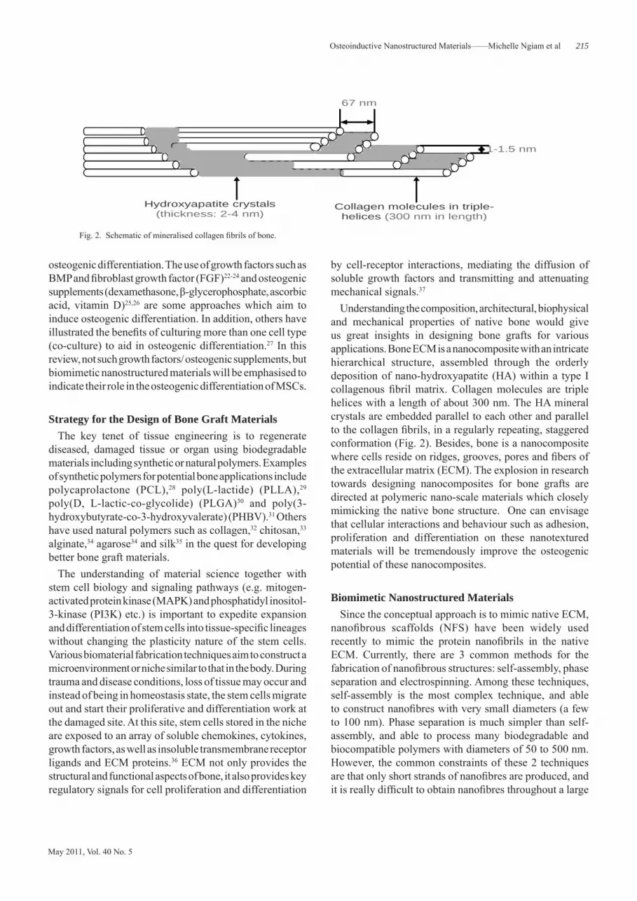
Understanding the composition, architectural, biophysical and mechanical properties of native bone would give us great insights in designing bone grafts for various applications. Bone ECM is a nanocomposite with an intricate hierarchical structure, assembled through the orderly deposition of nano-hydroxyapatite (HA) within a type I collagenous fi bril matrix. Collagen molecules are triple helices with a length of about 300 nm. The HA mineral crystals are embedded parallel to each other and parallel to the collagen fi brils, in a regularly repeating, staggered conformation (Fig. 2). Besides, bone is a nanocomposite where cells reside on ridges, grooves, pores and fi bers of the extracellular matrix (ECM). The explosion in research towards designing nanocomposites for bone grafts are directed at polymeric nano-scale materials which closely mimicking the native bone structure. One can envisage that cellular interactions and behaviour such as adhesion, proliferation and differentiation on these nanotextured materials will be tremendously improve the osteogenic potential of these nanocomposites.
Biomimetic Nanostructured MaterialsSince the conceptual approach is to mimic native ECM,
nanofi brous scaffolds (NFS) have been widely used recently to mimic the protein nanofi brils in the native ECM. Currently, there are 3 common methods for the fabrication of nanofi brous structures: self-assembly, phase separation and electrospinning. Among these techniques, self-assembly is the most complex technique, and able to construct nanofi bres with very small diameters (a few to 100 nm). Phase separation is much simpler than self-assembly, and able to process many biodegradable and biocompatible polymers with diameters of 50 to 500 nm. However, the common constraints of these 2 techniques are that only short strands of nanofi bres are produced, and it is really diffi cult to obtain nanofi bres throughout a large
Hydroxyapatite crystals (thickness: 2-4 nm)
Collagen molecules in triple-helices (300 nm in length)
67 nm
1-1.5 nm
Fig. 2. Schematic of mineralised collagen fi brils of bone.
Osteoinductive Nanostructured Materials——Michelle Ngiam et al

216
Annals Academy of Medicine
scaffold. Electrospinning is a technique used to fabricate polymeric nanofi bers by means of an electrostatic force. Electrospinning is a reliable method to fabricate long continuous strands of nanofi bres with diameters in the range of 50 ÷ 1000 nm, and these fi brous diameters can be controlled with a rather small deviation. Its fl exibility in terms of material selection and the ability to create various nanofi brous architectures (nonwoven fibre mesh, aligned fibre mesh, patterned fibre mesh and random 3-dimensional structures) have also made this process highly attractive for scaffold fabrication.38-40 Recently, a technique for fabrication and remodeling of 3D hierarchically organised nanofi brous assemblies using a dynamic liquid support system has been developed.41,42
To mimic the nanocomposite nature of bone, newer compositions of synthetic bone graft substitutes attempt to resemble the nano-HA and collagen fi brils composition of natural bone. Collagen, as one of the ECM proteins plays critical role in bone mineralisation, thus collagen is a prime candidate material for tissue-engineered graft material. Type I collagen has been used in several commercial products such as Collapat II (Biomet Inc.), Collagraft (Zimmer Inc.), Healos (Depuy Spine Inc.). Note that the above-mentioned commercial products are not tissue-
engineering nanofi brous scaffolds. As collagen has a rapid adsorption rate and possess weak mechanical strength, polymer additions are often incorporated for enhancing the mechanical properties of the material constructs. Besides, polymers by themselves lack cell recognition signals,43 and the addition of collagen provides the necessary binding sites for cell-material interactions. Polymer and collagen can be co-blended and then fabricated into nanofi brous scaffolds via electrospinning.40 In electrospinning a high voltage fi eld is applied to electrically-charge a liquid (material of interest: polymer, collagen, salts that can be fully dissolved in the appropriate solvents), resulting in nanofi bres. Calcium salts such HA,29 β-tricalcium phosphate (β-TCP) and calcium carbonate (CaCO3)
28 can also be incorporated to mimic the inorganic component of native bone and to improve the osteoconductivity of the material construct.
The reasons why these biomimetic nanostructured materials are considered as potential regulators for osteogenesis and their examples will be discussed in the following section.
Potential Regulators for OsteogenesisFigure 3 shows that regulating stem cell fate can be
b) Topographical cues - from Micro to Nano - 2D or 3D - Morphological structures
Osteoblasts Mineralised bone
Pits/Pores
Grooves/Ridges
Smooth/Flat surface
MSC
Attachment
Cell spreading
Pre-osteoblast
Media supplements
Osteoblasts Mineralised bone
a) Chemical cues
Osteoblasts Mineralised bone
Surface modification
Peptides/Functional groups
Osteoinductive Nanostructured Materials——Michelle Ngiam et al

May 2011, Vol. 40 No. 5
217
achieved through various means, as such chemical, topographical, mechanical and electrical or electromagnetic cues. For chemical cues, media supplements or peptides/ functional groups can be added into the environment to differentiate MSCs into osteoblasts. Besides, topographical cues such as size affect (micro/nano), architecture form (2D/3D) and morphological structures (pits/grooves/ridges/pores), etc are able to help MSCs differentiated. Additionally, various stress stimuli applied to substrate and/or cell construct, called mechanical cues, have been supposed to induce osteogenesis. Lastly, the differentiation of MSCs into osteoblasts can be stimulated by electrical and electromagnetic cues (through application of electrical or electromagnetic currents/fi elds to stimulate substrate and/or cell construct). In this review, we focus on the importance of topographical features and substrate characteristics of biomimetic nanostructured materials to inducing/enhancing MSC differentiation.
Nanofi brous scaffolds being in nanometer scale (in diameter) are said to resemble the ECM proteins, and such microenvironment is conducive for cellular interaction.
Nanotexture is said to infl uence cell activity. Cells are subjected to topographical features such as protein folding, collagen bending within a niche in vivo. Nanoscale disorder has shown to stimulate osteogenic stem cell differentiation without chemical treatments.44 Such geometric cues have demonstrated a dominant effect on adhesion, spreading, growth and differentiation of MSCs in several studies. Lateral spacing geometry of TiO2 nanotubes of 30 to 50 nm was reported to be the critical threshold for cell fate.45 Diameter (<15 nm) and spacing (<30 nm) was considered to be the effective length scale for augmenting integrin clustering and focal contact formation. Good evidence showed that smaller diameter nanotubes (15 nm) were associated with greater focal contact formation, stress fi ber (contractile actomyosin bundles or actin fi laments) assembly, cell spreading and osteocalcin differentiation compared to larger diameter nanotubes (100 nm). On the other hand, larger diameter nanotubes (>50 nm) resulted in the reduction in cellular activity, fewer focal contact and stress fi bers and even programmed cell death.45 In a separate study, it was shown that larger diameter nanotubes enhanced cell spreading compared to smaller diameter and
Fig. 3. Regulating cell fate through various cues. (a) Chemical (through use of media chemicals or surface modifi cation), (b) Topographical (through surface features [such as pits, grooves, ridges, pores etc.], architectural form [2D vs 3D] or size effect [micro, nano scale]), (c) Mechanical (through various stress stimuli applied to substrate and/or cell construct) and (d) Electrical and electromagnetic cues (through application of electrical or electromagnetic currents/fi elds to stimulate substrate and/or cell construct).
Osteoblasts Mineralised bone
d) Electrical or Electromagnetic cues
Osteoblasts Mineralised bone
c) Mechanical cues
Stiffness
Elasticity
Stretch
Compression
Vibration
Osteoinductive Nanostructured Materials——Michelle Ngiam et al
218
Annals Academy of Medicine
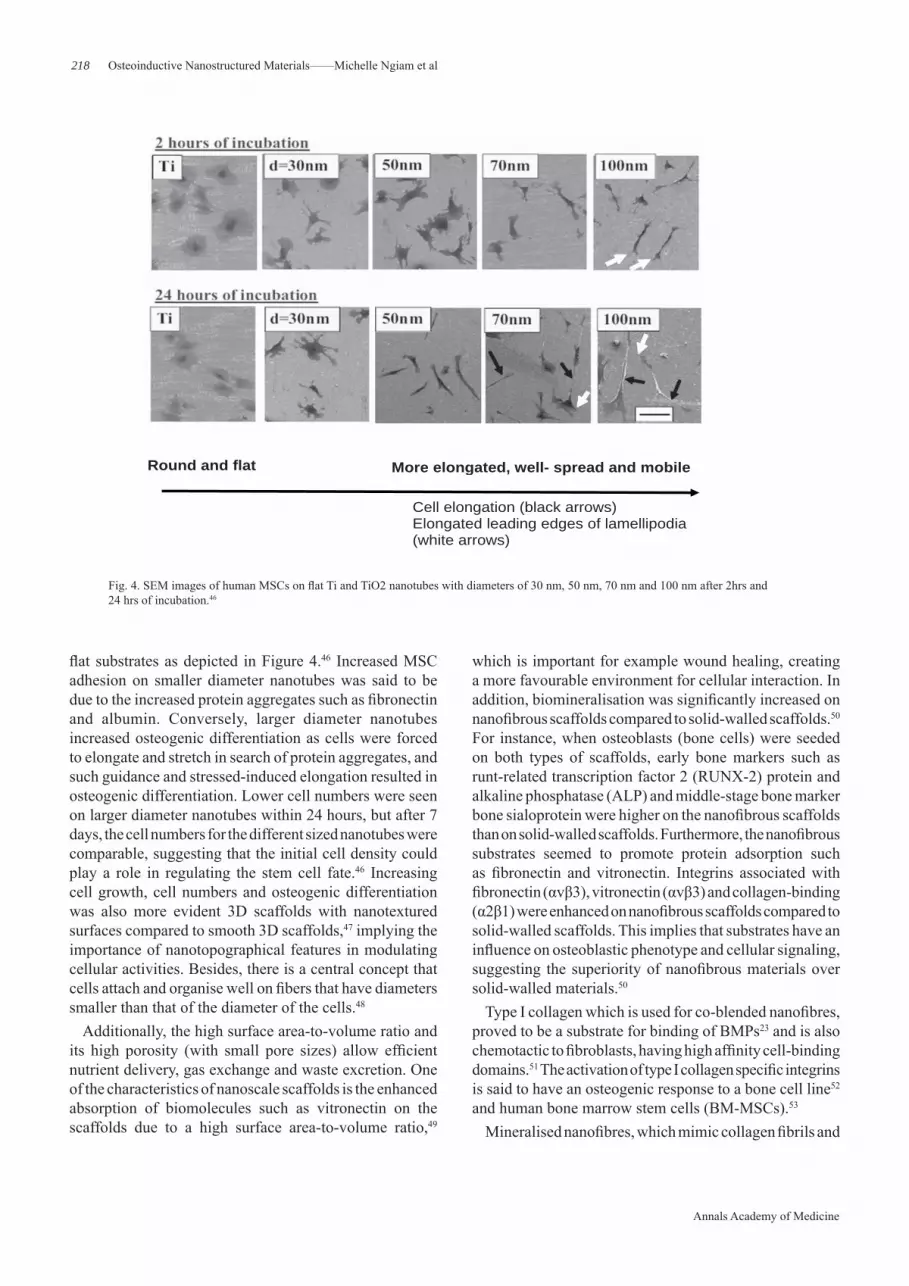
fl at substrates as depicted in Figure 4.46 Increased MSC adhesion on smaller diameter nanotubes was said to be due to the increased protein aggregates such as fi bronectin and albumin. Conversely, larger diameter nanotubes increased osteogenic differentiation as cells were forced to elongate and stretch in search of protein aggregates, and such guidance and stressed-induced elongation resulted in osteogenic differentiation. Lower cell numbers were seen on larger diameter nanotubes within 24 hours, but after 7 days, the cell numbers for the different sized nanotubes were comparable, suggesting that the initial cell density could play a role in regulating the stem cell fate.46 Increasing cell growth, cell numbers and osteogenic differentiation was also more evident 3D scaffolds with nanotextured surfaces compared to smooth 3D scaffolds,47 implying the importance of nanotopographical features in modulating cellular activities. Besides, there is a central concept that cells attach and organise well on fi bers that have diameters smaller than that of the diameter of the cells.48
Additionally, the high surface area-to-volume ratio and its high porosity (with small pore sizes) allow effi cient nutrient delivery, gas exchange and waste excretion. One of the characteristics of nanoscale scaffolds is the enhanced absorption of biomolecules such as vitronectin on the scaffolds due to a high surface area-to-volume ratio,49
which is important for example wound healing, creating a more favourable environment for cellular interaction. In addition, biomineralisation was signifi cantly increased on nanofi brous scaffolds compared to solid-walled scaffolds.50
For instance, when osteoblasts (bone cells) were seeded on both types of scaffolds, early bone markers such as runt-related transcription factor 2 (RUNX-2) protein and alkaline phosphatase (ALP) and middle-stage bone marker bone sialoprotein were higher on the nanofi brous scaffolds than on solid-walled scaffolds. Furthermore, the nanofi brous substrates seemed to promote protein adsorption such as fi bronectin and vitronectin. Integrins associated with fi bronectin (αvβ3), vitronectin (αvβ3) and collagen-binding (α2β1) were enhanced on nanofi brous scaffolds compared to solid-walled scaffolds. This implies that substrates have an infl uence on osteoblastic phenotype and cellular signaling, suggesting the superiority of nanofi brous materials over solid-walled materials.50
Type I collagen which is used for co-blended nanofi bres, proved to be a substrate for binding of BMPs23 and is also chemotactic to fi broblasts, having high affi nity cell-binding domains.51 The activation of type I collagen specifi c integrins is said to have an osteogenic response to a bone cell line52 and human bone marrow stem cells (BM-MSCs).53
Mineralised nanofi bres, which mimic collagen fi brils and
Fig. 4. SEM images of human MSCs on fl at Ti and TiO2 nanotubes with diameters of 30 nm, 50 nm, 70 nm and 100 nm after 2hrs and 24 hrs of incubation.46
Rouund and flaat MMore elong
Cell elonElongate(white ar
gated, well
ngation (blaed leading errows)
- spread a
ack arrows) edges of la
nd mobile
mellipodia
Osteoinductive Nanostructured Materials——Michelle Ngiam et al

May 2011, Vol. 40 No. 5
219
nano-HA in native bone, elevated osteoblastic activities compared to non-mineralised nanofi bres.30 Figure 5 shows the nanotextured surfaces of mineralized nanofi bres, where prominent groves and ridges are more evident on HA fi bers than non-HA fi bers.30 The importance of closely mimicking the natural composition of bone can be delineated in several studies.29,30,54 For instance, enhanced mineral deposition (57% higher) was observed when osteoblasts were grown on PLLA/Collagen/HA nanofi bres compared to PLLA/HA nanofi bres, suggestive of the synergistic effect of collagen and HA in osteogenic differentiation and bone mineralisation.54 Many studies have shown that BM-MSCs are capable of differentiating towards an osteoblastic lineage.32,37,55-57 It was also shown that when MSCs were cultured on HA surfaces, osteo-specifi c genes were up-regulated.55,57 Not only the viability of human MSCs was not affected, the expression of ALP, osteogenic genes and calcium mineralisation of the MSCs were elevated when the cells were cultured on blended PLGA and nano-HA nanofi bres.58 It was speculated that when the cells interacted with HA, potent inductive substances were released. Using this conditioned media after the initial culture, uncommitted MSCs were then cultured without the presence of HA and upregulation of osteo-specifi c genes were observed.57 Although some reports stated that HA induced osteogenic differentiation of MSCs and other cell types, there were also confl icting reports which saw an attenuation in osteogenic differentiation when cells were cultured on HA surfaces.59,60 This could be due to the physical and chemical characteristics of the HA material such as crystallinity and particle size etc.59
A landmark paper highlighted the importance of matrix stiffness and its infl uence in directing MSC commitment towards a specifi c lineage.61 Briefl y, soft matrices were associated with neurogenic differentiation, stiffer matrices were corresponded to myogenic differentiation and lastly rigid matrices were related with osteogenic differentiation.61
In a separate study, the stiffness of substrates (PEG-based materials) affected differentiation of pre-osteoblastic cells via mitogen-activated protein kinase (MAPK) activation.62
It was reported that such ECM rigidity regulated osteogenic differentiation involving MAPK activation downstream of the RhoA-ROCK signaling cascade.63 Early osteogenic differentiation markers, such as RUNX-2 and ALP expression were associated with stiffer materials.62,63
The elasticity of the substrates also impinges upon cell proliferation, where stiffer substrates resulted up to 10-fold increase in cell numbers compared to lower stiffness substrates.64 Interestingly, osteogenic differentiation of MSC were signifi cantly increased on collagen-I coated substrates with the highest modulus,64 suggesting that substrate elasticity alone did not direct stem cell fate, but
rather a network of factors such as the presence of integrins and integrin-receptor interactions was also likely at work. This highlights the importance of designing materials that are more closely related to the microenvironments found in native tissues.
Several studies have shown that the dimensionality of the substrate (2D vs 3D) has an impact on cell fate and signaling cascade. Three-dimensional scaffolds provide more precise, reproducible nano-topographical features and such nano-texturing is usually absent in 2D substrates. Certain stress mediators such as p38 and c-Jun N-terminal kinase (JNK) were signifi cantly activated in 3D calcium phosphate scaffolds, thereby indicating that cells response to environmental signals, triggering certain signaling pathways such as MAPK cascade. This phenomenon was less evident in 2D calcium phosphate scaffolds.65
Current Perspectives, Challenges and Future DirectionsThe substrate composition, dimensionality, mechanical
properties, nanotopographical cues, elasticity, biophysical characteristics, biochemical signaling regulatory networks are some important factors that affect the differentiation lineage of MSCs. Nevertheless, the mechanisms of stem cell biology and cell-material interactions needs to be further harnessed. Standardising culture techniques and conditions for the expansion and differentiation of stem cells, and also narrowing down to a few material substrates from the plethora of material choices is a gargantuan task. The arduous, time-intensive culture process of MSC expansion can also be daunting. Kinks need to be worked out, such as the establishment of more robust culture protocols (controlled expansion and differentiation into specifi c lineages), effective cell delivery systems to ensure cell survival, and designing the appropriate material carriers suitable for specifi c clinical conditions (e.g. trauma, spinal fusion, fractures, maxillofacial reconstruction, cranial and dental applications). It is also imperative to ameliorate scale-up and characterisation efforts for effective MSC-based therapies. Particularly, growth factors that are used to hasten MSC differentiation and directly isolating MSCs from various tissue sources may not give rise to one cell-type exclusivity. In some instances, it may be more important to have an enrichment of the cell of interest. Other challenges include matching the mechanical properties of the substrates to bone and supporting angiogenesis in tissue-engineered constructs. Although harvesting MSCs from bone marrow is the gold standard, a recent study showed that adipocytes that reside in bone marrow could antagonise the haematopoietic activity in the bone marrow niche.66 By suppressing marrow adipogenesis, haematopoietic activity may have been improved but balancing between osteogenesis and adipogenesis has to be considered as both osteoblasts and
Osteoinductive Nanostructured Materials——Michelle Ngiam et al

220
Annals Academy of Medicine
(a) (b)
(c) (d)
Fig. 5. Atomic force microscopy (AFM) images of nanotexturing of mineralized nanofi bers.30 (a) PLGA, (b) PLGA/Col, (c) 3D surface topography of (a) PLGA , (d) 3D surface topography of (b) PLGA/Col, (e) PLGA+HA, (f) PLGA/Col+HA, (g) 3D surface topography of (e) PLGA+HA and (h) 3D surface topography of (f) PLGA/Col+HA.
(e) (f)
(g) (h)
Osteoinductive Nanostructured Materials——Michelle Ngiam et al

May 2011, Vol. 40 No. 5
221
adipocytes originate from bone marrow MSCs and they have a reciprocal relationship.67 Other reasons stymieing the clinical application of tissue-engineered constructs on a wide scale is the lack of large clinical trials. Most in vivo work involves animal models and there should be concerted effort in carrying out clinical trials to elucidate the true performance of tissue-engineered graft materials. These unaddressed issues call for the global push for collaborative work which aims to accelerate its clinical application.
AcknowledgementsWe would like to acknowledge our funding sources, National Medical
Research Council, Singapore (NMRC/1151/2008) and the National University of Singapore, Singapore.
REFERENCES1. Greenwald AS, Boden SD, Goldberg VM, Khan Y, Laurencin CT, Rosier
RN; American Academy of Orthopaedic Surgeons. The Committee on Biological Implants. Bone-graft substitutes: facts, fi ctions, and applications. J Bone Joint Surg Am 2001;83:S98-S103.
2. Steinmann JC, Herkowitz HN. Pseuarthrosis of the spine. Clin Orthop 1992;284:80-90.
3. Lee K, Roper J, Wang J. Demineralized bone matrix and spinal arthrodesis. Spine J 2005;5:S217S-S2223.
4. U.S. Medical Devices Market Outlook 2008. Frost & Sullivan, 5:16-5:28.5. Desai BM. Osteobiologics. Am J Orthop 2007;36:8-11.6. Lee K, Roper J, Wang J. Demineralized bone matrix and spinal arthrodesis.
Spine J 2005;5:S217-S223.7. Muschler GF, Raut VP, Patterson TE, Wenke JC, Hollinger JO. The
design and use of animals models for translational research in bone tissue engineering and regenerative medicine. Tissue Eng Part B: Rev 2010;16:123-45.
8. Both SK, van der Muijsenberg AJ, van Blitterswijk CA, de Boer J, de Bruijn JD. A rapid and effi cient method for expansion of human mesenchymal stem cells. Tissue Eng 2007;13:3-9.
9. Tian XF, Heng BC, Ge X, Lu K, Rufaihah AJ, Fan VT, et al. Comparison of osteogenesis of human embryonic stem cells within 2D and 3D culture systems. Scand J Clin Lab Invest 2008;68:58-67.
10. Chan CK, Kumar TS, Liao S, Murugan R, Ngiam M, Ramakrishnan S. Biomimetic nanocomposites for bone graft applications. Nanomedicine (Lond) 2006;1:177-88.
11. Thomson JA, Itskovitz-Eldor J, Shapiro SS, Waknitz MA, Swiergiel JJ, Marshall VS, et al. Embryonic stem cells lines derived from human blastocysts. Science 1998;282:1145-7.
12. Evans M, Kaufman M. Establishment in culture of pluripotential cells from mouse embryos. Nature 1981;292:154-6.
13. Martin G. Isolation of a pluripotent cell line from early mouse embryos cultured in medium conditioned by tetracarcinoma stem cells. Proc Natl Acad Sci USA 1981;78:7634-8.
14. Bradley A, Evans M, Kaufman MH, Robertson E. Formation of germ-like chimaeras from embryo-derived teratocarcinoma cell lines. Nature 1984;309:255-6.
15. Pittenger MF, Mackey AM, Beck SC, Jaiswal RK, Douglas R, Mosca JD, et al. Multilineage potential of adult human mesenchymal stem cells. Science 1999;284:143-7.
16. Wang HS, Hung SC, Peng ST, Huang CC, Wei HM, Guo YJ, et al.
Mesechymal stem cells in the Wharton’s jelly of the human umbilical cord. Stem Cells 2004;22:1330-7.
17. Jeon O, Rhie JW, Kwon IK, Kim JH, Kim BS, Lee SH. In vivo bone formation following transplantation of human adipose-derived stromal cells that are not differentiated osteogenically. Tissue Eng Part A 2008;14:1285-94.
18. Gentleman E, Swain RJ, Evans ND, Boonrungsiman S, Jell G, Ball MD, et al. Comparative materials differences revealed in engineered bone as a function of cell-specifi c differentiation. Nat Mater 2009;8:763-70.
19. Chen Y, Shao JZ, Xiang LX, Dong XJ, Zhang GR. Mesenchymal stem cells: a promising candidate in regenerative medicine. Int J Biochem Cell Biol 2008;40:815-20.
20. Nakajima T, Iizuka H, Tsutsumi S, Kayakabe M, Takagishi K. Evaluation of posterolateral spinal fusion using mesenchymal stem cells: differences with or without osteogenic differentation. Spine 2007;32:2432-6.
21. Morishita T, Honoki K, Ohgushi H, Kotobuki N, Matsushima A, Takakura Y. Tissue engineering approach to the treatment of bone tumors: three cases of cultured bone grafts derived from patients’ mesenchymal stem cells. Artif Organs 2006;30:115-8.
22. Minamide A, Yoshida M, Kawakami M, Okada M, Enyo Y, Hashizume H, et al. The effects of bone morphogenetic protein and basic fi broblast growth factor on cultured mesenchymal stem cells for spine fusion. Spine (Phila Pa 1976) 2007;32:1067-71.
23. Reddi AH. Role of morphogenetic proteins in skeletal tissue engineering and regeneration. Nat Biotechnol 1998;16:247-52.
24. Liao SS, Guan K, Cui FZ, Shi SS, Sun TS. Lumbar spinal fusion with a mineralized collagen matrix and rhBMP-2 in a rabbit model. Spine 2003;28:1954-60.
25. Jørgensen NR, Henriksen Z, Sørensen OH, Civitelli R. Dexamethasone, BMP-2, and 1,25-dihydroxyvitamin D enhance a more differentiated osteoblast phenotype: validation of an in vitro model for human bone marrow-derived primary osteoblasts. Steroids 2004;69:219-26.
26. Gupta A, Leong DT, Bai HF, Singh SB, Lim TC, Hutmacher DW. Osteo-maturation of adipose-derived stem cells required the combined action of vitamin D3, beta-glycerophosphate, and ascorbic acid. Biochem Biophys Res Commun 2007;362:17-24.
27. Choong CS, Hutmacher DW, Triffi tt JT. Co-culture of bone marrow fibroblasts and endothelial cells on modified polycaprolactone substrates for enhanced potentials in bone tissue engineering. Tissue Eng 2006;12:2521-31.
28. Fujihara K, Kotaki M, Ramakrishna S. Guided bone regeneration membrane made of polyprolactone/calcium carbonated composite nano-fi bers. Biomaterials 2005;26:4139-47.
29. Ngiam M, Liao S, Patil AJ, Cheng Z, Yang F, Gubler MJ, et al. Fabrication of mineralized polymeric nanofi brous composites for bone graft materials. Tissue Eng Part A 2009;15:535-46.
30. Ngiam M, Liao S, Patil AJ, Cheng Z, Chan CK, Ramakrishna S. The fabrication of nano-hydroxyapatite on PLGA and PLGA/collagen nanofi brous composite scaffolds and their effects in osteoblastic behavior for bone tissue engineering. Bone 2009;45:4-16.
31. Ke Y, Wang YJ, Ren L, Zhao QC, Huang W. Modifi ed PHBV scaffolds by in situ UV polymerization: structural characteristic, mechanical properties and bone mesenchymal stem cell compatibility. Acta Biomater 2010;6:1329-36
32. Shih YRV, Chen CN, Tsai SW, Wang YJ, Lee OK. Growth of mesenchymal stem cells on electrospun type I collagen nanofi bers. Stem Cells 2006;24:2391-7.
33. Oliveira JM, Rodrigues MT, Silva SS, Malafaya PB, Gomes ME, Viegas CA, et al. Novel hydroxyapatite/chitosan bilayered scaffold for osteochondral tissue-engineering applications: Scaffold design and its performance when seeded with goat bone marrow stromal cells.
Osteoinductive Nanostructured Materials——Michelle Ngiam et al

222
Annals Academy of Medicine
Biomaterials 2006;27:6123-37.34. Awad H, Wickham M, Leddy H, Gimble J, Guilak F. Chondrogenic
differentiation of adipose-derived adult stem cells in agarose, alginate, and gelatin scaffolds. Biomaterials 2004;25:3211-22.
35. Li C, Vepari C, Jin HJ, Kim HJ, Kaplan DL. Electrospun silk-BMP-2 scaffolds for bone tissue engineering. Biomaterials 2006;27: 3115-24.
36. Lutolf MP, Gilbert PM, Blau HM. Designing materials to direct stem-cell fate. Nature 2009;462:433-41.
37. Dawson JI, Wahl DA, Lanham SA, Kanczler JM, Czernuszka JT, Oreffo ROC. Development of specifi c collagen scaffolds to support the osteogenic and chondrogenic differentiation of human bone marrow stromal cells. Biomaterials 2008;29:3105-16.
38. Vasita R, Katti DS. Nanofi bers and their applications in tissue engineering. Int J Nanomedicine 2006;1:15-30.
39. Saw S, Wang K, Yong T, Ramakrishna S. Polymeric nanofi bers in tissue engineering. In: Challa SSR Kumar, editor. Tissue, Cell and Organ Engineering. Wiley-VCH Verlag GmbH & Co, 2006.
40. Teo WE, Ramakrishna S. A review on electrospinning design and nanofi bre assemblies. Nanotechnology 2006;17:R89-R106.
41. Teo W, Gopal R, Ramaseshan R, Fujihara K, Ramakrishna S. A dynamic liquid support system for continuous electrospun yarn fabrication. Polymer 2007;48:3400-5.
42. Teo W, Liao S, Chan C, Ramakrishna S. Remodeling of three-dimensional hierarchically organized nanofi brous assemblies. Current Nanoscience 2008;4:361-9.
43. Kim BS, Mooney DJ. Development of biocompatible synthetic extracellular matrices for tissue engineering. Trends Biotechnol 1998;16:224-30.
44. Dalby MJ, Gadegaard N, Tare R, Andar A, Riehle MO, Herzyk P, et al. The control of human mesenchymal cell differentiation using nanoscale symmetry and disorder. Nat Mater 2007;6:997-1003.
45. Park J, Bauer S, von der Mark K, Schmuki P. Nanosize and vitality: TiO2 nanotube diameter directs cell fate. Nano Lett 2007;7:1686-91.
46. Oh S, Brammer KS, Li YS, Teng D, Engler AJ, Chien S, et al. Stem cell fate dictated solely by altered nanotube dimension. Proc Natl Acad Sci U S A 2009;106:2130-5.
47. Mata A, Kim EJ, Boehm CA, Fleischman AJ, Muschler GF, Roy S. A three-dimensional scaffold with precise micro-architecture and surface micro-textures. Biomaterials 2009;30:4610-7.
48. Laurencin CT, Ambrosio AM, Borden MD, Cooper JA. Tissue engineering: orthopedic applications. Annu Rev Biomed Eng 1999;1:19-46.
49. Woo KM, Chen VJ, Ma PX. Nano-fi brous scaffolding architecture selectively enhances protein adsorption contributing to cell attachment. J Biomed Mater Res A 2003;67:531-7.
50. Woo KM, Jun JH, Chen VJ, Seo J, Baek JH, Ryoo HM, et al. Nano-fi brous scaffolding promotes osteoblast differentiation and biomineralization. Biomaterials 2007;28:335-43.
51. Yang XB, Bhatnagar RS, Li S, Oreffo RO. Biomimetic collagen scaffolds for human bone cell growth and differentiation. Tissue Eng
2004;10:1148-59.52. Jikko A, Harris SE, Chen D, Mendrick DL, Damsky CH. Collagen integrin
receptors regulate early osteoblast differentiation induced by BMP-2. J Bone Miner Res 1999;14:1075-83.
53. Mizuno M, Fujisawa R, Kuboki Y. Type I collagen-induced osteoblastic differentiation of bone-marrow cells mediated by collagen-alpha2beta1 integrin interaction. J Cell Physiol 2000;184:207-13.
54. Prabhakaran MP, Venugopal J, Ramakrishna S. Electrospun nanostructured scaffolds for bone tissue engineering. Acta Biomater 2009;5:2884-93.
55. Bernhardt A, Lode A, Boxberger S, Pompe W, Gelinsky M. Mineralised collagen—an artifi cial, extracellular bone matrix--improves osteogenic differentiation of bone marrow stromal cells. J Mater Sci Mater Med 2008;19:269-75.
56. Ichinohe N, Takamoto T, Tabata Y. Proliferation, osteogenic differentiation, and distribution of rat bone marrow stromal cells in nonwoven fabrics by different culture methods. Tissue Eng Part A 2008;14:107-16.
57. Lin L, Chow KL, Leng Y. Study of hydroxyapatite osteoinductivity with an osteogenic differentiation of mesenchymal stem cells. J Biomed Mater Res A 2009;89:326-35.
58. Lee JH, Rim NG, Jung HS, Shin H. Control of osteogenic differentiation and mineralization of human mesenchymal stem cells on composite nanofi bers containing poly[lactic-co-(glycolic acid)] and hydroxyapatite. Macromol Biosci 2010;10:173-82.
59. Deligianni DD, Katsala ND, Koutsoukos PG, Missirlis YF. Effect of surface roughness of hydroxyapatite on human bone marrow cell adhesion, proliferation, differentiation and detachment strength. Biomaterials 2001;22:87-96.
60. Chou Y, Dunn J, Wu B. In vitro response of MC3T3-E1 pre-osteoblasts within three-dimensional apatite-coated PLGA scaffolds. J Biomed Mater Res B Appl Biomater 2005;75:81-90.
61. Engler AJ, Sen S, Sweeney HL, Discher DE. Matrix elasticity directs stem cell lineage specifi cation. Cell 2006;126:677-89.
62. Khatiwala CB, Peyton SR, Metzke M, Putnam AJ. The regulation of osteogenesis by ECM rigidity in MC3T3-E1 cells requires MAPK activation. J Cell Physiol 2007;211:661-72.
63. Khatiwala CB, Kim PD, Peyton SR, Putnam AJ. ECM compliance regulates osteogenesis by infl uencing MAPK signaling downstream of RhoA and ROCK. J Bone Miner Res 2009;24:886-98.
64. Rowlands AS, George PA, Cooper-White JJ. Directing osteogenic and myogenic differentiation of MSCs: interplay of stiffness and adhesive ligand presentation. Am J Physiol Cell Physiol 2008;295:C1037-44.
65. Appleford MR, Oh S, Cole JA, Carnes DL, Lee M, Bumgardner JD, et al. Effects of trabecular calcium phosphate scaffolds on stress signaling in osteoblast precursor cells. Biomaterials 2007;28:2747-53.
66. Naveiras O, Nardi V, Wenzel PL, Hauschka PV, Fahey F, Daley GQ. Bone-marrow adipocytes as negative regulators of the haematopoietic microenvironment. Nature 2009;460:259-63.
67. Nuttall ME, Gimble JM. Controlling the balance between osteoblastogenesis and adipogenesis and the consequent therapeutic implications. Curr Opin Pharmacol 2004;4:290-4.
Osteoinductive Nanostructured Materials——Michelle Ngiam et al


