Embed Size (px)
Citation preview
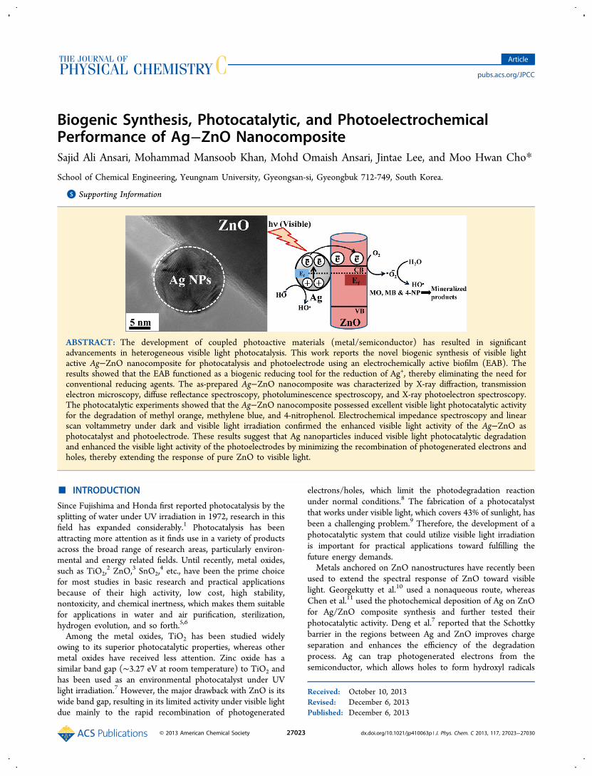
Biogenic Synthesis, Photocatalytic, and PhotoelectrochemicalPerformance of Ag−ZnO NanocompositeSajid Ali Ansari, Mohammad Mansoob Khan, Mohd Omaish Ansari, Jintae Lee, and Moo Hwan Cho*
School of Chemical Engineering, Yeungnam University, Gyeongsan-si, Gyeongbuk 712-749, South Korea.
*S Supporting Information
ABSTRACT: The development of coupled photoactive materials (metal/semiconductor) has resulted in significantadvancements in heterogeneous visible light photocatalysis. This work reports the novel biogenic synthesis of visible lightactive Ag−ZnO nanocomposite for photocatalysis and photoelectrode using an electrochemically active biofilm (EAB). Theresults showed that the EAB functioned as a biogenic reducing tool for the reduction of Ag+, thereby eliminating the need forconventional reducing agents. The as-prepared Ag−ZnO nanocomposite was characterized by X-ray diffraction, transmissionelectron microscopy, diffuse reflectance spectroscopy, photoluminescence spectroscopy, and X-ray photoelectron spectroscopy.The photocatalytic experiments showed that the Ag−ZnO nanocomposite possessed excellent visible light photocatalytic activityfor the degradation of methyl orange, methylene blue, and 4-nitrophenol. Electrochemical impedance spectroscopy and linearscan voltammetry under dark and visible light irradiation confirmed the enhanced visible light activity of the Ag−ZnO asphotocatalyst and photoelectrode. These results suggest that Ag nanoparticles induced visible light photocatalytic degradationand enhanced the visible light activity of the photoelectrodes by minimizing the recombination of photogenerated electrons andholes, thereby extending the response of pure ZnO to visible light.
■ INTRODUCTION
Since Fujishima and Honda first reported photocatalysis by thesplitting of water under UV irradiation in 1972, research in thisfield has expanded considerably.1 Photocatalysis has beenattracting more attention as it finds use in a variety of productsacross the broad range of research areas, particularly environ-mental and energy related fields. Until recently, metal oxides,such as TiO2,
2 ZnO,3 SnO2,4 etc., have been the prime choice
for most studies in basic research and practical applicationsbecause of their high activity, low cost, high stability,nontoxicity, and chemical inertness, which makes them suitablefor applications in water and air purification, sterilization,hydrogen evolution, and so forth.5,6
Among the metal oxides, TiO2 has been studied widelyowing to its superior photocatalytic properties, whereas othermetal oxides have received less attention. Zinc oxide has asimilar band gap (∼3.27 eV at room temperature) to TiO2 andhas been used as an environmental photocatalyst under UVlight irradiation.7 However, the major drawback with ZnO is itswide band gap, resulting in its limited activity under visible lightdue mainly to the rapid recombination of photogenerated
electrons/holes, which limit the photodegradation reactionunder normal conditions.8 The fabrication of a photocatalystthat works under visible light, which covers 43% of sunlight, hasbeen a challenging problem.9 Therefore, the development of aphotocatalytic system that could utilize visible light irradiationis important for practical applications toward fulfilling thefuture energy demands.Metals anchored on ZnO nanostructures have recently been
used to extend the spectral response of ZnO toward visiblelight. Georgekutty et al.10 used a nonaqueous route, whereasChen et al.11 used the photochemical deposition of Ag on ZnOfor Ag/ZnO composite synthesis and further tested theirphotocatalytic activity. Deng et al.7 reported that the Schottkybarrier in the regions between Ag and ZnO improves chargeseparation and enhances the efficiency of the degradationprocess. Ag can trap photogenerated electrons from thesemiconductor, which allows holes to form hydroxyl radicals
Received: October 10, 2013Revised: December 6, 2013Published: December 6, 2013
Article
pubs.acs.org/JPCC
© 2013 American Chemical Society 27023 dx.doi.org/10.1021/jp410063p | J. Phys. Chem. C 2013, 117, 27023−27030
that can then react with the organic species, resulting in theirdegradation.12−14 However, most reported methods useharmful chemicals, whose discharge into the environment is amajor issue of concern. Therefore, there is increasing need for anovel route for metal−metal oxide nanocomposite synthesis.Electrochemically active microorganisms form electrochemi-
cally active biofilms (EABs) on solid surfaces and have potentialapplications in bioenergy and chemical production.15 EABshave attracted considerable attraction in bio electrochemicalsystems (BESs), such as microbial fuel cells, where they act as aliving bioanode and produce excess electrons and protons bybiologically oxidizing a range of substrates. The flow of theseelectrons produces a considerable amount of electricity.16
Recently, it was reported that EABs could be used to reduce theband gap of metal oxide,17,18 synthesize noble metal nano-particles,19,20 hydrogen energy,21 and metal−semiconductornanocomposites with higher efficiency than other syntheticmethods.22−24
As part of an ongoing study of EABs, this work focused onthe use of EABs for the decoration of pure ZnO nanostructures(p-ZnO) by silver nanoparticles (AgNPs). This work reports avery simple, biogenic, and facile method for the fabrication ofAg−ZnO nanocomposite (Ag−ZnO) by EAB as a reducingtool, which is far superior and fast compared to other microbialsynthesis.25 The enhancement in photocatalytic activity wasattributed to the presence of AgNPs at the surface of p-ZnOand used for the degradation of methyl orange (MO),methylene blue (MB), and 4-nitrophenol (4-NP) in an aqueoussolution under visible light irradiation. A possible chargetransfer mechanism for the Ag−ZnO nanocomposite todegrade dyes, such as MO, MB, and 4-NP, is suggested. TheEIS experiment under dark and visible light irradiation furthersupported the visible light activity of the Ag−ZnO nano-composite, suggesting that Ag−ZnO is a good candidate forvisible light photocatalysis and photoelectrodes. Ag−ZnOshowed significantly high photocatalytic activity compared top-ZnO. The method reported in this work, which does notinvolve the use of hazardous chemicals, capping agents andreducing agent etc., is simple and has great potential forcommercialization.
■ EXPERIMENTAL SECTIONMaterials. Nanosized zinc oxide (nano ZnO), silver nitrate
(AgNO3, 99% pure), and methylene blue (MB) were purchasedfrom Sigma−Aldrich. Sodium acetate, methyl orange (MO), p-nitrophenol (4-NP) and sodium sulfate (Na2SO4) wereobtained from Duksan Pure Chemicals Co. Ltd. South Koreaand were used as received. Ethyl cellulose and α-terpineol werepurchased from KANTO Chemical Co., Japan and fluorine-doped transparent conducting oxide glass (FTO; F-dopedSnO2 glass; 7 Ω/sq) was purchased from Pilkington, U.S.Carbon paper (without wet proof, Fuel Cell Earth LLC, U.S.),and all other chemicals used in this study were of analyticalgrade and used as received. All solutions were prepared fromdeionized water obtained using a PURE ROUP 30 waterpurification system.Methods. A UV−vis-NIR spectrophotometer (VARIAN,
Cary 5000, U.S.) was used to record the diffuse reflectance/absorbance spectra (DRS) of the commercial pure ZnO (p-ZnO) and Ag−ZnO nanocomposite (Ag−ZnO) samples in therange of 200−800 nm. The photoluminescence (PL) of thesamples (Ag−ZnO and p-ZnO) was recorded over the range of200−800 nm using a Kimon, 1 K, Japan spectroscope. X-ray
photoelectron spectroscopy (XPS, ESCALAB 250) wasperformed using a monochromatized Al Kα X-ray source (hν= 1486.6 eV) with a 500 μm spot size. The binding energy ofC1s at 284.8 eV was used to calibrate the other bindingenergies. X-ray diffraction (XRD, PANalytical, X’pert PRO-MPD, Netherland) was carried out using Cu Kα radiation (λ =0.15405 nm). The XRD peaks of the crystalline phases werecompared with those of the standard compounds reported inthe JCPDS data file. The size and distribution of the Ag−ZnOand p-ZnO nanoparticles were observed by field emissiontransmission electron microscopy (FE-TEM, Tecnai G2 F20,FEI, U.S.) with an accelerating voltage of 200 kV combinedwith energy dispersive spectrometry (EDS). The photo-electrochemical and photocatalytic experiments were per-formed using a 400 W lamp with irradiating intensity of 31.0mW cm−2 (λ > 500 nm, 3M, U.S.). Electrochemical impedancespectroscopy (EIS) was performed in a three electrode cell witha 0.2 M Na2SO4 aqueous solution as the electrolyte using apotentiostat (VersaSTAT 3, Princeton Research, U.S.). Theworking electrodes were prepared as follows: 100 mg of p-ZnOand Ag−ZnO were suspended thoroughly using a conditioningmixer by adding ethyl cellulose as a binder and α-terpineol as asolvent for the paste, and then coated on FTO glass electrodeusing the doctor-blade method. The p-ZnO and Ag−ZnO-coated (FTO) glass substrates were used as the workingelectrode. Ag/AgCl (saturated with KCl) and a Pt gauge wereused as the reference and counter electrodes.
Electrochemically Active Biofilm (EAB) Preparation.The EABs were developed on plain carbon paper according toprevious reports.15−24 In a typical procedure, carbon paper(without wet proof, Fuel Cell Earth LLC) with a size of 2.5 ×4.5 cm2 was dipped into a mineral salt medium containingsodium acetate (1 g L−1) as the substrate and carbon source ina 250 mL bottle. Ten mL of anaerobic sludge (from a biogasplant in Paju, Korea) was added under strict anaerobicconditions by sparging N2 gas for 5 min to remove theenvironmental oxygen. All media, including the bacterialinoculum, were changed every two days under strict anaerobicconditions. This process was repeated for two weeks, and aliving EAB formed on the surface of the carbon paper.
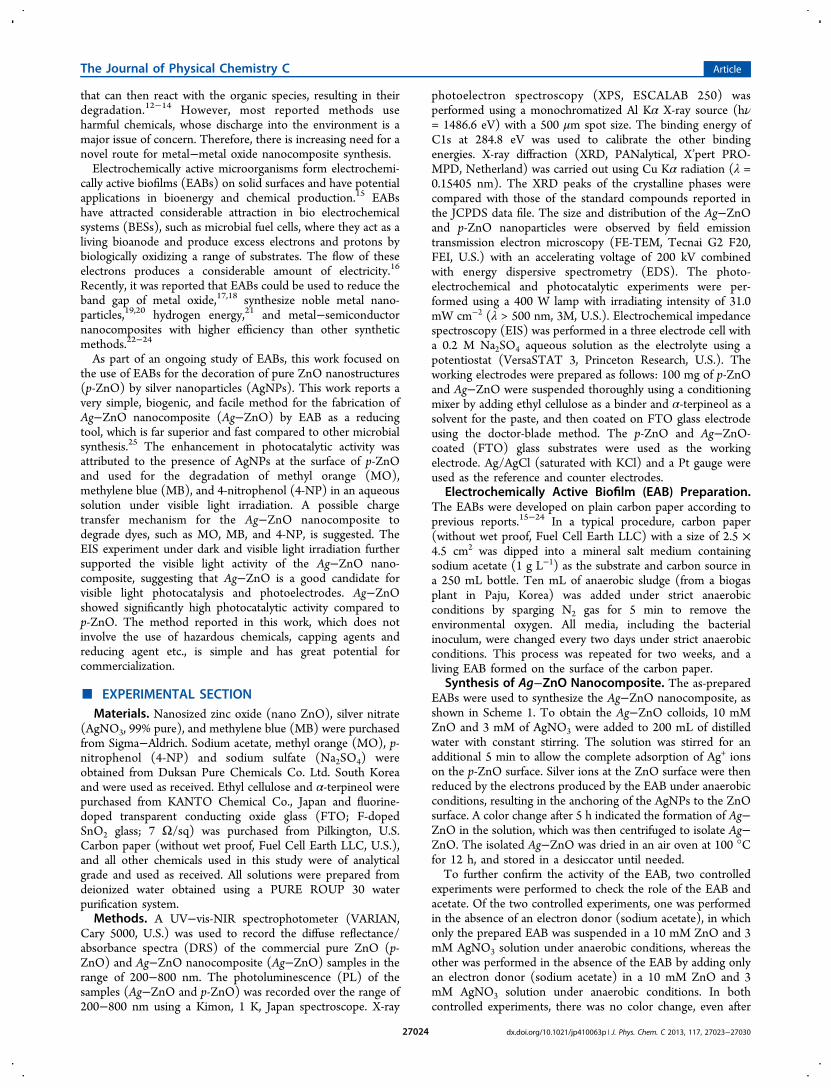
Synthesis of Ag−ZnO Nanocomposite. The as-preparedEABs were used to synthesize the Ag−ZnO nanocomposite, asshown in Scheme 1. To obtain the Ag−ZnO colloids, 10 mMZnO and 3 mM of AgNO3 were added to 200 mL of distilledwater with constant stirring. The solution was stirred for anadditional 5 min to allow the complete adsorption of Ag+ ionson the p-ZnO surface. Silver ions at the ZnO surface were thenreduced by the electrons produced by the EAB under anaerobicconditions, resulting in the anchoring of the AgNPs to the ZnOsurface. A color change after 5 h indicated the formation of Ag−ZnO in the solution, which was then centrifuged to isolate Ag−ZnO. The isolated Ag−ZnO was dried in an air oven at 100 °Cfor 12 h, and stored in a desiccator until needed.To further confirm the activity of the EAB, two controlled
experiments were performed to check the role of the EAB andacetate. Of the two controlled experiments, one was performedin the absence of an electron donor (sodium acetate), in whichonly the prepared EAB was suspended in a 10 mM ZnO and 3mM AgNO3 solution under anaerobic conditions, whereas theother was performed in the absence of the EAB by adding onlyan electron donor (sodium acetate) in a 10 mM ZnO and 3mM AgNO3 solution under anaerobic conditions. In bothcontrolled experiments, there was no color change, even after
The Journal of Physical Chemistry C Article
dx.doi.org/10.1021/jp410063p | J. Phys. Chem. C 2013, 117, 27023−2703027024
24 h. These experiments confirmed the role of EAB and acetatein the synthesis of the Ag−ZnO nanocomposite.Evaluation of Dye Degradation. The photocatalytic
activity of Ag−ZnO nanocomposite was evaluated by thedegradation of MO, MB, and 4-NP under visible lightirradiation (λ > 500 nm). In a typical process, 2 mg of eachp-ZnO and Ag−ZnO photocatalyst were added to threedifferent 20 mL MO (10 mg L−1), 20 mL MB (10 mg L−1),and 20 mL of 4-NP (5 mg L−1) solutions, and agitated for 30min in the dark to achieve the adsorption−desorptionequilibrium. The above suspension were irradiated with visiblelight and after a certain time interval (1 h), 2 mL of the solutionwas taken, and the catalyst was separated from the solution bycentrifugation to obtain a clear liquid from which UV−visspectra was measured. The decolorization efficiency of thephotocatalyst was determined using the formula reportedelsewhere.18,22
Photoelectrochemical Studies (EIS and LSV). Toexamine the photoelectrochemical response of the p-ZnO andas-synthesized Ag−ZnO, EIS and LSV experiments were carriedout under ambient conditions in the dark and under visiblelight irradiation in 50 mL of a 0.2 M Na2SO4 aqueous solutionat room temperature. For each electrode, EIS was firstperformed in the dark and later under visible light irradiation(λ > 500 nm) at 0.0 V and with a frequency ranging from 1 to104 Hz. The photocurrent response was obtained by LSV underdark and visible light irradiation at a scan rate of 50 mV/s overa potential range of −0.7 to 1.0 V.
■ RESULTS AND DISCUSSION
Proposed Reaction Mechanism and Its Confirmation.EABs function as a reducing tool and provide an excess ofelectrons and protons by biologically decomposing sodiumacetate.15−24 These electrons help in anchoring the metalnanoparticles to the surface of the ZnO nanostructure, asshown in Scheme 1. The advantage of this protocol is that itdoes not involve any external energy input, which makes thissynthesis highly useful and efficient in the field of nano-composite syntheses.15−24 Another exciting feature of thissynthesis is that it occurs in water at room temperature and
does not involve harmful chemicals, capping/reducing agents orsevere treatments.
→ ++ −AgNO Ag NO3(aq) (aq) 3 (aq) (1)
+ ̅ →+Ag e Ag(aq) (from EAB)0
(2)
+ →− +NO H HNO3 (aq) (from EAB) 3 (3)
To confirm the proposed mechanism and reaction stepsinvolved in the synthesis of Ag−ZnO, the pH of the solutionwas measured before and after completion of the reaction. ThepH of the reaction media decreased during the course of thereaction, and after 5 h, the pH changed from 7.9 to 6.8 due tothe formation of acid (HNO3) according to eqs 1, 2, and 3.Therefore, Ag+ was reduced by the EAB generated electronsand the formation of acid (HNO3) occurred by the EABgenerated protons.
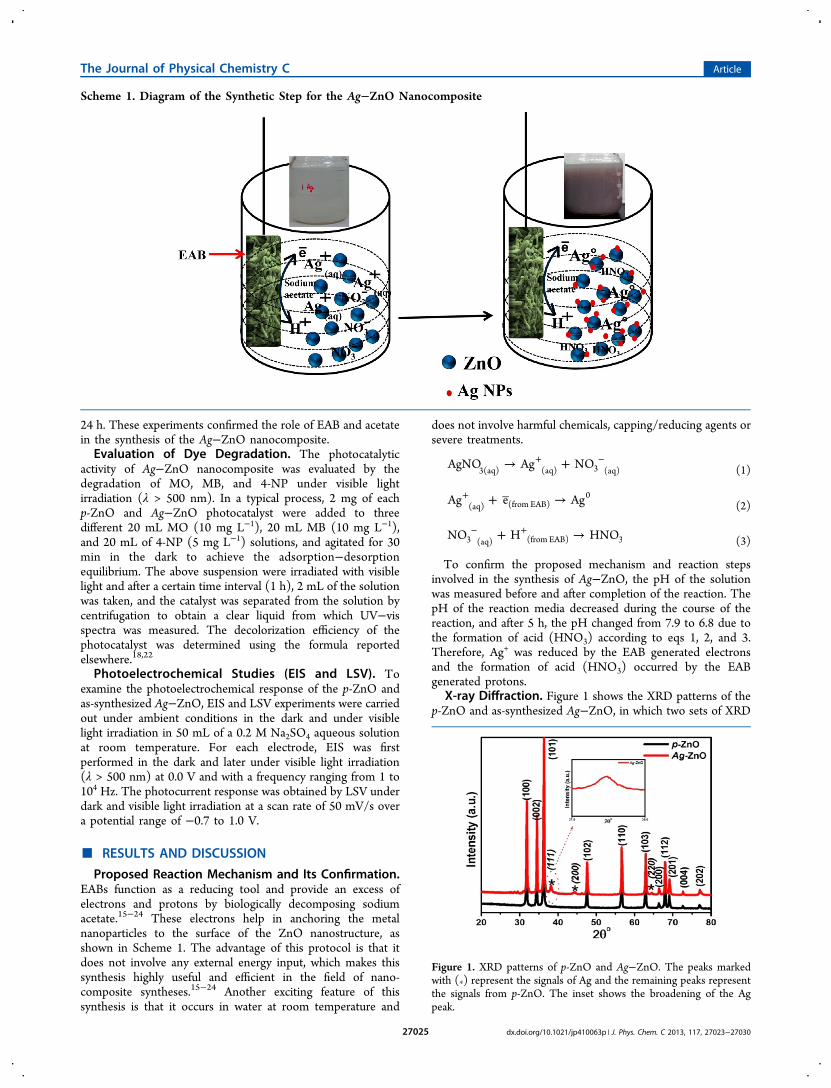
X-ray Diffraction. Figure 1 shows the XRD patterns of thep-ZnO and as-synthesized Ag−ZnO, in which two sets of XRD
Scheme 1. Diagram of the Synthetic Step for the Ag−ZnO Nanocomposite
Figure 1. XRD patterns of p-ZnO and Ag−ZnO. The peaks markedwith (*) represent the signals of Ag and the remaining peaks representthe signals from p-ZnO. The inset shows the broadening of the Agpeak.
The Journal of Physical Chemistry C Article
dx.doi.org/10.1021/jp410063p | J. Phys. Chem. C 2013, 117, 27023−2703027025
patterns are obtained: those unmarked were indexed to the wellcrystalline hexagonal wurtzite ZnO structure corresponding toJCPDS (36-1451), whereas the other peaks marked with “*”were assigned to face centered cubic (fcc) silver correspondingto JCPDS (04-0783).10,11 Compared to p-ZnO, the XRDpattern of ZnO after Ag anchoring was similar, suggesting thatsilver was not incorporated into the ZnO lattice. This can beattributed to the significantly larger ionic radius of Ag+ (126pm) than Zn2+ (74 pm). Therefore, a silver metallic phase wasformed successfully on the surface of ZnO. The crystallize size,which was calculated using the Scherrer formula, was ∼30 nmand ∼33.5 nm for p-ZnO and Ag−ZnO, respectively.22 Theincrease in the crystallite size of Ag−ZnO might be due to theanchoring of very small AgNPs at the surface of p-ZnO. The Ag(111) XRD peak is relatively weak and broad, as shown in theinset in Figure 1, indicating a small size and well dispersed Ag atthe surface of p-ZnO, which is also consistent with the HR-TEM image.Transmission Electron Microscope. Supporting Informa-
tion, SI, Figure S1 and Figure 2 shows low and high-resolution
TEM images of p-ZnO and as prepared Ag−ZnO, respectively.Figure 2c shows that the AgNPs are anchored to the ZnOsurface with a particle size of ∼7−12 nm. HRTEM (Figure 2d)revealed an interface and the continuity of lattice fringesbetween the AgNPs and ZnO. The lattice fringes of d = 0.26nm matched those of the (002) crystallographic plane of ZnO.The fringe spacing at 0.24 nm corresponds to the (111) planeof the face-centered cubic phase of AgNPs. HAADF-STEM ofp-ZnO and Ag−ZnO are shown in SI Figures S2 and S3,respectively, whereas SI Figures S4 and S5 show the EDXspectra of the p-ZnO and Ag−ZnO, corresponding to the Ag(K), Zn (K), and O (K) lines. The EDX-elemental spectra werealso supported by the elemental composition, as shown in SITables S1 and S2 for p-ZnO and Ag−ZnO, respectively. Thisfurther confirms the presence of the AgNPs in Ag−ZnOnanocomposite. EDX (SI Figure S5) also indicates the presenceof the AgNPs in Ag−ZnO nanocomposite. The HRTEMresults were in good agreement with the XRD result. Theseresults show that Ag−ZnO has highly crystalline structure witha small and relatively uniform distribution of AgNPs at the ZnOsurface, which is useful for the photocatalytic activities.
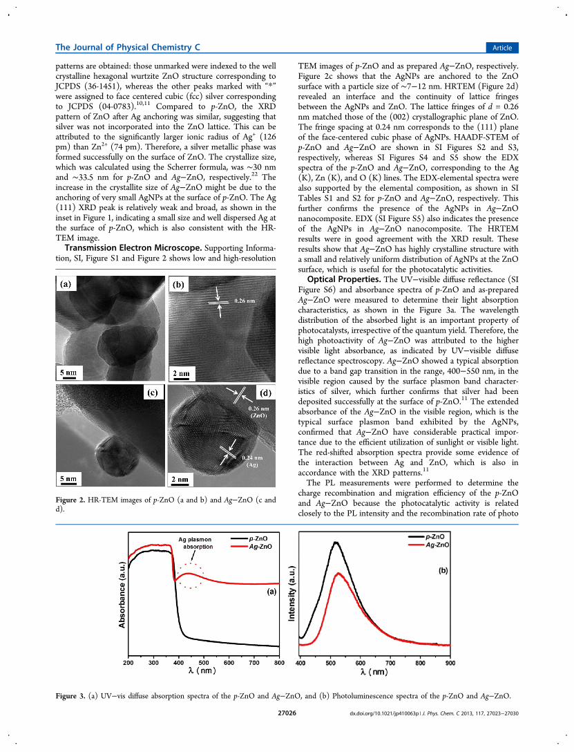
Optical Properties. The UV−visible diffuse reflectance (SIFigure S6) and absorbance spectra of p-ZnO and as-preparedAg−ZnO were measured to determine their light absorptioncharacteristics, as shown in the Figure 3a. The wavelengthdistribution of the absorbed light is an important property ofphotocatalysts, irrespective of the quantum yield. Therefore, thehigh photoactivity of Ag−ZnO was attributed to the highervisible light absorbance, as indicated by UV−visible diffusereflectance spectroscopy. Ag−ZnO showed a typical absorptiondue to a band gap transition in the range, 400−550 nm, in thevisible region caused by the surface plasmon band character-istics of silver, which further confirms that silver had beendeposited successfully at the surface of p-ZnO.11 The extendedabsorbance of the Ag−ZnO in the visible region, which is thetypical surface plasmon band exhibited by the AgNPs,confirmed that Ag−ZnO have considerable practical impor-tance due to the efficient utilization of sunlight or visible light.The red-shifted absorption spectra provide some evidence ofthe interaction between Ag and ZnO, which is also inaccordance with the XRD patterns.11
The PL measurements were performed to determine thecharge recombination and migration efficiency of the p-ZnOand Ag−ZnO because the photocatalytic activity is relatedclosely to the PL intensity and the recombination rate of photo
Figure 2. HR-TEM images of p-ZnO (a and b) and Ag−ZnO (c andd).
Figure 3. (a) UV−vis diffuse absorption spectra of the p-ZnO and Ag−ZnO, and (b) Photoluminescence spectra of the p-ZnO and Ag−ZnO.
The Journal of Physical Chemistry C Article
dx.doi.org/10.1021/jp410063p | J. Phys. Chem. C 2013, 117, 27023−2703027026
excited electrons and holes. Figure 3b shows the PL spectra ofp-ZnO and Ag−ZnO measured at an excitation wavelength of325 nm. The emission intensity of the PL spectrum of Ag−ZnO was lower than that of p-ZnO suggesting that theanchoring of AgNPs could quench the fluorescence from theZnO nanoparticles and prolong electron−hole pair lifetime.22,26A lower photoluminescence intensity means a lower electron−hole recombination rate, and hence a longer lifetime of thephotogenerated carriers.22,26 In general, the efficient chargeseparation and inhibited electron−hole recombination byAgNPs are favorable for enhancing the photocatalytic activityof p-ZnO. The PL spectra showed that anchoring AgNPs to thesurface of ZnO could effectively inhibit electrons and holesrecombination during the photocatalytic reaction under visiblelight irradiation.XPS Analysis. The surface chemical composition and
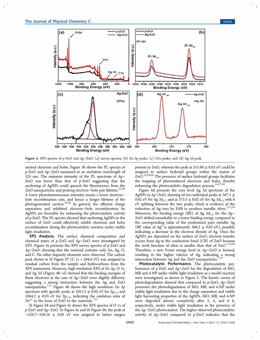
chemical states of p-ZnO and Ag−ZnO were investigated byXPS. Figure 4a presents the XPS survey spectra of p-ZnO andAg−ZnO, showing that the material contains only Zn, Ag, O,and C. No other impurity elements were observed. The carbonpeak shown in SI Figure S7 (C 1s = 284.8 eV) was assigned toresidual carbon from the sample and hydrocarbons from theXPS instrument. However, high-resolution XPS of Zn 2p, O 1s,and Ag 3d (Figure 4b−d) showed that the binding energies ofthese electrons in the case of Ag−ZnO were slightly different,suggesting a strong interaction between the Ag and ZnOnanoparticles.7,11 Figure 4b shows the high resolution Zn 2pspectrum with specific peaks at 1021.5 ± 0.02 eV for 2p3/2 and1044.2 ± 0.03 eV for 2p1/2, indicating the oxidation state ofZn2+ in the form of ZnO in the materials.7,11
SI Figure S8 and Figure 4c shows the XPS spectra of O 1s ofp-ZnO and Ag−ZnO. In Figure 4c and SI Figure S8, the peak at∼528.7−530.16 ± 0.05 eV was assigned to lattice oxygen
present in ZnO, whereas the peak at 531.60 ± 0.03 eV could beassigned to surface hydroxyl groups within the matrix ofZnO.14,18,26 The presence of surface hydroxyl groups facilitatesthe trapping of photoinduced electrons and holes, therebyenhancing the photocatalytic degradation process.14,27,28
Figure 4d presents the core level Ag 3d spectrum of theAgNPs in Ag−ZnO, showing of two individual peaks at 367.5 ±0.02 eV for Ag 3d5/2 and at 373.5 ± 0.02 eV for Ag 3d3/2 with 6eV splitting between the two peaks, which is evidence of thereduction of Ag ions by EAB to produce metallic silver.7,11,14
Moreover, the binding energy (BE) of Ag 3d5/2 for the Ag−ZnO shifted remarkably to a lower binding energy compared tothe corresponding value of the synthesized pure metallic Ag(BE value of Ag0 is approximately 368.2 ± 0.03 eV), possiblyindicating a decrease in the electron density of Ag. Once theAgNPs are deposited on the surface of ZnO, electron transferoccurs from Ag to the conduction band (CB) of ZnO becausethe work function of silver is smaller than that of ZnO.7,14,28
Therefore, a new Fermi energy level in Ag−ZnO is formed,resulting in the higher valence of Ag, indicating a stronginteraction between Ag and the ZnO nanoparticles.7,14
Photocatalytic Performance. The photocatalytic per-formance of p-ZnO and Ag−ZnO for the degradation of MO,MB and 4-NP under visible light irradiation as a model reactionwere investigated, as shown in Figure 5. The kinetic curves ofphotodegradation showed that compared to p-ZnO, Ag−ZnOpromotes the photodegradation of MO, MB, and 4-NP undervisible light irradiation due to the charge separation and visiblelight harvesting properties of the AgNPs. MO, MB, and 4-NPwere degraded almost completely after 5, 4, and 6 h,respectively, under visible light irradiation in the presence ofthe Ag−ZnO photocatalyst. The higher observed photocatalyticactivity of Ag−ZnO compared to p-ZnO indicates that the
Figure 4. XPS spectra of p-ZnO and Ag−ZnO: (a) survey spectra; (b) Zn 2p peaks; (c) O1s peaks; and (d) Ag 3d peak.
The Journal of Physical Chemistry C Article
dx.doi.org/10.1021/jp410063p | J. Phys. Chem. C 2013, 117, 27023−2703027027
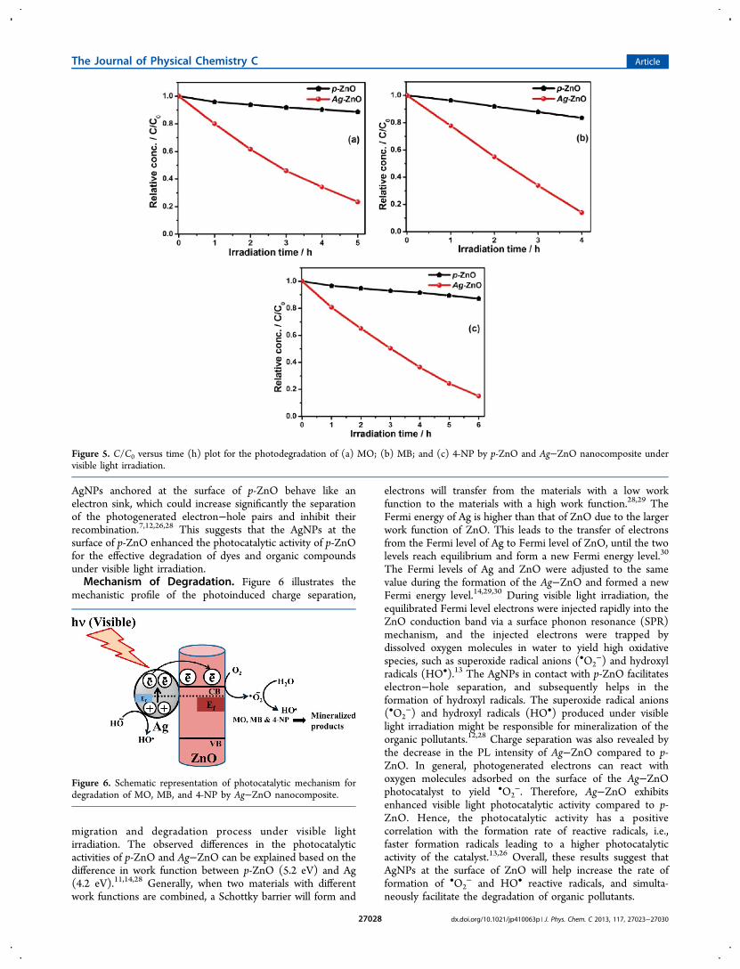
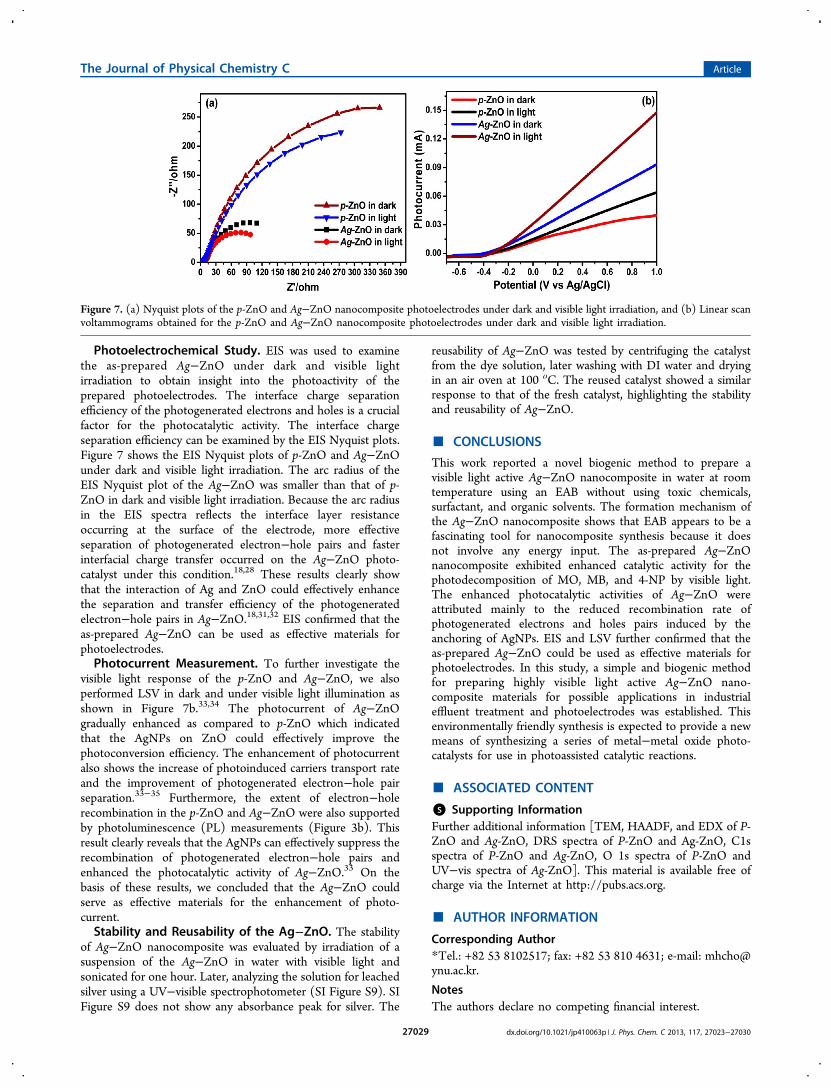
AgNPs anchored at the surface of p-ZnO behave like anelectron sink, which could increase significantly the separationof the photogenerated electron−hole pairs and inhibit theirrecombination.7,12,26,28 This suggests that the AgNPs at thesurface of p-ZnO enhanced the photocatalytic activity of p-ZnOfor the effective degradation of dyes and organic compoundsunder visible light irradiation.Mechanism of Degradation. Figure 6 illustrates the
mechanistic profile of the photoinduced charge separation,
migration and degradation process under visible lightirradiation. The observed differences in the photocatalyticactivities of p-ZnO and Ag−ZnO can be explained based on thedifference in work function between p-ZnO (5.2 eV) and Ag(4.2 eV).11,14,28 Generally, when two materials with differentwork functions are combined, a Schottky barrier will form and
electrons will transfer from the materials with a low workfunction to the materials with a high work function.28,29 TheFermi energy of Ag is higher than that of ZnO due to the largerwork function of ZnO. This leads to the transfer of electronsfrom the Fermi level of Ag to Fermi level of ZnO, until the twolevels reach equilibrium and form a new Fermi energy level.30
The Fermi levels of Ag and ZnO were adjusted to the samevalue during the formation of the Ag−ZnO and formed a newFermi energy level.14,29,30 During visible light irradiation, theequilibrated Fermi level electrons were injected rapidly into theZnO conduction band via a surface phonon resonance (SPR)mechanism, and the injected electrons were trapped bydissolved oxygen molecules in water to yield high oxidativespecies, such as superoxide radical anions (•O2
−) and hydroxylradicals (HO•).13 The AgNPs in contact with p-ZnO facilitateselectron−hole separation, and subsequently helps in theformation of hydroxyl radicals. The superoxide radical anions(•O2
−) and hydroxyl radicals (HO•) produced under visiblelight irradiation might be responsible for mineralization of theorganic pollutants.12,28 Charge separation was also revealed bythe decrease in the PL intensity of Ag−ZnO compared to p-ZnO. In general, photogenerated electrons can react withoxygen molecules adsorbed on the surface of the Ag−ZnOphotocatalyst to yield •O2
−. Therefore, Ag−ZnO exhibitsenhanced visible light photocatalytic activity compared to p-ZnO. Hence, the photocatalytic activity has a positivecorrelation with the formation rate of reactive radicals, i.e.,faster formation radicals leading to a higher photocatalyticactivity of the catalyst.13,26 Overall, these results suggest thatAgNPs at the surface of ZnO will help increase the rate offormation of •O2
− and HO• reactive radicals, and simulta-neously facilitate the degradation of organic pollutants.
Figure 5. C/C0 versus time (h) plot for the photodegradation of (a) MO; (b) MB; and (c) 4-NP by p-ZnO and Ag−ZnO nanocomposite undervisible light irradiation.
Figure 6. Schematic representation of photocatalytic mechanism fordegradation of MO, MB, and 4-NP by Ag−ZnO nanocomposite.
The Journal of Physical Chemistry C Article
dx.doi.org/10.1021/jp410063p | J. Phys. Chem. C 2013, 117, 27023−2703027028
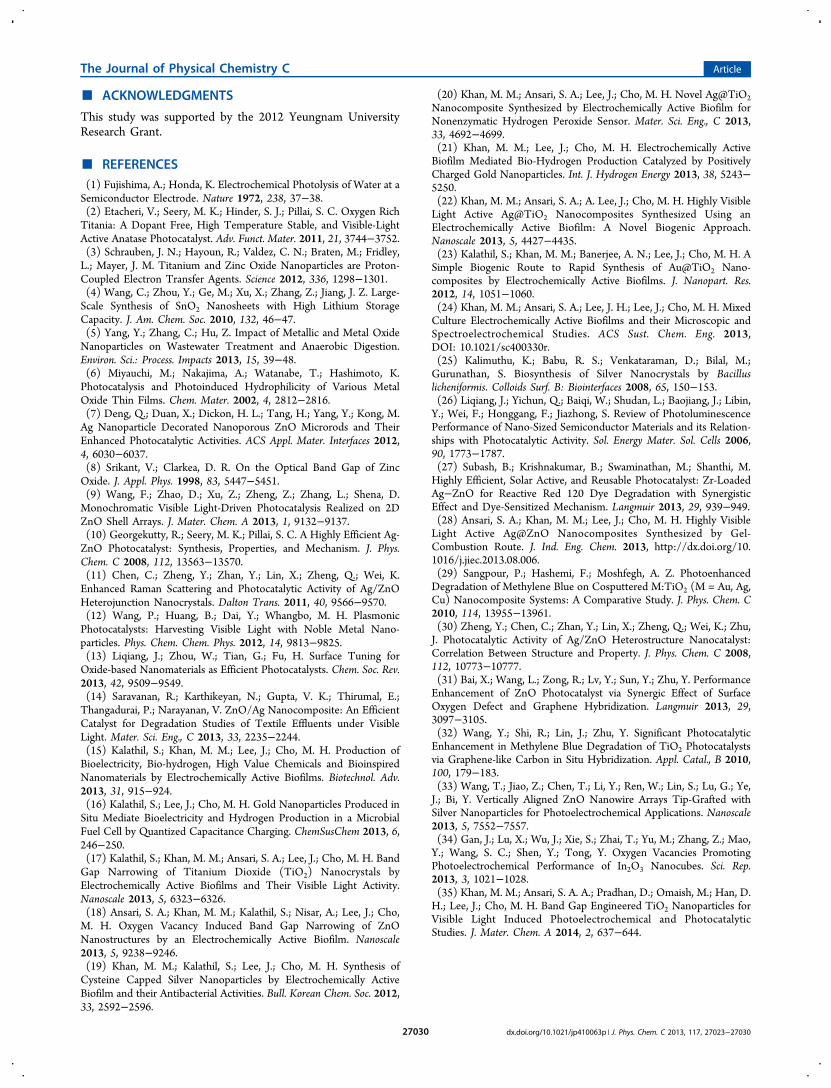
Photoelectrochemical Study. EIS was used to examinethe as-prepared Ag−ZnO under dark and visible lightirradiation to obtain insight into the photoactivity of theprepared photoelectrodes. The interface charge separationefficiency of the photogenerated electrons and holes is a crucialfactor for the photocatalytic activity. The interface chargeseparation efficiency can be examined by the EIS Nyquist plots.Figure 7 shows the EIS Nyquist plots of p-ZnO and Ag−ZnOunder dark and visible light irradiation. The arc radius of theEIS Nyquist plot of the Ag−ZnO was smaller than that of p-ZnO in dark and visible light irradiation. Because the arc radiusin the EIS spectra reflects the interface layer resistanceoccurring at the surface of the electrode, more effectiveseparation of photogenerated electron−hole pairs and fasterinterfacial charge transfer occurred on the Ag−ZnO photo-catalyst under this condition.18,28 These results clearly showthat the interaction of Ag and ZnO could effectively enhancethe separation and transfer efficiency of the photogeneratedelectron−hole pairs in Ag−ZnO.18,31,32 EIS confirmed that theas-prepared Ag−ZnO can be used as effective materials forphotoelectrodes.Photocurrent Measurement. To further investigate the
visible light response of the p-ZnO and Ag−ZnO, we alsoperformed LSV in dark and under visible light illumination asshown in Figure 7b.33,34 The photocurrent of Ag−ZnOgradually enhanced as compared to p-ZnO which indicatedthat the AgNPs on ZnO could effectively improve thephotoconversion efficiency. The enhancement of photocurrentalso shows the increase of photoinduced carriers transport rateand the improvement of photogenerated electron−hole pairseparation.33−35 Furthermore, the extent of electron−holerecombination in the p-ZnO and Ag−ZnO were also supportedby photoluminescence (PL) measurements (Figure 3b). Thisresult clearly reveals that the AgNPs can effectively suppress therecombination of photogenerated electron−hole pairs andenhanced the photocatalytic activity of Ag−ZnO.33 On thebasis of these results, we concluded that the Ag−ZnO couldserve as effective materials for the enhancement of photo-current.Stability and Reusability of the Ag−ZnO. The stability
of Ag−ZnO nanocomposite was evaluated by irradiation of asuspension of the Ag−ZnO in water with visible light andsonicated for one hour. Later, analyzing the solution for leachedsilver using a UV−visible spectrophotometer (SI Figure S9). SIFigure S9 does not show any absorbance peak for silver. The
reusability of Ag−ZnO was tested by centrifuging the catalystfrom the dye solution, later washing with DI water and dryingin an air oven at 100 οC. The reused catalyst showed a similarresponse to that of the fresh catalyst, highlighting the stabilityand reusability of Ag−ZnO.
■ CONCLUSIONS
This work reported a novel biogenic method to prepare avisible light active Ag−ZnO nanocomposite in water at roomtemperature using an EAB without using toxic chemicals,surfactant, and organic solvents. The formation mechanism ofthe Ag−ZnO nanocomposite shows that EAB appears to be afascinating tool for nanocomposite synthesis because it doesnot involve any energy input. The as-prepared Ag−ZnOnanocomposite exhibited enhanced catalytic activity for thephotodecomposition of MO, MB, and 4-NP by visible light.The enhanced photocatalytic activities of Ag−ZnO wereattributed mainly to the reduced recombination rate ofphotogenerated electrons and holes pairs induced by theanchoring of AgNPs. EIS and LSV further confirmed that theas-prepared Ag−ZnO could be used as effective materials forphotoelectrodes. In this study, a simple and biogenic methodfor preparing highly visible light active Ag−ZnO nano-composite materials for possible applications in industrialeffluent treatment and photoelectrodes was established. Thisenvironmentally friendly synthesis is expected to provide a newmeans of synthesizing a series of metal−metal oxide photo-catalysts for use in photoassisted catalytic reactions.
■ ASSOCIATED CONTENT
*S Supporting InformationFurther additional information [TEM, HAADF, and EDX of P-ZnO and Ag-ZnO, DRS spectra of P-ZnO and Ag-ZnO, C1sspectra of P-ZnO and Ag-ZnO, O 1s spectra of P-ZnO andUV−vis spectra of Ag-ZnO]. This material is available free ofcharge via the Internet at http://pubs.acs.org.
■ AUTHOR INFORMATION
Corresponding Author*Tel.: +82 53 8102517; fax: +82 53 810 4631; e-mail: [email protected].
NotesThe authors declare no competing financial interest.
Figure 7. (a) Nyquist plots of the p-ZnO and Ag−ZnO nanocomposite photoelectrodes under dark and visible light irradiation, and (b) Linear scanvoltammograms obtained for the p-ZnO and Ag−ZnO nanocomposite photoelectrodes under dark and visible light irradiation.
The Journal of Physical Chemistry C Article
dx.doi.org/10.1021/jp410063p | J. Phys. Chem. C 2013, 117, 27023−2703027029
■ ACKNOWLEDGMENTS
This study was supported by the 2012 Yeungnam UniversityResearch Grant.
■ REFERENCES(1) Fujishima, A.; Honda, K. Electrochemical Photolysis of Water at aSemiconductor Electrode. Nature 1972, 238, 37−38.(2) Etacheri, V.; Seery, M. K.; Hinder, S. J.; Pillai, S. C. Oxygen RichTitania: A Dopant Free, High Temperature Stable, and Visible-LightActive Anatase Photocatalyst. Adv. Funct. Mater. 2011, 21, 3744−3752.(3) Schrauben, J. N.; Hayoun, R.; Valdez, C. N.; Braten, M.; Fridley,L.; Mayer, J. M. Titanium and Zinc Oxide Nanoparticles are Proton-Coupled Electron Transfer Agents. Science 2012, 336, 1298−1301.(4) Wang, C.; Zhou, Y.; Ge, M.; Xu, X.; Zhang, Z.; Jiang, J. Z. Large-Scale Synthesis of SnO2 Nanosheets with High Lithium StorageCapacity. J. Am. Chem. Soc. 2010, 132, 46−47.(5) Yang, Y.; Zhang, C.; Hu, Z. Impact of Metallic and Metal OxideNanoparticles on Wastewater Treatment and Anaerobic Digestion.Environ. Sci.: Process. Impacts 2013, 15, 39−48.(6) Miyauchi, M.; Nakajima, A.; Watanabe, T.; Hashimoto, K.Photocatalysis and Photoinduced Hydrophilicity of Various MetalOxide Thin Films. Chem. Mater. 2002, 4, 2812−2816.(7) Deng, Q.; Duan, X.; Dickon, H. L.; Tang, H.; Yang, Y.; Kong, M.Ag Nanoparticle Decorated Nanoporous ZnO Microrods and TheirEnhanced Photocatalytic Activities. ACS Appl. Mater. Interfaces 2012,4, 6030−6037.(8) Srikant, V.; Clarkea, D. R. On the Optical Band Gap of ZincOxide. J. Appl. Phys. 1998, 83, 5447−5451.(9) Wang, F.; Zhao, D.; Xu, Z.; Zheng, Z.; Zhang, L.; Shena, D.Monochromatic Visible Light-Driven Photocatalysis Realized on 2DZnO Shell Arrays. J. Mater. Chem. A 2013, 1, 9132−9137.(10) Georgekutty, R.; Seery, M. K.; Pillai, S. C. A Highly Efficient Ag-ZnO Photocatalyst: Synthesis, Properties, and Mechanism. J. Phys.Chem. C 2008, 112, 13563−13570.(11) Chen, C.; Zheng, Y.; Zhan, Y.; Lin, X.; Zheng, Q.; Wei, K.Enhanced Raman Scattering and Photocatalytic Activity of Ag/ZnOHeterojunction Nanocrystals. Dalton Trans. 2011, 40, 9566−9570.(12) Wang, P.; Huang, B.; Dai, Y.; Whangbo, M. H. PlasmonicPhotocatalysts: Harvesting Visible Light with Noble Metal Nano-particles. Phys. Chem. Chem. Phys. 2012, 14, 9813−9825.(13) Liqiang, J.; Zhou, W.; Tian, G.; Fu, H. Surface Tuning forOxide-based Nanomaterials as Efficient Photocatalysts. Chem. Soc. Rev.2013, 42, 9509−9549.(14) Saravanan, R.; Karthikeyan, N.; Gupta, V. K.; Thirumal, E.;Thangadurai, P.; Narayanan, V. ZnO/Ag Nanocomposite: An EfficientCatalyst for Degradation Studies of Textile Effluents under VisibleLight. Mater. Sci. Eng., C 2013, 33, 2235−2244.(15) Kalathil, S.; Khan, M. M.; Lee, J.; Cho, M. H. Production ofBioelectricity, Bio-hydrogen, High Value Chemicals and BioinspiredNanomaterials by Electrochemically Active Biofilms. Biotechnol. Adv.2013, 31, 915−924.(16) Kalathil, S.; Lee, J.; Cho, M. H. Gold Nanoparticles Produced inSitu Mediate Bioelectricity and Hydrogen Production in a MicrobialFuel Cell by Quantized Capacitance Charging. ChemSusChem 2013, 6,246−250.(17) Kalathil, S.; Khan, M. M.; Ansari, S. A.; Lee, J.; Cho, M. H. BandGap Narrowing of Titanium Dioxide (TiO2) Nanocrystals byElectrochemically Active Biofilms and Their Visible Light Activity.Nanoscale 2013, 5, 6323−6326.(18) Ansari, S. A.; Khan, M. M.; Kalathil, S.; Nisar, A.; Lee, J.; Cho,M. H. Oxygen Vacancy Induced Band Gap Narrowing of ZnONanostructures by an Electrochemically Active Biofilm. Nanoscale2013, 5, 9238−9246.(19) Khan, M. M.; Kalathil, S.; Lee, J.; Cho, M. H. Synthesis ofCysteine Capped Silver Nanoparticles by Electrochemically ActiveBiofilm and their Antibacterial Activities. Bull. Korean Chem. Soc. 2012,33, 2592−2596.
(20) Khan, M. M.; Ansari, S. A.; Lee, J.; Cho, M. H. Novel Ag@TiO2Nanocomposite Synthesized by Electrochemically Active Biofilm forNonenzymatic Hydrogen Peroxide Sensor. Mater. Sci. Eng., C 2013,33, 4692−4699.(21) Khan, M. M.; Lee, J.; Cho, M. H. Electrochemically ActiveBiofilm Mediated Bio-Hydrogen Production Catalyzed by PositivelyCharged Gold Nanoparticles. Int. J. Hydrogen Energy 2013, 38, 5243−5250.(22) Khan, M. M.; Ansari, S. A.; A. Lee, J.; Cho, M. H. Highly VisibleLight Active Ag@TiO2 Nanocomposites Synthesized Using anElectrochemically Active Biofilm: A Novel Biogenic Approach.Nanoscale 2013, 5, 4427−4435.(23) Kalathil, S.; Khan, M. M.; Banerjee, A. N.; Lee, J.; Cho, M. H. ASimple Biogenic Route to Rapid Synthesis of Au@TiO2 Nano-composites by Electrochemically Active Biofilms. J. Nanopart. Res.2012, 14, 1051−1060.(24) Khan, M. M.; Ansari, S. A.; Lee, J. H.; Lee, J.; Cho, M. H. MixedCulture Electrochemically Active Biofilms and their Microscopic andSpectroelectrochemical Studies. ACS Sust. Chem. Eng. 2013,DOI: 10.1021/sc400330r.(25) Kalimuthu, K.; Babu, R. S.; Venkataraman, D.; Bilal, M.;Gurunathan, S. Biosynthesis of Silver Nanocrystals by Bacilluslicheniformis. Colloids Surf. B: Biointerfaces 2008, 65, 150−153.(26) Liqiang, J.; Yichun, Q.; Baiqi, W.; Shudan, L.; Baojiang, J.; Libin,Y.; Wei, F.; Honggang, F.; Jiazhong, S. Review of PhotoluminescencePerformance of Nano-Sized Semiconductor Materials and its Relation-ships with Photocatalytic Activity. Sol. Energy Mater. Sol. Cells 2006,90, 1773−1787.(27) Subash, B.; Krishnakumar, B.; Swaminathan, M.; Shanthi, M.Highly Efficient, Solar Active, and Reusable Photocatalyst: Zr-LoadedAg−ZnO for Reactive Red 120 Dye Degradation with SynergisticEffect and Dye-Sensitized Mechanism. Langmuir 2013, 29, 939−949.(28) Ansari, S. A.; Khan, M. M.; Lee, J.; Cho, M. H. Highly VisibleLight Active Ag@ZnO Nanocomposites Synthesized by Gel-Combustion Route. J. Ind. Eng. Chem. 2013, http://dx.doi.org/10.1016/j.jiec.2013.08.006.(29) Sangpour, P.; Hashemi, F.; Moshfegh, A. Z. PhotoenhancedDegradation of Methylene Blue on Cosputtered M:TiO2 (M = Au, Ag,Cu) Nanocomposite Systems: A Comparative Study. J. Phys. Chem. C2010, 114, 13955−13961.(30) Zheng, Y.; Chen, C.; Zhan, Y.; Lin, X.; Zheng, Q.; Wei, K.; Zhu,J. Photocatalytic Activity of Ag/ZnO Heterostructure Nanocatalyst:Correlation Between Structure and Property. J. Phys. Chem. C 2008,112, 10773−10777.(31) Bai, X.; Wang, L.; Zong, R.; Lv, Y.; Sun, Y.; Zhu, Y. PerformanceEnhancement of ZnO Photocatalyst via Synergic Effect of SurfaceOxygen Defect and Graphene Hybridization. Langmuir 2013, 29,3097−3105.(32) Wang, Y.; Shi, R.; Lin, J.; Zhu, Y. Significant PhotocatalyticEnhancement in Methylene Blue Degradation of TiO2 Photocatalystsvia Graphene-like Carbon in Situ Hybridization. Appl. Catal., B 2010,100, 179−183.(33) Wang, T.; Jiao, Z.; Chen, T.; Li, Y.; Ren, W.; Lin, S.; Lu, G.; Ye,J.; Bi, Y. Vertically Aligned ZnO Nanowire Arrays Tip-Grafted withSilver Nanoparticles for Photoelectrochemical Applications. Nanoscale2013, 5, 7552−7557.(34) Gan, J.; Lu, X.; Wu, J.; Xie, S.; Zhai, T.; Yu, M.; Zhang, Z.; Mao,Y.; Wang, S. C.; Shen, Y.; Tong, Y. Oxygen Vacancies PromotingPhotoelectrochemical Performance of In2O3 Nanocubes. Sci. Rep.2013, 3, 1021−1028.(35) Khan, M. M.; Ansari, S. A. A.; Pradhan, D.; Omaish, M.; Han, D.H.; Lee, J.; Cho, M. H. Band Gap Engineered TiO2 Nanoparticles forVisible Light Induced Photoelectrochemical and PhotocatalyticStudies. J. Mater. Chem. A 2014, 2, 637−644.
The Journal of Physical Chemistry C Article
dx.doi.org/10.1021/jp410063p | J. Phys. Chem. C 2013, 117, 27023−2703027030