Embed Size (px)
Citation preview
DOI: 10.1093/brain/awh239 Brain Page 1 of 16
Behavioural disorders induced by externalglobus pallidus dysfunction in primatesII. Anatomical study
Chantal Francois, David Grabli, Kevin McCairn, Caroline Jan, Carine Karachi, Etienne-C. Hirsch,Jean Feger and Leon Tremblay
Correspondence to: Chantal Francois, INSERM U289,
Hopital de la Salpetriere, 47 Bd de l’Hopital, 75013,
Paris, France
E-mail: [email protected]
INSERM U289: Neurologie et Therapeutique
experimentale, Hopital de la Salpetriere, 47 Bd de
l’Hopital, 75651 Paris cedex 13, France
SummaryThe anatomical organization of the basal ganglia supports
their involvement in movement and behavioural disor-
ders. Thus dyskinesia, attention deficit with or without
hyperactivity, and stereotyped behaviour can be induced
by microinjections of bicuculline, a GABAergic antago-
nist, into different parts of the external globus pallidus
(GPe) in monkeys. The aim of the present study was todetermine the anatomo-functional circuits inside the basal
ganglia which are specifically related to each of these beha-
vioural changes. For that, axonal tracers were injected in
the same pallidal sites where abnormal behaviours have
previously been obtained by bicuculline microinjections.
The labelling was mapped in the different basal ganglia
and matched with the topography of the cortico-striato-
pallidal projections already reported in the literatureand with the distribution of calbindin immunoreactivity.
Our results first show that the pallidal sites related to
dyskinesia, attention deficit with or without hyperactivity,
and stereotyped behaviour, were respectively in motor,
associative and limbic territories, defined as weak, mod-
erate and intensive calbindin immunoreactivity. The same
relationship was observed between the distribution of the
labelling in the different basal ganglia after tracer injec-tions performed in these different pallidal sites and the
anatomo-functional territories. Thus regarding the origin
of the circuits within the striatum, tracer injections
performed in the dyskinesia site labelled neurons located
in the posterior sensorimotor putamen, those performed
in the hyperactivity and/or attention deficit labelled neu-
rons in the laterodorsal putamen and caudate nucleus,
regions corresponding to associative and anterior motor
territories, while those performed in the stereotyped beha-
viour site labelled neurons in the ventral limbic striatum.Regarding the GPe output on the basal ganglia, the
different circuits also appeared underlined by different
anatomo-functional territories, even if a partial overlap
exists. Each of these anatomical circuits systematically
involves both the internal globus pallidus (GPi) and the
substantia nigra pars reticulata (SNr) but, whereas
movement circuit is mainly related to the GPi, stereotyped
behaviour is mainly related to the SNr. Additionally,subregions of the subthalamic nucleus were also sys-
tematically involved, depending on the movement or
behavioural disorder produced. These results demon-
strate that distinct circuits involving different anatomo-
functional territories of the basal ganglia, with partial
overlap, participate in different behavioural disorders
in monkeys. It seems likely that these neuronal circuits
are involved in pathologies like Tourette’s syndrome,attention deficit/hyperactivity disorders and obsessional
compulsive troubles. This study provides the basis for
further researches with a therapeutical viewpoint.
Keywords: basal ganglia; monkeys; stereotypy; hyperactivity; attentional deficit
Abbreviations: AC = anterior commissure; ADHD = attention deficit/hyperactivity disorder; BDA = biotin dextran amine;
Cd = caudate nucleus; Cb = calbindin; GPe = external globus pallidus; GPi = internal globus pallidus; PC = posterior commissure;
SNr = substantia nigra pars reticulata; STN = subthalamic nucleus; WGA-HRP = wheat germ agglutinin conjugated to
horseradish peroxidase
Received January 26, 2004. Revised May 3, 2004. Accepted May 13, 2004
Brain # Guarantors of Brain 2004; all rights reserved
Brain Advance Access published August 3, 2004 by guest on July 17, 2016
http://brain.oxfordjournals.org/D
ownloaded from
IntroductionIt is well established that the origin and distribution of corti-
costriatal inputs allow us to subdivide the striatum into three
anatomo-functional territories. They are referred to respect-
ively as (i) sensorimotor territory processing sensorial and
motor information; (ii) associative territory processing cog-
nitive information; and (iii) limbic territory processing emo-
tional and motivational information (Alexander et al., 1986;
Parent and Hazrati, 1995b; Haber et al., 2000). These
anatomo-functional territories occupy distinct regions without
clear-cut boundaries between each of them (Parent and
Hazrati, 1995a; Haber et al., 2000). This partition in three
territories is maintained within the basal ganglia, especially in
the external globus pallidus (GPe) regarding the distribution
of the striatopallidal projections (Parent et al., 1984; Smith
and Parent, 1986; Haber et al., 1990; Saint-Cyr et al., 1990;
Hedreen and DeLong, 1991; Francois et al., 1994a). Such
anatomical organization allows us to speculate that activities
of some cortical area will affect the neuronal activity in the
corresponding striatal and pallidal territories.
Functional imaging studies (PET or functional MRI) in
humans suggest that abnormal activities in different parts
of the cortex and basal ganglia are present in various behav-
ioural disorders. It was reported that patients expressing
attention deficit/hyperactivity disorder (ADHD) have a
reduced metabolism in the prefrontal cortex and in the asso-
ciative part of the striatum (Castellanos et al., 1996; Filipek
et al., 1997; Rubia et al., 1999; Castellanos, 2001) and in the
pallidum as a unit (Aylward et al., 1996; Castellanos et al.,
1996). Tourette’s syndrome which is characterized by motor
(tics) and behavioural disorders such as obsessive-compulsive
disorder and ADHD (for a review, see Jankovic, 2001) is
marked by abnormalities in various parts of the cortex, stria-
tum and thalamus (Scarone et al., 1992; Peterson et al., 1993;
Singer et al., 1993; Hyde et al., 1995; Eidelberg et al., 1997;
Moriarty et al., 1997; Peterson, 2001). Thus in these various
behavioural disorders, the dysfunction seems limited to the
cortex and striatum, whereas the involvement of the other
components of the basal ganglia is not obviously mentioned.
Only one clinical study reported cases of obsessive-
compulsive disorder with an emphasis on the involvement
of a subcortical structure other than the striatum, since
these troubles are attributed to a bilateral lesion restricted
to the pallidum (Laplane et al., 1984, 1989).
Generally, the functional imaging approach has been, until
now, without sufficient resolution to distinguish small ana-
tomical structures or subterritories. Moreover, this approach
shows the structures where abnormal changes in activity
occur, but it does not allow us to determine which of them
is at the origin of such changes. In a complementary way, a
pharmacological approach using microinjection of a suitable
pharmacological agent in animals allows us to induce a local
disturbance of the neuronal activity within a given structure.
Then, it can be stated that a functional relationship of causality
exists between this part of the structure where a dysfunction is
induced and the expression of abnormal movements or
behavioural disorders. Indeed, we previously observed in
monkeys that microinjections of bicuculline, an antagonist
of GABAergic receptor, provoked abnormal movement
(dyskinesia) and behavioural disorders (hyperactivity, attention
deficit or stereotypy) dependent on the injection site into the
anatomo-functional part of the GPe (see Grabli et al., 2004).
The aim of the present study was to determine the ana-
tomical relationships between the origin of the inputs and the
distribution of the output pathways which are related to the
neuronal population localized in the GPe where microinjec-
tions of bicuculline have been able to induce abnormal move-
ments or behavioural disorders in monkeys. To circumvent
this question, injections of axonal tracers were performed in
different sites of the GPe where the most characteristic effects
have been obtained. This approach allowed us to determine
which anatomo-functional circuit is implicated in the different
behavioural disorders. The axonal tracers used ensured
retrograde tracing in the striatum, and thus opened the way
to correlate our results with the numerous data in the literature
concerning the corticostriatal projection and functional MRI
results in human pathology. The anterograde transport of
tracers allowed us to compare the detailed patterns of axonal
ending distribution at both the output structures of the basal
ganglia represented by the internal globus pallidus (GPi) and
the pars reticulata of the substantia nigra (SNr). We also
focused our attention on the subthalamic nucleus (STN),
another output structure of the GPe. The delineation of ana-
tomo-functional territories in the GPe, as well as in different
basal ganglia structures, has been performed according to the
distribution of calbindin (Cb) immunohistochemistry, which
was shown to be correlated with the delineation of anatomo-
functional territories in the striatopallidal complex (Francois
et al., 1994b; Karachi et al., 2002). More generally, the delin-
eation of anatomo-functional territories was compared with
afferent territories already published in the literature using a
tract-tracing method. These results have been previously
reported in an abstract form (Francois et al., 2002) and are com-
pleted by their behavioural counterparts in Grabli et al. (2004).
Materials and methodsSubjectsThree adult male African green monkeys (Cercopithecus aethiops;
H, A and B) were used in both behavioural and anatomical studies
and have been supplemented with the anatomical observations per-
formed on one macaque (Macaca mulatta MM33). They weighed
between 5 and 7 kg and were aged between 5 and 10 years, as
determined by their age at entry in captivity, dentition and hair
appearance. All studies were carried out in accordance with
European Communities Council Directive of 1986 (86/609/EEC).
The animals were kept under standard conditions (12-h light cycles,
23�C and 50% humidity).
Microinjections of bicuculline and behaviouralanalysisThe behavioural study described in Grabli et al. (2004) was con-
ducted to assess the effects of microinjection of bicuculline into the
Page 2 of 16 C. Francois et al.
by guest on July 17, 2016http://brain.oxfordjournals.org/
Dow
nloaded from
different functional territories of the GPe. Briefly, to perform these
microinjections and finally the injections of tracers, a stainless steel
chamber containing a perforated grid was first positioned above the
animal’s dura. For that, monkeys were first tranquillized with keta-
mine hydrochloride (10 mg/kg), then anaesthetized through intratra-
cheal intubation (fluothane 1%, nitrogen protoxide 50% and oxygen
50%). The chamber was positioned with reference to the anterior and
posterior commissures (AC and PC, respectively) visualized by ven-
triculography (Percheron et al., 1986). Since the chamber was fixed
on the skull with reference to the ventricular system of coordinates,
this device allowed us to perform all microinjections in reference to
the stereotaxic coordinates of the various parts of the GPe (for more
precise description of the experimental set-up device, see Grabli
et al., 2004).
In a first step, abnormal movements and behavioural disorders
were induced by microinjections of bicuculline in several parts of the
GPe in the two hemispheres of each monkey. Then microinjection
sites related to the most typical changes were selected for this
anatomical study.
Injections of axonal tracersInjections of axonal tracers were performed in each GPe of the three
African green monkeys studied when all behavioural studies were
achieved. They consisted of 0.1–0.2 ml of wheat germ agglutinin
conjugated to horseradish peroxidase (WGA-HRP; Sigma, St Louis,
MO, USA) 10% in 0.1 M phosphate buffered saline (PBS, 0.01 M,
pH 7.4), or of 0.5–0.8 ml of biotin dextran amine (BDA) diluted
(10%) in PBS (0.01 M, pH 7.4) injected using the same specific
device used for bicuculline micro-injection. Moreover, in order to
compensate the weakness of injection in one site (site no. 37 in
monkey H), BDA was also injected in the GPe of the experimental
case MM33 which was not involved in the behavioural studies. In
that case, the injection site was exactly at the same coordinates as
those of monkey H.
Three days (for HRP-WGA tracer) and 10 days (for BDA tracer)
after injections, the animals were deeply anaesthetized and replaced
in the stereotaxic frame as described above. Two vertical and two
horizontal metal rods (one in each hemisphere) were introduced into
the brain perpendicular and parallel, respectively, to the plane
passing through the two ventricular landmarks. These landmarks
allowed us to define two ventricular planes, transverse and horizon-
tal, in order to analyse the brains post-mortem according to the
stereotaxic landmarks. The monkeys were then perfused transcar-
dially with 400 ml of saline (0.9% at 37�C) followed by 5 l of 4%
paraformaldehyde (in 0.1 M PBS, pH 7.4 at 4�C) and 1 l of PBS with
5% sucrose. The brains were removed from the skull, rinsed in PBS
complemented with 10% sucrose for 1 day and 20% sucrose for
1 day, then frozen and cut into 50-mm-thick sections transversely
with reference to the ventricular anterior and posterior commissures
on a freezing microtome.
Histological processingOn all monkeys, Cb was revealed on regularly interspaced (one in
five) free-floating sections using a monoclonal antiserum raised
against Cb (Sigma) diluted to 1 : 1.000 for 48 h at 4�C under gentle
agitation. The primary antibodies were revealed using the ABC
method (diluted 1 : 100; Vector Laboratories, Burlingame, CA,
USA) using diaminobenzidine tetrahydrochloride (DAB; 0.05%)
as peroxidase chromogen.
For the revelation of BDA, all sections were pretreated with 1%
Triton X-100 in PBS, then incubated using avidin–biotin complex
staining (ABC, Elite, Vector Laboratories) in PBS with 1% Triton for
48 h at 4�C. Sections were treated with nickel (0.2%)-DAB (0.05%)
as peroxidase chromagen. Cell bodies could easily be visualized in
our material as biocytin occurs in trace in eukaryotic organisms
(Smith, 1992). WGA-HRP was revealed using the method previously
described by Mesulam (1978). Briefly, sections were abundantly
rinsed in 0.1 M PBS, then in acetate buffer. Preincubation was per-
formed in 0.1% nitroferricyanure (Sigma) and 0.005% 3,3-5,5 tetra-
methyl-benzidine (Sigma) solution for 15 min, and hydrogen peroxide
was added at a concentration of 0.02–0.04% for about 15 min. Sec-
tions were then counterstained with green methyl solution (0.25%).
Cartographic methodsContours of cerebral structures were traced under the microscope
with the aid of an XY plotter connected to the stage of the micro-
scope. The anteroposterior position of each section was given by the
anteriority of each section along the AC–PC axis, taking AC as
the origin of the system of axes. The dorsoventral axis was given
by the holes corresponding to the horizontal rods. The mediolateral
axis was perpendicular to the dorsoventral axis. All sections were
thus transformed into maps drawn in relation to the AC–PC system of
coordinates, and the contours of structures mapped in different mon-
keys could be directly compared. The limits of anatomo-functional
territories shown in published figures for the striatopallidal projec-
tion (Parent et al., 1984; Smith and Parent, 1986; Haber et al., 1990;
Saint-Cyr et al., 1990; Hedreen and DeLong, 1991; Hazrati and
Parent, 1992; Flaherty and Graybiel, 1994; Francois et al., 1994a)
were transferred onto the corresponding cerebral maps of our series
drawn from Cb material.
All cartographic data obtained from the left hemisphere of mon-
keys were transferred to the right hemisphere for an easier compar-
ison. The illustrations were scanned into a computer from camera
lucida drawings or charts and finished using Adobe Photoshop or
Microsoft PowerPoint software.
ResultsOverview of abnormal movement andbehavioural disorders induced by bicucullinemicroinjections into the GPeIn the three monkeys studied, microinjections of bicuculline
performed into various parts of the GPe induced three differ-
ent types of behavioural disorders. Thus injections in the
posterior and lateral part of the GPe induced abnormal move-
ment referred to as dyskinesia, those performed in the antero-
dorsal part of the GPe produced hyperactivity and/or attention
deficit, while stereotyped behaviours where obtained after
injections in the anterior and medioventral portion of the
GPe. For the present study, different sites of bicuculline
microinjections that produced the most characteristic behav-
ioural changes were chosen to perform injections of axonal
tracer. The localization of these microinjection sites is illus-
trated in Fig. 1A, and their behavioural effects induced in the
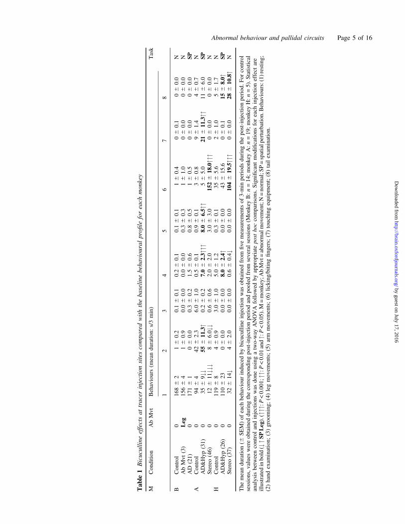
different monkeys are summarized in Table 1.
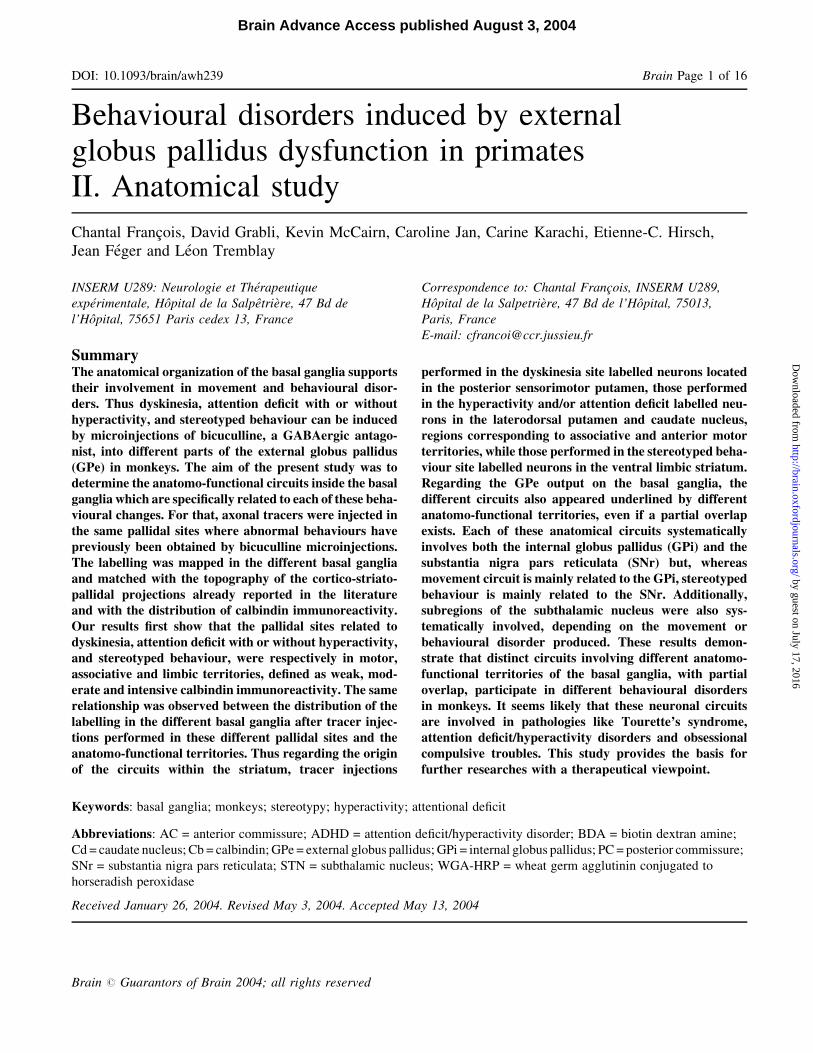
The effect obtained from site no. 3 in the right GPe of
monkey B (Fig. 1A) was leg dyskinesia without any
Abnormal behaviour and pallidal circuits Page 3 of 16
by guest on July 17, 2016http://brain.oxfordjournals.org/
Dow
nloaded from
behavioural modification (Table 1). This dyskinesia did not
modify the execution of the task. In the same monkey, site no.
21 in the left GPe (Fig. 1A) was retained as the injection of
bicuculline induced only a change in the spatial strategy in the
execution of the task, a trouble that we related to an attention
deficit. In monkey A, we have retained site no. 31 in the right
GPe, as it was associated with the production of a hyper-
activity with spatial strategy modification in the task, and
Fig. 1 Series of anteroposterior sequences of regularly interspaced transverse sections located with reference to the anterior commissuralreference point (AC) and centred on the pallidum. (A) Localization of sites of bicuculline microinjections within the GPe that produced themost characteristic behavioural changes and that were chosen to perform injections of axonal tracer. These pallidal sites elicited eitherstereotyped behaviour, or hyperactivity with attention deficit, or attention deficit alone, or abnormal movements. (B) Dark-fieldphotomicrographs of axonal tracer injections into four different cases (monkey A, left and right hemisphere, monkey B left and righthemisphere). (C) Dark-field photomicrographs of Cb-immunoreactive material. The distribution of immunoreactivity was directlycompared with the delineation of the functional territories of the striatopallidal complex (D) obtained by superimposition of data fromtracing studies in the literature (Parent et al., 1984; Smith and Parent, 1986; Haber et al., 1990; Saint-Cyr et al., 1990; Hedreen and DeLong,1991; Hazrati and Parent, 1992; Flaherty and Graybiel, 1994; Francois et al., 1994a) onto the same anteroposterior sequence of transversesections. The sensorimotor territory is represented in white, the associative territory in grey and the limbic territory in black. AC = anteriorcommissure; GPe = external globus pallidus; GPi = internal globus pallidus.
Page 4 of 16 C. Francois et al.
by guest on July 17, 2016http://brain.oxfordjournals.org/
Dow
nloaded from
Ta
ble
1B
icu
cull
ine
effe
cts
at
tra
cer
inje
ctio
nsi
tes
com
pa
red
wit
hth
eb
ase
lin
eb
eha
vio
ura
lp
rofi
lefo
rea
chm
on
key
MC
on
dit
ion
Ab
Mv
tB
ehav
iou
rs(m
ean
du
rati
on
:s/
3m
in)
Tas
k
12
34
56
78
BC
on
tro
l0
16
86
216
0.2
0.1
60
.10
.26
0.1
0.1
60
.116
0.4
06
0.1
06
0.0
NA
bM
vt
(3)
Leg
15
66
416
0.9
0.0
60
.00
.06
0.0
0.3
60
.316
1.0
06
0.0
06
0.0
NA
D(2
1)
01
716
106
0.0
0.3
60
.21
.56
0.6
0.8
60
.516
0.5
06
0.0
06
0.0
SP
AC
on
tro
l0
946
44
26
2.3
6.0
61
.00
.56
0.1
0.9
60
.136
0.8
96
1.4
46
0.7
NA
D&
Hy
p(3
1)
03
56
9##
556
11
.3"
0.2
60
.27
.06
2.3""
"8
.06
6.5""
56
5.0
216
11
.3""
116
6.0
SP
Ste
reo
(46
)0
126
11##
#86
6.0##
0.6
60
.62
.06
2.0
3.0
63
.01
526
18
.0""
"06
0.0
06
0.0
NH
Co
ntr
ol
01
196
846
0.9
3.0
61
.05
.06
1.2
0.3
60
.13
56
5.6
26
1.0
56
1.7
NA
D&
Hy
p(2
6)
01
106
23
06
0.0
0.0
60
.08
.06
2.4"
0.0
60
.04
36
15
.606
0.1
156
8.0"
SP
Ste
reo
(37
)0
326
14#
46
2.0
0.0
60
.00
.66
0.4#
0.0
60
.01
046
19
.5""
"06
0.0
286
10
.8"
N
Th
em
ean
du
rati
on
(6S
EM
)o
fea
chb
ehav
iou
rin
du
ced
by
bic
ucu
llin
ein
ject
ion
was
ob
tain
edfr
om
fiv
em
easu
rem
ents
of
3-m
inp
erio
ds
du
rin
gth
ep
ost
-in
ject
ion
per
iod
.F
or
con
tro
lse
ssio
ns,
val
ues
wer
eo
bta
ined
du
rin
gth
eco
rres
po
nd
ing
po
st-i
nje
ctio
np
erio
dan
dp
oo
led
fro
mse
ver
alse
ssio
ns
(Mo
nk
eyB
:n
=1
4;
mo
nk
eyA
:n
=1
9;
mo
nk
eyH
:n
=5
).S
tati
stic
alan
aly
sis
bet
wee
nco
ntr
ol
and
inje
ctio
ns
was
do
ne
usi
ng
atw
o-w
ayA
NO
VA
foll
ow
edb
yap
pro
pri
ate
po
sth
oc
com
par
iso
ns.
Sig
nifi
can
tm
od
ifica
tio
ns
for
each
inje
ctio
nef
fect
are
illu
stra
ted
inb
old
(#"S
PL
eg).
("""
:P<
0.0
01
;"":
P<
0.0
1an
d":
P<
0.0
5).
M=
mo
nk
ey;A
bM
vt=
abn
orm
alm
ov
emen
t;N
=n
orm
al;S
P=
spat
ialp
ertu
rbat
ion
.Beh
avio
urs
:(1
)re
stin
g;
(2)
han
dex
amin
atio
n;
(3)
gro
om
ing
;(4
)le
gm
ov
emen
ts;
(5)
arm
mo
vem
ents
;(6
)li
ckin
g/b
itin
gfi
ng
ers;
(7)
tou
chin
geq
uip
men
t;(8
)ta
ilex
amin
atio
n.
Abnormal behaviour and pallidal circuits Page 5 of 16
by guest on July 17, 2016http://brain.oxfordjournals.org/
Dow
nloaded from
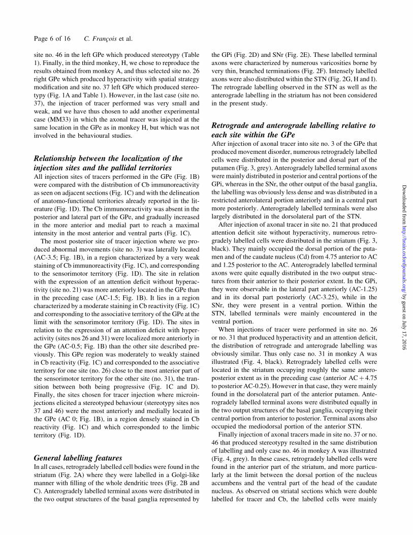
site no. 46 in the left GPe which produced stereotypy (Table
1). Finally, in the third monkey, H, we chose to reproduce the
results obtained from monkey A, and thus selected site no. 26
right GPe which produced hyperactivity with spatial strategy
modification and site no. 37 left GPe which produced stereo-
typy (Fig. 1A and Table 1). However, in the last case (site no.
37), the injection of tracer performed was very small and
weak, and we have thus chosen to add another experimental
case (MM33) in which the axonal tracer was injected at the
same location in the GPe as in monkey H, but which was not
involved in the behavioural studies.
Relationship between the localization of theinjection sites and the pallidal territoriesAll injection sites of tracers performed in the GPe (Fig. 1B)
were compared with the distribution of Cb immunoreactivity
as seen on adjacent sections (Fig. 1C) and with the delineation
of anatomo-functional territories already reported in the lit-
erature (Fig. 1D). The Cb immunoreactivity was absent in the
posterior and lateral part of the GPe, and gradually increased
in the more anterior and medial part to reach a maximal
intensity in the most anterior and ventral parts (Fig. 1C).
The most posterior site of tracer injection where we pro-
duced abnormal movements (site no. 3) was laterally located
(AC-3.5; Fig. 1B), in a region characterized by a very weak
staining of Cb immunoreactivity (Fig. 1C), and corresponding
to the sensorimotor territory (Fig. 1D). The site in relation
with the expression of an attention deficit without hyperac-
tivity (site no. 21) was more anteriorly located in the GPe than
in the preceding case (AC-1.5; Fig. 1B). It lies in a region
characterized by a moderate staining in Cb reactivity (Fig. 1C)
and corresponding to the associative territory of the GPe at the
limit with the sensorimotor territory (Fig. 1D). The sites in
relation to the expression of an attention deficit with hyper-
activity (sites nos 26 and 31) were localized more anteriorly in
the GPe (AC-0.5; Fig. 1B) than the other site described pre-
viously. This GPe region was moderately to weakly stained
in Cb reactivity (Fig. 1C) and corresponded to the associative
territory for one site (no. 26) close to the most anterior part of
the sensorimotor territory for the other site (no. 31), the tran-
sition between both being progressive (Fig. 1C and D).
Finally, the sites chosen for tracer injection where microin-
jections elicited a stereotyped behaviour (stereotypy sites nos
37 and 46) were the most anteriorly and medially located in
the GPe (AC 0; Fig. 1B), in a region densely stained in Cb
reactivity (Fig. 1C) and which corresponded to the limbic
territory (Fig. 1D).
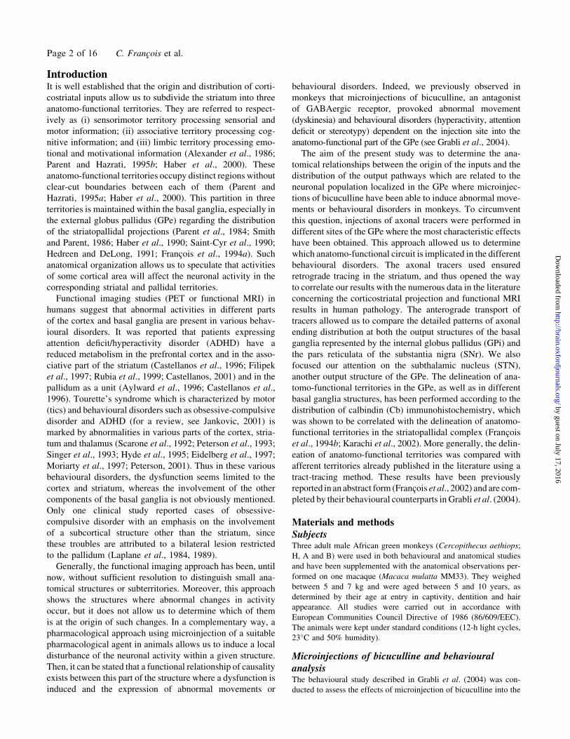
General labelling featuresIn all cases, retrogradely labelled cell bodies were found in the
striatum (Fig. 2A) where they were labelled in a Golgi-like
manner with filling of the whole dendritic trees (Fig. 2B and
C). Anterogradely labelled terminal axons were distributed in
the two output structures of the basal ganglia represented by
the GPi (Fig. 2D) and SNr (Fig. 2E). These labelled terminal
axons were characterized by numerous varicosities borne by
very thin, branched terminations (Fig. 2F). Intensely labelled
axons were also distributed within the STN (Fig. 2G, H and I).
The retrograde labelling observed in the STN as well as the
anterograde labelling in the striatum has not been considered
in the present study.
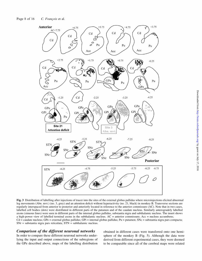
Retrograde and anterograde labelling relative toeach site within the GPeAfter injection of axonal tracer into site no. 3 of the GPe that
produced movement disorder, numerous retrogradely labelled
cells were distributed in the posterior and dorsal part of the
putamen (Fig. 3, grey). Anterogradely labelled terminal axons
were mainly distributed in posterior and central portions of the
GPi, whereas in the SNr, the other output of the basal ganglia,
the labelling was obviously less dense and was distributed in a
restricted anterolateral portion anteriorly and in a central part
more posteriorly. Anterogradely labelled terminals were also
largely distributed in the dorsolateral part of the STN.
After injection of axonal tracer in site no. 21 that produced
attention deficit site without hyperactivity, numerous retro-
gradely labelled cells were distributed in the striatum (Fig. 3,
black). They mainly occupied the dorsal portion of the puta-
men and of the caudate nucleus (Cd) from 4.75 anterior to AC
and 1.25 posterior to the AC. Anterogradely labelled terminal
axons were quite equally distributed in the two output struc-
tures from their anterior to their posterior extent. In the GPi,
they were observable in the lateral part anteriorly (AC-1.25)
and in its dorsal part posteriorly (AC-3.25), while in the
SNr, they were present in a ventral portion. Within the
STN, labelled terminals were mainly encountered in the
ventral portion.
When injections of tracer were performed in site no. 26
or no. 31 that produced hyperactivity and an attention deficit,
the distribution of retrograde and anterograde labelling was
obviously similar. Thus only case no. 31 in monkey A was
illustrated (Fig. 4, black). Retrogradely labelled cells were
located in the striatum occupying roughly the same antero-
posterior extent as in the preceding case (anterior ACþ 4.75
to posterior AC-0.25). However in that case, they were mainly
found in the dorsolateral part of the anterior putamen. Ante-
rogradely labelled terminal axons were distributed equally in
the two output structures of the basal ganglia, occupying their
central portion from anterior to posterior. Terminal axons also
occupied the mediodorsal portion of the anterior STN.
Finally injection of axonal tracers made in site no. 37 or no.
46 that produced stereotypy resulted in the same distribution
of labelling and only case no. 46 in monkey A was illustrated
(Fig. 4, grey). In these cases, retrogradely labelled cells were
found in the anterior part of the striatum, and more particu-
larly at the limit between the dorsal portion of the nucleus
accumbens and the ventral part of the head of the caudate
nucleus. As observed on striatal sections which were double
labelled for tracer and Cb, the labelled cells were mainly
Page 6 of 16 C. Francois et al.
by guest on July 17, 2016http://brain.oxfordjournals.org/
Dow
nloaded from
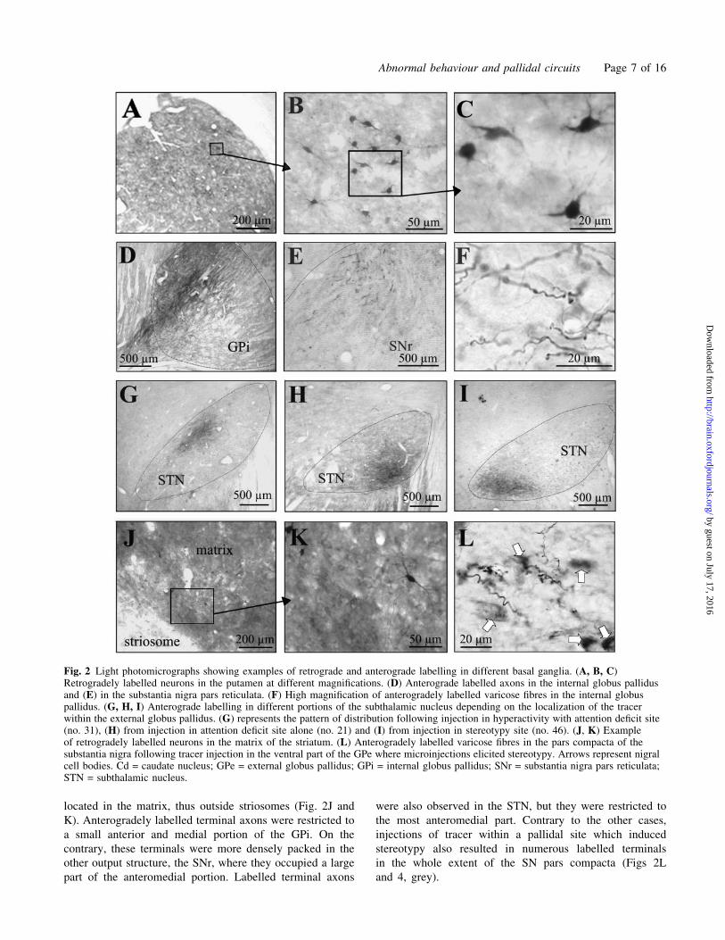
located in the matrix, thus outside striosomes (Fig. 2J and
K). Anterogradely labelled terminal axons were restricted to
a small anterior and medial portion of the GPi. On the
contrary, these terminals were more densely packed in the
other output structure, the SNr, where they occupied a large
part of the anteromedial portion. Labelled terminal axons
were also observed in the STN, but they were restricted to
the most anteromedial part. Contrary to the other cases,
injections of tracer within a pallidal site which induced
stereotypy also resulted in numerous labelled terminals
in the whole extent of the SN pars compacta (Figs 2L
and 4, grey).
Fig. 2 Light photomicrographs showing examples of retrograde and anterograde labelling in different basal ganglia. (A, B, C)Retrogradely labelled neurons in the putamen at different magnifications. (D) Anterograde labelled axons in the internal globus pallidusand (E) in the substantia nigra pars reticulata. (F) High magnification of anterogradely labelled varicose fibres in the internal globuspallidus. (G, H, I) Anterograde labelling in different portions of the subthalamic nucleus depending on the localization of the tracerwithin the external globus pallidus. (G) represents the pattern of distribution following injection in hyperactivity with attention deficit site(no. 31), (H) from injection in attention deficit site alone (no. 21) and (I) from injection in stereotypy site (no. 46). (J, K) Exampleof retrogradely labelled neurons in the matrix of the striatum. (L) Anterogradely labelled varicose fibres in the pars compacta of thesubstantia nigra following tracer injection in the ventral part of the GPe where microinjections elicited stereotypy. Arrows represent nigralcell bodies. Cd = caudate nucleus; GPe = external globus pallidus; GPi = internal globus pallidus; SNr = substantia nigra pars reticulata;STN = subthalamic nucleus.
Abnormal behaviour and pallidal circuits Page 7 of 16
by guest on July 17, 2016http://brain.oxfordjournals.org/
Dow
nloaded from
Comparison of the different neuronal networksIn order to compare these different neuronal networks under-
lying the input and output connections of the subregions of
the GPe described above, maps of the labelling distribution
obtained in different cases were transferred onto one hemi-
sphere of the monkey B (Fig. 5). Although the data were
derived from different experimental cases, they were deemed
to be comparable since all of the cerebral maps were related
Fig. 3 Distribution of labelling after injections of tracer into the sites of the external globus pallidus where microinjections elicited abnormalleg movements (Abn. mvt.) (no. 3, grey) and an attention deficit without hyperactivity (no. 21, black) in monkey B. Transverse sections areregularly interspaced from anterior to posterior and anteriorly located in reference to the anterior commissure (AC). Note that in two cases,labelled cell bodies (dots) were distributed in different parts of the putamen and of the caudate nucleus. Similarly anterogradely labelledaxons (sinuous lines) were seen in different parts of the internal globus pallidus, substantia nigra and subthalamic nucleus. The insert showsa high-power view of labelled terminal axons in the subthalamic nucleus. AC = anterior commissure; Acc = nucleus accumbens;Cd = caudate nucleus; GPe = external globus pallidus; GPi = internal globus pallidus; Pu = putamen; SNc = substantia nigra pars compacta;SNr = substantia nigra pars reticulata; STN = subthalamic nucleus.
Page 8 of 16 C. Francois et al.
by guest on July 17, 2016http://brain.oxfordjournals.org/
Dow
nloaded from
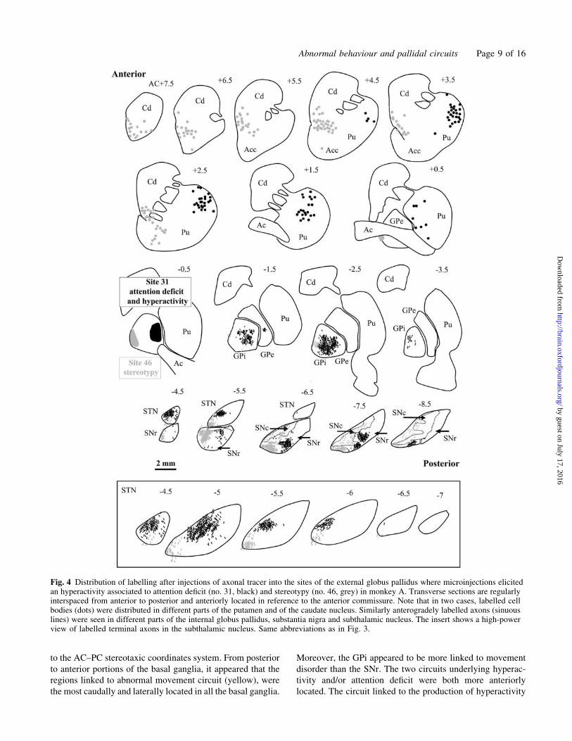
to the AC–PC stereotaxic coordinates system. From posterior
to anterior portions of the basal ganglia, it appeared that the
regions linked to abnormal movement circuit (yellow), were
the most caudally and laterally located in all the basal ganglia.
Moreover, the GPi appeared to be more linked to movement
disorder than the SNr. The two circuits underlying hyperac-
tivity and/or attention deficit were both more anteriorly
located. The circuit linked to the production of hyperactivity
Fig. 4 Distribution of labelling after injections of axonal tracer into the sites of the external globus pallidus where microinjections elicitedan hyperactivity associated to attention deficit (no. 31, black) and stereotypy (no. 46, grey) in monkey A. Transverse sections are regularlyinterspaced from anterior to posterior and anteriorly located in reference to the anterior commissure. Note that in two cases, labelled cellbodies (dots) were distributed in different parts of the putamen and of the caudate nucleus. Similarly anterogradely labelled axons (sinuouslines) were seen in different parts of the internal globus pallidus, substantia nigra and subthalamic nucleus. The insert shows a high-powerview of labelled terminal axons in the subthalamic nucleus. Same abbreviations as in Fig. 3.
Abnormal behaviour and pallidal circuits Page 9 of 16
by guest on July 17, 2016http://brain.oxfordjournals.org/
Dow
nloaded from
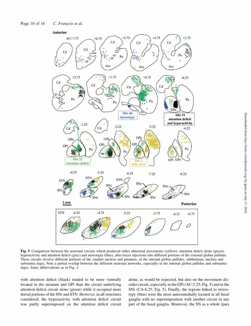
with attention deficit (black) tended to be more ventrally
located in the striatum and GPi than the circuit underlying
attention deficit circuit alone (green) while it occupied more
dorsal portions of the SNr and STN. However, in all structures
considered, the hyperactivity with attention deficit circuit
was partly superimposed on the attention deficit circuit
alone, as would be expected, but also on the movement dis-
order circuit, especially in the GPi (AC-2.25; Fig. 5) and in the
SNr (CA-6.25; Fig. 5). Finally, the regions linked to stereo-
typy (blue) were the most anteromedially located in all basal
ganglia with no superimposition with another circuit in any
part of the basal ganglia. Moreover, the SN as a whole (pars
Fig. 5 Comparison between the neuronal circuits which produced either abnormal movements (yellow), attention deficit alone (green),hyperactivity and attention deficit (grey) and stereotypy (blue), after tracer injections into different portions of the external globus pallidus.These circuits involve different portions of the caudate nucleus and putamen, of the internal globus pallidus, subthalamic nucleus andsubstantia nigra. Note a partial overlap between the different neuronal networks, especially in the internal globus pallidus and substantianigra. Same abbreviations as in Fig. 3.
Page 10 of 16 C. Francois et al.
by guest on July 17, 2016http://brain.oxfordjournals.org/
Dow
nloaded from
reticulata and compacta) appeared to be more linked to stereo-
typy than the GPi.
DiscussionBy using axonal tracer injected in restricted parts of the GPe,
where transitory behavioural disorders have previously been
produced by microinjection of bicuculline, we describe the
regions of the basal ganglia underlying these different behav-
ioural disorders. Basic principles that emerge are that (i) the
different behavioural disorders produced in monkeys are
underlined by different anatomical circuits within the basal
ganglia; (ii) these circuits involve different subregions of the
basal ganglia and appeared largely separated even if a partial
overlap exists within the output structures represented by the
GPi and SNr; (iii) each of these anatomical circuits system-
atically involves both the GPi and the SNr but not equally; (iv)
different subregions of the STN were also involved depending
on the movement or behavioural disorder produced.
Different structures of the basal gangliarelated to the GPeThe present tract-tracing study first confirms previous data
in monkeys concerning the retrograde labelling of cells in the
striatum and anterograde labelling of terminals within the
GPi, SNr and STN after tracer injection into the GPe
(Nauta and Mehler, 1966; Carpenter et al., 1968, 1981;
Hazrati et al., 1990; Shink et al., 1996). Indeed it had already
been reported that the pallidal terminals display large varico-
sities that were often closely apposed to the cell bodies and
proximal dendrites of STN, GPi and SNr (Sato et al., 2000).
The existence of preferential synaptic contacts with cell
bodies and proximal dendrites of SNr and entopeduncular
neurons in rats (the equivalent of the GPi of primates) has
been demonstrated using electron microscopy (Smith and
Bolam, 1991; Bolam and Smith, 1992). Altogether these
data suggest that all parts of the GPe project to the GPi
and SNr as well as to the STN. The present study confirms
such an organization, but also indicates that different
subregions of the GPe project to different anatomo-functional
territories of the basal ganglia. In such an organization, the
most anteromedial portion of the GPe appeared particular as it
was the only one subregion of the GPe which projects not only
to the SNr but also to the whole extent of the pars compacta.
As previously shown in monkeys, this projection appeared not
topographically organized (Haber et al., 1993).
The GPe was reported to also project to structures such
as the thalamic reticular nucleus and the pedunculopontine
nucleus, which are located outside of the basal ganglia. How-
ever, such projections are very sparse and even questioned
(Sato et al., 2000). This suggests that the different anatomo-
functional territories of the basal ganglia that were labelled in
our study are the main cerebral structures involved in the
behavioural effect produced. Our data are based on the deli-
neation of distinct anatomo-functional territories in the basal
ganglia as already done in the literature on the basis of tract-
tracing data and using the distribution of Cb immunoreactiv-
ity. Thus in the striatum and in the two pallidal segments,
but also in the SNr, the limbic territories were delineated
as regions of intense immunoreactivity, the associative terri-
tories as those of moderate Cb immunoreactivity, and the sen-
sorimotor territories as those of very weak immunoreactivity,
confirming previous data from our laboratory (Francois et al.,
1994b; Karachi et al., 2002). All these different anatomo-
functional territories of the pallidum and SNr appeared
implicated in circuits related to the expression of abnormal
movements and behavioural disorders. This is also the case
for the STN which thus appeared subdivided into different
functional subregions. This result may explain the role played
by the STN not only in motor control but also in cognitive and
motivational processes as reported in clinical and experi-
mental studies (Baunez and Robbins, 1997, 1999; Alegret
et al., 2001; Mallet et al., 2002; Funkiewiez et al., 2003).
In our study, it is not possible to determine if the extent of
the bicuculline injection site, and thus its physiological action,
can be superimposed on that of the axonal tracer injection
sites. However, injection of tracers performed within two
different sites of a given anatomo-functional territory of
the GPe where microinjections of bicuculline produced a
given behavioural effect, resulted in similar distribution of
labelling within the subregions of the basal ganglia. This thus
reinforces the result that every behavioural effect produced
is underlined by a given anatomo-functional subregion
within the basal ganglia.
Anatomical circuits underlying the movementand behavioural disordersRegions related to movement disorderThe present data first confirm that after injection of axonal
tracer into the posterior and central portion of the GPe where
microinjections produced leg dyskinesia, retrogradely
labelled cells were distributed in the posterior part of the
putamen. This striatal region is known to receive inputs
mainly from the motor, premotor and somatosensory cortex
(Kunzle, 1975; Flaherty and Graybiel, 1991; Inase et al.,
1996; Takada et al., 1998; Tokuno et al., 1999). These two
pallidal and striatal regions contain very weak Cb immuno-
reactivity, which is a characteristic feature of sensorimotor
territories (Francois et al., 1994b; Karachi et al., 2002). More-
over, we observed that the sensorimotor region of the GPe
projects to posterolateral parts of the SNr, but above all to
posterior portions of the GPi, all regions which are known to
receive inputs from the striatal sensorimotor territories
(Parent et al., 1984; Smith and Parent, 1986; Flaherty and
Graybiel, 1991, 1994; Hedreen and DeLong, 1991; Hazrati
and Parent, 1992; Francois et al., 1994a; Kaneda et al., 2002).
Electrophysiological evidence in support of the main sen-
sorimotor role of the GPi was provided by recording cells
after stimulation of motor cortical areas (Yoshida et al., 1993)
Abnormal behaviour and pallidal circuits Page 11 of 16
by guest on July 17, 2016http://brain.oxfordjournals.org/
Dow
nloaded from
and by retrograde transynaptic labelling of herpes virus from
motor cortical areas (Hoover and Strick, 1999). The dorso-
lateral portion of the STN where we found labelled GPe
terminations is also known to receive motor cortical
inputs (Nambu et al., 1996). It can thus be concluded that
movement disorder that can be induced by microinjection of
bicuculline in the GPe involves motor territories of all the
basal ganglia.
Regions related to hyperactivity and/or attentiondeficitThe striatal region occupied by neurons which project to the
GPe region where microinjections elicited spatial perturba-
tion in the task, suggesting attention deficit, without hyper-
activity (see Grabli et al., 2004) were located anteriorly in
dorsal portions of the putamen and of the caudate nucleus.
These striatal regions are reported to receive cortical inputs
from different areas: the associative parietal and dorsolateral
cortex (Yeterian and Pandya, 1991, 1993), the anterior part
of the motor and premotor cortex (Inase et al., 1996, 1999;
Takada et al., 1998), but above all the supplementary and
pre-supplementary motor areas (Inase et al., 1999; Kaneda
et al., 2002), the supplementary eye field (Shook et al., 1991;
Parthasarathy et al., 1992, Cui et al., 2003) and the motor
cingulate cortex (Takada et al., 2001). All these cortical areas
anterior to the primary motor cortex participate in cognitive
processes in addition to their well-known role in movement
planning and execution. In particular, the anterior part of the
premotor cortex, the dorsolateral prefrontal cortex and the
supplementary eye field which mainly project to the caudate
nucleus were reported to be specialized, among others, in
spatial attention (Corbetta et al., 1991; Corbetta and Shulman,
1998; Boussaoud, 2001).
Within the GPi, axonal fibres were located in the lateral part
anteriorly, and in its dorsal part posteriorly, while in the SNr
they were present in a ventral portion. These regions, which
appeared moderately stained on Cb immunoreactive sections,
were reported to receive striatal inputs from the most posterior
part of the associative territories, and from the most anterior
part of the sensorimotor territories (Parent et al., 1984; Smith
and Parent, 1986; Hedreen and DeLong, 1991; Hazrati and
Parent, 1992; Flaherty and Graybiel, 1994; Francois et al.,
1994a), and more specifically from striatal regions linked to
the supplementary motor area (Kaneda et al., 2002). These
regions contain neurons which are known to project via the
thalamus back to the associative prefrontal cortex as well as to
the supplementary and pre-supplementary motor cortex
(Middleton and Strick, 2000, 2002). Within the STN, axonal
fibres were confined to the anterior and ventral portion, in a
region known to receive cortical inputs mainly from the
supplementary and pre-supplementary motor areas (Takada
et al., 2001) but also, partly, from pre-supplementary and
supplementary eye field (Shook et al., 1991). Thus our results
certainly indicate that the attention deficit circuit alone is
underlined by subregions of the basal ganglia linked to the
cortical areas which play a major role in both motor planning
and attention processes.
Comparatively, hyperactivity with attention deficit circuit
involves roughly the same portions of the striatum, but the
labelling was slightly more ventrally located in the putamen
and did not reach the caudate nucleus. This striatal zone not
only receives cortical projection from the most anterior part of
the secondary motor areas such as the supplementary motor
cortex but also from the rostral cingulate motor cortex as
recently reported (Takada et al., 2001). Within the GPi,
GPe labelled axons occupied a more ventral portion, while
in the SNr they were more dorsally located than in the atten-
tion deficit circuit alone. As for the striatum, these regions,
and particularly that located in the GPi, tended to involve the
anterior part of the sensorimotor territories. The region impli-
cated in the STN was also more dorsally located than in the
preceding case and thus implicated a region which was
reported to receive inputs from the rostral cingulate motor
cortex (Takada et al., 2001). It may thus be concluded from
our results that the attention deficit with hyperactivity circuit
is underlined by subregions of the basal ganglia linked not
only to the supplementary and pre-supplementary motor areas
but also to the rostral cingulate motor cortex. These secondary
motor cortical areas are implicated not only in the execution
of the movement but also in higher-order cognitive aspects
of the movement such as motor selection or error detection
(Akkal et al., 2002; Isomura et al., 2003).
Even if it is difficult to compare attention deficit with
hyperactivity observed in monkeys with ADHD disorder
described in humans, it is interesting to note that functional
abnormalities in humans were visualized not only in prefron-
tal cortical areas (Amen et al., 1993; Amen and Carmichael,
1997; Spalletta et al., 2001) and more specifically in the
anterior cingulate cortex (Bush et al., 1999), but also in the
caudate nucleus (Castellanos et al., 1996; Filipek et al., 1997;
Rubia et al., 1999; Castellanos, 2001), in both caudate nucleus
and putamen (Vaidya et al., 1998; Teicher et al., 2000) and in
the pallidum as a unit (Aylward et al., 1996; Castellanos et al.,
1996). Our findings and these observations in humans strongly
suggest that ADHD as a whole represents a deficiency in parts
of the basal ganglia linked to associative prefrontal cortex and
to secondary motor cortical areas involved in attention pro-
cesses and motor planning.
Regions related to stereotyped behaviourThe striatal region labelled after injection of axonal tracer in
the region of the GPe where microinjections elicited a per-
sistent repetition of a single behaviour was ventromedial in
the striatum, in a region located at the limit between the dorsal
portion of the nucleus accumbens and the ventral part of the
head of the caudate nucleus. Even if neither cytoarchitectonic
nor immunohistochemical methods allows us to isolate the
nucleus accumbens from the other part of the striatum, this
striatal region is known to be the major structure that receives
limbic inputs from the orbitofrontal, anterior cingulate and
Page 12 of 16 C. Francois et al.
by guest on July 17, 2016http://brain.oxfordjournals.org/
Dow
nloaded from
insular cortices (Kunishio and Haber, 1994; Haber et al.,
1995; Chikama et al., 1997; Ferry et al., 2000), amygdala
and hippocampus (Russchen et al., 1985; Friedman et al.,
2002; Fudge and Haber, 2002), all regions which are
known to process emotional and motivational information.
The labelled GPe fibres originating in the site related to stereo-
typy occupied anterior and ventromedial located regions of
the GPi but above all of the SNr, regions which correspond to
limbic territories as seen on Cb-immunoreactive sections and
after comparison with a tracing study (Haber et al., 1990).
However it is clear from our results that the SNr is more linked
to limbic structures than the GPi. These regions of the output
structures of the basal ganglia are known to project to the
orbital frontal cortex via the medial part of the thalamus
(Ilinsky et al., 1985; Haber et al., 1993). In the STN,
which is free of Cb, the labelling region was also restricted
to the most rostromedial part of the nucleus, which confirms
the existence of a limbic pallidosubthalamic territory, as
recently described in our laboratory (C. Karachi, in press).
Our present anatomical evidence thus supports the early
suggestion that the pallidum could be involved in
obsessive-compulsive disorder as it was reported that
bilateral lesions in this structure can produce striking
obsessive-compulsive disorder-like behaviour (Laplane
et al., 1984, 1989).
Our data also confirm that one particular output target of
the ventral limbic GPe is represented by the dopaminergic
neurons of the SN pars compacta (Gerfen, 1992; Haber et al.,
1993; Lynd-Balta and Haber, 1994). These dopaminergic
neurons are considered to be the key for focusing attention
on significant and rewarding stimuli (Schultz, 1997). This
suggests an influence of the limbic GPe on the whole extent
of the striatum via the dopaminergic nigro-striatal projection
(Haber et al., 1993). Moreover, it was recently demonstrated
in the rat that striosomes, one of the two compartments of the
striatum which is thought to also project to the SNc (Graybiel,
1990; Gerfen, 1992), appear active when the animal performs
repetitive, stereotyped behaviours in response to dopamine
receptor agonists (Canales and Graybiel, 2000). The fact
that, in our study, neurons, labelled after injection in the
site where stereotypy was induced, were mainly found in
the matrix of the ventral striatum not in striosomes, suggests
that the output pathway of the matrix is also involved in
the production of stereotyped behaviour.
This stereotyped behaviour that was produced in our mon-
keys resembles tics and compulsive disorders described
in patients with Tourette’s syndrome or with obsessive-
compulsive disorder. Imaging studies from patients with
obsessive-compulsive disorder and with Tourette’s syndrome
reported abnormalities of volumes in the caudate nucleus
(Scarone et al., 1992; Peterson et al., 1993; Singer et al.,
1993; Hyde et al., 1995; Moriarty et al., 1997; Peterson,
2001). Abnormal neuronal activity has also been reported
at rest in the caudate nucleus but also in the ventral globus
pallidus, orbito-frontal and cingulate cortex (Eidelberg et al.,
1997; Rauch and Baxter, 1998; Saxena et al., 1998; Peterson,
2001; Rauch et al., 2001). Interestingly it was reported that
infusion of sera from patients with Tourette’s syndrome into
the ventral striatum of rats increased the rate of oral stereo-
typies (Taylor et al., 2002). Together, these data and our
present results provide strong evidence for an implication
of the limbic parts of all the basal ganglia in producing stereo-
typed behaviour.
Partial superimposition of the differentneuronal networksThere was no evidence of superimposition of the different
neuronal networks that produced abnormal behaviours and
movements within the striatum and the STN, except in limited
zones between the hyperactivity associated to attention deficit
circuit and the attention deficit circuit alone. On the other
hand, within the output structures of the basal ganglia, regions
which appeared linked to attention deficit alone overlapped
with some portions of the hyperactivity and attention deficit
circuits, as can be expected, but also with the movement
disorder circuit. This highly suggests the existence of a gra-
dient between movement, hyperactivity and attention deficit
circuits in the GPi and in the SNr. The notion that such a
gradient exists is supported by the existence of very long
dendrites especially in these two structures (Yelnik et al.,
1984, 1987), which allows convergence of information
coming from different functional striatal territories on a single
neuron. This was observed for pallidal neurons located in an
intermediate zone between the associative and the sensori-
motor territories and which respond to the stimulation of
both the caudate nucleus and the putamen (Tremblay and
Filion, 1989), and this may also be the case at the interface
between the limbic and associative territories. Such a pattern
gives the possibility of integration of functionally diverse
information, at least within the output structures of the
basal ganglia.
ConclusionOur approach allowed us to demonstrate that the different
behavioural disorders produced in monkeys by microinjec-
tions of bicuculline in different portions of the GPe are under-
lined by different anatomical circuits within the basal ganglia.
This provides strong evidence for the involvement of different
subregions of the basal ganglia (the two output structures:
GPi and SNr, and the STN) in producing abnormal movement
and behaviours. It can thus be hypothesized that different
portions of the basal ganglia organized in anatomo-functional
circuits play a role in the pathophysiology of syndromes such
as obsessive-compulsive disorder, Tourette’s syndrome or
ADHD. In such a hypothesis, these different subregions of
the basal ganglia could be considered as therapeutic targets.
Thus, high frequency stimulation, which is well known to
improve motor disorders of Parkinson’s disease when it is
centred in the sensorimotor territory of the STN (Yelnik
Abnormal behaviour and pallidal circuits Page 13 of 16
by guest on July 17, 2016http://brain.oxfordjournals.org/
Dow
nloaded from
et al., 2003) could also improve behavioural disorders when
other anatomo-functional subregions are targeted.
AcknowledgementsWe wish to thank Dominique Tande for expert participation
in surgical operations and immunohistochemical procedures,
and Nicholas Barton for checking the English. This work was
supported by the Institut National de la Sante et de la
Recherche Medicale (INSERM, France) and a grant from
The Tourette Syndrome Association (USA).
References
Akkal D, Bioulac B, Audin J, Burbaud P. Comparison of neuronal activity in
the rostral supplementary and cingulate motor areas during a task with
cognitive and motor demands. Eur J Neurosci 2002; 15: 887–904.
Alegret M, Junque C, Valldeoriola F, Vendrell P, Pilleri M, Rumia J, et al.
Effects of bilateral subthalamic stimulation on cognitive function in
Parkinson disease. Arch Neurol 2001; 58: 1223–7.
Alexander GE, DeLong MR, Strick PL. Parallel organization of functionally
segregated circuits linking basal ganglia and cortex. Annu Rev Neurosci
1986; 9: 357–81.
Amen DG, Carmichael BD. High-resolution brain SPECT imaging in ADHD.
Ann Clin Psychiatry 1997; 9: 81–6.
Amen DG, Paldi F, Thisted RA. Brain SPECT imaging. J Am Acad Child
Adolesc Psychiatry 1993; 32: 1080–1.
Aylward EH, Reiss AL, Reader MJ, Singer HS, Brown JE, Denckla MB.
Basal ganglia volumes in children with attention-deficit hyperactivity
disorder. J Child Neurol 1996; 11: 112–15.
Baunez C, Robbins TW. Bilateral lesions of the subthalamic nucleus induce
multipledeficits inanattentional taskinrats.EurJNeurosci1997;9:2086–99.
Baunez C, Robbins TW. Effects of transient inactivation of the subthalamic
nucleus by local muscimol and APV infusions on performance on the five-
choice serial reaction time task in rats. Psychopharmacology (Berl) 1999;
141: 57–65.
Bolam JP, Smith Y. The striatum and the globus pallidus send convergent
synaptic inputs onto single cells in the entopeduncular nucleus of the rat: a
double anterograde labelling study combined with postembedding immu-
nocytochemistry for GABA. J Comp Neurol 1992; 321: 456–76.
Boussaoud D. Attention versus intention in the primate premotor cortex.
Neuroimage 2001; 14: S40–5.
Bush G, Frazier JA, Rauch SL, Seidman LJ, Whalen PJ, Jenike MA, et al.
Anterior cingulate cortex dysfunction in attention-deficit/hyperactivity
disorder revealed by fMRI and the Counting Stroop. Biol Psychiatry
1999; 45: 1542–52.
Canales JJ, Graybiel AM. A measure of striatal function predicts motor
stereotypy. Nat Neurosci 2000; 3: 377–83.
Carpenter MB, Fraser RAR, Shriver JE. The organization of pallidosubtha-
lamic fibers in the monkey. Brain Res 1968; 11: 522–59.
Carpenter MB, Batton RR 3rd, Carleton SC, Keller JT. Interconnections and
organization of pallidal and subthalamic nucleus neurons in the monkey.
J Comp Neurol 1981; 197: 579–603.
Castellanos FX. Neural substrates of attention-deficit hyperactivity disorder.
Adv Neurol 2001; 85: 197–206.
Castellanos FX, Giedd JN, Marsh WL, Hamburger SD, Vaituzis AC,
Dickstein DP, et al. Quantitative brain magnetic resonance imaging in atten-
tion-deficit hyperactivity disorder. Arch Gen Psychiatry 1996; 53: 607–16.
Chikama M, McFarland NR, Amaral DG, Haber SN. Insular cortical projec-
tions to functional regions of the striatum correlate with cortical cytoarch-
itectonic organization in the primate. J Neurosci 1997; 17: 9686–705.
Corbetta M, Shulman GL. Human cortical mechanisms of visual attention
during orienting and search. Philos Trans R Soc Lond B Biol Sci 1998; 353:
1353–62.
Corbetta M, Miezin FM, Dobmeyer S, Shulman GL, Petersen SE. Selective
and divided attention during visual discriminations of shape, color, and
speed: functional anatomy by positron emission tomography. J Neurosci
1991; 11: 2383–402.
Cui DM, Yan YJ, Lynch JC. Pursuit subregion of the frontal eye field projects
to the caudate nucleus in monkeys. J Neurophysiol 2003; 89: 2678–84.
Eidelberg D, Moeller JR, Antonini A, Kazumata K, Dhawan V, Budman C,
et al. The metabolic anatomy of Tourette’s syndrome. Neurology 1997; 48:
927–34.
Ferry AT, Ongur D, An X, Price JL. Prefrontal cortical projections to the
striatum in macaque monkeys: evidence for an organization related to
prefrontal networks. J Comp Neurol 2000; 425: 447–70.
Filipek PA, Semrud-Clikeman M, Steingard RJ, Renshaw PF, Kennedy DN,
Biederman J. Volumetric MRI analysis comparing subjects having atten-
tion-deficit hyperactivity disorder with normal controls. Neurology 1997;
48: 589–601.
Flaherty AW, Graybiel AM. Corticostriatal transformations in the primate
somatosensory system. Projections from physiologically mapped body-part
representations. J Neurophysiol 1991; 66: 1249–63.
Flaherty AW, Graybiel AM. Input-output organization of the sensorimotor
striatum in the squirrel monkey. J Neurosci 1994; 14: 599–610.
Francois C, Yelnik J, Percheron G, Fenelon G. Topographic distribution of the
axonal endings from the sensorimotor and associative striatum in the maca-
que pallidum and substantia nigra. Exp Brain Res 1994a; 102: 305–18.
Francois C, Yelnik J, Percheron G, Tande D. Calbindin D-28k as a marker
for the associative cortical territory of the striatum in macaque. Brain
Res 1994b; 633: 331–6.
Francois C, Jan C, McCairn K, Grabli D, Hirsch EC, Feger J, et al. A primate
model of Tourette’s syndrome: anatomical analysis of the basal ganglia
territories involved in hyperactivity disorder with attention deficit (HD/AD)
and stereotypy [abstract]. Soc Neurosci Abstr 2002; 32: No. 663.6.
Friedman DP, Aggleton JP, Saunders RC. Comparison of hippocampal,
amygdala, and perirhinal projections to the nucleus accumbens: combined
anterograde and retrograde tracing study in the Macaque brain. J Comp
Neurol 2002; 450: 345–65.
Fudge JL, Haber SN. Defining the caudal ventral striatum in primates: cellular
and histochemical features. J Neurosci 2002; 22: 10078–82.
Funkiewiez A, Ardouin C, Krack P, Fraix V, Van Blercom N, Xie J, et al.
Acute psychotropic effects of bilateral subthalamic nucleus stimulation
and levodopa in Parkinson’s disease. Mov Disord 2003; 18: 524–30.
Gerfen CR. The neostriatal mosaic: multiple levels of compartmental orga-
nization. Trends Neurosci 1992; 15: 133–9.
Graybiel AM. Neurotransmitters and neuromodulators in the basal ganglia.
Trends Neurosci 1990; 13: 244–54.
Grabli D, McCarin K, Hirsch EC, Agid Y, Feger J, Francois C, et al. Beha-
vioural disorders induced by external globus pallidus dysfunction in pri-
mates: I Behavioural study. Brain/Advance Access published August 3,
2004, 10.1093/brain/awh220.
Haber SN, Lynd E, Klein C, Groenewegen HJ. Topographic organization
of the ventral striatal efferent projections in the rhesus monkey: an
anterograde tracing study. J Comp Neurol 1990; 293: 282–98.
Haber SN, Lynd-Balta E, Mitchell SJ. The organization of the descending
ventral pallidal projections in the monkey. J Comp Neurol 1993; 329:
111–28.
Haber SN, Kunishio K, Mizobuchi M, Lynd-Balta E. The orbital and medial
prefrontal circuit through the primate basal ganglia. J Neurosci 1995; 15:
4851–67.
Haber SN, Fudge JL, McFarland NR. Striatonigrostriatal pathways in pri-
mates form an ascending spiral from the shell to the dorsolateral striatum.
J Neurosci 2000; 20: 2369–82.
Hazrati LN, Parent A. The striatopallidal projection displays a high degree of
anatomical specificity in the primate. Brain Res 1992; 592: 213–27.
Hazrati LN, Parent A, Mitchell S, Haber SN. Evidence for interconnections
between the two segments of the globus pallidus in primates: a PHA-L
anterograde tracing study. Brain Res 1990; 533: 171–5.
Hedreen JC, DeLong MR. Organization of striatopallidal, striatonigral, and
nigrostriatal projections in the macaque. J Comp Neurol 1991; 304: 569–95.
Page 14 of 16 C. Francois et al.
by guest on July 17, 2016http://brain.oxfordjournals.org/
Dow
nloaded from
Hoover JE, Strick PL. The organization of cerebellar and basal ganglia out-
puts to primary motor cortex as revealed by retrograde transneuronal trans-
port of herpes simplex virus type 1. J Neurosci 1999; 19: 1446–63.
Hyde TM, Stacey ME, Coppola R, Handel SF, Rickler KC, Weinberger DR.
Cerebral morphometric abnormalities in Tourette’s syndrome: a quantita-
tive MRI study of monozygotic twins. Neurology 1995; 45: 1176–82.
Ilinsky IA, Jouandet ML, Goldman-Rakic PS. Organization of the nigrotha-
lamocortical system in the rhesus monkey. J Comp Neurol 1985; 236:
315–30.
Inase M, Sakai ST, Tanji J. Overlapping corticostriatal projections from the
supplementary motor area and the primary motor cortex in the macaque
monkey: an anterograde double labeling study. J Comp Neurol 1996; 373:
283–96.
Inase M, Tokuno H, Nambu A, Akazawa T, Takada M. Corticostriatal and
corticosubthalamic input zones from the presupplementary motor area in
the macaque monkey: comparison with the input zones from the supple-
mentary motor area. Brain Res 1999; 833: 191–201.
Isomura Y, Ito Y, Akazawa T, Nambu A, Takada M. Neural coding of
‘attention for action’ and ‘response selection’ in primate anterior
cingulate cortex. J Neurosci 2003; 23: 8002–12.
Jankovic J. Tourette’s syndrome. N Engl J Med 2001; 345: 1184–92.
Kaneda K, Nambu A, Tokuno H, Takada M. Differential processing patterns
of motor information via striatopallidal and striatonigral projections.
J Neurophysiol 2002; 88: 1420–32.
Karachi C, Francois C, Parain K, Bardinet E, Tande D, Hirsch EC, et al.
Three-dimensional cartography of functional territories in the human
striatopallidal complex using calbindin immunoreactivity. J Comp Neurol
2002; 450: 122–34.
Karachi C, Yelnik J, Tande D, Tremblay L, Hirsch EC, Francois C. An
anatomical substrate for non motor functions of the subthalamic nucleus
in primates. Mov Disord, in press.
Kunishio K, Haber SN. Primate cingulostriatal projection: limbic
striatal versus sensorimotor striatal input. J Comp Neurol 1994; 350:
337–56.
Kunzle H. Bilateral projections from precentral motor cortex to the putamen
and other parts of the basal ganglia. an autoradiographic study in Macaca
fascicularis. Brain Res 1975; 88: 195–209.
Laplane D, Baulac M, Widlocher D, Dubois B. Pure psychic akinesia with
bilateral lesions of basal ganglia. J Neurol Neurosurg Psychiatry 1984; 47:
377–85.
Laplane D, Levasseur M, Pillon B, Dubois B, Baulac M, Mazoyer B, et al.
Obsessive-compulsive and other behavioural changes with bilateral basal
ganglia lesions. A neuropsychological, magnetic resonance imaging and
positron tomography study. Brain 1989; 112: 699–725.
Lynd-Balta E, Haber SN. Primate striatonigral projections: a comparison of
the sensorimotor-related striatum and the ventral striatum. J Comp Neurol
1994; 345: 562–78.
Mallet L, Mesnage V, Houeto JL, Pelissolo A, Yelnik J, Behar C, et al.
Compulsions, Parkinson’s disease, and stimulation. Lancet 2002; 360:
1302–4.
Mesulam MM. Tetramethyl benzidine for horseradish peroxidase neuro-
histochemistry: a non-carcinognic blue reaction product with superior sen-
sitivity for visualizing neural afferents and efferents. J Histochem
Cytochem 1978; 26: 106–17.
Middleton FA, Strick PL. Basal ganglia and cerebellar loops: motor and
cognitive circuits. Brain Res Brain Res Rev 2000; 31: 236–50.
Middleton FA, Strick PL. Basal-ganglia ‘projections’ to the prefrontal cortex
of the primate. Cereb Cortex 2002; 12: 926–35.
Moriarty J, Varma AR, Stevens J, Fish M, Trimble MR, Robertson MM. A
volumetric MRI study of Gilles de la Tourette’s syndrome. Neurology
1997; 49: 410–15.
Nambu A, Takada M, Inase M, Tokuno H. Dual somatotopical representations
in the primate subthalamic nucleus: Evidence for ordered but reversed
body-map transformations from the primary motor cortex and the supple-
mentary motor area. J Neurosci 1996; 16: 2671–83.
Nauta WJH, Mehler WR. Projections of the lentiform nucleus in the monkey.
Brain Res 1966; 1: 3–42.
Parent A, Hazrati LN. Functional anatomy of the basal ganglia. 1. The cortico-
basal ganglia-thalamo-cortical loop. Brain Res Brain Res Rev 1995a;
20: 91–127.
Parent A, Hazrati LN. Functional anatomy of the basal ganglia. II. The place
of subthalamic nucleus and external pallidum in basal ganglia circuitry.
Brain Res Brain Res Rev 1995b; 20: 128–54.
Parent A, Bouchard C, Smith Y. The striatopallidal and striatonigral projec-
tions: two distinct fiber systems in primate. Brain Res 1984; 303: 385–90.
Parthasarathy HB, Schall JD, Graybiel AM. Distributed but convergent
ordering of corticostriatal projections: analysis of the frontal eye field
and the supplementary eye field in the macaque monkey. J Neurosci
1992; 12: 4468–88.
Percheron G, Francois C, Yelnik J. Instruments and techniques for the stereo-
tactic surgery based on the CA-CP ventricular system of coordinates in
monkeys. J Neurosci Methods 1986; 17: 89–99.
Peterson BS. Neuroimaging studies of Tourette syndrome: a decade of pro-
gress. Adv Neurol 2001; 85: 179–96.
Peterson B, Riddle MA, Cohen DJ, Katz LD, Smith JC, Hardin MT, et al.
Reduced basal ganglia volumes in Tourette’s syndrome using three-
dimensional reconstruction techniques from magnetic resonance images.
Neurology 1993; 43: 941–9.
Rauch SL, Baxter LR. Neuroimaging of OCD and related disorders. In: Jenike
MA, Baer L, Minichiello WE, editors. Obsessive-compulsive disorders:
practical management. 3rd ed. St Louis: Mosby; 1998. p. 289–317.
Rauch SL, Whalen PJ, Curran T, Shin LM, Coffey BJ, Savage CR, et al.
Probing striato-thalamic function in obsessive-compulsive disorder and
Tourette syndrome using neuroimaging methods. Adv Neurol 2001; 85:
207–24.
Russchen FT, Bakst I, Amaral DG, Price JL. The amygdalostriatal projections
in the monkey. An anterograde tracing study. Brain Res 1985; 329: 241–57.
Saint-Cyr JA, Ungerleider LG, Desimone R. Organization of visual cortical
inputs to the striatum and subsequent outputs to the pallido-nigral complex
in the monkey. J Comp Neurol 1990; 298: 129–56.
Sato F, Lavallee P, Levesque M, Parent A. Single-axon tracing study of
neurons of the external segment of the globus pallidus in primate. J
Comp Neurol 2000; 417: 17–31.
Saxena S, Brody AL, Schwartz JM, Baxter LR. Neuroimaging and frontal-
subcortical circuitry in obsessive-compulsive disorder. Br J Psychiatry
Suppl 1998; 35: 26–37.
Scarone S, Colombo C, Livian S, Abbruzzese M, Ronchi P, Locatelli M, et al.
Increased right caudate nucleus size in obsessive-compulsive disorder:
detection with magnetic resonance imaging. Psychiatry Res 1992; 45:
115–21.
Schultz W. Dopamine neurons and their role in reward mechanisms. Curr
Opin Neurobiol 1997; 7: 191–7.
Shink E, Bevan MD, Bolam JP, Smith Y. The subthalamic nucleus and the
external pallidum: two tightly interconnected structures that control the
output of the basal ganglia in the monkey. Neuroscience 1996; 73: 335–57.
Shook BL, Schlag-Rey M, Schlag J. Primate supplementary eye field. II.
Comparative aspects of connections with the thalamus, corpus striatum,
and related forebrain nuclei. J Comp Neurol 1991; 307: 562–83.
Singer HS, Reiss AL, Brown JE, Aylward EH, Shih B, Chee E, et al. Volu-
metric MRI changes in basal ganglia of children with Tourette’s syndrome.
Neurology 1993; 43: 950–6.
Smith Y. Anterograde tracing with PHA-L and biocytin at the electron micro-
scopic level. In: Bolam JP, Rickwood D, Hames BD, editors. Experimental
neuroanatomy. A practical approach. New York: Plenum Press; 1992.
p. 61–79.
Smith Y, Bolam JP. Convergence of synaptic inputs from the striatum and the
globus pallidus onto identified nigrocollicular cells in the rat: a double
anterograde labelling study. Neuroscience 1991; 44: 45–73.
Smith Y, Parent A. Differential connections of caudate nucleus and
putamen in the squirrel monkey (Saımiri sciureus). Neuroscience 1986;
18: 347–71.
Spalletta G, Pasini A, Pau F, Guido G, Menghini L, Caltagirone C. Prefrontal
blood flow dysregulation in drug naive ADHD children without structural
abnormalities. J Neural Transm 2001; 108: 1203–16.
Abnormal behaviour and pallidal circuits Page 15 of 16
by guest on July 17, 2016http://brain.oxfordjournals.org/
Dow
nloaded from
Takada M, Tokuno H, Nambu A, Inase M. Corticostriatal input zones from the
supplementary motor area overlap those from the contra- rather than
ipsilateral primary motor cortex. Brain Res 1998; 791: 335–40.
Takada M, Tokuno H, Hamada I, Inase M, Ito Y, Imanishi M, et al. Organiza-
tion of inputs from cingulate motor areas to basal ganglia in macaque
monkey. Eur J Neurosci 2001; 14: 1633–50.
Taylor JR, Morshed SA, Parveen S, Mercadante MT, Scahill L, Peterson BS,
et al. An animal model of Tourette’s syndrome. Am J Psychiatry 2002; 159:
657–60.
Teicher MH, Anderson CM, Polcari A, Glod CA, Maas LC, Renshaw PF.
Functional deficits in basal ganglia of children with attention-deficit/
hyperactivity disorder shown with functional magnetic resonance imaging
relaxometry. Nat Med 2000; 6: 470–73.
Tokuno H, Inase M, Nambu A, Akazawa T, Miyachi S, Takada M.
Corticostriatal projections from distal and proximal forelimb
representations of the monkey primary motor cortex. Neurosci Lett
1999; 269: 33–6.
Tremblay L, Filion M. Responses of pallidal neurons to striatal stimulation
in intact waking monkeys. Brain Res 1989; 498: 1–16.
Vaidya CJ, Austin G, Kirkorian G, Ridlehuber HW, Desmond JE,
Glover GH, et al. Selective effects of methylphenidate in attention deficit
hyperactivity disorder: a functional magnetic resonance study. Proc Natl
Acad Sci USA 1998; 95: 14494–9.
Yelnik J, Percheron G, Francois C. A Golgi analysis of the primate globus
pallidus. II. Quantitative morphology and spatial orientation of dendritic
arborizations. J Comp Neurol 1984; 227: 200–13.
Yelnik J, Francois C, Percheron G, Heyner S. Golgi study of the primate
substantia nigra. I. Quantitative morphology and typology of nigral neu-
rons. J Comp Neurol 1987; 265: 455–72.
Yelnik J, Damier P, Demeret S, Gervais D, Bardinet E, Bejjani BP, et al.
Localization of stimulating electrodes in patients with Parkinson
disease by using a three-dimensional atlas-magnetic resonance imaging
coregistration method. J Neurosurg 2003; 99: 89–99.
Yeterian EH, Pandya DN. Prefrontostriatal connections in relation to cortical
architectonic organization in rhesus monkeys. J Comp Neurol 1991; 312:
43–67.
Yeterian EH, Pandya DN. Striatal connections of the parietal
association cortices in rhesus monkeys. J Comp Neurol 1993; 332:
175–97.
Yoshida SI, Nambu A, Jinnai K. The distribution of the globus pallidus
neurons with input from various cortical areas in the monkeys. Brain
Res 1993; 611: 170–4.
Page 16 of 16 C. Francois et al.
by guest on July 17, 2016http://brain.oxfordjournals.org/
Dow
nloaded from


