Embed Size (px)
Citation preview
Linnaeus University DissertationsNr 303/2017
Carina Bunse
Bacterioplankton in the light of seasonality and environmental drivers
linnaeus university press
Lnu.seisbn: 978-91-88761-02-6 (print), 978-91-88761-03-3 (pdf )
Bacterioplankton in the light of seasonality and environm
ental driversC
arina Bunse

Bacterioplankton in the light of seasonality and environmental drivers

Linnaeus University Dissertations
No 303/2017
BACTERIOPLANKTON IN THE LIGHT OF SEASONALITY AND
ENVIRONMENTAL DRIVERS
CARINA BUNSE
LINNAEUS UNIVERSITY PRESS
Linnaeus University Dissertations
No 303/2017
BACTERIOPLANKTON IN THE LIGHT OF SEASONALITY AND
ENVIRONMENTAL DRIVERS
CARINA BUNSE
LINNAEUS UNIVERSITY PRESS
Abstract Bunse, Carina (2017). Bacterioplankton in the light of seasonality and environmental drivers:, Linnaeus University Dissertations No 303/2017, ISBN: 978-91-88761-02-6 (print), 978-91-88761-03-3 (pdf). Written in English with a summary in Swedish. Bacterioplankton are keystone organisms in marine ecosystems. They are important for element cycles, by transforming dissolved organic carbon and other nutrients. Bacterioplankton community composition and productivity rates change in surface waters over spatial and temporal scales. Yet, many underlying biological processes determining when, why and how bacterioplankton react to changes in environmental conditions are poorly understood. Here, I used experiments with model bacteria and natural assemblages as well as field studies to determine molecular, physiological and ecological responses allowing marine bacteria to adapt to their environment.
Experiments with the flavobacterium Dokdonia sp. MED134 aimed to determine how the metabolism of bacteria is influenced by light and different organic matter. Under light exposure, Dokdonia sp. MED134 expressed proteorhodopsin and adjusted its metabolism to use resources more efficiently when growing with lower-quality organic matter. Similar expression patterns were found in oceanic datasets, implying a global importance of photoheterotrophic metabolisms for the ecology of bacterioplankton.
Further, I investigated how the composition and physiology of bacterial assemblages are affected by elevated CO2 concentrations and inorganic nutrients. In a large-scale experiment, bacterioplankton could keep productivity and community structure unaltered by adapting the gene expression under CO2 stress. To maintain pH homeostasis, bacteria induced higher expression of genes related to respiration, membrane transport and light acquisition under low-nutrient conditions. Under high-nutrient conditions with phytoplankton blooms, such regulatory mechanisms were not necessary. These findings indicate that open ocean systems are more vulnerable to ocean acidification than coastal waters.
Lastly, I used field studies to resolve how bacterioplankton is influenced by environmental changes, and how this leads to seasonal succession of marine bacteria. Using high frequency sampling over three years, we uncovered notable variability both between and within years in several biological features that rapidly changed over short time scales. These included potential phytoplankton-bacteria linkages, substrate uptake rates, and shifts in bacterial community structure. Thus, high resolution time series can provide important insights into the mechanisms controlling microbial communities.
Overall, this thesis highlights the advantages of combining molecular and traditional oceanographic methodological approaches to study ecosystems at high resolution for improving our understanding of the physiology and ecology of microbial communities and, ultimately, how they influence biogeochemical processes. Keywords marine bacteria, marine microbiology, seasonal succession, ocean acidification, proteorhodopsin, photoheterotrophy, microbial time series
Bacterioplankton in the light of seasonality and environmental drivers Doctoral Dissertation, Department of Biology and Environmental Science, Linnaeus University, Kalmar, 2017 ISBN: 978-91-88761-02-6 (print), 978-91-88761-03-3 (pdf) Published by: Linnaeus University Press, 351 95 Växjö Printed by: DanagårdLiTHO, 2017
1
“The face of the sea is always changing. Crossed by colors, lights, and moving shadows, sparkling in the sun, mysterious in the
twilight, its aspects and its moods vary hour by hour. The surface waters move with the tides, stir to the breath of the winds, and rise
and fall to the endless, hurrying forms of the waves. Most of all, they change with the advance of the seasons.”
Rachel Carson, The sea around us
Svensk sammanfattning
Bakterier är nyckelorganismer i marina ekosystem. De är viktiga för näringsämnenas kretslopp tack vare sin förmåga att transformera löst organiskt kol och andra näringsämnen. Bakteriesamhällets artsammansättning och produktivitet i ytvattnen förändras ständigt i både tid och rum. Många underliggande biologiska processer såsom när, varför och hur bakterieplankton reagerar på årstider och andra miljöförändringar är fortfarande dåligt förstådda. I denna avhandling har jag gjort experiment med modellorganismer och naturliga bakteriesamhällen samt fältstudier för att studera molekylära, fysiologiska och ekologiska mekanismer som gör det möjligt för marina bakterier att anpassa sig till sin miljö.
Experiment med flavobakterien Dokdonia sp. MED134, syftade till att bestämma hur bakteriers metabolism påverkad av ljus och olika organiska substrat. Vid ljusexponering uttryckte Dokdonia sp. MED134 det ljuskänsliga proteinet proteorodopsin och andra gener. Särskilt vid tillväxt med substrat av lägre kvalitet så ledde detta till en tydlig anpassning av ämnesomsättningen. Liknande genuttrycksmönster återfanns även i havet, vilket tyder på att bakteriellt nyttjande av solljus kan vara globalt viktig för bakteriers ekologi.
Vidare undersökte jag hur naturliga bakteriesamhällen och bakteriers ämnesomsättning påverkas av förhöjda CO2–halter och tillgång på oorganiska näringsämnen. I ett storskaligt experiment visade vi att bakteriernas produktivitet och artsammansättning förblev stabil tack vare justeringar av genuttrycket som respons på CO2 stress. För att upprätthålla pH-homeostasen under förhöjda CO2 nivåer och låga näringsförhållanden, uttryckte bakterierna flera gener relaterade till respiration, membrantransport och fotosyntes. Vid förhöjda näringsförhållanden och växtplanktonblomningar, var sådana regleringsmekanismer inte nödvändiga. Detta visar att den näringsfattiga miljön i det öppna havet är känsligare för havsförsurning än kustnära områden.
Slutligen utförde jag fältstudier i Östersjön för att bestämma hur marina bakterier påverkas av miljöförändringar och hur det leder till säsongsmässiga successionsmönster. Med hjälp av högfrekvent provtagning hittade vi flera biologiska fenomen som varierade påtagligt och snabbt både mellan och inom år, såsom kopplingar mellan växtplankton och bakterier, näringsupptag och bakteriesamhällets sammansättning.
Denna avhandling visar de stora fördelarna med av att kombinera metoder i molekylärbiologi och mikrobiell oceanografi för att nå kunskap om marina bakteriers fysiologi och ekologi och hur de påverkar näringsämnenas kretslopp i havet.

3
Table of Contents List of Papers ........................................................................................... 4 Additional published papers not included in this thesis ........................... 5 List of Abbreviations ............................................................................... 5
Introduction ........................................................................................ 6 Role of bacterioplankton in marine ecosystems ...................................... 6 Functional diversity .................................................................................. 7
Photoautotrophy ................................................................................... 8 Heterotrophy ......................................................................................... 9 Photoheterotrophy – the light effect ................................................... 11
Microbial species concept ...................................................................... 12 Taxonomic diversity in the Baltic Sea ................................................... 13 Bacterioplankton community structure .................................................. 16
Temporal patterns ............................................................................... 16 Multiple climate change drivers (do bacteria care?) ............................. 18
Aims ................................................................................................. 20 A brief overview of methodology .................................................... 21
Sampling sites .................................................................................... 21 Cell enumeration and productivity estimates ..................................... 21 Model organism work: flavobacterium Dokdonia sp. MED134 ........ 22 16S rRNA gene amplicon data ........................................................... 23 Metatranscriptomics ........................................................................... 23 Method limitations in the field of microbial ecology ......................... 24
Results and Discussion .................................................................... 26 Light effects on model organism Dokdonia sp. MED134 ..................... 26 Bacterioplankton responses to elevated CO2 ......................................... 28
Elevated CO2 in high chlorophyll environments ................................ 28 Elevated CO2 in low chlorophyll environments ................................. 29
Proteorhodopsin photoheterotrophy in marine systems – future outlooks ................................................................................................................ 30 Seasonal bacterioplankton trends in Baltic Sea ..................................... 32 Dynamics in Bacterioplankton community structure ............................. 34 Microbial food web dynamics: insights and future outlooks ................. 37
Conclusions ...................................................................................... 39 My contribution to the individual papers ......................................... 41
Acknowledgements .......................................................................... 42 References ........................................................................................ 45
2
5
Additional published papers not included in this thesis Anna Godhe, Conny Sjöqvist, Silke Sildever, Josefin Sefbom, Sarah Harðardóttir, Mireia Bertos-‐Fortis, Carina Bunse, Susanna Gross, Emma Johansson, Per R. Jonsson, Saghar Khandan. Catherine Legrand, Inga Lips, Nina Lundholm, Karin E. Rengefors, Ingrid Sassenhagen, Sanna Suikkanen, Lisa Sundqvist and Anke Kremp. 2016. Physical barriers and environmental gradients cause spatial and temporal genetic differentiation of an extensive algal bloom. Journal of Biogeography 1;43(6):1130-42. Carina Bunse, Mireia Bertos-Fortis, Ingrid Sassenhagen, Silke Sildever, Conny Sjöqvist, Anna Godhe, Susanna Gross, Anke Kremp, Inga Lips, Nina Lundholm, Karin Rengefors, Josefin Sefbom, Jarone Pinhassi and Catherine Legrand. 2016. Spatio-temporal interdependence of bacteria and phytoplankton during a Baltic Sea spring bloom. Frontiers in Microbiology 7:517.
Luisa Hugerth, Markus V. Lindh, Conny Sjöqvist, Carina Bunse, Catherine Legrand, Jarone Pinhassi, Anders Andersson. 2016. Seasonal dynamics and interactions among Baltic Sea prokaryotic and eukaryotic plankton assemblages. Diva preprint urn:nbn:se:kth:diva-186160
List of Abbreviations autochthonous DOM: phytoplankton-derived dissolved organic matter allochthonous DOM: dissolved organic matter derived from terrestrial runoff DOM: dissolved organic matter DOC: dissolved organic carbon chl a: chlorophyll a LMO: Linnaeus Microbial Observatory, time-series station in the Baltic Sea POM: particulate organic matter RuBisCo: Ribulose-1,5-bisphosphate carboxylase/ oxygenase TCA cycle: tricarboxylic acid (TCA) cycle, also known as citric acid cycle OTU: operational taxonomic unit, used to group “species” based on gene similarity
4
List of Papers Paper I Joakim Palovaara, Neelam Akram, Federico Baltar, Carina Bunse, Jeremy Forsberg, Carlos Pedrós-Alió, José M. González, and Jarone Pinhassi. 2014. Stimulation of growth by proteorhodopsin phototrophy involves regulation of central metabolic pathways in marine planktonic bacteria. Proceedings of the National Academy of Sciences USA 111: E3650-E3658.
Paper II Carina Bunse, Daniel Lundin, Christofer MG Karlsson, Neelam Akram, Maria Vila-Costa, Joakim Palovaara, Lovisa Svensson, Karin Holmfeldt, José M. González, Eva Calvo, Carles Pelejero, Cèlia Marrasé, Mark Dopson, Josep M. Gasol, Jarone Pinhassi. 2016. Response of marine bacterioplankton pH homeostasis gene expression to elevated CO2. Nature Climate Change 6: 483-487.
Paper III Carina Bunse, Daniel Lundin, Markus V. Lindh, Johanna Sjöstedt, Stina Israelsson, Sandra Martinez-Garcia, Federico Baltar, Saraladevi Muthusamy, Benjamin Pontiller, Christofer M.G. Karlsson, Catherine Legrand and Jarone Pinhassi. Seasonality and co-occurrences of free-living Baltic Sea bacterioplankton. Manuscript
Paper IV Stina Israelsson*, Carina Bunse*, Federico Baltar, Mireia Bertos-Fortis, Emil Fridolfsson, Catherine Legrand, Elin Lindehoff, Markus V. Lindh, Sandra Martinez-Garcia and Jarone Pinhassi. Seasonal dynamics of Baltic Sea plankton activities: heterotrophic bacterial function under different biological and environmental change. Manuscript * These authors contributed equally to this work.
Paper V Carina Bunse and Jarone Pinhassi. 2017. Marine bacterioplankton seasonal succession dynamics. Trends in Microbiology 25: 494-505 Papers I and V were reprinted with the kind permission of the publishers. Following the Springer Nature licence-to-publish, Paper II is reprinted in the form of the final submitted manuscript - for the published and formatted version please visit the publisher’s homepage. Supplementary material of the published papers (I & II) can be found online on the publishers’ respective homepages.
7
land runoff (so called allochthonous organic matter), influencing mainly coastal carbon budgets (Tranvik 1992). Bacterioplankton, therefore, control the turnover rates and biogeochemical reactions of carbon, and other nutrients like phosphate and nitrogen (Kirchman 2008). Consequently, the total biomass and activities of marine microbes have massive impacts on global water nutrient cycling, marine food webs and productivity.
Functional diversity Bacterioplankton are functionally very diverse. Biogeochemical functions conducted by marine microbes include CO2 fixation and production of organic material, degradation of organic material, mineralizing and oxidizing dissolved organic matter for biomass production, control of prey, reducing N2 to ammonium (nitrogen fixation), oxidizing ammonium to nitrate (nitrification) or reducing nitrate to N2 (denitrification) (Kirchman 2008). The ‘functional groups’ conducting these tasks are divided into compartments like primary producers, heterotrophs, grazers and viruses, N2- fixers, nitrifiers and denitrifiers (Kirchman 2008). Bacterioplankton groups can be named based on their source of energy and carbon compounds used as building blocks, e.g. photoheterotroph or chemoautotroph. However, a strict classification of the functional groups is often not straightforward, as many organisms are mixotrophic, that is a combination of the above functions (Kirchman 2008). Further ‘minor’ functions can have massive impacts on microbial communities and food webs. For example, extracellular enzymes hydrolyze large molecules into smaller ones that can be transported into cells (Hoppe 1993). While several studies are currently redefining our knowledge of substrate exchanges and production of marine microbial communities (Amin, et al. 2012, Durham, et al. 2015), many underlying concepts, gene functions and traits are still to be explored; an exciting task for future studies. Functional diversity, and to some extent bacterioplankton phenotypes (that is biochemical or physiological properties), can be estimated from their gene repertoire and gene expression patterns. Gene expression patterns of different metabolic pathways or functional enzymes thus provide information of which biochemical reactions
Marine microbes (organisms smaller than 100 µm and only seen with the help of a microscope) include bacteria, archaea, viruses, protists and fungi (Fuhrman 2009, Kirchman 2008). In this thesis, I refer to components of the marine microbial community: bacterioplankton broadly describing marine prokaryotes, i.e. bacteria and archaea, and phytoplankton including photosynthetic eukaryotic algae, such as diatoms and dinoflagellates, as well as cyanobacteria.
6
Introduction
“If you don’t like bacteria, you’re on the wrong planet.” Stewart Brand
Role of bacterioplankton in marine ecosystems Marine microbes rule the oceans. Prokaryotes, including marine bacteria, owe their success to several properties: For example, they have populated the planet for a long time (~3.7 billion years (Nutman, et al. 2016)), and they are uniquely adapted to their specific habitat or niche. Marine prokaryotes can grow fast, are ubiquitously distributed and they display high cell numbers in the water column (Kirchman 2016). Marine prokaryotic cells can thrive in all oceanic habitats; from the deep sea to surface waters and from the polar seas to equatorial regions (Karl 2007, Kirchman 2008).
Traditionally, marine microbes are classified according to two main trophic states; photoautotrophs (organisms producing organic compounds via photosynthesis for carbon and energy) and heterotrophs (organisms gaining carbon and energy via degradation of organic compounds). During photosynthesis, carbon dioxide and water are transformed into glucose and oxygen and the synthesized organic material is subsequently used for biomass and growth of the primary producer. In the oceans, phytoplankton fix approximately 48.5 Pg carbon per year via primary production processes, which is equivalent to the carbon fixed by all terrestrial plants combined (Field, et al. 1998). Major portions of the organic material are released into the water continuously (as particulate organic matter or dissolved organic matter, POM or DOM respectively), through processes such as cell lysis, during ‘sloppy feeding’ by grazers or viral lysis (see for example (Fuhrman 1992, Wilhelm, et al. 1999)). Heterotrophic organisms in turn rely on these organic molecules for cellular carbon and energy production (Karl 2014) (Figure 3), and the bacterioplankton sequentially degrade organic material in the seawater (Arrigo 2005). They therefore play a major role in total nutrient turnover. Furthermore, these bacterioplankton also mineralize organic compounds from
9
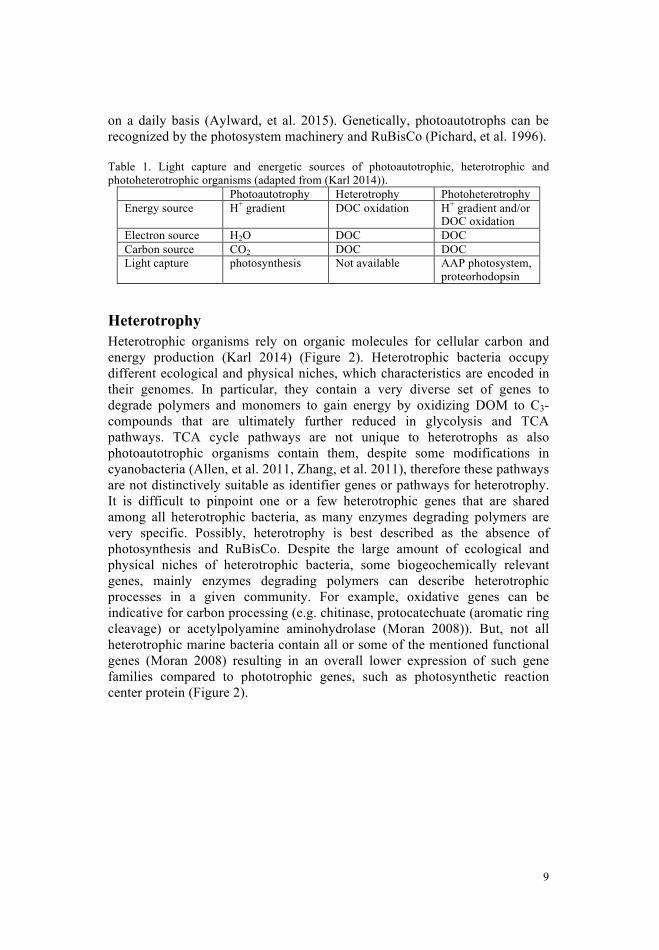
on a daily basis (Aylward, et al. 2015). Genetically, photoautotrophs can be recognized by the photosystem machinery and RuBisCo (Pichard, et al. 1996).
Table 1. Light capture and energetic sources of photoautotrophic, heterotrophic and photoheterotrophic organisms (adapted from (Karl 2014)).
Photoautotrophy Heterotrophy Photoheterotrophy Energy source H+ gradient DOC oxidation H+ gradient and/or
DOC oxidation Electron source H2O DOC DOC Carbon source CO2 DOC DOC Light capture photosynthesis Not available AAP photosystem,
proteorhodopsin
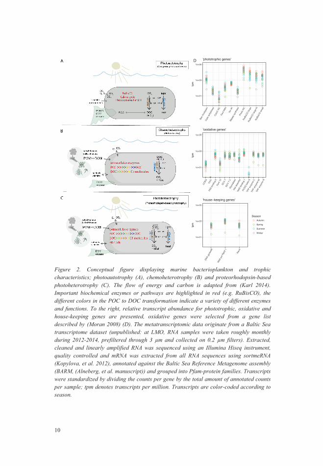
Heterotrophy Heterotrophic organisms rely on organic molecules for cellular carbon and energy production (Karl 2014) (Figure 2). Heterotrophic bacteria occupy different ecological and physical niches, which characteristics are encoded in their genomes. In particular, they contain a very diverse set of genes to degrade polymers and monomers to gain energy by oxidizing DOM to C3-compounds that are ultimately further reduced in glycolysis and TCA pathways. TCA cycle pathways are not unique to heterotrophs as also photoautotrophic organisms contain them, despite some modifications in cyanobacteria (Allen, et al. 2011, Zhang, et al. 2011), therefore these pathways are not distinctively suitable as identifier genes or pathways for heterotrophy. It is difficult to pinpoint one or a few heterotrophic genes that are shared among all heterotrophic bacteria, as many enzymes degrading polymers are very specific. Possibly, heterotrophy is best described as the absence of photosynthesis and RuBisCo. Despite the large amount of ecological and physical niches of heterotrophic bacteria, some biogeochemically relevant genes, mainly enzymes degrading polymers can describe heterotrophic processes in a given community. For example, oxidative genes can be indicative for carbon processing (e.g. chitinase, protocatechuate (aromatic ring cleavage) or acetylpolyamine aminohydrolase (Moran 2008)). But, not all heterotrophic marine bacteria contain all or some of the mentioned functional genes (Moran 2008) resulting in an overall lower expression of such gene families compared to phototrophic genes, such as photosynthetic reaction center protein (Figure 2).
8
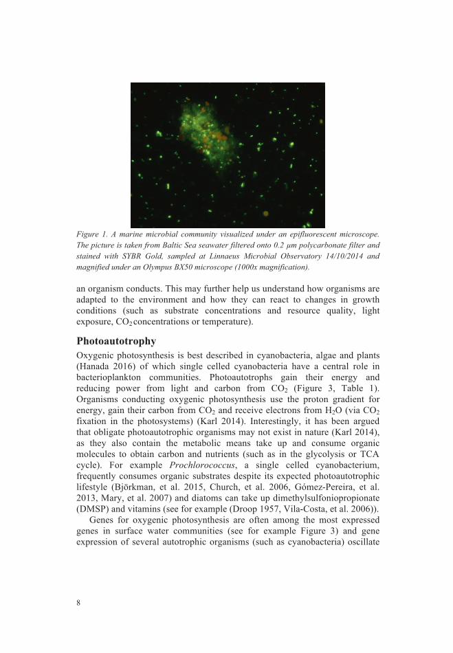
Figure 1. A marine microbial community visualized under an epifluorescent microscope. The picture is taken from Baltic Sea seawater filtered onto 0.2 µm polycarbonate filter and stained with SYBR Gold, sampled at Linnaeus Microbial Observatory 14/10/2014 and magnified under an Olympus BX50 microscope (1000x magnification).
an organism conducts. This may further help us understand how organisms are adapted to the environment and how they can react to changes in growth conditions (such as substrate concentrations and resource quality, light exposure, CO2 concentrations or temperature).
Photoautotrophy Oxygenic photosynthesis is best described in cyanobacteria, algae and plants (Hanada 2016) of which single celled cyanobacteria have a central role in bacterioplankton communities. Photoautotrophs gain their energy and reducing power from light and carbon from CO2 (Figure 3, Table 1). Organisms conducting oxygenic photosynthesis use the proton gradient for energy, gain their carbon from CO2 and receive electrons from H2O (via CO2 fixation in the photosystems) (Karl 2014). Interestingly, it has been argued that obligate photoautotrophic organisms may not exist in nature (Karl 2014), as they also contain the metabolic means take up and consume organic molecules to obtain carbon and nutrients (such as in the glycolysis or TCA cycle). For example Prochlorococcus, a single celled cyanobacterium, frequently consumes organic substrates despite its expected photoautotrophic lifestyle (Björkman, et al. 2015, Church, et al. 2006, Gómez-Pereira, et al. 2013, Mary, et al. 2007) and diatoms can take up dimethylsulfoniopropionate (DMSP) and vitamins (see for example (Droop 1957, Vila-Costa, et al. 2006)).
Genes for oxygenic photosynthesis are often among the most expressed genes in surface water communities (see for example Figure 3) and gene expression of several autotrophic organisms (such as cyanobacteria) oscillate
9
on a daily basis (Aylward, et al. 2015). Genetically, photoautotrophs can be recognized by the photosystem machinery and RuBisCo (Pichard, et al. 1996).
Table 1. Light capture and energetic sources of photoautotrophic, heterotrophic and photoheterotrophic organisms (adapted from (Karl 2014)).
Photoautotrophy Heterotrophy Photoheterotrophy Energy source H+ gradient DOC oxidation H+ gradient and/or
DOC oxidation Electron source H2O DOC DOC Carbon source CO2 DOC DOC Light capture photosynthesis Not available AAP photosystem,
proteorhodopsin
Heterotrophy Heterotrophic organisms rely on organic molecules for cellular carbon and energy production (Karl 2014) (Figure 2). Heterotrophic bacteria occupy different ecological and physical niches, which characteristics are encoded in their genomes. In particular, they contain a very diverse set of genes to degrade polymers and monomers to gain energy by oxidizing DOM to C3-compounds that are ultimately further reduced in glycolysis and TCA pathways. TCA cycle pathways are not unique to heterotrophs as also photoautotrophic organisms contain them, despite some modifications in cyanobacteria (Allen, et al. 2011, Zhang, et al. 2011), therefore these pathways are not distinctively suitable as identifier genes or pathways for heterotrophy. It is difficult to pinpoint one or a few heterotrophic genes that are shared among all heterotrophic bacteria, as many enzymes degrading polymers are very specific. Possibly, heterotrophy is best described as the absence of photosynthesis and RuBisCo. Despite the large amount of ecological and physical niches of heterotrophic bacteria, some biogeochemically relevant genes, mainly enzymes degrading polymers can describe heterotrophic processes in a given community. For example, oxidative genes can be indicative for carbon processing (e.g. chitinase, protocatechuate (aromatic ring cleavage) or acetylpolyamine aminohydrolase (Moran 2008)). But, not all heterotrophic marine bacteria contain all or some of the mentioned functional genes (Moran 2008) resulting in an overall lower expression of such gene families compared to phototrophic genes, such as photosynthetic reaction center protein (Figure 2).
8
Figure 1. A marine microbial community visualized under an epifluorescent microscope. The picture is taken from Baltic Sea seawater filtered onto 0.2 µm polycarbonate filter and stained with SYBR Gold, sampled at Linnaeus Microbial Observatory 14/10/2014 and magnified under an Olympus BX50 microscope (1000x magnification).
an organism conducts. This may further help us understand how organisms are adapted to the environment and how they can react to changes in growth conditions (such as substrate concentrations and resource quality, light exposure, CO2 concentrations or temperature).
Photoautotrophy Oxygenic photosynthesis is best described in cyanobacteria, algae and plants (Hanada 2016) of which single celled cyanobacteria have a central role in bacterioplankton communities. Photoautotrophs gain their energy and reducing power from light and carbon from CO2 (Figure 3, Table 1). Organisms conducting oxygenic photosynthesis use the proton gradient for energy, gain their carbon from CO2 and receive electrons from H2O (via CO2 fixation in the photosystems) (Karl 2014). Interestingly, it has been argued that obligate photoautotrophic organisms may not exist in nature (Karl 2014), as they also contain the metabolic means take up and consume organic molecules to obtain carbon and nutrients (such as in the glycolysis or TCA cycle). For example Prochlorococcus, a single celled cyanobacterium, frequently consumes organic substrates despite its expected photoautotrophic lifestyle (Björkman, et al. 2015, Church, et al. 2006, Gómez-Pereira, et al. 2013, Mary, et al. 2007) and diatoms can take up dimethylsulfoniopropionate (DMSP) and vitamins (see for example (Droop 1957, Vila-Costa, et al. 2006)).
Genes for oxygenic photosynthesis are often among the most expressed genes in surface water communities (see for example Figure 3) and gene expression of several autotrophic organisms (such as cyanobacteria) oscillate
11
Photoheterotrophy – the light effect Photoheterotrophic bacterioplankton can utilize light energy in several ways; aerobic anoxygenic photosynthesis, anaerobic anoxygenic photosynthesis or through proteorhodopsins-based energy capture from light. Despite that these organisms use different light capture systems, most of them use DOC as ATP, electron and carbon source, and light generated H+ gradient to obtain additional energy (Table 1), but see anaerobic phototrophic bacteria, that do not use DOC for carbon and energy (Yurkov, et al. 1998). Aerobic anoxygenic phototrophs (AAP) belong to many different phyla (Hanada 2016) and AAP bacteria have photosynthetic reaction centers with bacteriochlorophyll but not the capacity to oxidize water into oxygen (Hanada 2016, Yurkov, et al. 1998). Thus, light supplements the energetic needs for AAP bacteria, for example the increased proton gradient supports ATP production and substrate transport (Yurkov, et al. 1998).
Proteorhodopsins (PR) are light activated proton pumps that are embedded in the cell membrane of mainly prokaryotic cells and pump protons across the membrane when activated by light (Pinhassi, et al. 2016). Yet also some eukaryotic phytoplankton, fungi and viruses were described to contain proteorhodopsins (Brown, et al. 2006, Nagel, et al. 2002, Yutin, et al. 2012). Recently, several studies have investigated phylogeny, gene expression patterns and potential energetic advantages for PR-containing organisms. Proteorhodopsins are expressed by various bacterioplankton community members (Ottensen, et al. 2013). Generally, when light strikes the seven-helix protein, a conformational change in the inner chromophore pumps one ion through the channel (Sharma, et al. 2006). The most common proteorhodopsin type pumps protons out of the cytosol while other rhodopsin types can pump sodium ions out or even chloride ions in (Yoshizawa, et al. 2014). Upon light induced proton pumping, the resulting proton motive force across the membrane can in turn fuel the ATP synthase to build ATP (Béjà, et al. 2001). While for some described photoheterotrophic bacteria no growth stimulation was detected upon the growth in light, some bacteria showed responses when grown in light compared to darkness (Del Giorgio, et al. 2011, Gómez-Consarnau, et al. 2007, Gómez-Consarnau, et al. 2016, Kimura, et al. 2011). Some bacteria have been shown to have enhanced proteorhodopsin gene expression during daylight compared to night or have a prolonged survival in light compared to darkness (Akram, et al. 2013, Aylward, et al. 2015). Other bacteria, such as Psychroflexus torquis (Flavobacteria) show enhanced proteorhodopsin gene expression under salt stress (Feng, et al. 2013). In the photobacterium angustum S14 (Gammaproteobacterium) light effects are enhanced under higher cell biomass rather than low substrate availability (Courties, et al. 2015). It is now known that proteorhodopsin-containing bacteria are widespread in marine surface environments and comprise up to more than half of the bacterioplankton (Béjà, et al. 2001, Campbell, et al.
10
Figure 2. Conceptual figure displaying marine bacterioplankton and trophic characteristics; photoautotrophy (A), chemoheterotrophy (B) and proteorhodopsin-based photoheterotrophy (C). The flow of energy and carbon is adapted from (Karl 2014). Important biochemical enzymes or pathways are highlighted in red (e.g. RuBisCO), the different colors in the POC to DOC transformation indicate a variety of different enzymes and functions. To the right, relative transcript abundance for phototrophic, oxidative and house-keeping genes are presented, oxidative genes were selected from a gene list described by (Moran 2008) (D). The metatranscriptomic data originate from a Baltic Sea transcriptome dataset (unpublished: at LMO, RNA samples were taken roughly monthly during 2012-2014, prefiltered through 3 µm and collected on 0.2 µm filters). Extracted, cleaned and linearly amplified RNA was sequenced using an Illumina Hiseq instrument, quality controlled and mRNA was extracted from all RNA sequences using sortmeRNA (Kopylova, et al. 2012), annotated against the Baltic Sea Reference Metagenome assembly (BARM, (Alneberg, et al. manuscript)) and grouped into Pfam-protein families. Transcripts were standardized by dividing the counts per gene by the total amount of annotated counts per sample; tpm denotes transcripts per million. Transcripts are color-coded according to season.
1e+01
1e+03
1e+05
Bac r
hodo
psin
Carb
anhy
dras
eCy
toC R
CFe
r4 N
ifHIso
dhMa
late s
yntha
sePh
oto R
CRu
BisC
O lar
geRu
BisC
O lar
ge N
RuBi
sCO
small
tpm
'phototrophic genes'
1e+01
1e+03
1e+05
COXG ChiC
FAD
bindin
g 6Fe
r4 10
GCV
TGC
V T
CGl
yoxa
lase
Hist
deac
etyl
NAD
bindin
g 1PC
DO be
ta N
TPP
enzy
me C
TPP
enzy
me M
TPP
enzy
me N
tpm
'oxidative genes'
1e+01
1e+03
1e+05DN
A gy
rase
B
RNA
pol R
pb2 1
RecA
tpm
SeasonAutumn
Spring
Summer
Winter
'house−keeping genes'
D
11
Photoheterotrophy – the light effect Photoheterotrophic bacterioplankton can utilize light energy in several ways; aerobic anoxygenic photosynthesis, anaerobic anoxygenic photosynthesis or through proteorhodopsins-based energy capture from light. Despite that these organisms use different light capture systems, most of them use DOC as ATP, electron and carbon source, and light generated H+ gradient to obtain additional energy (Table 1), but see anaerobic phototrophic bacteria, that do not use DOC for carbon and energy (Yurkov, et al. 1998). Aerobic anoxygenic phototrophs (AAP) belong to many different phyla (Hanada 2016) and AAP bacteria have photosynthetic reaction centers with bacteriochlorophyll but not the capacity to oxidize water into oxygen (Hanada 2016, Yurkov, et al. 1998). Thus, light supplements the energetic needs for AAP bacteria, for example the increased proton gradient supports ATP production and substrate transport (Yurkov, et al. 1998).
Proteorhodopsins (PR) are light activated proton pumps that are embedded in the cell membrane of mainly prokaryotic cells and pump protons across the membrane when activated by light (Pinhassi, et al. 2016). Yet also some eukaryotic phytoplankton, fungi and viruses were described to contain proteorhodopsins (Brown, et al. 2006, Nagel, et al. 2002, Yutin, et al. 2012). Recently, several studies have investigated phylogeny, gene expression patterns and potential energetic advantages for PR-containing organisms. Proteorhodopsins are expressed by various bacterioplankton community members (Ottensen, et al. 2013). Generally, when light strikes the seven-helix protein, a conformational change in the inner chromophore pumps one ion through the channel (Sharma, et al. 2006). The most common proteorhodopsin type pumps protons out of the cytosol while other rhodopsin types can pump sodium ions out or even chloride ions in (Yoshizawa, et al. 2014). Upon light induced proton pumping, the resulting proton motive force across the membrane can in turn fuel the ATP synthase to build ATP (Béjà, et al. 2001). While for some described photoheterotrophic bacteria no growth stimulation was detected upon the growth in light, some bacteria showed responses when grown in light compared to darkness (Del Giorgio, et al. 2011, Gómez-Consarnau, et al. 2007, Gómez-Consarnau, et al. 2016, Kimura, et al. 2011). Some bacteria have been shown to have enhanced proteorhodopsin gene expression during daylight compared to night or have a prolonged survival in light compared to darkness (Akram, et al. 2013, Aylward, et al. 2015). Other bacteria, such as Psychroflexus torquis (Flavobacteria) show enhanced proteorhodopsin gene expression under salt stress (Feng, et al. 2013). In the photobacterium angustum S14 (Gammaproteobacterium) light effects are enhanced under higher cell biomass rather than low substrate availability (Courties, et al. 2015). It is now known that proteorhodopsin-containing bacteria are widespread in marine surface environments and comprise up to more than half of the bacterioplankton (Béjà, et al. 2001, Campbell, et al.
10
Figure 2. Conceptual figure displaying marine bacterioplankton and trophic characteristics; photoautotrophy (A), chemoheterotrophy (B) and proteorhodopsin-based photoheterotrophy (C). The flow of energy and carbon is adapted from (Karl 2014). Important biochemical enzymes or pathways are highlighted in red (e.g. RuBisCO), the different colors in the POC to DOC transformation indicate a variety of different enzymes and functions. To the right, relative transcript abundance for phototrophic, oxidative and house-keeping genes are presented, oxidative genes were selected from a gene list described by (Moran 2008) (D). The metatranscriptomic data originate from a Baltic Sea transcriptome dataset (unpublished: at LMO, RNA samples were taken roughly monthly during 2012-2014, prefiltered through 3 µm and collected on 0.2 µm filters). Extracted, cleaned and linearly amplified RNA was sequenced using an Illumina Hiseq instrument, quality controlled and mRNA was extracted from all RNA sequences using sortmeRNA (Kopylova, et al. 2012), annotated against the Baltic Sea Reference Metagenome assembly (BARM, (Alneberg, et al. manuscript)) and grouped into Pfam-protein families. Transcripts were standardized by dividing the counts per gene by the total amount of annotated counts per sample; tpm denotes transcripts per million. Transcripts are color-coded according to season.
1e+01
1e+03
1e+05
Bac r
hodo
psin
Carb
anhy
dras
eCy
toC R
CFe
r4 N
ifHIso
dhMa
late s
yntha
sePh
oto R
CRu
BisC
O lar
geRu
BisC
O lar
ge N
RuBi
sCO
small
tpm
'phototrophic genes'
1e+01
1e+03
1e+05
COXG ChiC
FAD
bindin
g 6Fe
r4 10
GCV
TGC
V T
CGl
yoxa
lase
Hist
deac
etyl
NAD
bindin
g 1PC
DO be
ta N
TPP
enzy
me C
TPP
enzy
me M
TPP
enzy
me N
tpm
'oxidative genes'
1e+01
1e+03
1e+05
DNA
gyra
seB
RNA
pol R
pb2 1
RecA
tpm
SeasonAutumn
Spring
Summer
Winter
'house−keeping genes'
D
13
heterotrophic bacteria populations (Rosselló-Móra, et al. 2015), using programs such as dada2 or Deblur (Amir, et al. 2017, Callahan, et al. 2016). These techniques use all “true” amplicon-sequences in a dataset, correcting sequencing errors using a statistical approach. Therefore, they are able to disentangle closely related populations (Amir, et al. 2017, Callahan, et al. 2016). In recent years, ‘high throughput sequencing’ instruments and techniques were developed that are faster, more efficient and cheaper compared to older methods (Caporaso, et al. 2012). Methods such as 16S rRNA gene amplicon techniques, are therefore now frequently used to annotate microbial datasets taxonomically and even metagenomic and metatranscriptomic analyses generate datasets of functionally annotated genes.
In summary, genetic techniques such as 16S rRNA gene amplicon techniques are a widely used approach to study bacterial taxonomic diversities, due to their economic and technical practicality. These detailed taxonomic techniques are currently painting a new picture of the vast microbial diversity existing on our planet.
Taxonomic diversity in the Baltic Sea Bacterioplankton communities are taxonomically diverse. Laboratory studies of model organisms provided insights about growth characteristics, genotypic and phenotypic traits of some marine bacteria classes, especially Alphaproteobacteria (such as the SAR11 clade and roseobacters) and Flavobacteria. For example, Alphaproteobacteria, can have distinct strategies: SAR11 clade organisms (such as Candidatus Pelagibacter ubique) have a streamlined genome (Giovannoni, et al. 2005). They mainly have transporters with a broad substrate range additional to a number of specific substrate targets, such as amino acids, osmolytes or N-compounds (Giovannoni, et al. 2005). They also contain many ABC transporters exhibiting high substrate affinities (Giovannoni, et al. 2005). Yet, despite the advances in deciphering Candidatus P. ubique behavior in cultures, other SAR11 strains remain to be cultivated to disentangle their growth characteristics. Other Alphaproteobacteria, such as roseobacters (e.g. Silicibacter pomeroyi) contain an overall large gene repertoire and can be cultivated (Brinkhoff, et al. 2008, Buchan, et al. 2005). Members of this lineage can degrade aromatic compounds, sulfidic compounds, DMSP or oxidize carbon monooxide (Buchan, et al. 2005). Yet, while members of this family (and possibly other bacterial phyla) are able to conduct all these functions, not all members contain the entire gene repertoire (Buchan, et al. 2005), making extrapolation from taxonomy (that is taxonomic annotation of organisms) to function (such as phenotypic traits) somewhat complicated. Thus, while some roseobacters are highly specialized for certain substrates, others can use a wide variety of DOM (Brinkhoff, et al. 2008). These different traits lead to an overall high
12
2008, de la Torré, et al. 2003, Pinhassi, et al. 2016). Therefore, their impact on biogeochemical nutrient cycling is potentially tremendous and requires further investigations.
Microbial species concept The classical biological species (CBS) concept was developed for interbreeding eukaryotic organisms; applying these CBS-rules and classification for asexually reproducing bacteria is difficult, if not impossible, and therefore a number of other species concepts have been proposed. I will not discuss how a ‘true bacterial species’ can, or should be, defined, but rather explain the practical taxonomic approach that is currently widely used in microbial oceanography to group bacterial diversity into “operational taxonomic units” (OTUs) or populations.
Traditionally, marine bacteria were classified using characteristics identified in monocultures, such as when grown on agar plates or in seawater cultures (Hagström, et al. 2017). With advances in DNA extraction and bioinformatics, DNA-DNA hybridization techniques were initially used to study relatedness between bacterial isolates (Rosselló-Móra, et al. 2015). Specifically, a degree of >70% cross-hybridization between genomic material of two isolates was classified as the same species (Rosselló-Móra, et al. 2015). Later, the DNA-DNA hybridization degree was translated to approximate nucleotide identity levels of the highly conserved small subunit of the ribosomal gene (16S rRNA gene) of bacteria (Pedros-Alio, et al. 2015, Rosselló-Móra, et al. 2015, Woese 1987). Whilst this method was first used for bacterial isolates and clones, it rapidly became a common tool for microbiologists working with environmental genetic material using amplicon-sequencing methodologies (e.g. (Sunagawa, et al. 2015)). Using this method, OTUs are clustered at a certain threshold (e.g. 97% sequence identity) of the 16S rRNA gene to classify them as the “same”. When applied to environmental samples, this led to the realization that only about 1% of marine bacteria are ‘culturable’ on standard media, that is, are able to be described and identified by traditional methods (Hagström, et al. 2017). This was later known as the great plate count anomaly (Hugenholtz 2002, Staley, et al. 1985). Some ecotypes (a sequence cluster coupled with distinct ecological attributes (Cohan 2006)) or populations can be classified at higher thresholds (Pedros-Alio, et al. 2015). For example, Prochlorococcus and Synechococcus 16S rRNA genes are within 96% identical, however they belong to different species (Rocarp, et al. 2002). Moreover, within the Prochlorococcus clade, many different ecotypes are described, which inhabit different ecological and spatial niches, typically exhibiting more than 97% 16S rRNA gene sequence identity (Kashtan, et al. 2014, Malmstrom, et al. 2010). To increase resolution, more conservative clustering thresholds (>98.7%) have been used to describe
15
Dokdonia sp. MED134 thus has a preference for peptides over polysaccharides (González, et al. 2011), explaining its growth in peptone medium. These three bacterial classes are abundant in seawater samples, and also in the Baltic Proper they display major roles, together with other marine bacteria (Lindh, et al. 2015) (Figure 3).
The Baltic Sea is a semi enclosed sea in northern Europe that displays a
salinity gradient from freshwater-like waters in the Bothnian Bay, salinities around 7 PSU in the Baltic Proper to marine salinities (~33 PSU) in the Skagerrak. In the Baltic Sea, two extensive phytoplankton blooms appear yearly, a spring bloom that is composed of dinoflagellates and diatoms, and a summer bloom of filamentous cyanobacteria (Karjalainen, et al. 2007, Klais, et al. 2011, Suikkanen, et al. 2007, Wasmund, et al. 1998, Wasmund, et al. 2011, Wasmund, et al. 2003). Sometimes, a smaller autumn bloom consisting of diatoms can be observed (Wasmund, et al. 2011). The intensity of spring blooms exhibits inter-annual variability and diatoms and dinoflagellates shift in dominance (roughly oscillating over decades) (Klais, et al. 2011, Wasmund, et al. 2011). During summer, filamentous cyanobacteria thrive in warm, stratified surface water conditions (Bertos-Fortis, et al. 2016, Karjalainen, et al. 2007, Legrand, et al. 2015). Because of the multiple phytoplankton bloom occurrences per year, the Baltic Sea is a suitable environment to study the response of heterotrophic bacteria to phytoplankton blooms and to investigate their impact on biogeochemical cycles (via for example heterotrophic production estimates) and consequences for the ecosystem.
In bacterioplankton community composition datasets in the Baltic Sea, the freshwater influence is particularly obvious given the abundance of Actinobacteria, which are associated with low saline and brackish waters (Dupont, et al. 2014, Herlemann, et al. 2011, Lindh, et al. 2015). Towards more saline waters in the southern Baltic Sea, Alphaproteobacteria and Gammaproteobacteria increase in relative abundance during summer (Dupont, et al. 2014). In the Western Baltic Proper, at Linnaeus University Observatory (LMO), Alphaproteobacteria, Betaproteobacteria and Gammaproteobacteria are the most abundant members of the proteobacterial clades (Figure 3 and (Dupont, et al. 2014, Herlemann, et al. 2011, Lindh, et al. 2015)). Class Flavobacteriia, Roseobacter clade species (Alphaproteobacteria) and Gammaproteobacteria, that are commonly following eukaryotic phytoplankton blooms (Buchan, et al. 2014, Bunse, et al. 2016, Pinhassi, et al. 2004, Riemann, et al. 2000, Teeling, et al. 2012, Teeling, et al. 2016), are also frequently observed in the Baltic Sea (Laas, et al. 2016, Lindh, et al. 2015). Not to be neglected are members of the phylum Planctomycetes and Verrucomicrobia, which were detected in elevated abundances during summer (Andersson, et al. 2010, Lindh, et al. 2015).
14
abundance of roseobacter and SAR11- clade bacteria in marine systems, yet occupying different niches (Brinkhoff, et al. 2008, Buchan, et al. 2005, Giebel, et al. 2011). Furthermore, other copiotrophic bacteria such as Flavobacteria are often found attached to particles, especially during and after phytoplankton blooms (Buchan, et al. 2014), which is encoded in gliding motility genes (González, et al. 2011). For the Dokdonia sp. MED134, for example, many lipoproteins and secretion proteins, carbohydrateester hydrolysis and many predicted peptidases are reported (González, et al. 2011).
Figure 3. All bacterial genera identified at Linnaeus Microbial Observatory (LMO) in the western Baltic Proper during 2011-2017. The LMO dataset is based on 16S rRNA amplicon sequences, analyzed with dada2 and drawn using the metacoder and ape packages in R (n=660) (Callahan, et al. 2016, Foster, et al. 2017, Paradis, et al. 2004). Each leaf node displays one genus, node color gradient shows the number of genera (observations) for each taxon, edges display the number of supertaxa. .
Root
Root
Bacteria
Actinobacteria
BacteroidetesCyanobacteria
Planctomycetes
Proteobacteria
Verrucomicrobia
Acidimicrobiia
Actinobacteria
AcidimicrobialesAcidimicrobiaceae
FrankialesSporichthyaceaehgcI_clade
Flavobacteriia
Flavobacteriales
CryomorphaceaeFlavobacteriaceaePolaribacter
uncultured
Chloroplast
Cyanobacteria
SubsectionI
SubsectionIVFamilyISynechococcus
FamilyI
Alphaproteobacteria
Betaproteobacteria
Gammaproteobacteria
RhodobacteralesRhodobacteraceae
Burkholderiales
Comamonadaceae
BAL58_marine_group
Alteromonadales
Oceanospirillales
Unclassified
1
186
689
1510
2650
4110
5890
num
ber o
f obs
erva
tions
Nodes
0.000
0.167
0.667
1.500
2.670
4.170
6.000
num
er o
f sup
erta
xa
Edges
17
expression patterns to regulate the turnover of DOM. This can be studied at different scales; from a broad scale such as ocean wide basins and overall biomass fluxes for a modelling perspective, to narrower scale location at high temporal resolution to understand the biological processes typical to different environments. One way to infer the appropriate scale is to consider water masses, time frames and distances from a bacterial viewpoint. Given a common non-motile bacterial cell is about ~0.4 µm in diameter, distances to other bacteria or aggregates which display potential food sources, are long. Brownian diffusion moves cells through the surrounding seawater so that they reach about 80 nl per day (Stocker 2012). Nutrients can then be taken up in the diffusion boundary layer, covering a few cell diameters (Stocker 2012). Swimming microbes on the other hand can reach a volume of about 0.8 ml a day, that is about 10 000 times the volume reached by non-motile bacteria (Stocker 2012). Furthermore, if bacteria can turn over half of their metabolite pool in one second, and express genes within minutes, this leads to growth rates of close to 1 h-1 in enriched medium (Hagström, et al. 2017, Shamir, et al. 2016). Notably, bacterial processing of DOM at the microscale ultimately influences ocean basin-scale carbon fluxes for example through the microbial loop, carbon storage, and carbon fixation itself (Azam 1998). Logically short timescales are important for microbes at the physiological level, such shifts can be semidiurnal (tides), diurnal (biological phototrophic production), weekly, monthly and seasonal (Gunderson, et al. 2016). Thus, also yearly timeframes remain of interest to oceanographers due to broad level factors such as seasons, and even beyond that, climate. Overall, marine seasons are driven by differences in light intensity and resulting temperatures that in turn impact on water chemistry and physical traits of seawater.
Figure 4. Conceptual summary of relevant timescales for bacterioplankton. Cellular and metabolic processes are very fast (< hour) while environmental changes (such as phytoplankton blooms, temperatures or seasons) occur at successively longer time frames. In the Baltic Proper at LMO, samples for this thesis were sampled approximately twice a week and bioassays were incubated for one to two hours, while transcriptomic samples were processed within half an hour.
second minute hour day week month year
doubling time, growth
mRNA turnovershifts in BCC seasonal succession
kinetic reactions genome copying
water mass changes
transcriptomics
rate incubations
LMO sampling
evolutionclimate change
gene expression
phytoplankton blooms
16
Bacterioplankton community structure Life and microbial communities are highly dynamic. The community structure of bacterioplankton (that is, species composition and the corresponding abundances of populations) changes with time, space, depth, interactions and nutrient availability (Fuhrman 2009, Fuhrman, et al. 2008). Photosynthetic bacteria have growth rates of about 0.75 d-1, as for the single celled cyanobacterium Synechococcus (Kirchman 2016). Marine heterotrophic bacteria in situ often reach growth rates between 0.1-0.3 d-1, with abundances of about half a million to millions of cells per millilitre (Kirchman 2016, Lindh, et al. 2015, Whitman, et al. 1998). Growth rates and cell abundances depend on the lifestyle of the bacterioplankton population, they can be oligotrophic (for example SAR11, that is adapted to low nutrient concentrations) or copiotrophic (e.g. members of the class Flavobacteria). In the low-nutrient (oligotrophic) ocean, oligotrophic bacteria can compete for resources, for example by using high-affinity and low capacity transport systems to exploit substrates (Hagström, et al. 2017). Copiotrophic bacteria (sometimes also called opportunistic bacteria) on the other hand can quickly respond to organic nutrient input, grow and build biofilms (Hagström, et al. 2017, Pinhassi, et al. 1997). Under nutrient-rich conditions in monocultures and under optimal temperatures, copiotrophic marine bacteria exhibit growth rates between 0.2 h-1 to 1 h-1 while oligotrophic marine bacteria show growth rates up to 0.02 h-1 in seawater cultures (Hagström, et al. 2017). Therefore, the potential for fast changes in microbial community structure are vast and become especially evident during times of nutrient input, such as during coastal phytoplankton blooms, where community structure can change within days (Lindh, et al. 2015, Needham, et al. 2016, Pinhassi, et al. 2004, Riemann, et al. 2000, Teeling, et al. 2012). Community compositions change depending on which bacteria grow actively and which die, but also functional capacities of communities can change. For example, in polar regions, shifts in community composition and functional capacities of microbial communities changed from a photosynthesis-fueled community during summer to a more chemotrophic community during dark winters (Ghiglione, et al. 2012, Grzymski, et al. 2012). Remarkably, such changes can appear at a single sampling location (Grzymski, et al. 2012), revealing a vast functional diversity of microbial communities throughout the year.
Temporal patterns Microbial communities change over time. Applying microbial ecology and molecular biology tool boxes on samples from marine microbial time-series, it is now possible to disentangle which populations in the microbial community are doing what, when and how across seasons. Included in this quest, and a main objective for microbial oceanographers and environmental biochemists, is to measure how bacterial communities, use their genetic repertoire and gene
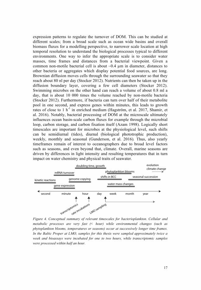
19
et al. 2017). Some studies conclude direct as well as indirect effects of elevated CO2 on bacterial community composition, respiration or productivity (Arnosti, et al. 2011, Grossart, et al. 2006, James, et al. 2017, Krause, et al. 2012, Piontek, et al. 2010), while others have found no significant effects on the bacterial communities (Allgaier, et al. 2008, Hartmann, et al. 2015, Oliver, et al. 2014, Roy, et al. 2008). As there is no consensus on how microbes will respond to ocean acidification or combined multiple stressors (Hutchins, et al. 2017, Pachauri, et al. 2014), one prominent hypothesis is that bacterioplankton and carbon export, via the microbial loop, will not be significantly altered in the future (Joint, et al. 2011). Yet, the IPCC report states that for “microbes, a conceptual foundation suitable to support an integrated understanding of climate impacts on individual species and communities is lacking” (Pachauri, et al. 2014). If marine bacteria can withstand the proton pressure of elevated CO2, for example with the help of proton pumps that are involved in bioenergetics (Joint, et al. 2011), it would be desirable to understand the underlying biochemical processes and enzymatic reactions. If they cannot household internal proton concentrations under predicted future ocean scenarios, this could lead to potential shifts in bacterial production and respiration rates, resulting differences in growth efficiencies, competition, abundances and community composition.
18
Multiple climate change drivers (do bacteria care?) Anthropogenic activities, such as combustion of fossil fuels, have increased CO2 concentration in the atmosphere and resulted in increased pCO2 values in seawater, particularly intensive in the last century (Doney, et al. 2009, Dore, et al. 2009, Pelejero, et al. 2010). Due to carbon chemistry in seawater, elevated CO2 absorption leads to a shift in the equilibrium from CO2 towards bicarbonate ions, resulting in increased proton concentration (reduced pH) (Doney, et al. 2009). A reduction of 0.3-0.4 pH, as projected for the end of this century by models of Haugan et al. (1996), could thus lead to up to 150% increase in H+ concentrations and 50% decrease of CO3
2- concentrations ((Doney, et al. 2009, Haugan, et al. 1996, Orr, et al. 2005) and references within). Marine microbial cells are separated to the seawater only through thin membranes, cell walls or calcified capsules. Higher proton concentrations in the surrounding seawater will consequently increase the proton pressure on the membrane. Ocean acidification directly affects calcification of several calcareous plankton (such as coccolithophores, reviewed in (Doney, et al. 2009) and (Hutchins, et al. 2017)). Phytoplankton photosynthesis might be expected to increase under predicted ocean acidification conditions, aided by potential decreasing needs for energetically costly CO2 concentrating mechanisms (Hutchins, et al. 2017). Yet, how ocean acidification will affect overall phytoplankton community composition and productivity are often hard to predict, also as they further depend on temperature and nutrients (Hutchins, et al. 2017).
Additional predicted climate changes include for example increased land runoff, changes in trace metal concentrations, and rising seawater temperatures (Boyd, et al. 2012). While these are global trends, it needs to be highlighted that many stressors differ locally, regionally and globally (Boyd, et al. 2012). For example, models for the Baltic Sea area indicate that warming and increased precipitation may have profound effects on ice cover and salinity in the brackish water system, while in the Mediterranean Sea decreased precipitation and warming are anticipated (Andersson, et al. 2015, Giorgi, et al. 2008). Future climate change and ocean acidification research is therefore urgent to determine the effect of multiple stressors on communities and ecosystems, rather than single driver and single species studies (Riebesell, et al. 2015).
In a climate change perspective, it is important to consider that natural bacterial communities are continuously exposed to changes in water chemistry, temperatures, as well as nutrient, substrate, O2 and CO2 concentrations. For example, during phytoplankton blooms, CO2 fixation leads to increases in pH (Joint, et al. 2011). Seasonal changes may increase surface water temperatures by up to 20°C during summer compared to winter (Ward,
21
A brief overview of methodology
“In one drop of water are found all the secrets of all the oceans.“ Kahlil Gibran
“When we’re 1,000 km from shore, and our $32,000-a-day cruise depends on getting one last profile to 4,000 m depth, we’ll find the duct tape, silicon sealant, and dry nitrogen to make things work.”
Richard Spinrad, A Sea of Microbes
Sampling sites Time-series data in this thesis derive from the Linnaeus Microbial Observatory (LMO), situated in the Western Baltic Proper, approximately 11 km off the coast of Kårehamn, Öland (N 56° 55.8540', E 17° 3.6420'). At LMO, surface water samples collected at a depth of 2 m were sampled approximately twice a week (during spring to autumn 2011-2013), once a month (2014), and bi-weekly (2015-2016). Water was transported to the laboratory in Kalmar in 10 l carboys where it was subsequently processed further for community composition analysis and bacterial activities (Paper III & IV). Water for the mesocosm experiment in paper II derived from the Mediterranean Sea in Blanes Bay (Figure 5).
Cell enumeration and productivity estimates Flow cytometry and microscopy were used for bacterial enumeration. For Flow cytometry analysis, water samples were fixed with formaldehyde (~3% final concentration) and preserved frozen until analysis. After staining the bulk bacterioplankton using SYBR Green or Syto13, instruments like FACSCalibur (Becton Dickinson) or Cube8 (Partec) were used to count the stained cells. For microscopic analysis, microbial cells were similarly fixed with formaldehyde, stained with SYBR Gold and filtered on black polycarbonate filters, mounted on a glass slide and counted under the epifluorescence microscope (Olympus BX50, 1000x magnification).
20
Aims
“Indeed, why not an issue on marine microbes? This field is fascinating, not only because it is the study of that which cannot be
seen with the naked eye but also because it is the study of organisms that are fundamental to the functioning and health of the ocean.”
L.M. Proctor and D.M. Karl, A Sea of Microbes
This thesis addresses how important environmental drivers influence bacterial responses using experiments with model species and natural assemblages as well as field studies. Further, this thesis aims to identify bacterioplankton succession patterns and activities as response to seasonal changes in the Baltic Proper.
Using laboratory approaches and a high-frequency time series study, I specifically aimed to: • determine how the metabolism of proteorhodopsin-containing,
flavobacterium Dokdonia sp. MED134 is influenced by light and different organic matter (using gene expression analysis) (Paper I)
• determine how the community composition and physiological processes of
marine bacteria are affected by elevated CO2 concentrations and inorganic nutrient loads (Paper II)
• determine how microbial activities and community composition are
influenced by temporal changes and growth conditions and how this leads to seasonality in microbial functioning in temperate marine systems (Paper III, IV, V)
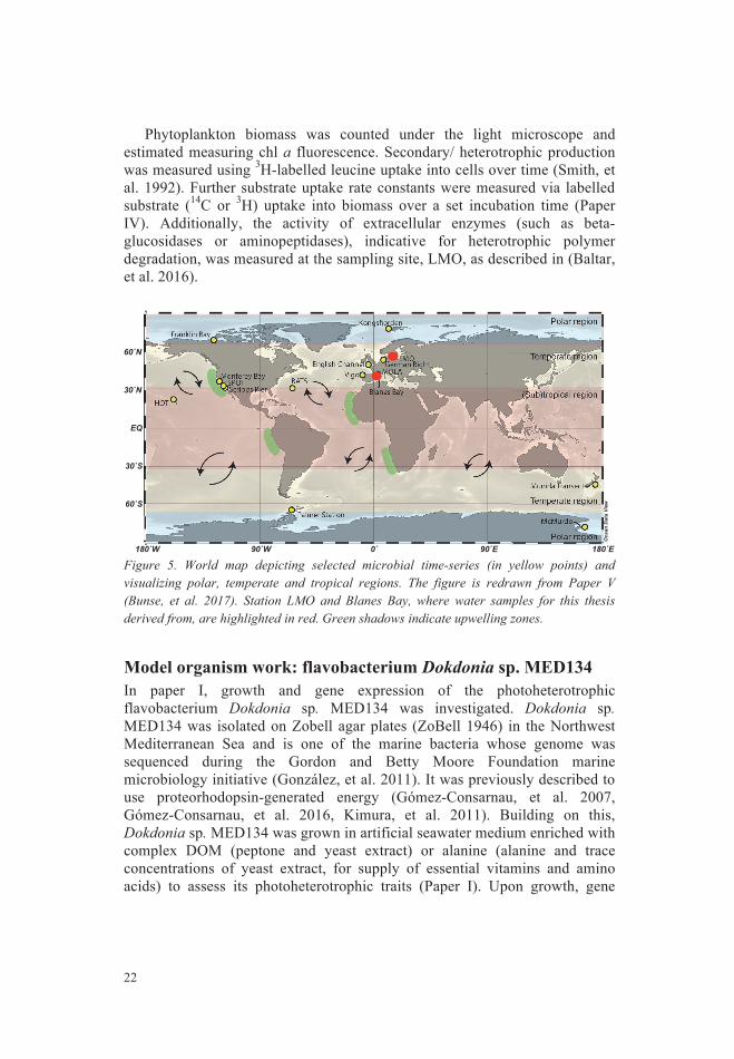
23
expression was monitored by harvesting cells, RNA extraction and RT-qPCR amplification of gene products using gene-specific primers (Paper I). Particulate organic carbon of harvested biomass was measured by harvesting cells of grown cultures on precombusted filters, drying and subsequent analysis for carbon content (Costech ECS 4010 Elemental Analyzer; Costech International). Bacterial production and bicarbonate uptake were measured using the above productivity estimates
16S rRNA gene amplicon data For bacterial community composition analysis, bacterioplankton cells were collected on 0.2 µm filters (Sterivex filters, after prefiltration through 3.0 µm polycarbonate filters) and DNA was extracted using protocols adapted from (Boström, et al. 2004, Lindh, et al. manuscript). The V3V4 region of the 16S rRNA gene was amplified using primers 341f-805r as described in (Hugerth, et al. 2014). Subsequently, Illumina adapters were attached to the amplicon product through a second PCR, and adapter-ligated DNA was pooled and cleaned according to Lindh et al. (unpublished manuscript). The resulting DNA libraries were sequenced on Illumina Miseq instruments at the SciLifeLab in Stockholm (using 2x300 bp).
For the LMO amplicon dataset described in this thesis, we wanted to be able to identify populations by distinct sequences of the 16S rRNA gene, and further to have a continuously running time-series dataset, to be able to add data of future sampling campaigns. Therefore, we decided to use curated sequences, instead of traditional clustering methods obtaining OTUs. Thus, after quality control, the resulting raw sequences were processed using the dada2 pipeline (Paper III) (Callahan, et al. 2016). Resulting sequences (populations from here) were further analyzed in Rstudio using a variety of packages, mainly dplyr, vegan, ggplot2, or Mfuzz (Kumar, et al. 2007, Oksanen, et al. 2007, Wickham 2009, Wickham, et al. 2015).
Metatranscriptomics For metatranscriptomic analysis of bacterioplankton communities in paper II, seawater from Blanes Bay (41°40'13.5"N, 2°48'00.6"E) was incubated under different nutrient and CO2 conditions for 9 days in 200 l tanks. Bacterioplankton biomass from day 9 was pre-filtered (3.0 µm) and harvested on 0.2 µm filters. RNA was extracted and processed using a protocol adapted from (Poretsky, et al. 2009). Briefly, after RNA extraction and remaining genomic DNA removal (RNEasy kit (Quiagen) and TURBO DNA-free kit (Ambion)), rRNA was depleted (mRNA-ONLY prokaryotic mRNA isolation kit (Epicenter Biotechnologies), MICROBExpress (Ambion) and MICROBEnrich (Ambion)). Then, the MessageAmp II-Bacteria kit (Ambion) was used to synthesise cDNA and linearly amplify the RNA. The resulting RNA was shotgun sequenced on an Illumina HiSeq2000 sequencing instrument.
22
Phytoplankton biomass was counted under the light microscope and estimated measuring chl a fluorescence. Secondary/ heterotrophic production was measured using 3H-labelled leucine uptake into cells over time (Smith, et al. 1992). Further substrate uptake rate constants were measured via labelled substrate (14C or 3H) uptake into biomass over a set incubation time (Paper IV). Additionally, the activity of extracellular enzymes (such as beta-glucosidases or aminopeptidases), indicative for heterotrophic polymer degradation, was measured at the sampling site, LMO, as described in (Baltar, et al. 2016).
Figure 5. World map depicting selected microbial time-series (in yellow points) and visualizing polar, temperate and tropical regions. The figure is redrawn from Paper V (Bunse, et al. 2017). Station LMO and Blanes Bay, where water samples for this thesis derived from, are highlighted in red. Green shadows indicate upwelling zones.
Model organism work: flavobacterium Dokdonia sp. MED134 In paper I, growth and gene expression of the photoheterotrophic flavobacterium Dokdonia sp. MED134 was investigated. Dokdonia sp. MED134 was isolated on Zobell agar plates (ZoBell 1946) in the Northwest Mediterranean Sea and is one of the marine bacteria whose genome was sequenced during the Gordon and Betty Moore Foundation marine microbiology initiative (González, et al. 2011). It was previously described to use proteorhodopsin-generated energy (Gómez-Consarnau, et al. 2007, Gómez-Consarnau, et al. 2016, Kimura, et al. 2011). Building on this, Dokdonia sp. MED134 was grown in artificial seawater medium enriched with complex DOM (peptone and yeast extract) or alanine (alanine and trace concentrations of yeast extract, for supply of essential vitamins and amino acids) to assess its photoheterotrophic traits (Paper I). Upon growth, gene
60˚S
30˚S
EQ
30˚N
60˚N
180˚W 90˚W 0˚ 90˚E 180˚E
Oce
an D
ata
View
Temperate region
Polar region
Polar region
Temperate region
(Sub)tropical regionSPOTScripps Pier
HOT
BATS
Franklin BayKongsfjorden
LMOEnglish Channel German Bight
Vigo
Blanes Bay
Monterey Bay
Palmer StationMcMurdo
Munida Transect
MOLA
25
sequencing projects (which do not amplify specific genes) often differ from amplicon datasets in community composition (Tessler, et al. 2017). Moreover, functional and taxonomic annotations are only as good as the databases to which they are annotated. Often, a high number of unknown genes may be present in samples, potentially limiting our interpretations. Furthermore, combined taxonomic and functional annotations are difficult to obtain and often not sufficiently detailed for non-model organisms or in situ assemblages. Despite their limitations, high throughput sequencing techniques have opened a “black box” of opportunities and advances that were hidden from scientists before the development of efficient extraction and deciphering of nucleic acid material. Supplemented with other methods, such as qPCR, FISH, activity measurements or bacterial abundances, they are good indicators and generate solid hypotheses that can be further investigated and tested. Thus, the supremacy and advantages of the mentioned methods (or the lack of alternatives) overpower their possible limitations.
24
Briefly, the bioinformatics pipeline involved quality control, trimming, ribosomal RNA filtering, assembly of the sequences, open reading frames (ORFs) were called on contigs (Paper II). These ORFs were annotated using M5NR SEED (Overbeek, et al. 2005), KEGG (Kanehisa, et al. 2007), and RefSeq (Overbeek, et al. 2005) databases using BLAST (Altschul, et al. 1997). For quantitative annotations, the original sequences were mapped back against annotated ORFs using Bowtie2 (Langmead, et al. 2012) and relative transcript abundances were calculated as counts per million (CPM). SEED categories were subsequently used for grouping functional genes into broader metabolic categories.
Method limitations in the field of microbial ecology All methods have limitations and uncertainties. The currently used methods, which have been employed here, have been evaluated and are in use by other groups or research fields, or might be traditional methods in oceanography. Regardless, interpreting the results should be conducted in consideration of the possible methodological drawbacks and biases. For example, in the field of microbial oceanography, filtration is central in collecting planktonic biomass, sterilize medium or to size-fractionate microbial communities. However, it remains a challenge to separate phytoplankton cells from bacterioplankton, and autotrophic cells from heterotrophic cells prior to incubation experiments. Fragile cells can be disrupted on filters, resulting in unintended cell lysis. Cells which are larger than the filter pore-size can be pushed through pores due to pressure, or conversely, cells can be caught on the filter due to organic material or particles, although their cell size should allow them to pass through pores. Further, the chosen filter pore size (in this thesis mostly 0.2 µm) dictates a threshold of which bacteria are included in the analysis, missing smaller cells (Brown, et al. 2015). Some of these limitations can be bypassed with cell-sorting flow cytometers or specific probes for fluorescent in situ hybridization (FISH) methods, yet these methods are mostly applied after incubation experiments, thus not entirely disentangling direct or indirect effects of e.g. multiple stressors in community experiments.
Several of the projects discussed in this thesis rely on ‘high throughput sequencing’ techniques, such as 16S rRNA gene amplicon techniques, metagenomic or metatranscriptomic analysis. These sequencing methods have revolutionized our understanding of microbial communities and are relatively straightforward to use, efficient and cheap, and allow comparing datasets from distantly related locations or over different time intervals. These methods are, however, biased with limitations, for example, nucleic acid extraction efficiencies and biases, time-dependent degradation of biological material, primer bias, PCR and sequencing efficiencies (see for example (Parada, et al. 2016)). Furthermore, the suitability of the 16S rRNA gene for relative abundance assessment of prokaryotic communities is problematic in itself as many bacterial taxa are polyploid (Soppa 2017). Consequently, shotgun-
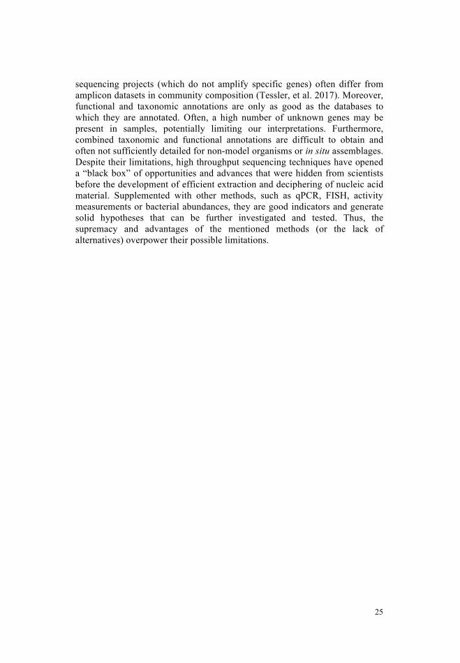
27
Figure 6. Relative transcript abundance and light intensity of the Monterey Bay dataset (A and B) and schematic TCA cycle (C) indicating the regulation of the studied genes, the figure is redrawn from Paper I (Palovaara, et al. 2014). Arrows indicate if genes were upregulated and 0s indicate no different expression upon growth in light.
decarboxylation steps of the traditional TCA cycle (Lorenz, et al. 2002) (Figure 6) and has been described for AAP bacteria (e.g. roseobacters (Moran, et al. 2007)). During growth in light in alanine medium, the gene expression of proteorhodopsin and the two glyoxylate shunt genes (isocitrate lyase and malate synthase) was enhanced in Dokdonia sp. MED134 (Paper I, Figure 6). In complex medium on the other hand, the glyoxylate shunt genes were only minimally expressed. The enhanced expression of the glyoxylate shunt genes isocitrate lyase and malate synthase in light in alanine medium suggests a relocation of the metabolic pathways from a more heterotrophic metabolism (TCA cycle centered) towards a photoheterotrophic and anaplerotic bicarbonate-fixing metabolism (Paper I). Interestingly, diel patterns of isocitrate lyase gene expression could be observed in the bacterioplankton community off Monterey Bay (Paper I, Figure 6). Subsequently, high glyoxylate shunt gene expression of several photoheterotrophic bacterioplankton clades (such as SAR11, SAR86 or SAR116) has been observed, with higher gene expression during day in the California coast and the North Pacific Subtropical Gyre (Aylward, et al. 2015). The glyoxylate shunt therefore seems to play an important role for proteorhodopsin-containing marine bacteria. Together, these results imply that photoheterotrophic bacterioplankton have the capacity for anaplerotic carbon fixation and glyoxylate shunt gene expression, that might aid them when nutrient quality and quantity is limiting in the environment.
26
Results and Discussion
Light effects on model organism Dokdonia sp. MED134 Physiological traits and gene expressions of metabolic pathways of photoheterotrophic marine bacteria are not much studied, thus hampering our understanding of how photoheterotrophic organisms impact on biogeochemical nutrient cycling. In paper I, we investigated light enhanced growth and bicarbonate uptake rates as an indication for anaplerotic carbon fixation of proteorhodopsin-containing Dokdonia sp. MED134. Estimates of the contribution of anaplerotic carbon fixation to bacterial biomass in lab cultures range from negligible 1.4% to substantial 15% (Roslev, et al. 2004, Tang, et al. 2009). In paper I, bicarbonate contributed between 5% and 31% of cell carbon and was elevated during growth in light for Dokdonia sp. MED134. In a previous study, also an aerobic anoxygenic phototrophic bacterium (Acidiphilium rubrum) was reported to have enhanced CO2 fixation rates in light (Kishimoto, et al. 1995). In situ estimations of anaplerotic carbon fixation rates of heterotrophic or photoheterotrophic bacterioplankton in surface waters are to my knowledge not existent. It is difficult to measure rates to precisely estimate the contribution of anaplerotic carbon fixation of bacterioplankton among phytoplankton communities, hampering our understanding of how abundant these metabolisms are in nature. This in turn leaves us with an incomplete picture of the carbon cycle in marine ecosystems, with special respect to the microbial loop. It could thus be that anaplerotic carbon dioxide fixation is so far overlooked, not significant or that it is already included in 14C primary production estimates (depending on methodology and e.g. filter sizes as discussed in the method section) but that we attribute it to phytoplankton primary production.
Interestingly, proteorhodopsin gene expression was low during exponential growth and increased during late exponential and stationary phase (Paper I). The glyoxylate shunt is a pathway that permits the use of C2-compounds to fill up cellular carbon supplies when glucose is limiting and bypasses
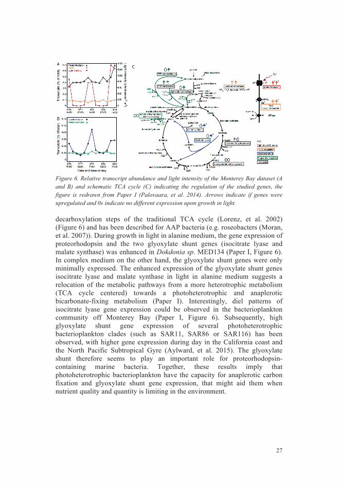
29
Figure 7. Transcripts from paper II, grouped into major SEED categories under low- and high- chlorophyll conditions, figure is redrawn from (Bunse, et al. 2016). Presented are mean relative RNA transcript abundances, normalized to counts per million (CPM), of duplicate samples. The colors represent control mesocosms (orange), compared to CO2 treated mesocosms (red), nutrient enriched control mesocosms (turquoise) and nutrient enriched and CO2 treated mesocosms (blue).
Elevated CO2 in low chlorophyll environments In our mesocosm experiment under low nutrient conditions (Low-chl), chl a values remained below 2.3 µg l-1 and pH decreased from 8.1 to ~7.8 (acidified Low-chl) and ~7.9 (control Low-chl) respectively (Paper II). Bacterioplankton community composition did not significantly differ between acidified and control mesocosms under Low-chl conditions (Baltar, et al. 2015). However, we detected more pronounced significant differences in relative transcript abundance compared to the High-chl treatment (>300 genes, 1.9% of total genes, EdgeR p<0.01). Transcripts of gene functions grouping into respiration, light harvesting, membrane transport, RNA and protein metabolism had a higher relative abundance in acidified Low-chl mesocosms compared to control mesocosms (Paper II). Many abundant genes that increased in acidified conditions could be associated with pH homeostasis mechanisms such as proton pumps (~20% of differentially abundant transcripts) (Paper II). Given similar bacterial abundance and productivity between acidified and control mesocosms (Paper II), these gene expression patterns would suggest that bacterioplankton possess the potential to physiologically adapt to elevated CO2 concentrations.
28
Bacterioplankton responses to elevated CO2 “The cell never acts; it reacts” Ernst Haeckel
Elevated CO2 in high chlorophyll environments Responses of bacterioplankton to climate stressors appears to differ between study systems (Hutchins, et al. 2017, Pachauri, et al. 2014). This has led to the proposal that marine bacteria and the functioning of the microbial loop might not be significantly different in the future ocean (Joint, et al. 2011). Yet, the underlying biochemical and physiological processes and stimuli with which marine bacteria respond to projected environmental drivers remain largely unknown. Ocean acidification experiments frequently study the combined effects of bacterioplankton and phytoplankton, for example in mesocosm experiments. In order to be able to study phytoplankton in sufficient cell numbers, growth is often stimulated by addition of inorganic nutrients. Consequently, DOM concentrations increase, that in turn can support heterotrophic bacterioplankton growth. As during phytoplankton growth, CO2 uptake and respiration lead to vast changes in pH over a short time (Joint, et al. 2011), phytoplankton-coupled bacteria may have a natural resilience to pH shifts. Given that the effects of ocean acidification on marine biota may be more severe under oligotrophic, rather than coastal areas (Duarte, et al. 2013), nutrient loads in experiments should be carefully assessed.
In paper II we tested if marine bacteria would respond to elevated CO2 in mesocosm experiments with Mediterranean seawater under high and low nutrient loads (High-chl, and Low-chl) for 9 days. Under elevated nutrient concentrations, phytoplankton bloomed, leading to high chl a values (~28 µg l-1 (High-chl), (Paper II)). Furthermore, pH values increased due to carbon dioxide fixation (~8.3 in High-chl-control and 8.1 in High-chl-acidified compared to 7.8 at day 3) and bacteria responded to the phytoplankton biomass with enhanced production and growth (Paper II). The bacterial community composition showed only minor differences in the elevated CO2 treatment compared to the control treatment (Baltar, et al. 2015). The transcriptomic response of the community resulted in <50 significantly differentially abundant transcripts upon acidification under High-chl (0.3% of total genes, EdgeR p<0.01), and among the significant genes, we did not detect obvious stress-related patterns. These results imply that bacteria in high nutrient conditions, such as in coastal waters or during phytoplankton blooms, might not be significantly affected by elevated CO2 concentrations. Yet, the gene expression patterns could be camouflaged by a high gene expression response to the declining phytoplankton bloom (Duarte, et al. 2013, Spilling 2007). Alternatively, it could reflect the overall elevated pH range upon phytoplankton growth, instead of a pH below a critical threshold that requires a transcriptomic regulation (Paper II).
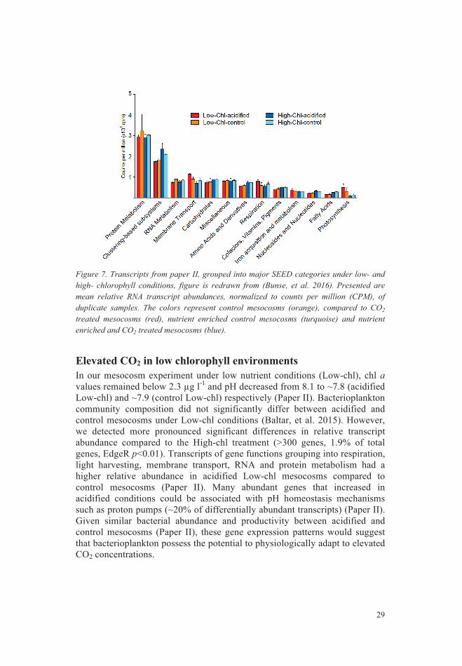
31
proteorhodopsin-resulting proton motive force could aid marine bacteria in different ways (Paper I&II). Given its occurrence across diverse phyla, high abundance in surface bacterioplankton genomes and its high gene expression levels, it is likely that even more proteorhodopsin-based functions exist in nature. Moreover, the latent role of photoheterotrophic bacteria for carbon fixation and energy turnover remains to be quantified in situ, seasonally and in different oceanic environments (for example using high-frequency time series and metatranscriptomics). Further recent instrumental advances, such as cell-sorting flow cytometry, single cell DNA and mRNA sequencing or FISH of functional genes – coupled to traditional oceanographic and microbiological techniques and experiments, promise great advances to uncover the functions and specific role of photoheterotrophic microbes.
Figure 8. Conceptual figure summarizing photoheterotrophic, metabolic traits of proteorhodopsin, analyzed in this thesis (Paper I & II). In paper II, under elevated CO2 in high chl a conditions (green, A), proteorhodopsin was similarly abundant in comparison to control conditions. Relative gene expression patterns of the bacterial community showed enhanced proteorhodopsin relative abundances under elevated CO2 and low chl a conditions (red, B), possibly due to enhanced pH homeostasis regulation (paper II). In paper I, Dokdonia sp. MED134 expressed TCA cycle enzymes, proteorhodopsin and anaplerotic reaction enzymes under light exposure in YEP medium (yellow C), and switched the TCA metabolism towards glyoxylate pathway expression in alanine medium (blue, D).
30
Furthermore, different bacterial families expressed distinct genes in response to acidification. For example, SAR11-clade bacteria mainly expressed a pyrophosphate-energized proton pump used to expel excess protons (Paper II). A study in the offshore Atlantic found no effects of short-term 2.5-fold acidification exposure on the productivity of SAR11, suggesting physiological resilience to acidification (Hartmann, et al. 2015). Our study could thus provide an explanation of how SAR11, and other bacteria, regulate their transcriptome to respond to, and potentially overcome challenges in pH. Interestingly, proteorhodopsin was highly abundant and significantly differently abundant upon acidification in Low-Chl, hinting to an important role in pH homeostasis regulation. Proteorhodopsin has been previously hypothesized, but not shown, to modulate responses of bacterioplankton to ocean acidification because of its mode of action: light-driven proton pumping (Fuhrman, et al. 2008).
Considering that the observed mechanisms to expel protons across the cell membrane against the proton gradient are energetically costly, bacteria would potentially have to assign more energy to these processes under acidification. Under long-term exposure to elevated CO2 concentrations, this could leave less energy for other processes, such as growth. Thus, despite unchanged bacterial production estimates in our nine-day experiment, increased proton expulsion and respiration rates could, under extended periods of elevated CO2, impact on bacterial growth efficiencies. In fact, a recent study showed that even short-term elevation of CO2 can increase respiration of microbial communities and subsequently increase the cell-specific DOC removal and loss of organic carbon for the ecosystem (James, et al. 2017). Yet, predictions of how bacteria will respond to ocean acidification remain difficult, particularly due to the tremendous functional diversities of heterotrophic bacterioplankton (Hutchins, et al. 2017). Future studies are recommended to further incorporate respiration measurements (in addition to productivity estimates) to explore the energetic cost of pH homeostasis under low-nutrient, open-ocean scenarios. Multiple stressors as well as a differentiation between direct and indirect effects of ocean change (Boyd, et al. 2016, Hutchins, et al. 2017, Riebesell, et al. 2015), have the potential to uncover underlying fundamental physiological traits and functions of microbes to ocean climate changes, and overall gradients in marine systems.
Proteorhodopsin photoheterotrophy in marine systems – future outlooks The role of proteorhodopsin for marine bacteria could be more diverse than previously hypothesized. Figure 8 summarizes the role of proteorhodopsin under the environmental conditions studied in this thesis. Depending on nutrient quality and quantity, bacterial growth stage or pH stress, the
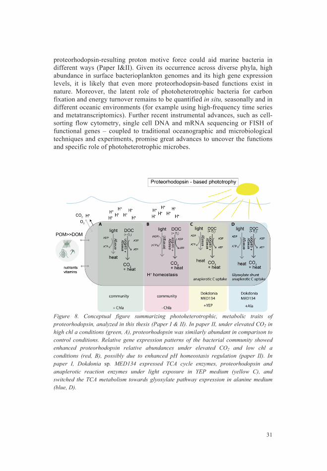
33
closer to a particle compared to another, it might have advantages to colonize it and grow, which would subsequently result in an earlier progression.
Figure 9. Boxplots grouped per month of environmental parameters (panel A) and bacterioplankton patterns (panel B), as well as dotplots of the most abundant population per class (one per class) (panel C). The data originate from the LMO time series (panel A, 2011-2014, Paper IV) and 3-0.2 µm filter fraction in 2011-2016 (panel B and C, Paper III). Note that the Chl a graph is cut at y-axis for visualization purposes and omits one data point (April 2011; 13.45 µg l-1). In panel B, bacterial relative abundances are summed by class (or phylum respectively) for each sampling date. The relative abundances of the most abundant populations are plotted against the day of year, blue lines indicate smoothed conditional means (method loess from ggplot2 package (Wickham 2009)). Abbreviations denote bacterial production (Bact. prod.), Glucose uptake rate constant K (Glucose K), bacterial biomass (Bact. Biom.). Numbers above temperature boxes indicate number of samples for each month of environmental samples (2011-2014), but see glucose K, while numbers above Flavobacteriia boxes indicate number of samples for 16S rRNA gene data (panel B and C, 2011-2016). Grey background panels indicate summer months.
3 2 6 19
25
2123
2424
209
6
0
5
10
15
20
3 6 9 12
Tem
pera
ture
(o C)
0
2
4
6
3 6 9 12
Chl a
(µg
l−1)
0.0
0.2
0.4
0.6
3 6 9 12
Bact.
pro
d. (µ
g C
l−1 h
−1)
2 2 14
65
5
4 5
73 3
0.00
0.05
0.10
0.15
3 6 9 12
Gluc
ose
K
0
50
100
150
200
3 6 9 12Month
Bact.
Biom
. (m
g C
m−3
)
Month
1e−04
1e−03
1e−02
0 100 200 300Day of Year
relat
ive a
bund
ance
(log
)
flavobacterial population
1e−04
1e−03
1e−02
0 100 200 300Day of Year
relat
ive a
bund
ance
(log
)
alphaproteobacterial population
1e−04
1e−03
1e−02
0 100 200 300Day of Year
relat
ive a
bund
ance
(log
)
gammaproteobacterial population
1e−04
1e−03
1e−02
0 100 200 300Day of Year
Relat
ive a
bund
ance
(log
) actinobacterial population
1e−04
1e−03
1e−02
0 100 200 300Day of Year
relat
ive a
bund
ance
(log
)
verrucomicrobial population
7
5 9
2025
25
25 27 27 20 1111
0.00
0.25
0.50
0.75
1.00
3 6 9 12
relat
ive a
bund
ance
class Flavobacteriia
0.0
0.1
0.2
0.3
3 6 9 12
relat
ive a
bund
ance
class Alphaproteobacteria
0.0
0.2
0.4
0.6
3 6 9 12
relat
ive a
bund
ance
class Gammaproteobacteria
0.0
0.1
0.2
0.3
0.4
0.5
3 6 9 12
relat
ive a
bund
ance
class Actinobacteria
0.0
0.1
0.2
0.3
3 6 9 12
relat
ive a
bund
ance
phylum Verrucomicrobia
CBA
32
Seasonal bacterioplankton trends in Baltic Sea Traditional time series have revealed persistent seasonal fluctuations and overturn of physical, chemical and some biological parameters in temperate marine areas (see for example Paper V, (Tiselius, et al. 2015, Wasmund, et al. 2011). Microbial time series have shown that, generally, seasonal succession of marine bacteria in temperate areas depends on environmental drivers such as light, day length, nutrient availability and especially temperature (Paper V) (Gilbert, et al. 2009, Gilbert, et al. 2012, Johnson, et al. 2006, Pinhassi, et al. 2000, Yung, et al. 2015). In addition to abiotic factors, biotic drivers have been identified including phytoplankton biomass, organic matter concentrations and quality, as well as biological interactions (Needham, et al. 2016, Pinhassi, et al. 2000, Teeling, et al. 2012, Teeling, et al. 2016).
In the LMO high-frequency dataset, we found overall predictable temperature and nutrient patterns and phytoplankton and bacterioplankton biomass dynamics. Seasonally, many environmental factors showed autocorrelation patterns; such as light and temperature, during spring when light and temperatures increased, phytoplankton grew and inorganic nutrients became depleted (Paper IV). During summer on the other hand, inorganic nutrient concentrations were low whereas temperatures and summer phytoplankton biomass were elevated (Paper IV). Dominating phyla of the eukaryotic phytoplankton spring bloom (diatoms and dinoflagellates) varied between 2011 and 2014; however, timing and succession of the spring bloom (in April) and summer bloom (early July) remained stable (Paper IV). In the Baltic Sea, decadal shifts in spring bloom dominance have been described with a recent tendency for dinoflagellate dominance over diatoms in the Western Baltic Proper (Legrand, et al. 2015, Suikkanen, et al. 2007, Wasmund, et al. 2011). We show that bacterioplankton displayed recurring seasonal trends at coarse taxonomic resolution, such as at class level (Figure 9, Paper III). On average, Flavobacteria had strongly elevated relative abundances during spring while Actinobacteria were relatively more abundant in late summer/autumn, and Verrucomicrobia peaked during summer but exhibited very low relative abundances during other times of the year (Figure 9, Paper III).
Repeating seasonal bacterioplankton community composition patterns would suggest that the community is predictable based upon seawater conditions (Fuhrman, et al. 2006). It would also imply that environmental parameters are more important than trophic interactions (Gilbert, et al. 2012). But do these deterministic effects explain all the fine-scale microbial population dynamics and productivities? Stochastic effects such as colonization, death, dispersal or migration could result in more variable patterns (Zhou, et al. 2017). For example, if, by chance, one bacterial cell is
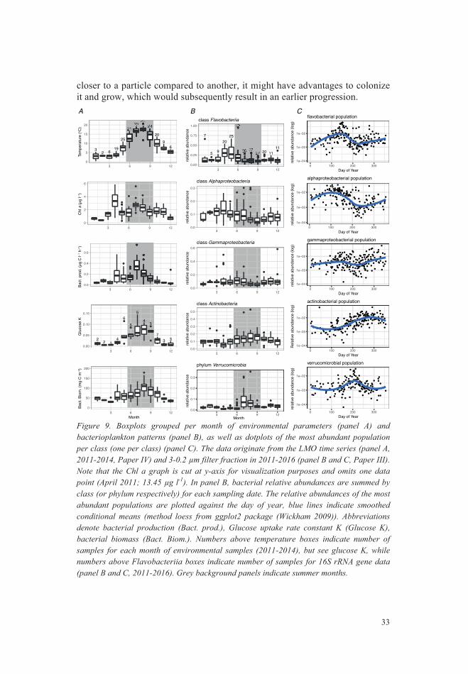
35
related bacterial populations can have a slightly different gene repertoire (as for example detected for actinobacterial acIV-MAGS (metagenomics amplified genomes) at LMO (Hugerth, et al. 2015)), a considerable part of the measured variability in substrate preference and bacterial activities in this dataset, could be related to shifting bacterial populations or ‘ecotypes’.
Table 2. Variability of relative abundances of dominant classes (and phylum Verrucomicrobia) in the free-living fraction at LMO over the time period 2011-2016, table is redrawn from Paper III.
Taxa Min Max Mean Variation Flavobacteriia 0.0089 0.949 0.1707 107-fold Gammaproteobacteria 0.0105 0.5845 0.0935 56-fold Alphaproteobacteria 0.0100 0.3021 0.0909 30-fold Actinobacteria 0.0035 0.5273 0.1254 150-fold Verrucomicrobia 0.0006 0.3538 0.0395 570-fold Cyanobacteria 0.0022 0.6226 0.1969 280-fold
A high frequency time-series study recently suggested that winter and
summer assemblages in temperate bacterioplankton communities resulted from switching between closely related strains with different temperature preferences (Ward, et al. 2017). Such variability and shifts in community structure might only to a lesser extend be detected in a community analysis at class level or at coarse time resolution such as monthly intervals (as presented in Figure 9B). Yet, even at class level, variability in community composition at LMO became evident; among summed relative abundances per sampling time point, Flavobacteriia varied 107-fold during the sampling period, Actinobacteria ranged 150-fold, Cyanobacteria varied 280-fold and verrucomicrobial abundances varied up to 570-fold (Table 2). To further unravel if bacterioplankton community structure could explain intra-annual variability in metabolic rates, we assessed whether distinct abundant bacterioplankton populations displayed seasonal preferences. Using the LMO 16S rRNA gene amplicon dataset of the free-living bacterioplankton community we further tested if bacterioplankton populations co-occurred with other populations of similar or dissimilar taxa.
Among the most abundant bacterioplankton populations, many populations were not stable over time (Paper III). For example, many Actinobacteria populations showed relatively elevated abundances during late summer, while some Flavobacteriia displayed relatively elevated abundances during spring or late summer, respectively (Figure 9, Paper III). Soft clustering analysis based on standardized relative abundances per population of high frequency sampling over three years (2011-2014) grouped populations of similar taxa together and clusters mostly consisted of populations from the same family (Paper III). Yet, populations displayed inter-annual variation in timing, amplitude and width of abundance peaks (Paper III).
34
In fact, in our study, bacterial abundances and activities (that is, heterotrophic productivity estimates, substrate uptake rate constants and enzyme activities) varied greatly throughout the year and exhibited higher values during summer compared to winter (Paper IV). Accordingly, bacterial production correlated with several environmental factors such as temperature, Chl a, bacterioplankton biomass, phytoplankton biomass such as cyanobacteria and flagellates and negatively to nutrients such as nitrate and phosphate (Spearman’s rho, p-value <0.001, Paper IV). Similarly, bacterial biomass correlated to multiple abiotic and biotic factors (temperature, salinity, phosphate, total N, DOC, Chl a, bacterial production, summer phytoplankton biomass (cyanobacteria and flagellates)) (Spearman’s rho, p-value <0.001, Paper IV). We did not detect noteworthy bacterioplankton growth prior to the spring bloom, indicating a reliance on phytoplankton-derived substrates for the heterotrophic bacterioplankton community. Furthermore, uptake rate constants of several substrates (such as glucose, amino acids and organic acids), correlated with temperature, bacterial production and cyanobacterial biomass (Spearman’s rho, p-value <0.001, Paper IV). This implies that a combination of abiotic and biotic parameters could represent important stimuli of microbial activities.
Dynamics in Bacterioplankton community structure Whereas seasonally recurring patterns in bacterioplankton community composition have been observed in several marine areas (Paper V, (Fuhrman, et al. 2006, Gilbert, et al. 2012)), also inter-annual variability has been detected when studying bacterial communities at higher temporal and taxonomic resolution (Gilbert, et al. 2012, Linz, et al. 2017, Teeling, et al. 2016). Accordingly, several underlying factors influencing ecotype dynamics, bacterioplankton evolution and niche expansion are still unidentified or possibly stochastic (Vergin, et al. 2013, Zhou, et al. 2017).
In paper IV, we observed a substantial variability over short time scales in many bacterial parameters. For example, microbial substrate uptake rate constants displayed the highest variability during times of water mass stratification during summer, i.e. presumably homogeneous surface water conditions (Paper IV). Among these variable patterns, stochastic effects could play a role for which populations bloom: for example, the history of the community, i.e. which populations were already growing, which were in a resting stage, or which populations were decreasing in cell numbers, just prior to the sampling time points. In Paper V, we hypothesized that preceding increasing abundances of populations could fuel further growth of populations in comparison to competitor populations as response to ‘disturbances’ such as nutrient input or changes in stratification. Further, assuming that closely
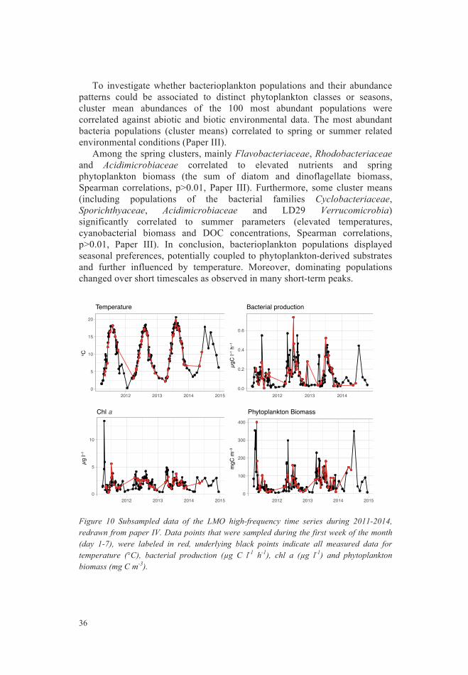
37
Assuming that different bacterial lineages have distinct genetic properties, it is possible that a substantial portion of the variability observed in heterotrophic rates in the Baltic Sea, is influenced by changes in bacterioplankton community composition.
Microbial food web dynamics: insights and future outlooks Going back to the original questions of who in the microbial community is doing what, when, and how; so far, as also shown in this thesis, we have begun to understand which bacterial classes and populations are abundant throughout the year in the Baltic Proper (Paper III & V). This thesis also investigated ‘what’ is done by the bacterioplankton community, such as bacterial biomass and activity estimations (heterotrophic production, substrate preferences and enzymatic activities) (Paper IV). Underlying metabolic and physiological processes (which population is doing what, and which functional genes do they possess to do it) are though more slowly disentangled, although ongoing analyses are addressing this issue (e.g. Figure 3, Figure 9, Paper V). It is possible that similar processes as discussed in Papers I and II are involved in driving community composition and heterotrophic activities, among other ‘yet-to-be-found’ and regulatory mechanisms. Further, co-occurrences between bacterial populations and correlation analysis to environmental parameters might provide further clues about possible biological interactions.
Summarizing, with the help of the high frequency sampling scheme at LMO, we identified several important characteristics of the phytoplankton and bacterioplankton assemblages (Paper III & IV):
• high inter-annual variation in spring phytoplankton bloom dynamics and
species dominance (i.e. shifts between diatoms and dinoflagellates) • a twin peak in filamentous cyanobacterial biomass during summer,
possibly consisting of different cyanobacterial species (paper IV) • distinct bacterioplankton seasonal patterns at coarse taxonomic rank (class
level) • different bacterial taxa respond to spring phytoplankton bloom compared
to summer phytoplankton bloom (e.g. Flavobacteria showed relatively higher abundances in spring, whereas Verrucomicrobia responded to summer bloom) (paper III)
36
To investigate whether bacterioplankton populations and their abundance patterns could be associated to distinct phytoplankton classes or seasons, cluster mean abundances of the 100 most abundant populations were correlated against abiotic and biotic environmental data. The most abundant bacteria populations (cluster means) correlated to spring or summer related environmental conditions (Paper III).
Among the spring clusters, mainly Flavobacteriaceae, Rhodobacteriaceae and Acidimicrobiaceae correlated to elevated nutrients and spring phytoplankton biomass (the sum of diatom and dinoflagellate biomass, Spearman correlations, p>0.01, Paper III). Furthermore, some cluster means (including populations of the bacterial families Cyclobacteriaceae, Sporichthyaceae, Acidimicrobiaceae and LD29 Verrucomicrobia) significantly correlated to summer parameters (elevated temperatures, cyanobacterial biomass and DOC concentrations, Spearman correlations, p>0.01, Paper III). In conclusion, bacterioplankton populations displayed seasonal preferences, potentially coupled to phytoplankton-derived substrates and further influenced by temperature. Moreover, dominating populations changed over short timescales as observed in many short-term peaks.
Figure 10 Subsampled data of the LMO high-frequency time series during 2011-2014, redrawn from paper IV. Data points that were sampled during the first week of the month (day 1-7), were labeled in red, underlying black points indicate all measured data for temperature (°C), bacterial production (µg C l-1 h-1), chl a (µg l-1) and phytoplankton biomass (mg C m-3).
0
5
10
15
20
2012 2013 2014 2015
o C
Temperature
0.0
0.2
0.4
0.6
2012 2013 2014
µgC
l−1 h
−1
Bacterial production
0
5
10
2012 2013 2014 2015
µg l−
1
Chl a
0
100
200
300
400
2012 2013 2014 2015
mgC
m−3
Phytoplankton Biomass
Figure 5
39
Conclusions
This thesis has contributed to a deepened understanding of when and how marine bacteria respond to environmental changes and gradients, and how such responses may partly explain the distribution and success of particular bacterial taxa in marine ecosystems. Overall, using laboratory approaches and a high-frequency time series study, I discovered that bacterioplankton communities reacted to changes in the environment at a transcriptional level, by altering productivity rates or by changes in community structure.
Under light exposure and different organic nutrient qualities, model
organism Dokdonia sp. MED134 upregulated the gene expression of proteorhodopsin and other genes to change its metabolism towards a more resourceful state. Similar metabolic functional traits of proteorhodopsin-containing bacterioplankton were found in several marine ecosystems and could further play important roles for marine bacteria to thrive and to encounter changing DOM gradients in natural ecosystems.
Under elevated CO2, bacterioplankton could keep their productivity and community structure unaltered by adapting the gene expression. To maintain pH homeostasis, bacteria induced higher expression of genes related to respiration, membrane transport and light acquisition, such as proteorhodopsin, under low-nutrient conditions. Under high-nutrient conditions with phytoplankton blooms, such regulatory mechanisms were not necessary, indicating that pelagic microbes are potentially more vulnerable to ocean acidification compared to populations in coastal habitats.
Using the LMO time series study in the Baltic Sea, this thesis discovered
pronounced temporal dynamics in many investigated microbial variables including latent phytoplankton-bacteria linkages, substrate uptake rates, and shifts in bacterial community structure. Environmental conditions, and especially seasonal physicochemical and biotic changes, highly impacted on microbial productivities and abundances. Also, bacterioplankton community
38
• different bacterial biomass and production dynamics following the spring diatom- and dinoflagellate bloom, compared to after the summer cyanobacterial bloom
• bacterial activities (heterotrophic production, substrate uptakes and extracellular enzyme activities) displayed high variance during stratified surface water conditions in summer
• the high variance in biological parameters, could lead to an over- or underestimation by monthly sampling (Figure 10, Paper IV)
Future research challenges and research directions could include
understanding underlying physiological processes shaping microbial communities. It would be desirable to further understand biological interactions between organisms, for example between phytoplankton and bacterioplankton populations, such as how succession is regulated by metabolic interdependences among planktonic communities (Paper V). Additionally, studies addressing the impact and specificity of grazers on seasonal bacterioplankton succession patterns, and the proportion of picocyanobacteria to bulk bacterioplankton abundances and productivities, have the potential to disentangle missing links in our understanding of Baltic Sea microbial communities. Also, outstanding research questions as defined in Paper V, such as which thresholds of physical, chemical, and biological factors are dependent on the transition between seasons and how marine bacteria communities will react to projected climate change scenarios. A paramount future line of research would be to study how specific seasonal transcription patterns of functional genes of different bacterioplankton populations relate to bacterioplankton activities and biogeochemical rates, for example photoheterotrophic traits. Here, high resolution sampling schemes focused across particular time periods could provide important insights into the regulation of marine ecosystems.
41
My contribution to the individual papers
Paper I Laboratory work and data analysis. Paper II Data analysis and writing of the manuscript. Paper III Time series sampling and laboratory work (2012-2015), study design, data analysis and interpretation, writing of the manuscript. Paper IV Time series sampling and laboratory work (2012-2014), data analysis and interpretation, writing of the manuscript. Paper V Literature research, data analysis, interpretation and writing of the manuscript.
40
composition changed seasonally at class level, despite detected inter-annual variability among genera and populations. Many abundant bacterioplankton populations displayed spring or summer preferences and accordingly correlated to seasonally changing environmental parameters, such as biomass of different phytoplankton phyla. Thus, high resolution time series can provide important insights into the interplay and scaling between factors ultimately determining the success and seasonal succession of different bacterial populations.
In this thesis, I highlight the advantages of combining methodological
approaches to study bacterioplankton at high resolution, for improving our understanding of mechanisms controlling microbial communities and biogeochemical processes. Such advances in knowledge of fundamental metabolic and physiological processes underlying plankton succession could further aid to obtain a four-dimensional picture of how microbial communities relate to the turnover of DOM in the oceans.
43
All my wonderful coauthors that, in one way or the other, have contributed to the success of this thesis: Thank you! (Graphic drawn using https://www.wordclouds.com/)
My colleagues made my work more than just work – a lot of fun on a daily
basis. Thank you, tack: Sabina, Anders, Henrik, Leon, Ymke, Kris, Emmelie, Conny and Christina for making the lab function, paperwork work, materials to magically appear, to happily process samples despite an already tight schedule, to week after week, month after month and year after year sample seawater - despite the weather - and still have time for a coffee or chat in the hall. I met some great Microbial Ecology PhD students along the way: Johanna, Hanna, Neelam, Markus, Devi, Christofer and Ben. Thank you all for fun moments, interesting discussions, exciting experiments, adventurous conference travels and experiencing late lab/office evenings, weekends and ‘recreational fieldwork’ together! To the experienced postdocs in our group, thank you for a pleasant atmosphere and sharing your wisdom and skills: Fede, Joakim, Lovisa, Sandra, Johanna, Hanna, John, Jürg and Stina! To all the short-term and longer-term group members, lab helpers, students and visitors: Jeremy, Matthias, Johanna, Therese, Anu, Josef, Camilla, Clara, Katrin, Daniela, Lingni, Elena, Marta, Emelie, Marcus – you all filled the lab with life and kept it running. Thank you all for your efforts, support and pleasant company in the lab. My second research group: Astrid, Martin, Elin, Karen, Emmelie, Elin, Fredrik, Mireia, Lina, Eva, Maurice, Shenhong, Elisabeth, Caroline and Kimberly. Thank you for sharing materials, stories, frustrations, green ‘phyto’ fingers and adventures during teaching, fieldwork and seminars.
http
s://w
ww.
wor
dclo
uds.c
om/
1 of
122
/11/
17 1
1:47
42
Acknowledgements
“An oceanographer is not (a) solitary sailor.“ L.M. Proctor
This thesis would not have been possible without the help of many people and for this, I am very thankful. I would like to thank my supervisor-team, for their guidance and countless opportunities that they provided me during my PhD studies. First and foremost: Jarone, thank you for giving me the opportunity to join your lab and for making science and projects work from ~7,500 km distance for a while. Thanks for occasionally giving me free hands to proceed side projects and thank you for your support through many ups and downs and trusting in my abilities as researcher! I learned a lot from you and am inspired to continue in this exciting research field. Catherine, thank you for taking me under your supervision as lonely Master student, for introducing me to beautiful phytoplankton and Kårehamn, encouraging social skills and engagement in outreach, countless Saturday afternoon chats and a great dose of women-power! José, thank you for interesting chats about projects, books and bioinformatics! Daniel, I would not have been able to finish this thesis without your mentoring, bioinformatics support, teaching of coding skills, insights into being a researcher and always having interesting biological background information. Vielen Dank!
I was very fortunate to be part of the projects ECOCHANGE,
PRODIVERSA and MESOTOMICS, and involved in collaborations with the Andersson lab at SciLifelab, Moran lab and Medeiros lab at UGA, the time-series crew in Kristineberg and the research center EEMiS. Also, the GENECO research school, Workshop on Genomics in Cesky Krumlov and 2014 CMORE Summer course in Hawaii were instructive, fun and very inspiring. I am very thankful for numerous opportunities, scientific meetings, interesting discussions and experiences and would like to thank everyone who made this possible and contributed to an inspiring scientific and social atmosphere in Kalmar and wherever I visited. I always felt truly welcome!

45
References
Akram, N., Palovaara, J., Forsberg, J., Lindh, M.V., Milton, D.L., Luo, H., et al. (2013) Regulation of proteorhodopsin gene expression by nutrient limitation in the marine bacterium Vibrio sp. AND4. Environ Microbiol 15: 1400-1415.
Allen, A.E., Dupont, C.L., Oborník, M., Horák, A., Nunes-Nesi, A., McCrow, J.P., et al. (2011) Evolution and metabolic significance of the urea cycle in photosynthetic diatoms. Nature 473: 203-207.
Allgaier, M., Riebesell, U., Vogt, M., Thyrhaug, R. and Grossart, H.P. (2008) Coupling of heterotrophic bacteria to phytoplankton bloom development at different pCO2 levels: a mesocosm study. Biogeosciences 5: 1007-1022.
Alneberg, J., Sundh, J., Bennke, C., Beier, S., Lundin, D., Hugerth, L.W., et al. (manuscript) BARM and BalticMicrobeDB: a reference metagenome and interface to meta-omic data for the Baltic Sea. https://githubcom/EnvGen:
Altschul, S.F., Madden, T.L., Schäffer, A.A., Zhang, J., Zhang, Z., Miller, W., et al. (1997) Gapped BLAST and PSI-BLAST: a new generation of protein database search programs. Nucleic Acids Res 25: 3389-3402.
Amin, S.A., Parker, M.S. and Armbrust, E.V. (2012) Interactions between diatoms and bacteria. Microb Mol Biol Rev, 76: 667-684.
Amir, A., McDonald, D., Navas-Molina, J.A., Kopylova, E., Morton, J.T., Xu, Z.Z., et al. (2017) Deblur rapidely resolves single-nucleotide community sequence patterns. mSystems 2: e00191-16.
Andersson, A., Meier, H.M., Ripszam, M., Rowe, O., Wikner, J., Haglund, P., et al. (2015) Projected future climate change and Baltic Sea ecosystem management. AMBIO 44: 345-356.
Andersson, A.F., Riemann, L. and Bertilsson, S. (2010) Pyrosequencing reveals contrasting seasonal dynamics of taxa within Baltic Sea bacterioplankton communities. ISME J 4: 171-181.
Arnosti, C., Grossart, H.P., Muhling, M., Joint, I. and Passow, U. (2011) Dynamics of extracellular enzyme activities in seawater under changed atmospheric pCO2: a mesocosm investigation. Aquat Microb Ecol 64: 285-298.
Arrigo, K.R. (2005) Marine microorganisms and global nutrient cycles. Nature 437: 349-356.
Aylward, F.O., Eppley, J.M., Smith, J.M., Chavez, F.P., Scholin, C.A. and DeLong, E.F. (2015) Microbial community transcriptional networks are conserved in three domains at ocean basin scales. Proc Natl Acad Sci USA 112: 5446-5448.
44
To the 7am morning coffee crew - Emil, Elias, Fredrik, Anu, little Håkan and Mariëlle: Thanks for great coffee, fun, fika and discussions about ponies, work, research papers, (alien) life, movies and general science. It was the lagom amount of nerdy for waking up. I will miss my breakfast club!
Best research organism; fishes, birds, bacteria? Who cares, as long as the
microbes win :P Thanks for taking me fishing, birding mushrooming and participate in the pike’s day: Oscar, Emil, Christofer, Nathalie, Andreas and Kocken; despite some weather interference in form of ice, frost, rain and stormy winds, I had a blast discovering Swedish nature and wildlife to keep my mind off work sometimes.
To my new (and old) international friends: Michelle, Anna, Håkan, Anu,
Mireia, Alexis, Jo, Andreas, Ulrika, Devi, Nayani, Lee, Batman, Conny, Susanna, Josefin, Ingrid, Daniela, Furong, Joacim, Daniel, Stephan, Xiaofen, Neus, Nathalie, Alina, Bea, Nini, Isi, Tamara and Paul: thank you for inspiration, mentoring, listening to my complains, support, birding, Friday beers and many adventures. We will meet each other somewhere around the planet!
A special thanks to four exceptional women: Bea - for encouraging me to
move to Sweden, Mireia - for guidance along the PhD path, Anu - for boosting my stubbornness to finish the thesis, and Michelle - for giving me the courage to always move on! I just kept swimming :)
Der größte Dank geht an meine Familie: ohne euch hätte ich dieses Buch
nicht schreiben können. Ich bin unendlich dankbar für eure immer-währende Geduld, Unterstützung, Ermutigung, Vertrauen und Liebe!
47
Callahan, B.J., McMurdie, P.J., Rosen, M.J., Han, A.W., Johnson, A.J.A. and Holmes, S.P. (2016) DADA2: High-resolution sample inference from Illumina amplicon data. Nat Methods 13: 581-583.
Campbell, B.J., Waidner, L.A., Cottrell, M.T. and Kirchman, D.L. (2008) Abundant proteorhodopsin genes in the North Atlantic Ocean. Environ Microbiol 10: 99-109.
Caporaso, J.G., Lauber, C.L., Walters, W.A., Berg-Lyons, D., Huntley, J., Fierer, N., et al. (2012) Ultra-high-throughput microbial community analysis on the Illumina HiSeq and MiSeq platforms. ISME J 6: 1621-1624.
Church, M.J., Ducklow, H.W., Letelier, R.M. and Karl, D.M. (2006) Temporal and vertical dynamics in picoplankton photoheterotrophic production in the subtropical North Pacific Ocean. Aquat Microb Ecol 45: 41-53.
Cohan, F.M. (2006) Towards a conceptual and operational union of bacterial systematics, ecology, and evolution. Philosophical transactions of the Royal Society of London Series B, Biological sciences 361: 1985-1996.
Courties, A., Riedel, T., Rapaport, A., Lebaron, P. and Suzuki, M.T. (2015) Light-driven increase in carbon yield is linked to maintenance in the proteorhodopsin-containing Photobacterium angustum S14. Front Microbiol 6: 688.
de la Torré, J.R., Christianson, L.M., Béja, O., Suzuki, M.T., Karl, D.M., Heidelberg, J., et al. (2003) Proteorhodopsin genes are distributed among divergent marine bacterial taxa. Proc Natl Acad Sci U S A 100: 12830-12835.
Del Giorgio, P.A., Condon, R.H., Bouvier, T., Longnecker, K., Bouvier, C., Sherr, E., et al. (2011) Coherent patterns in bacterial growth, growth efficiency, and leucine metabolism along a northeastern Pacific inshore-offshore transect. Limnol Oceanogr 56: 1-16.
Doney, S.C., Fabry, V.J., Feely, R.A. and Kleypas, J.A. (2009) Ocean acidification: the other CO2 problem. Ann Rev Mar Sci 1: 169-92.
Dore, J.E., Lukas, R., Sadler, D.W., Church, M.J. and Karl, D.M. (2009) Physical and biogeochemical modulation of ocean acidification in the central North Pacific. Proc Natl Acad Sci U S A 106: 12235-12240.
Droop, M. (1957) Auxotrophy and organic compounds in the nutrition of marine phytoplankton. Microbiology 16: 286-293.
Duarte, C.M., Hendriks, I.E., Moore, T.S., Olsen, Y.S., Steckbauer, A., Ramajo, L., et al. (2013) Is ocean acidification an open-ocean syndrome? Understanding anthropogenic impacts on seawater pH. ESTUAR COAST 36: 221-236.
Dupont, C.L., Larsson, J., Yooseph, S., Ininbergs, K., Goll, J., Asplund-Samuelsson, J., et al. (2014) Functional tradeoffs underpin salinity-driven divergence in microbial community composition. PloS ONE 9: e89549.
Durham, B.P., Sharma, S., Luo, H., Smith, C.B., Amin, S.A., Bender, S.J., et al. (2015) Cryptic carbon and sulfur cycling between surface ocean plankton. Proc Natl Acad Sci USA 112: 453-457.
Feng, S., Powell, S.M., Wilson, R. and Bowman, J.P. (2013) Light-stimulated growth of proteorhodopsin-bearing sea-ice psychrophile Psychroflexus torquis is salinity dependent. ISME J 7: 2206-2213.
Field, C.B., Behrenfeld, M.J., Randerson, J.T. and Falkowski, P. (1998) Primary production of the biosphere: integrating terrestrial and oceanic components. Science 281: 237-240.
46
Azam, F. (1998) Microbial Control of Oceanic Carbon Flux: The Plot Thickens. Science 280: 694-969.
Baltar, F., Legrand, C. and Pinhassi, J. (2016) Cell-free extracellular enzymatic activity is linked to seasonal temperature changes: a case study in the Baltic Sea. Biogeosciences 13: 2815-2821.
Baltar, F., Palovaara, J., Vila-Costa, M., Gasol, J.M., Salazar, G., Calvo, E., et al. (2015) Response of rare, common and abundant bacterioplankton to anthropogenic pertubations in a Mediterranean coastal site. FEMS Microbiol Ecol 91: fiv058.
Béjà, O., Spudich, E.N., Spudich, J.L., Leclerc, M. and DeLong, E.F. (2001) Proteorhodopsin phototrophy in the ocean. Nature 411: 786-789.
Bertos-Fortis, M., Farnelid, H.M., Lindh, M.V., Casini, M., Andersson, A., Pinhassi, J., et al. (2016) Unscrambling cyanobacteria community dynamics related to environmental factors. Front Microbiol 7: 625.
Björkman, K.M., Church, M.J., Doggett, J.K. and Karl, D.M. (2015) Differential assimilation of inorganic carbon and leucine by Prochlorococcus in the oligotrophic north Pacific subtropical gyre. Front Microbiol 6: 1401.
Boström, K.H., Simu, K., Hagström, Å. and Riemann, L. (2004) Optimization of DNA extraction for quantitative marine bacterioplankton community analysis. Limnol Oceanogr Methods 2: 365-373.
Boyd, P., Dillingham, P., Mcgraw, C., Armstrong, E., Cornwall, C., Feng, Y., et al. (2016) Physiological responses of a Southern Ocean diatom to complex future ocean conditions. Nat Clim Change 6: 207.
Boyd, P.W. and Hutchins, D.A. (2012) Understanding the responses of ocean biota to a complex matrix of cumulative anthropogenic change. Mar Ecol Prog Ser 470: 125-135.
Brinkhoff, T., Giebel, H.-A. and Simon, M. (2008) Diversity, ecology, and genomics of the Roseobacter clade: a short overview. Arch Microbiol 189: 531-539.
Brown, C.T., Hug, L.A., Thomas, B.C., Sharon, I., Castelle, C.J., Singh, A., et al. (2015) Unusual biology across a group comprising more than 15% of domain Bacteria. Nature 523: 208-211.
Brown, L.S. and Jung, K.-H. (2006) Bacteriorhodopsin-like proteins of eubacteria and fungi: the extent of conservation of the haloarchaeal proton-pumping mechanism. Photochem Photobiol Sci 5: 538-546.
Buchan, A., González, J.M. and Moran, M.A. (2005) Overview of the marine Roseobacter lineage. Appl Environ Microbiol 71: 5665-5677.
Buchan, A., LeCleir, G.R., Gulvik, C.A. and González, J.M. (2014) Master recyclers: features and functions of bacteria associated with phytoplankton blooms. Nat Rev Microbiol 12: 686-698.
Bunse, C., Bertos-Fortis, M., Sassenhagen, I., Sildever, S., Sjöqvist, C., Godhe, A., et al. (2016) Spatio-Temporal interdependence of bacteria ad phytoplankton during a Baltic Sea spring bloom. Front Microbiol 7: Article517.
Bunse, C., Lundin, D., Karlsson, C.M.G., Vila-Costa, M., Palovaara, J., Akram, N., et al. (2016) Response of marine bacterioplankton pH homeostasis gene expression to elevated CO2. Nature Clim Change 6: 483-487.
Bunse, C. and Pinhassi, J. (2017) Marine bacterioplankton seasonal succession dynamics. Trends Microbiol 25: 494-505.
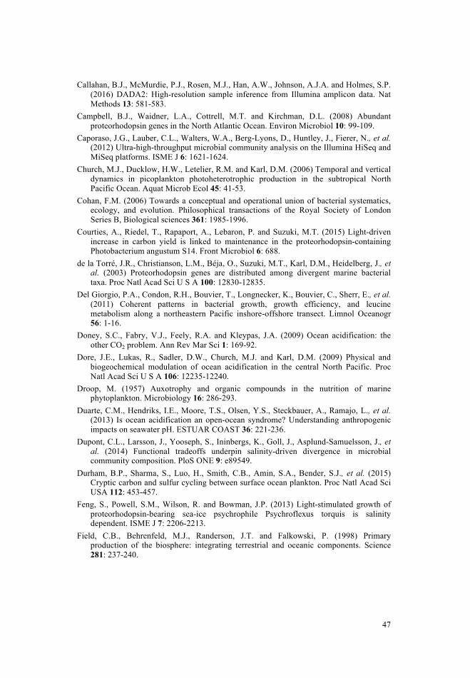
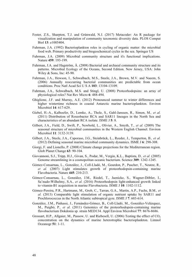
49
Grzymski, J.J., Riesenfeld, C.S., Williams, T.J., Dussaq, A.M., Ducklow, H.W., Erickson, M., et al. (2012) A metagenomic assessment of winter and summer bacterioplankton from Antarctica Peninsula coastal surface waters. ISME J 6: 1901-1915.
Gunderson, A.R., Armstrong, E.J. and Stillman, J.H. (2016) Multiple stressors in a changing world: the need for an improved perspective on physiological responses to the dynamic marine environment. Ann Rev Mar Sci 8: 357-378.
Hagström, Å., Azam, F., Berg, C. and Zweifel, U.-L. (2017) Isolates as models to study bacterial ecophysiology and biogeochemistry. Aquat Microb Ecol 80: 15-27.
Hanada, S. (2016) Anoxygenic photosynthesis -a photochemical reaction that does not contribute to oxygen reproduction-. Microbes Environ 31: 1-3.
Hartmann, M., Hill, P.G., Tynan, E., Achterberg, E.P., Leakey, R.J. and Zubkov, M.V. (2015) Resilience of SAR11 bacteria to rapid acidification in the high-latitude open ocean. FEMS Microbiol Ecol 92: fiv161.
Haugan, P.M. and Drange, H. (1996) Effects of CO2 on the ocean environment. Energy Convers Manag 37: 1019-1022.
Herlemann, D.P.R., Labrenz, M., Jürgens, K., Bertilsson, S., Waniek, J. and Andersson, A.F. (2011) Transitions in bacterial communities along the 2000 km salinity gradient of the Baltic Sea. ISME J 5: 1571-1579.
Hoppe, H.-G. (1993) Use of fluorogenic model substrates for extracellular enzyme activity (EEA) measurement of bacteria. Handbook of methods in aquatic microbial ecology: 423-431.
Hugenholtz, P. (2002) Exploring prokaryotic diversity in the genomic era. Genome biology 3: reviews0003.1.
Hugerth, L.W., Larsson, J., Alneberg, J., Lindh, M.V., Legrand, C., Pinhassi, J., et al. (2015) Metagenome-assembled genomes uncover a global brackish microbiome. Genome Biol 16: 279.
Hugerth, L.W., Wefer, H.A., Lundin, S., Jakobsson, H.E., Lindberg, M., Rodin, S., et al. (2014) DegePrime, a program for degenerate primer design for broad-taxonomic-range PCR in microbial ecology studies. Appl Environ Microbiol 80: 5116-5123.
Hutchins, D.A. and Fu, F. (2017) Microorganisms and ocean global change. Nature Microbiol 2: 17058.
James, A.K., Passow, U., Brzezinski, M.A., Parsons, R.J., Trapani, J.N. and Carlson, C.A. (2017) Elevated pCO2 enhances bacterioplankton removal of organic carbon. PloS ONE 12: e0173145.
Johnson, Z.I., Zinser, E.R., Coe, A., McNulty, N.P., Woodward, E.M.S. and Chisholm, S.W. (2006) Niche partitioning among Prochlorococcus ecotypes along ocean-scale environmental gradients. Science 311: 1737-1740.
Joint, I., Doney, S.C. and Karl, D.M. (2011) Will ocean acidification affect marine microbes? ISME J 5: 1-7.
Kanehisa, M., Araki, M., Goto, S., Hattori, M., Hirakawa, M., Itoh, M., et al. (2007) KEGG for linking genomes to life and the environment. Nucleic acids research 36: D480-D484.
Karjalainen, M., Engström-Öst, J., Korpinen, S., Peltonen, H., Pääkkönen, J.P., Rönkkönen, S., et al. (2007) Ecosystem Consequences of Cyanobacteria in the Northern Baltic Sea. AMBIO 36: 195-202.
Karl, D.M. (2007) Microbial oceanography: paradigms, processes and promise. Nat Rev Micro 5: 759-769.
48
Foster, Z.S., Sharpton, T.J. and Grünwald, N.J. (2017) Metacoder: An R package for visualization and manipulation of community taxonomic diversity data. PLOS Comput Biol 13: e1005404.
Fuhrman, J.A. (1992) Bacterioplankton roles in cycling of organic matter: the microbial food web. Primary productivity and biogeochemical cycles in the sea. Springer US
Fuhrman, J.A. (2009) Microbial community structure and it's functional implications. Nature 459: 193-199.
Fuhrman, J.A. and Hagström, Å. (2008) Bacterial and archaeal community structure and its patterns. Microbial Ecology of the Oceans, Second Edition. New Jersey, USA: John Wiley & Sons, Inc: 45-90.
Fuhrman, J.A., Hewson, I., Schwalbach, M.S., Steele, J.A., Brown, M.V. and Naeem, S. (2006) Annually reoccurring bacterial communities are predictable from ocean conditions. Proc Natl Acad Sci U S A 103: 13104-13109.
Fuhrman, J.A., Schwalbach, M.S. and Stingl, U. (2008) Proteorhodopsins: an array of physiological roles? Nat Rev Micro 6: 488-494.
Ghiglione, J.F. and Murray, A.E. (2012) Pronounced summer to winter differences and higher wintertime richness in coastal Antarctic marine bacterioplankton. Environ Microbiol 14: 617-629.
Giebel, H.-A., Kalhoefer, D., Lemke, A., Thole, S., Gahl-Janssen, R., Simon, M., et al. (2011) Distribution of Roseobacter RCA and SAR11 lineages in the North Sea and characteristics of an abundant RCA isolate. ISME J 5: 8.
Gilbert, J.A., Field, D., Swift, P., Newbold, L., Olivier, A., Smyth, T., et al. (2009) The seasonal structure of microbial communities in the Western English Channel. Environ Microbiol 11: 3132-3139.
Gilbert, J.A., Steele, J.A., Caporaso, J.G., Steinbrück, L., Reeder, J., Temperton, B., et al. (2012) Defining seasonal marine microbial community dynamics. ISME J 6: 298-308.
Giorgi, F. and Lionello, P. (2008) Climate change projections for the Mediterranean region. Glob Planet Change 63: 90-104.
Giovannoni, S.J., Tripp, H.J., Givan, S., Podar, M., Vergin, K.L., Baptista, D., et al. (2005) Genome streamlining in a cosmopolitan oceanic bacterium. Science 309: 1242-1245.
Gómez-Consarnau, L., González, J., Coll-Lladó, M., Gourdon, P., Pascher, T., Neutze, R., et al. (2007) Light stimulates growth of proteorhodopsin-containing marine Flavobacteria. Nature 445: 210-213.
Gómez-Consarnau, L., González, J.M., Riedel, T., Jaenicke, S., Wagner-Döbler, I., Sa¨nudo-Wilhelmy, S.A., et al. (2016) Proteorhodopsin light-enhanced growth linked to vitamin-B1 acquisition in marine Flavobacteria. ISME J 10: 1102-1112.
Gómez-Pereira, P.R., Hartmann, M., Grob, C., Tarran, G.A., Martin, A.P., Fuchs, B.M., et al. (2013) Comparable light stimulation of organic nutrient uptake by SAR11 and Prochlorococcus in the North Atlantic subtropical gyre. ISME J 7: 603-614.
González, J.M., Pinhassi, J., Fernández-Gómez, B., Coll-Lladó, M., González-Velázquez, M., Puigbò, P., et al. (2011) Genomics of the proteorhodopsin-containing marine flavobacterium Dokdonia sp. strain MED134. Appl Environ Microbiol 77: 8676-8686.
Grossart, H.P., Allgaier, M., Passow, U. and Riebesell, U. (2006) Testing the effect of CO2 concentration on the dynamics of marine heterotrophic bacterioplankton. Limnol Oceanogr 51: 1-11.
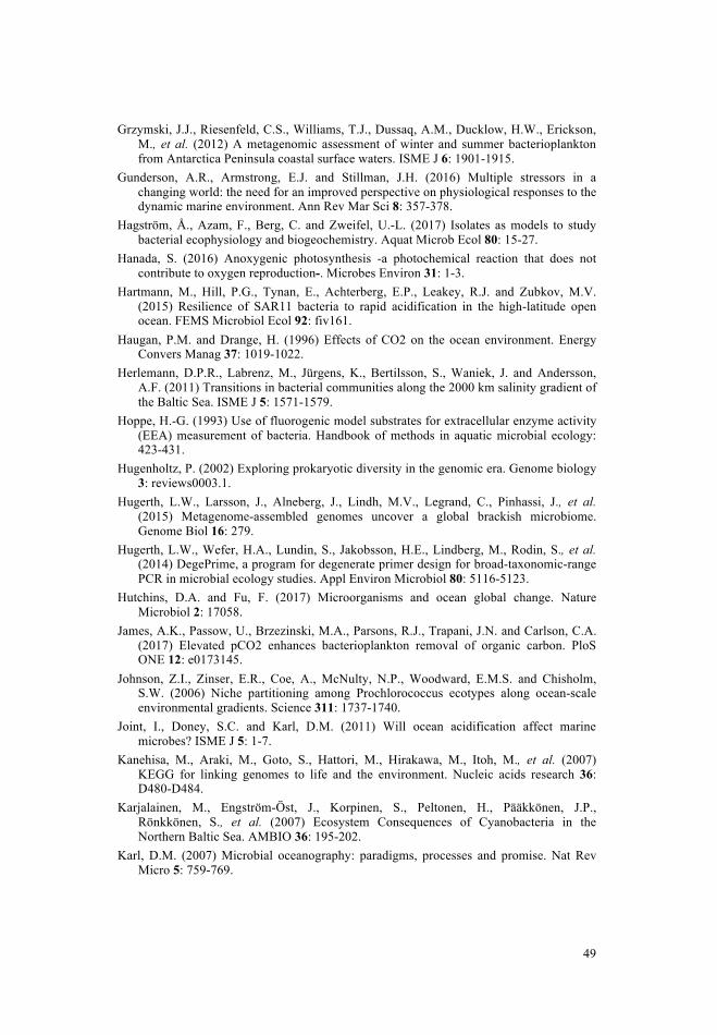
51
Malmstrom, R.R., Coe, A., Kettler, G.C., Martiny, A.C., Frias-Lopez, J., Zinser, E.R., et al. (2010) Temporal dynamics of Prochlorococcus ecotypes in the Atlantic and Pacific oceans. ISME J 4: 1252–1264.
Mary, I., Tarran, G.A., Warwick, P.E., Terry, M.J., Scanlan, D.J., Burkill, P.H., et al. (2007) Light enhanced amino acid uptake by dominant bacterioplankton groups in surface waters of the Atlantic Ocean. FEMS Microbiol Ecol 63: 36-45.
Moran, M., Belas, R., Schell, M., Gonzalez, J., Sun, F., Sun, S., et al. (2007) Ecological genomics of marine Roseobacters. Appl Environ Microbiol 73: 4559-4569.
Moran, M.A. (2008) Genomics and metagenomics of marine prokaryotes. Microbial Ecology of the Oceans, Second Edition New Jersey, USA: John Wiley & Sons, Inc.: 91-129.
Nagel, G., Ollig, D., Fuhrmann, M., Kateriya, S., Musti, A.M., Bamberg, E., et al. (2002) Channelrhodopsin-1: a light-gated proton channel in green algae. Science 296: 2395-2398.
Needham, D.M. and Fuhrman, J.A. (2016) Pronounced daily succession of phytoplankton, archaea and bacteria following a spring bloom. Nat Microbiol: 16005.
Nutman, A.P., Bennett, V.C., Friend, C.R., Van Kranendonk, M.J. and Chivas, A.R. (2016) Rapid emergence of life shown by discovery of 3,700-million-year-old microbial structures. Nature 537: 535-538.
Oksanen, J., Kindt, R., Legendre, P., O’Hara, B., Stevens, M.H.H., Oksanen, M.J., et al. (2007) Vegan: Community ecology package. R package version 117-5: http://cran.r-project.org/package=vegan.
Oliver, A.E., Lindsay, K., Newbold, L.K., Whiteley, A.S. and van der Gast, C.J. (2014) Marine bacterial communities are resistant to elevated carbon dioxide levels. Env Microbiol Rep 6: 574-582.
Orr, J., Fabry, V.J., Aumont, O., Bopp, L., Doney, S.C., Feely, R.A., et al. (2005) Anthropogenic ocean acidification over the twenty-first century and its impact on calcifying organisms. Nature 437: 681-6.
Ottensen, E., Young, C., Eppley, J., Ryan, J., Chavez, F., Scholin, C., et al. (2013) Pattern and synchrony of gene expression among sympatric marine microbial populations. Proc Natl Acad Sci U S A:
Overbeek, R., Begley, T., Butler, R.M., Choudhuri, J.V., Chuang, H.-Y., Cohoon, M., et al. (2005) The subsystems approach to genome annotation and its use in the project to annotate 1000 genomes. Nucleic Acids Res 33: 5691-5702.
Pachauri, R.K., Allen, M.R., Barros, V.R., Broome, J., Cramer, W., Christ, R., et al. (2014) Climate change 2014: synthesis report. Contribution of Working Groups I, II and III to the fifth assessment report of the Intergovernmental Panel on Climate Change. IPCC:
Palovaara, J., Akram, N., Baltar, F., Bunse, C., Forsberg, J., Pedrós-Alió, C., et al. (2014) Stimulation of growth by proteorhodopsin phototrophy involves regulation of central metabolic pathways in marine planktonic bacteria. Proc Natl Acad Sci U S A 111: E3650-E3658.
Parada, A.E., Needham, D.M. and Fuhrman, J.A. (2016) Every base matters: assessing small subunit rRNA primers for marine microbiomes with mock communities, time series and global field samples. Env Microbiol 18: 1403-1414.
Paradis, E., Claude, J. and Strimmer, K. (2004) APE: analyses of phylogenetics and evolution in R language. Bioinformatics 20: 289-290.
Pedros-Alio, C., Potvin, M. and Lovejoy, C. (2015) Diversity of planktonic microorganisms in the Arctic Ocean. Prog Oceanogr 139: 233-243.
50
Karl, D.M. (2014) Solar energy capture and transformation in the sea. Elem Sci Anth 2: 000021.
Kashtan, N., Roggensack, S.E., Rodrigue, S., Thompson, J.W., Biller, S.J., Coe, A., et al. (2014) Single-cell genomics reveals hundreds of coexisting subpopulations in wild Prochlorococcus. Science 344: 416-420.
Kimura, H., Young, C.R., Martinez, A. and DeLong, E.F. (2011) Light-induced transcriptional responses associated with proteorhodopsin-enhanced growth in a marine flavobacterium. ISME J 5: 1641.
Kirchman, D.L. (2016) Growth rates of microbes in the ocean. Annu Rev Mar Sci 8: 285-309.
Kirchman, D.L. (2008) Introduction Microbial Ecology of the Oceans, Second Edition. New Jersey, USA: John Wiley & Sons, Inc.
Kishimoto, N., Fukaya, F., Inagaki, K., Sugio, T., Tanaka, H. and Tano, T. (1995) Distribution of bacteriochlorophyll a among aerobic and acidophilic bacteria and light-enhanced CO2-incorporation in Acidiphilium rubrum. FEMS Microbiol Ecol 16: 291-296.
Klais, R., Tamminen, T., Kremp, A., Spilling, K. and Olli, K. (2011) Decadal-scale changes of dinoflagellates and diatoms in the anomalous Baltic Sea spring bloom. PloS ONE 6: e21567.
Kopylova, E., Noé, L. and Touzet, H. (2012) SortMeRNA: fast and accurate filtering of ribosomal RNAs in metatranscriptomic data. Bioinformatics 28: 3211-3217.
Krause, E., Wichels, A., Gimenez, L., Lunau, M., Schilhabel, M.B. and Gerdts, G. (2012) Small changes in pH have direct effects on marine bacterial community composition: a microcosm approach. PloS ONE: e47035.
Kumar, L. and Futschik, M. (2007) Mfuzz: a software package for soft clustering of microarray data. Bioinformation: 5-7.
Laas, P., Šatova, E., Lips, I., Lips, U., Simm, J., Kisand, V., et al. (2016) Near-bottom hypoxia impacts dynamics of bacterioplankton assemblage throughout water column of the Gulf of Finland (Baltic Sea). PloS ONE 11: e0156147.
Langmead, B. and Salzberg, S.L. (2012) Fast gapped-read alignment with Bowtie 2. Nat Methods 9: 357-359.
Legrand, C., Fridolfsson, E., Bertos-Fortis, M., Lindehoff, E., Larsson, P., Pinhassi, J., et al. (2015) Interannual variability of phyto-bacterioplankton biomass and production in coastal and offshore waters of the Baltic Sea. Ambio 44: S427-S438.
Lindh, M.V., Hugerth, L.W., Bunse, C., Bertos-Fortis, M., Israelsson, S., Martinez-Garcia, M., et al. (manuscript) High-resolution multiyear temporal dynamics of free-living compared to particle-associated Baltic Sea Proper bacterioplankton communities.
Lindh, M.V., Sjöstedt, J., Andersson, A.F., Baltar, F., Hugerth, L.W., Lundin, D., et al. (2015) Disentangling seasonal bacterioplankton population dynamics by high frequency sampling. Env Microbiol: 2459-76.
Linz, A., Crary, B.C., Shade, A., Owens, S., Gilbert, J., Knight, R., et al. (2017) Bacterial community composition and dynamics spanning five years in freshwater bog lakes. bioRxiv: 127035.
Lorenz, M.C. and Fink, G.R. (2002) Life and death in a macrophage: role of the glyoxylate cycle in virulence. Eukaryotic cell 1: 657-662.
53
Staley, J.T. and Konopka, A. (1985) Measurement of in situ activities of nonphotosynthetic microorganisms in aquatic and terrestrial habitats. Annu Rev Microbiol 39: 321-346.
Stocker, R. (2012) Marine microbes see a sea of gradients. Science 338: 628-633. Suikkanen, S., Laamnen, M. and Huttunen, M. (2007) Long-term changes in summer
phytoplankton communities of the open northern Baltic Sea. Estuar Coast Shelf Sci 71: 580-592.
Sunagawa, S., Coelho, L.P., Chaffron, S., Kultima, J.R., Labadie, K., Salazar, G., et al. (2015) Structure and function of the global ocean microbiome. Science 348: 1261359.
Tang, K.-H., Feng, X., Tang, Y.J. and Blankenship, R.E. (2009) Carbohydrate metabolism and carbon fixation in Roseobacter denitrificans OCh114. PLoS One 4: e7233.
Teeling, H., Fuchs, B.M., Becher, D., Klockow, C., Gardebrecht, A., Bennke, C.M., et al. (2012) Substrate-controlled succession of marine bacterioplankton populations induced by a phytoplankton bloom. Science 336: 608-611.
Teeling, H., Fuchs, B.M., Bennke, C.M., Krüger, K., Chafee, M., Kappelmann, L., et al. (2016) Recurring patterns in bacterioplankton dynamics during coastal spring algae blooms. eLIFE 5: e11888.
Tessler, M., Neumann, J.S., Afsinnekoo, E., Pineda, M., Hersch, R., Velho, L.F.M., et al. (2017) Large-scale differences in microbial biodiversity discovery between 16S amplicon and shotgun sequencing. Sci Rep 7: 6589.
Tiselius, P., Belgrano, A., Andersson, L. and Lindahl, O. (2015) Primary productivity in a coastal ecosystem: a trophic perspective on a long-term time series. J Plankton Res 38: 1092-1102.
Tranvik, L.J. (1992) Allochthonous dissolved organic matter as an energy source for pelagic bacteria and the concept of the microbial loop. Hydrobiologia 229: 107-114.
Vergin, K.L., Beszteri, B., Monier, A., Thrash, J.C., Temperton, B., Treusch, A.H., et al. (2013) High-resolution SAR11 ecotype dynamics at the Bermuda Atlantic Time-series Study site by phylogenetic placement of pyrosequences. ISME J 7: 1322-1332.
Vila-Costa, M., Simó, R., Harada, H., Gasol, J.M., Slezak, D. and Kiene, R.P. (2006) Dimethylsulfoniopropionate uptake by marine phytoplankton. Science 314: 652-654.
Ward, C.S., Yung, C.-M., Davis, K.M., Blinebry, S.K., Williams, T.C., Johnson, Z.I., et al. (2017) Annual community patterns are driven by seasonal switching between closely related marine bacteria. ISME J 11: 1412-1422.
Wasmund, N., Nausch, G. and Matthäus, W. (1998) Phytoplankton spring blooms in the southern Baltic Sea spatio-temporal development and long-term trends. J Plankton Res 20: 1099-1117.
Wasmund, N., Tuimala, J., Suikkanen, S., Vandepitte, L. and Kraberg, A. (2011) Long-term trends in phytoplankton composition in the western and central Baltic Sea. J Mar Sys 87: 145-159.
Wasmund, N. and Uhlig, S. (2003) Phytoplankton trends in the Baltic Sea. ICES J Mar Sci 60: 177-186.
Whitman, W.B., Coleman, D.C. and Wiebe, W.J. (1998) Prokaryotes: The unseen majority. Proc Natl Acad Sci U S A 95: 6578-6583.
Wickham, H. (2009) ggplot2: elegant graphics for data analysis. Springer New York: http://had.co.nz/ggplot2/book.
Wickham, H. and Francois, R. (2015) dplyr: A Grammar of Data Manipulation. R package version 043: http://cran.r-project.org/package=dplyr.
52
Pelejero, C., Calvo, E. and Hoegh-Guldberg, O. (2010) Paleo-perspectives on ocean acidification. Trends Ecol Evol 25: 332-344.
Pichard, S.L., Campbell, L., Kang, J.B., Tabita, F.R. and Paul, J.H. (1996) Regulation of ribulose bisphosphate carboxylase gene expression in natural phytoplankton communities. I. Diel rhythms Mar Ecol Prog Ser 139: 257-265.
Pinhassi, J., DeLong, E.F., Béjà, O., González, J.M. and Pedrós-Alió, C. (2016) Marine bacterial and archaeal ion-pumping rhodopsins: genetic diversity, physiology, and ecology. Microb Mol Biol Rev, 80: 929-954.
Pinhassi, J. and Hagström, Å. (2000) Seasonal succession in marine bacterioplankton. Aquat Microb Ecol 21: 245-256.
Pinhassi, J., Sala, M.M., Havskum, H., Peters, F., Guadayol, Ò., Malits, A., et al. (2004) Changes in bacterioplankton composition under different phytoplankton regimes. Appl Env Microbiol 70: 6753-6766.
Pinhassi, J., Zweifel, U.L. and Hagström, Å. (1997) Dominant marine bacterioplankton species found among colony-forming bacteria. Appl Environ Microbiol 63: 3359-3366.
Piontek, J., Lunau, M., Handel, N., Borchard, C., Wurst, M. and Engel, A. (2010) Acidification increases microbial polysaccharide degradation in the ocean. Biogeosciences 7: 1615-1624.
Poretsky, R.S., Hewson, I., Sun, S., Allen, A.E., Zehr, J.P. and Moran, M.A. (2009) Comparative day/night metatranscriptomic analysis of microbial communities in the North Pacific subtropical gyre. Environ Microbiol 11: 1358-1375.
Riebesell, U. and Gattuso, J.P. (2015) Lessons learned from ocean acidification research. Nat Clim Change 5: 12-14.
Riemann, L., Steward, G.F. and Azam, F. (2000) Dynamics of bacterial community composition and activity during a mesocosm diatom bloom. Appl Env Microbiol 66: 578-587.
Rocarp, G., Distel, D.L., Waterbury, J.B. and Chisholm, S.W. (2002) Resolution of prochlorococcus and synechococcus ecotypes by using 16S-23S ribosomal DNA internal transcribed spacer sequences. Appl Env Microbiol 68: 1180-1191.
Roslev, P., Larsen, M.B., Jørgensen, D. and Hesselsoe, M. (2004) Use of heterotrophic CO2 assimilation as a measure of metabolic activity in planktonic and sessile bacteria. J Microbiol Methods 59: 381-393.
Rosselló-Móra, R. and Amann, R. (2015) Past and future species definitions for Bacteria and Archaea. Syst Appl Microbiol 38: 209-216.
Roy, S. and Sharma, P. (2008) Analysis of all-optical light modulation in proteorhodopsin protein molecules. Optik 119: 192-202.
Shamir, M., Bar-On, Y., Phillips, R. and Milo, R. (2016) Snapshot: timescales in cell biology. Cell 164: 1302-1302.
Sharma, A.K., Spudich, J.L. and Doolittle, W.F. (2006) Microbial rhodopsins: functional versatility and genetic mobility. Trends Microbiol 14: 463-469.
Smith, D.C. and Azam, F. (1992) A simple, economical method for measuring bacterial protein synthesis rates in seawater using 3H-leucine. Mar Microb Food Webs 6: 107-114.
Soppa, J. (2017) Polyploidy and community structure. Nat Microbiol 2: 16261. Spilling, K. (2007) Dense sub-ice bloom of dinoflagellates in the Baltic Sea, potentially
limited by high pH. J Plankton Res 29: 895-901.
I
62
support. Perspectives on Psychological Science, 4(3), 236-255. doi: 10.1111/j.1745-6924.2009.01122.x
Van Humbeeck, L., Piers, R. D., Van Camp, S., Dillen L, Verhaeghe, S. T., van den
Noortgate N. J. (2013). Aged parents’ experiences during a critical illness trajectory and after the death of an adult child: A review of the literature. Palliative Medicine, 27(7):583-95. doi: 10.1177/0269216313483662
Wei, M., Liao, K. Y. H., Ku, T. Y., & Shaffer, P. A. (2011). Attachment, self‐compassion,
empathy, and subjective well‐being among college students and community adults. Journal of Personality, 79(1), 191-221. doi: 10.1111/j.1467-6494.2010.00677.x
Weiss, A., & Costa, P. T. (2005). Domain and facet personality predictors of all-cause mortality among medicare patients aged 65 to 100. Psychosomatic medicine, 67(5), 724-733. doi: 10.1097/01.psy.0000181272.58103.18
Wijngaards-de Meij, L., Stroebe, M. S., Schut, H. A. W., Stroebe, W., van den Bout, J., van der Heijden, P. G. M. (2005). Couples at risk following the death of their child: Predictors of grief versus depression. Journal of Consulting and Clinical Psychology, 73(4), 617-623. doi: 10.1037/0022-006x.73.4.617
Wijngaards-De Meij, L., Stroebe, M., Stroebe, W., Schut, H., Van den Bout, J., Van Der
Heijden, P.G., et al. (2008). The impact of circumstances surrounding the death of a child on parents’ grief. Death Studies, 32(3), 237-252. doi: 10.1080/07481180701881263
World Health Organization (WHO). (2006). Constitution of the World Health
Organization. Available at http://www.who.int/governance/eb/who_constitution_en.pdf Zimet, G. D., Dahlem, N. W., Zimet, S. G., & Farley, G. K. (1988). The multidimensional
scale of perceived social support. Journal of Personality Assessment, 52(1), 30-41. doi: 10.1207/s15327752jpa5201_2
54
Wilhelm, S.W. and Suttle, C.A. (1999) Viruses and nutrient cycles in the sea: viruses play critical roles in the structure and function of aquatic food webs. Bioscience 49: 781-788.
Woese, C.R. (1987) Bacterial evolution. Microbiol Rev 51: 221. Yoshizawa, S., Kumagai, Y., Kim, H., Ogura, Y., Hayashi, T., Iwasaki, W., et al. (2014)
Functional characterization of flavobacteria rhodopsins reveals a unique class of light-driven chloride pump in bacteria. Proc Natl Acad Sci U S A 111: 6732-6737.
Yung, C.M., Vereen, M.K., Herbert, A., Davis, K.M., Yang, J., Kantorowska, A., et al. (2015) Thermally adaptive tradeoffs in closely related marine bacterial strains. Environ Microbiol 17: 2421-2429.
Yurkov, V.V. and Beatty, J.T. (1998) Aerobic anoxygenic phototrophic bacteria. Microbiol Mol Biol Rev 62: 695-724.
Yutin, N. and Koonin, E.V. (2012) Proteorhodopsin genes in giant viruses. Biology direct 7: 34.
Zhang, S. and Bryant, D.A. (2011) The tricarboxylic acid cycle in cyanobacteria. Science 334: 1551-1553.
Zhou, J. and Ning, D. (2017) Stochastic community assembly: Does it matter in microbial ecology? Microbiol Mol Biol Rev 81: e000002-17.
ZoBell, C.E. (1946) Marine microbiology. A monograph on hydrobacteriology. Chronica Botánica Company, Waltham, Mass.


![Seasonality PM group[1]](https://img.dokumen.tips/doc/110x75/6316d7de7451843eec0a642e/seasonality-pm-group1.jpg)


