Embed Size (px)
Citation preview
Airway Inflammation during Clinical Remission of Atopic Asthma
Effect of anti-inflammatory therapy
Luchtwegontsteking tijdens klinische remissie van atopisch astma
Effect van ontstekingsremmende therapie

Van den Toorn, Leon Michie}
Airway inflammtion during clinical remission of atopic asthmaEffect of anti-inflammatory therapy
Thesis Erasmus MC Rotterdam. -With ref.- With summary in Dutch
ISBN 90-6734-095-2
Cover photograph: Patrick Jongmans, Clickfactor, Rotterdam
Printed by: Optima Grafische Communicatie, Rotterdam
© L.M. van den Toorn

Airway Inflammation during Clinical Remission of Atopic Asthma
Effect of anti-inflammatory therapy
Luchtwegontsteking tijdens klinische remissie van atopisch astma
Effect van ontstekingsremmende therapie
Proefschrift
ter verkrijging van de graad van doctor aan de Erasmus Universiteit Rotterdam
op gezag van de Rector Magnificos
Prof.dr.ir. J.H. van Remmel
en volgens besluit van het College voor Promoties. De open bare verdediging zal plaatsvinden op
woensdag 4 december 2002 om 11.45 uur door
Leon Michiel van den Toorn geboren te Amsterdam

Promotoren:
Overige !eden:
Copromotor:
Prof.dr. H. C. Hoogsteden
Prof.dr. J.C. de Jongste
Prof.dr. P.J. Sterk
Prof.dr. Th. H. van der Kwast
Prof.dr. E.J. Duiverman
Dr. J-B. Prins
The work presented in this thesis was performed at the Department of Pulmonary
Medicine and the Department of Paediatrics/Paediatric Respiratory Medicine,
Erasmus MC, Rotterdam, The Netherlands.
The Netherlands Asthma Foundation and Glaxo Smithkline are gratefully acknowledged
for their financial support of this work and printing of this thesis.

V oar Angelique

Contents
0 Preface
General introduction
1.1 1.2 1.3 1.4 1.5 1.6 1.7
Definition of asthma Classification of asthma Epidemiological aspects Pathogenesis Establishing the diagnosis Treatment of asthma Growing out of asthma
2 Aim of the study
3 Adolescents in clinical remission of atopic asthma have elevated
exhaled nitric oxide levels and bronchial hyperresponsiveness.
9
9 12 15 20 25 34 38
46
48
4 Airnay inflammation is present during clinical remission of atopic asthma. 72
5 Dyspnea perception during clinical remission of atopic asthma 99
6 Airway inflammation during clinical remission of atopic asthma 112
- Benefit from anti-inflammatory treatment?
7 Asthma remission~ does it exist? 135
8 Conclusions and recommendations for future research 155
9 Summary 157
1 0 Samenvatting 163
11 Abbreviations 171
12 Dankwoord 173
13 Curriculum Vitae 178
14 Publications 180

Chapter
Preface
"Panic.
Started too enthusiastic for a long run into the cold I stop, but it is too late. A wheezing
sound swells from my chest and accompanies the cough symphony. All my efforts to take
a deep breath are in vain. !feel like choking. More than ever, I'm conscious of one oft he
vital functions of the human body known as respiration".
The unique character of respiration is that it is not only regulated by automatic centers
located in the brainstem, but also by voluntary signals originating from the cortex. As a
consequence of this, we are also aware of the occurrence of a discrepancy between supply
and demand with respect to ventilatory function, a discrepancy very familiar to subjects
with asthma under the term shortness of breath. I have the disputable honor to be among
these subjects, that is to say when it comes to asthmatic symptoms experienced in early
childhood. Fortunately, symptoms completely disappeared in early adulthood and, to the
present day, never returned.

Have I grown out of asthma? Or are there merely no symptoms at the moment? And, if
the latter is true, do I carry the risk of a relapse later in life and, moreover, should I take
anti-inflammatory medication?
That is what this thesis is about

Chapter
General introduction
1.1 Definition of asthma
Asthma has been well recognized since ancient times. Although there may be reference to
asthma in Egyptian papyrus records from the second millennium BC, it was differentiated
as a discrete disorder of breathing by the Greeks.
Figure 1: Hippocrates
The description of asthma as a disease with airflow limitation as the main component
resulted in a definition derived from the first CIBA symposium in 1959:
"Asthma is a condition with widespread narrowing of the bronchial airways, which
changes in severity over short periods ~f time, either spontaneously or under treatment,
and is not due to cardiovascular disease"
9

Chapter 1 General introduction
This definition of asthma has been updated as aspects of its pathophysiology became
recognized. In 1987, for example, the American Thoracic Society emphasized bronchial
hyperresponsiveness in the definition of asthma ( 1 ). However, the growing evidence for
the central role of inflammation in the pathogenesis of asthma led the 1991 National
Asthma Education Program (Expert) Panel (NAEPP) Report from the National Institutes
of Health (NIH) to define asthma as a lung disease with the following characteristics:
Variable airflow obstruction.
2 Airway inflammation.
3 Increased airway responsiveness to a variety of stimuli.
The concept of airway inflammation as central element in asthma was incorporated in a
1995 workshop report, which was revised in 2002, from the Global Initiative for Asthma
(GINA), sponsored by the World Health Organization (WHO) and the NIH, where
asthma was finally defined as:
" ... a chronic inflammatory disorder of the airnays in which many cells and cellular
elements play a role. The chronic inflammation causes an associated increase in ainvay
hyperresponsiveness that leads to recurrent episodes of wheezing, breathlessness, chest
tightness, and coughing, particularly at night or in the early morning. These episodes are
usually associated with widespread but variable aiiflow limitation that is often
reversible, either spontaneously or with treatment. " In 1997, the NAEPP expert panel
report II guidelines subsequently included the variable presence of airway remodelling
aspects in the working definition of asthma.
10

Chapter 1 General introduction
Presumably, because of the heterogeneity of the syndrome there will never be an all
embracing definition of asthma, creating difficulty both in obtaining accurate numbers to
determine the prevalence of asthma and in comparing the results of different studies.
11

Chapter 1 General introduction
1.2 Classification of asthma
"Physicians think they do a lot for a patient when they give his disease a name"
Immanuel Kant
Clinically usable classifications of disease are dependent on good descriptions of tissue
morphology characteristics. So far, because of concerns with reference to the safety and
ethics of employing invasive methods such as flexible bronchoscopy to obtain bronchial
biopsies, the use of histopathology to define and classifY asthma has lagged behind.
Atopic and non-atopic forms of asthma
Asthma may be subdivided according to the presence or absence of atopy. Atopy is
defined as the enhanced sensitivity of individuals to common and otherwise innocuous
aeroallergens such as house dust mites, pollen and allergenic substances of animal origin.
Figure 2: A source of allergens
12

Chapter 1 General introduction
Classification of asthma according to severity
The GINA workshop has proposed the first classification of asthma based on clinical
features as well as treatment requirements. This classification makes use of asthmatic
symptoms, spirometry values, peakflow variability and the necessity of using short acting
~2-agonists. Asthma is categorized as either intermittent or persistent. Persistent asthma is
further subdivided into mild, moderate and severe. Although there is much overlap
among these groups- and some patients do not fit easily into one category- this
classification provides a useful guide for physicians in assessing their patients' asthma
severity and prescribing the correct treatment.
13

Chapter 1
Classification of asthma
II
m
IV
Clinical features
Intermittend Intermittent symptoms< once a week. Brief exacerbations. Nocturnal symptoms< twice/month. Normal lung function between episodes.
Mild persistend Symptoms> once a week, but< once a day. Nocturnal symptoms> twice a month, but< once a week. Normal lung function between episodes.
Moderate persistend Symptoms daily. Exacerbations affect activity and sleep. Nocturnal symptoms at least once a week. FEV 1 60- 80% predicted or PEF 60-80% of personal best
Severe persistent Symptoms daily. Frequent exacerbations. Frequent nocturnal asthma symptoms. FEV1 < 60% predicted or PEF < 60% of personal best
General introduction
Medication required
None necessary
Inhaled glucocorticosteroid (:5 500 )..lg BDP or equivalent) Consider theophy!line, cromone, or Jeukotriene modifier.
Inhaled g\ucocorticosteroid (200 ~ 1000 )..lg BDP or equivalent) plus long~acting.inhaled Pragonist. Consider theophylline, leukotriene modifier, long-acting oral P2-agonist or higher dose inhaled glucocorticosteroid.
Inhaled glucocorticosteroid ( > 1000 !J.g BDP or equivalent) plus long-acting.inhaled Pragonist plus theophylline, leukotriene modifier, long-acting oral Pragonist or oral glucocorticosteroid.
Based on US DHSS, NIH, NHLBI, Asthma Management and Prevention. Global Initiative for Asthma. A practical guide for public health officials and health care professionals. NIH publication no. 96-3659A, December 1995, revised in 2002.
14

Chapter 1 General introduction
1.3 Epidemiological aspects
Prevalence
The literature is replete with studies reporting the prevalence of asthma from different
countries around the world. However, comparison of these prevalence estimates, ranging
from 1% to 30%, has been difficult given the different methods used between studies.
One multicenter study, the International Study of Asthma and Allergy in Children
(ISAAC) has addressed this issue, and confirmed the wide variations in prevalence of
asthma symptoms, even though assessed with the same instruments, in children
worldwide (figure). There is substantial evidence that asthma prevalence is increasing
throughout the world (2).
N America
Latin America
WEurope
N+E
0 5 10
Figure 3: Prevalence(%) of asthma worldwide.50
15 20 25 30
15

Chapter 1 General introduction
The rate of increase in prevalence is such that it can only be rooted in environmental
changes and not in genetic factors, although it is assumed that the latter are important for
individual susceptibility to develop asthma (3). Next to the increase in asthma prevalence,
there has been a similar rise in asthma severity, as suggested from hospital admission
records (4).
Despite the improvement in asthma management, of which the emphasis on
antiinflammatory treatment may have played a major role in the gradual decline in
mortality (5), the reduction in deaths has been small and disappointing (6). There are
probably various reasons for the failure to further decrease mortality due to asthma,
including socioeconomic factors, lack of appreciation of disease severity by both patients
and doctors resulting in non-compliance, and an overall increase in patients with asthma.
Natural history of asthma
Our understanding of the natural history of asthma has improved through the more
specified definitions of asthma phenotypes that resulted from large longitudinal cohort
studies. Risk factors for the development of childhood asthma including atopic status (7),
genetic and familial factors (8), respiratory infections (9), and outdoor and indoor
pollution are now more clearly appreciated. New information on the relation of viral
wheezing episodes in infancy to later asthma-like syndromes is evolving (10, 11). Data
from these studies suggest that recurrent obstructive symptoms remit in a large number of
children who develop these symptoms during the first 3 years of life, and low lung
function seems to be the main risk factor for these transient episodes. On the other hand,
16

Chapter 1 General introduction
children who will go on to develop persistent wheezing beyond infancy and early
childhood usually have a family history of asthma and allergies and present with allergic
symptoms very early in life (12, 1 3). Fmihermore, epidemiological studies have shown
that between 30 and 70 percent of those children with (early onset) atopic asthma
markedly improve or become asymptomatic by early adulthood (13, 14). However, a
considerable proportion of asthmatics in "clinical remission" will have a relapse later in
life (14, 15), making the eventual "remission" rate in the middle aged and elderly small
and presumably dependent on initial degree of bronchial hyperresponsiveness (BHR),
Forced Expiratory Volume in one second (FEV 1), and smoking behaviour (16-18).
Besides, if asthma persists into adulthood, the likelihood of remission seems to be even
lower ( 18). It is intriguing whether early use of steroids or other newer modalities of
therapy can alter the natural history of asthma.
17

Chapter 1 General introduction
Persistent asthma
50%
Relapse 25%
Figure 4: Natural history of atopic asthma
Effect of asthma on lungfimcdon
Persistent remission
25%
Little is known about the effects of asthma on lung function, particularly early in life. In
studies concerning pulmonary function of asthmatic children in later adulthood,
persistently lower lung function values were found in subjects with more severe asthma
in childhood (19-21). Even so, the duration of asthma seems to correlate with the degree
of impairment in lung function. Baseline data from 1041 children with mild to moderate
asthma in the Childhood Asthma Management Program (CAMP) study found a
significant correlation between asthma duration and lower lung function, greater
bronchial hyperresponsiveness, more asthma symptoms, and greater use of medication. A
rapid decline in lung function of asthmatics as compared with healthy controls was
18

Chapter 1 General introduction
confirmed in another study, where the decline in FEV1 among subjects with asthma was
38 ml per year, compared with 22 ml per year in those without asthma (22).
19

Chapter 1 General introduction
1.4 Pathogenesis of asthma
Histology
The airways are lined with mucosa consisting of epithelium, basement
membrane/reticular basement membrane (RBM), and lamina propria. The subepithelium
is a cell-rich area 100 11m deep in the lamina propria. A spirally oriented smooth muscle
layer surrounds the bronchial mucosa.
Figure 6: Bronchial mucosa
Mucosal inflammation
"Inflammation is not itself considered to be a disease but a salutary operation .. but when
it cannot accomplish that salutary purpose. it does mischief"
John Hunter
20

Chapter 1 General introduction
Although the inflammatory basis of astluna is well established, the mechanisms involved
remain incompletely understood (23). Airway inflammation involves a complex network
of interactions between inflammatory and structural cells and their mediators. Fibreoptic
bronchoscopy has greatly improved our understanding of the pathogenesis of asthma
(24). It is, however, important to appreciate in this regard that astluna is a syndrome of
signs, symptoms, and laboratory abnormalities that is probably composed of many
diseases, each with its own genetic and biochemical characteristics. The common
denominator and primary physiologic abnormality in asthma is airway obstruction, which
is most likely due to cellular inflammation and subsequent cytokine production, oedema,
and smooth muscle infiltration with mast cells.
a
Figure 7: Flexible bronchoscopy (a) and example of an inside view (b). Published with permission of Dr SE Overbeek
b
21

Chapter 1 General introduction
Inflammation is an early event in asthma, and infiltrating cells are uniformly present in
airway biopsies obtained from newly diagnosed patients (25). Abundant evidence
suggests that allergen-reactive type 2 T helper (Th2) cells, with eosinophils and mast
cells as most important effector cells, orchestrate asthmatic airway inflammation (26, 27).
The level of eosinophilia in the airway wall is, hereby, apparently the most distinguishing
feature of bronchial asthma that seems to be correlated with asthma severity (28). Along
with interleukin (IL)-5, a Th2 cytokine, and eotaxin, a chemoattractant for eosinophils,
eosinophil-derived products such as major basic protein (MBP) have been put forward as
markers ofeosinophjl participation in the pathogenesis of asthma (29). Also, elevated
numbers of mast cells have been found in the bronchial mucosa of atopic and non-atopic
asthmatic subjects (30, 3 1 ). Release of mediators from mast cells, such as granule-
associated tryptase and chymase, leads to immediate bronchoconstriction and enhances
airway inflammation (3 1 ).
How numbers and activity levels of the different cell types and mediators in the bronchial
mucosa relate to less in vasive markers of airway inflammation, is, up ti ll now, still matter
of debate (32-34).
Figur e 8: Bronchial mucosa showing extensive deposition of eosinophil derived major basic protein (red) beneath the ret icular basement membrane.
22

Chapter 1 General introduction
Airway remodeling
In conjunction with, or because of, the inflammatory process structural changes occur
that are known as airway remodeling (23). These structural changes of the airway walls
occur early in the course of the disease (35, 36). As a result of airway remodeling,
alterations in the airway epithelium, reticular basement membrane, subepithelium and
submucosa develop, leading to thickening of the smooth muscle layer and airway walL
Moreover, these morphological changes may not be completely reversible (3 7). Attempts
to delineate the physiologic consequences of such specific structural aberrations are at an
early stage. Probably, (irreversible) airway narrowing and bronchial hyperresponsiveness
may result from these changes in the mucosa and submucosa (38-42). The exact
physiological consequences of airway wall thickening are, however, still incompletely
understood and require more detailed investigation (23, 43). This was also part of the
guidelines of the 1997 NAEPP expert panel report II, which stated that "the importance
of airway remodeling and the development of persistent airflow limitation need further
exploration and may have significant implications for the treatment of asthma".
Remodeling of the airulay mucosa and submucosa can be assessed by measurement of the
reticular basement membrane (RBM) thickness ( 44) and extent of epithelial shedding
(45). Also, collagen deposition (46) and microvascular proliferation (47) are quantifiable
aspects of airway remodeling.
23

Chapter 1 General introduction
Figure 9: RBM Figure 10: Epithelial desquamation
Epithelial shedding
24

Chapter 1 General introduction
1.5 Establishing the diagnosis
Traditionally, the presence of asthma is suggested by a compatible history of cough,
sputum, wheezing, chest tightness or breathlessness, particularly when the symptoms are
variable. Physical examination is of little value in recognizing asthmatics. Conversely,
pulmonary function measurements and measurement of the bronchial response to inhaled
stimuli can help in identifying asthmatic subjects. Up till now, diagnosis and treatment
decisions are mostly based on assessments of symptoms and simple measures of lung
function. Only recently it has been put fonvard to include aspects of airway inflammation
in the decision-making around diagnosis and therapy.
Lungfimction
Measurement of lung function, particularly the reversibility of bronchoconstriction,
provides an easy to obtain direct assessment of airflow limitation. According to GINA
guidelines, at least a 12 percent improvement in FEY 1, either spontaneously, or after
inhalation of a bronchodilatator, or in response to a trial of anti-inflammatory therapy
favors a diagnosis of asthma.
Bronchial hyperresponsiveness
Bronchial hyperresponsiveness (BHR) is one of the hallmarks of asthma and is often used
as an indicator of asthma severity. Several studies have shown a clear relationship
25

Chapter 1 General introduction
between BHR and airway inflammation in symptomatic asthma (33, 48, 49), although
there are clues that the relationship is not simple (32, 50). The degree of airway
responsiveness can be assessed with a variety of inhaled stimuli, such as methacholine
(MCh) or adenosine-Y-rnonophosphate (AMP). MCh induces airway constriction via
direct stimulation of the muscarin receptors on airway smooth muscle cells. AMP, on the
other hand, causes airway narrowing mainly through indirect mechanisms, in particular
stimulation of mast cells and activation of neuronal reflexes in the lung (48). Since the
presence of mast cells in the mucosa and airway smooth muscle cells is believed to play a
predominant role in atopic asthma, the bronchial response to AMP, in addition to the
response to MCh, may serve as indicator of the acute inflammatory process.
Figure 11: Lung function equipment with different doses of methacholine shown on the left
Nitric oxide
Nitric oxide synthase (NOS) is a newly identified enzyme system active in airway
epithelial- and endothelial cells, macrophages, neutrophils, mast cells, autonomic
26

Chapter 1 General introduction
neurons, smooth muscle cells and fibroblasts. Nowadays, nitric oxide (NO) is known as a
mediator of vasodilatation and bronchodilatation. The presence of an inducible form of
NOS (iN OS) in human lungs suggests that increased production of NO, probably brought
about by cytokines, may be relevant to the pathology of asthma (5!). Many studies
concerning atopic asthma demonstrate enhanced NO levels in exhaled air (52, 53).
Furthermore, exhaled NO (eNO) levels are lowered by anti-inflammatory therapy, which
offers opportunities to monitor compliance with- and effectiveness of treatment (54). The
measurement ofeNO can be performed repeatedly, even in children and patients with
severe airflow obstruction, in whom invasive techniques are not feasible or desirable.
Studies regarding eNO report a weak relationship between eNO levels and BHR (55, 56),
indicating the complex interrelationships between the mechanisms involved. An
increasing number of papers shows evidence that eNO is related to atopic asthma more
than to non-atopic asthma or atopy per se. It was recently demonstrated that atopic
asthmatic children had higher geometric mean eNO levels than non-atopic asthmatic
children, atopic non-asthmatic children, or non-atopic non-asthmatic children, suggesting
that both atopy and asthma are important in the context of elevated eNO levels (57). The
usefulness and specificity of eNO values with respect to the monitoring of airvvay
inflammation in atopic asthma are also still under investigation (58-62). It is proposed
that airway acidification leads to non-enzymatic NO formation independent of
inflammation, which may occur during acute severe asthma (63). Nevertheless, it is
attractive to speculate that this simple, non-invasive test could be used to monitor the
inflammatory status of the asthmatic airway during treatment with anti-inflammatory
27

Chapter 1 General introduction
medication (64-67). Additionally, exhaled NO may be useful as a diagnostic tool in
asthma.
The measurement of exhaled NO has been subject to alteration. In 1997, the European
Respiratory Society Task Force (68) published recommendations concerning the
measurement of NO in exhaled air. Briefly, eNO can be measured with a
chemiluminescence analyzer with a detection range of< 0.1 - 500,000 ppb. The
measurement circuit consists of a mouthpiece connected to a two-way non-rebreathing
valve through which subjects inhale ambient air or NO-free medical air, depending on the
ambient air NO concentration. Subjects inhale to TLC and immediately exhale for as long
as possible into a tube, with an in-line flow resistor to prevent contamination of exhaled
air with air from the upper airways, which have a high NO content (69). Exhalation is
performed at low flow, providing NO to diffuse properly from the lining epithelium to
the bronchial lumen when air passes through the conducting airways, thereby amplifying
the signal (70-72) A fine tube samples exhaled air from a side port situated directly after
the mouthpiece to conduct the air to the analyzer continuously.
Figure 12: The author experiences an exhaled NO measurement
28

Chapter 1 General introduction
Hydrogen peroxide in exhaled air
Exhaled air condensate provides a noninvasive means of obtaining samples from the
lower respiratory tract. Leukocytes involved in the asthma inflammatory cascade
(predominantly eosinophils, mast cells and neutrophils) release mediators, including
reactive oxygen species, i.e. superoxide anion that is dismutated to hydrogen peroxide
(H20 2). Hz02 in exhaled air has therefore been proposed as a marker of airway
inflammation (52, 73-75).
29

Chapter 1 General introduction
Induced sputum
Investigation of inflammatory mediators and cells in sputum induced by inhalation of
nebulized hypertonic saline is increasingly used to monitor airway inflammation in
asthma, and can be performed safely in subjects with moderate to severe asthma when
carried out under carefully monitored conditions (52, 62, 76). It is suggested that analysis
of induced sputum reveals infonnation qualitatively similar to that obtained by analysis
of bronchoalveolar lavage fluid or bronchial biopsy specimens (77). Sputum induction is
appreciated not only for being noninvasive and repeatable (78) but also for yielding
samples more concentrated and richer in airway secretions than those obtained by
bronchoscopy (79).
Blood eosinophils
It has been proposed that determination of numbers of eosinophils in peripheral blood
may also help to indicate the level of ainvay inflammation, thereby providing a surrogate,
less invasive marker to monitor asthma. Indeed, within a population of atopic asthmatics,
ainvay wall eosinophilia weakly correlates with eosinophilia in peripheral blood (34 ).
Flexible bronchoscopy
From all indices of ainvay inflammation, bronchial mucosal biopsy investigations still
provide the golden standard (80, 81). How numbers and activation state of the different
30
Chapter 1 General introduction
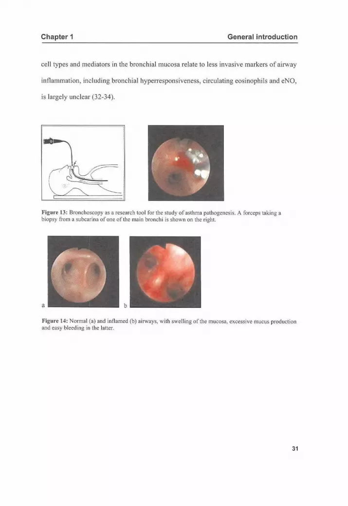
cell types and mediators in the bronchial mucosa relate to less invasive markers of airway
inflammation, including bronchial hypenesponsiveness, circulating eosinophils and eNO,
is largely unclear (32-34).
,...
Ql;. " ··. ·~~ . . ~.~ . ~~- - -
'···. -. .--.
Figure 13: Bronchoscopy as a research tool for the study of asthma pathogenesis. A fo rceps taki ng a biopsy from a subcarina of one of the main bronchi is shown on the right.
a b
Figure 14: Normal (a) and inflamed (b) airways, with swelling of the mucosa, excessive mucus production and easy bleeding in the latter.
31

Chapter 1 General introduction
Analysis of bronchial biopsy specimens
Immunostained sections of bronchial biopsies can be analyzed with computer-assisted
image analysis, a method which provides highly reproducible and reliable parameters of
asthmatic airway inflammation (82). After setting of the reticular basement membrane
and epithelial margins interactively, the program is primed to analyze the epithelium as
well as an area I 00 !!m below the reticular basement membrane. Artefacts are excluded
from analysis. The ratio of positive stained area divided by the total area analyzed is
taken as measure for each immunohistochemical staining.
Figure 15: Image analysis
32

Chapter 1 General introduction
c
Figure 16: Interactively setting of the reticular basement membrane (black, b), 100 ~-tm subepithelial zone (green area, c), and epithelial lining (red, d) in image analysis.
33

Chapter 1 General introduction
1.6 Treatment of asthma
Asthma is a chronic inflammatory disease of the airways involving a characteristic
picture of airway infiltration with lymphocytes, eosinophils, and mast cells, subepithelial
deposition of collagen, and hypertrophy and/or hyperplasia of smooth muscle cells,
goblet cells and submucosal glands. The consequences of this chronic inflammatory
process are still not understood in detail but include variable or persistent symptoms,
bronchial hyperresponsiveness, and attacks of airflow limitation that may require
emergency care or hospitalization and can even lead to death. Pathology studies have
shown that antiinflammatory therapy can reverse or suppress airway inflammation (35,
83-85), whereas prospective controlled clinical trials have demonstrated that it can also
diminish symptoms, reduce bronchial hyperreactivity, and reduce the frequency and
severity of exacerbations (86-88) as well as the rate of decline in lung function (37). It is
also highly likely, although it is not yet proven, that inhaled antiinflammatory therapy
reduces the risk of asthma fatality and prevents, retards or even reduces airway wall
remodeling (47, 89-92). These beneficial effects are easily shown in patients with
moderate to severe asthma. Although benefits are also applicable to patients with mild
asthma, it is less certain that the cost and harm of continuous antiinflammatory therapy
are justified for the mildest forms of the disease (65, 85, 93, 94). For these patients, the
most important issue that remains to be resolved is which factors lead to permanent
airflow obstruction and what the effects of early, sustained treatment are on the
possibility to reach complete sustained remission of asthma (89).
34

Chapter 1 General introduction
Inhaled corticosteroids have now become established as antiinflammatory therapy of
choice in patients with persistent asthma (5). Inhaled corticosteroids largely avoid the
adverse effects associated with oral steroids and are now also recommended in newly
detected disease (94). Several different inhaled corticosteroids are available as
therapeutic options for the treatment of asthma, including include fluticasone propionate,
beclomethasone dipropionate, and budesonide.
Other therapies
Other drugs widely used to treat asthma include beta 2-agonists, theophylline,
cromones, and anticholinergic agents. For acute asthma attacks, the inhaled beta 2-
agonists are the most effective bronchodilators (95). Short- acting forms, such as
salbutamol and terbutaline, give rapid relief; long-acting agents, i.e. salmeterol and
formoterol, provide sustained relief and reduce nocturnal and exercise-induced asthma
(96). Although some authors postulate that an effect on airway inflammation and
remodeling is lacking (97), others have shown that, in addition to their bronchodilator
action, long-acting ~2-agonists may also bring about changes in airway inflammation
and/or remodeling itself, including a decrease in mast cell mediator release (98) and in
airway wall vascularity (47, 99).
Adverse effects are of minor importance when these drugs are used properly (100). The
anticholinergic bronchodilators are more useful for treating COPD than chronic asthma.
These drugs have virtually no side effects, and their onset is slower and their action
35

Chapter 1 General introduction
longer than that of inhaled beta 2-agonists. Finally, the antileukotrienes seem to provide
some bronchodilation and have a minor effect on eosinophilia, with minimal side effects.
Combination therapy
The dose-response curve to inhaled corticosteroids is relatively flat, and there is a strong
scientific rationale for adding long-acting inhaled beta2-agonists, which may be
equivalent or preferable to increasing the dose of inhaled corticosteroids in patients with
moderate-to-severe asthma (88, 101, 102).
Mild asthma
The anticipated natural history of the disease may influence treatment considerations. If
mild asthma is destined to remain mild into old age, treatment is best determined by the
symptoms of asthma at that time. On the other hand, if the chronic eosinophilic
inflammatory process progresses over time to subepithelial fibrosis and irreversible
airflow limitation, the early initiation and prolonged continuation of antiinflammatory
therapy might be beneficial. Unfortunately, the ability to predict the outcome of mild
asthma is limited at present. As yet, no studies have been performed that provide long
term physiologic and histologic follow-up over many years in a large cohort of
individuals with mild asthma. At present, NAEPP guidelines indicate that patients with
mild intermittent asthma are best treated with intermittent use of an inhaled beta-2-
36

Chapter 1 General introduction
selective adrenergic agonist, where antiinflammatory medications are only needed when
asthmatic symptoms are reported more than twice a week (mild persistent asthma).
37

Chapter 1 General introduction
1. 7 Growing out of asthma
Epidemiological work has shown that symptoms of atopic asthma often disappear in
early adolescence (13). This apparent improvement eventually leads to cessation of
treatment and discontinuation of routine check-ups at the out-patient clinic. The
classification "clinical remission" is used to identify these subjects who are believed to
have outgrown their asthma. Unfortunately, 30 to 80% of these subjects in clinical
remission experiences a relapse of symptoms later in life (2). The factors responsible for
this high relapse rate are unknown. Several authors have demonstrated spirometry
abnormalities and/or bronchial hyperresponsiveness in subjects during clinical remission
of asthma ( 103-1 05). It is unclear whether these functional aberrations reflect ongoing
airway inflammation or merely indicate structural changes of the airways as a late
consequence of childhood asthma. Defining asthma as chronic inflammatory disease,
persistent airway inflammation during clinical remission of atopic asthma could possibly
account for the high relapse rate (1 06). On the other hand, from several studies it has
emerged that structural changes, known as airway remodelling, finally lead to thickening
of the airway wall (39, 41). A relationship between airway wall thickening and impaired
airflow has been made plausible (I 07, I 08). How the different aspects of airway wall
thickening contribute to altered airway function is, however, incompletely understood
(23, 43). If airway wall thickening is present in subjects during clinical remission of
asthma, it could at least in part account for the functional abnormalities found during
'remission', but even so for the high relapse rate. In particular ongoing airway
inflammation arises the question whether anti-inflammatory therapy should be continued
38

Chapter 1 General introduction
in subjects with apparently outgrown asthma. Monitoring airway inflammation,
preferably without the use of invasive techniques, will then be needed to show the effect
of treatment.
The discrepancy between the lack of symptoms and ongoing airway inflammation
remains an other problem to be resolved. It is well-known that the correlation between
symptom perception and other indices of asthmatic severity, such as the degree of airflow
obstruction, is poor (1 09, 11 0). Poor perception of airway narrowing may, on its turn,
itse1flead to undertreatment of asthma (111, 112). Thus, the concept ofb1unted
perception of dyspnea in subjects with apparently outgrown asthma is likely of great
clinical relevance.
39

Chapter 1 General introduction
REFERENCES
I. 1987. Standards for the diagnosis and care of patients with chronic obstructive pulmonary disease (COPD) and asthma. This official statement of the American Thoracic Society was adopted by the A TS Board of Directors, November 1986. Am Rev Respir Dis \36(1 ):225-44.
2. Strachan, D. P. 1999. The epidemiology of childhood asthma. Allergy 54(Suppl49):7-11. 3. Manian, P. 1997. Genetics of asthma: a review. Chest I 12(5): 1397-408. 4. Anderson, H. R. 1989. Increase in hospital admissions for childhood asthma: trends in referral,
severity, and readmissions from 1970 to 1985 in a health region of the United Kingdom. Thorax 44(8)o614-9.
5. Barnes, P. J. 1998. Efficacy of inhaled corticosteroids in asthma. J Allergy Clin /mmunol 1 02(4 Pt 1 )o531-8.
6. Turner~ Warwick, M. 199\. Can asthma be cured? Clin Exp Allergy 21(Suppl 1 ): 105-10. 7. Burrows, B. 1995. Allergy and the development of asthma and bronchial hyperresponsiveness.
Clin Exp Allergy 25(Suppl2): 15-6; discussion 17-8. 8. Martinez, F. D. 1997. Complexities of the genetics of asthma. Am J Respir Crit Care Med 156(4
Pt 2)oS 117-22. 9. Handzel, Z. T., W. W. Busse, J. B. Sedgwick, R. Vrtis. W. M. Lee, E. A. Kelly, and J. E. Gern.
1998. Eosinophils bind rhinovirus and activate virus-specific T cells. J /mmunol\60(3): 1279-84. 10. Castro-Rodriguez, J. A., C. J. Holberg, A. L Wright, and F. D. Martinez. 2000. A clinical index to
define risk of asthma in young children with recurrent wheezing. Am J Respir Crit Care Med 162(4 Pt 1)o1403-6.
II. Martinez, F. D. 1999. Recognizing early asthma. Allergy 54(Suppl 49):24-8. 12. Martinez, F. D., and P. J. Helms. 1998. Types of asthma and wheezing. Eur Respir J Suppl27:3s-
8s. 13. Barbee, R. A., and S. Murphy. 1998. The natural history of asthma. J Allergy Clin !mmunol1 02(4
Pt 2):S65-72. 14. Panhuysen, C. I., J. M. Vonk, G. H. Koeter, J.P. Schouten, R. van Altena, E. R. Bleecker, and D.
S. Postma. 1997. Adult patients may outgrow their asthma: a 25-year follow-up study. Am J Re:.pir Crit Care Med 155(4): 1267-72.
15. Kelly, W. J., I. Hudson, P. D. Phelan, M. C. Pain, and A. Olinsky. 1987. Childhood asthma in adult life: a further study at 28 years of age. Br Med J (Clin Res Ed) 294(6579):1059-62.
16. Gerritsen, J., G. H. Koeter, D. S. Postma, J.P. Schouten, W. M. van Aalderen, and K. Knol. 1991. Airway responsiveness in childhood as a predictor of the outcome of asthma in adulthood. Am Rev RespirDis 143(6):1468-9.
17. Grol, M. H., J. Gerritsen, J. M. Vonk, J.P. Schouten, G. H. Keeter, B. Rijcken, and D. S. Postma. 1999. Risk factors for growth and decline of lung function in asthmatic individuals up to age 42 years. A 30-year follow-up study. Am J Respir Crit Care Med 160(6):1830-7.
18. Renmark, E., E. Jonsson, and B. Lundback. !999. Remission of asthma in the middle aged and elderly: report from the Obstructive Lung Disease in Northern Sweden study. Thorax 54(7):611-3.
19. Godden, D. J., S. Ross, M. Abdalla, D. McMurray, A. Douglas, D. Oldman, J. A. Friend, J. S. Legge, and J. G. Douglas. 1994. Outcome of wheeze in childhood. Symptoms and pulmonary function 25 years later. Am J Respir Crit Care Med 149(1 ): I 06-12.
20. Jenkins, M.A., J. L Hopper, G. Bowes, J. B. Carlin, L. B. Flander, and G. G. Giles. 1994. Factors in childhood as predictors of asthma in adult life. Bmj 309(6947):90-3.
21. Martin, A. J., L.A. McLennan, L. I. Landau, and P. D. Phelan. 1980. The natural history of childhood asthma to adult life. Br Med 1280(6229): 1397-1400.
22. UlriL C. S. 1999. Outcome of asthma: longitudinal changes in lung function. Eur Respir J 13(4)o904-18.
23. Fahy, J. V., D. B. Corry, and H. A. Boushey. 2000. Airway inflammation and remodeling in asthma. Curr Opin Pulm Med 6(1 ): 15-20.
40

Chapter 1 General introduction
24. Djukanovic, R. 1996. Bronchoscopy as a research tool for the study of asthma pathogenesis and effects of antiasthma drugs. J Allergy Clin lmmuno/98(5 Pt 2):541-5; discussion S64-6.
25. Laitinen, L. A., A. Laitinen, A. Altraja, I. Virtanen, M. Kampe, B. G. Simonsson, S. E. Karlsson, L Hakansson, P. Venge, and H. Sillastu. 1996. Bronchial biopsy findings in intennittent or "early" asthma. J Allergy Clin lmmunol98(5 Pt 2):S3-6; discussion S33-40.
26. Robinson, D. S., A.M. Bentley, A. Hartnell, A. B. Kay, and S. R. Durham. 1993. Activated memory T helper cells in bronchoalveolar lavage fluid from patients with atopic asthma: relation to asthma symptoms, lung function, and bronchial responsiveness. Thorax 48(1):26-32.
27. Kon, 0. M., and A. B. Kay. 1999. T cells and chronic asthma.lnt Arch Allergy lmmunol 118(2-4)'133-5.
28. Bousquet, J., P. Chanez, J. Y. Lacoste, G. Barneon, N. Ghavanian, I. Enander, P. Venge, S. Ahlstedt, J. Simony-Lafontaine, P. Godard, and eta!. 1990. Eosinophilic inflammation in asthma [see comments]. N Eng! J Med323(15):1033-9.
29. Moqbel, R., J. Barkans, B. L. Bradley, S. R. Durham, and A. B. Kay. 1992. Application of monoclonal antibodies against major basic protein (BMK-13) and eosinophil cationic protein (EG I and EG2) for quantifying eosinophils in bronchial biopsies from atopic asthma. Clin Exp Allagy 22(2)o265-73.
30. Church, M. K., and F. Levi-Schaffer. 1997. The human mast cell.J Allergy Clin lmmunol 99(2)ol55-60.
31. Rossi, G. L, and D. Olivieri. 1997. Does the mast cell still have a key role in asthma? Chest !12(2)o523-9.
32. Crimi, E., A. Spanevello, M. Neri, P. W. Ind, G. A. Rossi, and V. Brusasco. 1998. Dissociation between airway inflammation and airway hyperresponsiveness in allergic asthma. Am J Respir Crit Care Med 157(1):4-9.
33. Moller, G. M., S. E. Overbeek, C. G. van Held en-Meeuwsen, H. C. Hoogsteden, and J. M. Bogaard. 1999. Eosinophils in the bronchial mucosa in relation to methacholine dose- response curves in atopic asthma. J Appl Physio/86(4): 1352-6.
34. Bousquet, J., C. J. Corrigan, and P. Venge. 1998. Peripheral blood markers: evaluation of inflammation in asthma. Eur Respir J Suppl26:42S-48S.
35. Busse, W. W. 1998. Inflammation in asthma: the cornerstone of the disease and target of therapy. J Allergy Clin lmmuno/102(4 Pt 2):S 17-22.
36. Holgate, S. 1993. Mediator and cytokine mechanisms in asthma. Thorax 48(2): 103-9. 37. Carter, P.M., T. L Heinly, S. W. Yates, and P. L Lieberman. 1997. Asthma: the irreversible
airways disease. J lnvestig Al!ergol Clin lmmuno/7(6):566-71. 38. Chetta, A., A. Foresi, M. Del Donno, G. Bertorelli, A. Pesci, and D. Olivieri. 1997. Airways
remodeling is a distinctive feature of asthma and is related to severity of disease. Chest !11(4)o852-7.
39. Gillis, H. L, and K. R. Lutchen. 1999. Ainvay remodeling in asthma amplifies heterogeneities in smooth muscle shortening causing hyperresponsiveness. J Appl Physiol 86(6):2001-12.
40. Vignola, A.M., P. Chanez, G. Bonsignore, P. Godard, and J. Bousquet 2000. Structural consequences of airway inflammation in asthma. J Allergy Clin lmmuno/105(2 Pt 2):514-517.
4 !. Djukanovic, R. 2000. Asthma: A disease of inflammation and repair. J Allergy C/in lmmunol I 05(2 Pt 2):522-526.
42. Boulet, L P., J. Chakir, J. Dube, C. Laprise, M. Boutet, and M. Laviolette. 1998. Airway inflammation and structural changes in airway hyper-responsiveness and asthma: an overview. Can Re.1pir J5(1):16-21.
43. Tiddens, H., M. Silverman, and A. Bush. 2000. The role of inflammation in airway disease: remodeling. Am J Respir Crit Care Med 162(2 Pt 2):S7-Sl0.
44. Sullivan, P., D. Stephens, T. Ansari, J. Costello, and P. Jeffery. 1998. Variation in the measurements of basement membrane thickness and inflammatory cell number in bronchial biopsies. Eur Respir J 12(4):811-5.
45. Jeffery, P. 1998. Structural alterations and inflammation of bronchi in asthma. lnt J Clin Pract Suppl96:5-14.
46. Wilson, J. W., and X. Li. 1997. The measurement of reticular basement membrane and submucosal collagen in the asthmatic airway. Clin Exp Allergy 27(4):363-71.
41

Chapter 1 General introduction
47. Orsida, B. E., C. Ward, X. Li, R. Bish, J. W. Wilson, F. Thien, and E. H. Walters. 2001. Effect of a long-acting beta2-agonist over three months on airway wall vascular remodeling in asthma. Am J Respir Crit Care Med 164(l):l\7-2l.
48. Polosa, R., and S. T. Holgate. 1997. Adenosine bronchoprovocation: a promising marker of allergic inflammation in asthma? Thorax 52(10):919-23.
49. Sont, J. K., L. N. Willems, E. H. Bel, J. H. van Krieken, J.P. Vandenbroucke, and P. J. Sterk. \999. Clinical control and histopathologic outcome of asthma when using ain.vay hyperresponsiveness as an additional guide to long-term treatment. Am J Respir Crit Care Med 159(4 Pt 1):1043-51.
50. Chetta, A., A. Foresi, M. Del Donno, G. F. Consigli, G. Bertorelli, A. Pesci, R. A. Barbee, and D. Olivieri. 1996. Bronchial responsiveness to distilled water and methacholine and its relationship to inflammation and remodeling of the airways in asthma. Am J Re~pir Crit Care Med 153(3):91 0-7.
51. Gaston, B., J. M. Drazen, J. Loscalzo, and J. S. Stamler. 1994. The biology of nitrogen oxides in the airways. Am J Respir Crit Care Med 149(2 Pt 1 ):538-51.
52. Horvath, I., L E. Donnelly, A. Kiss, S. A. Kharitonov, S. Lim, K. Fan Chung, and P. J. Barnes. 1998. Combined use of exhaled hydrogen peroxide and nitric oxide in monitoring asthma. Am J Respir Crit Care Med 158(4):1042-6.
53. Kharitonov, S. A., D. Yates, R. A. Robbins, R. Logan-Sinclair, E. A. Shineboume, and P. J. Barnes. 1994. Increased nitric oxide in exhaled air of asthmatic patients. Lancet 343(8890): 133-5.
54. van Rensen, E. L, K. C. Straathof, M.A. Veselic-Charvat, A. H. Zwinde1man, E. H. Bel, and P. J. Sterk. 1999. Effect of inhaled steroids on airway hyperresponsiveness, sputum eosinophils, and exhaled nitric oxide levels in patients with asthma. Thorax 54(5):403-408.
55. de Gouw, H. W., K. Grunberg, R. Schot, A. C. Kroes, E. C. Dick, and P. J. Sterk. 1998. Relationship between exhaled nitric oxide and aif\\'ay hyperresponsiveness following experimental rhinovirus infection in asthmatic subjects. Eur Respir J 11(1 ): 126-32.
56. Jatakanon, A., S. Lim, S. A. Kharitonov, K. F. Chung, and P. J. Barnes. 1998. Correlation between exhaled nitric oxide, sputum eosinophils, and methacholine responsiveness in patients with mild asthma. Thorax 53(2):91-5.
57. Frank, T. L., A. Adisesh, A. C. Pickering, J. F. Morrison, T. Wright, H. Francis, A. Fletcher, P. I. Frank, and P. Hannaford. 1998. Relationship between exhaled nitric oxide and childhood asthma. Am J Respir Crit Care Med 158(4):1032-6.
58. Mattes, J., K. Storm van's Gravesande, U. Reining, K. Alving, G. Ihorst, M. Henschen, and J. Kuehr. 1999. NO in exhaled air is correlated with markers of eosinophilic airway inflammation in corticosteroid-dependent childhood asthma. Eur Respir J 13(6): 1391-5.
59. Piacentini, G. L., A. Bodini, S. Costella, L. Vicentini, P. Mazzi, S. Sperandio, and A. L. Boner. 1999. Exhaled nitric oxide and sputum eosinophil markers of inflammation in asthmatic children [In Process Citation]. Eur Respir J 13(6):1386-90.
60. Silvestri, M., D. Spallarossa, V. Frangova Youmkova, E. Battistini, B. Fregor.ese, and G. A. Rossi. 1999. Orally exhaled nitric oxide levels are related to the degree of blood eosinophilia in atopic children with mild-intermittent asthma. Eur Respir J 13(2):321-6.
61. Lim, S., A. Jatakanon, S. Meah, T. Oates, K. F. Chung, and P. J. Barnes. 2000. Relationship between exhaled nitric oxide and mucosal eosinophilic inflammation in mild to moderately severe asthma. Thorax 55(3):184-188.
62. Berlyne, G. S., K. Parameswaran, D. Kamada, A. Efthimiadis, and F. E. Hargreave. 2000. A comparison of exhaled nitric oxide and induced sputum as markers of airway inflammation. J Allergy Clin Immuno! 106(4):638-44.
63. Hunt, J. F., K. Fang, R. Malik, A. Snyder, N. Malhotra, T. A. Platts-Mills, and B. Gaston. 2000. Endogenous airway acidification. Implications for asthma pathophysiology [see comments]. Am J Respir Crit Care Med 161(3 Pt 1):694-9.
64. Massaro, A. F., B. Gaston, D. Kita, C. Fanta, J. S. Stamler, and J. M. Drazen. 1995. Expired nitric oxide levels during treatment of acute asthma. Am J Respir Crit Care Med 152(2):800-3.
65. Jatakanon, A., S. Kharitonov, S. Lim, and P. J. Barnes. 1999. Effect of differing doses of inhaled budesonide on markers of ainvay inflammation in patients with mild asthma. Thorax 54(2): I 08-14.
42

Chapter 1 General introduction
66. Lim, S., A. Jatakanon, M. John, T. Gilbey, 0. c. BJ, K. F. Chung, and P. J. Barnes. 1999. Effect of inhaled budesonide on lung function and ainvay inflammation. Assessment by various inflammatory markers in mild asthma. Am J Respir Crit Care Med 159(1):22-30.
67. Silkoff, P. E., P. McClean, M. Spino, L. Erlich, A. S. Slutsky, and N. Zame!. 2001. DoseResponse Relationship and Reproducibility of the Fall in Exhaled Nitric Oxide After Inhaled Beclomethasone Dipropionate Therapy in Asthma Patients. Chest 119(5): 1322-1328.
68. Kharitonov, S., K. Alving, and P. J. Barnes. 1997. Exhaled and nasal nitric oxide measurements: recommendations. The European Respiratory Society Task Force. Eur Respir J I 0(7): 1683-93.
69. Kharitonov, S. A., and P. J. Barnes. 1997. Nasal contribution to exhaled nitric oxide during exhalation against resistance or during breath holding. Thorax 52(6):540-4.
70. Jorres, R. A. 2000. Modelling the production of nitric oxide within the human airways. Eur Respir Jl6(3P55-60.
71. Kroesbergen, A., Q. Jobsis, E. H. Bel, W. C. Hop, and J. C. de Jongste. 1999. Flow-dependency of exhaled nitric oxide in children with asthma and cystic fibrosis. Eur Respir J 14(4):871-5.
72. Silkoff, P. E., P. A. McClean, A. S. Slutsky, H. G. Furlott, E. Hotfstein, S. Wakita, K. R. Chapman, J.P. Szalai, and N. Zamel. 1997. Marked flow-dependence of exhaled nitric oxide using a new technique to exclude nasal nitric oxide. Am J Respir Crit Care Med 155(1 ):260-7.
73. Antczak, A., D. Nowak, B. Shariati, M. Krol, G. Piasecka, and Z. Kurmanowska. 1997. Increased hydrogen peroxide and thiobarbituric acid-reactive products in expired breath condensate of asthmatic patients. Eur Respir J 1 0(6): 1235-41.
74. Dahlman, A. W., H. R. Black, and J. A. Royall. 1993. Expired breath hydrogen peroxide is a marker of acute airway inflammation in pediatric patients with asthma. Am Rev Respir Dis 148(4 Pt 1):955-60.
75. Jobsis, Q .. H. C. Raatgeep, P. W. Hermans, and J. C. de Jongste. 1997. Hydrogen peroxide in exhaled air is increased in stable asthmatic children. Eur Respir J 1 0(3):519-21.
76. Fahy, J. V., H. A. Boushey, S.C. Lazarus, E. A. Mauger, R. M. Chemiack, V. M. Chinchilli, T. J. Craig, J. M. Drazen, J. G. Ford, J. E. Fish, E. Israel, M. Kraft, R. F. Lemanske, R. J. Martin, D. Mclean, S. P. Peters, C. Sorkness, and S. J. Szefler. 2001. Safety and Reproducibility of Sputum Induction in Asthmatic Subjects in a Multicenter Study. Am. J. Respir. Crit. Care iVIed. !63(6): 1470-1475.
77. Grootendorst, D. C., J. K. Sont, L. N. Willems, J. C. Kluin-Nelemans, J. H. Van Krieken, M. Veselic-Charvat, and P. J. Sterk. 1997. Comparison of inflammatory cell counts in asthma: induced sputum vs bronchoalveolar lavage and bronchial biopsies. Clin Exp Allergy· 27(7):769-79.
78. in 't Veen, J. C., H. W. de Gouv.', H. H. Smits, J. K. Sont, P. S. Hiemstra, P. J. Sterk, and E. H. Bel. 1996. Repeatability of cellular and soluble markers of inflammation in induced sputum from patients with asthma [see comments]. Eur Respir J 9(12):2441-7.
79. Fahy, J. V., H. Wong, J. Liu, and H. A. Boushey. 1995. Comparison of samples collected by sputum induction and bronchoscopy from asthmatic and healthy subjects. Am J Respir Crit Care Med !52( I ):53-8.
80. Jarjour, N. N., S. P. Peters, R. Djukanovic, and W. J. Calhoun. 1998.lnvestigative use of bronchoscopy in asthma. Am J Respir Crit Care Med 157(3 Pt 1):692-7.
81. Jeffery, P. K., A. Laitinen, and P. Venge. 2000. Biopsy markers of airway inflammation and remodelling. Respir Med94 Suppl F:S9-15.
82. Faul, J. L.. E. A. Demers, C. M. Burke, and L. W. Poulter. 1999. The reproducibility of repeat measures of airway inflammation in stable atopic asthma. Am J Respir Crit Care Med 160(5 Pt 1):1457-61.
83. Barnes, P. J. 1998. Current issues for establishing inhaled corticosteroids as the antiinflammatory agents of choice in asthma. J Allergy Clin lmmuno/1 01 (4 Pt 2):8427-33.
84. Laitinen, L.A., and A. Laitinen. 1994. Modulation of bronchial inflammation: corticosteroids and other therapeutic agents. Am J Respir Crit Care Med 150(5 Pt 2):S87-90.
85. Jatakanon, A., S. Lim, K. F. Chung, and P. J. Barnes. 1998. An inhaled steroid improves markers of ainvay inflammation in patients with mild asthma. Eur Respir J 12(5): 1084-8.
86. Djukanovic, R., J. W. Wilson, K. M. Britten, S. J. Wilson, A. F. Walls, W. R. Roche, P. H. Howarth, and S. T. Holgate. 1992. Effect of an inhaled corticosteroid on ain.vay inflammation and symptoms in asthma. Am Rev Respir Dis 145(3):669-74.
43

Chapter 1 General introduction
87. van Essen-Zandvliet, E. E., M.D. Hughes, H. J. Waalkens, E. J. Duivennan, S. J. Pocock, and K. F. Kerrebijn. 1992. Effects of22 months of treatment with inhaled corticosteroids and/or beta-2-agonists on lung function, airway responsiveness, and symptoms in children with asthma. The Dutch Chronic Non-specific Lung Disease Study Group. Am Rev Respir Dis 146(3):547-54.
88. Pauwels, R. A., C. G. Lofdahl, D. S. Postma, A. E. Tattersfield, 0. B. P, P. J. Barnes, and A. Ullman. 1997. Effect of inhaled formoterol and budesonide on exacerbations of asthma. Formoterol and Corticosteroids Establishing Therapy (FACET) International Study Group. N Eng! J Med337(20):1405-11.
89. Boushey, H. A. 1998. Effects of inhaled corticosteroids on the consequences o;:~asthma. J Allergy Clin Immuno/102( 4 Pt 2):S5~ \6.
90. Hoshino, M., M. Takahashi, Y. Takai, and J. Sim. 1999. Inhaled corticosteroids decrease subepithelial collagen deposition by modulation of the balance between matrix meta!loproteinase-9 and tissue inhibitor ofmetalloproteinase-1 expression in asthma. J Allergy Clin lmmuno/104(2 Pt 1)356-363.
91. Laitinen, A., A. Altraja, M. Kampe, M. Linden, I. Virtanen, and L.A. Laitinen. 1997. Tenascin is increased in airway basement membrane of asthmatics and decreased by an inhaled steroid. Am J Respir Crit Care Med 156(3 Pt 1):951-8.
92. Vanacker, N.J., E. Palmans, J. C. Kips, and R. A. Pauwels. 2001. Fluticasone inhibits but does not reverse allergen-induced structural airway changes. Am J Respir Crit Care Med 163(3 Pt 1):674-9.
93. van Grunsven, P.M., C. P. van Schayck, J. Molema, R. P. Akkermans, and C. van Wee\. 1999. Effect of inhaled corticosteroids on bronchia! responsiveness in patients with "corticosteroid naive" mild asthma: a meta-analysis. Thorax 54(4):316~22.
94. O'Byme, P.M. 1999. Inhaled corticosteroid therapy in newly detected mild asthma [In Process Citation]. Drugs 58(Suppl 4): 17~24; discussion 52.
95. Barnes, P. J. 1997. Current therapies for asthma. Promise and limitations. Chest 111(2 Suppl):17S-26S.
96. Moore, R. H., A. Khan, and B. F. Dickey. 1998. Long-acting inhaled beta2-agonists in asthma therapy. Chest 113(4):1095-\08.
97. Calhoun, W. J., K. L. Hinton, and J. J. K.ratzenberg. 2001. The effect ofsalmetero\ on markers of ain.vay inflammation following segmental allergen challenge. Am J Respir Crit Care Med 163(4):881-6.
98. Chong, L. K., E. Cooper, C. J. Vardey, and P. T. Peachell. 1998. Salmeterol inhibition of mediator release from human lung mast cells by beta-adrenoceptor-dependent and independent mechanisms. Br J Pharmaco/123(5): 1009-15.
99. McDonald, D. M. 200 I. Angiogenesis and remodeling of airway vasculature in chronic inflammation. Am J Respir Crit Care Med 164(10 Pt 2):S39-45.
100. Guhan, A. R., S. Cooper, J. Oborne, S. Lewis, J. Bennett, and A. E. Tattersfield. 2000. Systemic effects offormoterol and salmeterol: a dose-response comparison in healthy subjects. Thorax 55(8):650-6.
101. Barnes, P. J. 2002. Scientific rationale for inhaled combination therapy with long-acting beta2-agonists and corticosteroids. Eur Respir J 19(1):182-91.
J 02. Chung, K. F. 1998. The complementary role of glucocorticosteroids and long-acting betaadrenergic agonists. Allergy 53(42 Suppl):7-13.
103. Gruber, W,, E. Eber, B. Steinbrugger, M. Modi, E. Weinhandl, and M.S. Zach. 1997. Atopy, lung function and bronchial responsiveness in symptom-free paediatric asthma patients. Eur Respir J 10(5):1041-5.
104. Boulet, L. P., H. Turcotte, and A. Brochu. 1994. Persistence of airway obstruction and hyperresponsiveness in subjects with asthma remission. Chest 1 05(4): 1024-31.
I 05. Kerrebijn, K. F., A. C. Fioole, and R. D. van Bentve\d. 1978. Lung function in asthmatic children after year or more without symptoms or treatment. Br Med J 1(6117):886-8.
106. Warke, T. J., P. S. Fitch, V. Brown, R. Taylor, J.D. Lyons, M. Ennis, and M.D. Shields. 2002. Outgrown asthma does not mean no airways inflammation. Eur Re~pir J 19(2):284-7.
\07. Ward, C., D.P. Johns, R. Bish, M. Pais, D. W. Reid, C. Ingram, B. Feltis, and E. H. Walters. 2001. Reduced Airway Distensibility, Fixed Airflow Limitation, and Airway Wall Remodeling in Asthma. Am J Respir Crit Care Med 164(9):1718-1721.
44

Chapter 1 General introduction
!08. Pare, P. D., C. R. Roberts, T. R. Bai, and B. J. Wiggs. 1997. The functional consequences of airway remodeling in asthma. Monaldi Arch Chest Dis 52(6):589-96.
!09. Teeter, J. G., and E. R. Bleecker. 1998. Relationship between airway obstruction and respiratory symptoms in adult asthmatics. Chest 113(2):272-7.
110. Rietveld, S.l998. Symptom perception in asthma: a multidisciplinary review.} Asthma 35(2)ol37-46.
Ill. Boulet, L. P., P. Leblanc, and H. Turcotte. 1994. Perception scoring of induced bronchoconstriction as an index of awareness of asthma symptoms. Chest 1 05(5): 1430-3.
112. Bijl-Hofland, I. D., S. G. Cloosterman, H. T. Folgering, R. P. Akkermans, and C. P. van Schayck. 1999. Relation of the perception of airway obstruction to the severity of asthma. Thorax 54(1): 15-19.
45

Chapter
Aim of the study
"Everything hinges on the matter of evidence"
Car! Sagan
As stated in the introductory remarks, various questions arise from the epidemiological
fact that a great deal of subjects with apparently outgrown asthma experiences a relapse
of symptoms of asthma later in life. ln the present thesis, the next questions will be
addressed:
Are we able to produce convincing arguments of ongoing airway inflammation
and/or remodeling in subjects who are believed to have outgrown their asthma?
To address this question, we measured spirometry values, bronchial responsiveness to
different inhaled stimuli, and exhaled nitric oxide levels in adolescents in clinical
remission of atopic asthma. Clinical remission was defined as complete absence of
symptoms without the use of any medication in the year preceding the study. Chapter 3
describes the results of this study, wherein subjects in clinical remission were compared
with subjects with symptomatic asthma and healthy controls. In Chapter 4 invasively
obtained "proof' of ongoing airway inflammation and remodeling during clinical
remission of atopic asthma is discussed. By means of flexible bronchoscopy, biopsies
46

Chapter 2 Aim of the study
were obtained from the airway walls in all subjects from the three study groups. A
comparison of biopsy findings, including inflammatory cell type density and various
indices of airway remodeling, was made between the groups. Also, data were compared
with noninvasive markers of airway disease, such as exhaled nitric oxide levels and
bronchial responsiveness to inhaled stimuli.
2 If persistent airway inflammation can be demonstrated during clinical remission,
could blunted symptom perception explain the discrepancy between the lack of
symptoms and ongoing disease?
This question will be dealt with in chapter 5, where a study is described in which we
obtained "BORG" dyspnea scores from subjects in clinical remission and subjects with
symptomatic asthma during MCh and AMP provocation.
3 Would subjects with subclinical airway inflammation and remodelling benefit from
antiinflammatory treatment in the short-term?
In chapter 6, a double blind, longitudinal, placebo controlled study is described in which
subjects in clinical remission of atopic asthma are treated for three months with either the
salmeterol/flixotide propionate combination product (Seretide) or placebo. Again,
invasive- as well as non-invasive indices were obtained from all subjects before and after
treatment.
4 Asthma remission- does it exist?
This question will be dealt with in chapter 7. Whether "true" remission of asthma can be
reached with or without the aid of prolonged anti-inflammatory treatment is as yet
unknown. A review of relevant literature as well as a proposal to deal with subjects with
apparently outgrown asthma is given.
47

Chapter
Adolescents in Clinical Remission of Atopic Asthma have
Elevated Exhaled Nitric Oxide Levels and Bronchial
Hyperresponsiveness
LEON M VAN DEN TOORN u, MD, JAN-BAS PRINS 2, PhD, Msc, SHELLEY E
OVERBEEK 2, PhD, MD, HENK C HOOGSTEDEN 2, PhD, MD, AND JOHAN C DE
JONGSTE 1, PhD, MD
Depts of 1Paediatrics/Paediatric Respiratory Medicine and 2Pulmonary Medicine,
Erasmus MC, Rotterdam, the Netherlands.
Am J Respir Crit Care Med 2000: 62(3 Pt 1):953-7.
48

Chapter 3 Elevated Exhaled NO and BHR during remission
ABSTRACT
Symptoms of atopic asthma often decrease or even seem to disappear around
puberty. The aim of this study was to investigate whether this so-called clinical
remission is accompanied by remission of airway inflammation, since symptoms
relapse in a substantial proportion of subjects later in life.
To assess indicators of inflammation and/or structural damage of the ainvays, exhaled
nitric oxide (eNO) and bronchial responsiveness to adenosine-5'-monophosphate (AMP)
and methacholine (MCh) were determined in 21 subjects in clinical remission of atopic
asthma. Clinical remission was defined as complete absence of symptoms of asthma
without the use of any medication in the year preceding the study. Results were compared
with those of 21 patients with current asthma and 18 healthy control subjects. We found
significantly higher eNO values in the remission group than in healthy controls
(geometric mean 18.9 ppb and 1.0 ppb, respectively; p < 0.001) whereas eNO values of
the remission group and those of the subjects with current asthma (geometric mean 21.9
ppb) were similar (p ~ 0.09). The responsiveness to both AMP and MCh of subjects in
clinical remission was significantly higher as compared with responsiveness of healthy
controls, and lower than responsiveness of subjects with current asthma. A significant
correlation could be established between eNO and responsiveness to AMP, but not
between eNO and responsiveness to MCh. The results of this study are suggestive of
persistent airway inflammation during clinical remission of atopic asthma. We speculate
that subclinical inflammation is a risk factor for asthma relapse later in life, and that eNO
49

Chapter 3 Elevated Exhaled NO and BHR during remission
and responsiveness to both AMP and MCh can be used as different, non-invasive indices
of the inflammatory process of the airways.
50

Chapter 3 Elevated Exhaled NO and BHR during remission
INTRODUCTION
Asthma is a chronic inflammatory disorder of the airways, characterized by cellular
infiltration, cellular activity and cell damage, but also by oedema, vascular leakage, and
hypertrophy/hyperplasia of resident cells, as there are goblet cells and smooth muscles.
Structural changes of the airway walls occur early in the course of the disease (1, 2).
Epidemiological studies have shown that symptoms of atopic asthma often begin in early
childhood and improve or seem to disappear around puberty (3, 4). However, a
considerable proportion of asthmatics in "clinical remission" will have a relapse later in
life (4). Several studies have shown spirometric abnormalities and bronchial
hyperresponsiveness (BHR) to methacholine (MCh) or cold air challenge during clinical
remission of asthma (5, 6). It is unknown whether these functional abnormalities, which
are supposed to be indicative of asthma severity with respect to symptomatic asthma,
reflect persistent airway inflammation or merely indicate residual airway damage. These
considerations seem to be important, as it is reasonable to believe that persistent airway
inflammation during clinical remission of atopic asthma has substantial impact on the risk
of relapse at a later age. The question arises whether other available non-invasive
physiological techniques can give information about the presence of an ongoing
inflammatory process.
Nitric oxide synthase (NOS) is a newly identified enzyme system active in airway
epithelial- and endothelial cells, macrophages, neutrophils, mast cells, autonomic
neurons, smooth muscle cells, and fibroblasts. The existence of an inducible form of NOS
(iN OS) in human lungs suggests that increased production of NO, probably induced by
51

Chapter 3 Elevated Exhaled NO and BHR during remission
cytokines, may be relevant to the pathology of asthma (7). Many studies concerning
atopic asthma demonstrate elevated NO levels in exhaled air (8, 9). Furthermore, exhaled
NO (eNO) levels are decreased by antiinflammatory therapy, which offers opportunities
to monitor compliance with and effectiveness of treatment (10). The measurement of
eNO can be performed repeatedly, even in children and patients with severe airflow
obstruction, in whom invasive techniques are not feasible or desirable. Studies regarding
eNO reported a weak relationship between eNO levels and BHR (II, 12), indicating the
complexity of mechanisms involved in atopic asthma. The usefulness and specificity of
eNO values with respect to the monitoring of airway inflammation in atopic asthma are
still under investigation.
BHR is one of the hallmarks of asthma and is often used as an indicator of asthma
severity. Several studies have shown a significant correlation between BHR and airway
inflammation in symptomatic asthma (13-15). The degree of airway responsiveness can
be assessed with a variety of inhaled stimuli, such as MCh or adenosine-5'
monophosphate (AMP). MCh induces airway constriction via direct stimulation of the
muscarin receptors on airway smooth muscle cells. AMP, on the other hand, causes
airway narrowing mainly through indirect mechanisms, in particular stimulation of mast
cells and activation of neuronal reflexes in the lung (14). Since mast cells are believed to
play a predominant role in atopic asthma, the bronchial response to AMP, in addition to
the response to MCh, may serve as indicator of the inflammatory process.
The aim of this study was to measure eNO values and bronchial responsiveness to
MCh and AMP, each reflecting distinct parts of the inflammatory process in the airways,
in previously well-documented atopic asthmatic adolescents who were in clinical
52

Chapter 3 Elevated Exhaled NO and BHR during remission
remission for more than one year. Data were compared with data on adolescents with
current asthma and healthy control subjects.
53

Chapter 3 Elevated Exhaled NO and BHR during remission
METHODS
Subjects
Adolescents, 18 - 25 years of age, with atopic asthma were selected from the Sophia
Children's Hospital discharged patients files. Clinical remission was assumed if subjects
reported complete absence of cough, wheezing and breathlessness at rest and on exertion
and had not taken any medication in order to control asthmatic symptoms for at least 12
mo before to the study. Twenty-one eligible subjects in remission were included, and
compared with 21 patients with asthma who had persistent symptoms at least once a
month in the year preceding the study, and used inhaled B2-agonists on demand in order
to relieve symptoms. All subjects had a history of wheezing and chest tightness and were
previously diagnosed as having atopic asthma according to American Thoracic Society
(ATS) criteria (16). In the past, all had a provocation dose/concentration ofMCh or
histamine producing a 20 % fall in FEY 1 of S 150 ).lg (dosimeter method) or S 8 mg·mL~ 1
(2-min tidal breathing method), and/or had an FEY1 reversibility 2 12% of predicted
normal value. All had evidence of atopy defined by radio allergosorbent test (RAST)
Class 2 or higher for at least one common airborne allergen. Participating subjects were
lifelong nonsmokers, in stable clinical condition and did not take inhaled steroids,
including nasal steroids, or anti-allergic medication such as cromoglycate and
antihistamines for at least 1 yr before the study.
Eighteen healthy nonsmoking young adult volunteers were recruited via advertisement
and served as control subjects. They had a negative personal and first-degree family
history of asthma and atopy. Common exclusion criteria were an inability to perform lung
54

Chapter 3 Elevated Exhaled NO and BHR during remission
function tests reproducibly, perennial rhinitis, and other illnesses that may affect lung
function. None of the subjects in the study reported symptoms of respiratory infection in
the month prior to the study. The study was approved by the Medical Ethics Committee
of the Erasmus University and University Hospital Rotterdam.
55

Chapter 3 Elevated Exhaled NO and BHR during remission
Study Design
We performed a cross-sectional study with three visits on separate days. At the first
visit, subjects gave infonned written consent and were asked about asthmatic symptoms
and requirement for rescue medication during the past year, especially to avoid the
inclusion of subjects with mild symptoms of asthma in the remission group. For the same
reason, subjects were asked to complete Juniper's Quality of Life Questionnaire (17),
which was verified by the investigator on hidden minor symptoms. Also, physical
examination was performed and eNO and PD2o MCh determined. At Visit 2, scheduled at
least l dafter Visit 1, subjects underwent an AMP challenge test. At Visit 3, scheduled at
least 3 d after Visit 2, FEY 1 and FEY 1 reversibility were tested. The sequence and
intervals were chosen in order to avoid any influence of the AMP challenge on MHh
responsiveness and/or on FEY 1 ( 18). The maximal time allowed between Visits 1 and 3
was 3 wk.
Spirometry
Short acting Bragonists were not allowed within 8 hours before the test. Flow-volume
curves were obtained using a Lilly-type pneumotachograph (Masterlab Jaeger, Wiirzberg,
Germany). The best of three reproducible recordings ofFEV1 was expressed as
percentage of predicted normal and used for analysis. Reversibility was tested by
measuring FEY 1 before and 20 minutes after inhalation of 1 rng of terbutaline powder
(Bricanyl turbuhaler, ASTRA, Lund, Sweden), and expressed as increase in percentage of
predicted normal.
56

Chapter 3 Elevated Exhaled NO and BHR during remission
Nitric Oxide
Based on the recommendations of the European Respiratory Society Task Force (19),
eNO was measured in exhaled air by means of chemiluminescence (model280 nitric
oxide analyzer, Sievers, Boulder, CO) with a sensitivity of 0.1 ppb and a detection range
of< 0.1 - 500,000 ppb. The sampling flow was 0.2 Llmin and the response time 0.2 s.
Data were displayed continuously on a PC screen, and stored in a computer with a sample
frequency of 20 Hz for later analysis. The analyzer was calibrated regularly according to
the manufacturer1s guidelines, employing certified calibration gases containing 0 ppb,
100 ppb and 9 ppm NO (Hoekloos, Barendrecht, the Netherlands). The measurement
circuit consisted of a mouthpiece connected to a two-way nome breathing valve (Hans
Rudolph, Kansas City, MO), through which subjects inhaled ambient air (if ambient NO
was < I 0 ppb) or NO-free medical air (if ambient NO was > 10 ppb) while seated, not
wearing a noseclip. Subjects inhaled to TLC and immediately exhaled for as long as
possible into a tube with an in-line flow resistor (20 em H20 L-1 s-1, Hans Rudolph). This
was done at a flow corresponding to 5 % of subject vital capacity per second, with the aid
of a visual feedback display. A fine-bore Teflon tube continuously sampled exhaled air
from a side port situated directly after the mouthpiece to the analyzer. Water vapor was
absorbed by means of an NO-inert filter in the tube. Airflow was measured with a Lilly
type pneumotachograph (Masterlab Jaeger) positioned downstream of the resistor. An
end-expiratory plateau of at least 10 s, where flow varied± I 0 % of the target flow, was
the end point of the measurement. The test was done in triplicate and average eNO at the
plateau calculated by means of custom-made software.
57

Chapter 3 Elevated Exhaled NO and BHR during remission
Methacholine and Adenosine-5'-MonoPhosphate Challenge
Challenge tests were performed at the same time of day(± 1 h) according to the
dosimeter method validated by Birnie and coworkers (20). Short acting B2-agonists were not
allowed within 8 h before the test. Calibrated DeVilbiss 646 nebulizers (DeVilbiss Health
Care, Somerset, PA) were filled with 3 ml of the appropriate solutions. Subjects inhaled four
5-).ll volumes, using a French-Rosenthal dosimeter (Laboratory for Applied Immunology,
Fairfax, VA). After recording baseline values, the challenge started with inhalation of 0.9 %
NaCl. If a patient responded to saline or the lowest concentration of either MCh or AMP,
they were assigned a PD2o value of half the starting dose. Inhalation provocation tests were
performed using doubling concentrations of 0.15 to 78.4 mg/ml MCh bromide in phosphate
buffered saline (PBS) or 0.08 to 160 mg/ml AMP in normal saline (Sigma, St. Louis, MO).
Provocative doses causing a 20 % fall in FEV 1 (PD2o) from baseline were calculated by
means of linear interpolation of the logarithmic dose-response curve. IfFEV1 fell less than
20 % of the pre-challenge level at the highest dose administered, twice the highest dose was
arbitrarily used as the PD2o value. An MCh PD2o value of 1,000 ].lg, corresponding
approximately with 7.8 J.liDOl cumulative, was considered the cutoff value for bronchial
hyperresponsiveness to MCh.
Peak Expiratory Flow Rate (PEFR)
After being instructed by the investigator at visit 1, the patients recorded their peak
expiratory flow rate (PEFR) with a Personal Best peakflow meter (Respironics, Nantes,
France) twice daily at home during the period benveen Visit 1 and Visit 3. PEFR was
58

Chapter 3 Elevated Exhaled NO and BHR during remission
always recorded before bronchodilatation. Each measurement consisted of three attempts of
which the highest value was used for further analysis.
Statistical Analysis
Because of their highly skewed distributions, PD20 and eNO values were analyzed after
logarithmic transformation. Mean data were expressed as geometric mean e ± SEM (lnX)_
With respect to PD20 values, comparisons between groups were made by the Mann-\Vhitney
test for unpaired samples. Correlation between different tests was made by the Speannan
rank correlation test. The distributions of all other variables were not significantly different
from a standard normal distribution. Hence, parametric techniques (Student t test, Pearson
correlation coefficients) were applied. Mean values of these parameters were expressed as
means± SEM. A two-tailed p value ofless than 0.05 was considered significant. Data were
analyzed with the Statistical Package for the Social Science (SPSS, Chicago, IL) for
Windows version 8.0.
59

Chapter 3 Elevated Exhaled NO and BHR during remission
RESULTS
Sixty subjects completed the study (21 subjects with current asthma, 21 subjects in
clinical remission and 18 healthy controls). The male-to-female ratio was 15 to 6 in the
asthmatic group, 18 to 3 in the remission group, and 10 to 8 in the control group. Age did
not differ significantly between the groups (22 ± 2 yr in the asthmatic group, 21 ± 2 yr in the
remission group and 24 ± 1 yr in the control group). Duration of remission in the remission
group varied from I to 12 yr (median, 5 yr), All subjects of the asthma group and the
remission group, and t:\vo subjects of the control group, had positive RAST tests. Results of
eNO measurement, bronchial provocation tests and spirometry are summarized in Table 1.
TABLE L Exhaled NO and pulmonary function
Asthmatics Clinical Remission Controls
(n~21) (n~21) (n~JS)
eNO (ppb) 22e-"U.V* 14e ± o.l5 * le-" u. I
PD, MCH (~g) 94e=D.37 * 752e ± 031 ** 4,954 · e"' 0
PD, AMP (~g) l,llOe"' OJ/* 5,704e ± o.n *** 10,496e"' 0
FEY 1 (% of predicted) 88 ± 12 * 93 ± 15 t 105 ± 13
Reversibility FEY1 (%) 11 ±I* 7± 1 tt 4±1
Diurnal PEFR variation(%) 13±2 ll± l 8±1
Abbreviations: PD20 MCH: provocative dose of methacholine causing a 20% fall in FEY 1• PD10 AMP: provocative dose of adenosine-5' -monophosphate causing a 20 % fall in FEY 1. Reversibility FEY 1: change in FEY], expressed as increase in% of predicted normal value after administration of I mg terbutaline. eNO, PD2o MCH and PD20 AMP are expressed as geometric mean· e ± SEM(lnXJ.
All other variables are expressed as mean± SEM. * p < 0.001 versus healthy controls.** p < 0.001 versus healthy controls and asthmatics.*** p < 0.01 versus healthy controls and asthmatics. t p = 0.02 versus healthy controls. tt p < 0.02 versus healthy controls and asthmatics
60

Chapter 3 Elevated Exhaled NO and BHR during remission
Exhaled NO Values
Geometric mean eNO values in the remission group were significantly higher than in
healthy controls (mean, 18.9 and l.O ppb, respectively; p < 0.001), and slightly but not
significantly lower than those of asthmatics (2!.9 ppb; p ~ 0.09) (Table l; Figure l ).
Although there seemed to be a trend toward lower eNO values with longer duration of
remission, a significant correlation between these variables could not be established.
Figure 1: Exhaled NO values of currently asthmatic subjects, of subjects in clinical remission of atopic asthma, and of healthy controls. Each dot represents one subject. Horizontal bars represent geometric mean values. TheY-axis shows NO values logarithmically.
61

Chapter 3 Elevated Exhaled NO and BHR during remission
Bronchial Challenge Tests
In the remission group, II of21 subjects had a PD2o MCh ofless than 1,000 )lg,
compared with none ofthe control subjects. Of the subjects with asthma, 19 of21 showed a
PD2o MCh value below this leveL PD2o MCh values in the remission group were
significantly lower than in the control group (geometric mean, 751 and 4,954 ).!g,
respectively; p < 0,001), but higher than in the group of asthmatics (94 )lg; p < 0,001)
(Figure 2),
100000
p < 0.001 p < 0.001
10000 ~ lC ... bJ)
~ :t 1000 I u . '"I"' :; • I • = ! N
~ 100 .... ~ . .
10
•
Asthma Remission Controls (n = 21) (n = 21) (n = 18)
Figure 2: PD20 MCh (micrograms, not cumulative) values of currently asthmatic subjects, of subjects in clinical remission of atopic asthma, and of healthy controls. Non-responders are arbitrarily given a value of twice the highest dose administered. Horizontal bars represent geometric mean values. TheY -axis shows PD20 MCh values logarithmically.
62

Chapter 3 Elevated Exhaled NO and BHR during remission
The AMP challenge showed significantly lower PD2o AMP values in the remission group
than in the control group (geometric mean, 5,704 and 10,496 ~g. respectively; p < 0.01), but
higher values in the remission group than in asthmatics (1,110 ~g, p < 0.01) (Figure 3).
100000
10000
~ .:, ~ :.: 1000 < = c.N ~
100
10
X
+ •
Asthma (n ~ 21)
p < 0.01 p<O.Ol
Ill .... •
Remission (n ~ 21)
ill'
Controls (n ~ 18)
Figure 3: PD20 AMP (micrograms, not cumulative) values of currently asthmatic subjects, of subjects in clinical remission of atopic asthma, and of healthy controls. Non-responders are arbitrarily given a value of twice the highest dose administered. Horizontal bars represent geometric mean values. TheY-axis shows PD20
AMP values logarithmically.
In subjects in remission and those with asthma, PD2o MCh and PD2o Al\1P were highly
correlated (r = 0.72, p < 0.001). There was a significant inverse correlation between PD2o
AMP and eNO (r ~ -0.51; p ~ 0.00 I), while no correlation could be established between
PD2o MCh and eNO (r ~ -0.25; p ~ 0.1 ). There was no significant relationship between
fonnerly and currently assessed bronchial responsiveness, or between duration of remission
and bronchial responsiveness.
63

Chapter 3 Elevated Exhaled NO and BHR during remission
Spirometry and PEFR
Baseline FEV 1 values, expressed as percentage of predicted normal value, were
significantly reduced in subjects in remission compared to healthy control subjects (mean,
93 and 105 %, respectively; p = 0.02). No significant difference could be detected between
subjects in remission and subjects with asthma (mean, 88 %). Reversibility in FEV1 was
significantly different (p < 0.02) between the three groups (mean, 11, 7, and 4% in subjects
with asthma, subjects in remission and healthy control subjects, respectively). Mean diumal
PEFR variation was 13% in subjects with asthma, 11% in subjects in remission, and 8% in
control subjects, and showed no significant difference between the groups.
64
Chapter 3 Elevated Exhaled NO and BHR during remission
DISCUSSION
In this study, we demonstrated elevated levels of eNO and airway
hyperresponsiveness to MCh and AMP in adolescents in long-standing clinical remission
of atopic asthma. During remission, eNO levels were almost similar to those found in
subjects with current asthma. There was a significant correlation between eNO values
and AMP responsiveness, but not between eNO values and MCh responsiveness. A
significant relationship between duration of remission and degree of abnormalities could
not be established.
Studies regarding completely symptom-free subjects who had well-documented atopic
asthma in the past are scarce (6). In our study, subjects were regarded to be in clinical
remission when they reported complete absence of symptoms and were without treatment
for at least one yr before the study. This definition was applied to avoid the inclusion of
asthmatic subjects with mild symptoms in the remission group. The possibility that
subjects with mild symptoms were included in other studies on asthma remission cannot
be ruled out. In one of these studies, asthma was considered inactive when subjects
reported no asthma attacks or use of medication during the preceding year, and were
without frequent episodes of shortness of breath with wheezing (5). Following this
definition of remission, the investigators reported bronchial hyperresponsiveness to MCh
with or without airflow obstruction in subjects aged 18 - 61 years in remission.
Despite the relatively long median duration of remission, we found that subjects in
clinical remission had significantly elevated eNO levels, as compared with eNO levels
from healthy controls. Exhaled NO originates from several inflammatory cell types in the
65

Chapter 3 Elevated Exhaled NO and BHR during remission
airways, including epithelial- and endothelial cells and macrophages. Whether elevated
eNO levels are caused by enhanced activity of inflammatory cells expressing NOS,
potentially driven by inflammatory mediators or cytokines, and/or by enhanced diffusion
through the airway wall due to structural damage remains to be resolved. An increasing
number of papers shows evidence that eNO is related to atopic asthma more than to non
atopic asthma or atopy per se. It was recently demonstrated that atopic asthmatic children
had higher geometric mean eNO levels than non-atopic asthmatic children, atopic non
asthmatic children, or non-atopic non-asthmatic children, suggesting that both atopy and
asthma are important in the context of elevated eNO levels (21). In other studies
performed recently, eNO levels were significantly higher in atopic subjects with asthma
compared with levels from non-atopic asthmatic subjects (22, 23). Similar results were
obtained in patients with rhinitis, whereas no difference was found in eNO levels between
atopic and non-atopic control subjects (22). Horvath and Barnes found elevated eNO
levels in atopic asymptomatic subjects, but suggested that this finding merely reflected an
early stage of airway inflammation, possibly preceding the onset of asthmatic symptoms
(24). In our study, two subjects in the control group had positive RAST test, but had no
elevated eNO values. Thus, with reference to the suggestion of Horvath and Barnes, the
presence of elevated eNO levels in our atopic remission group might well be due to
subclinical airway inflammation. This is also supported by other studies in which a
positive correlation was reported between eNO levels and markers of eosinophilic airway
inflammation in induced sputum (8, 25). Others demonstrated an association between
eNO and exposure to relevant allergens (26), or a reduction in eNO following
antiinflammatory treatment (27). These study results provide sufficient indications that
66

Chapter 3 Elevated Exhaled NO and BHR during remission
airway inflammation is significantly associated with elevated eNO levels, thus providing
a tool in monitoring disease activity. However, a range of normality needs to be
established.
The eNO levels measured in this study tend to be somewhat lower than those
measured in other studies (8, 28). This may be caused by technical factors. It has been
shown that there is a marked flow-dependence of eNO values with lower values
measured at high flow-rates and vice versa (29). We standardized the expiratory flow as
5% of vital capacity per second, which resulted in an expiratory flow of approximately
0.25 to 0.30 ml/min, which is higher than the flow used in some other studies. It is not
clear whether such standardization of flow for lung volume is important when measuring
eNO. Recently it was demonstrated that the flow-eNO relationship differed between
asthmatic subjects and healthy individuals (30). This difference is, however, of minor
importance compared to the differences we now report between the various groups.
We believe that responsiveness to AMP may serve as indicator of airway
inflammation, although normal values for bronchial AMP responses have not been
determined yet. It was demonstrated a few years ago that experimental instillation of
AMP into an airway segment caused a prompt reduction in airway caliber, paralleled by a
significant rise in prostaglandin D2, histamine, and tryptase levels in the lavage fluid,
suggestive of mast cell degranulation (31 ). Since mast cells are believed to play a
predominant role in asthmatic airway inflammation, the response to AMP may reflect
different aspects of inflammation as compared with the bronchial response to direct
stimuli, such as methacholine or histamine. Results supporting this statement include a
more pronounced improvement of AMP responsiveness following avoidance of allergen
67

Chapter 3 Elevated Exhaled NO and BHR during remission
(32), and following antiinflammatory therapy (33), as compared with effect on MCh- or
histamine responsiveness. In recent years, it has been suggested that an exaggerated
ainvay response to direct stimuli, like MCh, and abnormal FEVl values, may exist or
persist independently of"active" airway inflammation (34). Probably, these indices are
also influenced by irreversible airway damage, caused by airway remodeling (35). Thus,
being associated with the more slowly responding elements of inflammation within the
airways. Our finding of a positive correlation between response to AMP and eNO levels,
but not between responsiveness to MCh and eNO levels, is in agreement with this.
The clinical relevance of our findings is as yet unknown. Although one would expect
the degree of abnonnalities to be related to the duration of remission, a significant
correlation could not be established. Probably, when subclinical airway inflammation is
present during adolescence, it may persist for several years with a continuous risk to
become clinically manifest again. Future longitudinal studies should assess the possible
benefits of prolonged disease monitoring and antiinflammatory treatment during the
asymptomatic phase. Also, the question arises whether or not indices associated with
different parts of the inflammatory process, such as eNO and responsiveness to AMP and
MCh, need to be included in the definition of asthma remission.
68

Chapter 3 Elevated Exhaled NO and BHR during remission
ACKNOWLEDGEMENTS:
We wish to thank Mrs. Simone Suelmann-Beckers, Marieke Taapken-Sipman, Edith
van Duyn-Van de Water and Aafke Lok-'t Lam ofthe Pulmonary function lab of the
Sophia Children's Hospital for their skilful assistance in performing lung function tests.
69
Chapter 3 Elevated Exhaled NO and BHR during remission
REFERENCES:
L Busse, W. W. 1998. Inflammation in asthma: the cornerstone of the disease and target of therapy. J Allergy Clin Immuno! 102(4 Pt 2):S 17-22.
2. Chemiaev, A., L Balabolkin, 0. Kozlik and I. Volkov. 1998. Bronchus pathomorphology in atopic bronchial asthma in children. Eur Resp J (Abstract 1031 ERS 1998) \2(suppl 28):~43s.
3. Barbee, R. A., and S. Murphy. 1998. The natural history of asthma. J Allergy Clin lnununo/102(4 Pt 2):865-72.
4. Panhuysen, C. I., J. M. Vonk, G. H. Keeter, J.P. Schouten, R. van Altena, E. R. Bleecker, and D. S. Postma. 1997. Adult patients may outgrow their asthma: a 25-year follow-up study. Am J Respir Crit Care Med 155(4):1267-72.
5. Gruber, \V .. E. Eber, B. Steinbmgger, M. Modl, E. Weinhandl, and M.S. Zach. 1997. Atopy, lung function and bronchial responsiveness in symptom-free paediatric asthma patients. Eur Respii· J 10(5):1041-5.
6. Kerrebijn, K. F., A. C. Fioole, and R. D. van Bentveld. 1978. Lung function in asthmatic children after year or more without symptoms or treatment. Br Med J 1(6117):886-8.
7. Gaston, B., J. M. Drazen, J. Loscalzo, and J. S. Stamler. 1994. The biology of nitrogen oxides in the ainvays. Am J Respir Crit Care Med 149(2 Pt 1):538-51.
8. Horvath, L L. E. Donnelly, A. Kiss, S. A. Kharitonov, S. Lim, K. Fan Chung, and P. J. Barnes. 1998. Combined use of exhaled hydrogen peroxide and nitric oxide in monitoring asthma. Am J Respir Crit Care Med 158(4):1042-6.
9. Kharitonov, S. A., D. Yates, R. A. Robbins, R. Logan-Sinclair, E. A. Shinebourne, and P. J. Barnes. 1994. Increased nitric oxide in exhaled air of asthmatic patients. Lancet 343(8890):133-5.
10. van Rensen, E. L., K. C. Straathof, M.A. Veselic-Charvat, A. H. Zwindennan, E. H. Bel, and P. J. Sterk. 1999. Effect of inhaled steroids on airway hYPerresponsiveness, sputum eosinophils, and exhaled nitric oxide levels in patients with asthma. Thora.\ 54(5):403-408.
II. de Gouw, H. W .. K. Grunberg. R. Schot, A. C. Kroes, E. C. Dick, and P. J. Sterk. ~998. Relationship betv..·een exhaled nitric oxide and airway hyperresponsiveness following experimental rhinovirus infection in asthmatic subjects. Eur Respir J II (I): 126-32.
12. Jatakanon, A., S. Lim, S. A. Kharitonov, K. F. Chung, and P. J. Bames. 1998. Correlation between exhaled nitric oxide, sputum eosinophils, and methacholine responsiveness in patients with mild asthma. Thora.\ 53(2):91-5.
13. Moller, G. M., S. E. Overbeek, C. G. van Helden-Meeuwsen, H. C. Hoogsteden, and J. M. Bogaard. 1999. Eosinophils in the bronchial mucosa in relation to methacholine dose- response curves in atopic asthma. J Appl Physio/86(4): 1352-6.
14. Polosa. R., and S. T. Holgate. 1997. Adenosine bronchoprovocation: a promising marker of allergic inflammation in asthma? Thorax 52(1 0):919-23.
15. Sont, J. K., LN. Willems, E. H. BeL J. H. van Krieken, J.P. Vandenbroucke, and P. J. Sterk. 1999. Clinical control and histopathologic outcome of asthma when using airway hyperresponsiveness as an additional guide to long-tenn treatment. Am J Respir Crit Care Med 159(4 Pt 1): 1043-51.
16. 1987. Standards fOr the diagnosis and care of patients with chronic obstructive pulmonary disease (COPD) and asthma. Am Rev Respir Dis 136( 1):225-44.
17. Juniper, E. F., G. H. Guyatt, R. S. Epstein, P. J. Ferrie, R. Jaeschke, and T. K. Hiller. 1992. Evaluation ofimpainnent of health related quality of life in asthma: development of a questionnaire for use in clinical trials. Thorax 47(2):76-83.
18. Rosati, G., F. E. Hargreave, and E. H. Ramsdale. 1989. Inhalation of adenosine 5'-monophosphate increases methacholine ait'\Vay responsiveness. J Appl Physio/67(2):792-6.
19. Kharitonov, S., K. Alving, and P. J. Barnes. 1997. Exhaled and nasal nitric oxide measurements: recommendations. The European Respiratory Society Task Force. Eur Respir J 10(7):1683-93.
20. Birnie, D., G. W. thoe Schwartzenberg, W. C. Hop, E. E. van Essen-Zandvliet, and K. F. Kerrebijn. 1990. Does the outcome ofthe tidal breathing and dosimeter methods of assessing bronchial responsiveness in children with asthma depend on age? Thorax 45(3):199-202.
70

Chapter 3 Elevated Exhaled NO and BHR during remission
21. Frank, T. L., A. Adisesh, A. C. Pickering, J. F. Morrison, T. Wright, H. Francis, A Fletcher, P. I. Frank, and P. Hannaford. 1998. Relationship between exhaled nitric oxide and childhood asthma. Am J Respir Crit Care Med 158(4):1 032-6.
22. Gratziou, C., M. Lignos, M. Dassiou, and C. Roussos. 1999. Influence of atopy on exhaled nitric oxide in patients with stable asthma and rhinitis. Eur Respir J 14(4):897-901.
23. Ludviksdottir, 0., C. Janson, M. Hagman, H. Hedenstrom, E. Bjomsson, and G. Boman. 1999. Exhaled nitric oxide and its relationship to airway responsiveness and atopy in asthma. BHR-Study Group. Re.spir Med93(8):552-6.
24. Horvath, I., and P. J. Barnes. 1999. Exhaled monoxides in asymptomatic atopic subjects. Clin Exp Allecgy 29(9): 1276-1280.
25. Mattes, J., K. Stann van's Gravesande, U. Reining, K. Alving, G. lhorst, M. Henschen, and J. Kuehr. 1999. NO in exhaled air is correlated with markers of eosinophilic airway inflammation in corticosteroid-dependent childhood asthma. Eur Respir J 13( 6): 1391-5.
26. Baraldi, E., S. Carra, C. Dario, N. Azzolin, R. Ongaro, G. Marcer, and F. Zacchello. 1999. Effect of natural grass pollen exposure on exhaled nitric oxide in asthmatic children. Am J Respir Crit Care Med 159(1 ):262-6.
27. Jatakanon, A., S. Kharitonov, S. Lim, and P.]. Barnes. 1999. Effect of differing doses of inhaled budesonide on markers of ain.vay inflammation in patients with mild asthma. Thorax 54(2): I 08-14.
28. Jatakanon, A., S. Lim, K. F. Chung, and P. J. Barnes. 1998. An inhaled steroid improves markers of airway inflammation in patients with mild asthma. Eur Respir J 12(5): 1084-8.
29. Silkoff, P. E., P. A. McClean, A. S. Slutsky, H. G. Furlott, E. Hoffstein, S. Wakita, K. R. Chapman, J. P. Szalai, and N. Zamel. 1997. Marked flow-dependence of exhaled nitric oxide using a new technique to exclude nasal nitric oxide. Am J Respir Crit Care Med 155(1):260-7.
30. Kroes bergen, A., Q. Jobsis, E. H. Bel, W. C. Hop, and J. C. de Jongste. 1999. Flow-dependency of exhaled nitric oxide in children with asthma and cystic. Eur Respir J 14(4):871-5.
31. Polosa, R., W. H. Ng, N. Crimi, C. Vancheri, S. T. Holgate, M. K. Church, and A. Mistretta. 1995. Release of mast-cell-derived mediators after endobronchial adenosine challenge in asthma. Am J Respir Crit Care Med 151(3 Pt 1):624-9.
32. Benckhuijsen, J.,]. W. van den Bos, E. van Velzen, R. de Bruijn, and R. Aalbers. 1996. Differences in the effect of allergen avoidance on bronchial hyperresponsiveness as measured by methacholine, adenosine 5'-monophosphate, and exercise in asthmatic children. Pediatr Pulmono/22(3): 147-53.
33. Taylor, D. A., M. W. Jensen, V. Kanabar, R. Engelstatter, V. W. Steinijans, P. J. Barnes, and B. J. O'Connor. 1999. A dose-dependent effect of the novel inhaled corticosteroid ciclesonide on ainvay responsiveness to adenosine-5'-monophosphate in asthmatic patients. Am J Respir Crit Care Med 160(1):23743.
34. Crimi, E., A. Spanevello, M. Neri, P. W. Ind, G. A. Rossi, and V. Brusasco. 1998. Dissociation between airway inflammation and airway hyperresponsiveness in allergic asthma. Am J Respir Crit Care Med 157(1):4-9.
35. Laprise, C., M. Laviolette, M. Boutet, and L. P. Boulet. 1999. Asymptomatic airway hyperresponsiveness: relationships with airway inflammation and remodelling. Eur Respir J 14( I ):63-73.
71

Chapter
Airways Inflammation is Present during Clinical Remission of
Atopic Asthma
LEON M VAN DEN TOORN u, SHELLEY E OVERBEEK 1,
JOHAN C DE JONGSTE 2, KAROLINA LEMAN 1 ,HENK C HOOGSTEDEN 1, AND
JAN-BAS PRINS 1
Dept of 1 Pulmonary Medicine and 2Paediatrics/Paediatric Respiratory Medicine,
Erasmus MC, Rotterdam, The Netherlands.
Am J Respir Crit Care Med 2001: 164(1 1):2107-13.
72

Chapter 4 Persistent airways inflammation during remission
ABSTRACT
Symptoms of atopic asthma often disappear around puberty. However, asthmatic
subjects in clinical remission will frequently have a relapse later in life. The aim of this
study was to investigate whether subjects in clinical remission of atopic asthma have
persistent airways inflammation and/or airways remodeling. Bronchial biopsies were
obtained from subjects in clinical remission, asthmatic subjects, and healthy controls. The
presence and/or activation state of eosinophils, mast cells, rnacrophages, T lymphocytes,
interleukin (IL)-5, eotaxin and inducible nitric oxide synthase (iN OS) were analyzed.
Results were compared with less invasive indicators of airways inflammation. Also
aspects of airways remodeling were determined.
Eosinophils, T cells, mast cells, and IL-5 were significantly elevated in the airways
mucosa of subjects in remission compared to controls. Also, blood eosinophil cell counts
were significantly higher in subjects in clinical remission. Blood eosinophil cell counts,
exhaled nitric oxide ( eNO) levels, and bronchial response to adenosine-5 '
monophosphate (AMP) correlated significantly with the quantity of tissue eosinophils.
Significant ainvays' remodeling was found in subjects in clinical remission.
In conclusion, our study shows ongoing airways inflammation and airways remodeling
in adolescents in clinical remission of atopic asthma. Subclinical airways inflammation
may well determine the risk of an asthma relapse later in life.
73

Chapter 4 Persistent airways inflammation during remission
INTRODUCTION
Symptoms of atopic asthma often begin in early childhood and mostly improve or
even seem to disappear around puberty (1, 2). However, a considerable proportion of
asthmatic subjects in clinical remission will have a relapse later in life (2). Several studies
have shown spirometric abnormalities and bronchial hyperresponsiveness (BHR) to
methacholine (MCh) or cold air challenge during clinical remission of asthma (3, 4). It is
unlmown whether these functional abnormalities, which are supposed to be indicative of
asthma severity with respect to symptomatic asthma, reflect persistent activity of the
airways inflammatory process or merely indicate structural changes of the airways as a
consequence of childhood asthma. These structural changes, known as airways
remodeling, are probably early events in the course of the disease which appear to
progress. The process of remodeling leads to thickening of the airway wall (5-7). The
exact physiologic consequences of airway wall thickening are, however, incompletely
understood (8). If airway wall thickening is present in subjects in clinical remission of
asthma, it could at least in part account for the functional abnormalities including
bronchial hyperresponsiveness found during 'remission'. On its turn, ongoing 'active'
airways inflammation could have substantial impact on the risk of a relapse later in life.
As a consequence, subjects with subclinical airways inflammation could benefit from
anti-inflammatory treatment (8-10). Recently we demonstrated elevated exhaled nitric
oxide (eNO) levels and BHR to both MCh and adenosine-5'-monophosphate (AMP)
during clinical remission of atopic asthma (11). With respect to eNO, increased
expression of the inducible form of nitric oxide synthase (iNOS) is thought to underlie
74

Chapter 4 Persistent airways inflammation during remission
these elevated eNO levels (12, 13). Studies regarding the precise relationship between
iN OS expression and eNO are, however, scarce (12).
Proof of persistent airways inflammation and remodeling during clinical remission
could be furnished from the analysis of bronchial biopsy specimens. Evidence has been
accumulated to suggest that allergen-reactive type 2 T helper (Th2) cells, with
eosinophils and mast cells as main effector cells, orchestrate asthmatic airways
inflammation ( 14, 15). The presence of eosinophils in the airway wall is apparently the
most reliable feature of bronchial asthma that seems to be related to asthma severity ( 16).
Along with interleukin (IL)-5, a Th2 cytokine, and eotaxin, a chemoattractant for
eosinophils, eosinophil-derived products such as major basic protein (MBP) have been
investigated as markers of eosinophil participation in the pathogenesis of asthma (17).
Also, elevated numbers of bronchial mast cells have been found in the airway mucosa of
atopic and non-atopic asthmatic subjects (18, 19). Release of mediators from mast cells,
such as granule-associated tryptase and chymase, leads to immediate bronchoconstriction
and enhances airways inflammation ( 19).
In healthy ainvays, the inflammatory response to e.g. inhaled allergens is suppressed by
alveolar macrophages. In asthma, however, a deviant function of alveolar macro phages
may contribute to the allergic immune response in the ainvays (20).
How numbers and activation state of these different cell types in the bronchial mucosa
relate to less invasive markers of ainvays inflammation, including bronchial
hyperresponsiveness (BHR), circulating eosinophils and eNO, is largely unclear (21-23).
75

Chapter4 Persistent airways inflammation during remission
Remodeling of the ainvays mucosa and submucosa can be assessed by measurement of
the reticular basement membrane (RBM) thickness (24) and extent of epithelial shedding
(25). Also, collagen deposition (26) is a quantifiable aspect of airways remodeling.
To establish whether subjects in clinical remission of atopic asthma suffer from ongoing
active ainvays inflammation and/or airways remodeling, we compared bronchial biopsy
specimens of subjects with a long-standing clinical remission of atopic asthma with those
of currently asthmatic subjects, and of healthy controls. Biopsy findings were compared
to noninvasive markers of ainvays disease.
76

Chapter 4 Persistent airways inflammation during remission
SUBJECTS, MATERIALS AND METHODS
Subjects
Adolescents with atopic asthma, 18- 25 years of age, were selected from the Sophia
Children's Hospital discharged patients files. Clinical remission of atopic asthma was
defined as reported complete absence of asthmatic symptoms in subjects not taking any
asthma medication for at least 12 months prior to the study. Eligible subjects were
compared with patients with asthma who had persistent symptoms and used inhaled 132-
agonists on demand in order to relieve symptoms. All subjects were previously diagnosed
as having atopic asthma according to ATS criteria (27). All subjects were lifelong non
smokers in stable clinical condition and did not take inhaled steroids or anti-allergic
medication. Healthy non-smoking adult volunteers without a history of asthma served as
controls.
The study was approved by the Medical Ethics Committee of the Erasmus Medical
Center Rotterdam.
Study design
We performed a cross-sectional study with three visits on separate days. Spirometry
values, eNO, MCh- and AMP responsiveness were obtained according to methods we
described earlier ( 11 ).
77
Chapter 4 Persistent airways inflammation during remission
Blood eosinophils
Venous blood eosinophil numbers were counted by means ofhemocytometry.
Reference values ranged from 0.04 to 0.1 x 10⁹ cells/liter.
Bronchoscopy
An experienced bronchoscopist (SEO) using an Olympus model BF IT 10 (Tokyo,
Japan) performed flexible bronchoscopy. At least five bronchial biopsies were obtained
from segmental divisions of the main bronchi.
Processing of bronchial biopsies
Bronchial biopsies were embedded in Tissue-Tek II OCT medium (Miles, Naperville,
Illinois), snap frozen in liquid nitrogen, and stored at -80°C. Serial tissue sections (6 J..Lm)
were cut on a HM-560 cryostat (Microm, Heidelberg, Germany). At least two sections
120 !J.rn apart from one biopsy specimen were placed on a poly-L-lysine-coated
microscopic slide (Sigma Diagnostics, St.Louis, MO). lmmunostaining was carried out
with a-CD4 (T helper cells), a-CDS (cytotoxic T cells), a-CD25 (activated cells) and a
CD69 (activated cells), a-CD68 (macrophages), a-MBP (Dako, Glostrup, Denmark), a
IL-5, a:-eotaxin, a-tryptase, a-chymase (Chemicon Bnmschwig Chemie, Amsterdam, the
Netherlands), a,-iNOS (Santa Cruz Biotechnology, Santa Cruz, CA), and a-collagen Ill.
Binding of the antibodies was detected by the immune-alkaline phosphatase anti
alkaline phosphatase (APAAP) method. Immunostained sections were analyzed with an
image analysis system (Quantimed, Leica, Rijswijk, the Netherlands). With respect to the
sub-epithelium, the program was set to analyze 100 ).lm below the reticular basement
78

Chapter 4 Persistent airways inflammation during remission
membrane (RBM). The ratio of positive stained area divided by the total area analyzed
was taken as measure for each immunohistochemical staining. RBM thickness was
interactively measured at 20 ~m intervals over a 1 mm RBM length (24). The occupancy
ofRBM with epithelium was plotted as ratio of occupied membrane length divided by the
total membrane length.
Statistical analysis
Because of the skewed distribution of data, results are expressed as median value± SEM.
Comparisons between groups were made by the Mann-\Vhitney test for unpaired samples.
Correlation between different indices was made by Spearman's rank correlation test. A two
tailed p-value of equal to or less than 0.05 was considered significant. Data were analyzed
using Statistical Package for the Social Science (SPSS, Chicago, USA).
79

Chapter 4 Persistent airways inflammation during remission
RESULTS
Fifty-four subjects completed the study (19 subjects with atopic asthma, 18 subjects in
clinical remission of atopic asthma, and 17 healthy controls). Subjects characteristics are
summarized in table 1. The median duration of clinical remission was 5 years.
TABLE l: Sub"ects' characteristics
Asthmatic subjects Remission subjects Healthy controls
N 19 18 17
Age (years) 22± 2 21 ::::2 24:::: I
Sex (male: female) 13:6 15:3 9:8
FEY 1 (% of predicted) 88 ± 12 93 ± 15 105 ± 13
FEY 1 reversibility 11 ± 1 7=1 4±1
Variables arc expressed as mean::::: SEM. Reversibility FEV 1: change in FEY~, expressed as increase in% of predicted normal value after administration of 1 mg terbutaline
Immunohistochemistry results are summarized in table 2.
80

Chapter 4 Persistent airways inflammation during remission
TABLE 2: Immunohistochemistry results
Asthmatic subjects Remission subjects Healthy controls
Epithelium
MBP 0.023 ± 0.0093' 0.0082 ± 0.0046* 0.0007±0.0013
Tryptase 0.0068 ± 0.0020t 0.0119±0.0029* 0.0025 ± 0.0009
Chymase 0.0100 ± 0.0035 0.013 ± 0.0030t 0.0067 ± 0.0016
CD4 0.001 ± 0.0004 0.0007 ± 0.0002 0.0009 ± 0.0013
CDS 0.025 ± 0.0045 0.018 ± 0.0068 0.0281 ± 0.0082
CD25 0.0093 ± 0.0050 0.0123 ± 0.0032 0.0058 ± 0.0039
CD69 0.0095±0.0116 0.0076 ± 0.0182 0.0123 ± 0.0093
CD68 0.029 ± 0.0054 0.038 ± 0.0067 0.031 ± 0.019
1L-5 0.038 ± 0.0097 0.036±0.018 0.017±0.014
Eo tax in 0.001 ± 0.0013 0.0002 ± 0.0045 0.0002 ± 0.0005
rNOS 0.12±0.02tt 0.15 ± 0.03 0.21 ± 0.03
Collagen III 0.078 ± 0.011 0.077 ± 0.024 0.062 ± 0.009
Sub-epithelium
MBP 0.12 ± 0.026* 0.07 ± 0.024* 0.0042 ± 0.0086
Tryptase 0.037 ± 0.0082tt 0.075 ± 0.012** 0.022±0.010
Chymase 0.014 ± 0.0038 0.018 ± 0.0056t 0.0069 ± 0.0033
CD4 0.0043 ± 0.0040 0.0025±0.0019 0.0041 ± 0.0027
CDS 0.022 ± 0.0040 0.014±0.0033 0.019±0.004
CD25 0.0094 ± 0.0126 0.0356 ± 0.0233t 0.0095 ± 0.0041
CD69 0.0062 ± 0.0198 0.0156 ± 0.0273 0.0085 ± 0.0063
CD68 0.058 ± 0.0069 0.069±0.013 0.079 ± 0.032
1L-5 0.025 ± 0.0061 0.022 ± 0.0074** 0.0066 ± 0.0081
Eo tax in 0.0011 ± 0.0006t 0.0001 ± 0.0087 0.0001 ± 0.0003
!NOS 0.041 ± 0.006 0.042 ± 0.0141 0.065 ± 0.016
Collagen III 0.16±0.014 0.19±0.016 0.17±0.019
Variables are expressed as median density (ratio of positively stained area and total area)± SEM. * p::::: 0.001 versus healthy controls, ** p::::: 0.01 versus healthy controls, t p::::: 0.05 versus healthy controls, tt p::::: 0.05 versus remission subjects.
In both epithelium and sub-epithelium, MBP density was significantly higher in subjects
in remission than in healthy controls (p<O.OOl and p=O.OOl, respectively; figure 1 and 2).
81

Chapter4 Persistent airways inflammation during remission
10
p < 0.001
8 p < 0.001
= == ~ ;§ "' 0,1 ~
0 • • ~ "t ~ • 0,01 ..... ~ Q. I
"' i :;: 0,001 I -
0,0001
Asthma Remission Controls (n = 19) (n =18) (n= 17)
10
s p < 0.001
= ·~ p = 0.001 -" -= • ;<::: • • ... " + ' 0,1 • I ..Q -= • • ~ ... • •
.-:: I • ~ 0,01 • • = I ... " "" I ~ =:i 0,001 • ~ • •
• 0,0001
Asthma Remission Controls (n ~ 19) (n ~ 18) (n ~ 17)
Figure 1: ratio of area of Major Basic Protein (MBP) staining and total area of epithelium (upper figure) and of sub-epithelium (lower figure) in bronchial biopsy specimens from currently asthmatic subjects, subjects in clinical remission of atopic asthma, and healthy controls. Each dot represents one subject. Horizontal bars represent median values. Y -axis shows values logarithmically.
82

Chapter 4 Persistent airways inflammation during remission
Figure 2: Bronchial biopsy specimen from a healthy control subject (a) and a subject in clinical remission of atopic asthma (b), immunostained with a-Major Basic Protein (MBP). Notice the increased MBPpositive area (the red stain) and epithelial shedding in the subject in clinical remission.
In bronchial epithelium of subjects in remission, there was more tryptase present than
in that of healthy controls {p=0.001). In sub-epithelium, however, there was more
tryptase detectable in subjects in remission than in control subjects and in patients with
asthma (p~O.OOS and p~0.025, respectively; figure 3 and 4).
83

Chapter 4 Persistent airways inflammation during remission
p "'0.022
8 p "'0.001 ~
~ 0,1 ,c ~ ·o. " 0
.£ I I I .....
:5 0,01 u "':'" ~ . ~ . . ~
" : ... ~ ~ ~ 0,001 c. t' : ,...
0,0001 Asthma Remission Controls
(n= 19) (n" 18) (n" 17)
E p = 0.025 p"" 0.008
·= 0 ;§ 0. ~
0,1 ..I.
,Q
.!. a c ..... -~ 0 u
"' 0,01 ~ .s 0.
" ~ '"'
0,001 Asthma Remission Controls (n = 19) (n"' 18) (n = 17)
Figure 3: ratio of area oftryptase staining and total area of epithelium (upper figure) and sub-epithelium (lower figure) in bronchial biopsy specimens ffom currently asthmatic subjects, subjects in clinical remission of atopic asthma, and healthy controls. Each dot represents one subject. Horizontal bars represent median values. Y-axis shows values logarithmically.
84

Chapter 4 Persistent airways inflammation during remission
aL_ ____________________________ ~
Figu re 4: Bronchial biopsy specimen (a-tryptase) from a healthy control subject (a) and a subject in clinical remission of atopic asthma (b). Notice the increased tryptase-positive area (the red stain) and epithelial shedding in the subject in clinical remission.
Chymase density was higher in bronchial epithelium and sub-epithelium of subjects in
remission than in that of healthy controls (p=0.044 and p=0.05, respectively).
There was no difference detectable in the presence of CD4+ and CDS+ cells in both
epithelium and sub-epithelium among the experimental groups. However, there were
more activated, CD25+ cells present in the sub-epithelium of subjects in remission
compared to control subjects (p=0.04). No such difference was detected for CD69.
CD68+ cells were equally represented in sub-epithelium and epithelium in all three
experimental groups.
In sub-epithelium, lL-5 density was significantly higher in subjects in remission than in
healthy controls (p=0.006). Eotaxin density was significantly higher in patients with
asthma than in healthy controls (p=0.05), while intennediate levels were detected in
subjects in remission. In the epithelium, densities of fL-5 and eotaxin were similar in all
three groups. Density ofiNOS was s ignificantly higher in sub-epithelium of healthy
85

Chapter4 Persistent airways inflammation during remission
subjects than in that of subjects in remission (p~0.05). In the epithelium, iN OS density
was higher in healthy subjects than in asthmatic subjects (p~0.05). No difference in iNOS
density could be detected between healthy control subjects and subjects in clinical
remission.
With respect to indices of remodeling, RBM thickness differed significantly between
subjects in remission and control subjects (10.9 ± 1.3 and 7.9 ± 1.0 ).UTI, respectively;
p<O.OO 1 ). Current asthmatic subjects showed a median thickness of 11.5 ± 1.5 J.Lm
(Figure 5).
p < 0.001 ~
17 s p < 0.001 :t ~
,. ~ ~
15 • ~
= "" • ·" 13
• • -= • -·E ..... • " II • i"' :;
8 " • • ·z • ~ 9 • I ~ • " = • 1" ·e
" 7 0 -'1
• 5 Asthma Remission Controls (n ~ 19) (n ~ 17) (n ~ 16)
Figure 5: Thickness of the reticular basement membrane (REM) in biopsy specimens from currently asthmatic subjects, subjects in clinical remission of atopic asthma, and healthy controls. Horizontal bars represent median values.
86

Chapter 4 Persistent airways inflammation during remission
Furthermore, in subjects in remission the RBM occupancy with epithelium differed
significantly from values obtained from control subjects (ratio 0.67 and 0.83,
respectively; p<0.003), and from values from current asthmatic subjects (median ratio
0.50, p<0.03) (Figure 6).
1,2 p~0.003
» p~0.03 <J c
" "" = 0,8 ..:..
<J • <J 0 . "' ... ·;::
0,6 " = ... " ·~
. - 0,4 " ... " c ·e 0,2
" .... 0
Asthma Remission Controls (n~l9) (n~IS) (n~l6)
Figure 6: Occupancy of the reticular basement membrane (RBM) with epithelium in biopsy specimens from currently asthmatic subjects, subjects in clinical remission of atopic asthma, and healthy controls. Results are plotted as ratio of the length of occupied RBM divided by the total membrane length. Horizontal bars represent median values.
Collagen type III density in biopsy specimens was not significantly different between the
groups. In peripheral blood, the number of eosinophils was significantly elevated in
subjects in remission compared with control subjects (0.22 ± 0.17 x 10⁹/l and 0.11 ±
0.045 x 10911, respectively; p<O.OOl; figure 7). Median eosinophil count in patients with
asthma was 0.30 ± 0.17 x 10⁹/l.
87

Chapter4 Persistent airways inflammation during remission
1 p < 0.001
0,9 p < 0.001
• 2
0,8 ~
0 0,7 -"' ~ ""
0,6
:a Q. 0,5 0
. • ,5 • ~ 0,4 0 • "' "' 0,3 0
• • - • .s ~ 0,2
• • • ...... • • •
0,1 I • + • •
0 Asthma Remission Controls (n = 19) (n = 18) (n = 17)
Figure 7: Eosinophil cell counts in blood samples from currently asthmatic subjects,
subjects in clinical remission of atopic asthma, and healthy controls. Each dot represents
one subject Horizontal bars represent median values.
In subjects in remission and those with asthma, a significant positive correlation was
found between blood eosinophil cell counts and MBP density in both bronchial
epithelium and sub-epithelium (r=0.41, p=O.Ol7 and r=0.45, p=0.009, respectively; figure
Sa). A similar correlation was observed for eNO values (11) and MBP density in
bronchial epithelium and sub-epithelium of subjects in remission and patients with
asthma (r=0.40, p=0.022 and r=0.35, p=0.043, respectively; figure Sb).
88

Chapter 4 Persistent airways inflammation during remission
Figure 8: Correlation between blood eosinophil cell count and Major Basic Protein (MBP) density in subepithelium (r=0.45. p=0.009) (upper figure), and bet\veen exhaled Nitric Oxide (eNO) and MBP density in the bronchial epithelium (r=0.40, p=0.022) (lower figure) in the population of subjects in remission and currently asthmatic subjects. Each dot represents one subject.
89

Chapter 4 Persistent airways inflammation during remission
PD2o AMP values correlated inversely with MBP density in sub-epithelium of subjects in
remission and subjects with asthma (r=-0.37, p=0.032). In contrast, no significant
correlation could be found between PD2o MCh values and MBP density in bronchial
biopsy specimens. In subjects in remission and those with asthma, thickness of the RBM
correlated with PDzo AMP (!=0.47, p~0.038), whereas the relationship with PDzo MCh
was not significant (!=0.36, p~O.l).
90

Chapter 4 Persistent airways inflammation during remission
DISCUSSION
In this study we demonstrated the presence of an ongoing active inflammatory process
in the ainvays during long-lasting clinical remission of atopic asthma, as reflected by
significantly increased levels ofMBP, tryptase, chymase, CD25 and IL-5 in bronchial
epithelium and/or sub-epithelium. In addition, eosinophil cell counts in blood samples
were significantly elevated during clinical remission compared with those in healthy
control subjects. Less invasive indicators of airways inflammation, such as blood
eosinophils, eNO and PD20 AMP showed a significant correlation with MBP density in
biopsy specimens from subjects in remission and patients with asthma. We also found
evidence of airways remodeling during clinical remission. There was a significant
correlation between RBM thickness and bronchial hyperresponsiveness to AMP.
Airways inflammation has been demonstrated in subjects with symptomatic asthma
(28, 29). However, studies regarding the presence of inflammation in bronchial biopsy
specimens from prolonged symptom-free subjects with a history of atopic asthma are
lacking. Therefore, we applied a strict definition of clinical remission, where subjects
were regarded in clinical remission if they reported complete absence of cough, chest
discomfort or breathlessness for at least one year preceding the study, and did not use any
medication in order to control asthma symptoms. In our study population, the median
duration of remission was 5 years with only one subject that had been in remission for
just one year. Not withstanding these strict inclusion criteria, we are the first to show
direct evidence of substantial active airways inflammation, suggesting persistent disease
in these otherwise asymptomatic subjects.
91

Chapter 4 Persistent airways inflammation during remission
Increased numbers of eosinophils, one of the most important effector cells in atopic
asthma, have been demonstrated in the bronchial mucosa of patients with asthma ( 17).
We present similar findings in the bronchial mucosa of subjects in clinical remission.
Also, elevated levels of tryptase- and chymase, constituents and markers of mast cells,
were detected in the bronchial mucosa of subjects in remission compared to those of
healthy controls. On the other hand, we found reduced tryptase density in the airway wall
mucosa of subjects with asthma as compared with subjects in remission. This is
suggestive of enhanced degranulation of mast cells in patients with asthma compared to
subjects in clinical remission. Upon degranulation, mediators are diluted out into the
tissue environment and rapidly degraded (30).
In the ainvay mucosa, there was no significant difference between the groups with
respect to the presence ofT cells. In previous studies, elevated T cell numbers where
found in the ain.vay mucosa, even in patients newly diagnosed with asthma (31 ). Others
reported no increase in total numbers ofT cells in bronchial mucosa specimens (32, 33),
or just an increase in activated T cell numbers (34, 35). We found elevated levels of
CD25 expression in bronchial sub-epithelium of subjects in remission compared to
healthy subjects, which was not demonstrated in patients with asthma. The number of
CD69+ cells was not elevated. In our study, patients with asthma were in stable phase of
disease and by definition the subjects in remission were without clinical symptoms all
together. Therefore, disease activity should be regarded low in both groups. This may
explain the absence of significantly elevated density ofT cells. However, we did find
increased levels of activated cells albeit not with an early marker of activation.
92

Chapter 4 Persistent airways inflammation during remission
We found similar CD68 levels in all three groups. This may indicate that total
macrophage numbers are relatively unimportant for asthma, and that a transformed and
activated subset of these cells may be responsible for disease activity (20).
The relationship between markers of inflammation in bronchial biopsy specimens and
other indices that presumably reflect distinct aspects of the inflammatory process,
including peripheral blood eosinophils, eNO and the degree of BHR to various inhaled
stimuli, is controversiaL It has been proposed that determination of numbers of
eosinophils in peripheral blood may help to indicate the level of airways inflammation
(23). In the present study, blood eosinophil numbers correlated significantly with ainvay
mucosal MBP density. Therefore, blood eosinophil cell numbers might serve as an
indicator of eosinophilic ainvays inflammation in otherwise symptom free subjects with a
history of atopic asthma.
A relationship between elevated cell numbers and/or cell activity in bronchial biopsy
specimens and BHR has been demonstrated in some other studies (22, 36). Especially
BHR with AMP as the inhaled stimulus has been proposed as a useful marker reflecting
at least part of the inflammatory process in asthma (37). We found a significant
correlation between PD2o AMP and MBP density in the ainvay wall mucosa in patients
with current asthma and subjects in clinical remission of asthma. However, such a
correlation could not be established between PD20 MCh values and MBP density in the
ainvay mucosa. This is in agreement with other reports where MCh responsiveness
appeared to be unrelated to indices of inflammation determined in bronchial biopsy
specimens, and more to structural changes of the ainvays (21, 38). Previously, we
reported that levels of eNO were significantly correlated with PD2o AMP, but not with
93

Chapter 4 Persistent airways inflammation during remission
PD2o MCh (11). This is in agreement with finding that both PD,o AMP and eNO are
related to the activity of cells known to play a role in the inflammatory process in asthma.
In contrast, MCh acts directly on smooth muscle (SM) cells in the airway walls.
Therefore, the response to MCh potentially reflects the structural changes found in
asthma (39). This is supported by the fact that bronchial hyperreactivity to MCh may
exist independently of active airways inflammation ( 40). The fact that MCh acts directly
on SM cells may explain the absence of a correlation between BHR to MCh and RBM
thickness. On the other hand, the positive correlation between BHR to AMP and RBM
thickness may be explained by the presumed contribution of inflammatory mediators to
the process of RBM remodeling.
Although epithelial desquamation may be an artifact of tissue sampling and not a true
pathologic feature of the disease (41 ), we were able to demonstrate that the denudation of
the RBM is much more progressive in patients with asthma than in subjects in remission.
In subjects in remission, denudation is more pronounced than in healthy control subjects.
We hypothesize that the severity of airways disease determines the susceptibility of the
epithelium for physical stress. Hence, the differences we observed between the groups
may be a reflection of the overall integrity of bronchial epithelium in the asthmatic
bronchus compared to the bronchus of subjects in remission and to that of healthy control
subjects.
Exhaled NO levels have already been shown to correlate with blood- and sputum
eosinophilia in atopic children and adults with mild-intermittent asthma (42-44). Our
study is the first to show a significant correlation between eNO levels and airway
eosinophilia, as reflected by MBP density in the ain.vay mucosa. Recently, eNO levels
94

Chapter 4 Persistent airways inflammation during remission
were reported not to correlate with mucosal eosinophils ( 45). This difference may be
explained by methodological factors, which include patient selection and -numbers.
Surprisingly, we found enhanced iNOS density in the ainvay wall mucosa from
healthy controls compared with asthmatics and subjects in remission. This is not in
agreement with the hypothesis of increased expression of iN OS in airway epithelium as
the cause of elevated eNO (12, 46). We speculate that iN OS unrelated mechanisms,
including enhanced diffusion of NO through damaged epithelium, or iNOS independent
NO generation under conditions of airway acidity (47) may explain this finding in
subjects with airways inflammation. Furthermore, other isofonns of NOS could account
for elevated NO production. Following an increase in NO, a negative feedback
mechanism might result in lower iN OS levels in the airway mucosa from asthmatic
subjects.
In conclusion, our study shows ongoing airways inflammation and airways remodeling
in adolescents in clinical remission of atopic asthma. We speculate that subclinical
airways inflammation may well determine the risk of asthma relapse later in life.
Furthermore, we hypothesize that subjects with evidence of airways inflammation during
"remission" could possibly benefit from anti-inflammatory treatment in the short- and/or
long-term. We propose that blood eosinophils, eNO and airway responsiveness to AMP,
each reflecting distinct parts of the inflammatory process, may be useful in the long-term
monitoring of asthmatic airways inflammation.
95

Chapter 4 Persistent airways inflammation during remission
REFERENCES
1. Barbee, R. A., and S. Murphy. 1998. The natural history of asthma. J Allergy Clin Jmmuno/102(4 Pt 2)565-72.
2. Panhuysen, C. L, J. M. Yank, G. H. Keeter, J.P. Schouten, R. van Altena, E. R. Bleecker, and D. S. Postma. 1997. Adult patients may outgrow their asthma: a 25-year follo\\1-up. Am J Respir Crit Care Med I 55(4):1267-72.
3. Gruber, W., E. Eber, B. Steinbrugger, M. Modl, E. Weinhandl, and M.S. Zach. 1997. Atopy, lung function and bronchial responsiveness in symptom-free paediatric asthma patients. Etrr Respir J 10(5):1041-5.
4. Kerrebijn, K. F., A. C. Fioole, and R. D. van Bentveld. 1978. Lung function in asthmatic children after year or more without symptoms or treatment. Br Med J I (6117):886-8.
5. Gillis, H. L., and K. R. Lutchen. 1999. Ainvay remodeling in asthma amplifies heterogeneities in smooth muscle shortening causing hyperresponsiveness. J App! Physio/86(6):200!-12.
6. Vignola, A.M., P. Chanez, G. Bonsignore, P. Godard, and J. Bousquet. 2000. Structural consequences of airway inflammation in asthma. J Allergy C/in Immuno/1 05(2 Pt 2):514-517.
7. Djukanovic, R. 2000. Asthma: A disease of inflammation and repair. J A!lergy Clin !mmuno! I 05(2 Pt 2):522-526.
8. Fahy, J. V., D. B. Corry, and H. A. Boushey. 2000. Airway inflammation and remodeling in asthma. Curr Opin Pulm Med 6(1):15-20.
9. Hoshino, M., M. Takahashi, Y. Takai, and J. Sim. 1999. Inhaled corticosteroids decrease subepithelial collagen deposition by modulation of the balance between matrix metalloproteinase-9 and tissue inhibitor ofmetalloproteinase-1 expression in asthma. J Allergy Clin Immuno! 104(2 Pt 1 ):356-363.
10. Laitinen, A., A. Altraja, M. Kampe, M. Linden, I. Virtanen, and L. A. Laitinen. 1997. Tenascin is increased in airway basement membrane of asthmatics and decreased by an inhaled steroid. Am J Respir Crit Care Med 156(3 Pt 1):951-8.
11. Van den Toom, L. M., J. B. Prins, S. E. Overbeek, H. C. Hoogsteden, and J. C. de Jongste. 2000. Adolescents in clinical remission of atopic asthma have elevated exhaled nitric oxide levels and bronchial hyperresponsiveness. Am J Respir Crit Care Med 162(3 Pt I ):953-7.
12. Barnes, P. J., and F. Y. Liew. 1995. Nitric oxide and asthmatic inflammation. lmmuno! Today 16(3):128-30.
13. Saleh, D., P. Ernst, S. Lim, P. J. Barnes, and A. Giaid. 1998. Increased fonnation of the potent oxidant peroxynitrite in the airways of asthmatic patients is associated with induction of nitric oxide synthase: effect of inhaled glucocorticoid. Faseb J 12(\1 ):929-37.
!4. Robinson, D. S., A.M. Bentley, A. Hartnell, A. B. Kay, and S. R. Durham. 1993. Activated memory T helper cells in bronchoalveolar lavage fluid from patients with atopic asthma: relation to asthma symptoms, lung function, and bronchial responsiveness. Thorax 48(1 ):26-32.
15. Kon, 0. M., and A. B. Kay. 1999. T cells and chronic asthma. Int Arch Allergy Immuno1118(2-4): !33-5.
16. Bousquet, J., P. Chanez, J. Y. Lacoste, G. Barneon, N. Ghavanian, I. Enander, P. Venge, S. Ahlstedt, J. Simony-Lafontaine, P. Godard, and eta!. 1990. Eosinophilic inflammation in asthma. N Eng/ J Med323(15):!033-9.
17. Moqbel, R., J. Barkans, B. L. Bradley, S. R. Durham, and A. B. Kay. 1992. Application of monoclonal antibodies against major basic protein (BMK-13) and eosinophil cationic protein (EGI and EG2) for quantifying eosinophils in bronchial biopsies from atopic asthma. Clin Exp Al/agy 22(2):265-73.
18. Church, M. K., and F. Levi-Schaffer. 1997. The human mast cell. J A!!ergy· C!in Immunol 99(2): 155-60.
19. Rossi, G. L., and D. Olivieri. \997. Does the mast cell still have a key role in asthma? Chest 112(2):523-9.
96

Chapter 4 Persistent airways inflammation during remission
20. Mautino, G., C. Henriquet, C. Gougat, A. Le Cam, J. M. Dayer, J. Bousquet, and F. Capony. 1999. Increased expression of tissue inhibitor ofmetalloproteinase-1 and loss of correlation with matrix metal\oproteinase-9 by macrophages in asthma. Lob Invest 79(1):39-47.
21. Crimi, E., A. Spanevello, M. Neri, P. W.lnd, G. A. Rossi, and V. Brusasco. 1998. Dissociation between airway inflammation and airway hyperresponsiveness in allergic asthma [see comments]. Am J Respir Crit Care Med 157(1):4-9.
22. MOller, G. M., S. E. Overbeek, C. G. van Helden-Meeuwsen, H. C. Hoogsteden, and J. M. Bogaard. 1999. Eosinophils in the bronchial mucosa in relation to methacholine dose- response curves in atopic asthma. J Appl Physiol86(4):1352-6.
23. Bousquet, J., C. J. Corrigan, and P. Venge. 1998. Peripheral blood markers: evaluation of inflammation in asthma. Eur Respir J Suppl26:42S-4SS.
24. Sullivan, P., D. Stephens, T. Ansari, J. Costello, and P. Jeffery. 1998. Variation in the measurements of basement membrane thickness and inflammatory cell number in bronchial biopsies. Eur Respir J 12(4):811-5.
25. Jeffery, P. 1998. Structural alterations and inflammation of bronchi in asthma.lnt J Clin Pract Suppl96:5-l4.
26. Wilson, J. W., and X. Li. 1997. The measurement of reticular basement membrane and submucosal collagen in the asthmatic airway. Clin Exp Allergy 27(4):363-71.
27. 1987. Standards for the diagnosis and care of patients with chronic obstructive pulmonary disease (COPD) and asthma. Am Rev Respir Dis 136(1 ):225-44.
28. Vignola, A.M., P. Chanez, A.M. Campbell, F. Souques, B. Lebel, L Enander, and J. Bousquet. 1998. Airway inflammation in mild intennittent and in persistent asthma. Am J Respir Crit Care Med 157(2):403-9.
29. Laitinen, L.A., A. Laitinen, A. Altraja, I. Virtanen, M. Kampe, B. G. Simonsson, S. E. Karlsson, L. Hakansson, P. Venge, and H. Si\lastu. 1996. Bronchial biopsy findings in intennittent or "early" asthma. J Allergy Clin Jmmunol98(5 Pt 2):S3-6; discussion S33-40.
30. Holgate, S. T. 1996. The immunophannacology of mild asthma. J Allergy Clin Jmmuno/98(5 Pt 2):57-16; discussion S33-40.
31. Laitinen, L A., A. Laitinen, and T. Haahtela. 1993. Airway mucosal inflammation even in patients with newly diagnosed asthma. Am Rev Respir Dis 147(3):697-704.
32. Bradley, B. L., M. Azzawi, M. Jacobson, B. Assoufi, J. V. Collins, A. M.lrani, LB. Schwartz, S. R. Durham, P. K. Jeffery, and A. B. Kay. 1991. Eosinophils, T-lymphocytes, mast cells, neutrophils, and macrophages in bronchial biopsy specimens from atopic subjects with asthma: comparison with biopsy specimens from atopic subjects without asthma and normal control subjects and relationship to bronchial hyperresponsiveness. J Allergy C/in lmmunol 88(4):661-74.
33. Bentley, A.M., Q. Meng, D. S. Robinson, Q. Hamid, A. B. Kay, and S. R. Durham. 1993. Increases in activated T lymphocytes, eosinophils, and cytokine mRNA expression for interleukin-5 and granulocyte/macrophage colony- stimulating factor in bronchial biopsies after allergen inhalation challenge in atopic asthmatics. Am J Respir Cell Mol Bio/8( 1):35-42.
34. Frew, A. J., J. St-Pierre, L M. Teran, A. Trefilietf, J. Madden, D. Peroni, K. M. Bodey, A. F. Walls, P. H. Howarth, M.P. Carroll, and S. T. Holgate. 1996. Cellular and mediator responses twenty-four hours after local endobronchial allergen challenge of asthmatic airways. J Allergy Clin lmmunol98(1): 133-43.
35. Gratziou, C., M. Carroll, S. Montefort, L. Teran, P. H. Howarth, and S. T. Holgate. 1996. Inflammatory and T -cell profile of asthmatic airways 6 hours after local allergen provocation. Am J Respir Crit Care Med 153(2):515-20.
36. Koshino, T., Y. Arai, Y. Miyamoto, Y. Sana, M. ltami, S. Teshima, K. Hirai, T. Takaishi, K. Ito, andY. Morita. 1996. Airway basophil and mast cell density in patients with bronchial asthma: relationship to bronchial hyperresponsiveness. J Asthma 33(2):89-95.
37. Polosa, R., and S. T. Holgate. 1997. Adenosine bronchoprovocation: a promising marker of allergic inflammation in asthma? Thorax 52(10):919-23.
38. Djukanovic, R., J. W. Wilson, K. M. Britten, S. J. Wilson, A. F. Walls, W. R. Roche, P. H. Howarth, and S. T. Holgate. 1990. Quantitation of mast cells and eosinophils in the bronchial mucosa of symptomatic atopic asthmatics and healthy control subjects using immunohistochemistry. Am Rev Respir Dis 142(4):863-71.
97

Chapter 4 Persistent airways inflammation during remission
39. Chetta, A., A. Foresi, M. Del Donna, G. F. Consigli, G. Bertorelli, A. Pesci, R. A. Barbee, and D. Olivieri. 1996. Bronchial responsiveness to distilled water and methacholine and its relationship to inflammation and remodeling of the airways in asthma. Am J Respir Crit Care Med 153(3):910-7.
40. Perin, P. V., D. Weldon, and S. J. McGeady. 1994. Objective indicators of severity of asthma. J Allergy Clin lmmuno/94(3 Pt 1):517-22.
41. Ordonez, C., R. Ferrando, D. M. Hyde, H. H. Wong, and J. V. Fahy. 2000. Epithelial desquamation in asthma. Artifact Or pathology?. Am J Respir Crit Care Med 162(6):2324-9.
42. Jatakanon, A., S. Lim, S. A. Kharitonov, K. F. Chung, and P. J. Barnes. 1998. Correlation between exhaled nitric oxide, sputum eosinophils, and methacholine responsiveness in patients with mild asthma. Thorax 53(2):91-5.
43. Horvath, I., L. E. Donnelly, A. Kiss, S. A. Kharitonov, S. Lim, K. Fan Chung, and P. J. Barnes. 1998. Combined use of exhaled hydrogen peroxide and nitric oxide in monitoring asthma. Am J Respir Crit Care Med 158(4): 1042-6.
44. Piacentini. G. L., A. Bodini, S. Costella, L. Vicentini, P. Mazzi, S. Sperandio, and A. L. Boner. 1999. Exhaled nitric oxide and sputum eosinophil markers of inflammation in asthmatic children. Eur Respir J 13(6): 1386-90.
45. Lim, S., A. Jatakanon, S. Meah, T. Oates, K. F. Chung, and P. J. Barnes. 2000. Relationship between exhaled nitric oxide and mucosal eosinophilic inflammation in mild to moderately severe asthma. Thorax 55(3):184-188.
46. Hamid, Q., D. R. Springall, V. Riveros-Moreno, P. Chanez, P. Howarth, A. Redington, J. Bousquet, P. Godard, S. Holgate, and J. M. Polak. 1993. Induction of nitric oxide synthase in asthma. Lancet 342(8886-8887): 1510-3.
47. Hunt, J. F., K. Fang, R. Malik, A. Snyder, N. Malhotra, T. A. Platts-Mills, and B. Gaston. 2000. Endogenous airway acidification. Implications for asthma pathophysiology. Am J Respir Crit Care Med 161(3 Pt 1):694-9.
98

Chapter
Dyspnea Perception during Clinical Remission of Atopic
Asthma
LEON M VAN DEN TOORN u, SHELLEY E OVERBEEK 2, JAN-BAS PRINS 2
HENKC HOOGSTEDEN 2, AND JOHAN C DE JONGSTE 1
Dept of 1 Paediatrics/Paediatric Respiratory Medicine and 2 Pulmonary Medicine,
Erasmus MC, Rotterdam, The Netherlands.
Eur Resp J 2002: 19(6): 1047-50.
99

Chapter 5 Dyspnea perception during remission
ABSTRACT
Symptoms of atopic asthma often disappear around puberty. We recently
demonstrated that this clinical remission is accompanied with ongoing ainvays
inflammation in most subjects. The discrepancy between lack of symptoms and persistent
airway inflammation suggests blunted perception of symptoms. In the present study,
young adults in clinical remission of atopic asthma assigned themselves a modified
BORG score during methacholine and adenosine-5' -monophosphate induced
bronchoconstriction. BORG scores of subjects in clinical remission were compared with
those of symptomatic asthmatic subjects. We found a marked variation in BORG scores
at 20% fall in FEV J. Significant differences in BORG scores between remission patients
and asthmatics could not be detected. We conclude that perception of dyspnea, induced
with methacholine- and adenosine challenge, is similar in young adults in clinical
remission of atopic asthma compared to that of patients with symptomatic asthma. Hence,
blunted perception seems to be an unlikely explanation for the discrepancy between lack
of symptoms and ongoing inflammation. Other factors, which include physical and
psychological ones, may play a role in the apparent absence of symptoms, thereby
potentially leading to undertreatment.
100

Chapter 5 Dyspnea perception during remission
INTRODUCTION
In asthma, symptoms often decrease or even seem to disappear around puberty.
Unfortunately, clinical remission is followed by a relapse later in life in a considerable
proportion of subjects (1, 2). We recently found ongoing airways inflammation during
clinical remission of atopic asthma (3, 4). Persistent airways inflammation during clinical
remission would possibly explain the high relapse rate. The relationship between dyspnea
perception and other indices of asthmatic severity, such as the degree of
bronchoconstriction, is poor (5, 6). Conversely, impaired dyspnea perception is especially
believed to play a role in severe asthma (7-9). Poor perception of airflow obstruction may
lead to undertreatment of asthma (1 0). Especially in view of the high relapse rate of
asthma later in life, diminished perception of dyspnea in subjects in clinical remission of
asthma is of great clinical relevance. Therefore, the hypothesis of our study was that
subjects in clinical remission of atopic asthma have blunted perception of dyspnea.
Dyspnea scores, including visual analogue scale (VAS) scores and (modified) BORG
scores, can be assessed during spontaneous asthma or during induced bronchoconstriction
(11). The aim of the present study was to compare dyspnea scores during induced
bronchoconstriction between young adults in clinical remission of asthma and currently
symptomatic asthmatic subjects. Since the mechanism ofbronchoconstriction differs
between direct stimuli (methacholine, MCh) and indirect stimuli (adenosine
monophosphate, AMP), and may give rise to different perception, , MCh as well as AMP
challenges were used in both groups.
101

Chapter 5 Dyspnea perception during remission
SUBJECTS, MATERIALS AND METHODS
Subjects
Young adults with atopic asthma, 18 - 25 years of age, were selected from the Sophia
Children1s Hospital discharged patients files. Clinical remission of atopic asthma was
defined as reported complete absence of cough, wheezing and breathlessness at rest and
on exertion in subjects not taking any asthma medication for at least 12 months prior to
the study. Approximately 80 percent of subjects in clinical remission showed elevated
nitric oxide levels, eosinophilic airway inflammation, and hyperresponsiveness to both
MCh and AMP (3, 4). All subjects responding with a 20 percent fall in FEV1 during
induced bronchoconstriction with MCh or AMP were considered eligible for the present
study. Ten eligible subjects in remission were included, and compared with ten patients
with asthma who had persistent symptoms, assessed with theSF-36 quality of
life/symptom score form, at least once a month in the year preceding the study, and used
inhaled ~2-agonists on demand in order to relieve symptoms. Subjects with current
asthma could thus be defined as having 'mild persistent asthma' according to GINA
guidelines. All subjects had a history of wheezing and chest tightness and were
previously diagnosed as having asthma according to ATS criteria (12). All were atopic
defined as radio allergosorbent test (RAST) class 2 or higher for at least one common
airborne allergen. All subjects were lifelong non-smokers in stable clinical condition and
did not take inhaled steroids, including nasal steroids, or anti-allergic medication like
cromoglycate and antihistamines for at least one year prior to the study.
102

Chapter 5 Dyspnea perception during remission
Common exclusion criteria were an inability to perfonn lung function tests reproducibly,
and illnesses that may affect lung function. None of the subjects in the study reported
symptoms of respiratory infection in the month prior to the study.
The study was approved by the Medical Ethics Committee of the Erasmus University
Medical Center Rotterdam.
Study design
We per fanned a cross-sectional study with two visits on separate days. At the first
visit, subjects gave informed written consent and were asked about their asthmatic
symptoms, history and requirement of rescue medication during the past year. Also,
physical examination was performed, followed by determination of MCh responsiveness.
At the second visit, scheduled at least l day after visit l, subjects underwent an AMP
challenge test. During the tests, subjects were asked to score their degree of
breathlessness after each dose given according to a modified BORG scale. The sequence
and intervals of the provocation tests were chosen in order to avoid any influence of the
AMP challenge on MCh responsiveness (13).
Methacholine and Adenosine-5'-MonoPhosphate challenge
Challenge tests were performed at the same time of day (± 1 hour) according to the
dosimeter method as described in detail earlier (3, 14). Short acting ~ragonists were not
allowed within 8 hours prior to the test. If a patient responded to saline or the lowest
concentration of either MCh or AMP, they were assigned a PDzo value of half the starting
dose. Inhalation provocation tests were performed using doubling concentrations of 0.15 to
103

Chapter 5 Dyspnea perception during remission
78.4 mg/ml MCh bromide in PBS or 0.08 to 160 mg/ml AMP in normal saline (Sigma
Chemical Co., St. Louis, MO). During MCh and AMP challenge, before each FEV1
measurement, perception of dyspnea was assessed on a modified BORG scale. A perception
score at 20 percent fall in FEV1 (PS2o) was obtained by interpolation of the two last points
on the perception/fall in FEV1 curve (10).
Statistical analysis
Data are expressed as mean PS2o ± SEM. Comparisons between groups were made by
Student's t-test for unpaired samples. A two-tailed p-value of less than 0.05 was considered
significant. Correlation was expressed as partial correlation coefficient. Data were analyzed
using Statistical Package for the Social Science (SPSS, Chicago, USA) for \Vindows TM
version 9.0.
104

Chapter 5 Dyspnea perception during remission
RESULTS
Twenty subjects could be assigned both a PS2o MCh value and a PS2o AMP value (10
subjects with currently symptomatic atopic asthma and 10 subjects in clinical remission
of asthma).
Subjects' characteristics are summarized in table I. The two groups were comparable
with respect to age, FEY 1, and PD2o AMP. PD2o MCh in currently symptomatic asthmatic
subjects was significantly lower than in subjects in clinical remission (p=0.034).
TABLE 1: Subject's characteristics
Asthmatic subjects
n 10
Age (range) 22.5 (19-25)
Sex (male : female) L3
FEV 1 (range) 90 ± 3 (80- 1 06)
PD2o MCh (range) 0.4 ± 0.3 (0.1 - 2.8)
PD20 AMP (range) 5.3 ± 2.3 (0.6- 27.3)
Remission subjects
10
20.6(19-24)
10' 0
91±4(63-107)
2.3 ± 1.1 (0.1 -9.9)
9.8 ± 2.6 (1.0- 22.0)
All subjects, including the ones in clinical remission, experienced some degree of
dyspnea at reaching a 20 percent decrease in FEV 1, even though there was a marked
within-group variation in PS20 (for instance 0.5 to 9 in the remission group with respect to
MCh provocation). Nevertheless, for both inhaled stimuli no significant difference
105

Chapter 5 Dyspnea perception during remission
between currently asthmatic subjects and subjects in clinical remission with respect to
PS,o could be detected (Figure 1 and 2).
10
4
3
2
0
• • ••••• -
• .. Asthma (n~IO)
•
• •
':"1' ... •
Remission (n~IO)
Figure 1: PS20 MCh values in subjects with asthma and subjects in clinical remission of atopic asthma. Each dot represents one subject. Horizontal bars represent median values.
7
6
5
e 4 N
[FJ ~ 3
2
0
•
• • • • •
.. •
Asthma (n~IO)
•
... ••
..
•
Remission (n~IO)
Figure 2: PS20 AMP values in subjects with asthma and subjects in clinical remission of atopic asthma. Each dot represents one subject. Horizontal bars represent median values.
106

Chapter 5 Dyspnea perception during remission
Mean PS,oMCh was 2.7 ± 1.1 (range 1-4) and 3.2 ± 2.5 (range 0.5-9) for currently
asthmatic subjects and subjects in remission, respectively, and 3.3 ± 1.3 (range 1-5)
versus 3.7 ± 1.9 (range 0.5-6) with respect to AMP induced bronchoconstriction. We did
not find a significant relationship between bronchial responsiveness, expressed as PD20,
and respiratory distress at reaching a 20 percent decrease in FEY 1 for both inhaled stimuli
(r=0.23 and p~0.34 for MCh; r=0.06 and p~0.81 for AMP; figure 3 and 4).
10
9
' 7
.!:: 6 u
:;; 5
0
eli 4 a. 3
2
0
0,01
7
5 0 • a. :;; 4 00 00 • <(
0
eli 3 • ll.
2 0 0
0
• 0
•
• • 00
0 0 0 0 ••
• 0 •
0 0 • • 0,1 10
PD20 MCh
•
•
10
•
'C=0:0_6l ~ •
0
•
20 30
Figure 3: Partial correlation coefficients between PD20 and PS20 with respect to MCh challenge in currently asthmatic subjects (o) and subjects in clinical remission of atopic asthma (•).
Figure 4: Partial correlation coefficients between PD20 and PS20 with respect to AMP challenge in currently asthmatic subjects (o) and subjects in clinical remission of atopic asthma ( • ).
107

Chapter 5 Dyspnea perception during remission
DISCUSSION
In this study we did not find significant differences in dyspnea perception between
currently asthmatic subjects and subjects in clinical remission of atopic asthma at 20
percent fall in FEV1 with MCh and AMP induced bronchoconstriction. MCh
responsiveness differed significantly between subjects in remission and currently
symptomatic asthmatics, whereas AMP responsiveness did not. All subjects experienced
some degree of dyspnea at reaching a 20 percent fall in FEY 1• Dyspnea scores varied
from 'very, very slight dyspnea' to 'almost extreme dyspnea'. A signi'ficant correlation
between dyspnea perception and degree of bronchial hyperresponsiveness could not be
detected.
The finding that MCh-, but not AMP responsiveness differs significantly between
subjects in clinical remission and currently asthmatic subjects, can be explained by a
difference in mechanism of action. Where MCh responsiveness may be influenced by
structural changes in the airways and active airway inflammation, AMP is believed to
reflect only active airway inflammation, including increased numbers of mast cells. Thus,
since currently asthmatic subjects may suffer from both more pronounced structural
changes and more active inflammation, as compared with subjects in remission, MCh
responsiveness may well separate the groups better than AMP responsiveness.
In 1994, Boulet and coworkers suggested that lack of symptoms may be insufficient as
a tool to determine that asthma is in true remission (15). Indeed, there is a weak
correlation between symptoms on the one hand and bronchial hyperresponsiveness and
airways inflammation on the other. Several investigators have pointed to the possibility
108

Chapter 5 Dyspnea perception during remission
of diminished perception of dyspnea in subjects with more severe grades of asthma,
potentially leading to undertreatment in these subjects (7-9). Such an inverse relationship
is not a consistent finding ( 1 0). Also in our study, there was no correlation between
dyspnea perception and PD2o. However, our study population had a limited range of
asthma severity.
Reduced dyspnea perception during clinical remission could possibly explain the
absence of symptoms despite persistent airways inflammation and bronchial
hyperresponsiveness (4) found in these subjects. Studies concerning symptom perception
in asymptomatic asthmatics are scarce. In the present study we included subjects in
clinical remission of atopic asthma supposedly having no grade of asthma at alL Given
the ongoing airways inflammation we recently documented in a considerable proportion
of those subjects (3, 4), it seemed of the greatest importance to show whether diminished
perception of symptoms is present. The results of the present study do not confirm that
blunted symptom perception explains the lack of symptoms. Subjects in clinical
remission are obviously capable to detect ainvay narrowing at a similar level as
compared with currently asthmatic subjects. On the other hand, others found a poor
relationship between perception of spontaneous' daily life' asthma and artificially induced
bronchoconstriction (I 0, II). It can therefore not be excluded that this is also true for
subjects in clinical remission of asthma. Thus, it might still be possible that, as a result of
biomechanical, pathophysiological or psychological factors, abnormal daily life
fluctuations in airflow are not interpreted as asthma by subjects in clinical remission (6,
16). A fast decline in FEV 1 as a result ofMCh or AMP induced bronchoconstriction is
probably less liable to interindividual differences, and thus could explain the present
109

Chapter 5 Dyspnea perception during remission
findings. Nevertheless, since dyspnea and other asthmatic symptoms seem to be an
interplay between the degree of asthmatic inflammation and many other influences
including physical and psychological factors ( 6, 7), we suggest that the absence of
asthmatic symptoms in subjects regarded as ex-asthmatics should be interpreted with
great care.
In conclusion, our study indicates that the lack of symptoms despite persistent
bronchial hyperresponsiveness and ongoing airways inflammation during clinical
remission of asthma seems not to be due to blunted symptom perception. Other factors,
which include physical and psychological ones, may play a role in the apparent absence
of symptoms in daily life. This is a potential factor leading to undertreatment.
110

Chapter 5 Dyspnea perception during remission
REFERENCES
l. Barbee RA, MurphyS. The natural history of asthma. J Allergy Clio Immunoll998;102(4 Pt 2)oS65-72.
2. Panhuysen CI, Yank JM, Keeter GH, Schouten JP, van Altena R, Bleecker ER, et al. Adult patients may outgrow their asthma: a 25-year follow-up study. Am J Respir Crit Care Med 1997; 155(4)o 1267-72.
3. Van den Toom LM, Prins JB, Overbeck SE, Hoogsteden HC, de Jongste JC. Adolescents in clinical remission of atopic asthma have elevated exhaled nitric oxide levels and bronchial hyperresponsiveness. Am J Respir Crit Care Med 2000; 162(3 Pt 1):953-7.
4. Van den Toom LM, Overbeck SE, De Jongste JC, Leman K, Hoogsteden HC, Prins J-B. Airways Inflammation is Present during Clinical Remission of Atopic Asthma. Am J Respir Crit Care Med 164(11)2107-13.
5. Teeter JG, Bleecker ER. Relationship betw·een airway obstruction and respiratory symptoms in adult asthmatics. Chest 1998; 113(2):272-7.
6. Rietveld S. Symptom perception in asthma: a multidisciplinary review. J Asthma 1998;35(2):137-46.
7. Chetta A, Gerra G, Foresi A, Zaimovic A, Del Donna M, Chittolini B, eta!. Personality profiles and breathlessness perception in outpatients with different gradings of asthma. Am J Respir Crit Care Med 1998;157(1):116-22.
8. Veen JC, Smits HH, Ravensberg AJ, Hiemstra PS, Sterk PJ, Bel EH. Impaired perception of dyspnea in patients with severe asthma. Relation to sputum eosinophils. Am J Respir Crit Care Med 1998;158(4)ol134-41.
9. Bijl-Hofland ID, Cloosterman SG, Folgering HT, Akkermans RP, van Schayck CP. Relation of the perception of airway obstruction to the severity of asthma. Thorax 1999;54(1 ): 15-19.
10. Boulet LP, Leblanc P, Turcotte H. Perception scoring of induced bronchoconstriction as an index of awareness of asthma symptoms. Chest 1994; I 05(5): 1430-3.
11. Boudreau D, Styhler A, Gray-Donald K, Martin JG. A comparison of breathlessness during spontaneous asthma and histamine- induced bronchoconstriction. Clio Invest Med 1995; 18(1):25-32.
12. Standards for the diagnosis and care of patients ·with chronic obstructive pulmonary disease (COPD) and asthma. Am Rev Respir Dis 1987; 136(1 ):225-44.
13. Rosati G, Hargreave FE, Ramsdale EH. Inhalation of adenosine 5'-monophosphate increases methacholine airway responsiveness. J Appl Physic\ 1989;67(2):792-6.
14. Birnie D, Schwartzenberg GW, Hop WC, van Essen-Zandvliet EE, Kerrebijn KF. Does the outcome of the tidal breathing and dosimeter methods of assessing bronchial responsiveness in children with asthma depend on age? Thorax 1990;45(3): 199-202.
15. Boulet LP, Turcotte H. Brochu A. Persistence of airway obstruction and hyperresponsiveness in subjects with asthma remission. Chest 1994;1 05(4):1 024-31.
16. Salome CM, Xuan W, Gray EJ, Belooussova E, Peat JK. Perception of airway narrowing in a general population sample. Eur Respir J 1997; J 0(5): 1052-8.
111

Chapter
Airway Inflammation during Clinical Remission of Atopic
Asthma- Benefit from Anti-Inflammatory Treatment?
LEON M VAN DEN TOORN u, JAN-BAS PRINS 1, JOHAN C DE JONGSTE 2
,
KAROLINA LEMAN 1, PAUL GH MULDER 3, HENK C HOOGSTEDEN 1,
AND SHELLEY E OVERBEEK 1
Depts. of 1 Pulmonary Medicine, 2Paediatrics/Paediatric Respiratory Medicine, and
3Biostatistics & Epidemiology, Erasmus MC, Rotterdam, the Netherlands.
Submitted
112

Chapter 6 Benefit from anti-inflammatory treatment
ABSTRACT
Symptoms of atopic asthma often disappear around puberty. In 30 to 80% of the
subjects with apparently outgrown asthma, however, symptoms will recur later in life.
This high relapse rate is possibly due to ongoing airway inflammation and remodeling
during clinical remission. We hypothesized that a-symptomatic subjects with ongoing
airway inflammation and remodeling may benefit from anti-inflammatory treatment.
A double-blind, randomized placebo-controlled trial was conducted in 28 subjects in
clinical remission of atopic asthma with bronchial hyperresponsiveness to methacholine
(MCh) as non-invasive indicator of ongoing airway inflammationlremodeling.
Intervention consisted of the salmeterol/fluticasone propionate combination product
(SFC 50/250 !Jg bid via the DiskusTM inhaler) or placebo for three months. The change
in lung function (FEV 1), bronchial response to MCh and adenosine monophosphate
(AMP), the fraction of nitric oxide in exhaled air (FENO), and quality of life scores
were measured. Also, bronchial biopsies were taken and cryo sections immunostained
for eosinophils (major basic protein; MBP) and mast cells (tryptase and chymase)
before and after treatment. The change in reticular basement membrane (RBM)
thickness, one of the parameters of airway remodeling, was also detennined.
SFC treatment improved PD,0 MCh (p~0.014) as well as PD2o AMP (p~O.O!I), and
reduced FENO (p<O.OOl) significantly. Furthennore, SFC treatment reduced tryptase in
the subepithelium of bronchial biopsy specimens (p~O.OI), and slightly reduced RBM
thickness (p=0.05). However, eosinophils in (sub)epithelium were affected not
significantly, as were chymase levels, blood eosinophils and quality of life scores.
113

Chapter 6 Benefit from anti-inflammatory treatment
In conclusion, we found that three months of treatment with combination therapy with
fluticasone propionate and salmeterol improved airway hyperresponsiveness and FENO
as non-invasive markers of airway inflammation, but had limited effect on inflammation
in bronchial biopsies. Long-tenn clinical trials are needed to show benefits in terms of a
reduced risk of relapse and/or of irreversible airflow obstruction later in life. If that
proves to be the case, anti-inflammatory therapy during clinical remission of atopic
asthma could be justified.
114

Chapter 6 Benefit from anti-inflammatory treatment
INTRODUCTION
Atopic asthma commonly starts in childhood and often seems to disappear in early
adolescence (1, 2). Unfortunately, a substantial proportion of asthmatic adolescents with
apparently outgrown asthma will have a relapse later in life, mostly before the age of 30
(2). Recently we and others found markers of airway inflammation and structural airway
changes in airway biopsy specimens and bronchoalveolar lavage (BAL) fluid from young
adults with a longstanding clinical remission of atopic asthma (3, 4). Ongoing
inflammation and remodeling possibly contribute to irreversible airway narrowing (5)
and bronchial hyperresponsiveness, thereby providing a basis for a future relapse of
asthma.
It is unclear whether the long-term prognosis of subjects in clinical remission of
asthma can be altered with prolonged anti-inflammatory therapy. There is ample evidence
that inhaled steroids decrease airway inflammation (6), whereas the effect on remodeling
is less evident (7, 8). An effect on subepithelial collagen deposition (9) and airway wall
vascularity (10) was, however, found. Hence, the question arises whether subjects in
clinical remission of atopic asthma could benefit from anti-inflammatory treatment (9,
11, 12). When it comes to "optimal" treatment of moderate to severe asthma, there is a
strong scientific rationale for combining inhaled steroids with long-acting ~2-agonists.
(13, 14). Therefore, it was the aim of this study to investigate the effect of a short course
of such asthma treatment on indices of airway inflammation and remodeling in young
adults in longstanding clinical remission of atopic asthma.
115

Chapter 6 Benefit from anti-inflammatory treatment
SUBJECTS, STUDY DESIGN AND METHODS
Subjects
Young adults, 18- 30 yrs of age, were selected from the Sophia Children1s Hospital
discharged atopic asthma patients files. Clinical remission of atopic asthma was defined
as reported complete absence of asthmatic symptoms while not taking any asthma or
allergy medication for at least 12 mo prior to the study. All subjects were previously
diagnosed as having atopic asthma according to ATS criteria (15). All subjects were
lifelong non-smokers in stable clinical condition. We conducted a double blind, parallel
group, randomized placebo-controlled trial in 28 subjects who were in clinical remission
of atopic asthma for a median duration of 5 yrs, with a PD2o methacholine (MCh)
cumulative less than or equal to 7.8 Jlmol at the beginning of the study.
The study was approved by the Medical Ethics Committee of the Erasmus Medical
Center Rotterdam.
Study design
All subjects underwent non-invasive and invasive measurements on 3 visits within 3
wks, followed by a 3-months' intervention period, after which all measurements were
repeated on 3 visits. At the first visit, subjects gave written informed consent and were
asked about asthmatic symptoms and requirement of rescue medication during the past
yr, especially in order to avoid the inclusion of subjects with mild asthma. Also, quality
of life scores were obtained. Thereafter, physical examination was performed and the
fraction of NO in exhaled air (FENO) and PD20 MCh determined. At visit 2, scheduled at
least 1 day after visit 1, subjects underwent an AMP challenge test. At visit 3, scheduled
116

Chapter 6 Benefit from anti-inflammatory treatment
at least 3 days after visit 2, FEY 1 and FEY 1 reversibility were tested. The sequence and
intervals were chosen in order to avoid any influence of the AMP challenge on MCh
responsiveness and/or on FEV 1 (16). All baseline measurements were used to obtain the
subjects' characteristics (table 1 ). For further analysis on a specific variable, only subjects
with pre- as well as post-treatment values (table 2 and 3) were used.
Spirometry values, MCh- and AMP responsiveness, FENO and blood eosinophils
were obtained according to methods we described earlier (17). These are summarized
briefly hereafter. Methods of obtaining quality oflife scores as well as collecting and
processing bronchial biopsies are described in more detail. Intervention consisted of the
salmeterol/fluticasone propionate combination product (SFC 250/50 ~g bid via the
Diskus™ inhaler) or placebo for 3 mo ± 2 wks. Therapy adherence and adverse effects
were recorded by the primary investigator. If more than 85 percent of the totally
prescribed number of doses \Vas emptied out of the device, therapy adherence was
arbitrarily considered adequate.
Bronchial hyperresponsiveness
Challenge tests were performed according to the dosimeter method validated by Birnie
and coworkers ( 18). Study medication was not allowed within 36 hrs prior to the test. After
recording baseline values, the challenge started with inhalation ofNaCl 0.9 %. Inhalation
provocation tests were performed using doubling concentrations of0.15 to 78.4 mg/ml MCh
bromide in PBS or 0.08 to 160 mg/ml AMP in nonnal saline (Sigma Chemical Co., St.
Louis, MO, USA). Cumulative provocative doses causing a 20% fall in FEV1 (PD2o) from
baseline were calculated by means of linear interpolation of the logarithmic dose-response
117

Chapter 6 Benefit from anti-inflammatory treatment
curve. IfFEV1 fell less than 20% of the pre-challenge level at the highest dose
administered, twice the cumulative total inhaled dose was arbitrarily used as the PD20 value.
Spirometry
Study medication was not allowed within 36 hrs prior to the test. Flow-volume curves
were obtained using a Lilly-type pneumotachograph (Masterlab Jaeger, Wlirzberg,
Gennany). The best of3 reproducible recordings ofFEV 1 was expressed as percentage of
predicted normal and used for analysis. Reversibility was tested by measuring FEY 1
before and 20 minutes after inhalation of 1 rng of terbutaline powder (Bricanyl
turbuhaler, ASTRA, Lund, Sweden), and expressed as increase in percentage of predicted
normal.
FENO
FENO was measured in exhaled air by means of chemiluminescence (model280 nitric
oxide analyser, Sievers, Boulder, CO) according to recommendations of a European
Respiratory Society Task Force (!9). Subjects inhaled to TLC and immediately exhaled for
as long as possible into a tube with an in-line flow resistor (20 em H20/Lisee, Rudolph,
MO). This was done at a flow corresponding to 5% of the subjects' vital capacity per
second.
Bronchoscopy
Premedication consisted ofterbutaline 1 mg by metered dose inhalator and atropine
500 f.Lg intramuscularly. After topical anesthesia with oxybuprocaine I Omg/ml to a
118

Chapter 6 Benefit from anti-inflammatory treatment
maximum of20 ml, and xylocaine 10%, an Olympus model BF IT 10 (Tokyo, Japan) was
passed by oral route. All bronchoscopies were performed by the same pulmonary
physician (S.E.O.) At least 5 bronchial biopsies were obtained with the Olympus Swing
Jaw forceps from segmental divisions of the main bronchi at both occasions.
Processing of bronchial biopsies
Bronchial biopsies were embedded in Tissue-Tek II OCT medium (Miles, Naperville,
Illinois), snap frozen in liquid nitrogen, and stored at -80°C. Serial tissue sections (6 !-liTI)
were cut on a HM-560 cryostat (Microm, Heidelberg, Germany). At least two sections
120 Jlm apart from one biopsy specimen were placed on a poly-L-lysine-coated
microscopic slide (Sigma Diagnostics, St.Louis, MO). Jmmunostaining was carried out
with o:-major basic protein (MBP, Sanbio BV, Uden, the Netherlands), o:-tryptase and o:
chymase (Chemicon Brunschwig Chemic, Amsterdam, the Netherlands). Binding of the
antibodies was detected by the immuno-alkaline phosphatase anti-alkaline phosphatase
(APAAP) method. Immunostained sections were analyzed with an image analysis system
(Quantimed, Leica, Rijswijk, the Netherlands). With respect to the sub-epithelium, the
program was set to analyze 100 f-Un below the RBM. The ratio of positive stained area
divided by the total area analyzed was taken as measure for each immunohistochemical
staining. RBM thickness was interactively measured at 20 ~m intervals over a 1 mm
RBM length (20).
119

Chapter 6 Benefit from anti-inflammatory treatment
Quality of life
Quality of life scores were obtained with the aid of a modified RAND Health
Insurance Study Questionnaire (21 ), the so-called RAND 36-item Health Survey, Dutch
Version. RAND 36 consists of eight different categories of general health, that is to say
physical functioning, social functioning, role limitations, emotional problems, mental
health, pain, general health perception and health change. Items are scored I to 5 or 6. A
higher score means better health. In each category, 100 is the maximum total score.
Statistical analysis
Lung function data (FEY1, FEY 1 reversibility, PD20 MCh and PD20 AMP) and quality
of life data (RAND 36) were parametrically analyzed using ANCOYA, where the
baseline measurement of the outcome variable considered was included as covariant in
comparing the two treatment groups (SFC and placebo). Within-subject changes from
baseline of these variables were tested using the paired t-test for either treatment group.
For the PD20 variables, the analyses were done after log2 transformation. PD20 MCh was
used as primary variable. The other outcome variables were analyzed by comparing
changes from baseline between the two treatment groups using the Mann-Whitney test
Within-subject changes from baseline of these other variables were tested using the
"Wilcoxon signed rank test for either treatment group. P-values below 0.05 were supposed
to denote statistical significance.
120

Chapter 6 Benefit from anti-inflammatory treatment
RESULTS
Both the salmeterol/fluticasone propionate combination (SFC) group and the placebo
group consisted of 14 subjects. Groups were well matched with respect to age, sex, FEY 1,
FEV 1 reversibility, bronchial response to MCh and AMP and FENO (table 1). One
subject in the placebo group refused the second bronchoscopy because of a negative
experience with the first one. One other subject, also in the placebo group, could not
undergo bronchoscopy because of a bleeding disorder. Therapy adherence was adequate
since none of the subjects had used less than 85 percent of the prescribed total number of
doses. Both treatments were well tolerated.
TABLE 1: Subjects' characteristics
SFC Placebo
n(male) 14(11) 14 (9)
Age (yr) 19.6 (18- 23) 20.4(18-24)
FEV1 (% pred) 94.1 (63 -119) 101.2 (87- 120)
FEY 1 reversibility(%) 9.9(0-31) 6.9 (0- 23)
PD::>o MCh (!J-mol) 1.7 (0.1 - 6.2) 3.3 (0.3- 7.4)
PD20 AMP (~uno!) 24.0 (1.0- 60.5) 23.1 (1.4-60.5)
FENO (ppb) 11.9 (7.2- 22.0) 9.5 (4.0-23.4)
SFC: salmeterollfluticasone propionate combination.All variables but FENO (median; range) are expressed as means (range). FENO: Fraction of nitric oxide in exhaled air.
SFC treatment for three months significantly improved PD2o MCh (p~O.Ol4, table 2,
fig 1) as well as PD20 AMP (p~O.Oll, table 2, fig 2), and reduced FENO (p<O.OOl, table
2, fig 3). Furthennore, SFC treatment reduced tryptase-positive cells in the bronchial
subepithelium (p~O.Ol, table 3, fig 4 and 5), whereas MBP-positive cells in both the
121

Chapter 6 Benefit from anti-inflammatory treatment
epithelium and subepithelium decreased slightly, but not significantly (table 3, fig 6).
Following SFC treatment, thickness of the RBM was reduced slightly (p~0.05, table 3,
fig 7). Compared to placebo, FEV 1 and FEV 1 reversibility, blood eosinophils, chymase
density and quality of life scores did not change as a result of three months of SFC
treatment. Results are summarized in table 2 and 3.
TABLE 2: Results pre- and post-treatment- non-invasive measurements
SFC Placebo Significance (p)
FEY 1 pre (% pred) 93.9 ± 4.6 (13) 101.5±2.7(13) NS
FEY 1 post(% pred) 99.9 ±2.7 (13) 101.7 ± 3.7 (13)
FEV 1 reversibility pre(%) 10.5 ± 2.9 (13) 6.5 ± 1.8 (13) NS
FEY 1 reversibility post(%) 4.7 ± 1.7 (13) 6.2 ± 1.2 (13)
PD20 MCh pre (!J.mol) 1.7 ± 0.5 (14) 3.3 ± 0.7 (14) 0.014
PD-20 MCh post ()-lmol) 19.8 ±4.8 (14) 6.3 ±2.9 (14)
PD20 AMP pre (J.unol) 24.0 ± 6.6 (14) 23.1 ± 5.7 (14) 0.011
PD::w AMP post (!.uno!) 44.8 ± 6.0 (14) 27.6 ± 6.9 (14)
FENO pee (ppb) 13.0 ± 1.4 (12) 9.5 ± 1.5 (12) <0.001
FENO post (ppb) 4.9 ± 1.8 (12) 10.8 ±2.2 (12)
Quality of life pre 89.9±4.2(12) 84.5 ± 4.3(13) NS
Quality oflife post 88.2 ± 4.5 (12) 79.8 ± 3.9 (13)
SFC: salmeterol/fluticasone propionate combination. Pre: pre-treatment. Post: post-treatment. FENO: Fraction
of nitric oxide in exhaled air. All variables but FENO (median± SEM) are expressed as means± SEM. Number of subjects between brackets. NS: not signiftcant.
122

Chapter 6 Benefit from anti-inflammatory treatment
TABLE 3: Results pre- and post-treatment -blood and biopsy measurements
SFC
Blood eosinophils pre (x 109/1) 0.3 ±0.05 (12)
Blood eos post (x 109/1) 0.4±0.07 (12)
MBP subepithelium pre* 0.016± 0.005 (II)
MBP subepithelium post* 0.008 ± 0.008 (II)
MBP epithelium pre* 0.017 ±0.003 (11)
MBP epithelium post* 0.006 ± 0.006 (11)
Tryptase subepithelium pre * 0.012 ± 0.004 (I 0)
Tryptase subepithe\ium post* 0.004 ± 0.001 (1 0)
Tryptase epithelium pre* 0.003 ± 0.0008 (10)
Tryptase epithelium post* 0.0009 ± 0.0009 (1 0)
Chymase subepithelium pre* 0.002 ± 0.001 (7)
Chymase subepithelium post* 0.0009 ± 0.003 (7)
Chymase epithelium pre* 0.001 ± 0.0003 (7)
Chymase epithelium post* 0.001 ± 0.0005 (7)
RBM thickness pre ().lm) 9.7 ± 0.8 (8)
RBM thickness post (!lm) 8.7 ± 1.3 (8)
SFC: salmeterol!fluticasone propionate combination.
Placebo
0.3 ± 0.08 (12)
0.3 ± 0.08 (12)
0.008 ± 0.005 (11)
0.009 ± 0.009 (11)
0.014 ± 0.004 (11)
0.017 ± 0.008 (11)
0.005 ± 0.002 (11)
0.005 ± 0.005 (11)
0.001 ±0.001 (11)
0.004 ± 0.001 (11)
0.004 ± 0.001 (9)
0.003± 0.001 (9)
0.0009 ± 0.0009 (9)
0.001 ± 0.0009 (9)
9.1 ±0.4 (11)
9.0 ± 1.1 (11)
Significance (p)
NS
NS
NS
0.01
NS
NS
NS
0.05
*The ratio of positive stained area divided by the total area analyzed was taken as measure for immunohistochemical stainings. Pre:pre-treatment. Post: post-treatment. RBM: thickness of the reticular basement membrane (~m). Values are expressed as medians± SEM. Number of subjects between brackets. NS: not significant.
123

Chapter 6 Benefit from anti-inflammatory treatment
50
45 sfc placebo
• 40 > p=0.014 ] 35 , E 30 , u 0 25 E
"' 20 ~ <..>
" 15 ,5
10 0.
5
0
p•e post p•e post
Figure 1: Cumulative PD::>o MCh values pre- and post-treatment for three months \Vith the salmeterol!fluticasone propionate combination product (sfc, n=14)) or placebo (n= 14). Each line represents one subject. Non-responders are arbitrarily given a value of twice the highest cumulative dose given. Horizontal bars represent mean values.
70
sfc placebo 60 .. p=0.011
. 2 50 .. "3 E ,
40 u 0 E
"' 30 0.
" <( 20 ,5 0.
10
0 pm post pm post
Figure 2: Cumulative PD20 AMP values pre- and post-treatment for three months with the salmeterollfluticasone propionate combination product (sfc, n=l4 )) or placebo (n= 14). Each line represents one subject. Non-responders are arbitrarily given a value of twice the highest cumulative dose given. Horizontal bars represent mean values.
124

Chapter 6 Benefit from anti-inflammatory treatment
30
sfc placebo 25 p<0.001
20
:0 ~ .e, 15 0 z w ~
10
5
0 pco post pee post
Figure 3: FENO pre- and post-treatment for three mo with the salmeterollfluticasone propionate combination product (sfc, n=l2) or placebo (n=l2). Each line represents one subject. Horizontal bars represent median values.
a b
Figure 4: Example of a biopsy specimen pre-(4a) and post-treatment (4b) from a subject in clinical remission with tryptase staining (mast cells) in red.
125

Chapter 6 Benefit from anti-inflammatory treatment
0,06
sfc placebo E 0,05 -~ .. p=0.01 ~ O,o4 ~ • ~ = • ""
0,03 •• c • 'C
0,02 • • .. ~
1:' 0,01 ....
0
pre post pre post
Figure 5: Tryptase density in the bronchial subepithelium pre- and post-treatment for three months \Vith the salmeterollfluticasone propionate combination product (sfc, n= I 0) or placebo (n= 11 ). Each line represents one subject. Horizontal bars represent median values.
0,12
sfc placebo 0,1
E NS •• .. 0,08 = ·c.
• .c = 0,06 • "' •• c • 0,04 'C
"-
"' " 0,02
0
pre post pre post
Figure 6: MBP density in the bronchial subepithelium pre- and post-treatment for three months with the salmeterollfluticasone propionate combination product (sfc, n= 11) or placebo (n= 11 ). Each line represents one subject. Horizontal bars represent median values. NS: not significant.
126

Chapter 6 Benefit from anti-inflammatory treatment
14 sfc placebo
~ 12 p=0.05 .!!l • E e 10 0
:§. 8 • • • 0
~ 6 0
£ :;; 4
"' "' 2
0
pre post pre post
Figure 7: Thickness of the reticular basement membrane (RBM) pre- and post-treatment for three months with the salmeterol/fluticasone propionate combination product (sfc, n=8) or placebo (n=ll ). Each line represents one subject. Horizontal bars represent median values. NS: not significant.
127

Chapter 6 Benefit from anti-inflammatory treatment
DISCUSSION
Many subjects in clinical remission of atopic asthma have ongoing ainvay
inflammation and remodeling (3, 4), which may predispose for a relapse later in life. The
present study shows that combined treatment with an inhaled steroid and a long-acting
Bz-agonist significantly reduced bronchial hyperresponsiveness and FENO levels,
whereas invasively obtained markers of inflammation and remodeling remained largely
unchanged.
Anti-inflammatory treatment normalized both MCh responsiveness and AMP
responsiveness in a substantial proportion of the subjects in the present study. Where
AMP responsiveness is believed to be related to active airway inflammation by inducing
mast cell degranulation, MCh acts through direct mechanisms on the airway smooth
muscle, thereby at least in part reflecting structural changes, i.e. airway remodeling, in
the airways (22). We recently found a significant relationship between AMP
responsiveness and eosinophilic airway inflammation (3). Hence, a nonnal response to
AMP suggests the absence of clinically significant active inflammation (23). Normal
MCh responsiveness, on the other hand, relates to the absence of manifest structural
changes in the airways as well. In a study ofKoh and colleagues, BHR to MCh was
unchanged in subjects in clinical remission of asthma after nine months of treatment with
inhaled budesonide (24). Whether this was due to more severe and irreversible airway
changes, or a different kind of study medication as compared with our treatment, is
unclear. Thus, next to FENO measurement, both MCh and AMP challenge might be
useful in monitoring disease activity and response to treatment.
128

Chapter 6 Benefit from anti-inflammatory treatment
A highly significant reduction in FENO levels was seen. More specifically, 10 out of
12 subjects treated with SFC reached FENO values within the normal range after three
months of treatment. It has been recently shown that FENO correlates with airway
mucosal eosinophilia (3, 25), BAL eosinophils (26) and sputum eosinophils in atopic
asthma (27). Therefore, FENO might be a useful tool in monitoring disease activity and
effectiveness of treatment (28-30). Our data suggest that FENO might be a more rapidly
responding marker of active inflammation than are tissue eosinophils.
With respect to markers of airway inflammation in tissue specimens, only tryptase
levels improved significantly. Since a central role in the inflammatory process has been
attributed to bronchial mast cells (31, 32), the demonstrated effect of anti-inflammatory
therapy on these cells may be of clinical relevance. Changes in MBP density, reflecting
eosinophil activity, showed a trend towards a reduction in the SFC group, but were not
significant. We speculate that MBP changes might reach significance when more subjects
would have been studied.
We found that the thickness of the reticular basement membrane (RBM), one of the
characteristics of airn"ay remodeling, was reduced in some subjects, but not in alL Overall
changes were of borderline significance. It can be argued that these changes may be the
result of reduction in RBM edema due to plasma leakage (12, 33, 34), which might also
be influenced by the addition of a long acting f32-agonist (LABA) to the study medication
(1 0). Ward et al. recently demonstrated a significant effect of fluticasone propionate on
thickness of the RBM after nine months of treatment in subjects with mild asthma (35).
Other characteristics of airway remodeling, such as subepithelial collagen deposition and
smooth muscle hyperplasia, presumably respond more slowly, if at all (8, 9). Therefore,
129

Chapter 6 Benefit from anti-inflammatory treatment
our intervention may have been too short to induce important changes in the RBM
thickness.
The choice of salmeterol plus fluticasone propionate was based on the concept of
optimal combination treatment as therapy of choice in moderate to severe asthma. In
recent years, an additive or even synergistic effect oflong acting P2-agonists (LABA) on
airway inflammation (14, 36) and ainvay vascularity (10) has been documented. Also,
LABA can improve well being by providing immediate relief. As we found no evidence
of subjective improvement or better quality of life in this study, the added value of the
long-acting Bragonist in subjects with subclinical asthma remains conceptual. It was,
however, not the aim of this study to elucidate which treatment would be best for the
subjects in clinical remission.
The present study also demonstrates that the improvement in bronchial
hyperresponsiveness, FENO values and mast cell activation is not accompanied by
improved quality of life. We previously reported that the perception of acute onset
airflow obstruction was intact in subjects in clinical remission of asthma (3). Presumably,
slow changes in the degree of airway inflammation and remodeling are not well
perceived by asthmatic subjects. Especially in patients with asthma in clinical remission
or with mild asthma, this has important implications for the way we make treatment
decisions, as the presence of symptoms is obviously not a sensitive indicator of ongoing
disease.
The clinical implications of our study seem to be rather complicated. The effects of
our intervention on hyperresponsiveness, FENO and tissue mast cells were significant
and might be beneficial to the patient. Any effect on the long-term prognosis is, however,
130

Chapter 6 Benefit from anti-inflammatory treatment
unclear. Moreover, it is questionable whether subjects who have no subjective benefit
from treatment will continue to take prescribed inhaled steroids for an unspecified time,
since adherence with treatment is already a worry in patients with symptomatic asthma.
Tn conclusion, our data show that three months of combination therapy improved
ainvay responsiveness and FENO as non-invasive markers of ain:vay inflammation, but
had limited effect on inflammation in bronchial biopsies, and did not affect quality of life
scores. Long-tenn controlled clinical trials are therefore needed to show benefits in terms
of a reduced risk of relapse and/or of irreversible airflow obstruction later in life. If that
proves to be the case, anti-inflammatory therapy during clinical remission of atopic
asthma could be justified.
131

Chapter 6 Benefit from anti-inflammatory treatment
ACKNOWLEDGEMENTS
We wish to thank Glaxo SmithKline B.V. for supporting this study, and the staff of the
pulmonary function lab of the Sophia Children's Hospital for their skilful assistance in
performing lung function tests.
132

Chapter 6 Benefit from anti-inflammatory treatment
REFERENCES
1. Barbee, R. A .. and S. Murphy. 1998. The natural history of asthma. J Allergy Clin lmmunoll 02(4 Pt 2)oS65-72.
2. Panhuysen, C. L, J. M. Vonk, G. H. Keeter, J.P. Schouten, R. van Altena, E. R. Bleecker, and D. S. Postma. 1997. Adult patients may outgrow their asthma: a 25~year follow-up study. Am J Respir Crit Care Med 155(4):1267-72.
3. Van den Toom, L. M., S. E. Overbeek, J. C. De Jongste, K. Leman, H. C. Hoogsteden, and J. B. Prins. 200 I. Airway Inflammation Is Present during Clinical Remission of Atopic Asthma. Am J Respir Crit Care Med 164(11):2107-13.
4. Warke, T. 1., P. S. Fitch, V. Brown, R. Taylor, J. D. Lyons, M. Ennis, and M. D. Shields. 2002. Outgrown asthma does not mean no airways inflammation. Eur Respir J 19(2):284-7.
5. Vignola, A. M., R. Gagliardo, A. Siena, G. Chiappara, M. R. Bonsignore, J. Bousquet, and G. Bonsignore. 2001. Airway remodeling in the pathogenesis of asthma. Curr Allergy Asthma Rep 1(2)ol08-15.
6. Barnes, P. J. 1998. Current issues for establishing inhaled corticosteroids as the antiinflammatory agents of choice in asthma. J Allergy Clin lmmuno!I 01 (4 Pt 2):5427-33.
7. Boushey, H. A. 1998. Effects of inhaled corticosteroids on the consequences of asthma. J Allergy Clin lmmuno/1 02(4 Pt 2):55-16.
8. Vanacker, N.J., E. Palmans, J. C. Kips, and R. A. Pauwels. 2001. Fluticasone inhibits but does not reverse allergen-induced structural airway changes. Am J Respir Crit Care Med 163(3 Pt I ):674-9.
9. Hoshino, M .. M. Takahashi, Y. Takai, and J. Sim. 1999. Inhaled corticosteroids decrease subepithelial collagen deposition by modulation of the balance between matrix metalloproteinase-9 and tissue inhibitor of metalloproteinase-1 expression in asthma. J Allergy Clin !mmunol 1 04(2 Pt l)o356-363.
!0. Orsida, B. E., C. Ward, X. Li. R. Bish, J. W. Wilson, F. Thien, and E. H. Walters. 2001. Effect of a long-acting beta2-agonist over three months on airway \vall vascular remodeling in asthma. Am J Respir Crit Care Med 164(1 ): 117-21.
II. Laitinen, A., A. Altraja, M. Kampe, M. Linden, I. Virtanen, and L.A. Laitinen. 1997. Tenascin is increased in airway basement membrane of asthmatics and decreased by an inhaled steroid. Am J Respir Crit Care Med 156(3 Pt l ):951-8.
\2. Fahy, J. V., D. B. Corry, and H. A. Boushey. 2000. Airway inflammation and remodeling in asthma. Curr Opin Pu!m Med 6(1):15-20.
13. Chung, K. F. 1998. The complementary role of glucocorticosteroids and long-acting betaadrenergic agonists. Allergy 53(42 Suppl):7-13.
14. Barnes, P. J. 2002. Scientific rationale for inhaled combination therapy with long-acting beta2-agonists and corticosteroids. Eur Respir J 19( I): 182-91.
15. 1987. Standards for the diagnosis and care of patients with chronic obstructive pulmonary disease (COPD) and asthma. Am Rev Respir Dis 136( 1 ):225-44.
16. Rosati, G .. F. E. Hargreave, and E. H. Ramsdale. 1989. Inhalation of adenosine 5'-monophosphate increases methacholine airway responsiveness. J Appl Physio/67(2):792-6.
17. Van den Toom, L. M .. J. B. Prins, S. E. Overbeek, H. C. Hoogsteden, and J. C. de Jongste. 2000. Adolescents in clinical remission of atopic asthma have elevated exhaled nitric oxide levels and bronchial hyperresponsiveness. Am J Respir Crit Care Med 162(3 Pt I ):953-7.
18. Birnie, D., G. W. thoe Schwartzenberg, W. C. Hop, E. E. van Essen-Zandvliet. and K. F. Kerrebijn. 1990. Does the outcome of the tidal breathing and dosimeter methods of assessing bronchial responsiveness in children with asthma depend on age? Thorax 45(3): 199-202.
19. Kharitonov, S., K. Alving, and P. J. Bames. 1997. Exhaled and nasal nitric oxide measurements: recommendations. The European Respiratory Society Task Force. Eur Respir J 1 0(7): 1683-93.
20. Sullivan, P., D. Stephens, T. Ansari. J. Costello, and P. Jeffery. 1998. Variation in the measurements of basement membrane thickness and inflammatory cell number in bronchial biopsies. Eur Respir J 12( 4):811-5.
133

Chapter 6 Benefit from anti-inflammatory treatment
21. Brook, R. H., J. E. Ware, Jr., A. Davies-Avery, A. L. Stewart, C. A. Donald, W. H. Rogers, K. N. Williams, and S. A. Johnston. 1979. Overview of adult health measures fielded in Rand's health insurance study. Med Care 17(7 Suppl):iii-x, 1-131.
22. Van Schaar, J., G. F. Joos, and R. A. Pauwels. 2000. Indirect bronchial hyperresponsiveness in asthma: mechanisms, pharmacology and implications for clinical research. Eur Re~pir J 16(3PI4-33.
23. Van den Berge, M., H. A. Kerstjens, R. J. Meijer, D. M. de Reus, G. H. Koeter, H. F. Kauffman, and D. S. Postma. 2001. Corticosteroid-induced improvement in the PC20 of adenosine monophosphate is more closely associated with reduction in airway inflammation than improvement in the PC20 of methacholine. Am J Respir Crit Care Med 164(7): 1127-32.
24. Koh, Y. Y., Y. H. Sun, H. S. Lim, C. K. Kim, and S. J. Hong. 2001. Effect of inhaled budesonide on bronchial hyperresponsiveness in adolescents with clinical remission of asthma. Chest 120(4),1140-6.
25. Payne, D. N., I. M. Adcock, N. M. Wilson, T. Oates, M. Scallan, and A. Bush. 2001. Relationship between exhaled nitric oxide and mucosal eosinophilic inflammation in children with difficult asthma, after treatment with oral prednisolone. Am J Respir Crit Care Med 164(8 Pt 1): 1376-81.
26. Warke, T. J., P. S. Fitch, V. Brown, R. Taylor, J.D. Lyons, M. Ennis, and M.D. Shields. 2002. Exhaled nitric oxide correlates with airway eosinophils in childhood asthma. Thorax 57(5):383-7.
27. Mattes, J., K. Storm van's Gravesande, U. Reining, K. Alving, G. Ihorst, M. Henschen, and J. Kuehr. 1999. NO in exhaled air is correlated with markers of eosinophilic airway inflammation in corticosteroid-dependent childhood asthma. Eur Respir J 13(6): 1391-5.
28. Colice, G. L. 2002. Categorizing asthma severity and monitoring control of chronic asthma. Curr Opin Pulm Med 8(1):4-8.
29. Kharitonov, S. A., and P. J. Barnes. 2000. Clinical aspects of exhaled nitric oxide. Eur Respir J 16(4P81-92.
30. van Rensen, E. L., K. C. Straathof, M.A. Veselic-Charvat, A. H. Zwinderman, E. H. Bel, and P. J. Sterle 1999. Effect of inhaled steroids on airway hyperresponsiveness, sputum eosinophils, and exhaled nitric oxide levels in patients with asthma. Thorax 54(5):403-408.
31. Page, S., A. J. Ammit, J. L. Black, and C. L. Armour. 2001. Human mast cell and airway smooth muscle cell interactions: implications for asthma. Am J Physiol Lung Cell Mol Physiol 281(6),Ll313-23.
32. Sommerhoff, C. P. 2001. Mast cell tryptases and airway remodeling. Am J Respir Crit Care Med 164(10 Pt 2),552-8.
33. McDonald, D. M. 2001. Angiogenesis and remodeling of airway vasculature in chronic inflammation. Am J Respir Crit Care Med 164(10 Pt 2):539-45.
34. Johnson, M. 1995. The anti-inflammatory profile of fluticasone propionate. Allergy 50(23 Suppi)J 14.
35. Ward, C., M. Pais, R. Bish, D. Reid, B. Feltis, D. Johns, and E. H. Walters. 2002. Airway inflammation, basement membrane thickening and bronchial hyperresponsiveness in asthma. Thorax 57(4):309-316.
36. Calhoun, W. J., K. L. Hinton, and J. J. Kratzenberg. 2001. The effect of salmeterol on markers of airway inflammation following segmental allergen challenge. Am J Respir Crit Care Med 163(4):881-6.
134

Chapter
Asthma Remission - Does it exist?
LEON M VAN DEN TOORN 1,2, SHELLEY E OVERBEEK 2, JAN-BAS PRINS 2,
HENK C HOOGSTEDEN 2, AND JOHAN C DE JONGSTE 1
Depts. of 1Paediatrics/Paediatric Respiratory Medicine and 2Pulmonary Medicine,
Erasmus MC, Rotterdam, the Netherlands.
Current Opinion in Pulmonwy Medicine- In press
135

Chapter 7 Asthma remission - Does it exist?
ABSTRACT
Subjects that are believed to have grown out of asthma often develop symptoms again
later in life. Ongoing airway inflammation may detennine the risk on later relapse,
although the mechanisms involved remain poorly understood. Additionally, patients with
childhood asthma may develop irreversible airflow obstruction, i.e. airway remodeling, as
a result of chronic airway inflammation. Recently both airway inflammation and
remodeling could be demonstrated in bronchial biopsy specimens from young adults who
considered themselves grown out of asthma. It is also shown that evidence of airway
inflammation and remodeling can be obtained non-invasively, thereby providing the
opportunity to monitor disease activity. lf chronic airway inflammation and/or
remodeling are consistent findings in asymptomatic subjects with a history of atopic
asthma, the question arises whether natural history can be positively altered with
prolonged anti-inflammatory therapy. Such benefit on the long-tenn prognosis is,
however, as yet not shown. Since epidemiological work has demonstrated that a certain
percentage of subjects with apparently outgrown atopic asthma remains asymptomatic,
without needing therapy, for the rest of their lives, it can be argued that "asthma
remission does exist". The question is whether this percentage can be increased with
prolonged anti-inflammatory therapy and regular control.
136

Chapter 7 Asthma remission - Does it exist?
INTRODUCTION
Symptoms of atopic asthma often disappear around puberty (1, 2). Unfortunately, 30
to 80% of subjects who apparently have grown out of asthma develops symptoms again
later in life (3). As yet, the underlying risk factors are unknown. Martinez and coworkers
(4) suggested that chronic airflow limitation may contribute to recurrence of symptoms in
adult life, even after long periods of clinical remission and especially among active
smokers. The mechanisms leading to this persistent airflow limitation are, however,
incompletely understood. Recently some authors have suggested that ongoing airway
inflammation is the principal cause of progressing airway abnormalities (5, 6) and,
consequently, the high relapse rate (7, 8).
In this review certain issues will be discussed, such as the concept that ongoing airway
inflammation and remodeling coincide during "remission" of asthma. Also the
discrepancy between ongoing inflammation and the temporary absence of symptoms will
be dealt with. Finally, the question whether the long-tenn prognosis of asymptomatic
subjects with persistent inflammation can be altered with anti-inflammatory therapy and
intensified control is addressed.
In the last paragraph, we make an effort to answer the question "will asthma ever go
away" and to propose a strategy for subjects who believe to have grown out of asthma.
137

Chapter 7 Asthma remission- Does it exist?
ONGOING AIRWAY INFLAMMATION DURING CLINICAL REMISSION
Few studies have looked at asymptomatic subjects with a history of asthma. In 1978
Kerrebijn et al (9). examined pulmonary function in 24 children who had not received
treatment for over a year nor had any symptoms of their asthma. Although clinical
recovery from asthma seemed to be apparent, increased bronchial smooth-muscle tone
was still present in these children. In 1994, Boulet et al ( 1 0) demonstrated that most of a
group of30 ex-asthmatics considered to be in asthma remission showed a persistent
increase in airway responsiveness to methacholine (MCh). It is, however, not clear
whether these abnonnalities were accompanied by ongoing airway inflammation or were
solely due to structural changes as a late sequel of childhood asthma. To answer this
question, we recently performed fiberoptic bronchoscopy in 18 subjects in clinical
remission of asthma, 19 subjects with symptomatic asthma, and 17 healthy controls (7).
Biopsies were taken from the subcarinae of the main bronchi. Immunohistochemical
staining showed a significantly increased density of eosinophils (major basic protein
staining) and mast cells (tryptase) in the bronchial mucosa of subjects in remission as
compared with that from healthy controls (figure I and 2). The authors thus demonstrated
the presence of ongoing inflammation despite the absence of symptoms.
Recently, Warke and colleagues performed bronchoalveolar lavage (BAL) in 35 nonnal
children, 10 children with outgrown viral wheeze, and 25 children with outgrown asthma
(8). They found elevated numbers of eosinophils in the BAL fluid of children who had
apparently outgrown their asthma compared to controls (p=0.002, figure 3). There was no
138

Chapter 7 Asthma remission - Does it exist?
relationship between the length of remission and degree of airways eosinophilia. They
speculated that ongoing airway inflammation might be a risk factor for future relapse .
. · ...
a .. ·· b
Figure 1: Major basic protein (MBP) density in a bronchial biopsy specimen from a subject in clinical remission or asthma (a) and from a healthy control (b). MBP is stained red.
10
s p < 0.001
= p = 0.001 ~
= • ·a • • '" + • • ' 0,1
"" • .... = a ~
£ • • I • ~ 0,01 • • = • ... '" '0
"' I ~ 0,001 • ~ • •
• 0,0001
Asthma Remission Controls (n = 19) (n = 18) (n = 17)
Figure 2: Ratio of area of major basic protein (MBP) staining and total area of sub-epithelium in bronchial biopsy specimens from currently asthmatic subjects, subjects in clinical remission of atopic asthma, and healthy controls. Each dot represents one subject. Horizontal bars represent median values. Van den Toorn et al. 2001; Am J Re.1pir Crit Care Med !64(11):2 107-13.
139
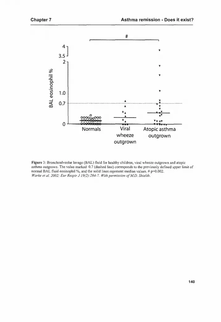
Chapter 7 Asthma remission - Does it exist?
.<: a. 0 c
·;;; 0 1.0 OJ
#
...J <(
"' 0.7 ..................................... . •
Normals Viral wheeze
outgrown
v
v ············ . ..,.'f ..... .
v v
Atopic asthma outgrown
Figure 3: Bronchoalveolar lavage (BAL) t1uid for healthy children, viral wheeze outgrown and atopic asthma outgrown. The value marked 0.7 (dashed line) corresponds to the previously defined upper limit of nonnal BAL fluid eosinophil%, and the solid lines represent median values.# p=0.002. Warke eta!. 2002; Eur Respir J 19(2):284~7. With permission of MD. Shields.
140

Chapter 7 Asthma remission -Does it exist?
AIRWAY REMODELING DURING CLINICAL REMISSION
The end result of asthmatic ainvay inflammation and its healing processes is an altered
structure referred to as airway remodeling. This altered airway structure includes fibrosis
and increased smooth muscle, mucus gland mass, vessel area, thickening of the reticular
basement membrane (RBM) and epithelial desquamation ( 11 ). As a consequence of these
structural changes, the airway wall in asthma is usually thickened, which may result in
permanent airflow obstruction. Cokugras et al demonstrated thickening of the RBM even
in 9-year old children with moderate asthma (12). Page and colleagues described recently
how human mast cells from asthmatic patients induce human ainvay smooth muscle
(HASM) cell proliferation. They proposed that mast cells fulfill a central role in the
remodeling process ( 13 ), which was also suggested by others ( 14, 15).
Invasively obtained "proof' of airway remodeling during clinical remission of asthma is
scarce. In our study, thickness of the RBM in ainvay biopsy specimens from subjects in
clinical remission of asthma differed significantly from those from healthy controls (7).
Also, occupancy of the RBM with epithelium differed between the study groups.
141

Chapter 7 Asthma remission • Does it exist?
MONITORING DISEASE ACTIVITY DURING CLINICAL REMISSION
Since flexible bronchoscopy is not feasible as a routine monitoring tool, non-invasive
markers that predict the degree of airway inflammation and/or remodeling are needed.
Possible indices of airway inflammation include the level of sputum eosinophils, the
presence of bronchial hyperresponsiveness (BHR) to inhaled stimuli, and exhaled nitric
oxide (eNO) levels.
In 1995, Fahy demonstrated that analysis of induced sputum in subjects with asthma
reveals infonnation about eosinophil percentages qualitatively similar to those obtained
by analysis ofBAL fluid (16). A close relationship between sputum eosinophils and
eosinophil percentage in BAL fluid could also be demonstrated in a study by
Grootendorst and colleagues (17). With respect to treatment decisions, Green and Pavord
could recently demonstrate that a management strategy directed at normalising the
sputum eosinophil count reduced asthma exacerbations (2002 ATS poster A320), which
finding for the first time provides "evidence" for the usefulness of monitoring ainvay
inflammation.
BHR can be measured with different kind of stimuli. Provocation with adenosine 5'
monophosphate (AMP), as an indirect ainvay challenge, has been suggested to be a better
marker of ainvay inflammation than direct challenges, such as provocation with
methacholine (MCh) (18, 19). Where MCh directly induces smooth muscle contraction,
AMP is thought to have its effect through the stimulation of bronchial mast cells. The
presence of mast cells in airway smooth muscle cells has recently been shown to be a
hallmark of asthma which is highly associated with disordered ainvay function (20). In a
142

Chapter 7 Asthma remission - Does it exist?
study by Van den Berge and coworkers, PC20 MCh and PC20 AMP were measured in 120
patients with atopic asthma (21 ). In contrast to PC,o MCh, PC2o AMP could be predicted
by the percentage of eosinophils in sputum. This is in agreement with our findings, where
the density of mucosal eosinophils correlated significantly with BHR to AMP and with
eNO levels, but not with BHR to MCh (7). Also when measuring the effect of allergen
avoidance (22, 23) and of anti-inflammatory therapy (24-26) on airway inflammation,
PC2o AMP has been proven to be a useful monitoring tool. On the other hand, Sont et a!.
could demonstrate a significant correlation between PC20 MCh and eosinophil number in
the lamina propria (27).
The response to MCh may, thus, provide infonnation about active inflammation as
well as structural ainvay changes as a consequence of airway remodeling (28-30). In a
study by Ward et al, it was shown that an early improvement in BHR to MCh with
fluticasone treatment was associated with early changes in inflammation, but the more
progressive and larger improvement was associated with later improvement in airway
remodeling (31 ).
Recent articles on the subject have evidenced of a close relationship between eNO
levels and exposure to relevant allergens (32, 33), or allergen avoidance (34) in atopic
asthmatic subjects. Furthermore, eNO positively correlates with the level of blood- and
sputum eosinophils in asthmatic subjects (35-37). The observation that eNO levels in
atopic asthmatic subjects are closely related to the eosinophil percentage in BAL fluid
(38) and in bronchial biopsy specimens (7, 39) from atopic asthmatic subjects indicates
sufficiently that elevated eNO levels are highly associated with eosinophilic airway
143

Chapter 7 Asthma remission -Does it exist?
inflammation, thereby providing a unique tool in monitoring disease activity. It also
seems to be justified to categorise asthma activity with the aid ofeNO levels (40, 41).
In conclusion, sputum eosinophils, eNO levels and BHR to AMP provide useful tools
in monitoring disease activity, whereas BHR to MCh may also or merely reflect
remodeling processes in the airways.
144

Chapter 7 Asthma remission - Does it exist?
SYMPTOM PERCEPTION DURING CLINICAL REMISSION
Blunted symptom perception would provide a plausible explanation for the
discrepancy between persistent ainvay inflammation and the absence of symptoms during
clinical remission. Several studies have shown that the relationship between asthmatic
symptoms and other indices of asthma severity is poor. Teeter et al. found asthma
symptoms to correlate poorly with the level of airway obstruction (42). A low degree of
"perceptiveness 10 for bronchoconstriction is especially demonstrated in subjects with
severe asthma (43, 44). Bijl-Hofland hypothesized that either patients with more severe
asthma show adaptation of "perceptiveness" for airway obstruction or, perhaps instead,
that low perceptiveness leads to more severe asthma as a result ofundertreatment (44). If,
after years of symptomatic asthma, adaptation or loss of perceptiveness plays a role in
subjects in clinical remission of asthma, it could possibly explain the discrepancy
between the lack of symptoms and the ongoing airway inflammation in these subjects. In
a study from our group, however, BORG-scores after a 20% fall in FEV1 during MCh
and AMP induced bronchoconstriction were not different between subjects in clinical
remission and subjects with symptomatic asthma (45), thereby making blunted dyspnea
perception not a vel)' likely explanation for the stated discrepancy. We suppose that the
weight given by patients to breathlessness depends on many other factors, including age,
where younger patients find it more troublesome than older ones ( 46). Besides, a slow
change in airflow obstruction is possibly less perceived than a fast deterioration due to
provocation with MCh or AMP. This was also shown by Rietveld, who demonstrated that
patients with prolonged airway obstruction perceived symptoms less well and were more
145

Chapter 7 Asthma remission -Does it exist?
vulnerable to negative effects of asthma than patients with acute onset airway obstruction
(47).
146

Chapter 7 Asthma remission -Does it exist?
RATIONALE FOR ANTI-INFLAMMATORY THERAPY DURING CLINICAL
REMISSION OF ASTHMA
Since some studies have evidenced of ongoing airway inflammation during clinical
remission of atopic asthma (7, 8), it has increasingly been a subject of debate that
prolonged anti-inflammatory therapy should be prescribed to these patients. Since airway
inflammation is considered the cornerstone of asthma ( 48), inhaled steroids are widely
accepted as first-choice therapy, even in the mildest fonns of disease (49). A down
regulating effect on several cell types in the bronchial mucosa (50, 51), sputum
eosinophils (52, 53), exhaled NO (53, 54) and bronchial hyperresponsiveness (55) has
been demonstrated. Airway remodeling aspects such as collagen deposition and thickness
of the RBM have also been shown to respond to some degree to anti-inflammatory
therapy (56, 57). If airway remodeling corresponds with progressive airway obstruction, a
smaller decline in lung function with anti-inflammatory therapy could possibly be
reached, as shown by Grol et al. (58). Several authors have focussed their attention to the
important concept that with the aid of inhaled corticosteroids natural history and final
outcome of asthma might be altered (59-61). Koh eta!. studied the effect of inhaled
budesonide on BHR to MCh of subjects in long-standing clinical remission of asthma
(62). After nine months of therapy no significant change on BHR was seen in this study.
It was suggested that the mechanisms underlying BHR in this clinical setting could be
different from those in symptomatic asthma. As BHR to MCh is in the first place thought
to reflect structural changes in the airways, the absence of any effect of anti-inflammatory
therapy on this parameter could be due to irreversible remodeling of the airways in the
147

Chapter 7 Asthma remission - Does it exist?
subjects studied. On the other hand, De Kluijver et al demonstrated that asymptomatic
worsening of airway inflammation in subjects with mild asthma could be prevented by
inhaled steroids (63). In a study from our group (64), a significant reduction in bronchial
hyperresponsiveness, exhaled nitric oxide levels, bronchial mast cells and thickness of
the reticular basement membrane was seen after three months of combination therapy
with fluticasone proprionate plus salrneterol in subjects in clinical remission of atopic
asthma. However, bronchial eosinophils, blood eosinophils, and quality of life were
unchanged.
Thus, it is still unclear whether subjects, who seem to have grown out of asthma, but
show evidence of ongoing disease, would benefit from prolonged anti-inflammatory
therapy in the long-term. Since several study results indicate that inhaled steroids can
positively influence progressive airflow limitation and long-term prognosis in
symptomatic asthmatics, this therapeutic modality should presumably also be (re)
considered in subjects with subclinical airway inflammation. Whether this approach is
really able to alter the natural history in subjects in clinical remission of asthma is as yet
not !mown. Besides, therapy adherence in asymptomatic young adults will presumably be
a major problem. Long-term controlled clinical trials are therefore needed to show
benefits in terms of a reduced risk of relapse and/or of irreversible airflow obstruction
later in life.
148

Chapter 7 Asthma remission - Does it exist?
DOES ASTHMA EVER GO AWAY?
There is ample evidence from the literature and our own studies that most young
adults who consider themselves grown out of asthma just appear to be asymptomatic. The
question remains whether true remission can be achieved, with or without the aid of anti
inflammatory therapy. It may be that the absence of any signs of airway inflammation
and airway remodeling is representative of true remission. This was only true for 10 or 15
percent of our population of asymptomatic subjects with a history of atopic asthma.
Epidemiological data show that a small percentage of young adults in clinical remission
remains asymptomatic for the rest of their lives. These may very well be the subjects in
true remission. It is largely unknown whether "additional remissions" can be achieved
with prolonged anti-inflammatory treatment, and, if so, when this treatment can safely be
stopped. Bahceciler and colleagues recently investigated which parameters would be
useful in deciding whether inhaled corticosteroids therapy could be stopped. They
demonstrated that only the duration of clinical remission prior to cessation of treatment
had proven to be a useful parameter ( 65). In addition to their finding, Sont eta! have
shown that a better control of airway inflammation can be achieved when airway
hyperresponsiveness is used as additional guide (66). Thus, long-term control at the
outpatient clinic with non-invasive measurements reflecting airway inflammation and
remodeling is presumably needed to decide whether anti-inflammatory therapy should be
started, continued or stopped.
We propose that adolescents who seem to have grown out of asthma should be monitored
for years after symptoms have disappeared, thereby using non-invasive measurements of
149

Chapter 7 Asthma remission -Does it exist?
ainvay inflammation and remodeling, such as exhaled nitric oxide levels, sputum
eosinophils and the response to MCh and/or AMP. Treatment might be safely withheld in
subjects without signs of airway inflammation or remodeling. In all other patients
continuation of anti-inflammatory therapy has the theoretical potential to positively alter
the prognosis of atopic asthma. Long-term studies are needed to examine the efficacy and
safety of treatment on asymptomatic subjects with chronic airway inflammation.
150

Chapter 7 Asthma remission - Does it exist?
REFERENCES
1. Barbee, R. A., and S. Murphy. 1998. The natural history of asthma. J Allergy Clin Jmnnmol!02(4 Pt 2),365-72.
2. Martin, A. J., L.A. McLennan, L I. Landau, and P. D. Phelan. 1980. The natural history of childhood asthma to adult life. Br Med J 280(6229):1397-1400.
3. Panhuysen, C. I., J. M. Vonk, G. H. Keeter, J.P. Schouten, R. van Altena, E. R. Bleecker, and D. S. Postma. 1997. Adult patients may outgrow their asthma: a 25-year follow-up study. Am J Respir Crit Care Med 155(4):1267-72.
4. Martinez, F. D. 2001. Links between pediatric and adult asthma. J Allergy Clin !mmuno/!07(5 Suppi)A49S-55S.
5. Fahy, J. V., D. B. Corry, and H. A. Boushey. 2000. Ainvay inflammation and remodeling in asthma. Curr Opin Pulm Med 6(1):15-20.
6. Vignola, A.M., P. Chanez, G. Bonsignore, P. Godard, and J. Bousquet. 2000. Structural consequences of airway inflammation in asthma. J Allergy Clin Jmmunol105(2 Pt 2):514-517.
7. Van den Toorn, L. M., S. E. Overbeek, J. C. De Jongste, K. Leman, H. C. Hoogsteden, and J. B. Prins. 200 I. Airway Inflammation Is Present during Clinical Remission of Atopic Asthma. Am J Respir Crir Care Med 164(11 ):21 07-13.
8. Warke, T. J., P. S. Fitch, V. Brown, R. Taylor, J.D. Lyons, M. Ennis, and M.D. Shields. 2002. Outgrovm asthma does not mean no airways inflammation. Eur Respir J 19(2):284-7.
9. Kerrebijn, K. F., A. C. Fioole, and R. D. van Bentveld. 1978. Lung function in asthmatic children after year or more without symptoms or treatment. Br Med J 1(6117):886-8.
I 0. Boulet, L. P., H. Turcotte, and A. Brochu. 1994. Persistence of airway obstruction and hyperresponsiveness in subjects with asthma remission. Chest 105(4):1024-31.
11. Vignola, A.M., R. Gagliardo, A. Siena, G. Chiappara, M. R. Bonsignore, J. Bousquet, and G. Bonsignore. 2001. Airway remodeling in the pathogenesis of asthma. Curr Allergy Asthma Rep 1(2),108-15.
12. Cokugras, H., N. Akcakaya, Seckin, Y. Camcioglu, N. Sarimurat, and F. Aksoy. 2001. Ultrastructural examination of bronchial biopsy specimens from children with moderate asthma. Thorax 56( I ):25-9.
13. Page, S., A. J. Ammit, J. L. Black, and C. L. Armour. 2001. Human mast cell and airway smooth muscle cell interactions: implications for asthma. Am J Physiol Lung Cell Mol Physiol 281 (6):L1313-23.
14. Rossi, G. L., and 0. Olivieri. 1997. Does the mast cell still have a key role in asthma? Chest 112(2P23-9.
15. Sommerhoff, C. P. 2001. Mast cell tryptases and airway remodeling. Am J Respir Crit Care Med 164(1 0 Pt 2),352-8.
16. Fahy, J. V., H. Wong, J. Liu, and H. A. Boushey. 1995. Comparison of samples collected by sputum induction and bronchoscopy from asthmatic and healthy subjects. Am J Respir Crit Care Med 152(1 )'53-8.
17. Grootendorst, D. C., J. K. Soot, L. N. Willems, J. C. Kluin-Nelemans, J. H. Van Krieken, M. Veselic-Charvat, and P. J. Sterk. 1997. Comparison of inflammatory cell counts in asthma: induced sputum vs bronchoalveolar lavage and bronchial biopsies. Clin Exp Allergy 27(7):769-79.
18. De Meer, G., D. Heederik, and D. S. Postma. 2002. Bronchial responsiveness to adenosine 5'monophosphate (AMP) and methacholine differ in their relationship with airway allergy and baseline FEV(l). Am J Respir Crir Care Med 165(3):327-31.
19. Polosa, R., S. Rorke, and S. T. Holgate. 2002. Evolving concepts on the value of adenosine hyperresponsiveness in asthma and chronic obstructive pulmonary disease. Thorax 57(7):649-654.
20. Brightling, C. E., P. Bradding, F. A. Symon, S. T. Holgate, A. J. Wardlaw, and I. D. Pavord. 2002. Mast-Cell Infiltration of Airway Smooth Muscle in Asthma. N Eng/ J Med 346(22):1699-1705.
21. Van den Berge, M., R. J. Meijer, H. A. Kerstjens, D. M. de Reus, G. H. Koeter, H. F. Kauffman, and D. S. Postma. 2001. PC(20) adenosine 5'-monophosphate is more closely associated with
151

Chapter 7 Asthma remission - Does it exist?
airway intlammation in asthma than PC(20) methacholine. Am J Respir Crit Care }vfed 163(7)o 1546-50.
22. van Velzen, E., J. W. van den Bos, J. A. Benckhuijsen, T. van Esse!, R. de Bruijn, and R. Aalbers. 1996. Effect of allergen avoidance at high altitude on direct and indirect bronchial hyperresponsiveoess and markers of inflammation in children with allergic asthma. Thorax 51 (6)o582-4.
23. Benckhuijsen, J., J. W. van den Bos, E. van Velzen, R. de Bruijn, and R. Aalbers. 1996. Differences in the effect of allergen avoidance on bronchial hyperresponsiveness as measured by methacholine, adenosine 5'-monophosphate, and exercise in asthmatic children. Pediatr Pulmonol 22(3)ol47-53.
24. O'Connor, B. J., S. M. Ridge, P. J. Sames, and R. W. Fuller. 1992. Greater effect of inhaled budesonide on adenosine 5'-monophosphate-induced than on sodium-metabisulfite-induced bronchoconstriction in asthma. Am Rev Respir Dis 146(3):560-4.
25. Taylor, D. A., M. W. Jensen, V. Kanabar, R. Engelstatter. V. W. Steinijans, P. J. Barnes. and B. J. O'Connor. 1999. A dose-dependent effect of the novel inhaled corticosteroid ciclesonide on airway responsiveness to adenosine-5'-monophosphate in asthmatic patients. Am J Respir Crit Care Med !60(l)o237-43.
26. Van den Berge, M., H. A. Kerstjens, R. J. Meijer, D. M. de Reus, G. H. Keeter. H. F. Kauffi11an, and D. S. Postma. 2001. Corticosteroid-induced improvement in the PC20 of adenosine monophosphate is more closely associated with reduction in airway inflammation than improvement in the PC20 of methacholine. Am J Respir Crit Care Med 164(7): 1127-32.
27. Sont, J. K., J. Han, J. M. van Krieken, C. E. Evertse, R. Hooijer, LN. Willems, and P. J. Sterk. 1996. Relationship between the inflammatory infiltrate in bronchial biopsy specimens and clinical severity of asthma in patients treated with inhaled steroids. Thorax 51 (5):496-502.
28. Crimi, E .. A. Spanevello, M. Neri, P. W. Ind, G. A. Rossi, and V. Brusasco. 1998. Dissociation between ainvay inflammation and airway hyperresponsiveness in allergic asthma. Am J Respir Crit Care Med 157(1):4-9.
29. Chetta. A., A. Foresi, M. Del Donna, G. F. Consigli, G. Bertorelli, A. Pesci, R. A. Barbee. and D. Olivieri. 1996. Bronchial responsiveness to distilled water and methacholine and its relationship to inflammation and remodeling of the airways in asthma. Am J Respir Crit Care Med 153(3):910-7.
30. Laprise, C., M. Laviolette, M. Boutet, and L P. Boulet. 1999. Asymptomatic airway hyperresponsiveness: relationships with airway inflammation and remodelling. Eur Respir J 14(l)o63-73.
31. Ward, C., M. Pais, R. Bish, D. Reid, B. Feltis, D. Johns, and E. H. Walters. 2002. Airway inflammation, basement membrane thickening and bronchial hypeiTesponsiveness in asthma. Thorax 57(4):309-316.
32. Baraldi, E .. S. Carra, C. Dario, N. Azzolin, R. Ongaro, G. Marcer, and F. Zacchello. 1999. Effect of natural grass pollen exposure on exhaled nitric oxide in asthmatic children. Am J Respir Crit Care Med 159(1 ):262-6.
33. Kharitonov. S. A,, 0. C. BJ, D. J. Evans, and P. J. Bames. 1995. Allergen-induced late asthmatic reactions are associated with elevation of exhaled nitric oxide. Am J Respir Crit Care Med 151(6)o!894-9.
34. Piacentini, G. L., A. Bodini, S. Costella, L. Vicentini, D. Peroni. L. Zanolla, and A. L Boner. 1999. Allergen avoidance is associated with a fall in exhaled nitric oxide in asthmatic children. J Allergy C/in Immuno/104(6): 1323-4.
35. Mattes, J., K. Storm van's Gravesande, U. Reining, K. Alving, G. lhorst, M. Henschen, and J. Kuehr. 1999. NO in exhaled air is correlated with markers of eosinophilic airway inllammation in corticosteroid-dependent childhood asthma. Eur Respir J 13(6): 1391-5.
36. Piacentini, G. L., A. Bodini, S. Costella, L. Vicentini, P. Mazzi, S, Sperandio, and A. L. Boner. 1999. Exhaled nitric oxide and sputum eosinophil markers of inflammation in asthmatic children [ln Process Citation]. Eur Respir J 13(6): 1386-90.
37. Silvestri, M., D. Spallarossa, V. Frangova Yourukova, E. Battistini, B. Fregonese, and G. A. Rossi. 1999. Orally exhaled nitric oxide levels are related to the degree of blood eosinophilia in atopic children with mild-intermittent asthma. Eur Re:,pir J 13(2):321-6.
152

Chapter 7 Asthma remission -Does it exist?
38. Warke, T. J., P. S. Fitch, V. Brown, R. Taylor, J.D. Lyons, M. Ennis, and M.D. Shields. 2002. Exhaled nitric oxide correlates \Vith airway eosinophils in childhood asthma. Thorax 57(5):383-7.
39. Payne, D. N., I. M. Adcock, N. M. Wilson, T. Oates, M. Scallan, and A. Bush. 2001. Relationship between exhaled nitric oxide and mucosal eosinophilic inflammation in children with difficult asthma, after treatment with oral prednisolone. Am J Respir Crit Care Med 164(8 Pt 1 ): 1376-81.
40. Co lice, G. L 2002. Categorizing asthma severity and monitoring control of chronic asthma. Curr Opin Pulm Med 8(1):4-8.
41. Jones, S. L., J. Kittelson, J. 0. Cowan, E. M. Flannery, R. J. Hancox. C. R. McLachlan, and D. R. Taylor. 2001. The predictive value of exhaled nitric oxide measurements in assessing changes in asthma control. Am J Respir Crit Care Med 164(5):738-43.
42. Teeter, J. G .. and E. R. Bleecker. 1998. Relationship between airway obstruction and respiratory symptoms in adult asthmatics [see comments]. Chest 113(2):272-7.
43. Veen, J. C., H. H. Smits, A. J. Ravensberg, P. S. Hiemstra, P. J. Sterk, and E. H. Bel. 1998. Impaired perception of dyspnea in patients with severe asthma. Relation to sputum eosinophils. Am J Respir Crit Care Med 158(4):1134-41.
44. Bijl-Hofland, l. D., S. G. Cloosterman, H. T. Folgering, R. P. Akkermans, and C. P. van Schayck. !999. Relation of the perception of airway obstruction to the severity of asthma. Thorax 54(1): 15-19.
45. Van den Toorn, L M., S. E. Overbeek, J.-B. Prins, H. C. Hoogsteden, and J. C. De Jongste. 2002. Dyspnea perception during clinical remission of atopic asthma. Eur Respir J 19(6):1 047-50.
46. Osman, L M., L. McKenzie, J. Cairns, J. A. Friend, D. J. Godden .. J. S. Legge, and J. G. Douglas. 2001. Patient weighting of importance of asthma symptoms. Thorax 56(2): 138-42.
47. Rietveld, S., and W. Everaerd. 2000. Perceptions of Asthma by Adolescents at Home. Chest 117(2):434-439.
48. Busse, W. W. 1998. Inflammation in asthma: the cornerstone of the disease and target of therapy. J Allergy Clin Jmmunol! 02(4 Pt 2):S 17-22.
49. Jatakanon, A .. S. Lim, K. F. Chung, and P. J. Barnes. 1998. An inhaled steroid improves markers of ainvay inflammation in patients with mild asthma. Eur Respir J 12(5): 1084-8.
50. Djukanovic, R., J. W. Wilson, K. M. Britten, S. J. Wilson, A. F. Walls, W. R. Roche, P. H. Howarth, and S. T. Holgate. !992. Effect of an inhaled corticosteroid on airway inflammation and symptoms in asthma. Am Rev Respir Dis 145(3):669-74.
51. MOller, G. M., S. E. Overbeek, C. G. Van Helden-Meeuwsen, J. M. Van Haarst E. P. Prens, P. G. Mulder, D. S. Postma, and H. C. Hoogsteden. \996. Increased numbers of dendritic cells in the bronchial mucosa of atopic asthmatic patients: downregulation by inhaled corticosteroids. Clin Exp Allergy 26(5):517-24.
52. Jatakanon, A., S. Kharitonov, S. Lim, and P. J. Barnes. 1999. Effect of differing doses of inhaled budesonide on markers of ainvay inflammation in patients with mild asthma. Thorax 54(2): I 08-14.
53. van Rensen, E. L., K. C. Straathot: M.A. Veselic-Charvat, A. H. Zwinderman, E. H. Bel, and P. J. Sterk. 1999. Effect of inhaled steroids on airway hyperresponsiveness, sputum eosinophils, and exhaled nitric oxide levels in patients with asthma. Thorax 54(5):403-408.
54. Kharitonov, S. A., D. H. Yates, K. F. Chung, and P. J. Barnes. 1996. Changes in the dose of inhaled steroid affect exhaled nitric oxide levels in asthmatic patients. Eur Respir J 9(2): 196-20 I.
55. van Grunsven, P.M., C. P. van Schayck, J. Mo\ema, R. P. Akkermans, and C. van Weel. 1999. Effect of inhaled corticosteroids on bronchial responsiveness in patients with "corticosteroid naive" mild asthma: a meta-analysis. Thorax 54(4):316-22.
56. Vanacker, N.J., E. Palmans, J. C. Kips, and R. A. Pauwels. 2001. Fluticasone inhibits but does not reverse allergen-induced structural airway changes. Am J Respir Crit Care Med 163(3 Pt 1 ):674-9.
57. Hoshino, M., M. Takahashi, Y. Takai, and J. Sim. 1999. Inhaled corticosteroids decrease subepithelial collagen deposition by modulation of the balance bet\veen matrix metalloproteinase-9 and tissue inhibitor ofmeta\loproteinase-1 expression in asthma. J Allergy Clin Jmmuno!I04(2 Pt 1):356-363.
58. Grol, M. H., J. Gerritsen. J. M. Vonk, J.P. Schouten, G. H. Keeter, B. Rijcken, and D. S. Postma. 1999. Risk factors for growth and decline of lung function in asthmatic individuals up to age 42 years. A 30-year follow-up study. Am J Respir Crit Care Med 160(6): 1830-7.
153

Chapter 7 Asthma remission- Does it exist?
59. Ulrik, C. S. !999. Outcome of asthma: longitudinal changes in lung function. Eur RespirJ 13(4):904-18.
60. Suissa, S., P. Ernst, S. Benayoun, M. Baltzan, and B. Cai. 2000. Low-Dose Inhaled Corticosteroids and the Prevention of Death from Asthma. N Eng! J Med 343(5):332-336.
61. Lange, P. 1999. Prognosis of adult asthma. Monaldi Arch Chest Dis 54(4):350-2. 62. Koh, Y. Y., Y. H. Sun, H. S. Lim, C. K. Kim, and S. J. Hong. 2001. Effect of inhaled budesonide
on bronchial hyperresponsiveness in adolescents with clinical remission of asthma. Chest 120(4):1140-6.
63. de Kluijver, J., C. E. Evertse, J. A. Schrumpf, H. van der Veen, A. H. Zwinderman, P. S. Hiemstra, K. F. Rabe, and P. J. Sterk. 2002. Asymptomatic Worsening of Airway Inflammation during LowDose Allergen Exposure in Asthma: Protection by Inhaled Steroids. Am. J. Respir. Crit. Care Med. 166(3):294-300.
64. Van den Toom, L. M., J. B. Prins, J. C. de Jongste, K. Leman, H. C. Hoogsteden, and S. E. Overbeek. 2002. Ainvay inflammation during clinical remission of atopic asthma- Benefit from anti-inllammatory treatment? Submitted.
65. Bahceciler, N. N., I. B. Bar!an, Y. Nuhoglu, and M. M. Basaran. 2002. Which factors predict success after discontinuation of inhaled budesonide therapy in children with asthma'? J Asthma 39(1):37-46.
66. Sont, J. K., L. N. Willems, E. H. Bel, J. H. van Krieken, J.P. Vandenbroucke, and P. J. Sterk. 1999. Clinical control and histopathologic outcome of asthma when using airway hyperresponsiveness as an additional guide to long-term treatment. Am J Respir Cl"it Care Med 159(4 Pt 1):1043-51.
154

Chapter
Conclusions and recommendations for future research
With reference to the questions, postulated in chapter 2, it can be concluded that:
Ongoing airway inflammation and remodeling can be demonstrated invasively
and non-invasively in the majority of subjects who are believed to have grown out
of asthma.
2 Blunted symptom perception is not a likely explanation for the discrepancy
between ongoing airway inflammation and absence of symptoms in subjects in
clinical remission of atopic asthma.
3 Anti-inflammatory treatment in subjects with subclinical airway inflammation
reduces bronchial hyperresponsiveness, exhaled NO levels, and bronchial mast
cells, but the effect on long-term prognosis is as yet not clear. Even so, the value
of prolonged monitoring of ain:vay inflammation and remodeling during clinical
remission of asthma has to be elucidated.
4 True asthma remission seems to be a rare phenomenon.
155

Chapter 8 Conclusion and future research
FUTURE RESEARCH
Long-term controlled clinical trials on prolonged anti-inflammatory therapy in subjects in
clinical remission of asthma are needed to show benefits in terms of a reduced risk of
relapse and/or of irreversible airflow obstruction later in life. If that proves to be the case,
anti-inflammatory therapy during clinical remission of atopic asthma could be justified.
156

Chapter
Summary
In this thesis, the presence of ongoing airway inflammation in subjects in clinical
remission of atopic asthma is demonstrated. Next to a discussion of non-invasive and
invasive methods which can be used to produce evidence of ain.vay inflammation, the
concept of blunted dyspnea perception to explain the discrepancy between absence of
symptoms and ongoing airway pathology in subjects with apparently outgrown asthma is
evaluated. Furthennore, the desirability to treat a-symptomatic subjects with ongoing
disease with anti-inflammatory therapy is dealt with. Finally, an answer is sought to the
question whether true remission in asthma is possible.
In Chapter 1, epidemiology and classification of atopic asthma are reviewed. Although
the presence and extent of symptoms of asthma are the basis of all classification systems,
it is emphasised that airway inflammation is the hallmark of atopic asthma. An overview
of non-invasive and invasive methods to evaluate ainvay inflammation in asthma, with
the bronchial biopsy histology as golden standard, is provided. Newer indices, such as
exhaled nitric oxide (eNO), are described in more detail. Next, treatment guidelines of
atopic asthma and possible future developments are discussed. It is concluded that
157

Chapter 9 Summary
asthma is dealt with. Although a great part of patients and doctors beheve that most
subjects with asthma will grow out of asthma in early adolescence, a considerable
proportion of subjects in clinical remission of asthma will have a relapse later in life. It is
suggested that ongoing ainvay inflammation and remodeling are the basis for this
continuing risk on relapsing symptoms. The question is raised whether ongoing airway
abnonnalities can indeed be demonstrated during clinical remission, and, if so, whether
this ongoing inflammation should be treated with anti-inflammatory therapy.
In Chapter 2, the aims of the studies are presented. This thesis aims to produce evidence
of persistent airway inflammation and remodeling in young adults who seem to have
outgrown their asthma. For this purpose, non-invasive as well as invasive methods are
evaluated. Besides, symptom perception was measured to find an explanation for the
discrepancy between the absence of symptoms and ongoing airway inflammation.
Finally, we examined whether subjects with subclinical airway inflammation could
benefit from anti-inflammatory treatment.
In Chapter 3, non-invasive evidence of persistent airway inflammation during clinical
remission of atopic asthma is demonstrated, as reflected by an elevated fraction of
exhaled nitric oxide (FENO) levels and bronchial hyperresponsiveness to adenosine
monophosphate (AMP) and methacholine (MCh). With respect to eNO, we found no
overlap between values of21 subjects in clinical remission of atopic asthma and those of
18 healthy controls. Values ofFENO in subjects in remission were similar to those of21
subjects with current asthma. A significantly higher AMP- as well as MCh
158

Chapter 9 Summary
responsiveness was present in the remission group as compared with the control group.
Furthermore, AMP responsiveness showed a significant inverse relationship with FENO
levels, probably suggesting that both indices are associated with active elements of
airway inflammation, \vhereas responsiveness to MCh may also reflect airway
remodeling.
In Chapter 4, invasive "proof' of persistent airway inflammation and airway remodeling
in the majority of subjects in clinical remission of atopic asthma complements the
findings of chapter 3. By means of flexible bronchoscopy, bronchial biopsies were taken
from 19 subjects with current atopic asthma, 18 subjects in clinical remission of atopic
asthma, and 17 healthy controls. Immunohistochemical staining of biopsy specimens
demonstrated an elevated density of eosinophils, T lymphocytes, mast cells, and IL-5 in
the airways mucosa of subjects in remission compared to controls. Also, blood eosinophil
cell counts were significantly elevated in subjects in clinical remission. Blood eosinophil
cell counts, FENO levels, and bronchial response to AMP correlate significantly with the
density of tissue eosinophils, indicating that these measurements could be used to
monitor airway inflammation with minimal burden to the patient.
MCh responsiveness did not show a relationship with the density of eosinophils in the
airway mucosa. Since MCh is believed to act directly on smooth muscle cells in the
airways, whereas AMP acts via activation of bronchial mast cells, it was concluded that
AMP- and MCh responsiveness probably relate to different aspects of the inflammatory
process.
159

Chapter 9 Summary
Surprisingly, inducible nitric oxide synthase (iN OS) activity in the airway mucosa from
healthy controls was higher than in subjects with asthma and subjects in remission. It
could thus well be that iNOS-unrelated mechanisms, including augmented diffusion of
NO through damaged epithelium, or iN OS independent NO generation under conditions
of ainvay acidity may explain this finding. Furthermore, other isoforms of NOS could be
responsible for elevated NO production.
In conclusion, both ongoing airway inflammation and remodeling were demonstrated
during clinical remission of atopic asthma, and can be monitored with non-invasive
measurements.
In Chapter 5, dyspnea perception was investigated in subjects in clinical remission of
atopic asthma and subjects with current asthma. The hypothesis was that blunted
symptom perception in subjects in clinical remission of asthma could explain the
discrepancy between the lack of symptoms and persistent airway inflammation. Twenty
subjects with bronchial hyperresponsiveness to MCh or AMP assigned themselves a
perception score of dyspnea on a modified BORG scale at reaching a 20% fall in FEY 1
(PS20) during MCh- or AMP challenge. Dyspnea perception was highly variable but did
not differ significantly between both groups, nor between challenge tests. It was
concluded that subjects in clinical remission are not different from currently asthmatic
subjects with respect to their perception acute changes in FEY 1• Perception of symptoms
in daily life might, however, be different from these results, since not only physical, but
also emotional and social factors are believed to play a role in the subjective feeling of
discomfort, and we can not exclude that daily life fluctuations in airflow limitation are
160
Chapter 9 Summary
not well perceived by subjects in clinical remission of asthma. Therefore, the absence of
symptoms in subjects with a past history of asthma does not mean that airway
inflammation is gone.
In Chapter 6, we assessed the effect of anti-inflammatory therapy in subjects in clinical
remission of atopic asthma. It was hypothesized that if ainvay inflammation and/or
remodeling are present during clinical remission despite a lack of symptoms, this could
well respond to the use of inhaled steroids. A double-blind, randomized, placebo
controlled trial with the salmeterollfluticasone propionate combination product (SFC
50/250 !-lg bid via the DiskusrM inhaler) was conducted in 28 subjects in clinical
remission with proven hyperresponsiveness to MCh as non-invasive indicator of
ongoing disease. SFC treatment for three months improved PD20 MCh as well as PD20
AMP, and nonnalized FENO in most subjects. SFC treatment also reduced mast cells
in subepithelium, had a minimal effect on the thickness of the reticular basement
membrane (RBM), but did not alter tissue eosinophils, blood eosinophils, and quality of
life. The added value of the long acting fh agonist (salmeterol) could not be extracted
from this study. It was suggested that airway hyperresponsiveness and FENO could
represent fast responding elements of airway pathology, whereas tissue markers of
inflammation and remodeling need more time than three months to respond. Based on
the results of this study, the use of prolonged anti-inflammatory therapy during clinical
remission of asthma is not justified. However, it encourages further investigation with
long-term studies. ·whether inhaled steroids might prevent a relapse of asthma later in
161

Chapter 9 Summary
life thus remains to be proven. Furthennore, major problems with therapy adherence in
subjects without symptoms can be anticipated.
In Chapter 7, an answer is sought to the question whether real asthma remission exists.
The majority of subjects with childhood asthma seems to grow out of asthma in early
adolescence. The appellation "clinical remission" is used in this context to define the
absence of symptoms without therapy. Current classification guidelines do not mention
this sizeable group of formerly asthmatic subjects who are at risk to develop symptoms
again later in life, mostly before the age of 30. More specifically, epidemiological studies
have shown that in about 30 to 80% of subjects in clinical remission of atopic asthma the
absence of symptoms is only temporary. We postulate that this is due to persistent airway
inflammation and remodeling. In about one fifth of the subjects in clinical remission in
our study, significant airway inflammation and/or remodeling could not be demonstrated.
Are these the subjects in true remission? If so, can we heighten this percentage with
prolonged anti-inflammatory therapy? In Chapter 7 a review of the literature on asthma
remission is given, thereby trying to find an answer to this questions.
162

Chapter
Samenvatting
Allergisch astma ontstaat meestal op jonge leeftijd, en wordt gekarakteriseerd door een
chronisch ontstekingsproces in de luchtwegen. Veel patienten laten rond de puberteit een
aanzienlijke vermindering van klachten zien, waarbij een groat dee! zelfs geheel
klachtenvrij wordt en veelal geen medicatie meer hoeft te gebruiken. Dit noemen we
"klinische remissie". In deze fase worden patienten uit de medische controle ontslagen,
en wordt er veelal gesproken van "over astma been groeien". Uit een aantal studies is
echter gebleken dat ongeveer de helft van deze ex-patienten op latere leeftijd, meestal al
voor bet dertigste levensjaar, een recidiefkrijgt. Tijdens het "goede" interval zijn door
sommige auteurs longfunctieafwijkingen vastgesteld, zoals verhoogde
luchtwegprikkelbaarheid en luchtwegobstructie. Het is echter onduidelijk of deze
afwijkingen verband houden met persisterende ontstekingsverschijnselen, dan wel met
structurele veranderingen in de luchtwegen als uiting van "restschade" ten gevolge van
astma op de kinderleeftijd. Tevens bestaat er onduidelijkheid over de relatie tussen de
aard en uitgebreidheid van de ontstekingsverschijnselen en de prognose van astma. Het
doel van ons onderzoek was nate gaan of bij person en met allergisch astma, die in
klinische remissie zijn gekomen, aldan niet een ontstekingsproces in de luchtwegen
163

Chapter 10 Samenvatting
bestaat, en of, indien dit het geval is, dergelijke person en gebaat zijn bij het gebruik van
ontstekingsremmende medicijnen. Tevens wordt er gezocht naar een verklaring voor de
discrepantie tussen de afwezigheid van klachten en de aanwezigheid van het
ontstekingsproces.
In hoofdstuk 1 worden de epidemiologische aspecten en classificatie van allergisch astma
besproken. Hoewel de aanwezigheid en uitgebreidheid van astmatische symptomen een
belangrijk uitgangspunt iijken te vormen voor alle classificatiesystemen, wordt
(chronische) \uchtwegontsteking tach als het kenmerkende element van allergisch- en
niet allergisch astma gezien.
Een overzicht van niet-invasieve en invasieve methoden om deze luchtwegontsteking te
bestuderen, met luchtwegbiopten als gouden standaard, wordt in dit hoofdstuk gegeven.
Nieuwere methoden, zoals het meten van de hoeveelheid stikstofoxide (NO) in
uitgeademde Iucht, worden in detail beschreven. Het hoofdstuk wordt vervolgd met een
uiteenzetting van de mogelijkheden van medicamenteuze therapie bij allergisch astma.
Geconcludeerd wordt dat tot op heden inhalatiesterorden nog altijd de therapie van eerste
keus vonnen. Als laatste wordt het gegeven van "over je astma heen groeien" nader
uitgewerkt. Het feit dat een groat percentage van patienten, die menen over hun astma te
zijn heen gegroeid, op latere leeftijd weer klachten krijgt, geeft a! aan dat de afwezigheid
van symptomen blijkbaar niet de afwezigheid van ziekte impliceert.
In hoofdstuk 2 worden de verschillende studies uiteengezet die als doe! hebben de
aanwezigheid van luchtwegontsteking en/of remodeling aan te tonen bij personen die van
164

Chapter 10 Samenvatting
mening zijn over hun astma te zijn heen gegroeid. Voor dit doe! worden zowel niet
invasieve als invasieve methoden geevalueerd. Daarnaast wordt er onderzocht of de
verklaring voor de discrepantie tussen het gebrek aan klachten en de aanwezige
luchtwegontsteking kan worden gezocht in veranderde syrnptoomperceptie. Als laatste
wordt gekeken ofpersonen met niet-manifeste luchtwegontsteking voordeel kunnen
hebben bij het gebruik van ontstekingsremrnende therapie.
In hoofdstuk 3 werd bij patienten in klinische remissie van allergisch astma, bij patienten
met actief astma en bij gezonden met niet-invasieve methoden gezocht naar persisterende
luchtwegontsteking aangedragen. Hierbij werd gebruik gemaakt van NO in uitgeademde
lucht en luchtweggevoeligheid voor adenosine monofosfaat (AMP) en metacholine
(MCh). Met betrekking tot de uitgeademde hoeveelheid NO vonden wij geen overlap
tussen meetwaarden van 21 personen in klinische remissie en die van 18 gezonde
controlepersonen. Meetwaarden van personen in remissie bleken nauwelijks verschillend
te zijn van die van 21 personen met symptomatisch astma.
Tevens werd er een verhoogde luchtweggevoeligheid voor zowel AMP als MCh in de
remissie groep gevonden, indien vergeleken met de controle groep. Daarnaast bestond er
een duidelijke omgekeerde relatie tussen de reactie op AMP en hoogte van de
uitgeademde NO hoeveelheid, hetgeen mogelijk impliceert dat beide variabelen
gerelateerd zijn aan luchtwegontsteking, terwijl de reactie op MCh mede ofvooral
beihvloed kan zijn door structurele afwijkingen.
165

Chapter 10 Samenvatting
In hoofdstuk 4 wordt invasief onderzoek naar bet bestaan van luchtwegontsteking en
structurele afwijkingen bij de personen in klinische remissie van allergisch astma
beschreven. Met behulp van flexibele bronchoscopie werden luchtwegmucosa biopten
verkregen van 19 personen met symptomatisch astma, 18 personen in klinische remissie,
en 17 gezonde controles. Irnmunohistochemische kleuring van bioptmateriaalliet een
verhoogde dichtheid van eosinofielen, T lymfocyten, mestcellen, en IL-5 in bet
luchtwegslijmvlies van personen in klinische remissie zien, wanneer werd vergeleken
met gezonde controles. Tevens was de hoeveelheid eosinofielen in het bloed verhoogd.
Er bleek een significant verband te zijn tussen eosinofielen aantal in bet bloed,
uitgeademde hoeveelheid NO, en reactie op AMP enerzijds, en de mate van
eosinofieleninfiltratie in het luchtwegslijmvlies anderzijds. Dit betekent dat dergelijke
meetmethoden waarschijnlijk gebruikt kunnen worden om de aanwezigheid van
luchtwegontsteking te controleren en te vervolgen, met mini male be lasting voor de
patient.
De reactie op MCh vertoonde echter geen relatie met de eosinofielen dichtheid in
luchtwegslijmvlies. Daar MCh een direct effect uitoefent op de gladde spieren in de
luchtwegen, terwijl AMP met name mestcel-degranulatie bewerkstelligt, kan worden
geconcludeerd dat de reactie op AMP en MCh aan verschillende aspecten van bet
ontstekingsproces gerelateerd zijn. Hierbij lijkt de reactie op MCh onatbankelijk van
actieve luchtwegontsteking verhoogd te kunnen zijn.
Een opvallende bevinding was tevens dat er een verhoogde activiteit van bet induceerbare
stikstof oxide synthase (iN OS) in de luchtwegmucosa van gezonde controles aanwezig
bleek te zijn, wanneer wordt vergeleken met de situatie in person en met symptomatisch
166

Chapter 10 Samenvatting
astma en person en in klinische remissie van astma. Dit was niet in overeenstemming met
de aanname dat verhoogde iNOS expressie in de luchtwegmucosa de bron vormt van de
verhoogde hoeveelheid NO in uitgeademde Iucht van personen met astma. Het is dus
mogelijk dat niet aan iN OS gerelateerde mechanisrnen, waaronder toegenomen diffusie
van NO door beschadigd epitheel ofveranderingen in de luchtwegzuurgraad,
verantwoordelijk zijn voor dit onvenvachte resultaat. Daamaast kunnen andere vonnen
van NOS een bijdrage leveren aan de verhoogde NO productie.
Concluderend, zowel actieve luchtwegontsteking als structurele veranderingen zijn
aantoonbaar bij personen in klinische remissie van atopisch astma, en kunnen worden
aangetoond middels niet-invasieve meetmethoden. De recente bevindingen van diverse
auteurs, waaronder verrninderde longfunctie en verhoogde luchtwegprikkelbaarheid
tijdens het klinische remissie kunnen gerelateerd zijn aan zowel actieve ontsteking als
structurele veranderingen in de luchtwegen.
In hoofdstuk 5 werd de perceptie van kortademigbeid onderzocht bij personen in
klinische remissie en bij personen met symptomatisch astma. De hypothese is dat
verminderde symptoomperceptie bij personen in kliniscbe remissie de discrepantie tussen
bet gebrek aan klachten en de persisterende luchtwegontsteking kan verklaren. Aan 20
personen in klinische remissie, met verhoogde gevoeligheid voor MCh of AMP, werd
gevraagd een kortademigheidscore toe te kennen bij bet bereiken van een 20 procent
daling in FEY 1 tijdens MCh- of AMP provocatie. Kortademigheidperceptie bleek grate
varia tie tussen personen te vertonen maar was niet duidelijk verschillend tussen personen
in klinische remissie en person en met symptomatisch astma voor zowel MCh als AMP.
167

Chapter 10 Samenvatting
Geconcludeerd werd dat person en in klinische remissie niet verschillen van personen met
symptomatisch astrna met betrekking tot de perceptie van een snelle opgelegde daling in
FEY 1- De perceptie van astmatische symptomen in het dagelijks Ieven zou echter anders
kunnen zijn, daar niet aileen lichamelijke, maar ook psychische en sociale factoren van
invloed kunnen zijn op de beleving van symptomen. Er kan in ieder geval worden
gesuggereerd dat de afwezigheid van symptomen bij personen met een voorgeschiedenis
van astma niet hoeft te betekenen dat het ziekteproces in de luchtwegen is verdwenen.
ln hoofdstuk 6 werd het effect van astrnabehandeling onderzocht bij personen in klinische
remissie van astma. Hierbij werd uitgegaan van de hypothese dat, indien
luchtwegontsteking en/of remodeling aanwezig blijven tijdens klinische remissie,
subklinische ontstekingsverschijnselen kunnen venninderen door het gebruik van
inhalatiesterorden. Een dubbelblinde, gerandomiseerde, placebo gecontroleerde studie
werd daarom verricht met het salmeterollfluticasone propionate combinatie product (SFC
50/250 !J.g 2dd via de Diskus™ inhaler) bij 28 personen in klinische remissie met
bewezen hyperreactiviteit voor MCh als niet-invasiefkenmerk van luchtwegpathologie.
SFC behandeling gedurende drie maanden venninderde in deze studie de
hyperreactiviteit voor MCh en AMP, en nonnaliseerde de uitgeademde hoeveelheid NO
bij de meeste person en in het onderzoek.
Tevens werd er na drie maanden een duidelijke vennindering van mestcellen in het
luchtwegslijmvlies, een geringe regressie van de dikte van de reticulaire
basaalmembraan (RBM), maar geen significante vennindering van weefsel
eosinofielen, bloed eosinofielen, en kwaliteit van leven gezien. De toegevoegde
168

Chapter 10 Samenvatting
waarde van een lang werkend 82- mimeticum kan uit de studieresultaten niet
geextraheerd worden. Er werk gesuggereerd dat luchtwegreactiviteit en uitgeademde
hoeveelheid NO mogelijk snel responderende elementen van luchtwegpathologie zijn,
terwijl weefsel eosinofielen en andere invasieve kenmerken wellicht meer dan drie
maanden nodig hebben om te reageren.Geconcludeerd werd dat de bevindingen van
deze studie het (opnieuw) voorschrijven van ontstekingsremmende medicijnen aan
personen in klinische remissie van astma niet rechtvaardigen. Wet moedigen deze
resultaten aan tot lange-tennijn studies. De vraag of inhalatiesteroi'den een recidief van
astma op latere leeftijd kunnen voorkomen blijft dus nog onbeantwoord. Daarnaast
zullen problemen met de therapietrouw bij personen zonder klachten dergelijk lange
tennijn onderzoek niet vereenvoudigen.
In hoofdstuk 7 wordt benadrukt dat een meerderheid van de jeugdige patienten met astma
in de vroege pubertijd over het astma been lijkt te groeien. De term "klinische remissie"
wordt in deze context gebruikt als aanduiding voor de afwezigheid van symptom en
zonder het gebruik van inhalatie medicatie gedurende tenminste 1 jaar.
Binnen de huidige classificatiesystemen is deze omvangrijke groep van volledig
klachtenvrije astmapatienten in het geheel niet terug te vinden, ondanks het feit dat dezen
vaak op latere leeftijd een recidiefvan astmatische klachten krijgen, over het algemeen
zelfs a] voor het dertigste levensjaar. Epidemiologische studies hebben aangetoond dat bij
30 tot 80% van personen in klinische remissie de afutezigheid van syrnptomen slechts
tijdelijk is. Er wordt naar voren gebracht dat persisterende luchtwegontsteking dit
aanzienlijke risico op een recidief zou kunnen veroorzaken. In ongeveer 20 procent van
169

Chapter 10 Samenvatting
de person en in klinische remissie in ons onderzoek konden geen luchtwegontsteking
en/of structurele afwijkingen meer worden aangetoond. Zijn dit dan de person en in echte
remissie? En zo ja, kunnen we dit I age percentage dan verhogen middels langdurige
ontstekingsremrnende behandeling? In hoofdstuk 7 wordt een overzicht van de
beschikbare literatuur over astma remissie gegeven, waarbij getracht wordt een antwoord
op deze vragen te geven.
170

Chapter
List of abbreviations
AMP
ATS
BHR
CRF
eNO
FENO
FE VI
HASM
IL-5
iN OS
MBP
MCh
PEFR
RAST
RBM
adenosine-5' -monophosphate
American Thoracic Society
bronchial hyperresponsiveness
clinical research file
exhaled nitric oxide
fraction of exhaled nitric oxide
forced expiratory volume s-1
human airway smooth muscle
interleukin-5
inducible nitric oxide synthase
major basic protein
methacholine
provocative dose resulting in 20 percent decrease in FEY 1
peak expiratory flow rate
perception score at reaching a 20 percent decrease in FEV1
radioallergoimmunosorbent test
reticular basement membrane
171

Chapter 11 List of abbreviations
SFC salmeterollfluticasone propionate combination
Th2 type 2 T-helper
TLC total lung capacity
172

Chapter
Dankwoord
Collega G. T. Verhoeven zei ooit: "bet is eenvoudiger om meerdere marathons uit te
lopen dan eenmaal te promoveren". Daar deze uitspraak na maar liefst vijfmarathons tot
stand is gekomen, moeten we wei aannemen dat het voltooien van een proefschrift een
uitputtende bezigheid is, die met een wazige blik op de finish en een wisselend gevoel
van verzuring gepaard kan gaan. In het zicht van- en vooral na het bereiken van de finish
slinkt het pijngevoel echter snel, en telt uitsluitend nog bet resultaat. Ook het volbrengen
van slechts twee marathons heeft de schrijver van dit proefschrift de overtuiging gegeven
dat ook het "finishgevoel" bij het promotieonderzoek een gevoel van voldoening moet
zijn. De blessures zijn echter van behoorlijke omvang en geven soms een nieuwe
dimensie aan het begrip "beroepsgerelateerde aandoeningen". Naast de alom bekende
muisarm was er bij mij sprake van een "microfilm-nystagmus" (na het beoordelen van
ongeveer 2000 patientendossiers op voorbij rollende microfilm) en "image analysis
hallucinaties" (het herkennen van een basaalmembraan in de trapleuning). Gelukkig
waren dergelijke ongemakken van tijdelijke aard en hebben zij er niet toe geleid dat voor
de finish moest worden uitgestapt. Dit proefschrift zou echter niet tot stand zijn gekomen
zonder de hulp van velen die nu snel doorlezen of ze daadwerkelijk met name worden
genoemd. In zijn algemeenheid verdienen het laboratorium longziekten in de hoogbouw,
173

Chapter 12 Dankwoord
de afdeling longfunctie en de mensen van het archiefvan het Sophia
Kinderziekenhuis, de verpleegkundigen in de bronchoscopiekamer, Glaxo SmithKline
B.V., en natuurlijk aile proefpersonen mijn speciale dank. Aan enkele personen wil ik
echter enige persoonlijke woorden wijden.
Prof. Dr H.C. Hoogsteden, beste Henk, ik wil je bedanken voor de mogelijkheid die je me
hebt geboden om dit onderzoek te verrichten, ook al sloeg de twijfel enigszins toe toen je
venneldde dat ik gerust elk weekend in het medisch archie£ door "mocht" werken, nadat
ik a! op zondag was komen opdraven voor het sollicitatiegesprek. Daarnaast heb je me
echter vee! vrijheid gegeven bij de invulling van het onderzoek, hetgeen ik zeer waardeer.
Prof. Dr J.C. de Jongste, beste Johan, je wonderbaarlijk snelle en secure correctie van
mijn wetenschappelijke probeersels hebben mij telkens aangenaam verrast. Je gewoonte
omje commentaar te starten met: "dit wordt weer een schitterend artikel" (om
vervolgens bijna elke zin met de rode stift te bestormen) heb ik als zeer prettig ervaren.
Ook je uitgebreide literatuurkennis en neus voor belangrijke ontwikkelingen zijn van
grote waarde geweest.
Dr S.E. Overbeek, beste Shelley, onze onderzoeksrelatie heeft een intens karakter
gekend. Je historische uitspraken tijdens de uitvoering van een bronchoscopie ("ik zie
niks!" of"wat is dat bloederig!") zal ik (en zullen de proefpersonen) niet snel vergeten.
We zullen bet wel nooit eens worden wie van ons tweeen bet opstapbankje bij de
bronchoscopie harder nodig heeft. Je enorme inzet en belangstelling voor bet onderzoek,
maar ook voor bet thuisfront, zijn bewonderenswaardig. Bedankt voor al je hulp!
Dr J-B Prins, beste Jan-Bas, op bet moment dat ik dit schrijfbenje al niet meer
werkzaam in de Erasmus Universiteit. Aanjou verliest het laboratorium een persoon met
174

Chapter 12 Dankwoord
vele kwaliteiten. Je bijzonder kritische noot en schijnbare onbekendheid met het woordje
"goed" hebben onze samenwerking echter niet altijd tot een gemakkelijke gemaakt. Na
een bezoek aanje kamer op de 22e vroeg ik me vaak af: "is er nog leven na(ast) het
promotieonderzoek?". Gelukkig heb je buiten bet werk Iaten zien ook over een andere en
gezellige persoonlijkheid te beschikken. Daamaast heb je op velerlei wijze, waaronder je
vakkundige begeleiding bij bet maken van voordrachten, jc waarde voor dit onderzoek
bewezen, waarvoor ikje veel dank verschuldigd ben.
Van de longafdeling verdient Joachim Aerts bet nag om nader aangehaald te worden.
Zijn bereidwilligheid om de laatste patienten voor bet onderzoek te zien zal ik niet
vergeten, oak al heeft het me enige tijd en hoofdbrekens gekost de juiste gegevens bij de
juiste patient te plaatsen. Joachim bedankt!
Van bet laboratorium longziekten in de hoogbouw verdienen twee mensen een extra
woord van dank: Joost Hegmans, jou wil ik bedanken voor je constante bereidwilligheid
te helpen met laboratoriumzaken waar ik te weinig verstand van heb. De te nemen
stappen bij het opstarten van de image analysis computer zullen wei nooit voor tangere
tijd in mijn geheugen gegrift blijven staan. Karolina Leman, hoewel we bet niet altijd
eens waren over de manier waarop een lijn tangs een basaalmembraan getrokken meet
worden, is bet vele snij- en kleurwerk datje samen met Sophia hebt verricht van
onschatbare waarde geweest.
Van de afdeling longfunctie in het Sophia Kinderziekenhuis wil ik Astrid, Simone,
Marieke, Aafke en Edith bedanken voor hun tomeloze inzet bij de longfunctiemetingen
en hun oneindige geduld wanneer ik weer eens een meting liefst direct of anderszins
tussendoor wilde.
175

Chapter 12 Dankwoord
Het Nederlands Astma Fonds dank ik voor de financiele ondersteuning van het
"hoofdonderzoek".
Van Glaxo SmithKline wil ik Pauline Bloemen en Martijn Akveld bedanken voor de
zorgvuldige en gezellige begeleiding van de interventiestudie. De kalme reactie van
Pauline op het feit dat ik een geleende laptop heb late crashen verdient aile !of Oat ze mij
vervolgens uit protest een andere computer in bruikleen heeft gegeven die nauwelijks
meer tot enigerlei bewerking te motiveren was, is volkomen begrijpelijk.
Bij de eenzame uren in het archie£ in het Sophia Kinderziekenhuis heeft Maarten mij
altijd met woord en daad terzijde gestaan.
Paul Mulder dank ik voor bet feit dat hij mij heeft bijgestaan in de soms moeilijk te
doorgronden brij van de statistiek. De uiting "ieder zijn vak" heb ik bier altijd voor ogen
gehouden.
Van het Sint Fransiscus Gasthuis wil ik niet nalaten Arie Rietveld te noernen die mijn
interesse voor de intensive care geneeskunde tot grate hoogten heeft weten te doen
stijgen. Arie, bet steeds weer kopieren van "Up to Date" heeft me weliswaar enige flessen
wijn gekost, maar weegt ruimschoots op tegen de vele plezierige en leerzame momenten
die ik op de IC hebben gehad. Ook dank ikje voor je min ofmeer subtiele pogingen om
mijn ecbtgenote te overtuigen van bet nut van een eigen snowboard. Daamaast wil ik van
betzelfde gasthuis Arjan Rudolphus bedanken voor zijn zachte drang om mij met links te
Iaten scopieren, en Youke Tan alsnog mijn verontschuldigingen aanbieden voor bet feit
dat ik een (oude) scoop ondanks bijtring door een patiente aan flarden heb Iaten bijten.
Tevens is uiteraard een woord van dank voor de vele proefpersonen die aan het
onderzoek hebben meegewerkt op zijn plaats. AI varieerde de reactie na een
176

Chapter 12 Dankwoord
bronchoscopie nog van "piece of cake" tot "never nooit meer", na de provocatietcsten
waren sommigen zelfs niet meer in staat om hun reactie te geven ( omdat ik bet nodig
vond de FEY 1 tot onder 40% van de beginwaarde te I a ten dalen). De bereidwilligheid om
bet gehele onderzoek te doorlopen was bij vrijwel a lie deelnemers daarom
indrukwekkend. Het feit dat een aantal van hen de in het vooruitzicht gestelde riante
vergoeding al ruimschoots hadden uitgegeven doet bier niets aan af.
Patrick Jongmans, bedankt voor je on twerp van de oms lag van het boekje en je hulp als
de computer weer eens niet deed wat ik wild e. Het tekentje dat je best paranimfwilde
zijn ondanks het feit datje dat erg dubieus vond klinken.
Het recht om als last but zeker not least in dit dankwoord te staan komt mijns inziens
zonder twijfel toe aan degene die tevergeefs bleef vragen of ze kon helpen om bet
onderzoek te bespoedigen. Lieve Angeli que, je hebt vaak Largo aileen moeten uitlaten
omdat ik weer eens achter de computer wilde duiken, maar nu is dat "stomme onderzoek"
toch eindelijk af. Ik beloofje dat we weer vaker samen leuke dingen gaan doen. Jij deed
me immers telkens weer realiseren dater nog leven is na(ast) bet promotieonderzoek.
177

Chapter
Curriculum Vitae
Leon Michie! van den Toorn werd geboren op 16 februari 1966 te Amsterdam. Het
diploma 0-VWO behaalde hij in 1984 aan het Bonhoeffer College te Castricum. In 1989
voltooide hij de studie medische biologic aan de Vrije Universiteit te Amsterdam. Tijdens
deze studie liep hij onderzoeksstages op de afdeling fysiologie (Dr G.C. van den Bos) en
interne geneeskunde (Prof Dr. R.O.B. Gans). In 1987 was hij tevens begonnen met de
studie geneeskunde aan dezelfde universiteit, hetgeen resulteerde in een met succes
afgelegd arts exam en in 1994. Vervolgens was hij gedurende twee jaar werkzaam als
AGNIO interne geneeskunde, longziekten en cardiologic in het Spaame Ziekenhuis te
Heemstede. Alhier werd zijn belangstelling voor de longziekten gewekt, hetgeen een
AGNIO-schap longziekten in bet Academisch Ziekenhuis te Rotterdam ten gevolge had.
Op 1 april 1997 werd tevens een start gemaakt met voorliggend promotieonderzoek
(promotoren Prof. Dr. H. C. Hoogsteden en Prof. Dr. J.C. de Jongste) op de afdelingen
longziekten (Erasmus MC Rotterdam) en kindergeneeskunde/subafdeling
kinder!ongziekten (Erasmus MC locatie Sophia). Vanaf 1 juli 2000 tot 1 maart 2002 heeft
hij als AGIO longziekten zijn vooropleiding interne geneeskunde in het St. Fransiscus
Gasthuis te Rotterdam doorlopen (opleiders Dr. Tjen en Prof. Dr. H. C. Hoogsteden).
178

Chapter 13 Curriculum Vitae
Sinds 1 maart 2002 is hij voor de vervolgopleiding longziekten weer werkzaam in bet
Erasmus MC.
179

Chapter
Publications
1. Van den Toorn, L M., J. B, Prins, S. E. Overbeck, H. C, Hoogsteden, and J. C, de Jongste. 2000. Adolescents in clinical remission of atopic asthma have elevated exhaled nitric oxide levels and bronchial hyperresponsiveness. Am. J. Respir. Crit, Care Med 162(3 Pt 1):953-7.
2. Van den Toorn, L M., S. E. Overbeck, J. C. De Jongste, K. Leman, H. C. Hoogsteden, and J. B. Prins. 200 l. Airnray Inflammation Is Present during Clinical Remission of Atopic Asthma. Am. J Respir. Crit. Care )\1ed. 164(11):2107-13.
3. Van den Toom, L. M., S. E. Overbeck, J. B. Prins, H. C. Hoogsteden, and J. C. De Jongste. 2002. Dyspnea perception during clinical remission of atopic asthma. Eur RespJ: 19(6):1047-50.
4. Van den Toorn, L M., J. B. Prins, J. C. de Jongste, K. Leman, H. C. Hoogsteden, and S. E. Overbeck. 2002. Airway inflammation during clinical remission of atopic asthma- Benefit from anti-inflammatory treatment? Submitted.
5. Van den Toorn, L. M., S. E. Overbeek, J. B. Prins, H. C. Hoogsteden, and J. C. De Jongste. 2002. Asthma remission- Does it exist? Current Opinion in Pulmonmy J\1edicine- in press.
180


