Embed Size (px)
Citation preview
Nutrients 2015, 7, 1426-1463; doi:10.3390/nu7031426
nutrients ISSN 2072-6643
www.mdpi.com/journal/nutrients
Review
Arginine and Citrulline and the Immune Response in Sepsis
Karolina A.P. Wijnands 1,†,*, Tessy M.R. Castermans 2,†, Merel P.J. Hommen 2,†,
Dennis M. Meesters 1 and Martijn Poeze 1
1 Department of Surgery, NUTRIM School for Nutrition, Toxicology and Metabolism,
Maastricht University Medical Center, Maastricht 6200 MD, The Netherlands;
E-Mails: [email protected] (D.M.M.); [email protected] (M.P.) 2 Department of Surgery, Maastricht University Medical Center, Maastricht 6200MD,
The Netherlands; E-Mails: [email protected] (T.M.R.C.);
[email protected] (M.P.J.H.)
† These authors contributed equally to this work.
* Author to whom correspondence should be addressed; E-Mail: [email protected];
Tel.: +31-433884502; Fax: +31-433884154.
Received: 18 October 2014 / Accepted: 26 January 2015 / Published: 18 February 2015
Abstract: Arginine, a semi-essential amino acid is an important initiator of the immune
response. Arginine serves as a precursor in several metabolic pathways in different organs.
In the immune response, arginine metabolism and availability is determined by the nitric
oxide synthases and the arginase enzymes, which convert arginine into nitric oxide (NO) and
ornithine, respectively. Limitations in arginine availability during inflammatory conditions
regulate macrophages and T-lymfocyte activation. Furthermore, over the past years more
evidence has been gathered which showed that arginine and citrulline deficiencies may underlie
the detrimental outcome of inflammatory conditions, such as sepsis and endotoxemia. Not
only does the immune response contribute to the arginine deficiency, also the impaired arginine
de novo synthesis in the kidney has a key role in the eventual observed arginine deficiency.
The complex interplay between the immune response and the arginine-NO metabolism is
further underscored by recent data of our group. In this review we give an overview of
physiological arginine and citrulline metabolism and we address the experimental and clinical
studies in which the arginine-citrulline NO pathway plays an essential role in the immune
response, as initiator and therapeutic target.
Keywords: arginine; citrulline; nitric oxide; sepsis; immunity
OPEN ACCESS
Nutrients 2015, 7 1427
1. Introduction
Sepsis and associated inflammatory conditions, such as bacteremia and endotoxemia; a condition
with increased presence of lipopolysaccharide (LPS), the essential component of the outer membrane of
Gram-negative bacteria, are associated with arginine deficiency [1–4]. This arginine deficiency is suggested
to be the result of a decreased arginine uptake and an impaired arginine de novo synthesis from citrulline,
in combination with an enhanced arginine catabolism by the upregulation of arginase and the inflammatory
nitric oxide synthase (iNOS; NOS2) in the immune response. As a result, strategies to improve the arginine
availability during inflammatory conditions have gathered widespread attention over the past decades.
However, this does not necessary mean that arginine deficiency occurs universally in all inflammation
or sepsis related illnesses. It is beyond the purpose of this review to determine whether arginine deficiency
is present in all these conditions. The purpose of this review is to provide an overview of the recent
literature that discusses the presence of arginine deficiency during sepsis and endotoxemia, and to
discuss the recent studies on improving the arginine and citrulline availability in relation to the immune
response. The focus of this review is on the role of the arginine-citrulline nitric oxide (NO) metabolism
during these inflammatory conditions, such as endotoxemia and sepsis. In addition, strategies to influence
the arginine and citrulline availability in endotoxemia will be discussed. Furthermore, to understand the
role of arginine and citrulline and the immune response during inflammatory conditions such as sepsis,
insight in the normal physiological processes of arginine and citrulline and the relevant enzymes that
maintain this metabolism is essential. Therefore, the important relevant metabolic pathways of arginine
in health will be highlighted.
2. Arginine during Physiological Conditions
Arginine is an amino acid that can be derived from dietary intake (approximately 4–6 g of arginine
per day) [5], from de novo synthesis from citrulline (10%–15% of the total arginine production) [6–11]
and through protein breakdown (approximately 80%) [7,10]. Arginine is poorly absorbed in the intestine,
with the jejunum as major absorption site, and exhibits a significant liver uptake [6,12]. The synthesized
arginine from citrulline accounts for 60% of the de novo whole-body arginine synthesis, however this
only represents 5%–15% of the total circulating arginine. This indicates that most of the plasma arginine
is derived from proteolysis and food intake [7–9,13–15].
Arginine plays a key role in several metabolic pathways; however it is beyond the purpose of this
review to discuss all the pathways of arginine during physiological conditions (see references [14,16–19]
for reviews). In this review only the arginine de novo synthesis-regulated anabolic and the arginase and
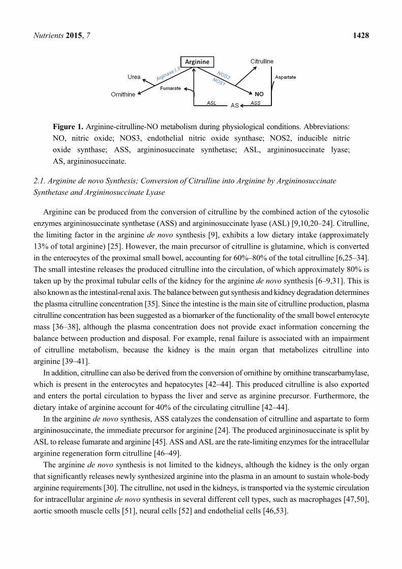
the NOS enzymes-regulated catabolic pathways of arginine will be discussed (Figure 1), since these
pathways play an essential role during inflammatory conditions.
Nutrients 2015, 7 1428
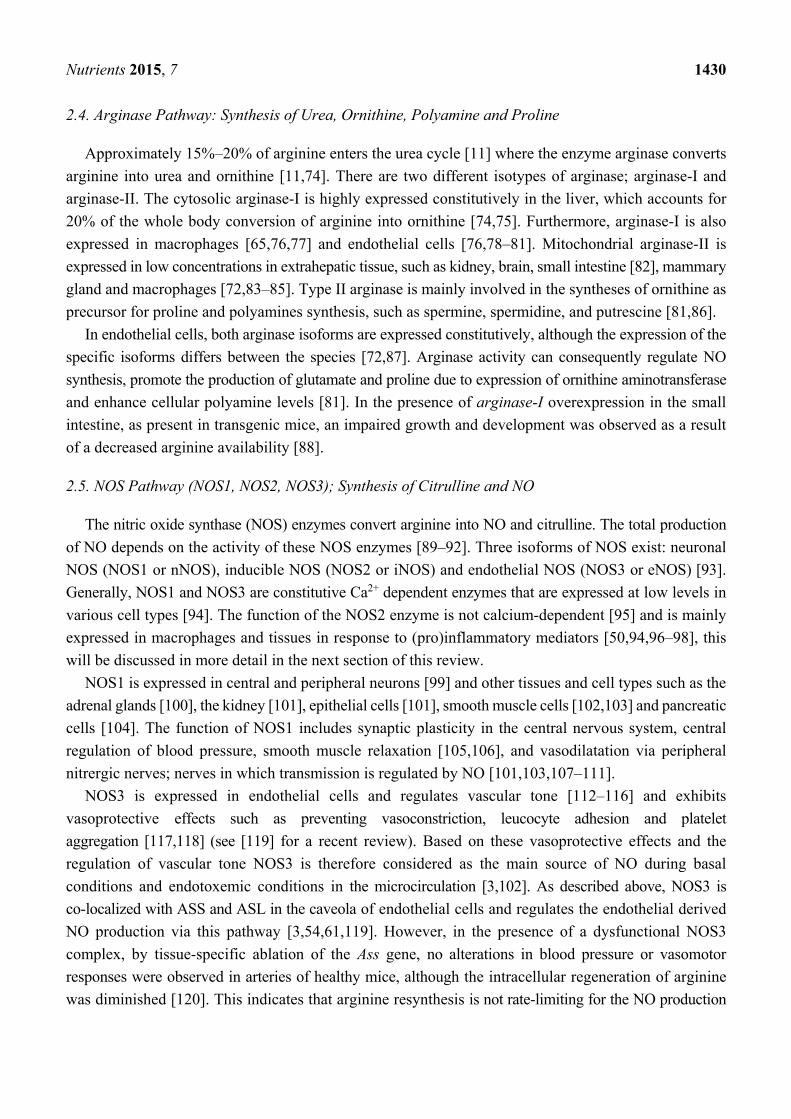
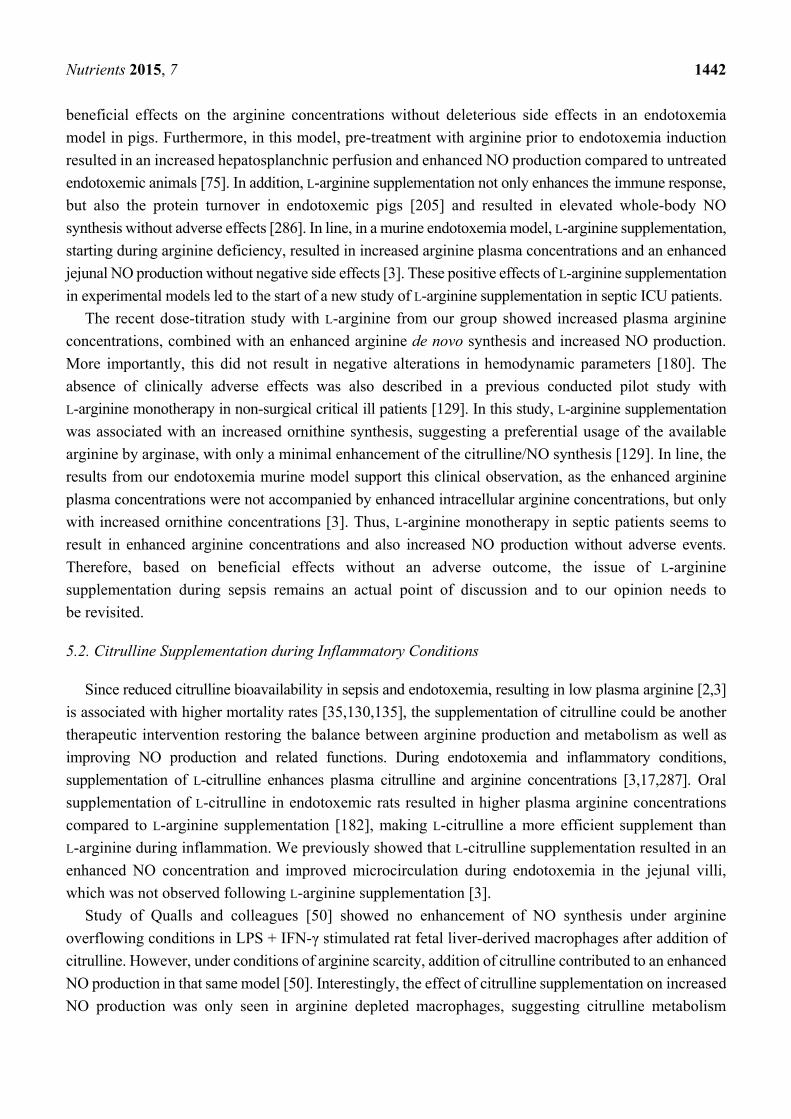
Figure 1. Arginine-citrulline-NO metabolism during physiological conditions. Abbreviations:
NO, nitric oxide; NOS3, endothelial nitric oxide synthase; NOS2, inducible nitric
oxide synthase; ASS, argininosuccinate synthetase; ASL, argininosuccinate lyase;
AS, argininosuccinate.
2.1. Arginine de novo Synthesis; Conversion of Citrulline into Arginine by Argininosuccinate
Synthetase and Argininosuccinate Lyase
Arginine can be produced from the conversion of citrulline by the combined action of the cytosolic
enzymes argininosuccinate synthetase (ASS) and argininosuccinate lyase (ASL) [9,10,20–24]. Citrulline,
the limiting factor in the arginine de novo synthesis [9], exhibits a low dietary intake (approximately
13% of total arginine) [25]. However, the main precursor of citrulline is glutamine, which is converted
in the enterocytes of the proximal small bowel, accounting for 60%–80% of the total citrulline [6,25–34].
The small intestine releases the produced citrulline into the circulation, of which approximately 80% is
taken up by the proximal tubular cells of the kidney for the arginine de novo synthesis [6–9,31]. This is
also known as the intestinal-renal axis. The balance between gut synthesis and kidney degradation determines
the plasma citrulline concentration [35]. Since the intestine is the main site of citrulline production, plasma
citrulline concentration has been suggested as a biomarker of the functionality of the small bowel enterocyte
mass [36–38], although the plasma concentration does not provide exact information concerning the
balance between production and disposal. For example, renal failure is associated with an impairment
of citrulline metabolism, because the kidney is the main organ that metabolizes citrulline into
arginine [39–41].
In addition, citrulline can also be derived from the conversion of ornithine by ornithine transcarbamylase,
which is present in the enterocytes and hepatocytes [42–44]. This produced citrulline is also exported
and enters the portal circulation to bypass the liver and serve as arginine precursor. Furthermore, the
dietary intake of arginine account for 40% of the circulating citrulline [42–44].
In the arginine de novo synthesis, ASS catalyzes the condensation of citrulline and aspartate to form
argininosuccinate, the immediate precursor for arginine [24]. The produced argininosuccinate is split by
ASL to release fumarate and arginine [45]. ASS and ASL are the rate-limiting enzymes for the intracellular
arginine regeneration form citrulline [46–49].
The arginine de novo synthesis is not limited to the kidneys, although the kidney is the only organ
that significantly releases newly synthesized arginine into the plasma in an amount to sustain whole-body
arginine requirements [30]. The citrulline, not used in the kidneys, is transported via the systemic circulation
for intracellular arginine de novo synthesis in several different cell types, such as macrophages [47,50],
aortic smooth muscle cells [51], neural cells [52] and endothelial cells [46,53].
Nutrients 2015, 7 1429
In the endothelial cells, ASS and ASL located in the caveola are part of the NOS3 complex, and
their activity is essential for maintenance of a functional NOS3 complex, to maintain sufficient NO
production [46,54] (Wijnands et al., unpublished data).
The intracellular arginine de novo synthesis in macrophages does not play a role during physiological
conditions, however in case of arginine starvation in inflammatory conditions this pathway may becomes
essential [50,55], which will be discussed in more detail later in this review.
2.2. Arginine and Citrulline Transport Systems
The produced arginine is transported by specific arginine transport systems into the cells, to provide
in the arginine demand of cells. The most important arginine transporter is y+ (for recent review [56–58]),
which consists of four different types of cationic amino acids transporters (CATs); CAT-1, CAT-2a and
CAT-2b, CAT-3 and CAT-4 [58,59]. Since CAT-2a, CAT-3 and CAT-4 have a very low affinity for
cationic amino acids arginine and citrulline, only CAT-1 and CAT-2b will be discussed in this review.
CAT-1 is expressed in almost all adult cells [57], such as endothelial cells [59–61] and epithelial cells.
In endothelial cells, CAT-1 is co-localized with NOS3 in the plasma membrane caveolae which facilitates
specifically arginine channeling for the endothelial NO production [62]. This co-localization results in
exclusive arginine availability for NOS3, preventing mixture of arginine in the intracellular pool [59–61].
CAT-2b is only expressed after induction with cytokines or lipopolysaccharide (LPS) treatment in
inflammatory cells and is highly associated with NOS2 expression [59,63] and will be discussed in more
detail in the next section of this review.
In basal conditions, arginine transport across the cell membrane of human monocytes [64] and murine
bone marrow-derived macrophages [63,65] is mainly mediated through y+ associated with CAT-1, while
after activation of the immune response, the increase in transport is due to CAT-2 [63,65].
Uptake from food-derived citrulline in the enterocytes of the small bowel is regulated by different
transport systems [66], such as B0,+, L, and b0,+ systems [67]. Presence of system L in the cell membrane
of intestinal epithelial cells allows passage of citrulline in both directions depending on the concentration
gradient [68]. Similar to system L, transporter systems b0,+ and B0,+ are also localized in the cell
membrane [67] regulating citrulline uptake. Also the main glutamine transporter in the intestine belongs
to transport system B0, the SLC6A19 or B0AT1 transporter, which is located in the epithelial cell layer
of the intestinal microvilli [69].
2.3. Catabolic Pathways of Arginine during Basal Conditions
Besides its dietary intake and endogenous production, arginine availability is also influenced by
arginine clearance. Arginine catabolism can be catalyzed by five different group of enzymes; arginases
(arginase-I and arginase-II) as part of the urea cycle, nitric oxide synthases (NOSs for the NO production),
arginine decarboxylase (ADC) and arginine:glycine amidinotransferase [70]. Through these pathways
arginine gives rise to ornithine, urea, polyamines, proline, NO and citrulline, proteins, glutamate, agmatine
and finally creatine. In this review only the arginase and NOS pathways during basal and inflammatory
conditions such as sepsis will be discussed (see [16,19,71–73] for additional reviews on these pathways).
Nutrients 2015, 7 1430
2.4. Arginase Pathway: Synthesis of Urea, Ornithine, Polyamine and Proline
Approximately 15%–20% of arginine enters the urea cycle [11] where the enzyme arginase converts
arginine into urea and ornithine [11,74]. There are two different isotypes of arginase; arginase-I and
arginase-II. The cytosolic arginase-I is highly expressed constitutively in the liver, which accounts for
20% of the whole body conversion of arginine into ornithine [74,75]. Furthermore, arginase-I is also
expressed in macrophages [65,76,77] and endothelial cells [76,78–81]. Mitochondrial arginase-II is
expressed in low concentrations in extrahepatic tissue, such as kidney, brain, small intestine [82], mammary
gland and macrophages [72,83–85]. Type II arginase is mainly involved in the syntheses of ornithine as
precursor for proline and polyamines synthesis, such as spermine, spermidine, and putrescine [81,86].
In endothelial cells, both arginase isoforms are expressed constitutively, although the expression of the
specific isoforms differs between the species [72,87]. Arginase activity can consequently regulate NO
synthesis, promote the production of glutamate and proline due to expression of ornithine aminotransferase
and enhance cellular polyamine levels [81]. In the presence of arginase-I overexpression in the small
intestine, as present in transgenic mice, an impaired growth and development was observed as a result
of a decreased arginine availability [88].
2.5. NOS Pathway (NOS1, NOS2, NOS3); Synthesis of Citrulline and NO
The nitric oxide synthase (NOS) enzymes convert arginine into NO and citrulline. The total production
of NO depends on the activity of these NOS enzymes [89–92]. Three isoforms of NOS exist: neuronal
NOS (NOS1 or nNOS), inducible NOS (NOS2 or iNOS) and endothelial NOS (NOS3 or eNOS) [93].
Generally, NOS1 and NOS3 are constitutive Ca2+ dependent enzymes that are expressed at low levels in
various cell types [94]. The function of the NOS2 enzyme is not calcium-dependent [95] and is mainly
expressed in macrophages and tissues in response to (pro)inflammatory mediators [50,94,96–98], this
will be discussed in more detail in the next section of this review.
NOS1 is expressed in central and peripheral neurons [99] and other tissues and cell types such as the
adrenal glands [100], the kidney [101], epithelial cells [101], smooth muscle cells [102,103] and pancreatic
cells [104]. The function of NOS1 includes synaptic plasticity in the central nervous system, central
regulation of blood pressure, smooth muscle relaxation [105,106], and vasodilatation via peripheral
nitrergic nerves; nerves in which transmission is regulated by NO [101,103,107–111].
NOS3 is expressed in endothelial cells and regulates vascular tone [112–116] and exhibits
vasoprotective effects such as preventing vasoconstriction, leucocyte adhesion and platelet
aggregation [117,118] (see [119] for a recent review). Based on these vasoprotective effects and the
regulation of vascular tone NOS3 is therefore considered as the main source of NO during basal
conditions and endotoxemic conditions in the microcirculation [3,102]. As described above, NOS3 is
co-localized with ASS and ASL in the caveola of endothelial cells and regulates the endothelial derived
NO production via this pathway [3,54,61,119]. However, in the presence of a dysfunctional NOS3
complex, by tissue-specific ablation of the Ass gene, no alterations in blood pressure or vasomotor
responses were observed in arteries of healthy mice, although the intracellular regeneration of arginine
was diminished [120]. This indicates that arginine resynthesis is not rate-limiting for the NO production
Nutrients 2015, 7 1431
in the endothelium of healthy arteries, and only becomes important in case of a disease state or in case of a
diminished availability of arginine.
The NOS enzymes require several cofactors for NO synthesis, of which tetrahydrobiopterin (BH4) is
the most important one [121,122]. BH4 stabilizes the NOS structure and enhances the binding of
L-arginine to NOS (see recent review [119]). Therefore, diminished availability or presence of BH4
results in NOS uncoupling, and results in superoxide production [122,123].
3. Arginine-NO Metabolism during Sepsis and Inflammatory Conditions
Sepsis and inflammatory conditions are characterized by organ dysfunction [124,125], as a result of
a misdistribution of blood flow and low peripheral vascular resistance especially at the microcirculatory
level which in turn results from the inflammation and endotoxemia [126–128]. Furthermore, sepsis is
considered to be an arginine deficient state [2–4,129] with arginine becoming a semi-essential amino
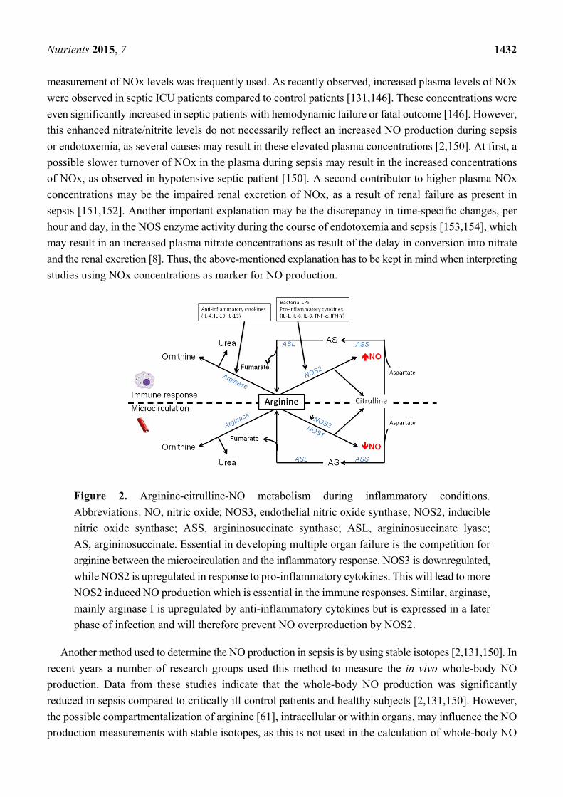
acid during stressed conditions such as sepsis [2,130] (see Figure 2). Although more arginine is being
released from protein breakdown [131], it is also accompanied by an enhanced consumption of arginine,
an impaired arginine de novo synthesis and a decreased supply of arginine, as observed in septic patients
compared with healthy adults and with non-septic intensive care unit patients, leading to an impaired
availability [1,2,131]. However, this does not necessarily mean that arginine deficiency occurs
universally in all septic or inflammatory conditions. It is beyond the purpose of this review to determine
whether arginine deficiency is present in all these conditions. Therefore, we focus on the available
literature on arginine deficiency in human sepsis and animal models, using prolonged endotoxemia to
mimic the arginine deficient state as observed in human septic patients. We previously observed
decreased arginine concentrations in murine endotoxemia and in septic ICU patients [3,132]. During
murine endotoxemia, arginine deficiency was present accompanied by a decreased NO production in the
jejunal tissue of these animals. As a result, an impaired microcirculation in the jejunal villi was
observed [3]. In the septic ICU patients, the decreased arginine concentrations were present independent
of the cause of sepsis [132].
Several causes may be responsible for this impaired arginine production or the enhanced catabolism
of arginine. The impaired arginine production can be the result of limited citrulline availability for
arginine de novo synthesis, which may result from decreased nutritional intake in the critically ill
patient [1,133–136], decreased uptake of protein by intestinal failure [2] or an impaired
glutamine-to-citrulline conversion despite adequate splanchnic glutamine uptake [137]. Furthermore, renal
failure can limit arginine de novo production from citrulline [39–41,138].
Arginine utilization is enhanced by the catabolic activity of arginase [2,139,140] and NOS2 or by an
increased protein synthesis during inflammatory conditions [71,76,131,141–144]. In the past, an enhanced
NO synthesis during the initial phase of sepsis was suggested. This was considered as observed by
enhanced plasma concentrations of nitrate/nitrite (NOx) [145,146]. Based on these increased plasma
nitrite and nitrate levels in septic patients, and on the fact that cytokine-induced NOS2 expression releases
more NO during experimental conditions compared to the other NOS enzymes, a key role for NOS2 in
the hemodynamic changes of sepsis was expected [147]. More recent studies indicate that the role of
NOS2 expression rates and excessive NO production in causing hypotension is overrated [148,149],
which will be discussed in more detail in Section 3.4. To estimate the NO production in septic patients,
Nutrients 2015, 7 1432
measurement of NOx levels was frequently used. As recently observed, increased plasma levels of NOx
were observed in septic ICU patients compared to control patients [131,146]. These concentrations were
even significantly increased in septic patients with hemodynamic failure or fatal outcome [146]. However,
this enhanced nitrate/nitrite levels do not necessarily reflect an increased NO production during sepsis
or endotoxemia, as several causes may result in these elevated plasma concentrations [2,150]. At first, a
possible slower turnover of NOx in the plasma during sepsis may result in the increased concentrations
of NOx, as observed in hypotensive septic patient [150]. A second contributor to higher plasma NOx
concentrations may be the impaired renal excretion of NOx, as a result of renal failure as present in
sepsis [151,152]. Another important explanation may be the discrepancy in time-specific changes, per
hour and day, in the NOS enzyme activity during the course of endotoxemia and sepsis [153,154], which
may result in an increased plasma nitrate concentrations as result of the delay in conversion into nitrate
and the renal excretion [8]. Thus, the above-mentioned explanation has to be kept in mind when interpreting
studies using NOx concentrations as marker for NO production.
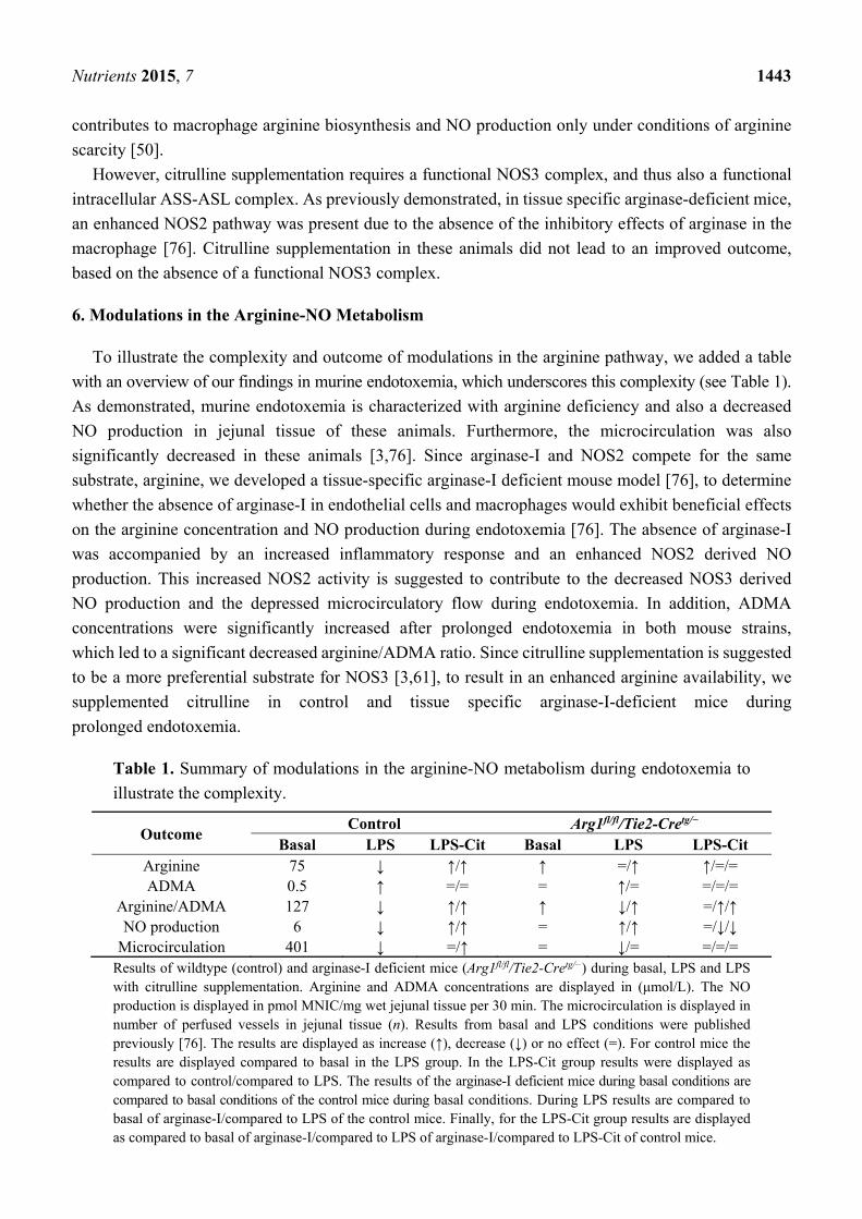
Figure 2. Arginine-citrulline-NO metabolism during inflammatory conditions.
Abbreviations: NO, nitric oxide; NOS3, endothelial nitric oxide synthase; NOS2, inducible
nitric oxide synthase; ASS, argininosuccinate synthase; ASL, argininosuccinate lyase;
AS, argininosuccinate. Essential in developing multiple organ failure is the competition for
arginine between the microcirculation and the inflammatory response. NOS3 is downregulated,
while NOS2 is upregulated in response to pro-inflammatory cytokines. This will lead to more
NOS2 induced NO production which is essential in the immune responses. Similar, arginase,
mainly arginase I is upregulated by anti-inflammatory cytokines but is expressed in a later
phase of infection and will therefore prevent NO overproduction by NOS2.
Another method used to determine the NO production in sepsis is by using stable isotopes [2,131,150]. In
recent years a number of research groups used this method to measure the in vivo whole-body NO
production. Data from these studies indicate that the whole-body NO production was significantly
reduced in sepsis compared to critically ill control patients and healthy subjects [2,131,150]. However,
the possible compartmentalization of arginine [61], intracellular or within organs, may influence the NO
production measurements with stable isotopes, as this is not used in the calculation of whole-body NO
Nutrients 2015, 7 1433
production [155]. As observed, this may also contribute to differences measured in NO concentration with
NOx or total body NO production [2,131,150]. Furthermore, the location of the expressed NOS-isoforms
within the organs may influence the measurement of the NO production. As described by Cauwels
et al. [148], non-hematopoietic cells are essential for the systemic NO production during inflammation,
such as the intestinal enterocytes, the Paneth cells or hepatocytes. Therefore, these different expression
sites, which may be differently affected by inflammation and sepsis, can also lead to unparalleled
concentrations of NO production. For example, nasal NO measurements is a good sensitive and specific
biomarker for nosocomial infection acquisition in the upper airway and respiratory tract [156–158]. In
patients with maxillary sinusitis and sepsis, nasal NO production was significantly reduced in the sinus
epithelium compared to control patients, suggested to be the result of reduced NOS2 mRNA
expression [157]. Also in patients with acute respiratory distress syndrome (ARDS) low NO levels were
measured, which as suggested by the authors may be the result of the rapid reaction of NO with other
molecular species, and therefore of less value to measure pulmonary inflammation [159]. As for patients
with pneumonia on mechanical ventilation, the NO levels were significantly increased, although basal
concentrations were significantly lower compared to other studies [151]. In line, in acute lung injury
patients increased urine NO was measured, which was associated with a better outcome, which may
reflect more perfused lung beds and associated decreased organ failure [160].
Another suggestion is the time point at which the NO production is measured which may influence
the differences in NO production and outcome of the abovementioned studies. This time-point is influenced
by the expression of the enzymes, the possible plateau phase of NOS expression or the maximum NO
production [153,161]. This plateau phase of NO production further depends on sufficient substrate
availability, in other words arginine availability and its co-factors [2,3,131]. As described in the clinical
sepsis studies and experimental studies with endotoxemia, arginine deficiency is related to sepsis and
also to a decreased NO production [2,3,129,131,146]. All the abovementioned factors may lead to the
discrepancy between the low NO production measured in tissue or plasma during sepsis and the increase
in nitrite/nitrate plasma or urine concentrations as measured in experimental and human sepsis. Thus,
measurements of total NO production during sepsis and endotoxemia may only be an estimation of the
total production or presence of NO in the tissues during sepsis and endotoxemia.
On the other hand, the production of NO by NOS3, especially for the endothelial cell, is decreased
during sepsis and prolonged endotoxemia [3,162]. Decreased arginine availability is suggested to play a
role in this downregulation of NOS3 and thereby in the decreased bioavailability of NO. This reduced
NO results in endothelial dysfunction [163,164].
The immune response also contributes to the arginine depletion. During infection and inflammation,
macrophages become active in response to a range of stimuli including damaged cells, pathogens and
inflammatory cytokines such as TNF-α [165]. Non-activated macrophages exhibit minimal utilization
of arginine and do not express NOS2 or arginase activity. Macrophages are classified based on their
two main functions resulting in 2 phenotypes; the M1 macrophages which “fight and kill” and the M2
macrophages which “restore and heal tissue” [166]. As part of the immune response, M1 and M2
macrophages stimulate T-lymphocytes to produce Th1 or Th2 cytokines to maintain the predominant
type of immune phenotype [166]. Upon activation by the immune response, M1-macrophages actively
import extracellular arginine to synthesize NO by NOS2 [50]. This enhanced NOS2 induced NO production
in peripheral blood mononuclear cells is likely to be associated with an increased arginine transport
Nutrients 2015, 7 1434
across the cell membrane [167]. Hypothetically, decreasing the arginine availability during sepsis may,
therefore function as a protective mechanism to decrease excessive NO production by NOS2 and to
regulate excessive adaptive (T-lymphocyte) immune responses, preventing possible excessive inflammation
during sepsis. Arginase, expressed by M2-macrophages, which is part of the anti-inflammatory response,
competes with NOS2 for arginine and therefore contributes to arginine deficiency for M1/Th1-type
macrophages [133,168,169] (see [170] for an excellent review on the competing arginine pathways in
macrophages). However, this not only results in less NO production by M1 macrophages, but also in an
impaired T-cell function [133,169], which is not beneficial during sepsis. The capacity of intracellular
arginine de novo synthesis in M1 macrophages partly preserves the arginine availability for NO
synthesis [171], and as will be discussed in more detail below, also presences NOS2 derived NO
synthesis, which is essential in the immune response as part of the host defense. As for the T cell function,
arginase-induced arginine depletion results in suppression of T-cell activation, proliferation and
differentiation in vitro [172,173] and in vivo [174–176]. However, in human T-cells, this arginine depletion
does not seem to impair the important aspects of T-cell function such as chemotaxis and cytotoxicity [169].
Therefore, these findings, strengthen the believe that a decreased arginine availability may not be beneficial
in sepsis, although a more balanced arginine availability may be essential, which preserves arginine for
both pathways in the immune response, and so maintains the balance between NOS2 and arginase in
inflammatory conditions.
As observed by our group, in tissue-specific arginase deficient mice, lacking arginase in hematopoietic
cells, NOS2 produced significantly more NO during endotoxemia in the tissues and in macrophages
compared to control [76]. However, this was not accompanied with an improved microcirculation in
these animals, which may suggest the importance of compartmentalization of arginine. Based on the different
expression levels and tissues in which the expressed NOS isoforms exist, this may lead to tissue specific
differences in arginine and NO production, and therefore a disbalance in the local (micro)circulation and
immune response. Thus, we believe sepsis and endotoxemia are not characterized by too much NO, but
by a maldistribution of the produced NO, leading to perfusion differences and eventually organ dysfunction
as a result of the impaired microcirculation.
3.1. Alterations in the Arginine and Citrulline Transport Systems in Sepsis
During sepsis or endotoxemia a shift in the expression of CAT transporters is observed as the increased
cytokine production and bacterial endotoxins result in a downregulation of CAT-1 transporters and
upregulation of CAT-2, mainly CAT-2b [56,58,167]. The upregulated CAT-2 is the main arginine
transporter for activated macrophages [50,63,65,177], which import large amounts of extracellular
arginine [63,65] for NOS2-derived NO synthesis [50,58,63]. CAT-2 is co-induced with NOS2 in these
macrophages, which results in shifting arginine towards NOS2 instead of NOS3 [56], to maintain the
required arginine availability for NOS2-derived NO production [178]. This shift in arginine transport is
suggested to be a part of the immune host response to produce increased amounts of NO, as a response
to endotoxemia and the bacterial load, to control pathogens [56,167]. In addition, LPS-induced macrophages
produce the cytokine TNF-α during infection or septicaemia, which is capable of activation of other
macrophages to participate in the host defence system [63,165].
Nutrients 2015, 7 1435
3.2. Decreased Citrulline Availability as Cause for Diminished Arginine de novo Synthesis in
Sepsis/Infection
Besides the observed decreased arginine concentration in sepsis and endotoxemia [2–4,129,132,179,180],
endotoxemia and inflammatory conditions are characterized by a reduced citrulline production and
bioavailability [2,131,181,182]. This reduced citrulline production and bioavailability may contribute to
the decreased arginine de novo synthesis during sepsis and endotoxemia [2,3,131]. These
low citrulline concentrations have been associated with higher mortality rates in this patient
population [35,130,135,183]. Furthermore, lower citrulline concentrations are associated with decreased
NO production [2]. Indeed, supplementation of citrulline during endotoxemia led to an enhanced NO
production in tissues of these animals [3].
A number of mechanisms can be responsible for this reduced citrulline production. At first, a
decreased nutritional intake may contribute to the observed low citrulline and arginine levels in the critically
ill. Moreover, a reduced uptake of glutamine by the enterocyte as part of the intestinal
failure [2,184] is suggested to contribute to the decreased citrulline synthesis [137], although normal
levels of glutamine and ornithine have also been reported in these patients [136,183]. Impaired
absorption of citrulline, arginine and glutamine as a result from a decreased blood flow in the
intestinal villi may be a factor in this reduced uptake by the enterocyte [185]. Also an impaired glutamine-
to-citrulline conversion plays a role in this citrulline deficiency [137].
As a compensation, ornithine transcarbamylase (OTC) may help to restore the citrulline availability
during conditions with low protein intake, such as sepsis and endotoxemia [73]. OTC converts ornithine
into citrulline, which can freely pass the liver to serve as a substrate for the arginine de novo synthesis
in the kidney [186]. Also a reduction of protein synthesis and ureagenesis may aid in restoring the citrulline
and arginine metabolism during these conditions [187].
3.3. Decreased Arginine Availability by Enhanced Arginase Activity in Sepsis
During inflammatory conditions, both arginase-I activity and arginase-II activity are induced [2] by
specific cytokines [188] in macrophages and neutrophils [83,179,189] (see Figure 2). The cytokines that
induce arginase activity are T-helper II produced cytokines, such as interleukins 4, 10 and 13, which
preferentially induce the expression of arginase-I [77,190,191].
The expression of arginase occurs at different time points during the course of endotoxemia. As
observed in an LPS-induced murine-endotoxemia model, arginase-I, already present in low levels before
LPS induction, was upregulated in activated murine peritoneal macrophages at 12 h after the onset of
endotoxemia. Arginase-I remained present during the total time course of the experiment with a peak
expression at 36 h after the onset [142]. On the contrary, arginase-II exhibited an increase in expression after
3 h of LPS stimulation with a peak expression at 6 h after induction and slowly decreased afterwards [142].
As described above, arginase-I and NOS2 compete for the same substrate, which may suggest that
the late onset of arginase-I expression is suggested to prevent the toxic effects of NOS2-induced
overproduction of NO [83]. Indeed, the different time points of peak expression of NOS2 and arginase
in murine endotoxemia models, after, respectively, 20 min, 4–6 h and 12 h for NOS2 [153] and
12–36 h for arginase may indicate this inhibitory effect [142,192–194]. Furthermore, the increased
Nutrients 2015, 7 1436
circulating neutrophils, with arginase containing granules [83] which are capable of releasing arginase
in the extracellular space in case of cell damage or phagocytosis, further contribute to the depletion of
extracellular arginine availability for NO synthesis during endotoxemia [133,143,179].
In endothelial cells both arginase-I and arginase-II are expressed during inflammatory conditions,
which compete with NOS3 for arginine, resulting in an impaired NOS3-derived NO production and
endothelial dysfunction [78–80,195,196]. We recently demonstrated that tissue-specific absence of
arginase-I, in endothelial cells and macrophages, resulted in increased arginine availability. However,
due to the increased NOS2 induced NO production this did not result in beneficial effects upon the
microcirculation during endotoxemia [76]. In addition, an increased arginase-II activity in endothelial
cells also resulted in enhanced arginine utilization. This led to limited substrate availability for NOS3;
resulting in NOS3 uncoupling accompanied by a reduced NO production and augmented superoxide
anions production [78–80].
3.4. Regulation of NOS2 during Sepsis/Inflammation
Historically, cytokine-induced NOS2 expression and consequential enhanced NO production
were suggested to be key factors in the development of sepsis-induced acute hemodynamic
changes [140,197–200] and end-organ damage [146,162,199,201]. The impact of NOS2 derived NO
production was recently re-evaluated in an experimental LPS endotoxemia model indicating no association
between NOS2 derived NO production and hypotension [148].
LPS and T-helper I cytokines, such as interleukin-1, 6 and 8, TNF and interferon-γ, can induce NOS2
expression as part of the cellular immune response [191,202–204]. On cellular level, sepsis results in a
cytokine-mediated induction of NOS2 in almost any cell type [198], but especially in macrophages [197].
Other cell types, including hepatocytes [205], aortic smooth muscle cells [51], vascular smooth muscle
cells [199,205,206], Paneth cells and enterocytes of the jejunum [148] also participate. As mentioned
above, the non-hematopoietic cells contribute to the systemic NO production, and not the hematopoietic
cells such as macrophages or endothelial cells [148,149].
This enhanced NOS2 expression plays a major role in the host defense mechanisms against various
intracellular pathogens [197], and represents a major cytotoxic principle by accomplishing successful
clearance and control of these intracellular pathogens [97,98]. Therefore, maintenance of NOS2 derived
NO is essential during sepsis and endotoxemia as part of the host defense mechanism, as inhibition results
in detrimental outcome, which will be discussed in Section 4.1.
3.5. Regulation and Expression of Constitutive NOS during Infection/Sepsis
During inflammatory conditions and especially during sepsis [207,208], NOS1 exhibits an important
immunoregulatory role, involving both pro-inflammatory and anti-inflammatory pathways [209–211].
As part of the pro-inflammatory pathway, NOS1-induced NO production is suggested to act as a free radical
and is prone to be converted into more reactive nitrogen species [209,212–214]. Endothelial cells express
both NOS1 and NOS3, with NOS1 localized in the nucleus and NOS3 in the cytosol [215]. Previous studies
already indicated an important role for NOS1 in the maintenance of the microvascularisation [102,105,106].
Furthermore, NOS1 localized in nucleus of endothelial cells, exhibits an anti-inflammatory role by
preventing cytokine production after TNF stimulation in cultured endothelial cells [106,215,216].
Nutrients 2015, 7 1437
As for NOS3, inflammatory conditions and the decreased arginine availability result in
NOS3 uncoupling [48,119,217,218], endothelial dysfunction [219], with an increased superoxide
production [119,220] and disturbed microcirculatory flow as a result [221–223].
Another important contributor to NOS3 uncoupling is the induction of asymmetric dimethylarginine
(ADMA). ADMA and NG-methyl-L-arginine (L-NMMA) are the most powerful endogenous
non-selective NOS inhibitors. ADMA competes with L-arginine for the active site of NOS and for
y+-mediated uptake into cells (see [224] for recent review). These methylarginines are eliminated from the
body by enzymatic degradation by dimethylaminohydrolase (DDAH) and renal excretion. Along with
arginine availability, endogenous inhibitors of NOS, including ADMA, may affect NO synthesis [131].
During sepsis increased ADMA levels have been observed which inhibit NOS derived NO production
resulting in an impaired microcirculation [225]. Furthermore, increased mortality in critically ill patients
has also been related to elevated ADMA levels [226,227]. The correlation between arginine and ADMA,
defined as the L-arginine/ADMA ratio, is decreased in adult sepsis patients [228] indicating a role of
ADMA in the modulation of NOS-regulated microcirculation.
3.6. ASS during Sepsis/Inflammation
Inflammation results in an upregulation of ASS expression, especially in macrophages [229,230] and
vascular smooth muscle cells [206,231]. This may indicate stimulation of the intracellular arginine de novo
synthesis to maintain the NOS2-derived NO production [47,229]. During the early phase of inflammation
and sepsis, with still sufficient extracellular arginine available, macrophages export approximately >98%
of the intracellular produced citrulline into the plasma [20,50]. The intracellular regeneration of citrulline,
therefore, only becomes important in case of arginine deficiency. During arginine deficiency, NO synthesis
in macrophages depends on the import of citrulline to maintain the NOS2-induced NO production [50,232].
Since intracellular accumulation of citrulline is thought to facilitate excessive arginine de novo synthesis,
which results in abundant amounts of NO, exporting citrulline is suggested to regulate the NO production
in macrophages [47,233,234]. Another important role of ASS as part of the innate immune system’s
defensive role, is the clearance and detoxification of LPS by binding to the active portion of LPS, lipid A,
which result in inactivation of LPS [235,236]. In addition, ASS released from the liver into the systemic
circulation neutralizes the extracellular LPS-induced cytotoxicity in response to inflammation [230].
Until know, no studies on ASL function during endotoxemia or inflammatory conditions have
been conducted.
4. Modulation of Enzymes
4.1. Modulation of NOS
Historically, excessive NO production by NOS2 [237,238] was suggested to play an important role
in the development of the key features of sepsis and endotoxemia, the systemic hypotension [239] and
vascular hyporeactivity to vasopressor agents [240,241]. The inhibition of the excess NO formation by
inhibiting NOS2 activity was therefore proposed as a treatment for endotoxic shock or sepsis in the past.
Over the past years multiple studies, in vitro and in vivo, have been conducted to investigate the role of
Nutrients 2015, 7 1438
selective NOS2 inhibition or deficiency, all using different sepsis or endotoxemia models with different
selective inhibitors, which resulted in inconclusive results as discussed in brief below [56,178].
Supplementation of the selective NOS2 inhibitor N-(3-(Aminomethyl)benzyl)acetamidine
(1400W) [242], did not affect systemic hemodynamic parameters during prolonged hyperdynamic
porcine endotoxemia [243], however was capable of attenuating the impaired intestinal oxygenation and
energy state [162]. In another porcine endotoxemia model, the supplementation of
the selective NOS2 inhibitor mercaptoethylguanadine (MEG) prevented hypotension and was capable
of decreasing the amount of expired NO, however this did not result in an improved hepatosplanchnic
metabolism [244].
Nω-nitro-L-arginine methyl ester (L-NAME) supplementation during porcine endotoxemia exhibited
detrimental effects on the liver perfusion, as it reduced the flow even further during endotoxemia, while
aminoethyl-isothiourea (AE-ITU) administration resulted in an improved liver blood flow [245].
Diminishing the arginine uptake for NOS2 in CAT-2 knock-out mice resulted in a reduced macrophage
NOS2 activity compared to healthy control mice [56,178]. Furthermore, genetic ablation of NOS2 in
mice exhibited a decrease mortality and enhanced microvasculature responsiveness in a cecal ligation
and puncture induced sepsis model [199]. In addition, in the absence of NOS2 a decreased defense against
bacterial inoculation was observed [246,247], which underlines that NOS2 also exhibits a protective role
during endotoxemia. In vitro studies in rat macrophages and lung cells pre-treated with selective inhibition
of NOS2, with AE-ITU and aminoguanidine (AG), resulted in significantly decreased NOS2 mRNA and
protein expression [248–250]. The supplementation of the selective NOS2 inhibitor ONO-1714 in a septic
lung injury model induced by cecal ligation and puncture in rats lead to a decreased NOS2 activity and
improved survival. However, this beneficial effect was only present when ONO-1714 was administered
12 h after the onset of sepsis [208].
In an ovine acute long injury model, representing sepsis, which was induced by inhalation of
Pseudomonas aeruginosa bacteria administration in the lungs, specific inhibition of NOS2 with AG did
not prevent the characteristic hypotension during endotoxemia, and still exhibited a significant increase
in nitrite production [251]. Also the investigation of a BBS-2, a more potent selective NOS2 inhibitor,
was not capable of reversing the sepsis induced vasodilatation [207], which may indicate that NOS2 may
not or only partially be involved in the vasodilatation during sepsis and inflammation. As recently observed
in an experimental sepsis model with LPS, excessive NOS2-derived NO production may not be accompanied
by hypotension or morbidity during inflammation [148]. This resulted in the usage of non-selective
NOS-inhibitors, to determine the role of the other NOS enzymes in sepsis.
Supplementation of the non-selective NOS inhibitor L-NAME in an experimental hyperdynamic
sepsis model in ewes lead to a normalized renal blood flow, indicating an important contribution of
the local NOS1 and NOS3 derived NO production in the hypotension [252]. In a rat endotoxemia
model, the beneficial and adverse effects of the selective NOS2 inhibitor AE-ITU and the
non-selective NOS-inhibitor L-NMMA were studied to determine the influence on organ failure as caused
by endotoxemia. Both AE-ITU and L-NMMA resulted in NOS2 inhibition and attenuated the liver
dysfunction and circulatory failure in the liver as caused by endotoxemia [253].
However, non-selective NOS inhibition also exhibited unfavorable side effects, such as a decreased
regional blood flow and accompanied decreased cardiac output as observed after the administration
of L-NMMA or L-NAME [254,255]. Also a higher vascular resistance and increased mortality
Nutrients 2015, 7 1439
in LPS treated dogs was demonstrated after administration of the non-selective NOS inhibitor
N omega-amino-L-arginine [256]. In line, prolonged inhibition of NOS by L-NAME, in patients with
severe shock, exhibited an increased blood pressure and vascular resistance during the course of
supplementation. However, the effect of L-NAME tended to decrease during the course of infusion,
which led to the conclusion that L-NAME supplementation only exhibited minor effects on the enhanced
mortality in the patients studied [257].
Furthermore, inhibition of NOS activity in patients suffering from sepsis using the non-specific NOS
inhibitor L-NG-methylarginine-hydrocholine was associated with an increased mortality rate [258]. Patients
supplemented with another non-selective NOS inhibitor, 546C88, exhibited a significantly higher mortality
rate, as a result of increased cardiovascular deaths compared to placebo treated patients within the seven
days follow-up [258]. In search of an explanation why this increased mortality was found, attention
focused on the function of the constitutive expression NOS3 and NOS1. The NO that is produced under
physiological conditions by these NOS isoforms regulates blood-flow distribution in organs. During
inflammation and sepsis, this NOS1 and NOS3-mediated NO production is reduced [259], reducing blood
flow to the individual organs, thereby compromising its function. Indeed, experimental evidence
indicates that inhibition of NOS1 and NOS3 activity during sepsis increases hepatic and splanchnic
damage [260]. Therefore, it is suggested that the upregulation of NOS2 during sepsis may compensate
for the downregulation of NOS1 and NOS3 with respect to organ perfusion [179]. However, as observed
in previous studies untimely or uncontrolled inhibition of non-selective NOS or selective NOS2 during
septic shock may induce splanchnic ischemia seen in some experiments [261]. Indeed, early inhibition
of NOS2 resulted in a trend toward decreased survival, whereas early inhibition of NOS1, by 7-nitroindazole
infusion, exhibited an improved survival in a murine pulmonary sepsis model [262]. On the contrary,
NOS1 inhibition during the early time course of this model, combined with a late NOS2 inhibition,
exhibited potential beneficial effects by decreasing the oxidative stress [263]. Therefore, we believe that
modulations in NOS enzymes activity during human sepsis need to be performed with great precautions
based on the diverse outcome of the several experimental models and the previous detrimental outcome
in human sepsis with NOS2 inhibition. The place at which and the time when the NO is produced may
be the key to better outcome in sepsis and endotoxemia. Thus, maintenance of good NOS isoforms function,
such as NOS3 in the microcirculation for tissue perfusion and NOS2 in the initial phase to fight the
pathogens may be essential. However, this complex regulation mechanism of the arginine-NO metabolism
remains to be investigated in future studies.
4.2. Modulation of Arginase during Inflammatory Conditions
Several methods are used to study inhibition of arginase activity, such as therapeutic intervention with
the non-specific arginase inhibitors L-valine or L-norvaline, or by the usage of (tissue-specific) arginase
deficient mice models. Supplementation of L-valine or L-norvaline resulted in arginase inhibition in
vitro, which led to an increased NO production in bovine pulmonary cells treated with LPS [79]. This
enhanced NO production was also observed after specific knock-down of arginase in an
in vitro experiment in thoracic aortas of rats [196]. Since arginase competes with NOS for the same
substrate, inhibition of arginase directly influences the NO production [79,139]. We recently
demonstrated that tissue specific arginase-I deficiency in macrophages and endothelial cells exhibit an
Nutrients 2015, 7 1440
increased NOS2 mediated NO production in a prolonged murine endotoxemic model, which was
abolished by NOS2 inhibition [76].
Arginase upregulation, as competitor for the substrate arginine, also contributes to endothelial
dysfunction, by NOS3 uncoupling resulting in a decreased NO production [78,87,264]. As previous
observed in vivo chronic inhibition of arginase by administration of 2(S)-amino-6-boronohexanoicacid
restored the endothelial function in the vessels of the rats supplemented with the arginase inhibitor
compared to control [80].
4.3. Modulation of ASS in Sepsis
Absence or reduction of ASS is detrimental due to a decreased NO production [55,206]. In conditions
in which extracellular arginine is depleted from the environment, activated macrophages of ASS-deficient
mice were not able to maintain its function as a fail-safe system to sustain NOS2-derived NO production
[50]. Also in endothelial cells ASS exhibits an essential role as selective reduction of ASS protein levels
by siRNA demonstrated a decreased cell viability and was accompanied by a 80% decrease in NO
production in endothelial cells [46]. ASS deficient mice, as developed by Patejunas et al. [265] in 1994,
resulted in animals with high levels of plasma citrulline, accompanied by hyperammonemia, which
eventually lead to early dead within 24–48 h after birth [265]. Injection of a recombinant adenovirus
carrying human Ass cDNA in these ASS deficient mice ameliorated this neonatal crisis, and resulted in
an expanded life span of approximately 16 days [266].
In heterozygotic ASS deficient mice, ASS+/− mice, significantly increased plasma citrulline and decreased
tissue arginine concentrations were observed [267]. Furthermore, these results were accompanied by a
decreased inflammatory response upon liver injury by pyrazole, as demonstrated by a decreased nitrosative
stress, decreased TNF-α production, impaired apoptosis, and diminished neutrophil infiltration [267],
which may be related to the role of ASS as part of the innate immune systems in the clearance and
detoxification of LPS [235,236]. In our developed tissue-specific ASS deficient mice, with ASS deficiency
in endothelial cells and macrophages [120], we observed similar results during prolonged endotoxemia
in macrophages (Wijnands et al., unpublished data). ASS deficient macrophages exhibited a significant
decreased nitrite production upon LPS stimulation compared to littermates and also a decreased
inflammatory response with decreased TNF-α production.
4.4. Modulation of ASL in Sepsis
The role of ASL modulations in sepsis has not been investigated thus far. However, recently ASL
deficiency was studied in necrotizing enterocolitis, for which a enterocyte-specific heterozygotic deletion
of ASL mouse model was developed (Aslflox/flox; VillinCretg/+ or CKO). This study showed that in the
absence of ASL in the enterocytes an increased pro-inflammatory state and enhanced apoptosis occurred
in these animals [268]. Another recent study, investigating ASL−/− mice, showed early mortality within
48 h after birth in all homozygotes as a result of elevated plasma ammonia, argininosuccinic acid,
glutamine, and citrulline concentrations to toxic concentrations [48]. In addition, significantly lower
concentrations of arginine and nitrite production were observed in the ASL deficient animals, accompanied
by an incompetent NOS3 complex formation, indicating the importance of intracellular regeneration of
citrulline into arginine [48].
Nutrients 2015, 7 1441
5. Enhancement of Arginine and Citrulline Availability in Sepsis and Inflammatory Conditions
with Supplementation
5.1. Arginine Supplementation during Inflammatory Conditions
Over the past decades, supplementation of L-arginine was considered a logical therapeutic option to
restore the decreased arginine levels in septic and critical ill patients, as increased arginine concentrations
may restore the important physiologic processes, including organ perfusion, immune function, protein
synthesis and wound healing [88,269]. Several studies have evaluated the effects of L-arginine
supplementation in a variety of conditions, at first in healthy controls. Arginine supplemented in a dosage
of three to eight grams a day, rarely resulted in unwanted side effects, suggesting the supplementation
of arginine to be safe [270]. However, given in a higher dosage, exceeding nine grams, human subjects
experience gastro-intestinal discomfort, vomiting and diarrhea. The severity of these side effects seem
to be dosage dependent [12].
Previous meta-analysis and systematic reviews evaluated the value of enhanced nutrition as a
possible additional treatment in surgical patients and critically ill patients. Most studies using arginine
as supplement [271,272] investigated a combination of different nutrition compounds [134,273], such
as omega-3-fatty-acids, glutamine [31,274,275], or nucleotides [275], unfortunately all with different
results. A prospective, randomized, double-blind, placebo-controlled study in ICU patients, used oral
arginine supplementation added to the enteral nutrition which resulted in enhanced arginine and ornithine
concentrations, possibly by an increased arginase activity in this patient population without an enhancement
of the NO production [276]. As for the clinical outcome, in a randomized control trial enteral formula
enriched with arginine, and other select nutrients, showed no differences in hospital mortality, infectious
complications or ICU length of stay between the arginine supplemented group and the placebo treated
ICU patients [277]. In line, in another study in critically ill trauma patients [278], no differences in the
arginine supplemented versus placebo treated group on clinical outcomes were observed. However, a
study in severe sepsis patients has proven otherwise [279] as a significantly higher mortality was observed
in the enteral supplemented patients with arginine containing diets [279]. Although all studies agree that
enhancing the metabolic response in patients and especially the critical ill is necessary to improve
outcome and to reduce mortality and complications [280–283], the different results of these studies led
to the questionability of the effectiveness of supplementation, and in this case also of arginine
supplementation [282]. Therefore, experts in the field suggested not to use arginine supplementation in
critical ill patients, as this may result in an enhanced mortality rate [284,285]. However, this questionability
and the assumption of an increased mortality especially after arginine supplementation in sepsis and
critical ill patients may be confounded by grouping different formulas and different types of patients
together. As previously observed in elective surgical patients, a significant reduction in infectious
complications and a trend towards improved mortality in patients supplemented with nutrition containing
high arginine concentrations was present [281], suggesting these negative results of arginine to be patient
population and interventional driven [281]. Therefore, good-quality studies examining L-arginine
monotherapy are essential to define the clinical usage of L-arginine in the critical ill patients.
Prior to the usage of L-arginine monotherapy in critical ill patients, our research group tested
L-arginine monotherapy in experimental sepsis/endotoxemia models. L-arginine supplementation exhibited
Nutrients 2015, 7 1442
beneficial effects on the arginine concentrations without deleterious side effects in an endotoxemia
model in pigs. Furthermore, in this model, pre-treatment with arginine prior to endotoxemia induction
resulted in an increased hepatosplanchnic perfusion and enhanced NO production compared to untreated
endotoxemic animals [75]. In addition, L-arginine supplementation not only enhances the immune response,
but also the protein turnover in endotoxemic pigs [205] and resulted in elevated whole-body NO
synthesis without adverse effects [286]. In line, in a murine endotoxemia model, L-arginine supplementation,
starting during arginine deficiency, resulted in increased arginine plasma concentrations and an enhanced
jejunal NO production without negative side effects [3]. These positive effects of L-arginine supplementation
in experimental models led to the start of a new study of L-arginine supplementation in septic ICU patients.
The recent dose-titration study with L-arginine from our group showed increased plasma arginine
concentrations, combined with an enhanced arginine de novo synthesis and increased NO production.
More importantly, this did not result in negative alterations in hemodynamic parameters [180]. The
absence of clinically adverse effects was also described in a previous conducted pilot study with
L-arginine monotherapy in non-surgical critical ill patients [129]. In this study, L-arginine supplementation
was associated with an increased ornithine synthesis, suggesting a preferential usage of the available
arginine by arginase, with only a minimal enhancement of the citrulline/NO synthesis [129]. In line, the
results from our endotoxemia murine model support this clinical observation, as the enhanced arginine
plasma concentrations were not accompanied by enhanced intracellular arginine concentrations, but only
with increased ornithine concentrations [3]. Thus, L-arginine monotherapy in septic patients seems to
result in enhanced arginine concentrations and also increased NO production without adverse events.
Therefore, based on beneficial effects without an adverse outcome, the issue of L-arginine
supplementation during sepsis remains an actual point of discussion and to our opinion needs to
be revisited.
5.2. Citrulline Supplementation during Inflammatory Conditions
Since reduced citrulline bioavailability in sepsis and endotoxemia, resulting in low plasma arginine [2,3]
is associated with higher mortality rates [35,130,135], the supplementation of citrulline could be another
therapeutic intervention restoring the balance between arginine production and metabolism as well as
improving NO production and related functions. During endotoxemia and inflammatory conditions,
supplementation of L-citrulline enhances plasma citrulline and arginine concentrations [3,17,287]. Oral
supplementation of L-citrulline in endotoxemic rats resulted in higher plasma arginine concentrations
compared to L-arginine supplementation [182], making L-citrulline a more efficient supplement than
L-arginine during inflammation. We previously showed that L-citrulline supplementation resulted in an
enhanced NO concentration and improved microcirculation during endotoxemia in the jejunal villi,
which was not observed following L-arginine supplementation [3].
Study of Qualls and colleagues [50] showed no enhancement of NO synthesis under arginine
overflowing conditions in LPS + IFN-γ stimulated rat fetal liver-derived macrophages after addition of
citrulline. However, under conditions of arginine scarcity, addition of citrulline contributed to an enhanced
NO production in that same model [50]. Interestingly, the effect of citrulline supplementation on increased
NO production was only seen in arginine depleted macrophages, suggesting citrulline metabolism
Nutrients 2015, 7 1443
contributes to macrophage arginine biosynthesis and NO production only under conditions of arginine
scarcity [50].
However, citrulline supplementation requires a functional NOS3 complex, and thus also a functional
intracellular ASS-ASL complex. As previously demonstrated, in tissue specific arginase-deficient mice,
an enhanced NOS2 pathway was present due to the absence of the inhibitory effects of arginase in the
macrophage [76]. Citrulline supplementation in these animals did not lead to an improved outcome,
based on the absence of a functional NOS3 complex.
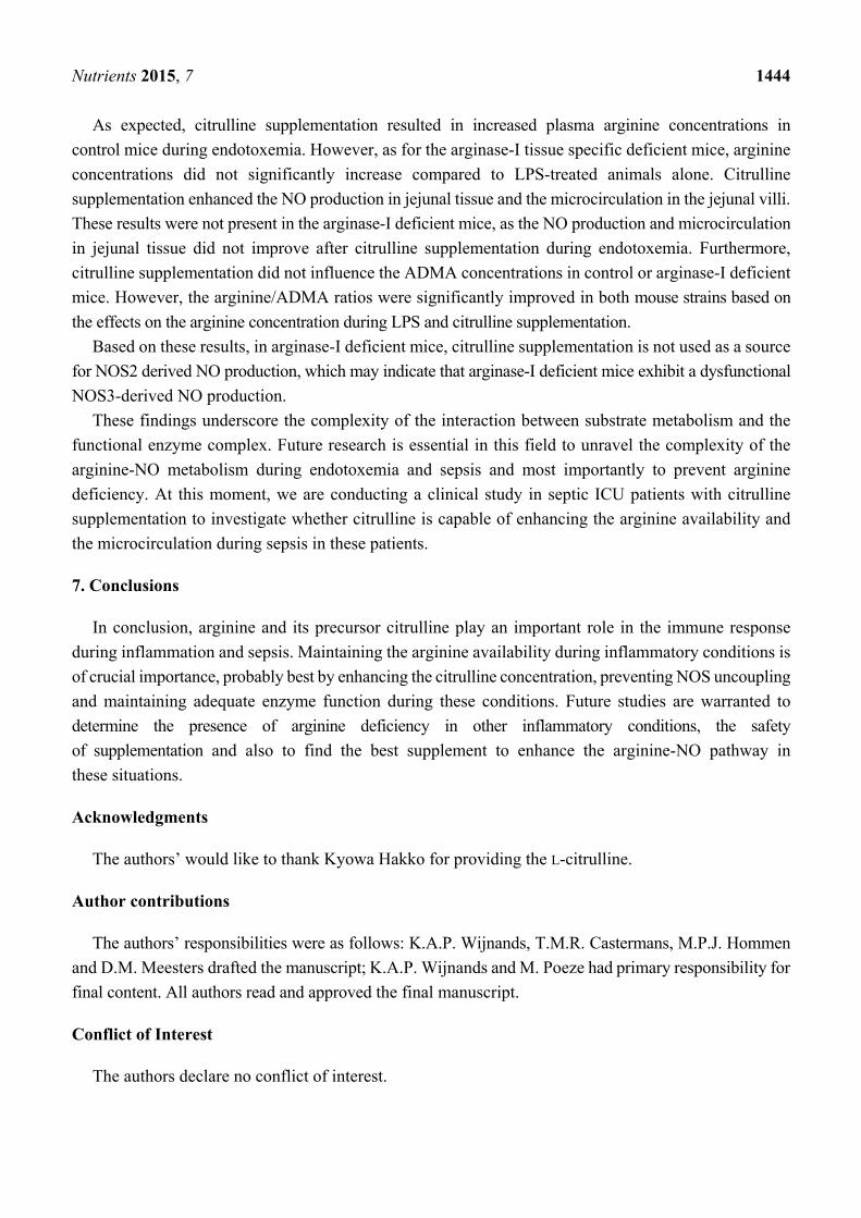
6. Modulations in the Arginine-NO Metabolism
To illustrate the complexity and outcome of modulations in the arginine pathway, we added a table
with an overview of our findings in murine endotoxemia, which underscores this complexity (see Table 1).
As demonstrated, murine endotoxemia is characterized with arginine deficiency and also a decreased
NO production in jejunal tissue of these animals. Furthermore, the microcirculation was also
significantly decreased in these animals [3,76]. Since arginase-I and NOS2 compete for the same
substrate, arginine, we developed a tissue-specific arginase-I deficient mouse model [76], to determine
whether the absence of arginase-I in endothelial cells and macrophages would exhibit beneficial effects
on the arginine concentration and NO production during endotoxemia [76]. The absence of arginase-I
was accompanied by an increased inflammatory response and an enhanced NOS2 derived NO
production. This increased NOS2 activity is suggested to contribute to the decreased NOS3 derived
NO production and the depressed microcirculatory flow during endotoxemia. In addition, ADMA
concentrations were significantly increased after prolonged endotoxemia in both mouse strains,
which led to a significant decreased arginine/ADMA ratio. Since citrulline supplementation is suggested
to be a more preferential substrate for NOS3 [3,61], to result in an enhanced arginine availability, we
supplemented citrulline in control and tissue specific arginase-I-deficient mice during
prolonged endotoxemia.
Table 1. Summary of modulations in the arginine-NO metabolism during endotoxemia to
illustrate the complexity.
Outcome Control Arg1fl/fl/Tie2-Cretg/−
Basal LPS LPS-Cit Basal LPS LPS-Cit Arginine 75 ↓ ↑/↑ ↑ =/↑ ↑/=/= ADMA 0.5 ↑ =/= = ↑/= =/=/=
Arginine/ADMA 127 ↓ ↑/↑ ↑ ↓/↑ =/↑/↑ NO production 6 ↓ ↑/↑ = ↑/↑ =/↓/↓
Microcirculation 401 ↓ =/↑ = ↓/= =/=/= Results of wildtype (control) and arginase-I deficient mice (Arg1fl/fl/Tie2-Cretg/−) during basal, LPS and LPS with citrulline supplementation. Arginine and ADMA concentrations are displayed in (μmol/L). The NO production is displayed in pmol MNIC/mg wet jejunal tissue per 30 min. The microcirculation is displayed in number of perfused vessels in jejunal tissue (n). Results from basal and LPS conditions were published previously [76]. The results are displayed as increase (↑), decrease (↓) or no effect (=). For control mice the results are displayed compared to basal in the LPS group. In the LPS-Cit group results were displayed as compared to control/compared to LPS. The results of the arginase-I deficient mice during basal conditions are compared to basal conditions of the control mice during basal conditions. During LPS results are compared to basal of arginase-I/compared to LPS of the control mice. Finally, for the LPS-Cit group results are displayed as compared to basal of arginase-I/compared to LPS of arginase-I/compared to LPS-Cit of control mice.
Nutrients 2015, 7 1444
As expected, citrulline supplementation resulted in increased plasma arginine concentrations in
control mice during endotoxemia. However, as for the arginase-I tissue specific deficient mice, arginine
concentrations did not significantly increase compared to LPS-treated animals alone. Citrulline
supplementation enhanced the NO production in jejunal tissue and the microcirculation in the jejunal villi.
These results were not present in the arginase-I deficient mice, as the NO production and microcirculation
in jejunal tissue did not improve after citrulline supplementation during endotoxemia. Furthermore,
citrulline supplementation did not influence the ADMA concentrations in control or arginase-I deficient
mice. However, the arginine/ADMA ratios were significantly improved in both mouse strains based on
the effects on the arginine concentration during LPS and citrulline supplementation.
Based on these results, in arginase-I deficient mice, citrulline supplementation is not used as a source
for NOS2 derived NO production, which may indicate that arginase-I deficient mice exhibit a dysfunctional
NOS3-derived NO production.
These findings underscore the complexity of the interaction between substrate metabolism and the
functional enzyme complex. Future research is essential in this field to unravel the complexity of the
arginine-NO metabolism during endotoxemia and sepsis and most importantly to prevent arginine
deficiency. At this moment, we are conducting a clinical study in septic ICU patients with citrulline
supplementation to investigate whether citrulline is capable of enhancing the arginine availability and
the microcirculation during sepsis in these patients.
7. Conclusions
In conclusion, arginine and its precursor citrulline play an important role in the immune response
during inflammation and sepsis. Maintaining the arginine availability during inflammatory conditions is
of crucial importance, probably best by enhancing the citrulline concentration, preventing NOS uncoupling
and maintaining adequate enzyme function during these conditions. Future studies are warranted to
determine the presence of arginine deficiency in other inflammatory conditions, the safety
of supplementation and also to find the best supplement to enhance the arginine-NO pathway in
these situations.
Acknowledgments
The authors’ would like to thank Kyowa Hakko for providing the L-citrulline.
Author contributions
The authors’ responsibilities were as follows: K.A.P. Wijnands, T.M.R. Castermans, M.P.J. Hommen
and D.M. Meesters drafted the manuscript; K.A.P. Wijnands and M. Poeze had primary responsibility for
final content. All authors read and approved the final manuscript.
Conflict of Interest
The authors declare no conflict of interest.
Nutrients 2015, 7 1445
References
1. Argaman, Z.; Young, V.R.; Noviski, N.; Castillo-Rosas, L.; Lu, X.M.; Zurakowski, D.; Cooper, M.;
Davison, C.; Tharakan, J.F.; Ajami, A.; et al. Arginine and nitric oxide metabolism in critically ill
septic pediatric patients. Crit. Care Med. 2003, 31, 591–597.
2. Luiking, Y.C.; Poeze, M.; Ramsay, G.; Deutz, N.E. Reduced citrulline production in sepsis is
related to diminished de novo arginine and nitric oxide production. Am. J. Clin. Nutr. 2009, 89,
142–152.
3. Wijnands, K.A.; Vink, H.; Briede, J.J.; van Faassen, E.E.; Lamers, W.H.; Buurman, W.A.; Poeze, M.
Citrulline a more suitable substrate than arginine to restore no production and the microcirculation
during endotoxemia. PLoS One 2012, 7, e37439, doi:10.1371/journal.pone.0037439.
4. Davis, J.S.; Anstey, N.M. Is plasma arginine concentration decreased in patients with sepsis?
A systematic review and meta-analysis. Crit. Care Med. 2011, 39, 380–385.
5. Heys, S.D.; Gardner, E. Nutrients and the surgical patient: Current and potential therapeutic
applications to clinical practice. J. R. Coll. Surg. Edinb. 1999, 44, 283–293.
6. Windmueller, H.G.; Spaeth, A.E. Source and fate of circulating citrulline. Am. J. Physiol. 1981,
241, E473–E480.
7. Yu, Y.M.; Burke, J.F.; Tompkins, R.G.; Martin, R.; Young, V.R. Quantitative aspects of interorgan
relationships among arginine and citrulline metabolism. Am. J. Physiol. 1996, 271, E1098–E1109.
8. Castillo, L.; Chapman, T.E.; Sanchez, M.; Yu, Y.M.; Burke, J.F.; Ajami, A.M.; Vogt, J.; Young, V.R.
Plasma arginine and citrulline kinetics in adults given adequate and arginine-free diets. Proc. Natl.
Acad. Sci. USA 1993, 90, 7749–7753.
9. Dhanakoti, S.N.; Brosnan, J.T.; Herzberg, G.R.; Brosnan, M.E. Renal arginine synthesis: Studies
in vitro and in vivo. Am. J. Physiol. 1990, 259, E437–E442.
10. Featherston, W.R.; Rogers, Q.R.; Freedland, R.A. Relative importance of kidney and liver in synthesis
of arginine by the rat. Am. J. Physiol. 1973, 224, 127–129.
11. Castillo, L.; Beaumier, L.; Ajami, A.M.; Young, V.R. Whole body nitric oxide synthesis in healthy
men determined from [15N]arginine-to-[15N]citrulline labeling. Proc. Natl. Acad. Sci. USA 1996,
93, 11460–11465.
12. Grimble, G.K. Adverse gastrointestinal effects of arginine and related amino acids. J. Nutr. 2007,
137, 1693S–1701S.
13. Morris, S.M., Jr. Regulation of enzymes of the urea cycle and arginine metabolism. Annu. Rev. Nutr.
2002, 22, 87–105.
14. Wu, G.; Morris, S.M., Jr. Arginine metabolism: Nitric oxide and beyond. Biochem. J. 1998, 336,
1–17.
15. Thibault, R.; Flet, L.; Vavasseur, F.; Lemerle, M.; Ferchaud-Roucher, V.; Picot, D.; Darmaun, D.
Oral citrulline does not affect whole body protein metabolism in healthy human volunteers: Results
of a prospective, randomized, double-blind, cross-over study. Clin. Nutr. 2011, 30,
807–811.
16. Wu, G.; Bazer, F.W.; Davis, T.A.; Kim, S.W.; Li, P.; Marc Rhoads, J.; Carey Satterfield, M.;
Smith, S.B.; Spencer, T.E.; Yin, Y. Arginine metabolism and nutrition in growth, health and
disease. Amino Acids 2009, 37, 153–168.
Nutrients 2015, 7 1446
17. Cynober, L. Pharmacokinetics of arginine and related amino acids. J. Nutr. 2007, 137,
1646S–1649S.
18. Morris, S.M., Jr. Arginine metabolism: Boundaries of our knowledge. J. Nutr. 2007, 137,
1602S–1609S.
19. Morris, S.M., Jr. Enzymes of arginine metabolism. J. Nutr. 2004, 134, 2743S–2747S.
20. Hammermann, R.; Bliesener, N.; Mossner, J.; Klasen, S.; Wiesinger, H.; Wessler, I.; Racke, K.
Inability of rat alveolar macrophages to recycle L-citrulline to L-arginine despite induction of
argininosuccinate synthetase mRNA and protein, and inhibition of nitric oxide synthesis by
exogenous L-citrulline. Naunyn. Schmiedebergs Arch. Pharmacol. 1998, 358, 601–607.
21. Ratner, S.; Petrack, B. Biosynthesis of urea. III. Further studies on arginine synthesis from citrulline.
J. Biol. Chem. 1951, 191, 693–705.
22. Levillain, O.; Hus-Citharel, A.; Morel, F.; Bankir, L. Localization of arginine synthesis along rat
nephron. Am. J. Physiol. 1990, 259, F916–F923.
23. Wakui, H.; Komatsuda, A.; Itoh, H.; Kobayashi, R.; Nakamoto, Y.; Miura, A.B. Renal
argininosuccinate synthetase: Purification, immunohistochemical localization, and elastin-binding
property. Ren. Physiol. Biochem. 1992, 15, 1–9.
24. Rochovansky, O.; Ratner, S. Biosynthesis of urea. XII. Further studies on argininosuccinate
synthetase: Substrate affinity and mechanism of action. J. Biol. Chem. 1967, 242, 3839–3849.
25. Van de Poll, M.C.; Ligthart-Melis, G.C.; Boelens, P.G.; Deutz, N.E.; van Leeuwen, P.A.;
Dejong, C.H. Intestinal and hepatic metabolism of glutamine and citrulline in humans. J. Physiol.
2007, 581, 819–827.
26. Fujita, T.; Yanaga, K. Association between glutamine extraction and release of citrulline and glycine
by the human small intestine. Life Sci. 2007, 80, 1846–1850.
27. Peters, J.H.; Wierdsma, N.J.; Teerlink, T.; van Leeuwen, P.A.; Mulder, C.J.; van Bodegraven, A.A.
The citrulline generation test: Proposal for a new enterocyte function test. Aliment. Pharmacol. Ther.
2008, 27, 1300–1310.
28. Wu, G.; Knabe, D.A.; Flynn, N.E. Synthesis of citrulline from glutamine in pig enterocytes.
Biochem. J. 1994, 299 (Pt 1), 115–121.
29. Wu, G. Synthesis of citrulline and arginine from proline in enterocytes of postnatal pigs. Am. J. Physiol.
1997, 272, G1382–G1390.
30. Van de Poll, M.C.; Siroen, M.P.; van Leeuwen, P.A.; Soeters, P.B.; Melis, G.C.; Boelens, P.G.;
Deutz, N.E.; Dejong, C.H. Interorgan amino acid exchange in humans: Consequences for arginine
and citrulline metabolism. Am. J. Clin. Nutr. 2007, 85, 167–172.
31. Ligthart-Melis, G.C.; van de Poll, M.C.; Boelens, P.G.; Dejong, C.H.; Deutz, N.E.;
van Leeuwen, P.A. Glutamine is an important precursor for de novo synthesis of arginine in
humans. Am. J. Clin. Nutr. 2008, 87, 1282–1289.
32. Ligthart-Melis, G.C.; van de Poll, M.C.; Dejong, C.H.; Boelens, P.G.; Deutz, N.E.;
van Leeuwen, P.A. The route of administration (enteral or parenteral) affects the conversion of
isotopically labeled L-[2–15N]glutamine into citrulline and arginine in humans. JPEN J. Parenter
Enteral. Nutr. 2007, 31, 343–348.

Nutrients 2015, 7 1447
33. Boelens, P.G.; Melis, G.C.; van Leeuwen, P.A.; ten Have, G.A.; Deutz, N.E. Route of administration
(enteral or parenteral) affects the contribution of L-glutamine to de novo L-arginine synthesis in mice:
A stable-isotope study. Am. J. Physiol. Endocrinol. Metab. 2006, 291, E683–E690.
34. Boelens, P.G.; van Leeuwen, P.A.; Dejong, C.H.; Deutz, N.E. Intestinal renal metabolism of
L-citrulline and L-arginine following enteral or parenteral infusion of L-alanyl-L-[2,15N]glutamine or
L-[2,15N]glutamine in mice. Am. J. Physiol. Gastrointest Liver Physiol. 2005, 289, G679–G685.
35. Piton, G.; Manzon, C.; Monnet, E.; Cypriani, B.; Barbot, O.; Navellou, J.C.; Carbonnel, F.;
Capellier, G. Plasma citrulline kinetics and prognostic value in critically ill patients.
Intensive Care Med. 2010, 36, 702–706.
36. Jianfeng, G.; Weiming, Z.; Ning, L.; Fangnan, L.; Li, T.; Nan, L.; Jieshou, L. Serum citrulline is a
simple quantitative marker for small intestinal enterocytes mass and absorption function in short
bowel patients. J. Surg. Res. 2005, 127, 177–182.
37. Crenn, P.; Coudray-Lucas, C.; Cynober, L.; Messing, B. Post-absorptive plasma citrulline
concentration: A marker of intestinal failure in humans. Transplant Proc. 1998, 30, 2528,
doi:10.1016/S0041-1345(98)00711-8.
38. Crenn, P.; Coudray-Lucas, C.; Thuillier, F.; Cynober, L.; Messing, B. Postabsorptive plasma
citrulline concentration is a marker of absorptive enterocyte mass and intestinal failure in humans.
Gastroenterology 2000, 119, 1496–1505.
39. Lau, T.; Owen, W.; Yu, Y.M.; Noviski, N.; Lyons, J.; Zurakowski, D.; Tsay, R.; Ajami, A.;
Young, V.R.; Castillo, L. Arginine, citrulline, and nitric oxide metabolism in end-stage renal
disease patients. J. Clin. Investig. 2000, 105, 1217–1225.
40. Ceballos, I.; Chauveau, P.; Guerin, V.; Bardet, J.; Parvy, P.; Kamoun, P.; Jungers, P. Early alterations
of plasma free amino acids in chronic renal failure. Clin. Chim. Acta 1990, 188, 101–108.
41. Levillain, O.; Parvy, P.; Hassler, C. Amino acid handling in uremic rats: Citrulline, a reliable marker
of renal insufficiency and proximal tubular dysfunction. Metabolism 1997, 46, 611–618.
42. Marini, J.C. Arginine and ornithine are the main precursors for citrulline synthesis in mice.
J. Nutr. 2012, 142, 572–580.
43. Tomlinson, C.; Rafii, M.; Ball, R.O.; Pencharz, P.B. Arginine can be synthesized from enteral
proline in healthy adult humans. J. Nutr. 2011, 141, 1432–1436.
44. Marini, J.C.; Didelija, I.C.; Castillo, L.; Lee, B. Plasma arginine and ornithine are the main
citrulline precursors in mice infused with arginine-free diets. J. Nutr. 2010, 140, 1432–1437.
45. Ratner, S.; Anslow, W.P., Jr.; Petrack, B. Biosynthesis of urea. VI. Enzymatic cleavage of
argininosuccinic acid to arginine and fumaric acid. J. Biol. Chem. 1953, 204, 115–125.
46. Goodwin, B.L.; Solomonson, L.P.; Eichler, D.C. Argininosuccinate synthase expression is required
to maintain nitric oxide production and cell viability in aortic endothelial cells. J. Biol. Chem. 2004,
279, 18353–18360.
47. Nussler, A.K.; Billiar, T.R.; Liu, Z.Z.; Morris, S.M., Jr. Coinduction of nitric oxide synthase and
argininosuccinate synthetase in a murine macrophage cell line. Implications for regulation of nitric
oxide production. J. Biol. Chem. 1994, 269, 1257–1261.
48. Erez, A.; Nagamani, S.C.; Shchelochkov, O.A.; Premkumar, M.H.; Campeau, P.M.; Chen, Y.;
Garg, H.K.; Li, L.; Mian, A.; Bertin, T.K.; et al. Requirement of argininosuccinate lyase for systemic
nitric oxide production. Nat. Med. 2011, 17, 1619–1626.
Nutrients 2015, 7 1448
49. Cao, M.; George, T.J.; Prima, V.; Nelson, D.; Svetlov, S. Argininosuccinate synthase as a novel
biomarker for inflammatory conditions. Biomarkers 2013, 18, 242–249.
50. Qualls, J.E.; Subramanian, C.; Rafi, W.; Smith, A.M.; Balouzian, L.; DeFreitas, A.A.; Shirey, K.A.;
Reutterer, B.; Kernbauer, E.; Stockinger, S.; et al. Sustained generation of nitric oxide and control
of mycobacterial infection requires argininosuccinate synthase 1. Cell Host Microbe. 2012, 12,
313–323.
51. Wileman, S.M.; Mann, G.E.; Pearson, J.D.; Baydoun, A.R. Role of L-citrulline transport in nitric oxide
synthesis in rat aortic smooth muscle cells activated with LPS and interferon-gamma. Br. J.
Pharmacol. 2003, 140, 179–185.
52. Schmidlin, A.; Fischer, S.; Wiesinger, H. Transport of L-citrulline in neural cell cultures. Dev.
Neurosci. 2000, 22, 393–398.
53. Hilderman, R.H.; Casey, T.E.; Pojoga, L.H. P(1),P(4)-Diadenosine 5′-tetraphosphate modulates
L-arginine and L-citrulline uptake by bovine aortic endothelial cells. Arch. Biochem. Biophys. 2000,
375, 124–130.
54. Flam, B.R.; Eichler, D.C.; Solomonson, L.P. Endothelial nitric oxide production is tightly coupled
to the citrulline-no cycle. Nitric Oxide 2007, 17, 115–121.
55. Hattori, Y.; Campbell, E.B.; Gross, S.S. Argininosuccinate synthetase mrna and activity are
induced by immunostimulants in vascular smooth muscle. Role in the regeneration or arginine for
nitric oxide synthesis. J. Biol. Chem. 1994, 269, 9405–9408.
56. Schwartz, D.; Schwartz, I.F.; Gnessin, E.; Wollman, Y.; Chernichovsky, T.; Blum, M.; Iaina, A.
Differential regulation of glomerular arginine transporters (CAT-1 and CAT-2) in
lipopolysaccharide-treated rats. Am. J. Physiol. Renal. Physiol. 2003, 284, F788–F795.
57. Closs, E.I.; Boissel, J.P.; Habermeier, A.; Rotmann, A. Structure and function of cationic amino
acid transporters (CATs). J. Membr. Biol. 2006, 213, 67–77.
58. Fotiadis, D.; Kanai, Y.; Palacin, M. The SLC3 and SLC7 families of amino acid transporters.
Mol. Aspects Med. 2013, 34, 139–158.
59. Verrey, F.; Closs, E.I.; Wagner, C.A.; Palacin, M.; Endou, H.; Kanai, Y. CATs and HATs: The
SLC7 family of amino acid transporters. Pflugers Arch. 2004, 447, 532–542.
60. Cynober, L.A. Plasma amino acid levels with a note on membrane transport: Characteristics,
regulation, and metabolic significance. Nutrition 2002, 18, 761–766.
61. Shen, L.J.; Beloussow, K.; Shen, W.C. Accessibility of endothelial and inducible nitric oxide
synthase to the intracellular citrulline-arginine regeneration pathway. Biochem. Pharmacol. 2005,
69, 97–104.
62. McDonald, K.K.; Zharikov, S.; Block, E.R.; Kilberg, M.S. A caveolar complex between the
cationic amino acid transporter 1 and endothelial nitric-oxide synthase may explain the “Arginine
paradox”. J. Biol. Chem. 1997, 272, 31213–31216.
63. Yeramian, A.; Martin, L.; Serrat, N.; Arpa, L.; Soler, C.; Bertran, J.; McLeod, C.; Palacin, M.;
Modolell, M.; Lloberas, J.; et al. Arginine transport via cationic amino acid transporter 2 plays a
critical regulatory role in classical or alternative activation of macrophages. J. Immunol. 2006, 176,
5918–5924.
Nutrients 2015, 7 1449
64. Rotoli, B.M.; Bussolati, O.; Sala, R.; Barilli, A.; Talarico, E.; Gazzola, G.C.; Dall’Asta, V.
Infgamma stimulates arginine transport through system y+L in human monocytes. FEBS Lett. 2004,
571, 177–181.
65. Yeramian, A.; Martin, L.; Arpa, L.; Bertran, J.; Soler, C.; McLeod, C.; Modolell, M.; Palacin, M.;
Lloberas, J.; Celada, A. Macrophages require distinct arginine catabolism and transport systems
for proliferation and for activation. Eur. J. Immunol. 2006, 36, 1516–1526.
66. Vadgama, J.V.; Evered, D.F. Characteristics of L-citrulline transport across rat small intestine
in vitro. Pediatr. Res. 1992, 32, 472–478.
67. Bahri, S.; Curis, E.; el Wafi, F.Z.; Aussel, C.; Chaumeil, J.C.; Cynober, L.; Zerrouk, N.
Mechanisms and kinetics of citrulline uptake in a model of human intestinal epithelial cells.
Clin. Nutr. 2008, 27, 872–880.
68. Chen, J.; Zhu, Y.; Hu, M. Mechanisms and kinetics of uptake and efflux of L-methionine in an
intestinal epithelial model (Caco-2). J. Nutr. 1994, 124, 1907–1916.
69. Pochini, L.; Scalise, M.; Galluccio, M.; Indiveri, C. Membrane transporters for the special amino
acid glutamine: Structure/function relationships and relevance to human health. Front. Chem.
2014, 2, 61, doi:10.3389/fchem.2014.00061.
70. Nagata, N.; Endo, F.; Matsuda, I. Ornithine carbamoyltransferase (OCT) in the jejunal mucosa, as
a reference of the liver oct. Clin. Chim. Acta 1983, 134, 155–166.
71. Luiking, Y.C.; ten Have, G.A.; Wolfe, R.R.; Deutz, N.E. Arginine de novo and nitric oxide
production in disease states. Am. J. Physiol. Endocrinol. Metab. 2012, 303, E1177–E1189.
72. Morris, S.M., Jr. Recent advances in arginine metabolism: Roles and regulation of the arginases.
Br. J. Pharmacol. 2009, 157, 922–930.
73. Curis, E.; Nicolis, I.; Moinard, C.; Osowska, S.; Zerrouk, N.; Benazeth, S.; Cynober, L. Almost all
about citrulline in mammals. Amino Acids 2005, 29, 177–205.
74. Yu, H.; Yoo, P.K.; Aguirre, C.C.; Tsoa, R.W.; Kern, R.M.; Grody, W.W.; Cederbaum, S.D.;
Iyer, R.K. Widespread expression of arginase I in mouse tissues. Biochemical and physiological
implications. J. Histochem. Cytochem. 2003, 51, 1151–1160.
75. Poeze, M.; Bruins, M.J.; Kessels, F.; Luiking, Y.C.; Lamers, W.H.; Deutz, N.E. Effects of
L-arginine pretreatment on nitric oxide metabolism and hepatosplanchnic perfusion during porcine
endotoxemia. Am. J. Clin. Nutr. 2011, 93, 1237–1247.
76. Wijnands, K.A.; Hoeksema, M.A.; Meesters, D.M.; van den Akker, N.M.; Molin, D.G.; Briede, J.J.;
Ghosh, M.; Kohler, S.E.; van Zandvoort, M.A.; de Winther, M.P.; et al. Arginase-1 deficiency
regulates arginine concentrations and NOS2-mediated no production during endotoxemia. PLoS
One 2014, 9, e86135, doi:10.1371/journal.pone.0086135.
77. Louis, C.A.; Mody, V.; Henry, W.L., Jr.; Reichner, J.S.; Albina, J.E. Regulation of arginase
isoforms I and II by IL-4 in cultured murine peritoneal macrophages. Am. J. Physiol. 1999, 276,
R237–R242.
78. Berkowitz, D.E.; White, R.; Li, D.; Minhas, K.M.; Cernetich, A.; Kim, S.; Burke, S.;
Shoukas, A.A.; Nyhan, D.; Champion, H.C.; et al. Arginase reciprocally regulates nitric oxide
synthase activity and contributes to endothelial dysfunction in aging blood vessels. Circulation 2003,
108, 2000–2006.
Nutrients 2015, 7 1450
79. Chicoine, L.G.; Paffett, M.L.; Young, T.L.; Nelin, L.D. Arginase inhibition increases nitric oxide
production in bovine pulmonary arterial endothelial cells. Am. J. Physiol. Lung Cell Mol. Physiol.
2004, 287, L60–L68.
80. Kim, J.H.; Bugaj, L.J.; Oh, Y.J.; Bivalacqua, T.J.; Ryoo, S.; Soucy, K.G.; Santhanam, L.; Webb, A.;
Camara, A.; Sikka, G.; et al. Arginase inhibition restores NOS coupling and reverses endothelial
dysfunction and vascular stiffness in old rats. J. Appl. Physiol. 2009, 107, 1249–1257.
81. Li, H.; Meininger, C.J.; Hawker, J.R., Jr.; Haynes, T.E.; Kepka-Lenhart, D.; Mistry, S.K.;
Morris, S.M., Jr.; Wu, G. Regulatory role of arginase I and II in nitric oxide, polyamine, and proline
syntheses in endothelial cells. Am. J. Physiol. Endocrinol. Metab. 2001, 280, E75–E82.
82. Ozaki, M.; Gotoh, T.; Nagasaki, A.; Miyanaka, K.; Takeya, M.; Fujiyama, S.; Tomita, K.; Mori, M.
Expression of arginase II and related enzymes in the rat small intestine and kidney. J. Biochem.
1999, 125, 586–593.
83. Mori, M. Regulation of nitric oxide synthesis and apoptosis by arginase and arginine recycling.
J. Nutr. 2007, 137, 1616S–1620S.
84. Morris, S.M., Jr.; Bhamidipati, D.; Kepka-Lenhart, D. Human type II arginase: Sequence analysis
and tissue-specific expression. Gene 1997, 193, 157–161.
85. Gotoh, T.; Sonoki, T.; Nagasaki, A.; Terada, K.; Takiguchi, M.; Mori, M. Molecular cloning of
cdna for nonhepatic mitochondrial arginase (arginase II) and comparison of its induction with nitric
oxide synthase in a murine macrophage-like cell line. FEBS Lett. 1996, 395, 119–122.
86. Vockley, J.G.; Jenkinson, C.P.; Shukla, H.; Kern, R.M.; Grody, W.W.; Cederbaum, S.D. Cloning
and characterization of the human type II arginase gene. Genomics 1996, 38, 118–123.
87. Buga, G.M.; Singh, R.; Pervin, S.; Rogers, N.E.; Schmitz, D.A.; Jenkinson, C.P.; Cederbaum, S.D.;
Ignarro, L.J. Arginase activity in endothelial cells: Inhibition by NG-hydroxy-L-arginine during
high-output NO production. Am. J. Physiol. 1996, 271, H1988–H1998.
88. De Jonge, W.J.; Hallemeesch, M.M.; Kwikkers, K.L.; Ruijter, J.M.; de Gier-de Vries, C.;
van Roon, M.A.; Meijer, A.J.; Marescau, B.; de Deyn, P.P.; Deutz, N.E.; et al. Overexpression of
arginase I in enterocytes of transgenic mice elicits a selective arginine deficiency and affects skin,
muscle, and lymphoid development. Am. J. Clin. Nutr. 2002, 76, 128–140.
89. Mayer, B.; John, M.; Bohme, E. Purification of a Ca2+/calmodulin-dependent nitric oxide synthase
from porcine cerebellum. Cofactor-role of tetrahydrobiopterin. FEBS Lett. 1990, 277, 215–219.
90. Schmidt, H.H.; Pollock, J.S.; Nakane, M.; Gorsky, L.D.; Forstermann, U.; Murad, F. Purification
of a soluble isoform of guanylyl cyclase-activating-factor synthase. Proc. Natl. Acad. Sci. USA
1991, 88, 365–369.
91. Forstermann, U.; Pollock, J.S.; Schmidt, H.H.; Heller, M.; Murad, F. Calmodulin-dependent
endothelium-derived relaxing factor/nitric oxide synthase activity is present in the particulate and
cytosolic fractions of bovine aortic endothelial cells. Proc. Natl. Acad. Sci. USA 1991, 88, 1788–1792.
92. Stuehr, D.J.; Gross, S.S.; Sakuma, I.; Levi, R.; Nathan, C.F. Activated murine macrophages secrete
a metabolite of arginine with the bioactivity of endothelium-derived relaxing factor and the chemical
reactivity of nitric oxide. J. Exp. Med. 1989, 169, 1011–1020.
93. Knowles, R.G.; Moncada, S. Nitric oxide synthases in mammals. Biochem. J. 1994, 298,
249–258.
Nutrients 2015, 7 1451
94. Bredt, D.S.; Snyder, S.H. Isolation of nitric oxide synthetase, a calmodulin-requiring enzyme.
Proc. Natl. Acad. Sci. USA 1990, 87, 682–685.
95. Moncada, S.; Higgs, A. The L-arginine-nitric oxide pathway. N. Engl. J. Med. 1993, 329,
2002–2012.
96. Mulsch, A.; Bassenge, E.; Busse, R. Nitric oxide synthesis in endothelial cytosol: Evidence for a
calcium-dependent and a calcium-independent mechanism. Naunyn. Schmiedebergs Arch. Pharmacol.
1989, 340, 767–770.
97. Hibbs, J.B., Jr.; Taintor, R.R.; Vavrin, Z.; Rachlin, E.M. Nitric oxide: A cytotoxic activated
macrophage effector molecule. Biochem. Biophys. Res. Commun. 1988, 157, 87–94.
98. Hibbs, J.B., Jr.; Taintor, R.R.; Vavrin, Z. Macrophage cytotoxicity: Role for L-arginine deiminase
and imino nitrogen oxidation to nitrite. Science 1987, 235, 473–476.
99. Downen, M.; Zhao, M.L.; Lee, P.; Weidenheim, K.M.; Dickson, D.W.; Lee, S.C. Neuronal nitric
oxide synthase expression in developing and adult human CNS. J. Neuropathol. Exp. Neurol. 1999,
58, 12–21.
100. Dun, N.J.; Dun, S.L.; Wu, S.Y.; Forstermann, U. Nitric oxide synthase immunoreactivity in rat
superior cervical ganglia and adrenal glands. Neurosci. Lett. 1993, 158, 51–54.
101. Schmidt, H.H.; Gagne, G.D.; Nakane, M.; Pollock, J.S.; Miller, M.F.; Murad, F. Mapping of neural
nitric oxide synthase in the rat suggests frequent co-localization with nadph diaphorase but not
with soluble guanylyl cyclase, and novel paraneural functions for nitrinergic signal transduction.
J. Histochem. Cytochem. 1992, 40, 1439–1456.
102. Kavdia, M.; Popel, A.S. Contribution of nNOS- and eNOS-derived NO to microvascular smooth
muscle no exposure. J. Appl. Physiol. 2004, 97, 293–301.
103. Toda, N.; Okamura, T. The pharmacology of nitric oxide in the peripheral nervous system of blood
vessels. Pharmacol. Rev. 2003, 55, 271–324.
104. Schmidt, H.H.; Warner, T.D.; Ishii, K.; Sheng, H.; Murad, F. Insulin secretion from pancreatic
B cells caused by L-arginine-derived nitrogen oxides. Science 1992, 255, 721–723.
105. Han, G.; Ma, H.; Chintala, R.; Miyake, K.; Fulton, D.J.; Barman, S.A.; White, R.E. Nongenomic,
endothelium-independent effects of estrogen on human coronary smooth muscle are mediated by
type I (neuronal) NOS and PI3-kinase-Akt signaling. Am. J. Physiol. Heart Circ. Physiol. 2007,
293, H314–H321.
106. Seddon, M.D.; Chowienczyk, P.J.; Brett, S.E.; Casadei, B.; Shah, A.M. Neuronal nitric oxide
synthase regulates basal microvascular tone in humans in vivo. Circulation 2008, 117, 1991–1996.
107. Förstermann, U.; Closs, E.I.; Pollock, J.S.; Nakane, M.; Schwarz, P.; Gath, I.; Kleinert, H. Nitric oxide
synthase isozymes. Characterization, purification, molecular cloning, and functions. Hypertension
1994, 23, 1121–1131.
108. Förstermann, U.; Sessa, W.C. Nitric oxide synthases: Regulation and function. Eur. Heart J. 2012,
33, 829–837, 837a–837d.
109. Bohme, G.A.; Bon, C.; Stutzmann, J.M.; Doble, A.; Blanchard, J.C. Possible involvement of nitric
oxide in long-term potentiation. Eur. J. Pharmacol. 1991, 199, 379–381.
110. Togashi, H.; Sakuma, I.; Yoshioka, M.; Kobayashi, T.; Yasuda, H.; Kitabatake, A.; Saito, H.;
Gross, S.S.; Levi, R. A central nervous system action of nitric oxide in blood pressure regulation.
J. Pharmacol. Exp. Ther. 1992, 262, 343–347.
Nutrients 2015, 7 1452
111. Bult, H.; Boeckxstaens, G.E.; Pelckmans, P.A.; Jordaens, F.H.; van Maercke, Y.M.; Herman, A.G.
Nitric oxide as an inhibitory non-adrenergic non-cholinergic neurotransmitter. Nature 1990, 345,
346–347.
112. Stone, J.R.; Marletta, M.A. Spectral and kinetic studies on the activation of soluble guanylate
cyclase by nitric oxide. Biochemistry 1996, 35, 1093–1099.
113. Rapoport, R.M.; Draznin, M.B.; Murad, F. Endothelium-dependent relaxation in rat aorta may be
mediated through cyclic GMP-dependent protein phosphorylation. Nature 1983, 306, 174–176.
114. Forstermann, U.; Mulsch, A.; Bohme, E.; Busse, R. Stimulation of soluble guanylate cyclase by
an acetylcholine-induced endothelium-derived factor from rabbit and canine arteries. Circ. Res.
1986, 58, 531–538.
115. Shesely, E.G.; Maeda, N.; Kim, H.S.; Desai, K.M.; Krege, J.H.; Laubach, V.E.; Sherman, P.A.;
Sessa, W.C.; Smithies, O. Elevated blood pressures in mice lacking endothelial nitric oxide synthase.
Proc. Natl. Acad. Sci. USA 1996, 93, 13176–13181.
116. Huang, P.L.; Huang, Z.; Mashimo, H.; Bloch, K.D.; Moskowitz, M.A.; Bevan, J.A.;
Fishman, M.C. Hypertension in mice lacking the gene for endothelial nitric oxide synthase. Nature
1995, 377, 239–242.
117. Radomski, M.W.; Palmer, R.M.; Moncada, S. The anti-aggregating properties of vascular
endothelium: Interactions between prostacyclin and nitric oxide. Br. J. Pharmacol. 1987, 92,
639–646.
118. Murohara, T.; Asahara, T.; Silver, M.; Bauters, C.; Masuda, H.; Kalka, C.; Kearney, M.; Chen, D.;
Symes, J.F.; Fishman, M.C.; et al. Nitric oxide synthase modulates angiogenesis in response to
tissue ischemia. J. Clin. Investig. 1998, 101, 2567–2578.
119. Kietadisorn, R.; Juni, R.P.; Moens, A.L. Tackling endothelial dysfunction by modulating NOS
uncoupling: New insights into its pathogenesis and therapeutic possibilities. Am. J. Physiol.
Endocrinol. Metab. 2012, 302, E481–E495.
120. Chennupati, R.; Meens, M.J.; Marion, V.; Janssen, B.J.; Lamers, W.H.; de Mey, J.G.; Kohler, S.E.
Endothelial arginine resynthesis contributes to the maintenance of vasomotor function in male
diabetic mice. PLoS One 2014, 9, e102264, doi:10.1371/journal.pone.0102264.
121. Chatterjee, A.; Black, S.M.; Catravas, J.D. Endothelial nitric oxide (NO) and its pathophysiologic
regulation. Vascul. Pharmacol. 2008, 49, 134–140.
122. Crabtree, M.J.; Channon, K.M. Synthesis and recycling of tetrahydrobiopterin in endothelial
function and vascular disease. Nitric Oxide 2011, 25, 81–88.
123. Stroes, E.; Hijmering, M.; van Zandvoort, M.; Wever, R.; Rabelink, T.J.; van Faassen, E.E. Origin
of superoxide production by endothelial nitric oxide synthase. FEBS Lett. 1998, 438,
161–164.
124. Vincent, J.L. Clinical sepsis and septic shock—Definition, diagnosis and management principles.
Langenbecks Arch. Surg. 2008, 393, 817–824.
125. Levy, M.M.; Fink, M.P.; Marshall, J.C.; Abraham, E.; Angus, D.; Cook, D.; Cohen, J.; Opal, S.M.;
Vincent, J.L.; Ramsay, G.; et al. 2001 SCCM/ESICM/ACCP/ATS/SIS international sepsis
definitions conference. Crit. Care Med. 2003, 31, 1250–1256.
126. McCuskey, R.S. Hepatic and splanchnic microvascular responses to inflammation and shock.
Hepatogastroenterology 1999, 46, 1464–1467.
Nutrients 2015, 7 1453
127. Ince, C. The microcirculation is the motor of sepsis. Crit. Care 2005, 9, S13–S19.
128. Schmidt, H.; Secchi, A.; Wellmann, R.; Bach, A.; Bohrer, H.; Gebhard, M.M.; Martin, E. Effect
of endotoxemia on intestinal villus microcirculation in rats. J. Surg. Res. 1996, 61, 521–526.
129. Tadie, J.M.; Cynober, L.; Peigne, V.; Caumont-Prim, A.; Neveux, N.; Gey, A.; Guerot, E.; Diehl, J.L.;
Fagon, J.Y.; Tartour, E.; et al. Arginine administration to critically ill patients with a low nitric
oxide fraction in the airways: A pilot study. Intensive Care Med. 2013, 39, 1663–1665.
130. Freund, H.; Atamian, S.; Holroyde, J.; Fischer, J.E. Plasma amino acids as predictors of the severity
and outcome of sepsis. Ann. Surg. 1979, 190, 571–576.
131. Kao, C.C.; Bandi, V.; Guntupalli, K.K.; Wu, M.; Castillo, L.; Jahoor, F. Arginine, citrulline and
nitric oxide metabolism in sepsis. Clin. Sci. 2009, 117, 23–30.
132. Wijnands, K.A.P.; Poeze, M.; Luiking, Y.C.; Breedveld, P.; Dejong, C.H.C.; Ramsay, G.;
Deutz, N.E.P. Reduced arginine production and arginase activity are independent of the cause of
sepsis. In proceedings of the 21st ESICM Annual Congress, Lisbon, Portugal, 21–24 September 2008.
133. Bansal, V.; Ochoa, J.B. Arginine availability, arginase, and the immune response. Curr. Opin. Clin.
Nutr. Metab. Care 2003, 6, 223–228.
134. Calder, P.C. Immunonutrition in surgical and critically ill patients. Br. J. Nutr. 2007, 98,
S133–S139.
135. Galban, C.; Montejo, J.C.; Mesejo, A.; Marco, P.; Celaya, S.; Sanchez-Segura, J.M.; Farre, M.;
Bryg, D.J. An immune-enhancing enteral diet reduces mortality rate and episodes of bacteremia in
septic intensive care unit patients. Crit. Care Med. 2000, 28, 643–648.
136. Ware, L.B.; Magarik, J.A.; Wickersham, N.; Cunningham, G.; Rice, T.W.; Christman, B.W.;
Wheeler, A.P.; Bernard, G.R.; Summar, M.L. Low plasma citrulline levels are associated with acute
respiratory distress syndrome in patients with severe sepsis. Crit. Care 2013, 17, R10,
doi:10.1186/cc11934.
137. Kao, C.; Hsu, J.; Bandi, V.; Jahoor, F. Alterations in glutamine metabolism and its conversion to
citrulline in sepsis. Am. J. Physiol. Endocrinol. Metab. 2013, 304, E1359–E1364.
138. Tizianello, A.; de Ferrari, G.; Garibotto, G.; Gurreri, G.; Robaudo, C. Renal metabolism of amino
acids and ammonia in subjects with normal renal function and in patients with chronic renal
insufficiency. J. Clin. Investig. 1980, 65, 1162–1173.
139. Durante, W.; Johnson, F.K.; Johnson, R.A. Arginase: A critical regulator of nitric oxide synthesis
and vascular function. Clin. Exp. Pharmacol. Physiol. 2007, 34, 906–911.
140. Ochoa, J.B.; Udekwu, A.O.; Billiar, T.R.; Curran, R.D.; Cerra, F.B.; Simmons, R.L.;
Peitzman, A.B. Nitrogen oxide levels in patients after trauma and during sepsis. Ann. Surg. 1991,
214, 621–626.
141. Bruins, M.J.; Soeters, P.B.; Deutz, N.E. Endotoxemia affects organ protein metabolism differently
during prolonged feeding in pigs. J. Nutr. 2000, 130, 3003–3013.
142. Salimuddin; Nagasaki, A.; Gotoh, T.; Isobe, H.; Mori, M. Regulation of the genes for arginase
isoforms and related enzymes in mouse macrophages by lipopolysaccharide. Am. J. Physiol. 1999,
277, E110–E117.
143. Darcy, C.J.; Woodberry, T.; Davis, J.S.; Piera, K.A.; McNeil, Y.R.; Chen, Y.; Yeo, T.W.;
Weinberg, J.B.; Anstey, N.M. Increased plasma arginase activity in human sepsis: Association
with increased circulating neutrophils. Clin. Chem. Lab. Med. 2014, 52, 573–581.
Nutrients 2015, 7 1454
144. Luiking, Y.C.; Engelen, M.P.; Deutz, N.E. Regulation of nitric oxide production in health and
disease. Curr. Opin. Clin. Nutr. Metab. Care 2010, 13, 97–104.
145. Gomez-Jimenez, J.; Salgado, A.; Mourelle, M.; Martin, M.C.; Segura, R.M.; Peracaula, R.;
Moncada, S. L-arginine: Nitric oxide pathway in endotoxemia and human septic shock.
Crit. Care Med. 1995, 23, 253–258.
146. Martin, G.; Asensi, V.; Montes, A.H.; Collazos, J.; Alvarez, V.; Perez-Is, L.; Carton, J.A.;
Taboada, F.; Valle-Garay, E. Endothelial (NOS3 E298D) and inducible (NOS2 exon 22) nitric
oxide synthase polymorphisms, as well as plasma NOX, influence sepsis development. Nitric
Oxide 2014, 42, 79–86.
147. Groeneveld, A.B.; Hartemink, K.J.; de Groot, M.C.; Visser, J.; Thijs, L.G. Circulating endothelin
and nitrate-nitrite relate to hemodynamic and metabolic variables in human septic shock. Shock
1999, 11, 160–166.
148. Cauwels, A.; Bultinck, J.; de Zwaef, R.; Vandendriessche, B.; Magez, S.; Brouckaert, P. Nitric
oxide production by endotoxin preparations in TLR4-deficient mice. Nitric Oxide 2014, 36, 36–43.
149. Bultinck, J.; Sips, P.; Vakaet, L.; Brouckaert, P.; Cauwels, A. Systemic no production during (septic)
shock depends on parenchymal and not on hematopoietic cells: In vivo inos expression pattern in
(septic) shock. Faseb. J. 2006, 20, 2363–2365.
150. Villalpando, S.; Gopal, J.; Balasubramanyam, A.; Bandi, V.P.; Guntupalli, K.; Jahoor, F. In vivo
arginine production and intravascular nitric oxide synthesis in hypotensive sepsis. Am. J. Clin. Nutr.
2006, 84, 197–203.
151. Adrie, C.; Monchi, M.; Dinh-Xuan, A.T.; Dall’Ava-Santucci, J.; Dhainaut, J.F.; Pinsky, M.R.
Exhaled and nasal nitric oxide as a marker of pneumonia in ventilated patients. Am. J. Respir. Crit.
Care Med. 2001, 163, 1143–1149.
152. Luiking, Y.C.; Deutz, N.E. Isotopic investigation of nitric oxide metabolism in disease. Curr. Opin.
Clin. Nutr. Metab. Care 2003, 6, 103–108.
153. Braulio, V.B.; Ten Have, G.A.; Vissers, Y.L.; Deutz, N.E. Time course of nitric oxide production
after endotoxin challenge in mice. Am. J. Physiol. Endocrinol. Metab. 2004, 287, E912–E918.
154. Hallemeesch, M.M.; Janssen, B.J.; de Jonge, W.J.; Soeters, P.B.; Lamers, W.H.; Deutz, N.E. NO
production by cNOS and iNOS reflects blood pressure changes in LPS-challenged mice. Am. J.
Physiol. Endocrinol. Metab. 2003, 285, E871–E875.
155. Van Eijk, H.M.; Luiking, Y.C.; Deutz, N.E. Methods using stable isotopes to measure nitric oxide
(NO) synthesis in the L-arginine/NO pathway in health and disease. J. Chromatogr. B Anal.
Technol. Biomed. Life Sci. 2007, 851, 172–185.
156. Tadie, J.M.; Trinquart, L.; Janniere-Nartey, C.; Guerot, E.; Louis, B.; Fagon, J.Y.; Diehl, J.L.;
Delclaux, C. Prediction of nosocomial infection acquisition in ventilated patients by nasal nitric
oxide: Proof-of-concept study. Shock 2010, 34, 217–221.
157. Deja, M.; Busch, T.; Bachmann, S.; Riskowski, K.; Campean, V.; Wiedmann, B.; Schwabe, M.;
Hell, B.; Pfeilschifter, J.; Falke, K.J.; et al. Reduced nitric oxide in sinus epithelium of patients
with radiologic maxillary sinusitis and sepsis. Am. J. Respir. Crit. Care Med. 2003, 168, 281–286.
158. Lundberg, J.O.; Farkas-Szallasi, T.; Weitzberg, E.; Rinder, J.; Lidholm, J.; Anggaard, A.; Hokfelt, T.;
Lundberg, J.M.; Alving, K. High nitric oxide production in human paranasal sinuses. Nat. Med.
1995, 1, 370–373.
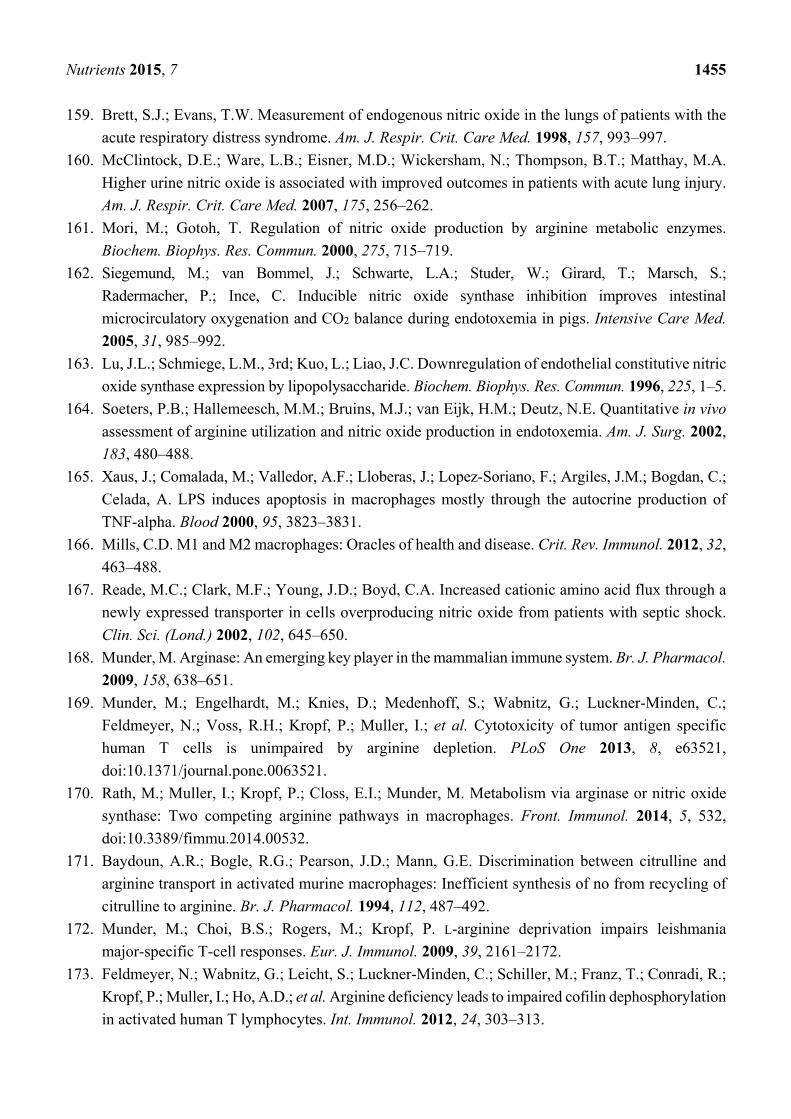
Nutrients 2015, 7 1455
159. Brett, S.J.; Evans, T.W. Measurement of endogenous nitric oxide in the lungs of patients with the
acute respiratory distress syndrome. Am. J. Respir. Crit. Care Med. 1998, 157, 993–997.
160. McClintock, D.E.; Ware, L.B.; Eisner, M.D.; Wickersham, N.; Thompson, B.T.; Matthay, M.A.
Higher urine nitric oxide is associated with improved outcomes in patients with acute lung injury.
Am. J. Respir. Crit. Care Med. 2007, 175, 256–262.
161. Mori, M.; Gotoh, T. Regulation of nitric oxide production by arginine metabolic enzymes.
Biochem. Biophys. Res. Commun. 2000, 275, 715–719.
162. Siegemund, M.; van Bommel, J.; Schwarte, L.A.; Studer, W.; Girard, T.; Marsch, S.;
Radermacher, P.; Ince, C. Inducible nitric oxide synthase inhibition improves intestinal
microcirculatory oxygenation and CO2 balance during endotoxemia in pigs. Intensive Care Med.
2005, 31, 985–992.
163. Lu, J.L.; Schmiege, L.M., 3rd; Kuo, L.; Liao, J.C. Downregulation of endothelial constitutive nitric
oxide synthase expression by lipopolysaccharide. Biochem. Biophys. Res. Commun. 1996, 225, 1–5.
164. Soeters, P.B.; Hallemeesch, M.M.; Bruins, M.J.; van Eijk, H.M.; Deutz, N.E. Quantitative in vivo
assessment of arginine utilization and nitric oxide production in endotoxemia. Am. J. Surg. 2002,
183, 480–488.
165. Xaus, J.; Comalada, M.; Valledor, A.F.; Lloberas, J.; Lopez-Soriano, F.; Argiles, J.M.; Bogdan, C.;
Celada, A. LPS induces apoptosis in macrophages mostly through the autocrine production of
TNF-alpha. Blood 2000, 95, 3823–3831.
166. Mills, C.D. M1 and M2 macrophages: Oracles of health and disease. Crit. Rev. Immunol. 2012, 32,
463–488.
167. Reade, M.C.; Clark, M.F.; Young, J.D.; Boyd, C.A. Increased cationic amino acid flux through a
newly expressed transporter in cells overproducing nitric oxide from patients with septic shock.
Clin. Sci. (Lond.) 2002, 102, 645–650.
168. Munder, M. Arginase: An emerging key player in the mammalian immune system. Br. J. Pharmacol.
2009, 158, 638–651.
169. Munder, M.; Engelhardt, M.; Knies, D.; Medenhoff, S.; Wabnitz, G.; Luckner-Minden, C.;
Feldmeyer, N.; Voss, R.H.; Kropf, P.; Muller, I.; et al. Cytotoxicity of tumor antigen specific
human T cells is unimpaired by arginine depletion. PLoS One 2013, 8, e63521,
doi:10.1371/journal.pone.0063521.
170. Rath, M.; Muller, I.; Kropf, P.; Closs, E.I.; Munder, M. Metabolism via arginase or nitric oxide
synthase: Two competing arginine pathways in macrophages. Front. Immunol. 2014, 5, 532,
doi:10.3389/fimmu.2014.00532.
171. Baydoun, A.R.; Bogle, R.G.; Pearson, J.D.; Mann, G.E. Discrimination between citrulline and
arginine transport in activated murine macrophages: Inefficient synthesis of no from recycling of
citrulline to arginine. Br. J. Pharmacol. 1994, 112, 487–492.
172. Munder, M.; Choi, B.S.; Rogers, M.; Kropf, P. L-arginine deprivation impairs leishmania
major-specific T-cell responses. Eur. J. Immunol. 2009, 39, 2161–2172.
173. Feldmeyer, N.; Wabnitz, G.; Leicht, S.; Luckner-Minden, C.; Schiller, M.; Franz, T.; Conradi, R.;
Kropf, P.; Muller, I.; Ho, A.D.; et al. Arginine deficiency leads to impaired cofilin dephosphorylation
in activated human T lymphocytes. Int. Immunol. 2012, 24, 303–313.
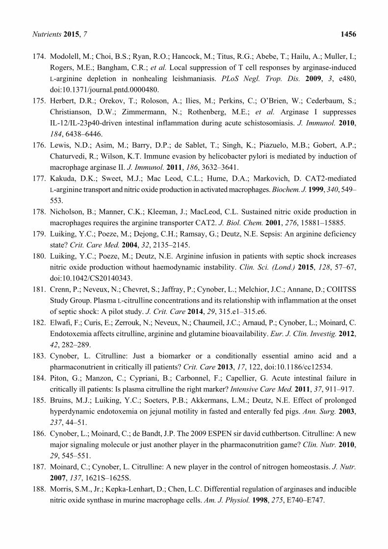
Nutrients 2015, 7 1456
174. Modolell, M.; Choi, B.S.; Ryan, R.O.; Hancock, M.; Titus, R.G.; Abebe, T.; Hailu, A.; Muller, I.;
Rogers, M.E.; Bangham, C.R.; et al. Local suppression of T cell responses by arginase-induced
L-arginine depletion in nonhealing leishmaniasis. PLoS Negl. Trop. Dis. 2009, 3, e480,
doi:10.1371/journal.pntd.0000480.
175. Herbert, D.R.; Orekov, T.; Roloson, A.; Ilies, M.; Perkins, C.; OʼBrien, W.; Cederbaum, S.;
Christianson, D.W.; Zimmermann, N.; Rothenberg, M.E.; et al. Arginase I suppresses
IL-12/IL-23p40-driven intestinal inflammation during acute schistosomiasis. J. Immunol. 2010,
184, 6438–6446.
176. Lewis, N.D.; Asim, M.; Barry, D.P.; de Sablet, T.; Singh, K.; Piazuelo, M.B.; Gobert, A.P.;
Chaturvedi, R.; Wilson, K.T. Immune evasion by helicobacter pylori is mediated by induction of
macrophage arginase II. J. Immunol. 2011, 186, 3632–3641.
177. Kakuda, D.K.; Sweet, M.J.; Mac Leod, C.L.; Hume, D.A.; Markovich, D. CAT2-mediated
L-arginine transport and nitric oxide production in activated macrophages. Biochem. J. 1999, 340, 549–
553.
178. Nicholson, B.; Manner, C.K.; Kleeman, J.; MacLeod, C.L. Sustained nitric oxide production in
macrophages requires the arginine transporter CAT2. J. Biol. Chem. 2001, 276, 15881–15885.
179. Luiking, Y.C.; Poeze, M.; Dejong, C.H.; Ramsay, G.; Deutz, N.E. Sepsis: An arginine deficiency
state? Crit. Care Med. 2004, 32, 2135–2145.
180. Luiking, Y.C.; Poeze, M.; Deutz, N.E. Arginine infusion in patients with septic shock increases
nitric oxide production without haemodynamic instability. Clin. Sci. (Lond.) 2015, 128, 57–67,
doi:10.1042/CS20140343.
181. Crenn, P.; Neveux, N.; Chevret, S.; Jaffray, P.; Cynober, L.; Melchior, J.C.; Annane, D.; COIITSS
Study Group. Plasma L-citrulline concentrations and its relationship with inflammation at the onset
of septic shock: A pilot study. J. Crit. Care 2014, 29, 315.e1–315.e6.
182. Elwafi, F.; Curis, E.; Zerrouk, N.; Neveux, N.; Chaumeil, J.C.; Arnaud, P.; Cynober, L.; Moinard, C.
Endotoxemia affects citrulline, arginine and glutamine bioavailability. Eur. J. Clin. Investig. 2012,
42, 282–289.
183. Cynober, L. Citrulline: Just a biomarker or a conditionally essential amino acid and a
pharmaconutrient in critically ill patients? Crit. Care 2013, 17, 122, doi:10.1186/cc12534.
184. Piton, G.; Manzon, C.; Cypriani, B.; Carbonnel, F.; Capellier, G. Acute intestinal failure in
critically ill patients: Is plasma citrulline the right marker? Intensive Care Med. 2011, 37, 911–917.
185. Bruins, M.J.; Luiking, Y.C.; Soeters, P.B.; Akkermans, L.M.; Deutz, N.E. Effect of prolonged
hyperdynamic endotoxemia on jejunal motility in fasted and enterally fed pigs. Ann. Surg. 2003,
237, 44–51.
186. Cynober, L.; Moinard, C.; de Bandt, J.P. The 2009 ESPEN sir david cuthbertson. Citrulline: A new
major signaling molecule or just another player in the pharmaconutrition game? Clin. Nutr. 2010,
29, 545–551.
187. Moinard, C.; Cynober, L. Citrulline: A new player in the control of nitrogen homeostasis. J. Nutr.
2007, 137, 1621S–1625S.
188. Morris, S.M., Jr.; Kepka-Lenhart, D.; Chen, L.C. Differential regulation of arginases and inducible
nitric oxide synthase in murine macrophage cells. Am. J. Physiol. 1998, 275, E740–E747.
Nutrients 2015, 7 1457
189. Tabuchi, S.; Gotoh, T.; Miyanaka, K.; Tomita, K.; Mori, M. Regulation of genes for inducible
nitric oxide synthase and urea cycle enzymes in rat liver in endotoxin shock. Biochem. Biophys.
Res. Commun. 2000, 268, 221–224.
190. Corraliza, I.M.; Soler, G.; Eichmann, K.; Modolell, M. Arginase induction by suppressors of nitric
oxide synthesis (IL-4, IL-10 and PGE2) in murine bone-marrow-derived macrophages. Biochem.
Biophys. Res. Commun. 1995, 206, 667–673.
191. Modolell, M.; Corraliza, I.M.; Link, F.; Soler, G.; Eichmann, K. Reciprocal regulation of the nitric
oxide synthase/arginase balance in mouse bone marrow-derived macrophages by Th1 and Th2
cytokines. Eur. J. Immunol. 1995, 25, 1101–1104.
192. Hey, C.; Boucher, J.L.; Vadon-Le Goff, S.; Ketterer, G.; Wessler, I.; Racke, K. Inhibition of
arginase in rat and rabbit alveolar macrophages by N omega-hydroxy-D,L-indospicine, effects on
L-arginine utilization by nitric oxide synthase. Br. J. Pharmacol. 1997, 121, 395–400.
193. Hecker, M.; Nematollahi, H.; Hey, C.; Busse, R.; Racke, K. Inhibition of arginase by
NG-hydroxy-L-arginine in alveolar macrophages: Implications for the utilization of L-arginine for
nitric oxide synthesis. FEBS Lett. 1995, 359, 251–254.
194. Chang, C.I.; Liao, J.C.; Kuo, L. Arginase modulates nitric oxide production in activated macrophages.
Am. J. Physiol. 1998, 274, H342–H348.
195. Bivalacqua, T.J.; Hellstrom, W.J.; Kadowitz, P.J.; Champion, H.C. Increased expression of
arginase II in human diabetic corpus cavernosum: In diabetic-associated erectile dysfunction.
Biochem. Biophys. Res. Commun. 2001, 283, 923–927.
196. White, A.R.; Ryoo, S.; Li, D.; Champion, H.C.; Steppan, J.; Wang, D.; Nyhan, D.; Shoukas, A.A.;
Hare, J.M.; Berkowitz, D.E. Knockdown of arginase I restores NO signaling in the vasculature of
old rats. Hypertension 2006, 47, 245–251.
197. Tumer, C.; Bilgin, H.M.; Obay, B.D.; Diken, H.; Atmaca, M.; Kelle, M. Effect of nitric oxide on
phagocytic activity of lipopolysaccharide-induced macrophages: Possible role of exogenous
L-arginine. Cell Biol. Int. 2007, 31, 565–569.
198. Rafiee, P.; Ogawa, H.; Heidemann, J.; Li, M.S.; Lamirand, T.H.; Fisher, P.J.; Graewin, S.J.;
Dwinell, M.B.; Johnson, C.P.; Shaker, R.; et al. Isolation and characterization of human esophageal
microvascular endothelial cells: Mechanisms of inflammatory activation. Am. J. Physiol.
Gastrointest. Liver Physiol. 2003, 285, G1277–G1292.
199. Hollenberg, S.M.; Broussard, M.; Osman, J.; Parrillo, J.E. Increased microvascular reactivity
and improved mortality in septic mice lacking inducible nitric oxide synthase. Circ. Res 2000, 86,
774–778.
200. Kubes, P.; McCafferty, D.M. Nitric oxide and intestinal inflammation. Am. J. Med. 2000, 109,
150–158.
201. Cobb, J.P.; Hotchkiss, R.S.; Swanson, P.E.; Chang, K.; Qiu, Y.; Laubach, V.E.; Karl, I.E.;
Buchman, T.G. Inducible nitric oxide synthase (iNOS) gene deficiency increases the mortality of
sepsis in mice. Surgery 1999, 126, 438–442.
202. Groeneveld, P.H.; Kwappenberg, K.M.; Langermans, J.A.; Nibbering, P.H.; Curtis, L. Relation
between pro- and anti-inflammatory cytokines and the production of nitric oxide (NO) in severe
sepsis. Cytokine 1997, 9, 138–142.
Nutrients 2015, 7 1458
203. Hesse, M.; Modolell, M.; la Flamme, A.C.; Schito, M.; Fuentes, J.M.; Cheever, A.W.; Pearce, E.J.;
Wynn, T.A. Differential regulation of nitric oxide synthase-2 and arginase-1 by type 1/type 2
cytokines in vivo: Granulomatous pathology is shaped by the pattern of L-arginine metabolism.
J. Immunol. 2001, 167, 6533–6544.
204. Pinsky, M.R.; Vincent, J.L.; Deviere, J.; Alegre, M.; Kahn, R.J.; Dupont, E. Serum cytokine levels
in human septic shock. Relation to multiple-system organ failure and mortality. Chest 1993, 103,
565–575.
205. Bruins, M.J.; Lamers, W.H.; Meijer, A.J.; Soeters, P.B.; Deutz, N.E. In vivo measurement of
nitric oxide production in porcine gut, liver and muscle during hyperdynamic endotoxaemia.
Br. J. Pharmacol. 2002, 137, 1225–1236.
206. Xie, L.; Gross, S.S. Argininosuccinate synthetase overexpression in vascular smooth muscle cells
potentiates immunostimulant-induced no production. J. Biol. Chem. 1997, 272, 16624–16630.
207. Enkhbaatar, P.; Murakami, K.; Traber, L.D.; Cox, R.; Parkinson, J.F.; Westphal, M.; Esechie, A.;
Morita, N.; Maybauer, M.O.; Maybauer, D.M.; et al. The inhibition of inducible nitric oxide
synthase in ovine sepsis model. Shock 2006, 25, 522–527.
208. Okamoto, I.; Abe, M.; Shibata, K.; Shimizu, N.; Sakata, N.; Katsuragi, T.; Tanaka, K. Evaluating
the role of inducible nitric oxide synthase using a novel and selective inducible nitric oxide
synthase inhibitor in septic lung injury produced by cecal ligation and puncture. Am. J. Respir.
Crit. Care Med. 2000, 162, 716–722.
209. Enkhbaatar, P.; Lange, M.; Nakano, Y.; Hamahata, A.; Jonkam, C.; Wang, J.; Jaroch, S.; Traber, L.;
Herndon, D.; Traber, D. Role of neuronal nitric oxide synthase in ovine sepsis model. Shock 2009,
32, 253–257.
210. Gocan, N.C.; Scott, J.A.; Tyml, K. Nitric oxide produced via neuronal nos may impair
vasodilatation in septic rat skeletal muscle. Am. J. Physiol. Heart Circ. Physiol. 2000, 278,
H1480–H1489.
211. Ortiz, F.; Garcia, J.A.; Acuna-Castroviejo, D.; Doerrier, C.; Lopez, A.; Venegas, C.; Volt, H.;
Luna-Sanchez, M.; Lopez, L.C.; Escames, G. The beneficial effects of melatonin against heart
mitochondrial impairment during sepsis: Inhibition of inos and preservation of nnos. J. Pineal. Res.
2014, 56, 71–81.
212. Miller, M.J.; Sandoval, M. Nitric oxide. III. A molecular prelude to intestinal inflammation.
Am. J. Physiol. 1999, 276, G795–G799.
213. Beck, P.L.; Xavier, R.; Wong, J.; Ezedi, I.; Mashimo, H.; Mizoguchi, A.; Mizoguchi, E.;
Bhan, A.K.; Podolsky, D.K. Paradoxical roles of different nitric oxide synthase isoforms in colonic
injury. Am. J. Physiol. Gastrointest Liver Physiol. 2004, 286, G137–G147.
214. Qu, X.W.; Thaete, L.G.; Rozenfeld, R.A.; Zhu, Y.; de Plaen, I.G.; Caplan, M.S.; Hsueh, W.
Tetrahydrobiopterin prevents platelet-activating factor-induced intestinal hypoperfusion and necrosis:
Role of neuronal nitric oxide synthase. Crit. Care Med. 2005, 33, 1050–1056.
215. Chakrabarti, S.; Chan, C.K.; Jiang, Y.; Davidge, S.T. Neuronal nitric oxide synthase regulates
endothelial inflammation. J. Leukoc. Biol. 2012, 91, 947–956.
216. Bachetti, T.; Comini, L.; Curello, S.; Bastianon, D.; Palmieri, M.; Bresciani, G.; Callea, F.; Ferrari, R.
Co-expression and modulation of neuronal and endothelial nitric oxide synthase in human
endothelial cells. J. Mol. Cell Cardiol. 2004, 37, 939–945.
Nutrients 2015, 7 1459
217. Domenico, R. Pharmacology of nitric oxide: Molecular mechanisms and therapeutic strategies.
Curr. Pharm. Des. 2004, 10, 1667–1676.
218. Forstermann, U.; Li, H. Therapeutic effect of enhancing endothelial nitric oxide synthase (eNOS)
expression and preventing eNOS uncoupling. Br. J. Pharmacol. 2011, 164, 213–223.
219. Venugopal, S.K.; Devaraj, S.; Yuhanna, I.; Shaul, P.; Jialal, I. Demonstration that C-reactive
protein decreases enos expression and bioactivity in human aortic endothelial cells. Circulation
2002, 106, 1439–1441.
220. Moens, A.L.; Kietadisorn, R.; Lin, J.Y.; Kass, D. Targeting endothelial and myocardial dysfunction
with tetrahydrobiopterin. J. Mol. Cell Cardiol. 2011, 51, 559–563.
221. Ince, C.; Sinaasappel, M. Microcirculatory oxygenation and shunting in sepsis and shock.
Crit. Care Med. 1999, 27, 1369–1377.
222. Derikx, J.P.; Poeze, M.; van Bijnen, A.A.; Buurman, W.A.; Heineman, E. Evidence for intestinal
and liver epithelial cell injury in the early phase of sepsis. Shock 2007, 28, 544–548.
223. Chen, K.; Inoue, M.; Wasa, M.; Fukuzawa, M.; Kamata, S.; Okada, A. Expression of endothelial
constitutive nitric oxide synthase mRNA in gastrointestinal mucosa and its downregulation by
endotoxin. Life Sci. 1997, 61, 1323–1329.
224. Leiper, J.; Vallance, P. Biological significance of endogenous methylarginines that inhibit nitric
oxide synthases. Cardiovasc. Res. 1999, 43, 542–548.
225. Leiper, J.; Murray-Rust, J.; McDonald, N.; Vallance, P. S-nitrosylation of dimethylarginine
dimethylaminohydrolase regulates enzyme activity: Further interactions between nitric oxide synthase
and dimethylarginine dimethylaminohydrolase. Proc. Natl. Acad. Sci. USA 2002, 99, 13527–13532.
226. Nijveldt, R.J.; Teerlink, T.; van der Hoven, B.; Siroen, M.P.; Kuik, D.J.; Rauwerda, J.A.;
van Leeuwen, P.A. Asymmetrical dimethylarginine (ADMA) in critically ill patients: High plasma
ADMA concentration is an independent risk factor of ICU mortality. Clin. Nutr. 2003, 22, 23–30.
227. Nijveldt, R.J.; Teerlink, T.; van Leeuwen, P.A. The asymmetrical dimethylarginine
(ADMA)-multiple organ failure hypothesis. Clin. Nutr. 2003, 22, 99–104.
228. Davis, J.S.; Darcy, C.J.; Yeo, T.W.; Jones, C.; McNeil, Y.R.; Stephens, D.P.; Celermajer, D.S.;
Anstey, N.M. Asymmetric dimethylarginine, endothelial nitric oxide bioavailability and mortality in
sepsis. PLoS One 2011, 6, e17260, doi:10.1371/journal.pone.0017260.
229. Kawahara, K.; Gotoh, T.; Oyadomari, S.; Kajizono, M.; Kuniyasu, A.; Ohsawa, K.; Imai, Y.;
Kohsaka, S.; Nakayama, H.; Mori, M. Co-induction of argininosuccinate synthetase, cationic amino
acid transporter-2, and nitric oxide synthase in activated murine microglial cells. Brain Res. Mol.
Brain Res. 2001, 90, 165–173.
230. Prima, V.; Wang, A.; Molina, G.; Wang, K.K.; Svetlov, S.I. Inhibition of LPS toxicity by hepatic
argininosuccinate synthase (ASS): Novel roles for ASS in innate immune responses to bacterial
infection. Int. Immunopharmacol. 2011, 11, 1180–1188.
231. Nagasaki, A.; Gotoh, T.; Takeya, M.; Yu, Y.; Takiguchi, M.; Matsuzaki, H.; Takatsuki, K.;
Mori, M. Coinduction of nitric oxide synthase, argininosuccinate synthetase, and argininosuccinate
lyase in lipopolysaccharide-treated rats. RNA blot, immunoblot, and immunohistochemical
analyses. J. Biol. Chem. 1996, 271, 2658–2662.

Nutrients 2015, 7 1460
232. Bryk, J.; Ochoa, J.B.; Correia, M.I.; Munera-Seeley, V.; Popovic, P.J. Effect of citrulline and
glutamine on nitric oxide production in RAW 264.7 cells in an arginine-depleted environment.
JPEN J. Parenter Enteral. Nutr. 2008, 32, 377–383.
233. Mori, M.; Gotoh, T.; Nagasaki, A.; Takiguchi, M.; Sonoki, T. Regulation of the urea cycle enzyme
genes in nitric oxide synthesis. J. Inherit. Metab. Dis. 1998, 21, 59–71.
234. Wu, G.Y.; Brosnan, J.T. Macrophages can convert citrulline into arginine. Biochem. J. 1992, 281,
45–48.
235. Satoh, M.; Iwahori, T.; Sugawara, N.; Yamazaki, M. Liver argininosuccinate synthase binds to
bacterial lipopolysaccharides and lipid a and inactivates their biological activities. J. Endotoxin. Res.
2006, 12, 21–38.
236. Satoh, M.; Ando, S.; Shinoda, T.; Yamazaki, M. Clearance of bacterial lipopolysaccharides and
lipid a by the liver and the role of argininosuccinate synthase. Innate Immun. 2008, 14, 51–60.
237. Hibbs, J.B., Jr.; Westenfelder, C.; Taintor, R.; Vavrin, Z.; Kablitz, C.; Baranowski, R.L.;
Ward, J.H.; Menlove, R.L.; McMurry, M.P.; Kushner, J.P.; et al. Evidence for cytokine-inducible
nitric oxide synthesis from L-arginine in patients receiving interleukin-2 therapy. J. Clin. Investig.
1992, 89, 867–877.
238. Nathan, C.; Xie, Q.W. Nitric oxide synthases: Roles, tolls, and controls. Cell 1994, 78, 915–918.
239. Kirkeboen, K.A.; Strand, O.A. The role of nitric oxide in sepsis—An overview.
Acta Anaesthesiol. Scand. 1999, 43, 275–288.
240. Bersten, A.D.; Hersch, M.; Cheung, H.; Rutledge, F.S.; Sibbald, W.J. The effect of various
sympathomimetics on the regional circulations in hyperdynamic sepsis. Surgery 1992, 112, 549–561.
241. Thiemermann, C. Nitric oxide and septic shock. Gen. Pharmacol. 1997, 29, 159–166.
242. Garvey, E.P.; Oplinger, J.A.; Furfine, E.S.; Kiff, R.J.; Laszlo, F.; Whittle, B.J.; Knowles, R.G.
1400W is a slow, tight binding, and highly selective inhibitor of inducible nitric-oxide synthase
in vitro and in vivo. J. Biol. Chem. 1997, 272, 4959–4963.
243. Pittner, A.; Nalos, M.; Asfar, P.; Yang, Y.; Ince, C.; Georgieff, M.; Bruckner, U.B.; Radermacher, P.;
Froba, G. Mechanisms of inducible nitric oxide synthase (iNOS) inhibition-related improvement
of gut mucosal acidosis during hyperdynamic porcine endotoxemia. Intensive Care Med. 2003, 29,
312–316.
244. Ploner, F.; Radermacher, P.; Theisen, M.; Tugtekin, I.F.; Matejovic, M.; Stehr, A.; Szabo, C.;
Southan, G.J.; Georgieff, M.; Bruckner, U.B.; et al. Effects of combined selective inos inhibition
and peroxynitrite blockade during endotoxemia in pigs. Shock 2001, 16, 130–136.
245. Saetre, T.; Hovig, T.; Roger, M.; Gundersen, Y.; Aasen, A.O. Hepatocellular damage in porcine
endotoxemia: Beneficial effects of selective versus non-selective nitric oxide synthase inhibition?
Scand. J. Clin. Lab. Investig. 2001, 61, 503–512.
246. Wei, X.Q.; Charles, I.G.; Smith, A.; Ure, J.; Feng, G.J.; Huang, F.P.; Xu, D.; Muller, W.; Moncada,
S.; Liew, F.Y. Altered immune responses in mice lacking inducible nitric oxide synthase. Nature
1995, 375, 408–411.
247. MacMicking, J.D.; Nathan, C.; Hom, G.; Chartrain, N.; Fletcher, D.S.; Trumbauer, M.; Stevens, K.;
Xie, Q.W.; Sokol, K.; Hutchinson, N.; et al. Altered responses to bacterial infection and endotoxic
shock in mice lacking inducible nitric oxide synthase. Cell 1995, 81, 641–650.
Nutrients 2015, 7 1461
248. Southan, G.J.; Szabo, C.; Thiemermann, C. Isothioureas: Potent inhibitors of nitric oxide synthases
with variable isoform selectivity. Br. J. Pharmacol. 1995, 114, 510–516.
249. Misko, T.P.; Moore, W.M.; Kasten, T.P.; Nickols, G.A.; Corbett, J.A.; Tilton, R.G.; McDaniel, M.L.;
Williamson, J.R.; Currie, M.G. Selective inhibition of the inducible nitric oxide synthase by
aminoguanidine. Eur. J. Pharmacol. 1993, 233, 119–125.
250. Ruetten, H.; Thiemermann, C. Prevention of the expression of inducible nitric oxide synthase
by aminoguanidine or aminoethyl-isothiourea in macrophages and in the rat. Biochem. Biophys.
Res. Commun. 1996, 225, 525–530.
251. Murakami, K.; McGuire, R.; Jodoin, J.; Katahira, J.; Schmalstieg, F.C.; Traber, L.D.; Traber, D.L.
Aminoguanidine did not attenuate septic changes and acute lung injury in sheep. In proceedings of
the 7th World Congress for Microcirculation, Sydney, Australia, 19–22 August 2001;
pp. 465–469.
252. Ishikawa, K.; Calzavacca, P.; Bellomo, R.; Bailey, M.; May, C.N. Effect of selective inhibition of
renal inducible nitric oxide synthase on renal blood flow and function in experimental hyperdynamic
sepsis. Crit. Care Med. 2012, 40, 2368–2375.
253. Thiemermann, C.; Ruetten, H.; Wu, C.C.; Vane, J.R. The multiple organ dysfunction syndrome
caused by endotoxin in the rat: Attenuation of liver dysfunction by inhibitors of nitric oxide synthase.
Br. J. Pharmacol. 1995, 116, 2845–2851.
254. Bone, H.G.; Waurick, R.; van Aken, H.; Booke, M.; Prien, T.; Meyer, J. Comparison of the
haemodynamic effects of nitric oxide synthase inhibition and nitric oxide scavenging in endotoxaemic
sheep. Intensive Care Med. 1998, 24, 48–54.
255. Zhang, H.; Rogiers, P.; Smail, N.; Cabral, A.; Preiser, J.C.; Peny, M.O.; Vincent, J.L. Effects of
nitric oxide on blood flow distribution and O2 extraction capabilities during endotoxic shock.
J. Appl. Physiol. 1997, 83, 1164–1173.
256. Cobb, J.P.; Natanson, C.; Hoffman, W.D.; Lodato, R.F.; Banks, S.; Koev, C.A.; Solomon, M.A.;
Elin, R.J.; Hosseini, J.M.; Danner, R.L. N omega-amino-L-arginine, an inhibitor of nitric oxide
synthase, raises vascular resistance but increases mortality rates in awake canines challenged with
endotoxin. J. Exp. Med. 1992, 176, 1175–1182.
257. Avontuur, J.A.; Tutein Nolthenius, R.P.; van Bodegom, J.W.; Bruining, H.A. Prolonged inhibition
of nitric oxide synthesis in severe septic shock: A clinical study. Crit. Care Med. 1998, 26, 660–667.
258. Lopez, A.; Lorente, J.A.; Steingrub, J.; Bakker, J.; McLuckie, A.; Willatts, S.; Brockway, M.;
Anzueto, A.; Holzapfel, L.; Breen, D.; et al. Multiple-center, randomized, placebo-controlled,
double-blind study of the nitric oxide synthase inhibitor 546c88: Effect on survival in patients with
septic shock. Crit. Care Med. 2004, 32, 21–30.
259. Scott, J.A.; Mehta, S.; Duggan, M.; Bihari, A.; McCormack, D.G. Functional inhibition of
constitutive nitric oxide synthase in a rat model of sepsis. Am. J. Respir. Crit. Care Med. 2002,
165, 1426–1432.
260. Kilbourn, R.G.; Szabo, C.; Traber, D.L. Beneficial versus detrimental effects of nitric oxide
synthase inhibitors in circulatory shock: Lessons learned from experimental and clinical studies.
Shock 1997, 7, 235–246.
Nutrients 2015, 7 1462
261. Saetre, T.; Smiseth, O.A.; Scholz, T.; Carlsen, H.; Nordsletten, L.; Fahlstrom, E.; Aasen, A.O.
Nitric oxide synthase inhibition reduces venous return in porcine endotoxemia. Am. J. Physiol.
1996, 271, H1325–H1332.
262. Lange, M.; Hamahata, A.; Traber, D.L.; Nakano, Y.; Traber, L.D.; Enkhbaatar, P. Specific inhibition
of nitric oxide synthases at different time points in a murine model of pulmonary sepsis. Biochem.
Biophys. Res. Commun. 2011, 404, 877–881.
263. Lange, M.; Connelly, R.; Traber, D.L.; Hamahata, A.; Cox, R.A.; Nakano, Y.; Bansal, K.;
Esechie, A.; von Borzyskowski, S.; Jonkam, C.; et al. Combined neuronal and inducible nitric oxide
synthase inhibition in ovine acute lung injury. Crit. Care Med. 2009, 37, 223–229.
264. Vanhoutte, P.M. Arginine and arginase: Endothelial no synthase double crossed? Circ. Res. 2008,
102, 866–868.
265. Patejunas, G.; Bradley, A.; Beaudet, A.L.; O’Brien, W.E. Generation of a mouse model for
citrullinemia by targeted disruption of the argininosuccinate synthetase gene. Somat Cell Mol. Genet.
1994, 20, 55–60.
266. Ye, X.; Whiteman, B.; Jerebtsova, M.; Batshaw, M.L. Correction of argininosuccinate synthetase
(AS) deficiency in a murine model of citrullinemia with recombinant adenovirus carrying human
AS cDNA. Gene Ther. 2000, 7, 1777–1782.
267. Lu, Y.; Leung, T.M.; Ward, S.C.; Nieto, N. Partial deletion of argininosuccinate synthase
protects from pyrazole plus lipopolysaccharide-induced liver injury by decreasing nitrosative
stress. Am. J. Physiol. Gastrointest Liver Physiol. 2012, 302, G287–G295.
268. Premkumar, M.H.; Sule, G.; Nagamani, S.C.; Chakkalakal, S.; Nordin, A.; Jain, M.; Ruan, M.Z.;
Bertin, T.; Dawson, B.C.; Zhang, J.; et al. Argininosuccinate lyase in enterocytes protects from
development of necrotizing enterocolitis. Am. J. Physiol. Gastrointest Liver Physiol. 2014. 307,
G347–G354.
269. Barbul, A.; Lazarou, S.A.; Efron, D.T.; Wasserkrug, H.L.; Efron, G. Arginine enhances wound
healing and lymphocyte immune responses in humans. Surgery 1990, 108, 331–336.
270. Boger, R.H. The pharmacodynamics of L-arginine. Altern. Ther. Health Med. 2014, 20, 48–54.
271. Vissers, Y.L.; Debats, I.B.; Luiking, Y.C.; Jalan, R.; van der Hulst, R.R.; Dejong, C.H.; Deutz, N.E.
Pros and cons of L-arginine supplementation in disease. Nutr. Res. Rev. 2004, 17, 193–210.
272. Bode-Boger, S.M.; Boger, R.H.; Galland, A.; Tsikas, D.; Frolich, J.C. L-arginine-induced vasodilation
in healthy humans: Pharmacokinetic-pharmacodynamic relationship. Br. J. Clin. Pharmacol. 1998,
46, 489–497.
273. Bertolini, G.; Luciani, D.; Biolo, G. Immunonutrition in septic patients: A philosophical view of
the current situation. Clin. Nutr. 2007, 26, 25–29.
274. Deutz, N.E. The 2007 ESPEN Sir David Cuthbertson lecture: Amino acids between and within
organs. The glutamate-glutamine-citrulline-arginine pathway. Clin. Nutr. 2008, 27, 321–327.
275. Grover, Z.; Tubman, R.; McGuire, W. Glutamine supplementation for young infants with severe
gastrointestinal disease. Cochrane Database Syst. Rev. 2007, CD005947, doi:10.1002/14651858.
CD005947.pub3.
276. Preiser, J.C.; Berre, P.J.; van Gossum, A.; Cynober, L.; Vray, B.; Carpentier, Y.; Vincent, J.L.
Metabolic effects of arginine addition to the enteral feeding of critically ill patients. JPEN J.
Parenter Enteral. Nutr. 2001, 25, 182–187.
Nutrients 2015, 7 1463
277. Kieft, H.; Roos, A.N.; van Drunen, J.D.; Bindels, A.J.; Bindels, J.G.; Hofman, Z. Clinical outcome
of immunonutrition in a heterogeneous intensive care population. Intensive Care Med. 2005, 31,
524–532.
278. Tsuei, B.J.; Bernard, A.C.; Barksdale, A.R.; Rockich, A.K.; Meier, C.F.; Kearney, P.A.
Supplemental enteral arginine is metabolized to ornithine in injured patients. J. Surg. Res. 2005,
123, 17–24.
279. Bertolini, G.; Iapichino, G.; Radrizzani, D.; Facchini, R.; Simini, B.; Bruzzone, P.; Zanforlin, G.;
Tognoni, G. Early enteral immunonutrition in patients with severe sepsis: Results of an interim
analysis of a randomized multicentre clinical trial. Intensive Care Med. 2003, 29, 834–840.
280. Zheng, Y.M.; Li, F.; Zhang, M.M.; Wu, X.T. Glutamine dipeptide for parenteral nutrition in
abdominal surgery: A meta-analysis of randomized controlled trials. World J. Gastroenterol. 2006,
12, 7537–7541.
281. Heyland, D.K.; Novak, F.; Drover, J.W.; Jain, M.; Su, X.; Suchner, U. Should immunonutrition
become routine in critically ill patients? A systematic review of the evidence. JAMA 2001, 286,
944–953.
282. Cynober, L. Can arginine and ornithine support gut functions? Gut 1994, 35, S42–S45.
283. Stechmiller, J.K.; Treloar, D.; Allen, N. Gut dysfunction in critically ill patients: A review of the
literature. Am. J. Crit. Care 1997, 6, 204–209.
284. Heyland, D.K.; Dhaliwal, R.; Drover, J.W.; Gramlich, L.; Dodek, P. Canadian clinical practice
guidelines for nutrition support in mechanically ventilated, critically ill adult patients. JPEN J.
Parenter Enteral. Nutr. 2003, 27, 355–373.
285. Dhaliwal, R.; Cahill, N.; Lemieux, M.; Heyland, D.K. The canadian critical care nutrition guidelines
in 2013: An update on current recommendations and implementation strategies. Nutr. Clin. Pract.
2014, 29, 29–43.
286. Bruins, M.J.; Soeters, P.B.; Lamers, W.H.; Deutz, N.E. L-arginine supplementation in pigs
decreases liver protein turnover and increases hindquarter protein turnover both during and after
endotoxemia. Am. J. Clin. Nutr. 2002, 75, 1031–1044.
287. Schwedhelm, E.; Maas, R.; Freese, R.; Jung, D.; Lukacs, Z.; Jambrecina, A.; Spickler, W.;
Schulze, F.; Boger, R.H. Pharmacokinetic and pharmacodynamic properties of oral L-citrulline and
L-arginine: Impact on nitric oxide metabolism. Br. J. Clin. Pharmacol. 2008, 65, 51–59.
© 2015 by the authors; licensee MDPI, Basel, Switzerland. This article is an open access article
distributed under the terms and conditions of the Creative Commons Attribution license
(http://creativecommons.org/licenses/by/4.0/).


