Embed Size (px)
Citation preview

Applications of LC-MS in PET Radioligand Development andMetabolic Elucidation
Ying Ma, Dale O. Kiesewetter*, Lixin Lang, Dongyu Gu, and Xiaoyuan Chen*
Laboratory of Molecular Imaging and Nanomedicine (LOMIN), National Institute of BiomedicalImaging and Bioengineering (NIBIB), National Institutes of Health (NIH), Bethesda, MD 20892,USA
AbstractPositron emission tomography (PET) is a very sensitive molecular imaging technique that whenemployed with an appropriate radioligand has the ability to quantititate physiological processes ina non-invasive manner. Since the imaging technique detects all radioactive emissions in the fieldof view, the presence and biological activity of radiolabeled metabolites must be determined foreach radioligand in order to validate the utility of the radiotracer for measuring the desiredphysiological process. Thus, the identification of metabolic profiles of radiolabeled compounds isan important aspect of design, development, and validation of new radiopharmaceuticals and theirapplications in drug development and molecular imaging. Metabolite identification for differentchemical classes of radiopharmaceuticals allows rational design to minimize the formation andaccumulation of metabolites in the target tissue, either through enhanced excretion or minimizedmetabolism. This review will discuss methods for identifying and quantitating metabolites duringthe pre-clinical development of radiopharmaceuticals with special emphasis on the application ofLC/MS.
KeywordsPositron emission tomography (PET); Radiopharmaceutical; LC/MS/MS; Metabolite
INTRODUCTIONPositron emission tomography (PET) is the most sensitive external imaging technique in themolecular imaging arsenal that can be applied to living patients [1, 2]. The technique imagesthe co-linear annihilation photons resulting from decay of positron-emitting radionuclides.Positron-emitting radionuclides most commonly introduced into molecules for PET are C-11(t½ 20.4 min), and F-18 (t½ 119.8 min). Other radionuclide used in some research centers areBr-76 (t½ 16.2 h), Cu-64 t½ 12.7 h), Ga-68 (t½ 68 min), and I-124 (t½ 4.18 d). The shorthalf-lives of the more commonly used C-11 and F-18 create challenges to their applicationin PET. The first challenge is the need to incorporate the radionuclide into more complicatedchemical structures. The short half-lives require that the incorporation be performed as closeto the final synthetic step as possible. The second major challenge is completing a radio-chemical synthesis, purification, and conducting pharmacokinetic imaging studies before thedecay of the radionuclide affects the radioactive counting statistics.
© 2010 Bentham Science Publishers Ltd.*Address correspondence to these authors at the 31 Center Dr, Suite 1C14, NIBIB/NIH, Bethesda, MD 20892-2281; USA; Tel:/Fax:301-451-4246; [email protected]; and 10 Center Drive MSC 1180, NIBIB/NIH, Bethesda, MD 20892; USA; Tel/Fax:301-451-3531; [email protected].
NIH Public AccessAuthor ManuscriptCurr Drug Metab. Author manuscript; available in PMC 2013 April 18.
Published in final edited form as:Curr Drug Metab. 2010 July ; 11(6): 483–493.
NIH
-PA Author Manuscript
NIH
-PA Author Manuscript
NIH
-PA Author Manuscript

The true power of the PET technique can be exploited by attaching these positron-emittingradionuclides to molecules that have high affinity to biological targets or participate inspecific biological transformations (i.e. the specific phosphorylation of 2-[18F]fluorodeoxy-glucose that results in accumulation of radioactive emission from cells that take up highquantities of glucose [3]). However, the PET scanner detects only coincident photons; thereis no information concerning the chemical structure to which the radionuclide is attached. Inorder to validate a new radioligand for its intended target, assurance that the radioactiveemissions derive primarily from the parent radioligand is required. Knowledge of metabolicrate and the structure of radiolabeled metabolites can allow prediction of the propensity ofthe metabolites to confound image interpretation [4, 5]. Therefore, identification ofmetabolic profiles of labeled compounds is an important aspect of design, development, andvalidation of new radiopharmaceuticals.
The metabolism profile of a potential radioligand in mice or rats is important for all of thepreclinical validation studies for the purpose of determining specificity or selectivity for thetarget. However, the fact that metabolism may be very different in humans requires somemethod for assessing species differences [6]. The rodent may turn out to be a very poormodel on which to base preclinical studies. This review will focus on methods of metaboliteidentification, species differences of metabolic profiles, and the application of highperformance liquid chromatography/mass spectrometry (LC/MS) in the preclinical designand evaluation of novel radiopharmaceuticals.
METABOLITE IDENTIFICATIONThe pharmaceutical industry expends a great deal of effort to identify metabolites of its drugcandidates in order to understand absorption, distribution, metabolism, excretion, andtoxicity (AD-MET) [7]. Identification of metabolites allows rational pharmaceutical designto minimize formation of undesirable metabolites that may affect drug safety. In addition,knowledge of the metabolite structure may allow a study of the contribution ofpharmacologically active or toxic metabolites to the overall pharmacological response. Thepharmaceutical industry integrated LC/MS into its drug discovery process several years ago[8]. Identification of metabolites of radiopharmaceuticals, which is complicated by the shorthalf-lives of the radionuclides and the very low dose of the compound [typically less than 10nmol] administered, may provide suggestions as to the most appropriate position in themolecule to attach the radiolabel and allow the synthesis and study of the bio-distribution ofthe metabolite(s) [9, 10].
Quantitative whole-body autoradiography (QWBA) [11] and MS imaging [12, 13] havebeen used to evaluate drug concentrations ex vivo. QWBA relies on the preparation ofimages showing the distribution of radioactivity in the whole body. Although the parentdrug may constitute some of the radioactivity measured, it is probable that radiolabeledmetabolites will also be present in an unknown amount. The separation and evaluation ofmixtures of parent compound and metabolites can only be performed by other analyticalmethods. MS imaging has the potential to deliver highly parallel, multiplexed data on thespecific localization of parent and metabolites in tissue samples directly, and to measure andmap the variations of these ions during development and disease progression or treatment.However, the sensitivity of MALDI –MSI, even with the recently introduced nanostructureinitiator mass spectrometry (NIMS), make it challenging to analyze radiopharmaceuticalsand their metabolites at the radiotracer level.
Although pharmacokinetic analysis of radiotracers can be conducted even if more than onechemical entity is observed in the target tissue, the analysis is much simpler when onlyparent radioligand is present. The time dependent concentration and the relative biological
Ma et al. Page 2
Curr Drug Metab. Author manuscript; available in PMC 2013 April 18.
NIH
-PA Author Manuscript
NIH
-PA Author Manuscript
NIH
-PA Author Manuscript

activity (receptor affinity or rate of enzymatic conversion) of each radiolabeled componentmust be known. Usually this information is unknown and thus the presence of metabolites inthe tissue of interest is a major reason for discontinuing further studies of new ligands [14].
Normal metabolic processes on small drug-like molecules (MW < 1000) usually results inthe generation of more polar species. The blood brain barrier (BBB) generally prohibitsmore polar metabolites from crossing into the brain. Traditionally, the observation ofmetabolites that cross the BBB results in a search for a new position to insert theradionuclide or the radioligand is discarded for a different chemical class. The traditionalmethod for evaluating metabolites in the brain was to extract rat or mouse brain tissue andevaluate the radioactive components by thin layer chromatography (TLC) or HPLC. Withthe increasing application of PET to targets outside the brain, this natural restrictionprovided by the BBB has been lost.
The inherent inability of PET to differentiate between a parent compound and its metabolitesconfounds the interpretation of images and may impact the identification of thepathologically induced biochemical changes under investigation. Cytochrome P450 isoformsplay a major role in mammalian xenobiotic biotransformation [15]. The utility of liverhepatocytes to generate phase 1 (i.e. oxidation by cytochrome P450) and phase 2 metabolites(i.e. glucuronidation of hydroxyl groups) of proposed radioligands followed by the analysisof the metabolites has been exploited to determine metabolic pathways [4, 16, 17]. The useof liver microsomes, which produce predominately first order metabolites, has also beenused extensively [18]. Hepatocytes obtained from Sprague-Dawley rats give fairly consistentresults from lot to lot because of the homogeneous genetic makeup of these animals. Humanhepatocytes, on the other hand, are somewhat more variable between lots. More recently,commercial vendors of these products are providing hepatocytes that are pooled from 10–50individual samples; which provides more consistent results. Once these cells have been usedto generate a metabolism profile, LC/MS can be applied to identify the possible structuresand quantitate the metabolism rate of the parent radioligand.
LC/MS has the unique combination of sensitivity and mass selectivity to provide sensitivedetection and mass to charge ratio (m/z) data that can be used to propose metabolitestructure [7]. In addition, the ability to conduct LC/MS/MS studies provides even greaterpower for assignment of possible structure. The various m/z components from collisioninduced fragmentation of the parent ion can provide information on the structure of theparent ion. Multiple reaction monitoring (MRM) is an LC/MS/MS technique that allowsscanning for several daughter ions from a single parent ion. Since many metabolites derivefrom oxidation, some fragment peaks of metabolites may be the same as the parent. Thusmetabolites may be detected and identified based on observation of the daughter ions fromthe parent.
The combination of two general classes of experiments, metabolite generation withhepatocytes from various species and LC/MS techniques, provides a general approach topredict the rate of metabolism, predict species differences, and assign chemical structures tometabolites. The structural identity of metabolites can be utilized to direct the design ofimproved radioligands with a new metabolic profile that improves the utility as an imagingagent. The extreme sensitivity of LC/MS can allow in vivo biodistribution studies of non-radiolabeled compounds at very low mass doses. In the following sections, we will discussspecific examples that illustrate the application of these techniques to preclinical studieswith PET radioligands.
Ma et al. Page 3
Curr Drug Metab. Author manuscript; available in PMC 2013 April 18.
NIH
-PA Author Manuscript
NIH
-PA Author Manuscript
NIH
-PA Author Manuscript
Muscarinic Receptor LigandsThe muscarinic receptor partial agonist 3-(3-(3-fluoropropyl) thio)-1,2,5-thiadiazol-4-yl-1,2,5,6-tetrahydro-1-methylpyridine (FP-TZTP) [19-23] is a potential tracer forevaluating M2 muscarinic receptor concentration as a function of Alzheimer’s disease. Anaccurate plasma input function was required for the compartment modeling for analysis ofthis M2 receptor binding radiotracer, because there is no region in the brain with low or zeroconcentration of M2 receptor that could be used as a reference region.
We incubated cultured rat hepatocytes with [18F]FP-TZTP [24]. The time course ofmetabolite formation from the time dependent analysis of the no-carrier-added [18F]FP-TZTP revealed that the radiolabeled parent compound was nearly consumed within 2 h,concomitant with the formation of radiolabeled metabolites based on radiochemicaldetection Fig. (1). The experiment was repeated with the addition of carrier FP-TZTP toprovide additional mass to more easily identify the metabolites by LC/MS Fig. (2). Two ofthe compounds showed a mass increase of 16 from the parent ligand. This is consistent withoxidation of nitrogen (Met-1) or sulfur (Met-2). Another metabolite showed a mass decreaseof 14, consistent with N-demethylation (Met-3). Another metabolite showed a mass decreaseof 4, which can be rationalized as oxidation of the N-methyl tetrahydropyridine ring into anN-methylpyridinium structure (Met-4). One metabolite showed a mass increase of 15 overthe metabolite Met-4, and can be rationalized as the cyclic amide Met-5. For four of thesemetabolites, Met-1, Met-2, Met-3, and Met-4, the structures were confirmed by comparisonof retention time and LC/MS/MS fragmentation with chemically synthesized authenticstandards. This observed metabolic profile was very similar in human hepatocytes [24].
Mass spectral detection is usually not quantitative among different structures as the massspectral response is compound specific. We relied on the HPLC radiochromatogram forquantitation with an on-line radio detector between UV and MS. N-oxidation (Met-1) wasthe major radioactive metabolite. In addition to these major metabolites of FP-TZTPidentified above, two more polar radioactivity peaks were found in the radiochromatogram.The peak eluting at the solvent front was assigned as [18F]fluoride based on co-elution withauthentic [18F]fluoride. The remaining peak which eluted just after fluoride (Fig. (2)) wasnot identified as we could not arrive at a structural assignment based on the LC/MS/MSdata.
We observed that the rat metabolite profile in vivo of plasma extracted radioactivity wassimilar to that found with hepatocytes in vitro. As previously stated, the imaging applicationof [18F]FP-TZTP required a plasma input function corrected for the parent concentration inhuman. Since we knew the identification of the metabolites, we developed a two-step liquid-liquid extraction procedure that allowed quantification of parent [18F]FP-TZTPconcentration in plasma [25]. The plasma was added to KCl-NaOH buffer followed byextraction with organic solvent (Hexane:EtOAc = 4:1). After mixing, the layers wereseparated by centrifugation. The aqueous phase was frozen in dry ice; the organic phase wascollected and treated with acetic anhydride. The organic fraction was extracted with 0.1 MHCl. The aqueous phase contained unmetabolized parent compound. After the firstextraction step, the organic soluble material contains both [18F]FP-TZTP and thedemethylated metabolite (Met-3), while the N-oxide metabolite (Met-1), Met-4, and Met-2remained in the aqueous layer Fig. (3a). Acetylation of the secondary amine of Met-3, bytreating the organic extract with acetic anhydride, formed an amide and allowed extractionof the parent in acidic aqueous media Fig. (3b). When using pure parent [18F]FP-TZTP, thetotal extraction efficiency of parent in this study for the two extraction steps was 92.0 ±2.8%.
Ma et al. Page 4
Curr Drug Metab. Author manuscript; available in PMC 2013 April 18.
NIH
-PA Author Manuscript
NIH
-PA Author Manuscript
NIH
-PA Author Manuscript

We also analyzed a second compound, an [18F] analogue of xanomeline [26] in thisthiadiazole series of muscarinic ligands, one that possesses some selectivity for M1. Thiscompound differs structurally from FP-TZTP in that it lacks the side chain sulfur and has alonger alkyl chain. Similar to [18F]FP-TZTP, the major metabolite of [18F]xanomeline wasidentified as the N-oxide, consistent with our observations with FP-TZTP and with literaturefrom the group at Eli Lilly [27, 28] Fig. (4). Since [18F]xanomeline lacks the sulfur atom,the sulfoxide metabolite was avoided. The other metabolites were also consistent with ourresults from FP-TZTP, demethylation of the tertiary amine, dehydrogenation of the tetra-hydropyridine ring, and its oxidation product, respectively. Rat, monkey and humanhepatocytes produced a similar metabolite profile. We also compared the metabolism rate inhepatocytes of the three species using LC/MS and radio-chromatography and found that therelative metabolism rate is monkey > rat > human [26].
Serotonin 5HT1A Receptor Ligand CompoundsIn contrast to FP-TZTP, the class of serotonin compounds based onWAY100635 (N-(2-(1-(4-(2-methoxyphenyl)piperazinyl)ethyl))-N-(2-pyridinyl)cyclohexanecarboxamide) Fig. (5)demonstrated a significant species difference. This example demonstrates the utility ofmetabolite information to guide radioligand design. WAY-100635 was developed as aradioligand for static measurement of 5-HT1A receptor distribution in the human brainutilizing PET in patients with psychiatric and neurological disorders, such as anxiety,depression, and Alzheimer’s disease [29]. This compound was first radiolabeled with an 11Cmethyl group on the phenolic methyl position [30]. A radiometabolite was observed in thebrain and identified as WAY100634 [31, 32], which was the result of amide hydrolysis. Thisradiolabeled metabolite was problematic in that it displayed specific receptor mediateduptake as well as non-specific uptake. With the knowledge of this metabolite formation, theradiolabel was moved into the cyclohexanecarboxylate portion of the structure with theexpectation that the radiolabeled metabolite would be a polar carboxylic acid. Amidehydrolysis by metabolic enzymes, presumably located in liver, formed [11C]cyclohexane-carboxylic acid as the primary radiometabolite, which gained only transient and low accessto brain [31]. The location of the radiolabel in the carboxyl carbonyl resulted in a muchimproved carbon-11 labeled radiopharmaceutical. In an effort to reduce the amidehydrolysis, one research group has reported preparing more hindered amide groups thancyclohexanecarboxamide [33]. Another analogous radioligand, (R)-[11C]RWAY [14], wasdeveloped that displays an amide that resisted enzymatic hydrolysis. (R)[11C]RWAY gaveblood and urine radiometabolites that were less lipophilic than the parent molecule [34–36].
We developed [18F]fluorine analogues of WAY100635 in order to evaluate the potentialbenefits of the longer-lived radionuclide and to evaluate the use of ligands with slightlylower affinity and slower pharmacokinetics that may be sensitive to endogenous ligandconcentration. Placing the fluorine in the cyclohexane ring resulted in formation of differentisomers. We examined the relationship between structure and metabolism [9, 10, 37, 38].The first four compounds, prepared both chemically and radiochemically, were 4-cis, 4-trans, 3-cis, and 3-trans isomers of N-{2-[4-(2-metho-xyphenyl)piperazinyl]ethyl}-N-(pyridin-2-yl) -fluorocyclohexane-carboxamides (FCWAY’s) Fig. (5) [10]. The higheraffinity 4-trans-[18F]FCWAY was most promising for measuring receptor density given itshigh hippocampus to cerebellum ratio in rat, whereas 3-cis-[18F]FCWAY could be sensitiveto competition by endogenous serotonin ligand due to the lower affinity and faster clearancefrom the brain [9].
The in vitro hepatocyte metabolism of 4-trans-FCWAY revealed a very complex mixture ofmetabolites Fig. (6) [39]. Both rat and human species showed common pathways ofoxidation, de-fluorination, and dealkylation. In addition, human hepatocytes showed amidehydrolysis. Oxidation with formation of a phenol was the major metabolic pathway for rat
Ma et al. Page 5
Curr Drug Metab. Author manuscript; available in PMC 2013 April 18.
NIH
-PA Author Manuscript
NIH
-PA Author Manuscript
NIH
-PA Author Manuscript
hepatocytes and amide hydrolysis was the major metabolic pathway for human hepatocytes.The position of the oxidation was suggested based on LC/MS/MS. The phenolic metabolitewas identified based on the fact that the daughter ion containing the anisole ring had anincrease of 16 mass units compared to the daughter ion of parent FCWAY. It is important topoint out that many of the metabolites no longer contain fluorine. For imaging purposes,only radiolabeled metabolites are of concern.
The 4-trans-[18F]FCWAY was studied extensively by us in rhesus monkeys and eventuallyapplied to human studies. The metabolite 4-trans-fluorocyclohexane carboxylic acid wasfound to have a very low uptake into the brain, but uptake sufficient that correction of thePET data for its presence in the brain improved the statistical fit to the pharmacokineticmodel [37]. Metabolic de-fluorination of [18F] fluoride was not insignificant and presentedno problems in PET data interpretation in the rhesus monkey. However in human, the higheramount of defluorination, probably due to the higher amount of amide hydrolysis, coupledwith a thicker skull caused spill-over of radioactive counts into the brain from the skulluptake. This created difficulties in interpretation of PET data when the brain region ofinterest was located near the skull (i.e. cortex) but not with brain regions farther from theskull. We utilized our knowledge of the structure of the metabolites to develop a procedureto extract parent FCWAY into 20% ethyl acetate in hexane from pH 12.5 buffer, leaving themajor radiolabeled metabolites, 4-fluoro-cyclohexanecarboxylic acid and the phenol in theaqueous phase layer Fig. (7) [25]. Therefore, we can determine parent concentration inplasma using a single extraction by counting the radioactivity of organic phase whichcontained only parent compound. The parent fraction obtained from this extractionprocedure was used to generate the metabolite corrected plasma input function for analyzingPET imaging data according to mathematical models [33, 40].
The extraction procedure has advantages over chromatography for the parent concentrationbecause of its simplicity and speed. The expectation is that clinical plasma samples may notcontain sufficient mass for detection and quantification. For quantitative analysis,radioactivity is the more sensitive detection method. HPLC flow radioactivity detectors aretypically not sensitive enough for plasma extract samples, particularly at late time pointsfollowing injection. Thus, radioactivity counting requires the use of a gamma counter andthe parent radioligand quantification will require an efficient separation from radiolabeledmetabolites. Standard analytical HPLC separation and fraction collection was necessaryprior to gamma counting. The validated extraction procedure allows more samples to beprocessed and counted compared with standard HPLC during the short half-life of F-18 andC-11 radioisotopes. With continued improvements in HPLC/MS sensitivity, this techniquemay eventually prove superior to extraction.
We further evaluated the metabolism of 3-cis and 3-trans-FCWAY in rat, monkey andhuman hepatocytes and obtained similar profiles and species differences as observed with 4-trans-FCWAY [6]. The metabolism of this class of compounds undergoes a transition fromaromatic oxidation to amide hydrolysis as the major metabolic pathway as one progresses upthe species ladder from rat to human. In human, hydrolysis of the amide linkage was themajor metabolic pathway. In monkey, both pathways (oxidation and amide hydrolysis) wereobserved.
We also examined 4-fluorobenzamide analogues, FBWAY, FPWAY, and MeFBWAY Fig.(5), because arylfluorides tend to resist defluorination [41]. These in vitro metabolismstudies indicated that hydrolysis of the amide linkage was the major metabolic pathway forFPWAY and FBWAY in human hepatocytes, whereas aromatic oxidation is the majormetabolic pathway for MeFBWAY. The unique metabolite that was observed in thesecompounds was O-demethylation of the aromatic methoxy moiety. The comparative
Ma et al. Page 6
Curr Drug Metab. Author manuscript; available in PMC 2013 April 18.
NIH
-PA Author Manuscript
NIH
-PA Author Manuscript
NIH
-PA Author Manuscript

metabolic rate, measured by decrease in parent compound, in human hepatocytes wasFPWAY > FBWAY > MeFBWAY. In rat hepatocytes, aromatic oxidation was the majormetabolic pathway for all three analogs and the rate was similar for all of the analogues.
In rat, monkey and human hepatocyte assays, the relative rate of defluorination was 4-trans= 4-cis ≈ 3-trans ≫ 3-cis-FCWAY ≈ FBWAY. This result corresponds well with in vivobone uptake studies. Based on our results with this short series of WAY analogues,defluorination seems to correspond to the amount of amide hydrolysis leading us tohypothesize that defluorination proceeds from the fluorocyclohexane carboxylic acid. Thelow defluorination of the aromatic acids was expected based on previous literature and thelow defluorination of cis-3-fluoro was not predicted.
In order to demonstrate the potential for metabolite quantification of FPWAY in vivo, weanalyzed plasma and brain tissue extracts following i.v. administration to rats using a MRMmass scanning procedure. The plasma extracts obtained at 30 min exhibited mass spectralsignals consistent with parent compound (A), oxidation (B), amide hydrolysis metabolites(C), and demethylation (D) (Fig. (8)) as expected from our previous metabolism study. Inthis same experiment, extracts of the rat brain presented mass spectral signals consistentwith parent FPWAY, oxidation, and demethylation metabolites.
Radiolabeled PaclitaxelPaclitaxel (PAC) is a complex diterpene natural product that has application as achemotherapeutic agent in a number of solid tumors [42]. However, the P-glycoprotein (P-gp) efflux pump reduces the effectiveness of paclitaxel in treating some tumors.Measurement of the in vivo concentration of a positron-emitting PAC derivative in tumorscould non-invasively predict, prior to chemotherapy, therapeutic efficacy. The derivative[18F]-Paclitaxel ([18F]FPAC) was sensitive to the presence or absence of P-gp in knockoutmice and sensitive to administration of XR9576, a P-gp modulator in monkeys [43, 44].
Metabolism studies of [18F]FPAC, [76Br]BPAC and [124I]IPAC were conducted usingcryopreserved hepatocytes from rat and human tissue. In human hepatocytes, [18F]FPACparent compound represented approximately 50% of the radioactivity after 4 h. A singleradioactive metabolite was observed that represented the remainder of the radioactivity. Thismetabolite is believed to be the known 6-hydroxy paclitaxel analog by analysis of daughterions by HPLC/MS/MS. [76Br]BPAC and [124I]IPAC showed the same profile with a singlemetabolite that is also consistent with hydroxylation. The t1/2 for parent compound was 172,408, and 547 min for [18F]FPAC, [76Br]BPAC, and [124I]IPAC, respectively.
In rat hepatocytes, three metabolites were observed for each of the radioligands (Fig. (9)).The major metabolite, in the case of FPAC was identified as N-(4-fluorobenzoyl)phenylisoserine (4), product of hydrolysis of the C-13 ester of paclitaxel. The othermetabolites were the products of hydroxylation (2) and de-acylation (3). The rates ofmetabolism were similar for [76Br]BPAC and [124I]IPAC, with calculated t1/2 values of 277and 270 min, respectively. The fluoro analogue was metabolized more rapidly, as observedin human hepatocytes, with a t1/2 of 194 min.
Radiometal Complexes of AzamacrocyclesRadiometals are of significant interest for PET radiochemistry; they exhibit a variety of half-lives and positron energies that are complimentary to the more common radioisotopes.Labeling with metals requires the use of a bifunctional chelating group that provides forattachment to biological targeting molecules and chelation of the metals. Theazamacrocycles are very often used as chelators for radiometals such as Cu-64 as theyprovide highly stable complexes. The stability of the metal chelate in vivo is of high
Ma et al. Page 7
Curr Drug Metab. Author manuscript; available in PMC 2013 April 18.
NIH
-PA Author Manuscript
NIH
-PA Author Manuscript
NIH
-PA Author Manuscript

importance as the exchange of the metal leads to background radioactivity emissions in ananalogous fashion to the metabolic loss of F-18 that has been described above.
An LC-MS methodology for separation and characterization of radiometal-labeledcomplexes would be advantageous because of the requirement of high sensitivity fordetection and characterization of these radiometal complexes at or near the tracer level [45].Incorporation of the metal into the desired ligand may be confirmed by comparing theretention times of radiolabeled species with the appropriately characterized natural metalcomplexes confirmed by LC-MS. This radio-LC-MS approach was used to confirm theidentities of 99mTc-Sestamibi [46] and other 99mTc radiopharmaceuticals [47, 48], a processthat is usually achieved at the tracer level only indirectly by assessment of RP-HPLCretention times. Boswell et al. [45] described the confirmation of the formation ofdesired 64Cu-azamacrocyclic complexes at the tracer level, identification of 64Cu-labeledimpurities, and investigation of the extent of 64Cu-azamacrocyclic complex metabolism invivo.
The metabolic fates of the 64Cu-labeled azamacrocycles Fig. (10) were investigated byanalysis of samples extracted following in vivo administration to rats. This wasaccomplished by analysis of liver extracts obtained from rats at 4 h following intravenousinjection of highly concentrated (5 mg/mL) carrier-added 64Cu-CB-TE2A (4,11-bis(carboxymethyl)-1,4,8,1-tetraazabicyclo[6.6.2] hexadcane) or 64Cu-TETA (1,4,8,11-tetraazacyclotetradecane-1,4,8,11-tetraacetic acid). The liver extracts were analyzed byradio-HPLC-MS. Because the HPLC elution times of 64Cu-TETA and 64Cu-CB-TE2A wereextremely sensitive to the concentration of the injection, LC/MS was essential in identifyingthe chemical species eluting with the radioactivity. The resulting radioactivity and LC/MSchromatograms indicated that 64Cu-TETA was metabolized to a significant extent in ratliver while a significant proportion of 64Cu-CB-TE2A remained intact.
The Use of LC/MS to Evaluate BiodistributionThe value of LC-MS as a complementary research tool to evaluate the primarybiodistribution of PET radiotracers has been demonstrated. The extremely high sensitivity ofnewly developed LC/MS equipment allowed quantitative determination of the distributionof radiopharmaceuticals in target tissue at injected masses similar to the radiotracer level[49]. Because LC/MS analyzes the stable nuclides, samples could be stored and used toconduct metabolite assays at a later time; this is not possible using radiodetection of short-lived PET radionuclides. In addition, analyses may be conducted multiple times with thesame sample to provide more precision and accuracy. High sensitivity and high resolutionLC/MS instrumentation has been applied in PET radiopharmaceutical development toprovide quantitative measurement of the mass of radiotracers extracted from tissues of rats[50, 51].
We employed the highly sensitive Waters Q-TOF premier MS coupled with an AcquityUPLC system to demonstrate that LC-MS can generate ex vivo biodistribution data for the5-HT1A PET ligand FPWAY without the need to radiolabel45. Quantitative measurement ofparent compound concentration in various tissues by LC-MS, following an administeredmass dose of 1 nmol/250 g rat, was compared with quantitative data obtained by gammacounting of co-injected radioactive [18F]FPWAY (0.5–1 mCi/250 g rat, specific acitivity).The six regions of rat brain [hippocampus (Hp), cortex (Cx), cerebellum (Cb), caudate (Cd),brain stem (BS), and thalamus (Th)] and plasma (P) were processed for quantitativemeasurement of parent compound concentration by LC-MS. The data were converted to (A)the differential uptake ratio DUR (%ID/g × body weight/100) and (B) the brain tissue-specific binding ratio [(DURtissue/DURcerebellum) − 1] to allow direct comparison with dataobtained by gamma counting of the coinjected radioactive [18F]FPWAY Fig. (11). The
Ma et al. Page 8
Curr Drug Metab. Author manuscript; available in PMC 2013 April 18.
NIH
-PA Author Manuscript
NIH
-PA Author Manuscript
NIH
-PA Author Manuscript
cerebellum had the lowest uptake because it has no 5-HT1A receptor, therefore cerebellumserved as the reference tissue for specific binding. The brain tissue specific uptake ratios andbinding ratio determined by the two methods differed by less than 20% and was determinedto be not significant [paired t-test, p>0.05]. The differential uptake ratio (DUR) and the braintissue specific binding ratio calculated using the LC-MS method showed a high degree ofcorrelation with the values obtained by standard radioactivity measurements of co-injected[18F]FPWAY. This concordance demonstrates the high sensitivity of LC/MS and validatesLC/MS as a new tool for evaluating biodistribution of potential new molecular imagingprobes.
CONCLUSIONSThe development of radiopharmaceuticals for PET is complicated by the short half-lives ofthe radionuclides, the very low dose administered to animals for in vivo imaging, and thecomplex sample matrices provided by tissue extraction. Because LC/MS is generally moresensitive than UV detection and more selective than online γ-radioactivity detection, thetechnique has been employed in the evaluation of PET radiopharmaceuticals. Hepatocytesfrom various species were used to generate metabolites of PET radiotracers and LC/MS wasemployed to analyze the resulting mixtures. LC/MS data can be used to probe speciesvariations in both the identity of metabolites and the rate of metabolism. The combination ofLC and MS/MS can provide both structural information for identification of metabolites andhigh selectivity for accurate quantitation. Knowledge of the metabolites’ structures can beused to design radiotracers that have improved properties with respect to stability andbiodistribution. Finally, analysis of in vitro hepatocyte incubations with LC/MS provides anefficient and sensitive method for screening radiotracer candidates without the need forradiolabeling.
AcknowledgmentsFinancial support for this work was provided by the intramural program of the National Institute of BiomedicalImaging and Bio-engineering (NIBIB), a component of the National Institutes of Health (NIH), U.S.A.
ABBREVIATIONS
PET Positron emission tomography
LC/MS Liquid chromatography/mass spectrometry
ADMET Absorption, distribution, metabolism, excretion, and toxicity
QWBA Quantitative whole-body autoradiography
NIMS Nanostructure initiator mass spectrometry
MALDI MSI Matrix-assisted laser desorption/ionization mass spectrometric imaging
MRM Multiple reaction monitoring
BBB Blood brain barrier
DUR Differential uptake ratio
TLC Thin layer chromatography
FP-TZTP 3-(3-(3-fluoropropyl)thio)-1,2,5-thiadiazol-4-yl-1,2,5,6-tetrahydro-1-methylpyridine
FCWAY N-{2-[4-(2-methoxyphenyl)piperazinyl]ethyl}-N-(pyridin-2-yl) –fluorocyclohexanecarboxamide
Ma et al. Page 9
Curr Drug Metab. Author manuscript; available in PMC 2013 April 18.
NIH
-PA Author Manuscript
NIH
-PA Author Manuscript
NIH
-PA Author Manuscript

PAC Paclitaxel
P-gp P-glycoprotein
CB-TE2A 4,11-bis(carboxymethyl)-1,4,8,1-tetraazabicyclo[6.6.2]hexadcane
TETA 1,4,8,11-tetraazacyclotetradecane-1,4,8,11-tetraacetic acid
References1. Ter-Pogossian MM, Phelps ME, Hoffman EJ, Mullani NA. A positron-emission transaxial
tomograph for nuclear imaging (PETT). Radiology. 1975; 114(1):89–98. [PubMed: 1208874]
2. Phelps ME, Hoffman EJ, Mullani NA, Ter-Pogossian MM. Application of annihilation coincidencedetection to transaxial reconstruction tomography. J Nucl Med. 1975; 16(3):210–24. [PubMed:1113170]
3. Fowler JS, Ido T. Initial and subsequent approach for the synthesis of 18FDG. Semin Nucl Med.2002; 32(1):6–12. [PubMed: 11839070]
4. Giron MC, Portolan S, Bin A, Mazzi U, Cutler CS. Cyto-chrome P450 and radiopharmaceuticalmetabolism. Q J Nucl Med Mol Imaging. 2008; 52(3):254–66. [PubMed: 18475251]
5. Ma Y, Kiesewetter D, Lang L, Eckelman WC. Application of LC-MS to the analysis of newradiopharmaceuticals. Mol Imaging Biol. 2003; 5(6):397–403. [PubMed: 14667494]
6. Ma Y, Lang L, Kiesewetter D, Jagoda E, Eckelman WC. Species differences in metabolites of PETligands: serotonergic 5-HT1A receptor antagonists 3-trans-FCWAY and 3-cis-FCWAY. Nucl MedBiol. 2006; 33(8):1013–9. [PubMed: 17127175]
7. Chu I, Nomeir AA. Utility of mass spectrometry for in-vitro ADME assays. Curr Drug Metab. 2006;7(5):467–77. [PubMed: 16787156]
8. Korfmacher WA. Principles and applications of LC-MS in new drug discovery. Drug Discov Today.2005; 10(20):1357–67. [PubMed: 16253874]
9. Lang L, Jagoda E, Ma Y, Sassaman MB, Eckelman WC. Synthesis and in vivo biodistribution ofF-18 labeled 3-cis-, 3-trans-, 4-cis-, and 4-trans-fluorocyclohexane derivatives of WAY 100635.Bioorg Med Chem. 2006; 14(11):3737–48. [PubMed: 16488611]
10. Lang L, Jagoda E, Schmall B, Sassaman M, Ma Y, Eckelman WC. Fluoro analogs ofWAY-100635 with varying pharmacokinetics properties. Nucl Med Biol. 2000; 27(5):457–62.[PubMed: 10962250]
11. Chay SH, Pohland RC. Comparison of quantitative whole-body autoradiographic and tissuedissection techniques in the evaluation of the tissue distribution of [14C]daptomycin in rats. JPharm Sci. 1994; 83(9):1294–9. [PubMed: 7830246]
12. Yanes O, Woo HK, Northen TR, Oppenheimer SR, Shriver L, Apon J, Estrada MN, Potchoiba MJ,Steenwyk R, Manchester M, Siuzdak G. Nanostructure initiator mass spectrometry: tissue imagingand direct biofluid analysis. Anal Chem. 2009; 81(8):2969–75. [PubMed: 19301920]
13. Goodwin RJ, Pennington SR, Pitt AR. Protein and peptides in pictures: imaging with MALDI massspectrometry. Proteomics. 2008; 8(18):3785–800. [PubMed: 18712772]
14. McCarron JA, Zoghbi SS, Shetty HU, Vermeulen ES, Wikstrom HV, Ichise M, Yasuno F, HalldinC, Innis RB, Pike VW. Synthesis and initial evaluation of [11C](R)-RWAY in monkey-a new,simply labeled antagonist radioligand for imaging brain 5-HT1A receptors with PET. Eur J NuclMed Mol Imaging. 2007; 34(10):1670–82. [PubMed: 17579853]
15. Nelson DR. Cytochrome P450 and the individuality of species. Arch Biochem Biophys. 1999;369(1):1–10. [PubMed: 10462435]
16. Lahoz A, Donato MT, Castell JV, Gomez-Lechon MJ. Strategies to in vitro assessment of majorhuman CYP enzyme activities by using liquid chromatography tandem mass spectrometry. CurrDrug Metab. 2008; 9(1):12–9. [PubMed: 18220567]
17. Lahoz A, Donato MT, Montero S, Castell JV, Gomez-Lechon MJ. A new in vitro approach for thesimultaneous determination of phase I and phase II enzymatic activities of human hepatocytepreparations. Rapid Commun Mass Spectrom. 2008; 22(2):240–4. [PubMed: 18088071]
Ma et al. Page 10
Curr Drug Metab. Author manuscript; available in PMC 2013 April 18.
NIH
-PA Author Manuscript
NIH
-PA Author Manuscript
NIH
-PA Author Manuscript
18. Matusch A, Meyer PT, Bier D, Holschbach MH, Woitalla D, Elmenhorst D, Winz OH, Zilles K,Bauer A. Metabolism of the A1 adenosine receptor PET ligand [18F]CPFPX by CYP1A2:implications for bolus/infusion PET studies. Nucl Med Biol. 2006; 33(7):891–8. [PubMed:17045169]
19. Ravasi L, Kiesewetter DO, Shimoji K, Lucignani G, Eckelman WC. Why does the agonist[(18)F]FP-TZTP bind preferentially to the M(2) muscarinic receptor? Eur J Nucl Med MolImaging. 2006; 33(3):292–300. [PubMed: 16333673]
20. Shimoji K, Esaki T, Itoh Y, Ravasi L, Cook M, Jehle J, Jagoda EM, Kiesewetter DO, Schmidt K,Sokoloff L, Eckelman WC. Inhibition of [18F]FP-TZTP binding by loading doses of muscarinicagonists P-TZTP or FP-TZTP in vivo is not due to agonist-induced reduction in cerebral bloodflow. Synapse. 2003; 50(2):151–63. [PubMed: 12923818]
21. Jagoda EM, Kiesewetter DO, Shimoji K, Ravasi L, Yamada M, Gomeza J, Wess J, Eckelman WC.Regional brain uptake of the muscarinic ligand, [18F]FP-TZTP, is greatly decreased in M2receptor knockout mice but not in M1, M3 and M4 receptor knockout mice. Neuropharmacology.2003; 44(5):653–61. [PubMed: 12668051]
22. Kiesewetter DO, Vuong BK, Channing MA. The automated radiosynthesis of [18F]FP-TZTP.Nucl Med Biol. 2003; 30(1):73–7. [PubMed: 12493545]
23. Carson RE, Kiesewetter DO, Jagoda E, Der MG, Herscovitch P, Eckelman WC. Muscariniccholinergic receptor measurements with [18F]FP-TZTP: control and competition studies. J CerebBlood Flow Metab. 1998; 18(10):1130–42. [PubMed: 9778190]
24. Ma Y, Kiesewetter DO, Jagoda EM, Huang BX, Eckelman WC. Identification of metabolites offluorine-18-labeled M2 muscarinic receptor agonist, 3-(3-[(3-fluoropropyl)thio]-1,2,5-thiadiazol-4-yl)-1,2,5,6-tetrahydro-1-methylpyridine, produced by human and rat hepatocytes. JChromatogr B Analyt Technol Biomed Life Sci. 2002; 766(2):319–29.
25. Ma Y, Kiesewetter DO, Lang L, Der M, Huang B, Carson RE, Eckelman WC. Determination of[18F]FCWAY, [18F]FP-TZTP, and their metabolites in plasma using rapid and efficient liquid-liquid and solid phase extractions. Nucl Med Biol. 2003; 30(3):233–40. [PubMed: 12745014]
26. Kiesewetter DO, Jagoda EM, Shimoji K, Ma Y, Eckelman WC. Evaluation of[18F]fluoroxanomeline {5-{4-[(6-[18F]-fluorohexyl)oxy]-1,2,5-thiadiazol-3-yl}-1-methyl-1,2,3,6-tetrahydropyridine} in muscarinic knockout mice. Nucl Med Biol. 2007; 34(2):141–52. [PubMed:17307122]
27. Murphy AT, Bonate PL, Kasper SC, Gillespie TA, De-Long AF. Determination of xanomeline inhuman plasma by ion-spray tandem mass spectrometry. Biol Mass Spectrom. 1994; 23(10):621–5.[PubMed: 7986832]
28. Murphy AT, Kasper SC, Gillespie TA, Delong AF. Determination of xanomeline and activemetabolite, N-desmethylxanomeline, in human plasma by liquid chromatography-atmosphericpressure chemical ionization mass spectrometry. J Chromatogr B Biomed Appl. 1995; 668(2):273–80. [PubMed: 7581862]
29. Cliffe IA. A retrospect on the discovery of WAY-100635 and the prospect for improved 5-HT(1A)receptor PET radioligands. Nucl Med Biol. 2000; 27(5):441–7. [PubMed: 10962248]
30. Pike VW, MJ, Hume SP, Ashworth S, Opacka-Juffry JSO. Pre-clinical development of aradioligand for studies of central 5-HT1A receptors in vivo-[11C]WAY-100635. Med Chem Res.1994; 5:207–227.
31. Osman S, Lundkvist C, Pike VW, Halldin C, McCarron JA, Swahn CG, Farde L, Ginovart N,Luthra SK, Gunn RN, Bench CJ, Sargent PA, Grasby PM. Characterisation of the appearance ofradioactive metabolites in monkey and human plasma from the 5-HT1A receptor radioligand,[carbonyl-11C]WAY-100635--explanation of high signal contrast in PET and an aid tobiomathematical modelling. Nucl Med Biol. 1998; 25(3):215–23. [PubMed: 9620626]
32. Osman S, Lundkvist C, Pike VW, Halldin C, McCarron JA, Swahn CG, Ginovart N, Luthra SK,Bench CJ, Grasby PM, Wikstrom H, Barf T, Cliffe IA, Fletcher A, Farde L. Characterization ofthe radioactive metabolites of the 5-HT1A receptor radioligand, [O-methyl-11C]WAY-100635, inmonkey and human plasma by HPLC: comparison of the behaviour of an identified radioactivemetabolite with parent radioligand in monkey using PET. Nucl Med Biol. 1996; 23(5):627–34.[PubMed: 8905828]
Ma et al. Page 11
Curr Drug Metab. Author manuscript; available in PMC 2013 April 18.
NIH
-PA Author Manuscript
NIH
-PA Author Manuscript
NIH
-PA Author Manuscript
33. Gallezot JD, Nabulsi N, Neumeister A, Planeta-Wilson B, Williams WA, Singhal T, Kim S,Maguire RP, McCarthy T, Frost JJ, Huang Y, Ding YS, Carson RE. Kinetic modeling of theserotonin 5-HT(1B) receptor radioligand [(11)C]P943 in humans. J Cereb Blood Flow Metab.30(1):196–210. [PubMed: 19773803]
34. Zhang XY, Yasuno F, Zoghbi SS, Liow JS, Hong J, McCarron JA, Pike VW, Innis RB.Quantification of serotonin 5-HT1A receptors in humans with [11C](R)-(−)-RWAY:radiometabolite(s) likely confound brain measurements. Synapse. 2007; 61(7):469–77. [PubMed:17415792]
35. Liow JS, Lu S, McCarron JA, Hong J, Musachio JL, Pike VW, Innis RB, Zoghbi SS. Effect of a P-glycoprotein inhibitor, Cyclosporin A, on the disposition in rodent brain and blood of the 5-HT1Areceptor radioligand, [11C](R)-(−)-RWAY. Synapse. 2007; 61(2):96–105. [PubMed: 17117422]
36. Yasuno F, Zoghbi SS, McCarron JA, Hong J, Ichise M, Brown AK, Gladding RL, Bacher JD, PikeVW, Innis RB. Quantification of serotonin 5-HT1A receptors in monkey brain with [11C](R)-(−)-RWAY. Synapse. 2006; 60(7):510–20. [PubMed: 16952161]
37. Carson RE, Wu Y, Lang L, Ma Y, Der MG, Herscovitch P, Eckelman WC. Brain uptake of theacid metabolites of F-18-labeled WAY 100635 analogs. J Cereb Blood Flow Metab. 2003; 23(2):249–60. [PubMed: 12571456]
38. Carson RE, Lang L, Watabe H, Der MG, Adams HR, Jagoda E, Herscovitch P, Eckelman WC.PET evaluation of [(18)F]FCWAY, an analog of the 5-HT(1A) receptor antagonist, WAY-100635.Nucl Med Biol. 2000; 27(5):493–7. [PubMed: 10962257]
39. Ma Y, Lang L, Kiesewetter DO, Jagoda E, Sassaman MB, Der M, Eckelman WC. Liquidchromatography-tandem mass spectrometry identification of metabolites of two 5-HT1Aantagonists, N-[2-[4-(2-methoxylphenyl)piperazino]ethyl]-N-(2-pyridyl) trans- and cis-4-fluorocyclohexanecarboxamide, produced by human and rat hepatocytes. J Chromatogr B BiomedSci Appl. 2001; 755(1–2):47–56. [PubMed: 11393732]
40. Bonne O, Bain E, Neumeister A, Nugent AC, Vythilingam M, Carson RE, Luckenbaugh DA,Eckelman W, Herscovitch P, Drevets WC, Charney DS. No change in serotonin type 1A receptorbinding in patients with posttraumatic stress disorder. Am J Psychiatry. 2005; 162(2):383–5.[PubMed: 15677606]
41. Ma Y, Lang L, Kiesewetter DO, Eckelman WC. Liquid chromatography-tandem massspectrometry identification of metabolites of three phenylcarboxyl derivatives of the 5-HT(1A)antagonist, N-(2-(4-(2-methoxyphenyl)-1-piperazinyl)ethyl)-N-(2-pyridyl) trans-4-fluorocyclohexanecarboxamide (FCWAY), produced by human and rat hepatocytes. J ChromatogrB Analyt Technol Biomed Life Sci. 2002; 780(1):99–110.
42. Horwitz SB. Mechanism of action of taxol. Trends Pharmacol Sci. 1992; 13(4):134–6. [PubMed:1350385]
43. Kurdziel KA, Kiesewetter DO, Carson RE, Eckelman WC, Herscovitch P. Biodistribution,radiation dose estimates, and in vivo Pgp modulation studies of 18F-paclitaxel in nonhumanprimates. J Nucl Med. 2003; 44(8):1330–9. [PubMed: 12902425]
44. Kiesewetter DO, Jagoda EM, Kao CH, Ma Y, Ravasi L, Shimoji K, Szajek LP, Eckelman WC.Fluoro-, bromo-, and iodopaclitaxel derivatives: synthesis and biological evaluation. Nucl MedBiol. 2003; 30(1):11–24. [PubMed: 12493538]
45. Boswell CA, McQuade P, Weisman GR, Wong EH, Anderson CJ. Optimization of labeling andmetabolite analysis of copper-64-labeled azamacrocyclic chelators by radio-LC-MS. Nucl MedBiol. 2005; 32(1):29–38. [PubMed: 15691659]
46. Verduyckt T, Kieffer D, Huyghe D, Cleynhens B, Verbeke K, Verbruggen A, Bormans G. Identityconfirmation of 99mTc-MAG3, 99mTc-sestamibi and 99mTc-ECD using radio-LC-MS. J PharmBiomed Anal. 2003; 32(4–5):669–78. [PubMed: 12899957]
47. Liu S, Ziegler MC, Edwards DS. Radio-LC-MS for the characterization of 99mTc-labeledbioconjugates. Bioconjug Chem. 2000; 11(1):113–7. [PubMed: 10639093]
48. Vanderghinste D, Van Eeckhoudt M, Terwinghe C, Mortelmans L, Bormans GM, VerbruggenAM, Vanbilloen HP. An efficient HPLC method for the analysis of isomeric purity oftechnetium-99mexametazime and identity confirmation using LC-MS. J Pharm Biomed Anal.2003; 32(4–5):679–85. [PubMed: 12899958]
Ma et al. Page 12
Curr Drug Metab. Author manuscript; available in PMC 2013 April 18.
NIH
-PA Author Manuscript
NIH
-PA Author Manuscript
NIH
-PA Author Manuscript

49. Ma Y, Lang L, Reyes L, Tokugawa J, Jagoda EM, Kiesewetter DO. Application of highly sensitiveUPLC-MS to determine biodistribution at tracer doses: validation with the 5-HT1A ligand[(18)F]FPWAY. Nucl Med Biol. 2009; 36(4):389–93. [PubMed: 19423006]
50. Chernet E, Martin LJ, Li D, Need AB, Barth VN, Rash KS, Phebus LA. Use of LC/MS to assessbrain tracer distribution in preclinical, in vivo receptor occupancy studies: dopamine D2, serotonin2A and NK-1 receptors as examples. Life Sci. 2005; 78(4):340–6. [PubMed: 16139310]
51. Barth VN, Chernet E, Martin LJ, Need AB, Rash KS, Morin M, Phebus LA. Comparison of ratdopamine D2 receptor occupancy for a series of antipsychotic drugs measured using radiolabeledor nonlabeled raclopride tracer. Life Sci. 2006; 78(26):3007–12. [PubMed: 16434058]
Ma et al. Page 13
Curr Drug Metab. Author manuscript; available in PMC 2013 April 18.
NIH
-PA Author Manuscript
NIH
-PA Author Manuscript
NIH
-PA Author Manuscript
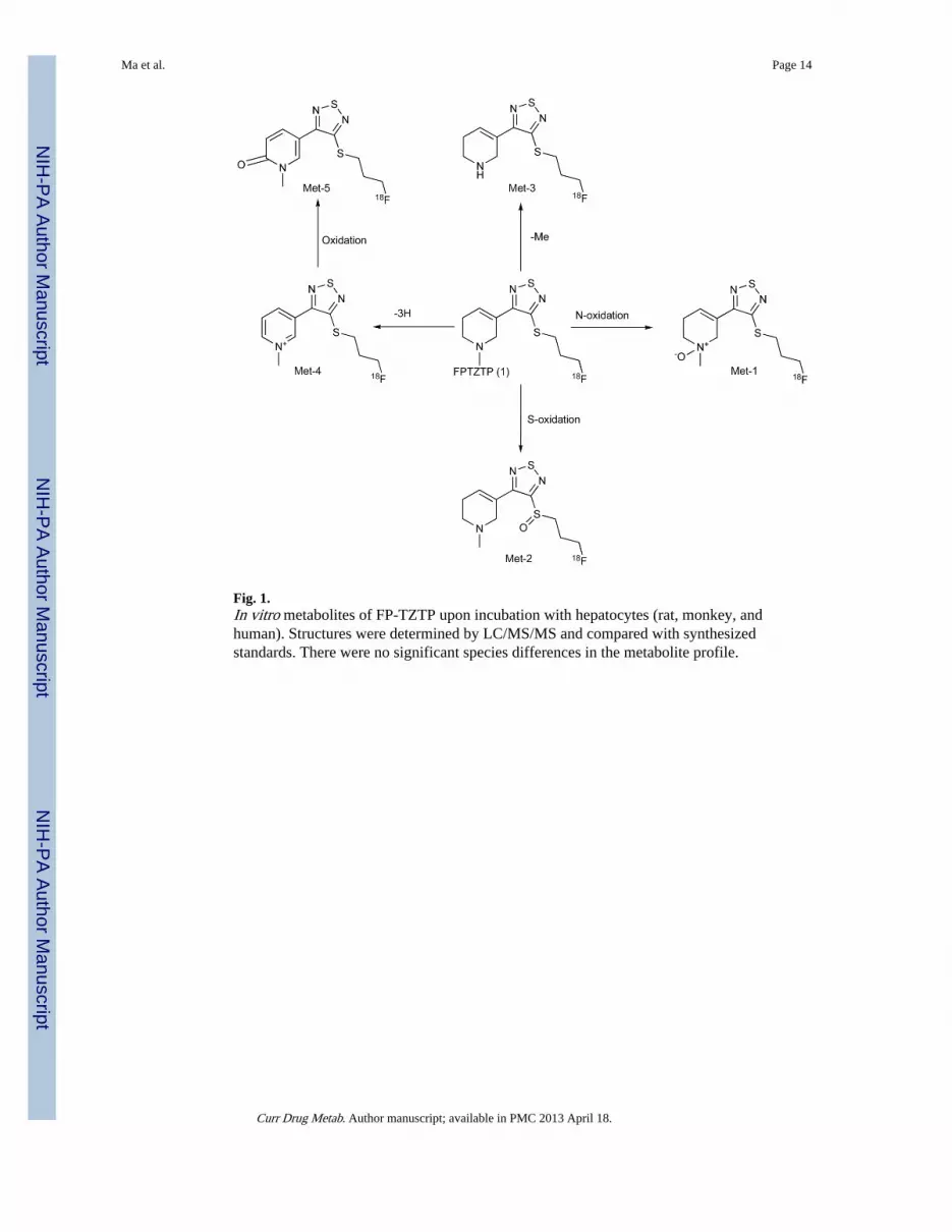
Fig. 1.In vitro metabolites of FP-TZTP upon incubation with hepatocytes (rat, monkey, andhuman). Structures were determined by LC/MS/MS and compared with synthesizedstandards. There were no significant species differences in the metabolite profile.
Ma et al. Page 14
Curr Drug Metab. Author manuscript; available in PMC 2013 April 18.
NIH
-PA Author Manuscript
NIH
-PA Author Manuscript
NIH
-PA Author Manuscript
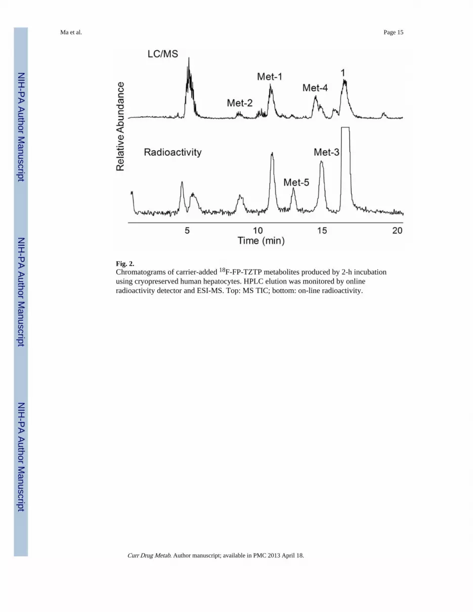
Fig. 2.Chromatograms of carrier-added 18F-FP-TZTP metabolites produced by 2-h incubationusing cryopreserved human hepatocytes. HPLC elution was monitored by onlineradioactivity detector and ESI-MS. Top: MS TIC; bottom: on-line radioactivity.
Ma et al. Page 15
Curr Drug Metab. Author manuscript; available in PMC 2013 April 18.
NIH
-PA Author Manuscript
NIH
-PA Author Manuscript
NIH
-PA Author Manuscript

Fig. 3.Quantitative extraction of parent FP-TZTP from its metabolites in plasma by a two-step two-phase solvent system. (A) Radio-HPLC chromatograms of organic and aqueous phases of(A) first extraction and (B) second extraction. The peaks are labeled with the proposedstructure.
Ma et al. Page 16
Curr Drug Metab. Author manuscript; available in PMC 2013 April 18.
NIH
-PA Author Manuscript
NIH
-PA Author Manuscript
NIH
-PA Author Manuscript
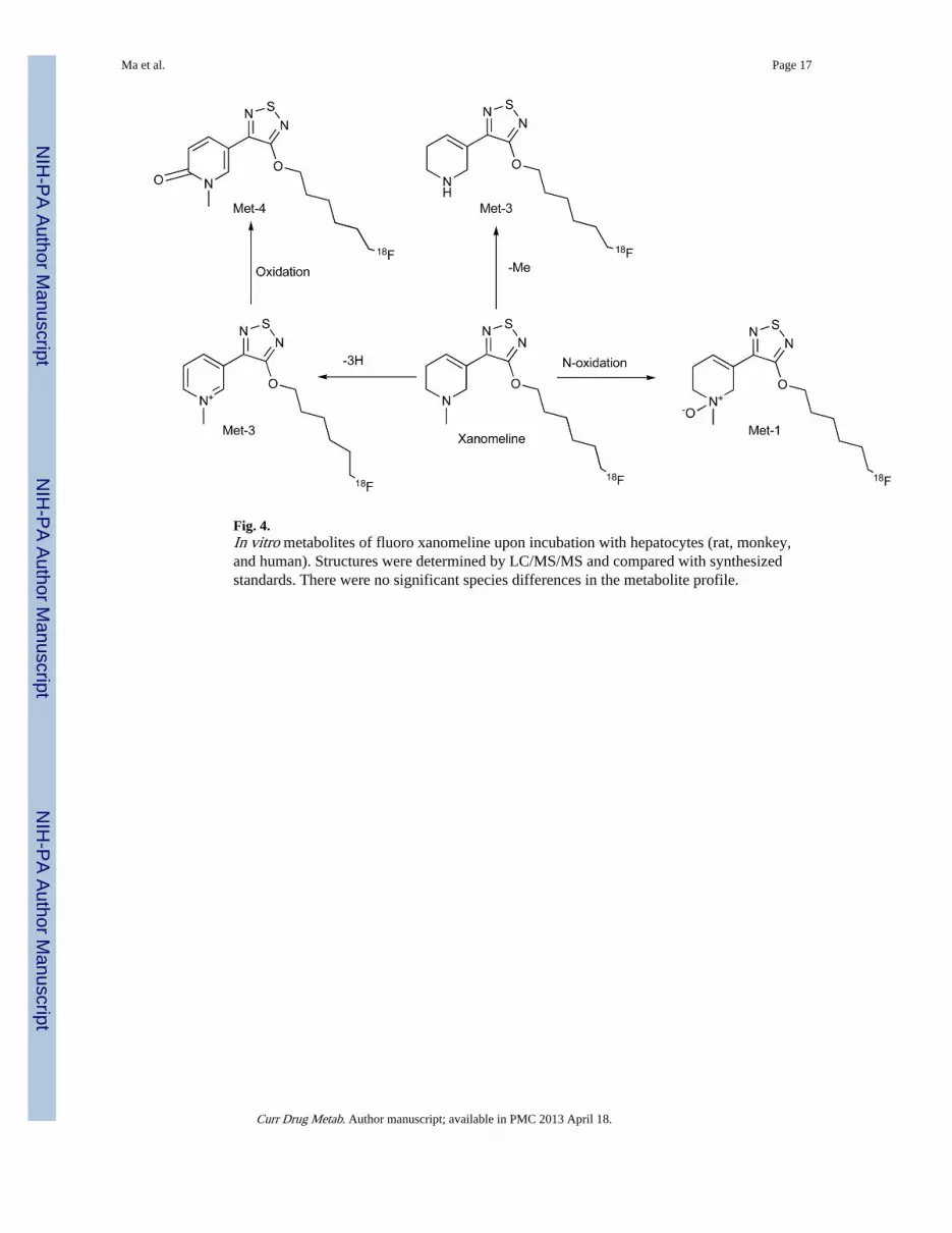
Fig. 4.In vitro metabolites of fluoro xanomeline upon incubation with hepatocytes (rat, monkey,and human). Structures were determined by LC/MS/MS and compared with synthesizedstandards. There were no significant species differences in the metabolite profile.
Ma et al. Page 17
Curr Drug Metab. Author manuscript; available in PMC 2013 April 18.
NIH
-PA Author Manuscript
NIH
-PA Author Manuscript
NIH
-PA Author Manuscript
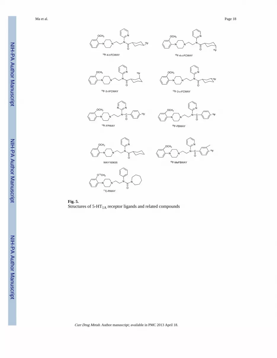
Fig. 5.Structures of 5-HT1A receptor ligands and related compounds
Ma et al. Page 18
Curr Drug Metab. Author manuscript; available in PMC 2013 April 18.
NIH
-PA Author Manuscript
NIH
-PA Author Manuscript
NIH
-PA Author Manuscript
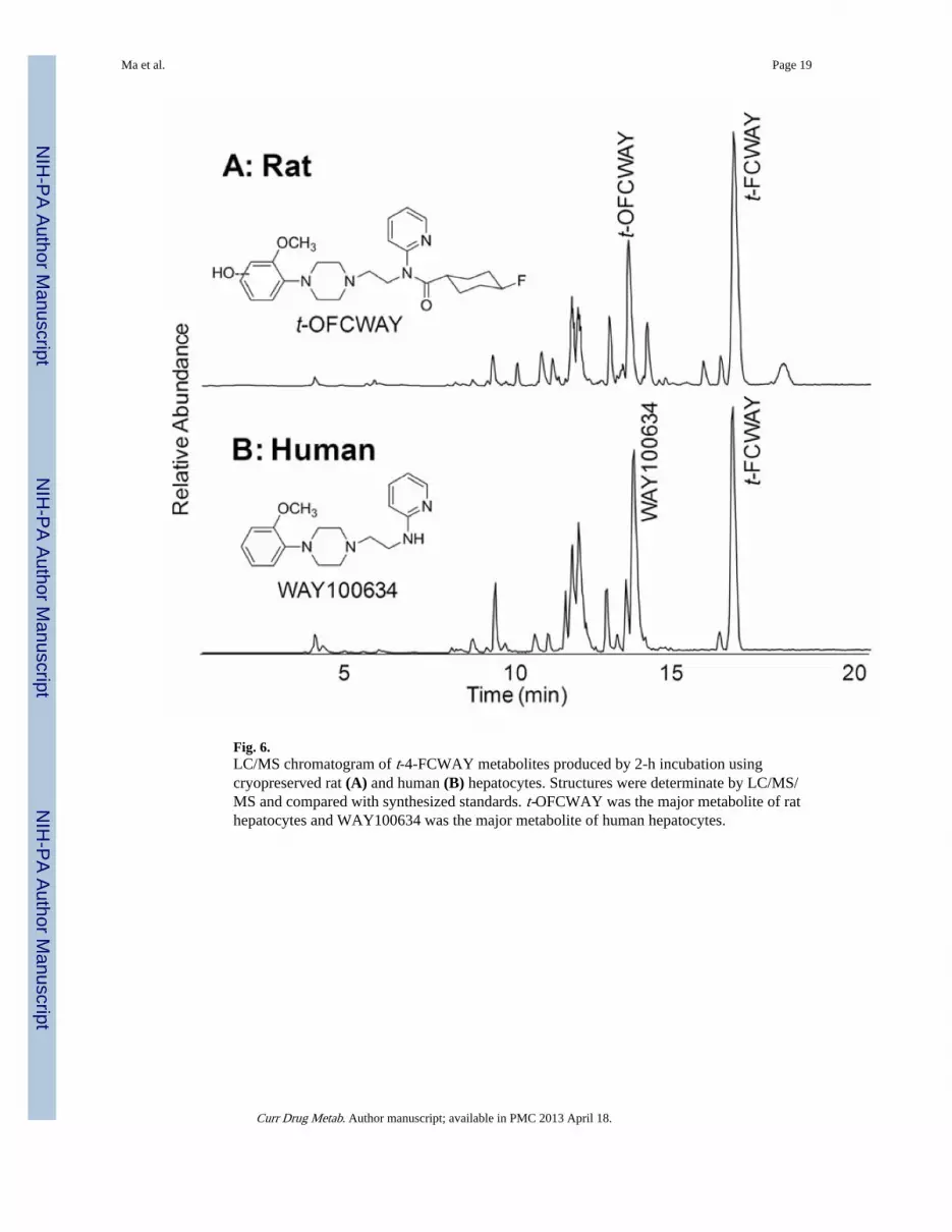
Fig. 6.LC/MS chromatogram of t-4-FCWAY metabolites produced by 2-h incubation usingcryopreserved rat (A) and human (B) hepatocytes. Structures were determinate by LC/MS/MS and compared with synthesized standards. t-OFCWAY was the major metabolite of rathepatocytes and WAY100634 was the major metabolite of human hepatocytes.
Ma et al. Page 19
Curr Drug Metab. Author manuscript; available in PMC 2013 April 18.
NIH
-PA Author Manuscript
NIH
-PA Author Manuscript
NIH
-PA Author Manuscript

Fig. 7.(A) Radiochromatogram of carrier-added 18F-t-4-FCWAY metabolites after incubation withhuman hepatocytes. Free fluoride and FC (4-trans-fluorocyclohexanecarboxylic acid) werethe major radiolabeled metabolites. (B) 18F-t-4-FCWAY standard.
Ma et al. Page 20
Curr Drug Metab. Author manuscript; available in PMC 2013 April 18.
NIH
-PA Author Manuscript
NIH
-PA Author Manuscript
NIH
-PA Author Manuscript
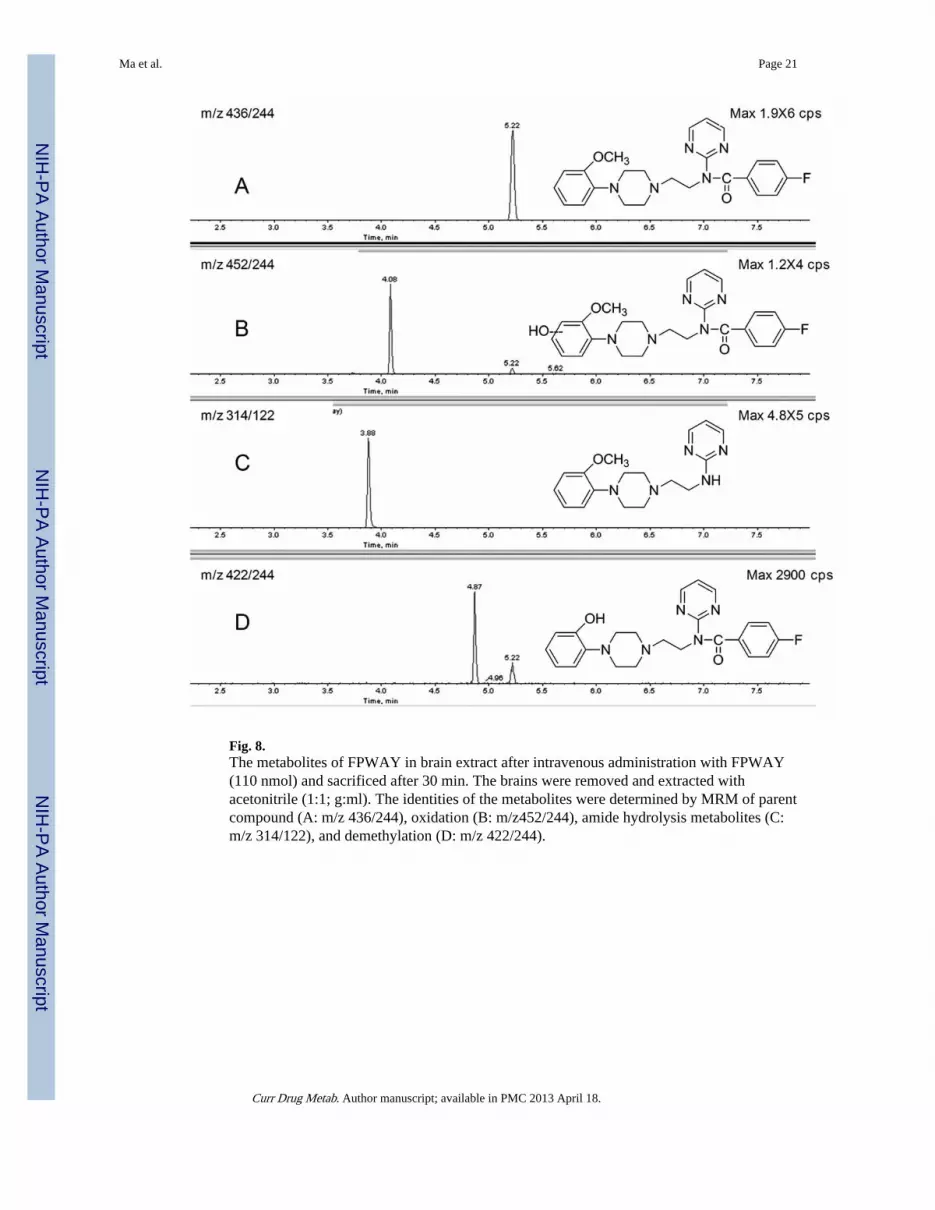
Fig. 8.The metabolites of FPWAY in brain extract after intravenous administration with FPWAY(110 nmol) and sacrificed after 30 min. The brains were removed and extracted withacetonitrile (1:1; g:ml). The identities of the metabolites were determined by MRM of parentcompound (A: m/z 436/244), oxidation (B: m/z452/244), amide hydrolysis metabolites (C:m/z 314/122), and demethylation (D: m/z 422/244).
Ma et al. Page 21
Curr Drug Metab. Author manuscript; available in PMC 2013 April 18.
NIH
-PA Author Manuscript
NIH
-PA Author Manuscript
NIH
-PA Author Manuscript
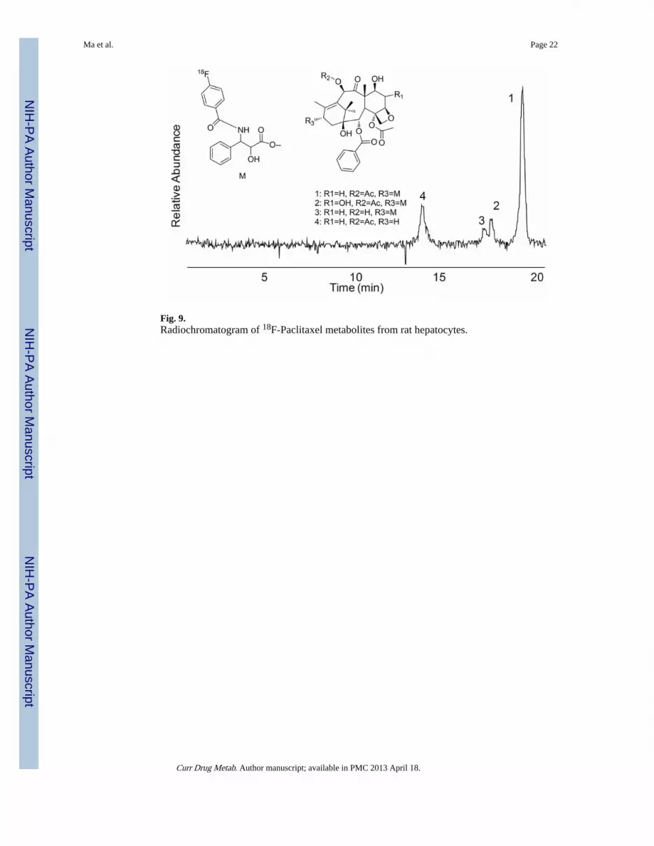
Fig. 9.Radiochromatogram of 18F-Paclitaxel metabolites from rat hepatocytes.
Ma et al. Page 22
Curr Drug Metab. Author manuscript; available in PMC 2013 April 18.
NIH
-PA Author Manuscript
NIH
-PA Author Manuscript
NIH
-PA Author Manuscript

Fig. 10.Structural comparison of H2CB-TE2A, H4TETA (top), and structural representations of thecorresponding 64Cu-labeled complexes based on solved crystal structures.
Ma et al. Page 23
Curr Drug Metab. Author manuscript; available in PMC 2013 April 18.
NIH
-PA Author Manuscript
NIH
-PA Author Manuscript
NIH
-PA Author Manuscript

Fig. 11.The biodistribution studies of [18F]FPWAY. The six regions of rat brain [hippocampus(Hp), cortex (Cx), cerebellum (Cb), caudate (Cd), brain stem (BS), and thalamus (Th)] andplasma (P) were processed for quantitative measurement of parent compound concentrationby LC-MS. The data (black bar) were then converted to (A) the differential uptake ratioDUR (%ID/g × body weight/100) and (B) the brain tissue-specific binding ratio [(DURtissue/DURcerebellum) − 1] to allow direct comparison with data (white bar) obtained by gammacounting of the coinjected radioactive [18F]FPWAY. The DUR and the brain tissue-specificbinding ratio calculated using the LC-MS method were highly correlated to the valuesobtained by standard radioactivity measurements of [18F]FPWAY.
Ma et al. Page 24
Curr Drug Metab. Author manuscript; available in PMC 2013 April 18.
NIH
-PA Author Manuscript
NIH
-PA Author Manuscript
NIH
-PA Author Manuscript

![[3H]-Methyllycaconitine: a high affinity radioligand that labels invertebrate nicotinic acetylcholine receptors](https://img.dokumen.tips/doc/110x75/6356f439ea2708a6d301d78b/3h-methyllycaconitine-a-high-affinity-radioligand-that-labels-invertebrate-nicotinic.jpg)


![Radioligand Binding Assays: Application of [125I]Angiotensin II Receptor Binding](https://img.dokumen.tips/doc/110x75/635cf688095e4caf22057ea4/radioligand-binding-assays-application-of-125iangiotensin-ii-receptor-binding.jpg)


