Embed Size (px)
Citation preview
�����������������
Citation: Lagashetti, A.C.; Singh,
S.K.; Dufossé, L.; Srivastava, P.; Singh,
P.N. Antioxidant, Antibacterial and
Dyeing Potential of Crude Pigment
Extract of Gonatophragmium triuniae
and Its Chemical Characterization.
Molecules 2022, 27, 393. https://
doi.org/10.3390/molecules27020393
Academic Editor: Jacqueline
Aparecida Takahashi
Received: 13 December 2021
Accepted: 5 January 2022
Published: 8 January 2022
Publisher’s Note: MDPI stays neutral
with regard to jurisdictional claims in
published maps and institutional affil-
iations.
Copyright: © 2022 by the authors.
Licensee MDPI, Basel, Switzerland.
This article is an open access article
distributed under the terms and
conditions of the Creative Commons
Attribution (CC BY) license (https://
creativecommons.org/licenses/by/
4.0/).
molecules
Article
Antioxidant, Antibacterial and Dyeing Potential of CrudePigment Extract of Gonatophragmium triuniae and ItsChemical CharacterizationAjay C. Lagashetti 1,2, Sanjay K. Singh 1,2,* , Laurent Dufossé 3,* , Pratibha Srivastava 2,4 and Paras N. Singh 1,2
1 National Fungal Culture Collection of India (NFCCI), Biodiversity and Palaeobiology Group,MACS’ Agharkar Research Institute, G.G. Agarkar Road, Pune 411004, India;[email protected] (A.C.L.); [email protected] (P.N.S.)
2 Faculty of Science, Savitribai Phule Pune University, Pune 411007, India3 CHEMBIOPRO Chimie et Biotechnologie des Produits Naturels, ESIROI Département Agroalimentaire,
Université de la Réunion, F-97490 Sainte-Clotilde, Ile de La Réunion, France4 Bioprospecting Group, MACS’ Agharkar Research Institute, G.G. Agarkar Road, Pune 411004, India;
[email protected]* Correspondence: [email protected] (S.K.S.); [email protected] (L.D.);
Tel.: +91-20-25325103 (S.K.S.); +33-66-8731906 (L.D.)
Abstract: Filamentous fungi synthesize natural products as an ecological function. In this study, aninteresting indigenous fungus producing orange pigment exogenously was investigated in detail asit possesses additional attributes along with colouring properties. An interesting fungus was isolatedfrom a dicot plant, Maytenus rothiana. After a detailed study, the fungal isolate turned out to be aspecies of Gonatophragmium belonging to the family Acrospermaceae. Based on the morphological,cultural, and sequence-based phylogenetic analysis, the identity of this fungus was confirmed asGonatophragmium triuniae. Although this fungus grows moderately, it produces good amounts ofpigment on an agar medium. The fermented crude extract isolated from G. triuniae has shownantioxidant activity with an IC50 value of 0.99 mg/mL and antibacterial activity against Gram-positive bacteria (with MIC of 3.91 µg/mL against Bacillus subtilis, and 15.6 µg/mL and 31.25 µg/mLfor Staphylococcus aureus and Micrococcus luteus, respectively). Dyeing of cotton fabric mordantedwith FeSO4 using crude pigment was found to be satisfactory based on visual observation, sug-gesting its possible use in the textile industry. The orange pigment was purified from the crudeextract by preparative HP-TLC. In addition, UV-Vis, FTIR, HRMS and NMR (1H NMR, 13C NMR),COSY, and DEPT analyses revealed the orange pigment to be “1,2-dimethoxy-3H-phenoxazin-3-one”(C14H11NO4, m/z 257). To our understanding, the present study is the first comprehensive report onGonatophragmium triuniae as a potential pigment producer, reporting “1,2-dimethoxy-3H-phenoxazin-3-one” as the main pigment from the crude hexane extract. Moreover, this is the first study reportingantioxidant, antibacterial, and dyeing potential of crude extract of G. triuniae, suggesting possiblepotential applications of pigments and other bioactive secondary metabolites of the G. triuniae intextile and pharmaceutical industry.
Keywords: Gonatophragmium triuniae; pigments; bioactivity; dyeing; chemical characterization
1. Introduction
Ascomycetous filamentous fungi are known to produce bio-pigments extensively usedas colourants, additives, colour intensifiers, antimicrobial, antioxidants, etc., in differentindustries such as food, beverages, cosmetics, textiles, and pharmaceuticals [1–3]. Naturalpigments/colours are gaining increased attention currently because of the adverse effectsof synthetic colours on humans and the environment. Most of the synthetic colours havebeen found to be toxic, allergic, and carcinogenic to human beings, and hazardous to theenvironment [4–6]. This has increased the need for safe, natural, eco-friendly pigments as
Molecules 2022, 27, 393. https://doi.org/10.3390/molecules27020393 https://www.mdpi.com/journal/molecules
Molecules 2022, 27, 393 2 of 23
an alternative to synthetic pigments. Increasing demand for natural pigments necessitatesa greater need to explore colours or pigments from safe, natural sources, especially frommicrobes (bacteria, fungi, algae, lichens, and actinomycetes).
Fungi are currently emerging as a better and excellent source of natural pigments.Many researchers have reported fungi of different taxonomic groups exhibiting the pro-duction of pigments of diverse chemicals classes such as carotenoids, flavins, ubiquinones,anthraquinones, quinines, phenazines, etc. [1,7–10]. Due to the additional attributes ofthese “mycopigments” such as antimicrobial, anticancer, antioxidant, cytotoxicity, activity,etc. in addition to colouring property, they are being extensively used for a wide range ofapplications in food, textiles, medicines, paints, cosmetics, and electronics [8,9]. Some fun-gal pigments such as azaphilones, astaxanthin, Arpink Red, riboflavin, β-carotene isolatedfrom Monascus, Xanthophyllomyces dendrorhous, Penicillium oxalicum, Ashbya gossypii, andBlakeslea trispora, respectively, are already in the market for their commercial and industrialapplications [11]. Numerous studies have reported the dyeing potential of fungal pigmentsand suggested their possible use in the textile industry for dyeing different types of textilefabrics like cotton, silk, wool, etc. [7,8,12,13].
Literature indicates that extensive studies have been done worldwide on produc-tion, optimization, and applications of pigments from common ascomycetous fungi likeMonascus, Talaromyces, Aspergillus, Penicillium, Fusarium, etc. [8,9]. Besides these conven-tional ones, several other genera, such as Epicoccum, Trichoderma, Alternaria, Chaetomium,etc., are reported to have good pigment production potential [7,14–18]; however, severalgenera of filamentous ascomycetes are still unknown and unexplored for their pigmentproduction potential and exploitations. These unexplored fungi might prove to be a hiddentreasure of novel bio-active pigments having a variety of applications.
Taking this view into account, we have planned the present research in which wehave isolated an uncommon fungus, Gonatophragmium triuniae, from infected leaves of theMaytenus rothiana, a plant endemic to central Maharashtra (Western Ghats). Interestingly,it was found that this rare fungus produces a very good extracellular orange pigmenton solid media [potato dextrose agar medium (PDA)] as well as in liquid media [potatodextrose (PD) broth]. For the characterization of pigment, pure culture of G. triuniae wassubjected to flask level fermentation in PD broth, and pigments were extracted from theculture filtrate using Hexane and dried. The dried Hexane extract was then examinedfor its antimicrobial and antioxidant properties and also assessed for its dyeing potentialon cotton fabric using two mordants (Alum & FeSO4). Finally, by preparative thin-layerchromatography (TLC), we purified an orange pigment from the crude pigment extract andidentified it as “1,2-dimethoxy-3H-phenoxazin-3-one” based on ultra-violet (UV); Fouriertransform infrared (FTIR); high-resolution mass (HRMS) spectroscopy; and 1H & 13C NMR,COSY, and DEPT analysis.
Several natural and synthetic phenoxazines are well known for their bioactivity anddyeing properties. These phenoxazines were found to exhibit antioxidant, antibacterial,anti-proliferative, and anti-tumoral activities [19]. 3H-phenoxazin-3-one and its derivativesexhibiting numerous biological activities (antimicrobial, anticancer, antitumor, antiviral,antitubercular, anticoccidial, antineoplastic, phytotoxic, and cell growth-stimulating) havebeen reported from different microorganisms such as actinomycetes, lichens, and fungi [20].Phenoxazine class of pigments such as Phenoxazone, Pycnosanguin, Cinnabarine, O-acetylcinnabarine, 2-Amino-9-formylphenoxazone-1-carbonic acid, and 9-Hydroxymethyl-2-methylaminophenoxazone-1-carbonic acid methyl ester have been described from thefungus Pycnoporus sanguineus [21]. Similarly, Chandrananimycins A-C belonging to class3H-phenoxazin-3-one, exhibiting antitumor activity against colon cancer (CCL HT29);breast cancer (LCL H460, CNCL SF268, MACL MCF-7); lung cancer (LXFA 526L, LXFL529L); melanoma (MEXF 514L); and kidney tumor cells (PRCL PC3M, RXF 631L) hasalso been reported from Actinomadura sp. [22,23]. One of the recent studies evaluated theantibacterial activity of the synthesized derivatives of 3H-phenoxazin-3-one [20]. Basedon the history of Phenoxazin class of pigments, their promising bioactivity, and their
Molecules 2022, 27, 393 3 of 23
dyeing property, we may consider present compound 1,2-dimethoxy-3H-phenoxazin-3-oneisolated from G. triuniae NFCCI 4873 as a good colourant as well as a potential candidatefor application in the textile and pharmaceutical industry.
2. Results and Discussion2.1. Morphological Identification
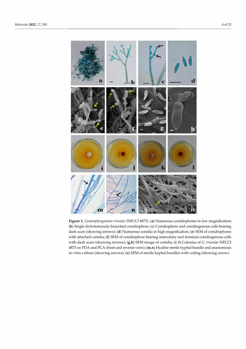
Leaf lesions, amphigenous, necrotic spots single or irregular in concentric rings, laterspots unite to form large spots. Margin: greyish-white; center: white. Colonies hypophyl-lus, velvety, brown. Mycelium superficial. Hyphae branched, septate, pale olivaceousto subhyaline, smooth-walled, up to 6.5 µm wide. Stroma and hyphopodia are absent.Conidiophores arising from superficial hyphae, dichotomously branched, multi-septate(6–8), the width of conidiophore gradually decreasing towards the length; macronema-tous to mononematous, erect, smooth-walled, highly geniculate, nodose, basal half partof conidiophore olivaceous brown and subhyaline to light olivaceous towards the apex,55–145 × 3.22–7 µm. Conidiogenous cells integrated, polyblastic, terminal to intercalary,swollen towards the apex, variable in size: 10–20 µm long, bearing 10–15 loci, scars thick-ened and darkened, dentate or plate-like about 1 µm diameter. Conidia solitary, dry,holoblastic, acropleurogenous, clavate, cylindrical, straight to slightly curved, 0–1 septate,smooth-walled, subhyaline to light olivaceous, base narrowly truncate, apex obtuse, hilumthickened and darkened, 6–15 × 2–3.6 µm (Figure 1).
Colonies on Potato Dextrose Agar (PDA), slow-growing, reach 26–29 mm diameterafter 4 weeks of incubation at 25 ◦C; front view of colony light yellow (4A4), circular,raised, aerial mycelium slightly cottony, margin smooth. Reverse dark brown (7F8) withdiffusible yellowish orange (5B8) pigment in entire media. Mycelium branched, septate,smooth-walled, with frequent anastomosis, hyaline, sterile. Colonies on Potato Carrot Agar(PCA), reach 27–28 mm diameter after 4 weeks at 25 ◦C; front view of colony grey (6C1),circular, raised, slightly cottony with margin smooth. Reverse dark brown (6F8) and withdiffusible yellowish orange (5B8) pigment in entire media. Mycelium branched, septate,smooth-walled, anastomosis, hyaline, sterile (Figure 1).
2.2. Molecular Identification and Phylogeny
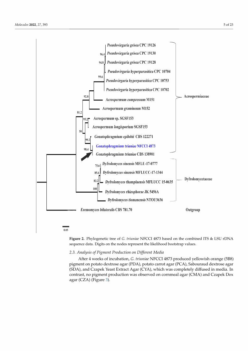
Mega Blast analysis of ITS sequence of G. triuniae NFCCI 4873 showed 100% identitywith type strain G. triuniae CBS138901; whereas LSU sequence showed 99.74% identity withG. epiloblii CPC 34889 and 99.61% similarity with G. triuniae CBS 138901. A phylogenetictree was constructed based on combined ITS & LSU rDNA sequence data of a total of19 genetically-related isolates, which shows that our isolate was clustered with G. triuniaeCBS138901 with a very strong bootstrap value of 98.4 (Figure 2). Therefore, based oncombined morphological and molecular phylogenetic analysis, the present isolate wasidentified as G. triuniae.
Molecules 2022, 27, 393 4 of 23Molecules 2022, 27, x FOR PEER REVIEW 4 of 23
Figure 1. Gonatophragmium triuniae (NFCCI 4873): (a) Numerous conidiophores in low magnifica‐
tion; (b) Single dichotomously branched conidiophore; (c) Conidiophore and conidiogenous cells
bearing dark scars (showing arrows); (d) Numerous conidia in high magnification; (e) SEM of co‐
nidiophores with attached conidia; (f) SEM of conidiophore bearing intercalary and terminal conid‐
iogenous cells with dark scars (showing arrows); (g,h) SEM image of conidia; (i–l) Colonies of G.
triuniae NFCCI 4873 on PDA and PCA (front and reverse view); (m,n) Hyaline sterile hyphal bundle
and anastomosis in‐vitro culture (showing arrows); (o) SEM of sterile hyphal bundles with coiling
(showing arrow).
2.2. Molecular Identification and Phylogeny
Mega Blast analysis of ITS sequence of G. triuniae NFCCI 4873 showed 100% identity
with type strain G. triuniae CBS138901; whereas LSU sequence showed 99.74% identity
with G. epiloblii CPC 34889 and 99.61% similarity with G. triuniae CBS 138901. A phyloge‐
netic tree was constructed based on combined ITS & LSU rDNA sequence data of a total
of 19 genetically‐related isolates, which shows that our isolate was clustered with G. triu‐
niae CBS138901 with a very strong bootstrap value of 98.4 (Figure 2). Therefore, based on
combined morphological and molecular phylogenetic analysis, the present isolate was
identified as G. triuniae.
Figure 1. Gonatophragmium triuniae (NFCCI 4873): (a) Numerous conidiophores in low magnification;(b) Single dichotomously branched conidiophore; (c) Conidiophore and conidiogenous cells bearingdark scars (showing arrows); (d) Numerous conidia in high magnification; (e) SEM of conidiophoreswith attached conidia; (f) SEM of conidiophore bearing intercalary and terminal conidiogenous cellswith dark scars (showing arrows); (g,h) SEM image of conidia; (i–l) Colonies of G. triuniae NFCCI4873 on PDA and PCA (front and reverse view); (m,n) Hyaline sterile hyphal bundle and anastomosisin-vitro culture (showing arrows); (o) SEM of sterile hyphal bundles with coiling (showing arrow).
Molecules 2022, 27, 393 5 of 23
Molecules 2022, 27, 393 5 of 24
Figure 2. Phylogenetic tree of G. triuniae NFCCI 4873 based on the combined ITS & LSU rDNA
sequence data. Digits on the nodes represent the likelihood bootstrap values.
2.3. Analysis of Pigment Production on Different Media
After 4 weeks of incubation, G. triuniae NFCCI 4873 produced yellowish orange (5B8)
pigment on potato dextrose agar (PDA), potato carrot agar (PCA), Sabouraud dextrose
agar (SDA), and Czapek Yeast Extract Agar (CYA), which was completely diffused in
media. In contrast, no pigment production was observed on cornmeal agar (CMA) and
Czapek Dox agar (CZA) (Figure 3).
Figure 2. Phylogenetic tree of G. triuniae NFCCI 4873 based on the combined ITS & LSU rDNAsequence data. Digits on the nodes represent the likelihood bootstrap values.
2.3. Analysis of Pigment Production on Different Media
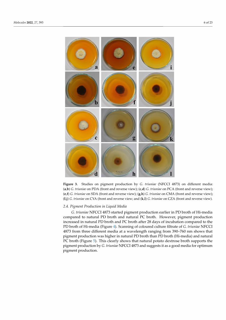
After 4 weeks of incubation, G. triuniae NFCCI 4873 produced yellowish orange (5B8)pigment on potato dextrose agar (PDA), potato carrot agar (PCA), Sabouraud dextrose agar(SDA), and Czapek Yeast Extract Agar (CYA), which was completely diffused in media. Incontrast, no pigment production was observed on cornmeal agar (CMA) and Czapek Doxagar (CZA) (Figure 3).
Molecules 2022, 27, 393 6 of 23Molecules 2022, 27, x FOR PEER REVIEW 6 of 23
Figure 3. Studies on pigment production by G. triuniae (NFCCI 4873) on different media: (a,b) G.
triuniae on PDA (front and reverse view); (c,d) G. triuniae on PCA (front and reverse view); (e,f) G.
triuniae on SDA (front and reverse view); (g,h) G. triuniae on CMA (front and reverse view); (I,j) G.
triuniae on CYA (front and reverse view; and (k,l) G. triuniae on CZA (front and reverse view).
2.4. Pigment Production in Liquid Media
G. triuniae NFCCI 4873 started pigment production earlier in PD broth of Hi‐media
compared to natural PD broth and natural PC broth. However, pigment production in‐
creased in natural PD broth and PC broth after 28 days of incubation compared to the PD
broth of Hi‐media (Figure 4). Scanning of coloured culture filtrate of G. triuniae NFCCI
4873 from three different media at a wavelength ranging from 390–760 nm shows that
pigment production was higher in natural PD broth than PD broth (Hi‐media) and natural
PC broth (Figure 5). This clearly shows that natural potato dextrose broth supports the
pigment production by G. triuniae NFCCI 4873 and suggests it as a good media for opti‐
mum pigment production.
Figure 3. Studies on pigment production by G. triuniae (NFCCI 4873) on different media:(a,b) G. triuniae on PDA (front and reverse view); (c,d) G. triuniae on PCA (front and reverse view);(e,f) G. triuniae on SDA (front and reverse view); (g,h) G. triuniae on CMA (front and reverse view);(I,j) G. triuniae on CYA (front and reverse view; and (k,l) G. triuniae on CZA (front and reverse view).
2.4. Pigment Production in Liquid Media
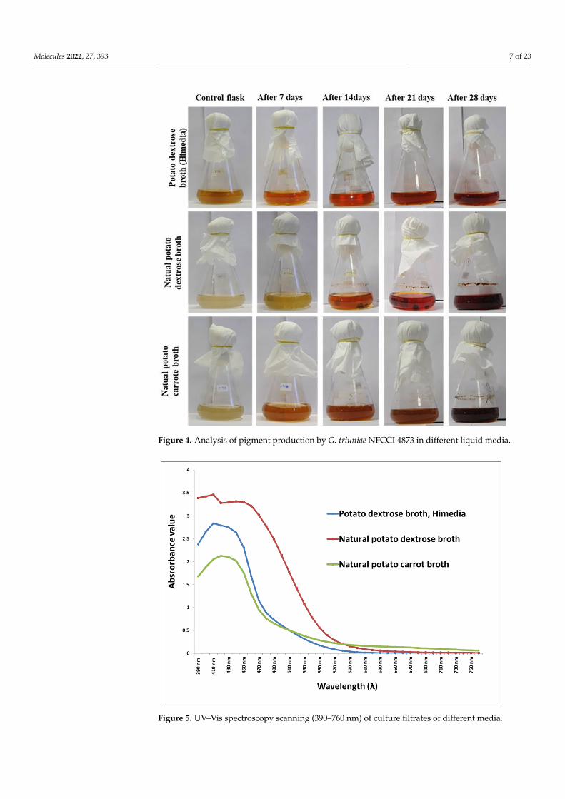
G. triuniae NFCCI 4873 started pigment production earlier in PD broth of Hi-mediacompared to natural PD broth and natural PC broth. However, pigment productionincreased in natural PD broth and PC broth after 28 days of incubation compared to thePD broth of Hi-media (Figure 4). Scanning of coloured culture filtrate of G. triuniae NFCCI4873 from three different media at a wavelength ranging from 390–760 nm shows thatpigment production was higher in natural PD broth than PD broth (Hi-media) and naturalPC broth (Figure 5). This clearly shows that natural potato dextrose broth supports thepigment production by G. triuniae NFCCI 4873 and suggests it as a good media for optimumpigment production.
Molecules 2022, 27, 393 7 of 23Molecules 2022, 27, x FOR PEER REVIEW 7 of 23
Figure 4. Analysis of pigment production by G. triuniae NFCCI 4873 in different liquid media.
Figure 5. UV–Vis spectroscopy scanning (390–760 nm) of culture filtrates of different media.
Figure 4. Analysis of pigment production by G. triuniae NFCCI 4873 in different liquid media.
Molecules 2022, 27, x FOR PEER REVIEW 7 of 23
Figure 4. Analysis of pigment production by G. triuniae NFCCI 4873 in different liquid media.
Figure 5. UV–Vis spectroscopy scanning (390–760 nm) of culture filtrates of different media.
Figure 5. UV–Vis spectroscopy scanning (390–760 nm) of culture filtrates of different media.
Molecules 2022, 27, 393 8 of 23
2.5. Fermentation and Extraction of Pigments
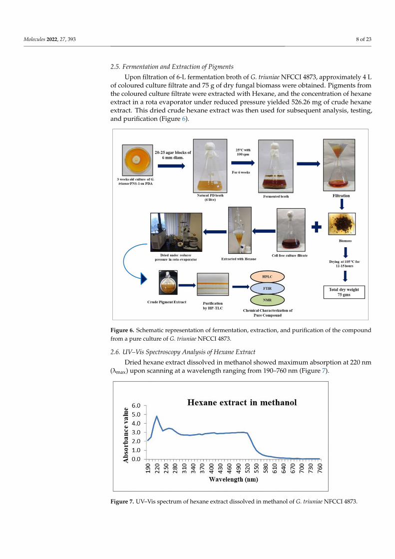
Upon filtration of 6-L fermentation broth of G. triuniae NFCCI 4873, approximately 4 Lof coloured culture filtrate and 75 g of dry fungal biomass were obtained. Pigments fromthe coloured culture filtrate were extracted with Hexane, and the concentration of hexaneextract in a rota evaporator under reduced pressure yielded 526.26 mg of crude hexaneextract. This dried crude hexane extract was then used for subsequent analysis, testing,and purification (Figure 6).
Molecules 2022, 27, x FOR PEER REVIEW 8 of 23
2.5. Fermentation and Extraction of Pigments
Upon filtration of 6‐L fermentation broth of G. triuniae NFCCI 4873, approximately 4
L of coloured culture filtrate and 75 g of dry fungal biomass were obtained. Pigments from
the coloured culture filtrate were extracted with Hexane, and the concentration of hexane
extract in a rota evaporator under reduced pressure yielded 526.26 mg of crude hexane
extract. This dried crude hexane extract was then used for subsequent analysis, testing,
and purification (Figure 6).
Figure 6. Schematic representation of fermentation, extraction, and purification of the compound
from a pure culture of G. triuniae NFCCI 4873.
2.6. UV–Vis Spectroscopy Analysis of Hexane Extract
Dried hexane extract dissolved in methanol showed maximum absorption at 220 nm
(λmax) upon scanning at a wavelength ranging from 190–760 nm (Figure 7).
Figure 7. UV–Vis spectrum of hexane extract dissolved in methanol of G. triuniae NFCCI 4873.
Figure 6. Schematic representation of fermentation, extraction, and purification of the compoundfrom a pure culture of G. triuniae NFCCI 4873.
2.6. UV–Vis Spectroscopy Analysis of Hexane Extract
Dried hexane extract dissolved in methanol showed maximum absorption at 220 nm(λmax) upon scanning at a wavelength ranging from 190–760 nm (Figure 7).
Molecules 2022, 27, x FOR PEER REVIEW 8 of 23
2.5. Fermentation and Extraction of Pigments
Upon filtration of 6‐L fermentation broth of G. triuniae NFCCI 4873, approximately 4
L of coloured culture filtrate and 75 g of dry fungal biomass were obtained. Pigments from
the coloured culture filtrate were extracted with Hexane, and the concentration of hexane
extract in a rota evaporator under reduced pressure yielded 526.26 mg of crude hexane
extract. This dried crude hexane extract was then used for subsequent analysis, testing,
and purification (Figure 6).
Figure 6. Schematic representation of fermentation, extraction, and purification of the compound
from a pure culture of G. triuniae NFCCI 4873.
2.6. UV–Vis Spectroscopy Analysis of Hexane Extract
Dried hexane extract dissolved in methanol showed maximum absorption at 220 nm
(λmax) upon scanning at a wavelength ranging from 190–760 nm (Figure 7).
Figure 7. UV–Vis spectrum of hexane extract dissolved in methanol of G. triuniae NFCCI 4873. Figure 7. UV–Vis spectrum of hexane extract dissolved in methanol of G. triuniae NFCCI 4873.
Molecules 2022, 27, 393 9 of 23
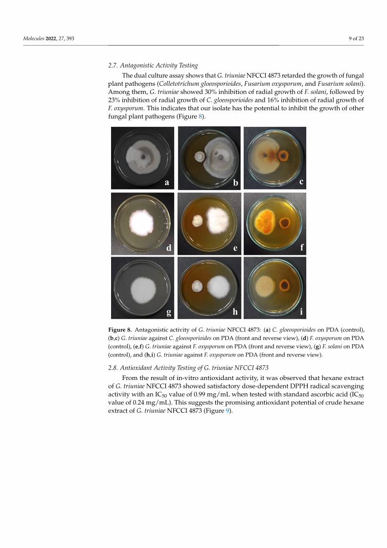
2.7. Antagonistic Activity Testing
The dual culture assay shows that G. triuniae NFCCI 4873 retarded the growth of fungalplant pathogens (Colletotrichum gloeosporioides, Fusarium oxysporum, and Fusarium solani).Among them, G. triuniae showed 30% inhibition of radial growth of F. solani, followed by23% inhibition of radial growth of C. gloeosporioides and 16% inhibition of radial growth ofF. oxysporum. This indicates that our isolate has the potential to inhibit the growth of otherfungal plant pathogens (Figure 8).
Molecules 2022, 27, x FOR PEER REVIEW 9 of 23
2.7. Antagonistic Activity Testing
The dual culture assay shows that G. triuniae NFCCI 4873 retarded the growth of
fungal plant pathogens (Colletotrichum gloeosporioides, Fusarium oxysporum, and Fusarium
solani). Among them, G. triuniae showed 30% inhibition of radial growth of F. solani, fol‐
lowed by 23% inhibition of radial growth of C. gloeosporioides and 16% inhibition of radial
growth of F. oxysporum. This indicates that our isolate has the potential to inhibit the
growth of other fungal plant pathogens (Figure 8).
Figure 8. Antagonistic activity of G. triuniae NFCCI 4873: (a) C. gloeosporioides on PDA (control), (b,c)
G. triuniae against C. gloeosporioides on PDA (front and reverse view), (d) F. oxysporum on PDA (con‐
trol), (e,f) G. triuniae against F. oxysporum on PDA (front and reverse view), (g) F. solani on PDA
(control), and (h,i) G. triuniae against F. oxysporum on PDA (front and reverse view).
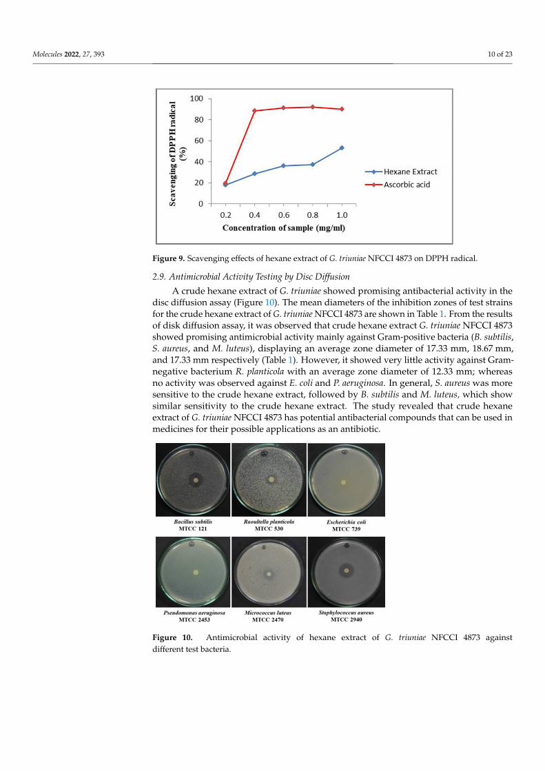
2.8. Antioxidant Activity Testing of G. triuniae NFCCI 4873
From the result of in‐vitro antioxidant activity, it was observed that hexane extract of
G. triuniae NFCCI 4873 showed satisfactory dose‐dependent DPPH radical scavenging ac‐
tivity with an IC50 value of 0.99 mg/mL when tested with standard ascorbic acid (IC50 value
of 0.24 mg/mL). This suggests the promising antioxidant potential of crude hexane extract
of G. triuniae NFCCI 4873 (Figure 9).
Figure 8. Antagonistic activity of G. triuniae NFCCI 4873: (a) C. gloeosporioides on PDA (control),(b,c) G. triuniae against C. gloeosporioides on PDA (front and reverse view), (d) F. oxysporum on PDA(control), (e,f) G. triuniae against F. oxysporum on PDA (front and reverse view), (g) F. solani on PDA(control), and (h,i) G. triuniae against F. oxysporum on PDA (front and reverse view).
2.8. Antioxidant Activity Testing of G. triuniae NFCCI 4873
From the result of in-vitro antioxidant activity, it was observed that hexane extractof G. triuniae NFCCI 4873 showed satisfactory dose-dependent DPPH radical scavengingactivity with an IC50 value of 0.99 mg/mL when tested with standard ascorbic acid (IC50value of 0.24 mg/mL). This suggests the promising antioxidant potential of crude hexaneextract of G. triuniae NFCCI 4873 (Figure 9).
Molecules 2022, 27, 393 10 of 23Molecules 2022, 27, x FOR PEER REVIEW 10 of 23
Figure 9. Scavenging effects of hexane extract of G. triuniae NFCCI 4873 on DPPH radical.
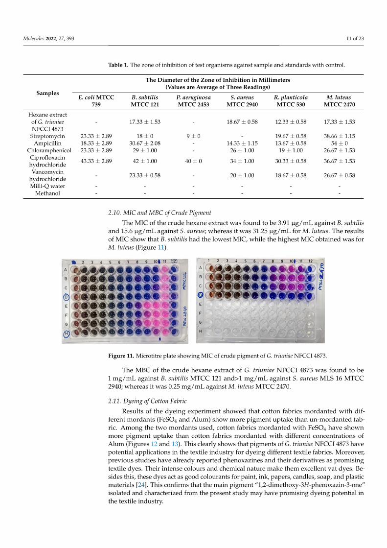
2.9. Antimicrobial Activity Testing by Disc Diffusion
A crude hexane extract of G. triuniae showed promising antibacterial activity in the
disc diffusion assay (Figure 10). The mean diameters of the inhibition zones of test strains
for the crude hexane extract of G. triuniae NFCCI 4873 are shown in Table 1. From the
results of disk diffusion assay, it was observed that crude hexane extract G. triuniae NFCCI
4873 showed promising antimicrobial activity mainly against Gram‐positive bacteria (B.
subtilis, S. aureus, and M. luteus), displaying an average zone diameter of 17.33 mm, 18.67
mm, and 17.33 mm respectively (Table 1). However, it showed very little activity against
Gram‐negative bacterium R. planticola with an average zone diameter of 12.33 mm;
whereas no activity was observed against E. coli and P. aeruginosa. In general, S. aureus
was more sensitive to the crude hexane extract, followed by B. subtilis and M. luteus, which
show similar sensitivity to the crude hexane extract. The study revealed that crude hexane
extract of G. triuniae NFCCI 4873 has potential antibacterial compounds that can be used
in medicines for their possible applications as an antibiotic.
Figure 10. Antimicrobial activity of hexane extract of G. triuniae NFCCI 4873 against different test
bacteria.
Figure 9. Scavenging effects of hexane extract of G. triuniae NFCCI 4873 on DPPH radical.
2.9. Antimicrobial Activity Testing by Disc Diffusion
A crude hexane extract of G. triuniae showed promising antibacterial activity in thedisc diffusion assay (Figure 10). The mean diameters of the inhibition zones of test strainsfor the crude hexane extract of G. triuniae NFCCI 4873 are shown in Table 1. From the resultsof disk diffusion assay, it was observed that crude hexane extract G. triuniae NFCCI 4873showed promising antimicrobial activity mainly against Gram-positive bacteria (B. subtilis,S. aureus, and M. luteus), displaying an average zone diameter of 17.33 mm, 18.67 mm,and 17.33 mm respectively (Table 1). However, it showed very little activity against Gram-negative bacterium R. planticola with an average zone diameter of 12.33 mm; whereasno activity was observed against E. coli and P. aeruginosa. In general, S. aureus was moresensitive to the crude hexane extract, followed by B. subtilis and M. luteus, which showsimilar sensitivity to the crude hexane extract. The study revealed that crude hexaneextract of G. triuniae NFCCI 4873 has potential antibacterial compounds that can be used inmedicines for their possible applications as an antibiotic.
Molecules 2022, 27, x FOR PEER REVIEW 10 of 23
Figure 9. Scavenging effects of hexane extract of G. triuniae NFCCI 4873 on DPPH radical.
2.9. Antimicrobial Activity Testing by Disc Diffusion
A crude hexane extract of G. triuniae showed promising antibacterial activity in the
disc diffusion assay (Figure 10). The mean diameters of the inhibition zones of test strains
for the crude hexane extract of G. triuniae NFCCI 4873 are shown in Table 1. From the
results of disk diffusion assay, it was observed that crude hexane extract G. triuniae NFCCI
4873 showed promising antimicrobial activity mainly against Gram‐positive bacteria (B.
subtilis, S. aureus, and M. luteus), displaying an average zone diameter of 17.33 mm, 18.67
mm, and 17.33 mm respectively (Table 1). However, it showed very little activity against
Gram‐negative bacterium R. planticola with an average zone diameter of 12.33 mm;
whereas no activity was observed against E. coli and P. aeruginosa. In general, S. aureus
was more sensitive to the crude hexane extract, followed by B. subtilis and M. luteus, which
show similar sensitivity to the crude hexane extract. The study revealed that crude hexane
extract of G. triuniae NFCCI 4873 has potential antibacterial compounds that can be used
in medicines for their possible applications as an antibiotic.
Figure 10. Antimicrobial activity of hexane extract of G. triuniae NFCCI 4873 against different test
bacteria.
Figure 10. Antimicrobial activity of hexane extract of G. triuniae NFCCI 4873 againstdifferent test bacteria.
Molecules 2022, 27, 393 11 of 23
Table 1. The zone of inhibition of test organisms against sample and standards with control.
Samples
The Diameter of the Zone of Inhibition in Millimeters(Values are Average of Three Readings)
E. coli MTCC739
B. subtilisMTCC 121
P. aeruginosaMTCC 2453
S. aureusMTCC 2940
R. planticolaMTCC 530
M. luteusMTCC 2470
Hexane extractof G. triuniaeNFCCI 4873
- 17.33 ± 1.53 - 18.67 ± 0.58 12.33 ± 0.58 17.33 ± 1.53
Streptomycin 23.33 ± 2.89 18 ± 0 9 ± 0 - 19.67 ± 0.58 38.66 ± 1.15Ampicillin 18.33 ± 2.89 30.67 ± 2.08 - 14.33 ± 1.15 13.67 ± 0.58 54 ± 0
Chloramphenicol 23.33 ± 2.89 29 ± 1.00 - 26 ± 1.00 19 ± 1.00 26.67 ± 1.53Ciprofloxacinhydrochloride 43.33 ± 2.89 42 ± 1.00 40 ± 0 34 ± 1.00 30.33 ± 0.58 36.67 ± 1.53
Vancomycinhydrochloride - 23.33 ± 0.58 - 20 ± 1.00 18.67 ± 0.58 26.67 ± 0.58
Milli-Q water - - - - - -Methanol - - - - - -
2.10. MIC and MBC of Crude Pigment
The MIC of the crude hexane extract was found to be 3.91 µg/mL against B. subtilisand 15.6 µg/mL against S. aureus; whereas it was 31.25 µg/mL for M. luteus. The resultsof MIC show that B. subtilis had the lowest MIC, while the highest MIC obtained was forM. luteus (Figure 11).
Molecules 2022, 27, x FOR PEER REVIEW 11 of 23
Table 1. The zone of inhibition of test organisms against sample and standards with control.
Samples
The Diameter of the Zone of Inhibition in Millimeters
(Values are Average of Three Readings)
E. coli
MTCC 739
B. subtilis
MTCC 121
P. aeruginosa
MTCC 2453
S. aureus
MTCC 2940
R. planticola
MTCC 530
M. luteus
MTCC 2470
Hexane extract of G. triuniae NFCCI
4873 ‐ 17.33 ± 1.53 ‐ 18.67 ± 0.58 12.33 ± 0.58 17.33 ± 1.53
Streptomycin 23.33 ± 2.89 18 ± 0 9 ± 0 ‐ 19.67 ± 0.58 38.66 ± 1.15
Ampicillin 18.33 ± 2.89 30.67 ± 2.08 ‐ 14.33 ± 1.15 13.67 ± 0.58 54 ± 0
Chloramphenicol 23.33 ± 2.89 29 ± 1.00 ‐ 26 ± 1.00 19 ± 1.00 26.67 ± 1.53
Ciprofloxacin hydrochloride 43.33 ± 2.89 42 ± 1.00 40 ± 0 34 ± 1.00 30.33 ± 0.58 36.67 ± 1.53
Vancomycin hydrochloride ‐ 23.33 ± 0.58 ‐ 20 ± 1.00 18.67 ± 0.58 26.67 ± 0.58
Milli‐Q water ‐ ‐ ‐ ‐ ‐ ‐
Methanol ‐ ‐ ‐ ‐ ‐ ‐
2.10. MIC and MBC of Crude Pigment
The MIC of the crude hexane extract was found to be 3.91 μg/mL against B. subtilis
and 15.6 μg/mL against S. aureus; whereas it was 31.25 μg/mL for M. luteus. The results of
MIC show that B. subtilis had the lowest MIC, while the highest MIC obtained was for M.
luteus (Figure 11).
The MBC of the crude hexane extract of G. triuniae NFCCI 4873 was found to be
1mg/mL against B. subtilis MTCC 121 and>1 mg/mL against S. aureus MLS 16 MTCC 2940;
whereas it was 0.25 mg/mL against M. luteus MTCC 2470.
Figure 11. Microtitre plate showing MIC of crude pigment of G. triuniae NFCCI 4873.
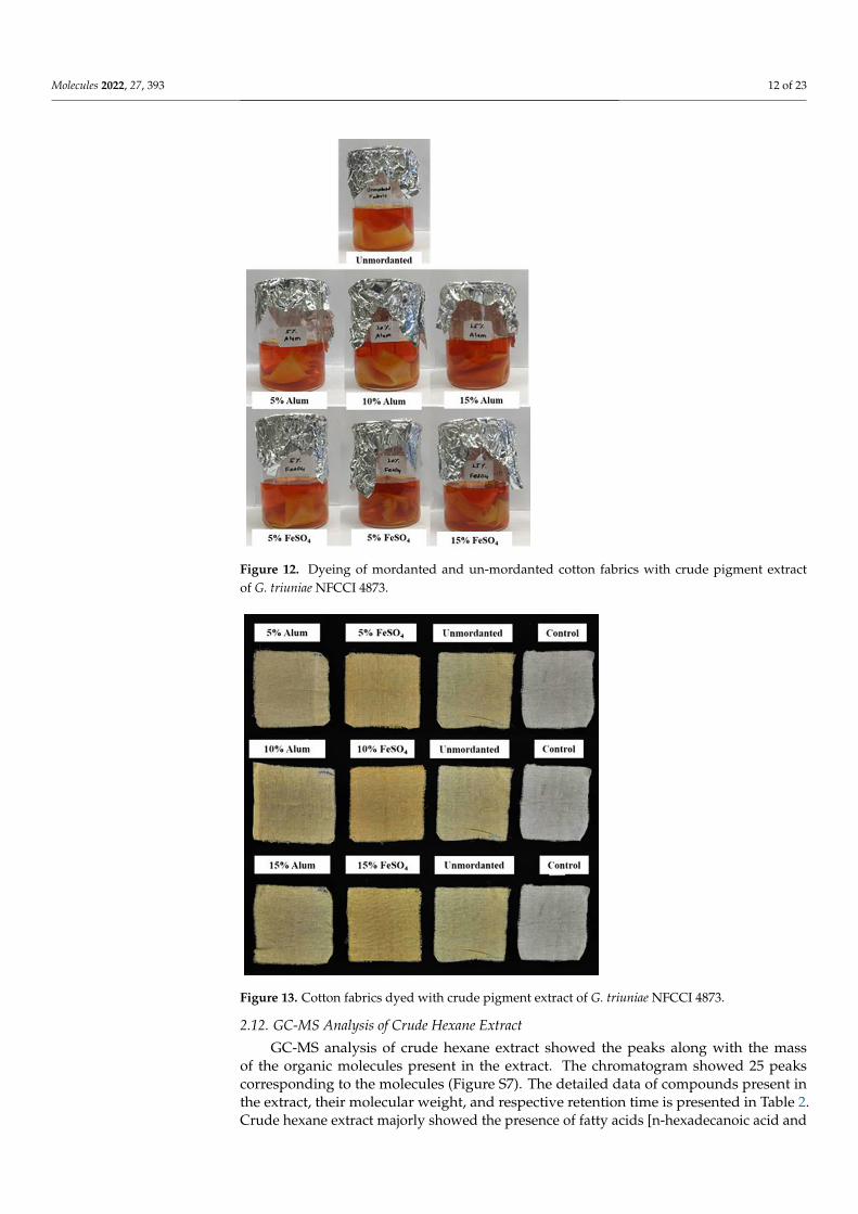
2.11. Dyeing of Cotton Fabric
Results of the dyeing experiment showed that cotton fabrics mordanted with differ‐
ent mordants (FeSO4 and Alum) show more pigment uptake than un‐mordanted fabric.
Among the two mordants used, cotton fabrics mordanted with FeSO4 have shown more
pigment uptake than cotton fabrics mordanted with different concentrations of Alum
(Figures 12 and 13). This clearly shows that pigments of G. triuniae NFCCI 4873 have po‐
tential applications in the textile industry for dyeing different textile fabrics. Moreover,
previous studies have already reported phenoxazines and their derivatives as promising
textile dyes. Their intense colours and chemical nature make them excellent vat dyes. Be‐
sides this, these dyes act as good colourants for paint, ink, papers, candles, soap, and plas‐
tic materials [24]. This confirms that the main pigment “1,2‐dimethoxy‐3H‐phenoxazin‐3‐
one” isolated and characterized from the present study may have promising dyeing po‐
tential in the textile industry.
Figure 11. Microtitre plate showing MIC of crude pigment of G. triuniae NFCCI 4873.
The MBC of the crude hexane extract of G. triuniae NFCCI 4873 was found to be1 mg/mL against B. subtilis MTCC 121 and>1 mg/mL against S. aureus MLS 16 MTCC2940; whereas it was 0.25 mg/mL against M. luteus MTCC 2470.
2.11. Dyeing of Cotton Fabric
Results of the dyeing experiment showed that cotton fabrics mordanted with dif-ferent mordants (FeSO4 and Alum) show more pigment uptake than un-mordanted fab-ric. Among the two mordants used, cotton fabrics mordanted with FeSO4 have shownmore pigment uptake than cotton fabrics mordanted with different concentrations ofAlum (Figures 12 and 13). This clearly shows that pigments of G. triuniae NFCCI 4873 havepotential applications in the textile industry for dyeing different textile fabrics. Moreover,previous studies have already reported phenoxazines and their derivatives as promisingtextile dyes. Their intense colours and chemical nature make them excellent vat dyes. Be-sides this, these dyes act as good colourants for paint, ink, papers, candles, soap, and plasticmaterials [24]. This confirms that the main pigment “1,2-dimethoxy-3H-phenoxazin-3-one”isolated and characterized from the present study may have promising dyeing potential inthe textile industry.
Molecules 2022, 27, 393 12 of 23Molecules 2022, 27, x FOR PEER REVIEW 12 of 23
Figure 12. Dyeing of mordanted and un‐mordanted cotton fabrics with crude pigment extract of G.
triuniae NFCCI 4873.
Figure 13. Cotton fabrics dyed with crude pigment extract of G. triuniae NFCCI 4873.
2.12. GC‐MS Analysis of Crude Hexane Extract
GC‐MS analysis of crude hexane extract showed the peaks along with the mass of the
organic molecules present in the extract. The chromatogram showed 25 peaks correspond‐
ing to the molecules (Figure S7). The detailed data of compounds present in the extract,
their molecular weight, and respective retention time is presented in Table 2. Crude hex‐
ane extract majorly showed the presence of fatty acids [n‐hexadecanoic acid and
Figure 12. Dyeing of mordanted and un-mordanted cotton fabrics with crude pigment extractof G. triuniae NFCCI 4873.
Molecules 2022, 27, x FOR PEER REVIEW 12 of 23
Figure 12. Dyeing of mordanted and un‐mordanted cotton fabrics with crude pigment extract of G.
triuniae NFCCI 4873.
Figure 13. Cotton fabrics dyed with crude pigment extract of G. triuniae NFCCI 4873.
2.12. GC‐MS Analysis of Crude Hexane Extract
GC‐MS analysis of crude hexane extract showed the peaks along with the mass of the
organic molecules present in the extract. The chromatogram showed 25 peaks correspond‐
ing to the molecules (Figure S7). The detailed data of compounds present in the extract,
their molecular weight, and respective retention time is presented in Table 2. Crude hex‐
ane extract majorly showed the presence of fatty acids [n‐hexadecanoic acid and
Figure 13. Cotton fabrics dyed with crude pigment extract of G. triuniae NFCCI 4873.
2.12. GC-MS Analysis of Crude Hexane Extract
GC-MS analysis of crude hexane extract showed the peaks along with the massof the organic molecules present in the extract. The chromatogram showed 25 peakscorresponding to the molecules (Figure S7). The detailed data of compounds present inthe extract, their molecular weight, and respective retention time is presented in Table 2.Crude hexane extract majorly showed the presence of fatty acids [n-hexadecanoic acid and
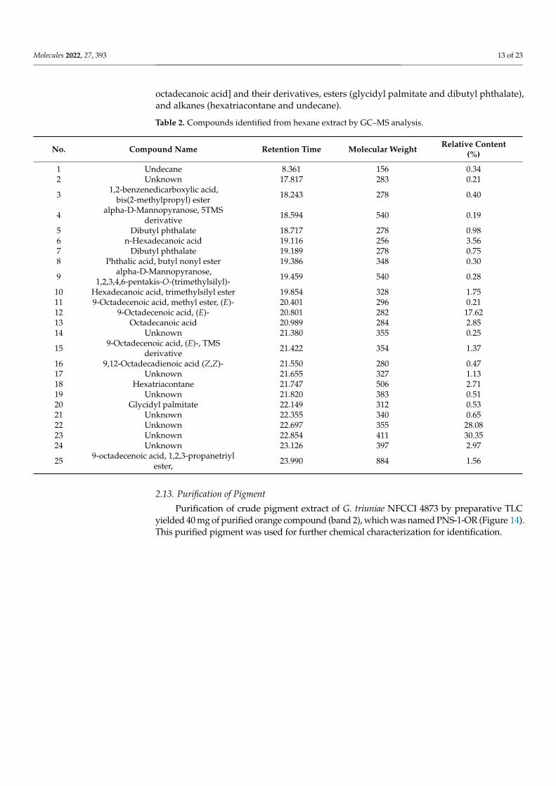
Molecules 2022, 27, 393 13 of 23
octadecanoic acid] and their derivatives, esters (glycidyl palmitate and dibutyl phthalate),and alkanes (hexatriacontane and undecane).
Table 2. Compounds identified from hexane extract by GC–MS analysis.
No. Compound Name Retention Time Molecular Weight Relative Content(%)
1 Undecane 8.361 156 0.342 Unknown 17.817 283 0.21
3 1,2-benzenedicarboxylic acid,bis(2-methylpropyl) ester 18.243 278 0.40
4 alpha-D-Mannopyranose, 5TMSderivative 18.594 540 0.19
5 Dibutyl phthalate 18.717 278 0.986 n-Hexadecanoic acid 19.116 256 3.567 Dibutyl phthalate 19.189 278 0.758 Phthalic acid, butyl nonyl ester 19.386 348 0.30
9 alpha-D-Mannopyranose,1,2,3,4,6-pentakis-O-(trimethylsilyl)- 19.459 540 0.28
10 Hexadecanoic acid, trimethylsilyl ester 19.854 328 1.7511 9-Octadecenoic acid, methyl ester, (E)- 20.401 296 0.2112 9-Octadecenoic acid, (E)- 20.801 282 17.6213 Octadecanoic acid 20.989 284 2.8514 Unknown 21.380 355 0.25
15 9-Octadecenoic acid, (E)-, TMSderivative 21.422 354 1.37
16 9,12-Octadecadienoic acid (Z,Z)- 21.550 280 0.4717 Unknown 21.655 327 1.1318 Hexatriacontane 21.747 506 2.7119 Unknown 21.820 383 0.5120 Glycidyl palmitate 22.149 312 0.5321 Unknown 22.355 340 0.6522 Unknown 22.697 355 28.0823 Unknown 22.854 411 30.3524 Unknown 23.126 397 2.97
25 9-octadecenoic acid, 1,2,3-propanetriylester, 23.990 884 1.56
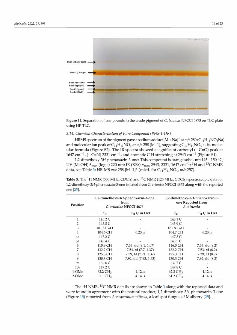
2.13. Purification of Pigment
Purification of crude pigment extract of G. triuniae NFCCI 4873 by preparative TLCyielded 40 mg of purified orange compound (band 2), which was named PNS-1-OR (Figure 14).This purified pigment was used for further chemical characterization for identification.
Molecules 2022, 27, 393 14 of 23Molecules 2022, 27, x FOR PEER REVIEW 14 of 23
Figure 14. Separation of compounds in the crude pigment of G. triuniae NFCCI 4873 on TLC plate
using HP‐TLC.
2.14. Chemical Characterization of Pure Compound (PNS‐1‐OR)
HRMS spectrum of the pigment gave a sodium adduct [M + Na]+ at m/z 280
(C14H11NO4Na) and molecular ion peak of C14H11NO4 at m/z 258 [M+1], suggesting
C14H11NO4 as its molecular formula (Figure S2). The IR spectra showed a significant car‐
bonyl (−C=O) peak at 1647 cm−1, (−C=N) 2331 cm−1, and aromatic C‐H stretching at 2943
cm−1 (Figure S1).
1,2‐dimethoxy‐3H‐phenoxazin‐3‐one: This compound is orange solid. mp 145−150
°C; UV (MeOH) λmax (log ε) 220 nm; IR (KBr) νmax 2943, 2331, 1647 cm−1; 1H and 13C NMR
data, see Table 3; HR‐MS m/z 258 [M+1]+ (calcd. for C14H11NO4, m/z 257).
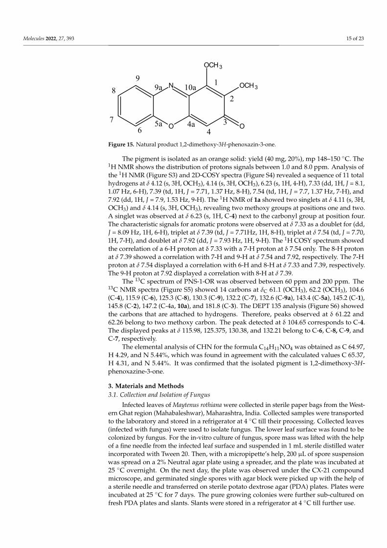
The 1H NMR, 13C NMR details are shown in Table 3 along with the reported data and
were found in agreement with the natural product, 1,2‐dimethoxy‐3H‐phenoxazin‐3‐one
(Figure 15) reported from Acrospermum viticola, a leaf spot fungus of Mulberry [25].
Table 3. The 1H NMR (500 MHz, CDCl3) and 13C NMR (125 MHz, CDCl3) spectroscopic data for 1,2‐
dimethoxy‐3H‐phenoxazin‐3‐one isolated from G. triuniae NFCCI 4873 along with the reported one
[25].
Position
1,2‐dimethoxy‐3H‐phenoxazin‐3‐one from
G. triuniae NFCCI 4873
1,2‐dimethoxy‐3H‐phenoxazin‐3‐one Re‐
ported from A. viticola
δC δH (J in Hz) δC δH (J in Hz)
1 145.2 C ‐ 145.1 C ‐
2 145.8 C ‐ 145.9 C ‐
3 181.8 C=O ‐ 181.8 C=O ‐
4 104.6 CH 6.23, s 104.7 CH 6.23, s
4a 147.2 C ‐ 147.3 C ‐
5a 143.4 C ‐ 143.5 C ‐
6 115.9 CH 7.33, dd (8.1, 1.07) 116.0 CH 7.33, dd (8.2)
7 132.2 CH 7.54, td (7.7, 1.37) 132.2 CH 7.53, td (8.2)
8 125.3 CH 7.39, td (7.71, 1.37) 125.3 CH 7.39, td (8.2)
9 130.3 CH 7.92, dd (7.93, 1.53) 130.3 CH 7.92, dd (8.2)
9a 132.6 C 132.7 C ‐
10a 147.2 C 147.8 C ‐
1‐OMe 62.2 CH3 4.12, s 62.3 CH3 4.12, s
2‐OMe 61.1 CH3 4.14, s 61.2 CH3 4.14, s
Figure 14. Separation of compounds in the crude pigment of G. triuniae NFCCI 4873 on TLC plateusing HP-TLC.
2.14. Chemical Characterization of Pure Compound (PNS-1-OR)
HRMS spectrum of the pigment gave a sodium adduct [M + Na]+ at m/z 280 (C14H11NO4Na)and molecular ion peak of C14H11NO4 at m/z 258 [M+1], suggesting C14H11NO4 as its molec-ular formula (Figure S2). The IR spectra showed a significant carbonyl (−C=O) peak at1647 cm−1, (−C=N) 2331 cm−1, and aromatic C-H stretching at 2943 cm−1 (Figure S1).
1,2-dimethoxy-3H-phenoxazin-3-one: This compound is orange solid. mp 145−150 ◦C;UV (MeOH) λmax (log ε) 220 nm; IR (KBr) νmax 2943, 2331, 1647 cm−1; 1H and 13C NMRdata, see Table 3; HR-MS m/z 258 [M+1]+ (calcd. for C14H11NO4, m/z 257).
Table 3. The 1H NMR (500 MHz, CDCl3) and 13C NMR (125 MHz, CDCl3) spectroscopic data for1,2-dimethoxy-3H-phenoxazin-3-one isolated from G. triuniae NFCCI 4873 along with the reportedone [25].
Position
1,2-dimethoxy-3H-phenoxazin-3-onefrom
G. triuniae NFCCI 4873
1,2-dimethoxy-3H-phenoxazin-3-one Reported from
A. viticola
δC δH (J in Hz) δC δH (J in Hz)
1 145.2 C - 145.1 C -2 145.8 C - 145.9 C -3 181.8 C=O - 181.8 C=O -4 104.6 CH 6.23, s 104.7 CH 6.23, s
4a 147.2 C - 147.3 C -5a 143.4 C - 143.5 C -6 115.9 CH 7.33, dd (8.1, 1.07) 116.0 CH 7.33, dd (8.2)7 132.2 CH 7.54, td (7.7, 1.37) 132.2 CH 7.53, td (8.2)8 125.3 CH 7.39, td (7.71, 1.37) 125.3 CH 7.39, td (8.2)9 130.3 CH 7.92, dd (7.93, 1.53) 130.3 CH 7.92, dd (8.2)
9a 132.6 C 132.7 C -10a 147.2 C 147.8 C -
1-OMe 62.2 CH3 4.12, s 62.3 CH3 4.12, s2-OMe 61.1 CH3 4.14, s 61.2 CH3 4.14, s
The 1H NMR, 13C NMR details are shown in Table 3 along with the reported data andwere found in agreement with the natural product, 1,2-dimethoxy-3H-phenoxazin-3-one(Figure 15) reported from Acrospermum viticola, a leaf spot fungus of Mulberry [25].
Molecules 2022, 27, 393 15 of 23Molecules 2022, 27, x FOR PEER REVIEW 15 of 23
O
N
O
OCH3
OCH31
2
34
4a5a6
7
8
99a 10a
Figure 15. Natural product 1,2‐dimethoxy‐3H‐phenoxazin‐3‐one.
The pigment is isolated as an orange solid: yield (40 mg, 20%), mp 148–150 °C. The 1H NMR shows the distribution of protons signals between 1.0 and 8.0 ppm. Analysis of
the 1H NMR (Figure S3) and 2D‐COSY spectra (Figure S4) revealed a sequence of 11 total
hydrogens at δ 4.12 (s, 3H, OCH3), 4.14 (s, 3H, OCH3), 6.23 (s, 1H, 4‐H), 7.33 (dd, 1H, J =
8.1, 1.07 Hz, 6‐H), 7.39 (td, 1H, J = 7.71, 1.37 Hz, 8‐H), 7.54 (td, 1H, J = 7.7, 1.37 Hz, 7‐H),
and 7.92 (dd, 1H, J = 7.9, 1.53 Hz, 9‐H). The 1H NMR of 1a showed two singlets at δ 4.11
(s, 3H, OCH3) and δ 4.14 (s, 3H, OCH3), revealing two methoxy groups at positions one
and two. A singlet was observed at δ 6.23 (s, 1H, C‐4) next to the carbonyl group at posi‐
tion four. The characteristic signals for aromatic protons were observed at δ 7.33 as a dou‐
blet for (dd, J = 8.09 Hz, 1H, 6‐H), triplet at δ 7.39 (td, J = 7.71Hz, 1H, 8‐H), triplet at δ 7.54
(td, J = 7.70, 1H, 7‐H), and doublet at δ 7.92 (dd, J = 7.93 Hz, 1H, 9‐H). The 1H COSY spec‐
trum showed the correlation of a 6‐H proton at δ 7.33 with a 7‐H proton at δ 7.54 only. The
8‐H proton at δ 7.39 showed a correlation with 7‐H and 9‐H at δ 7.54 and 7.92, respectively.
The 7‐H proton at δ 7.54 displayed a correlation with 6‐H and 8‐H at δ 7.33 and 7.39, re‐
spectively. The 9‐H proton at 7.92 displayed a correlation with 8‐H at δ 7.39.
The 13C spectrum of PNS‐1‐OR was observed between 60 ppm and 200 ppm. The 13C
NMR spectra (Figure S5) showed 14 carbons at δC 61.1 (OCH3), 62.2 (OCH3), 104.6 (C‐4),
115.9 (C‐6), 125.3 (C‐8), 130.3 (C‐9), 132.2 (C‐7), 132.6 (C‐9a), 143.4 (C‐5a), 145.2 (C‐1), 145.8
(C‐2), 147.2 (C‐4a, 10a), and 181.8 (C‐3). The DEPT 135 analysis (Figure S6) showed the
carbons that are attached to hydrogens. Therefore, peaks observed at δ 61.22 and 62.26
belong to two methoxy carbon. The peak detected at δ 104.65 corresponds to C‐4. The
displayed peaks at δ 115.98, 125.375, 130.38, and 132.21 belong to C‐6, C‐8, C‐9, and C‐7,
respectively.
The elemental analysis of CHN for the formula C14H11NO4 was obtained as C 64.97,
H 4.29, and N 5.44%, which was found in agreement with the calculated values C 65.37,
H 4.31, and N 5.44%. It was confirmed that the isolated pigment is 1,2‐dimethoxy‐3H‐
phenoxazine‐3‐one.
3. Materials and Methods
3.1. Collection and Isolation of Fungus
Infected leaves of Maytenus rothiana were collected in sterile paper bags from the
Western Ghat region (Mahabaleshwar), Maharashtra, India. Collected samples were
transported to the laboratory and stored in a refrigerator at 4 °C till their processing. Col‐
lected leaves (infected with fungus) were used to isolate fungus. The lower leaf surface
was found to be colonized by fungus. For the in‐vitro culture of fungus, spore mass was
lifted with the help of a fine needle from the infected leaf surface and suspended in 1 mL
sterile distilled water incorporated with Tween 20. Then, with a micropipette’s help, 200
μL of spore suspension was spread on a 2% Neutral agar plate using a spreader, and the
plate was incubated at 25 °C overnight. On the next day, the plate was observed under
the CX‐21 compound microscope, and germinated single spores with agar block were
picked up with the help of a sterile needle and transferred on sterile potato dextrose agar
(PDA) plates. Plates were incubated at 25 °C for 7 days. The pure growing colonies were
Figure 15. Natural product 1,2-dimethoxy-3H-phenoxazin-3-one.
The pigment is isolated as an orange solid: yield (40 mg, 20%), mp 148–150 ◦C. The1H NMR shows the distribution of protons signals between 1.0 and 8.0 ppm. Analysis ofthe 1H NMR (Figure S3) and 2D-COSY spectra (Figure S4) revealed a sequence of 11 totalhydrogens at δ 4.12 (s, 3H, OCH3), 4.14 (s, 3H, OCH3), 6.23 (s, 1H, 4-H), 7.33 (dd, 1H, J = 8.1,1.07 Hz, 6-H), 7.39 (td, 1H, J = 7.71, 1.37 Hz, 8-H), 7.54 (td, 1H, J = 7.7, 1.37 Hz, 7-H), and7.92 (dd, 1H, J = 7.9, 1.53 Hz, 9-H). The 1H NMR of 1a showed two singlets at δ 4.11 (s, 3H,OCH3) and δ 4.14 (s, 3H, OCH3), revealing two methoxy groups at positions one and two.A singlet was observed at δ 6.23 (s, 1H, C-4) next to the carbonyl group at position four.The characteristic signals for aromatic protons were observed at δ 7.33 as a doublet for (dd,J = 8.09 Hz, 1H, 6-H), triplet at δ 7.39 (td, J = 7.71Hz, 1H, 8-H), triplet at δ 7.54 (td, J = 7.70,1H, 7-H), and doublet at δ 7.92 (dd, J = 7.93 Hz, 1H, 9-H). The 1H COSY spectrum showedthe correlation of a 6-H proton at δ 7.33 with a 7-H proton at δ 7.54 only. The 8-H protonat δ 7.39 showed a correlation with 7-H and 9-H at δ 7.54 and 7.92, respectively. The 7-Hproton at δ 7.54 displayed a correlation with 6-H and 8-H at δ 7.33 and 7.39, respectively.The 9-H proton at 7.92 displayed a correlation with 8-H at δ 7.39.
The 13C spectrum of PNS-1-OR was observed between 60 ppm and 200 ppm. The13C NMR spectra (Figure S5) showed 14 carbons at δC 61.1 (OCH3), 62.2 (OCH3), 104.6(C-4), 115.9 (C-6), 125.3 (C-8), 130.3 (C-9), 132.2 (C-7), 132.6 (C-9a), 143.4 (C-5a), 145.2 (C-1),145.8 (C-2), 147.2 (C-4a, 10a), and 181.8 (C-3). The DEPT 135 analysis (Figure S6) showedthe carbons that are attached to hydrogens. Therefore, peaks observed at δ 61.22 and62.26 belong to two methoxy carbon. The peak detected at δ 104.65 corresponds to C-4.The displayed peaks at δ 115.98, 125.375, 130.38, and 132.21 belong to C-6, C-8, C-9, andC-7, respectively.
The elemental analysis of CHN for the formula C14H11NO4 was obtained as C 64.97,H 4.29, and N 5.44%, which was found in agreement with the calculated values C 65.37,H 4.31, and N 5.44%. It was confirmed that the isolated pigment is 1,2-dimethoxy-3H-phenoxazine-3-one.
3. Materials and Methods3.1. Collection and Isolation of Fungus
Infected leaves of Maytenus rothiana were collected in sterile paper bags from the West-ern Ghat region (Mahabaleshwar), Maharashtra, India. Collected samples were transportedto the laboratory and stored in a refrigerator at 4 ◦C till their processing. Collected leaves(infected with fungus) were used to isolate fungus. The lower leaf surface was found to becolonized by fungus. For the in-vitro culture of fungus, spore mass was lifted with the helpof a fine needle from the infected leaf surface and suspended in 1 mL sterile distilled waterincorporated with Tween 20. Then, with a micropipette’s help, 200 µL of spore suspensionwas spread on a 2% Neutral agar plate using a spreader, and the plate was incubated at25 ◦C overnight. On the next day, the plate was observed under the CX-21 compoundmicroscope, and germinated single spores with agar block were picked up with the help ofa sterile needle and transferred on sterile potato dextrose agar (PDA) plates. Plates wereincubated at 25 ◦C for 7 days. The pure growing colonies were further sub-cultured onfresh PDA plates and slants. Slants were stored in a refrigerator at 4 ◦C till further use.
Molecules 2022, 27, 393 16 of 23
3.2. Morphological Identification and Deposition of Fungal Culture
Necrotic lesions on the infected leaves were marked. Scrape mount slides wereprepared in lactophenol cotton blue and observed under a microscope; based on theliterature, the fungus was identified as Gonatophragmium sp. Similarly, slides were preparedin lactophenol cotton blue mount from axenic culture. Microphotographs were taken usingCarl Zeiss AXIO-10 microscope, and scanning electron microscopic (SEM) images weretaken using images ZEISS EVO MA 15 Scanning electron microscope at 20 KV.
A pure culture of G. triuniae was inoculated on potato dextrose agar (PDA) and potatocarrot agar (PCA) to study cultural and microscopic characters. Plates were incubated at25 ◦C for 14 days, and cultural and microscopic characters were noted upon completionof incubation. Slide culture [26,27] and grass leaf technique [28] were used to get thesporulation, but no sporulation was observed.
Live and pure fungal culture of G. triuniae were deposited in the National FungalCulture Collection of India (NFCCI), Agharkar Research Institute, Pune, under the accessionnumber NFCCI 4873. Voucher culture G. triuniae NFCCI 4873 was deposited in AjrekarMycological Herbarium (AMH), Agharkar Research Institute, Pune, with accession numberAMH 10289.
3.3. Molecular Identification and Phylogeny
For the identification and authentication of fungal culture up to species level, molecularcharacterization was done. The fungal genomic DNA was isolated following the standardprotocol [29]. Then by polymerase chain reaction (PCR), the ITS (internal transcribed spacer)and LSU (large subunit) regions of rDNA were amplified from the extracted genomic DNAusing the primers ITS-4 & ITS-5 [30] and LR-0R & LR-7 [31], respectively. FavorPrepTM PCRPurification Kit (Favorgen, Biotech Corporation, Taiwan) was used to purify PCR products.Purified PCR products were subjected for sequencing by the BigDye Terminator v3.1 CycleSequencing Kit (Applied Biosystems, Waltham, MA, USA) and ABI Avant 3100 automatedDNA sequencer (Applied Biosystems, USA). The manually edited sequences of ITS andLSU regions of rDNA of our fungal isolate were deposited in the nucleotide sequencedatabase of NCBI (Gene Bank Accession Numbers: ITS- MW193329 and LSU- MW144438).
The ITS and LSU rDNA sequences of our fungal isolate were subjected to MegaBLASTn sequence homology searches. Based on the BLASTn search results, geneti-cally related species, including genus Gonatophragmium, Acrospermum, Pseudovirgaria, andDyfrolomyces, were chosen to construct the phylogenetic tree (Table 4). Phylogenetic anal-ysis of G. triuniae NFCCI 4873 was performed based on a combined ITS and LSU rDNAsequence data of a total of 19 fungal cultures. The Eremomyces bilateralis CBS 781.70 waschosen as an out-group. With the help of the MUSCLE algorithm, multiple sequencealignment was performed in MEGA 7 [32]. Phylogenetic tree of G. triuniae NFCCI 4873 wasconstructed based on combined data of ITS & LSU rDNA sequences in IQ-TREE multicoreversion 1.6.11 [33] using the Maximum Likelihood method with best-fit model TN+F+G4.Selection of the best-fit model was done using the ModelFinder employed in IQ-TREE.
3.4. Analysis of Pigment Production on Different Media
A pure culture of G. triuniae NFCCI 4873 was inoculated on different media such aspotato dextrose agar (PDA), potato carrot agar (PCA), Sabouraud dextrose agar (SDA),Czapek Dox agar (CZA), cornmeal agar (CMA), and Czapek Yeast Extract Agar (CYA) induplicates and incubated at 25 ◦C for 28 days to assess the pigment production potential.After incubation, the colour of the pigment diffused in media was recorded using theMethuen handbook of colour [34].
3.5. Analysis of Pigment Production in Liquid Media
The culture of G. triuniae was also tested for its pigment production ability in differentliquid media such as potato dextrose broth (PDB, Hi-media), natural potato dextrosebroth (n-PDB), and natural potato carrot broth (n-PCB). Four agar blocks of a pure culture
Molecules 2022, 27, 393 17 of 23
of G. triuniae (6 mm diameter) from 20-days-old PDA culture plate were inoculated ina 250 mL Erlenmeyer flask containing 100 mL media (each media in the separate flask),and flasks were incubated at 25 ◦C with 150 rpm. All tests were performed in duplicates.All flasks were observed intermittently after every week from the date of inoculation forpigment production, and observations were noted down. After 4 weeks of incubation,culture broths were filtered, and absorption spectrum analyses of the coloured filtrateswere performed using a UV–VIS spectrophotometer (Shimadzu UV-2450). The absorbanceof coloured culture filtrates was recorded in the visible light range from 390–760 nm with a10 mm optical pathlength and 0.1 nm resolution.
Table 4. GenBank accession numbers of taxa used for phylogenetic analysis.
Sr. No. Fungal Culture StrainGenBank Accession No.
ITS LSU
1 Gonatophragmiumtriuniae NFCCI 4873 MW193329 MW144438
2 Gonatophragmiumtriuniae CBS 138901 NR_137932 NG_058117
3 Gonatophragmiumepilobii CBS 122271 MH863183 MH874728
4 Acrospermum sp. SGSF153 MK335823 MK754265
5 Acrospermumlongisporium MFLU 17-2849 - NG_064506
6 Acrospermumgramineum M152 - EU940085
7 Acrospermumcompressum M151 - EU940084
8 Pseudovirgaria grisea CPC 19130 JF957607 JF957612
9 Pseudovirgariahyperparasitica CPC 10702 EU041765 EU041822
10 Pseudovirgariahyperparasitica CPC 10704 EU041766 EU041823
11 Pseudovirgariahyperparasitica CPC 10753 EU041767 EU041824
12 Pseudovirgaria grisea CPC 19126 JF957605 JF95761013 Pseudovirgaria grisea CPC 19128 JF957606 JF95761114 Dyfrolomyces sinensis MFLU 17-0777 - NG_06450715 Dyfrolomyces sinensis MFLUCC 17-1344 - MG836699
16 Dyfrolomycestiomanensis NTOU3636 - KC692156
17 Dyfrolomycesrhizophorae JK 5456A - GU479799
18 Dyfrolomycesthamplaensis MFLUCC 15-0635 - KX925435
19 Eremomyces bilateralis CBS 781.70 NR_145364 NG_059206
3.6. Fermentation and Extraction of Pigments
G. triuniae culture was subjected to flask scale fermentation in a total of 6 L (four flaskscontaining 1.5 L of media) of natural potato dextrose broth (PD broth). Each flask wasinoculated with 20–25 mycelial disks (6 mm diameter) of G. triuniae from 3-weeks-old PDAculture plate using a cork borer and incubated at 25 ◦C with 100 rpm for 4–6 weeks. Afterincubation, the coloured culture broth was filtered through pre-weighed blotting paper, andculture filtrate was collected in a separate flask. Later, the pigments from the culture filtrateswere extracted thrice with an equal volume of Hexane. With the help of a separating funnel,the Hexane part was separated from the culture filtrate. The separated hexane part wasevaporated to dryness under reduced pressure in a rota evaporator (Heidolph, Schwabach,Germany). The resulting concentrated hexane extract was used for further experiments.
Molecules 2022, 27, 393 18 of 23
Finally, biomass collected in a pre-weighed blotting paper was dried at 105 ◦C for 12–15 hand weighed to measure the yield of biomass concentration [35].
3.7. UV–VIS Spectroscopy Analysis of Hexane Extract
UV-Visible spectroscopic analyses of the crude hexane extracts were performed using aUV-VIS spectrophotometer (Shimadzu UV-2450). The crude hexane extract was evaporatedto dryness and then dissolved in methanol solvent, and absorbance was recorded in therange of 190–760 nm with a 0.1 nm resolution and 10 mm optical path length.
3.8. Antagonistic Activity of G. triuniae NFCCI 4873
Antagonistic activity of G. triuniae against three fungal pathogens (Colletotrichumgloeosporioides, Fusarium oxysporum & Fusarium solani) was evaluated by dual culture tech-nique [36]. Mycelial disks of 6 mm diameter were excised from the edge of actively growingculture of G. triuniae and fungal pathogens and inoculated on opposite ends of PDA platesequidistant from the periphery (each pathogen separately with test culture). For control,PDA plates were inoculated with a pathogen without test culture. Plates were then incu-bated at 25 ◦C for 14 days. Experiments were performed in duplicates. After completion ofincubation, the radial growth of each fungal pathogen on PDA plates was measured, andpercentage inhibition of radial growth (PIRG) of fungal pathogens was calculated relativeto the control plate using the following formula:
PIRG = [(R1 − R2)/R1] × 100
where R1 is the radial growth of the fungal pathogen in the control plate, and R2 is theradial growth of the pathogen in the presence of test culture (G. triuniae) [37].
3.9. In-vitro Antioxidant Activity of Crude Pigment
In-vitro antioxidant activity of hexane extracts was tested using DPPH radical scaveng-ing method [38]. Different concentrations (0.2, 0.4, 0.6, 0.8, and 1.0 mg/mL) of the extractand standard solution (Ascorbic acid, Sigma, USA) were used, and for that, dilutions wereprepared in methanol. For the assay, 10 µL of extract or standard solution was added to200 µL of 0.1 mM DPPH in methanol solution in a 96-well microtitre plate (Thermofisher,Waltham, MA, USA). All reactions were performed in triplicates. The plate was then incu-bated at 37 ◦C for 30 min in the dark. After incubation, the absorbance of the solution ineach well was measured at 490 nm using a Synergy HT Multi-detection microplate reader[BioTek, Winooski, VT, USA]. The percentage of radical scavenging activity of the hexaneextract was calculated by the following formula:
DPPH radical scavenging activity (%) = [(OD control − OD sample)/OD control] × 100
where OD means optical density or absorbance value. The IC50 value (concentration ofsample required to scavenge 50% of free radicals) of hexane extract was determined.
3.10. Antimicrobial Activity of Crude Hexane Extract
Crude pigment sample was screened against a panel of test organisms, includingEscherichia coli (MTCC 739), Bacillus subtilis (MTCC 121), Staphylococcus aureus MLS 16(MTCC 2940), Pseudomonas aeruginosa (MTCC 2453), Raoultella planticola (MTCC 530), andMicrococcus luteus (MTCC 2470). The test strains were procured from Microbial TypeCulture Collection (MTCC), CSIR-Institute of Microbial Technology (IMTECH), Chandigarh,India. Ciprofloxacin hydrochloride (1 mg/mL), vancomycin hydrochloride (1 mg/mL),chloramphenicol (1 mg/mL), streptomycin (1 mg/mL), and ampicillin (1 mg/mL) againstbacteria, were used as positive controls.
Molecules 2022, 27, 393 19 of 23
3.11. Antimicrobial Activity by Disc Diffusion Method
The antimicrobial activity of the hexane extract of G. triuniae was tested using the diskdiffusion method. The standardized microbial inoculum of the test strain was prepared ina saline solution (~106–108 CFU/mL) from the 24 h old culture plates. The culture mediaused was Muller-Hinton agar (MHA). A direct colony suspension method was used forthe inoculum preparation in which well-isolated colonies from 24 h old culture plateswere selected and suspended in sterile saline. Then saline suspensions of the test cultureswere adjusted to achieve turbidity equivalent to a 0.5 McFarland standard. This was doneby adjusting the turbidity of the cell suspensions between 0.08 and 0.12 AU in a UV-VisSpectrophotometer (Shimadzu UV-2450) at 625 nm as recommended by CLSI guidelines.This results in a suspension with approximately 1–2 × 10⁸ colony-forming units (CFU)/mLfor Escherichia coli ATCC®a 25922. The resulted suspension was added in sterile moltenMuller-Hinton agar (0.5 mL/100 mL of media). The final concentration of the cells in themedia was 0.5–1.0 × 10⁸ CFU/mL. Then media was poured in Petri plates and allowed tosolidify [39].
Sterile Whatman filter paper discs impregnated with known amounts of the test sam-ple (1mg/mL of dried hexane extract dissolved in methanol) were placed on the surfaceof an agar plate that was inoculated with a standardized suspension of microorganismsthat were to be tested. Standard antibiotics like ampicillin (1 mg/mL), ciprofloxacin hy-drochloride (1 mg/mL), streptomycin (1 mg/mL), vancomycin hydrochloride (1 mg/mL),and chloramphenicol (1 mg/mL) were used as positive controls. Paper discs impregnatedwith only methanol and sterile water were used as negative controls. Plates were left atroom temperature for 1–2 h for the diffusion of samples and then incubated at 37 ◦C for24 h. After completion of incubation, all plates were observed for the zone of inhibition.The diameters of the zone of inhibition were measured in millimeters, including the diskdiameter (6 mm). All experiments were performed in triplicates.
3.12. Determination of Minimum Inhibitory Concentration (MIC) and Minimum BactericidalConcentration (MBC)3.12.1. Preparation of Pigment Stock
Forty milligrams of crude pigment was dissolved in 1 mL of DMSO in a 2 mL Eppen-dorf tube, and a tube was mixed well. Then this 1 mL solution was then diluted 10 times insterile Muller–Hinton broth (MHB), and the resultant solution was stored in the refrigeratorat 4 ◦C until further use.
3.12.2. Preparation of Standardized Inoculum
The standard inoculum (0.5 McFarland) of Gram-positive bacteria (Bacillus subtilis,Staphylococcus aureus MLS 16, and Micrococcus luteus) was prepared by the direct colonysuspension method as recommended by CLSI guidelines in which the OD625 value wasadjusted to the equivalent of 108 CFU/mL in a Shimadzu UV-2450 UV–VIS spectropho-tometer [39].
3.12.3. MIC and MBC Experiment
The minimum inhibitory concentration (MIC) of the crude pigment extract was as-sessed using the standard method [39]. The MIC of the crude pigment extract was deter-mined by the broth microdilution method in a 96-well plate as per the CLSI recommendedprotocol. All the wells of the microtitre plate from columns 2–11 were added with 50 µL ofsterile Muller–Hinton broth (MHB). The last (12th) column was added with 100 µL of MHBas sterility control. One hundred microlitres of pigment solution (2 mg/mL) was addedto the first column of the 96-well microtitre plate. Then, using a micropipette, a two-foldserial dilution was performed by transferring 50 µL of the pigment solution from the firstwell to the succeeding well and up to the 10th well, and the final 50 µL of the solutionwas discarded. Standardized inoculums of bacteria were diluted 1:150 times in sterile MHbroth to get 106 CFU/mL concentrations of bacteria. Then, 50 µL of microbial suspension
Molecules 2022, 27, 393 20 of 23
(106 CFUs/mL) was added in each well from well 2–11; while in rows D and H, from well1–10, there was no addition of bacterial suspension, which were treated as pigment control.Plates were incubated at 37 ◦C for 20 h. Each test was performed in triplicates.
After incubation at 37 ◦C for 20 h, 30 µL of 0.01% resazurin was added to each well,and then plates were further incubated for 2 h at 37 ◦C for the change of colour. Thewell-containing lowest concentration of pigment showing no colour change was consideredas the MIC value.
After 20 h of incubation at 37 ◦C, 10 µL solution from each well (2–11) of the 96-wellmicrotitre plate was plated on Muller–Hinton agar plate, and plates were incubated at37 ◦C for 24 h. After completion of incubation, plates were observed for the growth ofbacteria. The lowest concentrations that completely kill the bacteria and do not showgrowth on the MHA plate were considered minimum bactericidal concentration (MBC).
3.13. Dyeing of Cotton with a Crude Pigment of G. triuniae NFCCI 48733.13.1. Textile Fabric
Raw cotton fabric was collected from the local market, Pune. Cotton fabric was thencut into 10 × 10 cm pieces (each weighing 1 gm) and used for the dyeing experiment.
3.13.2. Scouring
Twelve cotton fabric pieces of 10 × 10 cm (12 g) were pre-soaked in milli-Q water andthen cooked in 2.5 L of Milli-Q water containing 20 mL of non-ionic detergent (Triton-X-100)for 1 h at 70 ◦C in a water bath to remove oil and dirt. Scoured cotton fabrics were thenrinsed thoroughly with running water and air-dried [40].
3.13.3. Mordanting
Scoured cotton fabric pieces were mordanted by the pre-mordanting technique [40,41].Cotton fabric pieces (10 × 10 cm) were mordanted with different concentrations (5%, 10%,and 15% w/w of fabric) of Alum and FeSO4 for 45 min at 70 ◦C with a 1:20 material to liquidratio (MLR). After completing mordanting, fabric pieces were rinsed in running water andfinally allowed to air dry.
3.13.4. Preparation of Dye Bath
One hundred milligrams of dried crude pigment of G. triuniae NFCCI 4873 was re-dissolved in 500 mL of Milli-Q water using a magnetic stirrer. The resultant colouredsolution was used as a dye bath for dyeing cotton fabric.
3.13.5. Dyeing
Unmordanted and pre-mordanted cotton fabric pieces were dyed with a crude pigmentsolution of G. triuniae NFCCI 4873. Cotton fabric pieces (10 × 10 cm) were dyed at a materialto liquor ratio (MLR) of 1:50 at 70 ◦C for 45 min in a water bath. The pH of the dye bathwas not controlled. Dyed fabric pieces were then treated with 1% acetic acid and washedthoroughly in running water. The dyed fabric pieces were then rinsed with cold water anddried overnight in the shed [40].
3.14. GC–MS Analysis
The dried hexane extract was subjected to derivatization using N-Methyl-N-(trimethylsilyl)trifluoroacetamide [MSTFA] as a silylating agent. For derivatization, about 5 mg of samplewas dissolved in 100 µL of pyridine in a glass vial followed by the addition of 100 µL ofMSTFA. The solution was mixed well, heated at 60 ◦C for 20 min, and cooled to roomtemperature. After derivatization, the sample was subjected to gas chromatography-massspectrometric analysis using GCMS-TQ8030 (Shimadzu, Nakagyo-ku, Kyoto, Japan). Afused silica column [RTX-5MS (30 m × 0.25 mm × 0.25 µm)] was used. The temperature ofthe column was programmed from 50 to 280 ◦C at a rate of 10 ◦C/min. The injection porttemperature was set at 280 ◦C, and a split ratio of 1:40 was used for the analysis. Helium
Molecules 2022, 27, 393 21 of 23
was used as the carrier gas at a flow rate of 1.0 mL/min. Electron ionization source of 70 eVand a mass range of m/z 35–800 U was used for MS detection. The resultant MS peaksin the GC-MS chromatogram were identified by comparing and matching the mass andmass fragmentation pattern with the reference mass and mass fragmentation pattern in theNIST05 MS library.
3.15. Purification of a Compound by HP-TLC
Two-hundred milligrams of crude pigment extract of G. triuniae NFCCI 4873 wasdissolved in 4 mL of acetone, and compounds present in crude extract were separatedon silica plates using HP-TLC (CAMAAG). Hexane:ethyl Acetate (40:60) was used as themobile phase; thin aluminium silica plates (Merck) were used as the stationary phase. Afterseparating compounds through HP-TLC, the orange band separated on plates was cut,and the compound attached to silica was extracted with acetone. Acetone extracts werecollected in a round bottom flask and dried using a rota evaporator under reduced pressureusing Heidolph rota evaporator. Finally, the weight of the dried, purified pigment wasmeasured and recorded.
3.16. Chemical Characterization of Purified Orange Compound (PNS-1-OR)
The high-resolution mass spectrum (HRMS) of the pure orange pigment labeledas PNS-1-OR was recorded on a Bruker IMPACT HD. FTIR spectrum of pigment wasrecorded on a Shimadzu-IRAffinity-1 FTIR spectrophotometer in the frequency range4000–400 cm−1. Pure compound was subjected to 1H (500 MHz) and 13C (125 MHz) NMRin Bruker 500 MHz NMR instrument using CDCl3 as a solvent for dissolving sample andTMS as internal standard. Chemical shifts (δ-values) are given in parts per million (ppm),and the coupling constants (J-values) are given in hertz (Hz).
4. Conclusions
Many fungi of different taxonomic groups producing a wide variety of pigmentsof different colours and chemical classes have been reported by researchers across theworld. Among them, some fungal pigments find their application in different industriespossessing promising colouring properties. The present study also reports one of theunconventional fungi, i.e., G. triuniae, showing very good pigment production potential.Moreover, this is the first experimental work reporting pigments and other secondarymetabolites from the fungus G. triuniae. Based on the results of the antibacterial activity ofthe crude pigment extract, we conclude that the crude pigment extract shows the presenceof antibiotic compounds, exhibiting antibacterial activity against Gram-positive bacteria.In addition to this, the DPPH radical scavenging activity of the crude pigment extractconfirmed the presence of antioxidant compounds in the crude pigment extract. Suchbioactivities (antibacterial and antioxidant) of the crude pigment extract have elevatedthe G. triuniae as a promising source of bioactive compounds for their possible use in themedicine and pharmaceutical industry. Besides this, the dyeing property of the crudepigment extract revealed the potential use of pigments of G. triuniae in the textile industryfor dyeing different types of fabrics.
The purification of crude pigment extract of G. triuniae finally yielded into a majororange-colored phenoxazine class pigment, which was characterized and identified as1,2-dimethoxy-3H-phenoxazin-3-one (C14H11NO4, M.W. 257), based on UV-Vis, FTIR,HRMS, and NMR analysis. Although this pigment was already described from fungusA. viticola, this is the first study reporting the phenoxazine class of pigment from fungusG. triuniae. Considering the previous studies describing dyeing potential and bioactivityof phenoxazines, we may finally conclude that the orange pigment “1,2-dimethoxy-3H-phenoxazin-3-one” is a promising colourant and possible bioactive compound of G. triuniae,having future applications in the textile and pharmaceutical industry.
Molecules 2022, 27, 393 22 of 23
Supplementary Materials: FTIR (Figure S1), HR-MS (Figure S2), 1H NMR (Figure S3), COSY (Figure S4),13C NMR (Figure S5), and DEPT-135 (Figure S6) spectra for compound 1,2-dimethoxy-3H-phenoxazin-3-one and GC-MS chromatogram (Figure S7) of hexane extract (PDF).
Author Contributions: Conceptualization, A.C.L., S.K.S.; Formal analysis, A.C.L., P.S.; Methodology,A.C.L.; Supervision, S.K.S., L.D. and P.N.S.; Writing—original draft, A.C.L.; Writing—review &editing, S.K.S., L.D., P.S. and P.N.S. All authors have read and agreed to the published version ofthe manuscript.
Funding: This research received no external funding.
Institutional Review Board Statement: Not applicable.
Informed Consent Statement: Not applicable.
Data Availability Statement: Not applicable.
Acknowledgments: We thank Prashant Dhakephalkar, MACS-Agharkar Research Institute, Pune, forproviding the necessary facilities and encouragement to carry out the research work. A.C. Lagashettiacknowledges CSIR (Council of Scientific and Industrial Research), New Delhi, for granting SeniorResearch Fellowship (SRF), and S.P. Pune University, Pune, for granting permission to register for thePh.D. degree. We acknowledge technical support and help by S.B. Gaikwad, N.S. Gaikwad, and S.Bagale. Laurent Dufossé deeply thanks the Conseil Régional de La Réunion, Réunion island, IndianOcean, for continuous financial support of research activities dedicated to microbial pigments.
Conflicts of Interest: The authors declare no conflict of interest.
References1. Rao, M.P.; Xiao, M.; Li, W.J. Fungal and Bacterial Pigments: Secondary Metabolites with Wide Applications. Front. Microbiol. 2017,
8, 1113.2. Mansour, R. Natural Dyes and Pigments: Extraction and Applications. In Handbook of Renewable Materials for Coloration & Finishing;
Scrivener Publishing LLC: Beverly, MA, USA, 2018; Volume 9, pp. 75–102.3. Ranaweera, S.J.; Ampemohotti, A.A.L.T.; Arachchige, U.S. Advantages, and Considerations for the Applications of Natural Food
Pigments in the Food Industry. J. Res. Technol. Eng. 2020, 1, 8–18.4. Arora, S. Textile Dyes: Its Impact on Environment and Its Treatment. J. Bioremediation Biodegrad. 2014, 5, 1000e146. [CrossRef]5. Ardila-Leal, L.D.; Poutou-Piñales, R.A.; Pedroza-Rodríguez, A.M.; Quevedo-Hidalgo, B.E. A Brief History of Colour, The
Environmental Impact of Synthetic Dyes and Removal by Using Laccases. Molecules 2021, 26, 3813. [CrossRef] [PubMed]6. Manzoor, J.; Sharma, M. Impact of Textile Dyes on Human Health and Environment. In Impact of Textile Dyes on Public Health and
the Environment; IGI Global: Hershey, PA, USA, 2020; pp. 162–169.7. Caro, Y.; Venkatachalam, M.; Lebeau, J.; Fouillaud, M.; Dufossé, L. Pigments and Colorants from Filamentous Fungi. In Fungal
Metabolites; Merillon, J.-M., Ramawat, K.G., Eds.; Springer International Publishing: Cham, Switzerland, 2017; pp. 499–568.8. Lagashetti, A.C.; Dufossé, L.; Singh, S.K.; Singh, P.N. Fungal Pigments and Their Prospects in Different Industries. Microorganisms
2019, 7, 604. [CrossRef]9. Ramesh, C.; Vinithkumar, N.V.; Kirubagaran, R.; Venil, C.K.; Dufossé, L. Multifaceted Applications of Microbial Pigments: Current
Knowledge, Challenges and Future Directions for Public Health Implications. Microorganisms 2019, 7, 186. [CrossRef] [PubMed]10. Khan, A.A.; Alshabi, A.M.; Alqahtani, Y.S.; Alqahtani, A.M.; Bennur, R.S.; Shaikh, I.A.; Muddapur, U.M.; Shakeel Iqubal, S.M.;
Mohammed, T.; Dawoud, A.; et al. Extraction and Identification of Fungal Pigment from Penicillium europium Using DifferentSpectral Studies. J. King Saud Univ.–Sci. 2021, 33, 101437. [CrossRef]
11. Dufossé, L. Microbial Production of Food Grade Pigments. Food Technol. Biotechnol. 2006, 44, 313–323.12. Kumar, A.; Vishwakarma, H.S.; Singh, J.; Dwivedi, S.; Kumar, M. Microbial Pigments: Production and Their Applications in
Various Industries. Int. J. Pharm. Chem. Biol. Sci. 2015, 5, 203–212.13. Sajid, S.; Akbar, N. Applications of Fungal Pigments in Biotechnology. Pure Appl. Biol. 2018, 7, 922–930. [CrossRef]14. Mapari, S.A.; Meyer, A.S.; Thrane, U. Evaluation of Epicoccum nigrum for Growth, Morphology, and Production of Natural
Colorants in Liquid Media and on a Solid Rice Medium. Biotechnol. Lett. 2008, 30, 2183–2190. [CrossRef]15. Sibero, M.T.; Triningsih, D.W.; Radjasa, O.K.; Sabdono, A.; Trianto, A. Evaluation of Antimicrobial Activity and Identification
of Yellow Pigmented Marine Sponge-Associated Fungi from Teluk Awur, Jepara, Central Java. Indones. J. Biotechnol. 2016, 21,1–11. [CrossRef]
16. Heo, Y.M.; Kim, K.; Kwon, S.L.; Na, J.; Lee, H.; Jang, S.; Kim, C.H.; Jung, J.; Kim, J.J. Investigation of Filamentous Fungi ProducingSafe, Functional Water-Soluble Pigments. Mycobiology 2018, 46, 269–277. [CrossRef]
17. Wang, W.; Liao, Y.; Chen, R.; Hou, Y.; Ke, W.; Zhang, B.; Gao, M.; Shao, Z.; Chen, J.; Li, F. Chlorinated Azaphilone Pigments withAnti-microbial and Cytotoxic Activities Isolated from the Deep Sea-Derived Fungus Chaetomium sp. NA–S01–R1. Mar. Drugs2018, 16, 61. [CrossRef] [PubMed]

Molecules 2022, 27, 393 23 of 23
18. Kalra, R.; Conlan, X.A.; Goel, M. Fungi as A Potential Source of Pigments: Harnessing Filamentous Fungi. Front. Chem. 2020,8, 369. [CrossRef] [PubMed]
19. Polak, J.; Wlizło, K.; Pogni, R.; Petricci, E.; Graz, M.; Szałapata, K.; Osinska-Jaroszuk, M.; Kapral-Piotrowska, J.; Pawlikowska-Pawlega, B.; Jarosz-Wilkołazka, A. Structure and Bioactive Properties of Novel Textile Dyes Synthesised by Fungal Laccase. Int. J.Mol. Sci. 2020, 21, 2052. [CrossRef] [PubMed]
20. Sudhakar, C.; Reddy, M.K.; Rajyalakshmi, K.; Raju, K.R. Antibacterial Activity of Substituted 2-Bromo-1, 4-Dimethoxy-3H-Phenoxazin-3-Ones. J. Chem. Pharm. Res. 2016, 8, 571–574.
21. Achenbach, H.; Blümm, E. Investigation of the Pigments of Pycnoporus sanguineus-Pycnosanguin and New Phenoxazin-3-Ones.Arch. Pharm. 1991, 324, 3–6. [CrossRef]
22. Maskey, R.P.; Li, F.; Qin, S.; Fiebig, H.H.; Laatsch, H. Chandrananimycins A approximately C: Production of Novel AnticancerAntibiotics from A Marine Actinomadura sp. Isolate M048 by Variation of Medium Composition and Growth Conditions. J. Antibiot.2003, 56, 622–629. [CrossRef]
23. Mondal, A.; Bose, S.; Banerjee, S.; Patra, J.K.; Malik, J.; Mandal, S.K.; Kilpatrick, K.L.; Das, G.; Kerry, R.G.; Fimognari, C.;et al. Marine Cyanobacteria and Microalgae Metabolites—A Rich Source of Potential Anticancer Drugs. Mar. Drugs 2020,18, 476. [CrossRef]
24. Onoabedje, E.A.; Okoro, U.C.; Knight, D.W. Rapid Access to New Angular Phenothiazine and Phenoxazine Dyes. J. Heterocycl.Chem. 2017, 54, 206–214. [CrossRef]
25. Kinjo, J.E.; Yokomizo, K.; Awata, Y.; Shibata, M.; Nohara, T.; Teramine, T.; Takahashi, K. Structures of Phytotoxins, AV–Toxins C,D, and E, Produced by Zonate Leaf Spot Fungus of Mulberry. Tetrahedron Lett. 1987, 28, 3697–3698. [CrossRef]
26. Riddell, R.W. Permanent Stained Mycological Preparations Obtained by Slide Culture. Mycologia 1950, 42, 265–270. [CrossRef]27. Senanayake, I.C.; Rathnayaka, A.R.; Marasinghe, D.S.; Calabon, M.S.; Gentekaki, E.; Lee, H.B.; Hurdeal, V.G.; Pem, D.; Dis-
sanayake, L.S.; Wijesinghe, S.N.; et al. Morphological Approaches in Studying Fungi: Collection, Examination, Isolation,Sporulation, and Preservation. Mycosphere 2020, 11, 2678–2754. [CrossRef]
28. Srinivasan, M.C.; Chidambaram, P.; Mathur, S.B.; Neergaard, P. A Simple Method for Inducing Sporulation in Seed–Borne Fungi.Trans. Br. Mycol. Soc. 1971, 56, 31–35. [CrossRef]
29. Aamir, S.; Sutar, S.; Singh, S.K.; Baghela, A. A Rapid and Efficient Method of Fungal Genomic DNA Extraction, Suitable for PCRBased Molecular Methods. Plant Pathol. Quar. 2015, 5, 74–81. [CrossRef]
30. White, T.J.; Bruns, T.D.; Lee, S.; Taylor, J. Amplification and Direct Sequencing of Fungal Ribosomal RNA Genes for Phylogenetics.In PCR Protocols, A Guide to Methods and Applications; Innis, M.A., Gelfand, D.H., Sninsky, J.J., White, T.J., Eds.; Academic Press:San Diego, CA, USA, 1990; pp. 315–322.
31. Vilgalys, R.; Hester, M. Rapid Genetic Identification and Mapping of Enzymatically Amplified Ribosomal DNA from SeveralCryptococcus Species. J. Bacteriol. 1990, 172, 4238–4246. [CrossRef]
32. Kumar, S.; Stecher, G.; Tamura, K. MEGA7, Molecular Evolutionary Genetics Analysis Version 7.0 for Bigger Datasets. Mol. Biol.Evol. 2016, 33, 1870–1874. [CrossRef]
33. Nguyen, L.T.; Schmidt, H.A.; Von Haeseler, A.; Minh, B.Q. IQ–TREE: A Fast and Effective Stochastic Algorithm for EstimatingMaximum-Likelihood Phylogenies. Mol. Biol. Evol. 2015, 32, 268–274. [CrossRef]
34. Kornerup, A.; Wanscher, J.H. Methuen Handbook of Colour, 3rd ed.; Eyre Methuen Ltd.: London, UK, 1978; pp. 1–252.35. Velmurugan, P.; Kim, M.J.; Park, J.S.; Karthikeyan, K.; Lakshmanaperumalsamy, P.; Lee, K.J.; Park, Y.J.; Oh, B.T. Dyeing of Cotton
Yarn with Five Water Soluble Fungal Pigments Obtained from Five Fungi. Fibers Polym. 2010, 11, 598–605. [CrossRef]36. Morton, D.J.; Stroube, W.H. Antagonistic and Stimulatory Effects of Soil Microorganisms Upon Sclerotium rolfsii. Phytopathology
1955, 45, 417–420.37. Rabha, A.J.; Naglot, A.; Sharma, G.D.; Gogoi, H.K.; Veer, V. In Vitro Evaluation of Antagonism of Endophytic Colletotrichum
gloeosporioides Against Potent Fungal Pathogens of Camellia sinensis. Indian J. Microbiol. 2014, 54, 302–309. [CrossRef] [PubMed]38. Jinesh, V.K.; Jaishree, V.; Badami, S.; Shyam, W. Comparative Evaluation of Antioxidant Properties of Edible and Non–Edible
Leaves of Anethum graveolens Linn. Indian J. Nat. Prod. Resour. 2010, 1, 168–173.39. Balouiri, M.; Sadiki, M.; Ibnsouda, S.K. Methods for In Vitro Evaluating Anti-microbial Activity: A Review. J. Pharm. Anal. 2016,
6, 71–79. [CrossRef] [PubMed]40. Devi, S.; Karuppan, P. Reddish Brown Pigments from Alternaria alternata for Textile Dyeing and Printing. Indian J. Fibre Text. Res.
2015, 40, 315–319.41. Janani, L.; Hillary, L.; Phillips, K. Mordanting Methods for Dyeing Cotton Fabrics with Dye from Albizia coriaria Plant Species. Int.
J. Sci. Res. Publ. 2014, 4, 1–6.