Embed Size (px)
Citation preview
Additional bioactive guanidine alkaloids from the Mediterranean spongeCrambe crambe{
Stephanie Bondu,a Gregory Genta-Jouve,a Marta Leiros,b Carmen Vale,b Jean-Marie Guigonis,c Luis M.
Botanab and Olivier P. Thomas*a
Received 6th January 2012, Accepted 6th January 2012
DOI: 10.1039/c2ra00045h
The full chemical reinvestigation of the Mediterranean marine sponge Crambe crambe led to the
isolation and structural characterization of 11 crambescin derivatives, including 8 new compounds,
together with the known crambescidin 816. HRMS/MS studies allowed the complete assignment of
the alkyl chain lengths of these guanidine alkaloids while the absolute configurations of all
compounds were inferred from the comparison between experimental and theoretical circular
dichroism spectra. Crambescidin 816 was proven to be more cytotoxic against neuronal cell lines than
crambescin C1.
Introduction
Marine sponges of the order Poecilosclerida are known to
produce a large array of structurally diverse bioactive polycyclic
guanidine alkaloids.1 This family of sponge marine natural
products is today considered as a chemotaxonomic marker of the
Crambeidae family.2 Crambe crambe (Schmidt, 1862) is a red
encrusting marine sponge, widely distributed in the Western
Mediterranean Sea but also in the Macaronesian archipelagos.
The first chemical studies on this sponge date back to the early
90s where crambines A, B, C1 and C2 were first isolated by the
group of Braekman.3 At the same time, the polycyclic
crambescidins 800, 816, 830, 844, isocrambescidin 800 and
crambidine, which are oxidized analogues of the previously
described ptilomycalin A,4 were reported by the groups of
Braekman and Rinehart in the same sponge.5 We decided to
reinvestigate the entire secondary metabolome of this sponge
because several structural revisions have been added later for the
crambines, mainly on the basis of chemical synthetic studies.6
Our interest in these compounds also arose from the vast array
of biological activities mainly associated with the polycyclic
crambescidin derivatives.7
As proposed earlier by the group of Rinehart,6b we will change
the name crambines to crambescins because crambin has been
previously ascribed to a peptide.8 While crambescidins were
patented due to their highly interesting cytotoxic and antiviral
activities,9 there are only limited data on the pharmaceutical
potential of crambescins.6b We describe herein the isolation and
structural elucidation of 11 crambescin derivatives (1–11)
together with crambescidin 816 (12) from the sponge C. crambe,
including for the first time the determination of the absolute
configurations deduced from comparisons between experimental
and theoretical electronic circular dichroism (ECD) spectra.
Four of these compounds have been reported earlier but usually
in a mixture.3a A further biological evaluation of these alkaloids
was performed against cortical neurons.
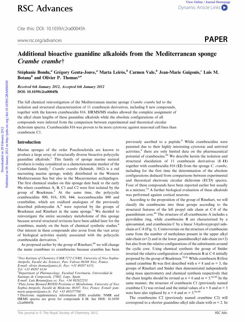
According to the proposition of the group of Rinehart, we will
classify the crambescins into three groups according to the
structural features of the left propyl side chain at C-8 of the
guanidinium core.6b The structure of all crambescins A includes a
pyrrolidine ring, while crambescins B are characterized by a
spiroaminal, and crambescins C by a linear 3-hydroxypropyl side-
chain at C-8 (Fig. 1). Controversies on the structure of crambescins
came from the number of methylenes present in the upper alkyl
side-chain (n+2) and in the lower guanidinoalkyl side-chain (m+2)
but also from the relative configurations of the substituents around
the cyclic core. Using chemical synthesis the group of Snider
inverted the relative configuration of crambescin B at C-8 initially
proposed by the group of Braekman.3b,6c While crambescin B (first
named crambine B) was first described with n = 8 and m = 3, the
groups of Rinehart and Snider then demonstrated independently
using mass spectrometry and chemical synthesis respectively that
the chain lengths should be revised as n = 6 and m = 5.6a,6b In the
same manner, the structure of crambescin C1 (previously named
crambine C1) was revised and the initial values of n = 8 and m = 3
were here also replaced by n = 6 and m = 5.6a,6b
The crambescins C2 (previously named crambine C2) will
correspond to a shorter guanidino alkyl side chain with m = 2. In
aNice Institute of Chemistry UMR 7272 CNRS, University of Nice Sophia-Antipolis, Faculte des Sciences, Parc Valrose 06108 Nice, France.E-mail: [email protected]; Fax: +33 49207 6151;Tel: +33 49207 6134bDepartment of Pharmacology, Facultad Veterinaria, Universidad deSantiago de Compostela, 27002, Lugo, Spain.E-mail: [email protected]; Fax: +34 982822233cPlate forme Bernard ROSSI Proteome et Metabolome, University of NiceSophia-Antipolis, Faculte de Medecine, 06107, Nice, France. E-mail: [email protected]; Tel: +33 493377708{ Electronic supplementary information (ESI) available: NMR andHRMS spectra are given for compounds 1–11. See DOI: 10.1039/c2ra00045h
RSC Advances Dynamic Article Links
Cite this: DOI: 10.1039/c2ra00045h
www.rsc.org/advances PAPER
This journal is � The Royal Society of Chemistry 2012 RSC Adv.
Dow
nloa
ded
on 2
8 Fe
brua
ry 2
012
Publ
ishe
d on
09
Janu
ary
2012
on
http
://pu
bs.r
sc.o
rg |
doi:1
0.10
39/C
2RA
0004
5HView Online / Journal Homepage
the same manner, we propose to use crambescin A2 and
crambescin B2 for the analogues with m = 2 because their
structures strongly differ from the usual crambescins with m
higher than four. Thus, most batzelladines, a large family of
crambescin ‘dimers’, all incorporate a crambescin A2 unit.10 The
biological evaluations and full structural assignments (including
the absolute configurations of these compounds) were hampered
by the difficulties associated with the purification processes, as
crambescins were usually isolated as a complex mixture of
homologues with added methylene units. We then decided to
purify all compounds to give additional data on the pure
crambescins.
Results and discussion
After an organic extraction of the lyophilized material with
CH2Cl2/MeOH (1 : 1), the extract was first submitted to fractiona-
tion by reversed phase Vacuum Liquid Chromatography. The
methanolic fraction was further purified by two successive HPLC
on semi-preparative C18 and analytical C3Phenyl reversed phase to
yield 11 pure crambescin derivatives and crambescidin 816. The 1H
NMR and HRMS spectra were used to confirm the high purity of
each isolated compound (.95%).
Each compound was then assigned to a crambescin family on
the basis of characteristic signals on their 1H NMR spectra. The
non-equivalent methylenes resonating at dH 2.99 and 3.33 (H-9),
2.11 and 2.23 (H-10) and 3.67 and 3.82 (H-11) ppm were
reminiscent of a crambescin A skeleton for compounds 1–4.
Compounds 6–8 were members of the crambescin B group due
to the absence of the signal at 4.40 (dd, H-13) ppm and the
presence of a characteristic signal at 2.99 (d, H-7) ppm.
Compounds 9–11 were part of the crambescin C family because
of the characteristic signals of the hydroxypropyl side chain
methylenes at 2.80 and 2.83 (H2-9), 1.81 (H2-10) and 3.61 (H2-11)
ppm (Table 1). Compound 5 exhibited unusual signals in its 1H
NMR spectrum which required deeper NMR analyses.
Comparison of the 1H NMR spectra for compounds 1–4
revealed clear differences in the intensity of the broad signals
around dH 1.30 and 1.40 ppm, assigned to the aliphatic side
chains protons of both the alkyl side chain at C-13 (n+2) and the
guanidinoalkyl chain at C-6 (m+2). These data were inferred to
differences in the lengths of both alkyl side chains. This
explanation may also account for the differences in the chemical
shifts at dH 4.25 and 4.22 (H2-5) ppm for 2, while these value
were dH 4.21 and 4.17 ppm for compound 4.
However, it was not possible to give clear data on the chain
lengths by NMR spectroscopy, due to signal overlapping and
imprecision in the signal integrations. As previously reported by
the group of Rinehart, mass spectrometry proved to be the
method of choice to assess the side-chain lengths.6b To be more
confident with our results, we performed (+)-HRESIMS/MS
analyses of each pure compound. All four crambescidin A
analogues differ by one or two methylene units (differences of 14
or 28 Da) (Table 2). Fragmentation analyses on compound 1 at
Table 1 1H NMR data of crambescin derivatives (500 MHz, CD3OD)
Position
Crambescin
A2 2 A1 4 5 B1 7 C1 10
2 3.23 3.17 3.16 3.16 3.173 1.68 1.60 1.61 1.59 1.60
3A 1.42 1.41 1.38 1.383B 1.42 1.41 1.38 1.413C 1.42 1.41 1.38 1.43
4 1.75 1.69 1.80 1.67 1.715 4.25 4.21 4.36 4.17 4.20
4.22 4.17 4.13 4.177 2.999 3.33 3.31 3.57 2.12 2.83
2.99 2.95 2.07 2.8010 2.23 2.23 2.39 2.10 1.81
2.11 2.10 2.0511 3.82 2.82 4.26 4.02 3.61
3.67 3.66 3.9313 4.40 4.39 3.86 4.4214 1.56 1.60 3.07 1.59 1.6015 1.41 1.41 1.72 1.47 1.4116 1.30 1.30 1.41 1.30 1.30
17-(15+n) 1.25–1.35 1.25–1.35 1.25–1.35 1.25–1.35 1.25–1.3516+n 0.90 0.90 0.90 0.90 0.90
Fig. 1 Crambescins and crambescidin skeletons.
Table 2 Crambescins isolated from C. crambe
Compound Crambescin m/z [M+H]+ m n
1 A2 449.36017 2 82 A2 463.37552 2 93 A2 477.39197 2 104 A1 463.37531 5 65 (Didehydro) A1 461.35976 5 66 B1 467.37051 4 67 B1 481.38629 5 68 B1 495.40207 6 69 C1 467.37027 4 6
10 C1 481.38635 5 611 C1 495.40182 6 6
RSC Adv. This journal is � The Royal Society of Chemistry 2012
Dow
nloa
ded
on 2
8 Fe
brua
ry 2
012
Publ
ishe
d on
09
Janu
ary
2012
on
http
://pu
bs.r
sc.o
rg |
doi:1
0.10
39/C
2RA
0004
5H
View Online
m/z 449.36017 [M+H]+ yielded two major fragments at m/z
336.26456 and 241.16585 corresponding to the molecular
formulae C19H34N3O2 and C11H21N4O2, respectively and were
consistent with the fragmentation scheme shown in Fig. 2.
The fragment peak at m/z 336.26456 [M–C5H11N3]+ indicated
the loss of a guanidine alkyl chain with m = 2 (Fig. 2). The
fragment at m/z 241.16285 [M–C13H24N2]+ was consistent with
an upper alkyl chain length of n = 8. The occurrence of the
analogous fragments at m/z 350.28018 and m/z 364.39654 in the
(+)-HRESIMS/MS spectra of compounds 2 and 3, respectively,
implied that both compounds had the same guanidinoalkyl chain
length (m = 2). The additional methylene units were then located
on the upper alkyl side chain which was confirmed by the
presence of the same fragments for all three compounds at m/z
241. Consequently, compounds 1–3 are all members of the
crambescin A2 family. The (+)-HRESIMS/MS mass fragmenta-
tion pattern of the molecular ion corresponding to compound 4
at m/z 463.37531 was completely different from those observed
for compounds 1–3. The presence of an intense fragment ion at
m/z 283.21280 was only consistent with a longer guanidinoalkyl
chain (m = 5). The length of the upper alkyl chain was then
deduced to be n = 6. According to this study, compound 4 is the
first isolated member of the crambescin A1 family and was
proven to be an isomer of compound 2, a member of the
crambescin A2 family, which underlined the importance of the
HRMS/MS study. In the original paper of the group of
Braekman only crambescins A2 (crambine A) were described
in a mixture.3b
The molecular formula of compound 5 was determined to be
C25H44N6O2 by HRESIMS (D 20.2 ppm by internal calibra-
tion), indicating seven degrees of unsaturation, one more than
compounds 2 and 4. The characteristic signals of crambescin A
were absent in the 1H NMR spectrum of 5 and the signals of the
methylenes at C-5 (dH, 4.36, t), C-9 (dH, 3.57, t) and C-11 (dH,
4.26, t) became equivalent and deshielded comparing to the data
obtained for 2 and 4. Furthermore, the upper alkyl chain was
connected to a quaternary carbon at C-13 (dC 180 ppm) due to
the H-14/C-13 HMBC. In consequence the additional unsatura-
tion was located at C-13 which induced an aromatized planar
pyrimidine cycle. The length of the upper alkyl chain was
estimated as being n = 6 from the integration of signals at dH
1.31 ppm (10H) assigned to H2-17 to H2-21 (Table 1). The length
of the guanidinoalkyl chain was assessed by the integration of
the signal at 1.41 ppm (8H) which corresponds to H2-3A, H2-3B
and H2-3C and H2-16, thus suggesting m = 5. The lengths of
the alkyl chains at C-13 and C-6 were further confirmed by
(+)-HRESIMS/MS fragmentation pattern of the molecular peak
at m/z 231.18352 [M+2H]2+ which gave two majors fragments at
m/z 306.21762 and m/z 156.14943, in accordance with the
molecular formulae C17H26N2O3 and C8H18N3, respectively.
The fragment peak at m/z 306.21762 [M–C8H17N3]+ corre-
sponds to the loss of a molecular ion of 155 Da, a 7–guanidino-1-
heptene. This result is consistent with m = 5 and n = 6 for both
alkyl chains and we could conclude that 5 is an oxidized form of
compound 4, belonging to the crambescin A1 family. 5 is then a
didehydrocrambescin A1 derivative. A dehydrocrambine A has
already been reported from an unknown sponge of the genus
Monanchora in Palau.11 The described dehydrocrambine A is an
oxidized equivalent of crambescin A2 (named crambine A) they
also isolated from that sponge. Aromatization into a pyrimidine
is not rare in this family of compounds.12 Just like for the
crambescin A2 derivative found in Monanchora sp., compound
4 was also isolated together with its oxidized form 5 which
reinforced the idea that an oxidation could partially take place
during the purification process, especially in the acidic conditions
required for the HPLC purification.
The 1H NMR spectra of compounds 6–8 first indicated that
they belong to the crambescin B family, especially for the
occurrence of the small doublet at 2.99 (J = 4 Hz, H-7) ppm.
The chain lengths were assigned on the basis of HRMS/MS data.
The fragmentation pattern differed from crambescins A and
both guanidines remain intact during the fragmentation whereas
the presence of the pyrrolidine in crambescins A induced a
fragmentation of the cyclic guanidine (Fig. 2).
Fragments were then observed at m/z 270.18124, 284.19693
and 298.21896 for compounds 6, 7 and 8 respectively, which
Fig. 2 Fragmentation pattern of crambescins.
This journal is � The Royal Society of Chemistry 2012 RSC Adv.
Dow
nloa
ded
on 2
8 Fe
brua
ry 2
012
Publ
ishe
d on
09
Janu
ary
2012
on
http
://pu
bs.r
sc.o
rg |
doi:1
0.10
39/C
2RA
0004
5H
View Online
indicated a change in the chain lengths for the guanidino alkyl
chain with m = 4, 5 and 6 respectively and a common upper alkyl
chain with n = 6 for all crambescin B1 derivatives. The relative
configurations for compounds 6–8 were assigned by interpreta-
tion of the coupling constant between H-7 and H-13 which was
reported as 4–4.2 Hz in the cis isomers and 11.5–12 Hz in the
trans isomer.6a All isolated crambescins B1 had JH7–H13 close to
4.2 Hz revealing a cis configuration between H-7 and H-13.
Comparison of the chemical shifts for the proton of the spiro
cycle with the previously described crambescins B1 (crambine B)
led us to propose the same relative configurations at C-8 than
those proposed by Snider.6a
The 1H NMR spectra of compounds 9–11 indicated that they
belong to the crambescin C family, especially for the occurrence
of the triplet at 3.61 (H2-11) ppm. The same fragmentation
patterns as those obtained for crambescins B were observed
which allowed us to assign the chain lengths of all three
crambescin C derivatives. Here also, differences between the
three compounds 9–11 were identified in the length of the lower
guanidine alkyl chain with m values of 4, 5 and 6 while the upper
alkyl chain at C-13 remain unchanged with the same n = 6. In
consequence these compounds are all crambescin C1 analogues.
At this point, it is interesting to note that most of the crambescin
A derivatives are from the crambescin A2 family while
crambescins B1 and C1 are only found in the sponge C. crambe.
In contrast to the second study of the group of Braekman,3a we
did not manage to identify a sufficient quantity of crambescin
C2. The presence of short guanidino alkyl chains with m = 2 in
crambescin A2 is remarkable and much more if we observe that
all batzelladine derivatives incorporate only this monomer.12
This observation raises the question of the biosynthetic path-
ways leading to these compounds, as studied first by the group of
Snider,6a and then by the group of Quinn for the mirabilins,
which are tricyclic monomers, analogues of the mono or bicyclic
crambescins.13 The addition of a guanidine to a unique polyketide
chain is questionable as no enzyme has been described to perform
such transformation. We rather propose the involvement of an
arginine or a homologue for the guanidines. Indeed, we previously
shown that the 2-aminoimidazole side chain of oroidin originates
from homoarginine,14 and, recently, the group of Molinski
proposed the same origin for a guanidinated derivative of the
polyacetylene family.15 We will address this issue for crambescins
by in vivo biosynthetic experiments.
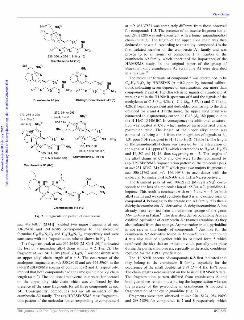
With all pure crambescins in hand, we were able to assign the
absolute configurations of the three families A, B and C using
ECD and comparison with theoretical values as already reported
by us for other families of marine natural products.16 We
anticipated strong Cotton effects due to the presence of
chromophoric groups (nAp* transition of the enone) placed in
the vicinity of asymmetric centers as for the asymmetric center at
C-13 always substituted by the guanidine and an enone in the
cases of crambescins A and C. Experimental ECD spectra were
then compared to the calculated ECD spectra of both
enantiomers. As illustrated on Fig. 3, there is a good overlapping
between both experimental and theoretical ECD spectra for one
enantiomer of the three different kinds of crambescins. All our
results converge to suggest an absolute S configuration at C-13
which is in accordance with the only result obtained on the
biosynthetic congener crambidine using asymmetric chemical
synthesis.17 The only discrepancy we observed for these
11 compounds was for the ECD spectrum of one crambescin
C1 (9) where the signs of the Cotton effects opposite to the one
measured for 10 and 11. Nevertheless in this case the intensities
of the bands were very low and the other results obtained for
10 and 11 seemed more reliable.
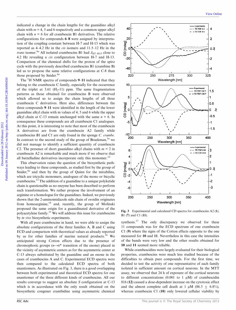
While crambescidins were largely evaluated for their biological
properties, crambescins were much less studied because of the
difficulties to obtain pure compounds. For the first time, we
decided to test the activity of one representative of each family
isolated in sufficient amount on cortical neurons. In the MTT
assay, we observed that 24 h of exposure of the cortical neurons
to different concentrations (0.001 to 1 mM) of crambescidin
816 (12) caused a dose-dependent increase on the cytotoxic effect
and the almost complete cell death at 1 mM (86.3 ¡ 6.8%),
whereas crambescin C1 (10) only lowered cellular viability by
Fig. 3 Experimental and calculated CD spectra for crambescins A2 (1),
B1 (7) and C1 (11).
RSC Adv. This journal is � The Royal Society of Chemistry 2012
Dow
nloa
ded
on 2
8 Fe
brua
ry 2
012
Publ
ishe
d on
09
Janu
ary
2012
on
http
://pu
bs.r
sc.o
rg |
doi:1
0.10
39/C
2RA
0004
5H
View Online
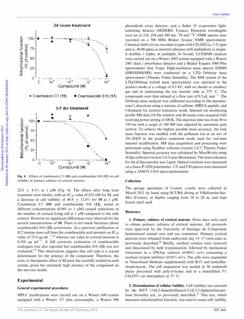
22.6 ¡ 4.1% at 1 mM (Fig. 4). The effects after long term
treatment were similar, with an IC50 value of 432 nM for 12, and
a decrease in cell viability of 40.8 ¡ 12.6% for 10 at 1 mM.
Crambescin C1 (10) and crambescidin 816 (12), tested at
different concentrations (0.001 to 1 mM) caused reductions in
the number of cortical living cell at 1 mM compared to the cells
control. However no significant differences were observed for the
several concentrations of 10. There is not much literature about
crambescidin 816 (12) cytotoxicity. In a previous publication in
IC2 murine mast cell lines the crambescidin acid presents an IC50
value of 25.0 mg ml21,18 whereas our value in cortical neurons is
0.354 mg ml21. A full cytotoxic evaluation of crambescidin
analogues was also reported but crambescidin 816 (12) was not
evaluated.19 This observation suggests that cell type is a crucial
determinant for the potency of the compound. Therefore, the
toxic or therapeutic effect of 12 must the carefully studied in each
system, given the extremely high potency of the compound on
the nervous model.
Experimental
General experimental procedures
HPLC purifications were carried out on a Waters 600 system
equipped with a Waters 717 plus autosampler, a Waters 996
photodiode array detector, and a Sedex 55 evaporative light-
scattering detector (SEDERE, France). Detection wavelengths
were set at 214, 254 and 280 nm. 1H and 13C NMR spectra were
recorded on a 500 MHz Bruker Avance NMR spectrometer.
Chemical shifts (d) are recorded in ppm with CD3OD (dH 3.31 ppm
and dC 49.00 ppm) as internal reference with multiplicity (s singlet,
d doublet, t triplet, m multiplet, br broad). LCESIMS analyses
were carried out on a Waters 2695 system equipped with a Waters
2487 dual l absorbance detector and a Bruker Esquire 3000 Plus
spectrometer (Ion Trap). High-resolution mass spectra ESIMS
(HRESIMS/MS) were conducted on a LTQ Orbitrap mass
spectrometer (Thermo Fisher Scientific). The MSI system of the
LTQ-Orbitrap hybrid mass spectrometer was operated in the
positive mode at a voltage of 4.5 kV, with no sheath or auxiliary
gas and in maintaining the ion transfer tube at 275 uC. The
compounds were then infused at a flow rate of 0.5 mL min21. The
Orbitrap mass analyzer was calibrated according to the manufac-
turer’s directions using a mixture of caffeine, MRFA peptide, and
Ultramark for positive ionization mode. Selected ion monitoring
profile MS data (10 Da window and 40 scans) were acquired with
resolving power setting of 100 K. The injection time was from 50 to
350 ms with a target of 100 000 ions adjusted by automatic gain
control. To achieve the highest possible mass accuracy, the lock
mass function was enabled with the pollutant ion at an m/z of
391.28429 in the positive ionization mode used for real-time
internal recalibration. MS data acquisition and processing were
performed using Xcalibur software (version 2.0.7; Thermo Fisher
Scientific). Spectral accuracy was calculated by MassWorks using
sClips software (version 2.0; Cerno Bioscience). The mass tolerance
for the sClips searches was 2 ppm. Optical rotations were measured
on a Jasco P-1020 polarimeter. UV and CD spectra were measured
using a JASCO J-810 spectropolarimeter.
Collection
The sponge specimens of Crambe crambe were collected in
March 2011 by hand using SCUBA diving at Villefranche-Sur-
Mer (France), at depths ranging from 10 to 20 m, and kept
frozen until used.
Bioassays
1. Primary cultures of cortical neurons. Swiss mice were used
to obtain primary cultures of cortical neurons. All protocols
were approved by the University of Santiago de Compostela
Institutional animal care and use committee. Primary cortical
neurons were obtained from embryonic day 15–17 swiss mice as
previously described.20 Briefly, cerebral cortices were removed
and dissociated by mild trypsinisation, followed by mechanical
trituration in a DNAse solution (0.005% w/v) containing a
soybean trypsin inhibitor (0.05% w/v). The cells were suspended
in Neurobasal Medium supplemented with B-27 and penicillin-
streptomycin. The cell suspension was seeded in 96 multiwell
plates precoated with poly-D-lysine and in a humidified 5%
CO2/95% air atmosphere at 37 uC.
2. Determination of cellular viability. Cell viability was assessed
by the MTT (3-[4,5-dimethylthiazol-2-yl]-2,5-diphenyltetrazo-
lium bromide) test, as previously described.21 This test, which
measures mitochondrial function, was used to assess cell viability
Fig. 4 Effects of crambescin C1 (10) and crambescidin 816 (12) on cell
viability in primary cultures of cortical neurons.
This journal is � The Royal Society of Chemistry 2012 RSC Adv.
Dow
nloa
ded
on 2
8 Fe
brua
ry 2
012
Publ
ishe
d on
09
Janu
ary
2012
on
http
://pu
bs.r
sc.o
rg |
doi:1
0.10
39/C
2RA
0004
5H
View Online
as it has been shown that in neuronal cells there is a good
correlation between a drug-induced decrease in mitochondrial
activity and its cytotoxicity.22 The assay was performed in
cultures grown in 96 well plates and exposed to different
concentrations (0.001 mM, 0.01 mM, 0.1 mM and 1 mM) of 10 and
12 added to the culture medium. Cultures were maintained in the
presence of the toxin at 37 uC in humidified 5% CO2/95% air
atmosphere for 24 h or five days in vitro (3–7 div). Saponin was
used as cellular death control and its absorbance was subtracted
from the other data. After the exposure time, cells were rinsed
and incubated for 60 min with a solution of MTT (500 mg ml21)
dissolved in Locke’s buffer. After washing off excess MTT, the
cells were disaggregated with 5% sodium dodecyl sulfate and the
absorbance of the colored formazan salt was measured at
595 nM in a spectrophotometer plate reader.
Extraction and isolation
The samples of Crambe crambe were freeze dried and then ground
to obtain a dry powder (15 g) which was exhaustively extracted
with a mixture of MeOH/CH2Cl2 (1 : 1, v/v) to yield 2.4 g of crude
extract after concentration under reduced pressure. The crude
extract was fractionated by Reverse Phase Vacuum Liquid
Chromatography over RP-18 silica gel (200 g) with a step gradient
of H2O (F1 fraction, 581 mg), MeOH/H2O 2/1 v/v (F2 fraction
286 mg), MeOH/H2O 3/1 v/v (F3 fraction, 284 mg), MeOH (F4
fraction, 767 mg) and DCM (F5 fraction, 450 mg) (500 mL each).
The F2 (140 mg, 100 mg ml21) and F3 (160 mg, 100 mg ml21)
fractions were then subjected to semi-preparative HPLC purifica-
tion (Phenomenex Luna C6-Phenyl, 250 6 10 mm, 5 mm) eluted
with a gradient of H2O/ACN/TFA : isocratic step from 0 min to
5 min (90 : 10 : 0.1) and then gradient steps from 5 min to 6 min
(from 90 : 10 : 0.1 to 70 : 30 : 0.1) and from 6 to 36 min (from
70 : 30 : 0.1 to 55 : 45 : 0.1) (flow rate: 3.0 mL min21, injection
volume: 100 mL) to give 9 and 13 fractions respectively, named
from F2P1 to F2P9 and F3P1 to F3P13. The fractions F2P2 (tr
21.5 min), F2P3 (tr 23.0 min), F2P4 (tr 26.0 min), F2P5
(tr 28.0 min) and F2P9 (tr 36.0 min) were further subjected to
analytical HPLC purification (XSelect C6-Phenyl, 250 6 4.6 mm,
5 mm). Fractions were eluted with a gradient of H2O/ACN/TFA:
isocratic step from 0 min to 5 min (90 : 10 : 0.1) and then gradient
steps from 5 min to 6 min (from 90 : 10 : 0.1 to 68 : 32 : 0.1) and
isocratic step from 6 to 30 min (68 : 32 : 0.1) (flow rate: 0.80 mL
min21, injection volume: 30 mL). This step yielded the major
components of the F2P2, F2P3, F2P4 and F2P9 fractions and so
to yield compound 9 (1.2 mg), 10 (11.1 mg), 11 (2.4 mg), 12
(10.2 mg), respectively, and, in the case of F2P5 fraction, to afford
5 (tr 24.5 min, 0.8 mg), 6 (tr 25.2 min, 0.9 mg), 4 (tr 26.5 min,
0.9 mg) and 7 (tr 28.2 min, 1.1 mg). Similar analytical HPLC
analyses were performed on fractions F3PP7 (tr 29.0 min), F3P8
(tr 30.5 min), F3P9 (tr 32.0 min) and F3P12 (tr 37.0 min) and let to
afford 1 (2.0 mg), 8 (1.1 mg), 2 (2.0 mg), 3 (1.3 mg), respectively.
At the total, twelve guanidine alkaloids were purified. All of them
were identified by a combination of spectroscopic methods (1D
and 2D NMR, HRESIMS).
Norcrambescin A2 (1)
[a]20D +7.1 (c 0.2, MeOH); UV (MeOH) lmax (log e) 288 nm (2.7);
CD (MeOH, c 4.46 6 1024 M) De (lmax nm) +0.66 (209) –2.22
(248) –0.99 (291); ESIMS m/z 449.3 [M+H]+; HRESIMS m/z
449.36017 [M+H]+ (Calc for C24H45O2N6, 449.35985,
D 20.70 ppm). 1H NMR (500 MHz) and 13C NMR
(125 MHz, CD3OD) (lH, J, 13C) C-1 (158.6), H2C-2 (3.23 t,
6.8; 42.1), H2C-3 (1.68 m; 26.6), H2C-4 (1.75 m; 27.1), H2C-5
(4.22 m, and 4.23 m; 65.1), C-6 (166.1), C-7 (103.2), C-8 (152.7),
H2C-9 (2.99 ddd, 9.5/ 9.8/18.3 and 3.30; 31.9), H2C-10 (2.11 m
and 2.23 m; 22.9), H2C-11 (3.67 ddd, 7.2/9.7/9.93 and 3.82 ddd,
2.8/ 9.1/9.4; 49.2), C-12 (153.0), HC-13 (4.40 dd, 7.1; 51.3), H2C-
14 (1.56 m; 37.5), H2C-15 (1.41 m; 25.2), H2C-16 to H2C-21
(1.25–1.35 br s; 30.0–31.0), H2C-22 (1.25–1.35 br s; 33.1), H2C-23
(1.29 br s; 23.8), H3C-24 (0.90 t, 6.8; 14.3).
Crambescin A2 (2)
[a]20D +12.1 (c 0.18, MeOH); UV (MeOH) lmax 287 nm (log e 2.7);
CD (MeOH, c 4.33 6 1024 M) De (lmax nm) –1.86 (249) –0.81
(288); ESIMS m/z 463.3 [M+H]+; HRESIMS m/z 463.37552
[M+H]+ (Calc for C24H45O2N6, 463.37550, D 20.04 ppm).
Homocrambescin A2 (3)
(3) [a]20D +35.9 (c 0.1, MeOH); UV (MeOH) lmax 288 nm (log e 2.3);
CD (MeOH, c 4.20 6 1024 M) De (lmax nm) +0.58 (207) –1.93 (249)
–0.85 (288); ESIMS m/z 477.0 [M+H]+; HRESIMS m/z 477.39197
[M+H]+ (Calc for C26H49O2N6, 477.39115, D –1.71 ppm).
Crambescin A1 (4)
[a]20D +8.9 (c 0.06, MeOH); UV (MeOH) lmax (log e) 289 nm (1.5);
CD (MeOH, c 4.84 6 1024 M) De (lmax nm) 20.10 (218) 20.06
(288) +0.08 (330); ESIMS m/z 463.2 [M+H]+; HRESIMS m/z
463.37531 [M+H]+ (Calc for C25H47O2N6, 463.37550, D
–0.42 ppm). 1H NMR (500 MHz) and 13C NMR (125 MHz,
CD3OD) (lH, J, 13C) C-1 (158.6), H2C-2 (3.17 t, 6.9; 42.5), H2C-3
(1.60 m; 29.8), H2C-3A (1.42 br s; 27.6), H2C-3B (1.42 br s; 29.9),
H2C-3C (1.42 br s; 27.1), H2C-4 (1.69 m; 29.8), H2C-5 (4.17 m,
17.4 and 4.21 m, 17.4; 65.6), C-6 (166.9), C-7 (100.4), C-8 (155.1),
H2C-9 (2.95 ddd, 9.1/ 9.5/18.9 and 3.31; 31.9), H2C-10 (2.10 m
and 2.23 m; 22.9), H2C-11 (3.66 ddd, 7.5/9.5/10.3 and 3.82 ddd,
2.8/9.5/10.3; 49.5), C-12 (154.4), HC-13 (4.39 m; 51.4), H2C-14
(1.60 m; 37.6), H2C-15 (1.30 br m; 25.2), H2C-16 to H2C-19
(1.30 br m; 30.35, 30.44, 30.60, 30.64), H2C-20 (1.30 br m; 33.1),
H2C-21 (1.30 br m; 23.8), H3C-22 (0.90 t, 7.4; 14.5).
Didehydrocrambescin A1 (5)
ESIMS m/z 461.3 [M+H]+; HRESIMS m/z 461.35976 [M+H]+
(Calcd for C25H45N6O2 461.35985 found 461.35976, D –0.20 ppm).1H NMR (500 MHz) and 13C NMR (125 MHz, CD3OD) (lH, J,13C) C-1 (158.5), H2C-2 (3.16 t, 6.6; 42.5), H2C-3 (1.61 m; 30.1),
H2C-3A (1.41 br s; 28.9), H2C-3B (1.41 br s; 29.9), H2C-3C (1.41 br
s; 27.3), H2C-4 (1.80 m; 29.9), H2C-5 (4.36 t, 7.6; 67.0), C-6 (165.9),
C-7 (111.7), C-8 (170.6), H2C-9 (2.57 t, 8.6 and 3.30; 35.6), H2C-10
(2.05 m and 2.39 q; 20.8), H2C-11 (4.26 t, 9.1; 53.1), C-13 (180.2),
H2C-14 (3.07 t, 9.5; 38.2), H2C-15 (1.72 m; 30.0), H2C-16 (1.41 br
m; 27.3), H2C-17 to H2C-19 (1.31 br m; 30.0-31.0), H2C-20 (1.31 br
m; 32.8), H2C-21 (1.31 br m; 23.6), H3C-22 (0.90 t, 7.5; 14.5).
Norcrambescin B1 (6)
[a]20D 2114.0 (c 0.05, MeOH); UV (MeOH) lmax (log e) 285 nm
(1.8), 326 nm (1.9); CD (MeOH, c 4.03 6 1024M) De (lmax nm)
RSC Adv. This journal is � The Royal Society of Chemistry 2012
Dow
nloa
ded
on 2
8 Fe
brua
ry 2
012
Publ
ishe
d on
09
Janu
ary
2012
on
http
://pu
bs.r
sc.o
rg |
doi:1
0.10
39/C
2RA
0004
5H
View Online
20.53 (211) +0.75 (247) +0.55 (326); ESIMS m/z 467.3 [M+H]+;
HRESIMS m/z 467.37051 [M+H]+ (Calc for C24H47O3N6,
467.37042, D +0.21 ppm).
Crambescin B1 (7)
[a]20D 2116.3 (c 0.04, MeOH); UV (MeOH) lmax (log e) 286 nm
(1.9), 325 nm (1.9); CD (MeOH, c 3.58 6 1024 M) De (lmax nm)
–0.51 (211) +0.74 (247) +0.53 (328); ESIMS m/z 481.2 [M+H]+;
HRESIMS m/z 481.38629 [M+H]+ (Calc for C25H49O3N6,
481.38607, D +0.47 ppm). 1H NMR (500 MHz) and 13C NMR
(125 MHz, CD3OD) (lH, J, 13C) C-1 (158.6), H2C-2 (3.16 t, 7.3;
42.6), H2C-3 (1.59 m; 29.7), H2C-3A (1.38 br s; 27.8), H2C-3B
(1.38 br s; 30.3), H2C-3C (1.38 br s; 27.1), H2C-4 (1.67 m; 29.9),
H2C-5 (4.13 m and 4.17 m; 66.3), C-6 (169.9), HC-7 (2.99 d, 4.3;
50.2), C-8 (89.9), H2C-9 (2.07 m and 2.12, m; 36.1), H2C-10
(2.10 m; 25.7), H2C-11 (3.93 m and 4.02 m; 68.9), C-12 (155.2),
HC-13 (3.86 ddd, 4.3/6.7/7.3; 49.7), H2C-14 (1.59 m; 33.1), H2C-
15 (1.47 m; 26.5), H2C-16 to H2C-19 (1.31 br m; 30.0–31.0), H2C-
20 (1.31 br m; 32.8), H2C-21 (1.31 br m; 23.7), H3C-22 (0.90 t,
7.1; 14.4).
Homocrambescin B1 (8)
[a]20D 2145.5 (c 0.07, MeOH); UV (MeOH) lmax (log e) 284 nm
(1.8), 326 nm (1.8); CD (MeOH, c 1.34 6 1024 M) De (lmax nm)
–0.90 (211) +1.42 (247) +0.90 (326); ESIMS m/z 495.3 [M+H]+;
HRESIMS m/z 495.40207 [M+H]+ (Calc for C26H51O3N6,
495.40172, D 20.71 ppm).
Norcrambescin C1 (9)
[a]20D +76.3 (c 0.08, MeOH); UV (MeOH) lmax (log e) 279 nm
(2.3); CD (MeOH, c 5.15 6 1024 M) De (lmax nm) +0.08 (207) –
0.20 (247); ESIMS m/z 467.5 [M+H]+; HRESIMS m/z 467.37027
[M+H]+ (Calc for C24H47O3N6, 467.37042, D 20.31 ppm).
Crambescin C1 (10)
[a]20D +33.1 (c 0.39, MeOH); UV (MeOH) lmax (log e) 278 nm
(2.8); CD (MeOH, c 1.39 6 1024 M) De (lmax nm) 20.48 (213)
+2.24 (247); ESIMS m/z 481.5 [M+H]+; HRESIMS m/z
481.38635 [M+H]+ (Calc for C25H49N6O3, 481.38607, D
0.59551). 1H NMR (500 MHz) and 13C NMR (125 MHz,
CD3OD) (lH, J, 13C) C-1 (159.1), H2C-2 (3.17 t, 7.0; 42.8), H2C-3
(1.60 m; 30.9), H2C-3A (1.41 br s; 27.9), H2C-3B (1.41 br s; 30.4),
H2C-3C (1.41 br s; 27.4), H2C-4 (1.71 m; 30.2), H2C-5 (4.17 m
and 4.20 m; 66.3), C-6 (166.5), C-7 (106.4), C-8 (149.4), H2C-9
(2.80 m and 2.83, m; 29.1), H2C-10 (1.81q; 32.5), H2C-11 (3.61 t,
6.5; 62.5), C-12 (153.9), HC-13 (4.42 dd, 7.5/5.0; 51.5), H2C-14
(1.60 m; 37.4), H2C-15 (1.41 m; 25.4), H2C-16 to H2C-19 (1.30 br
m; 30.0-32.0), H2C-20 (1.30 br m; 33.4), H2C-21 (1.30 br m;
24.7), H3C-22 (0.90 t, 6.5; 15.1).
Homocrambescin C1 (11)
[a]20D +62.7 (c 0.13, MeOH); UV (MeOH) lmax (log e) 279 nm
(2.9); CD (MeOH, c 0.68 6 1024 M) De (lmax nm) –0.58 (214)
+3.03 (249); ESIMS m/z 495.0 [M+H]+; HRESIMS m/z
495.40182 [M+H]+ (Calc for C26H51O3N6, 495.40172, D
+0.22 ppm).
Crambescidin 816 (12)
As reported in ref. 8.
Theoretical calculations of the electronic dichroism spectra
Quantum chemical calculations have been performed for all
examined compounds 1–11. Conformational analysis was
performed using the conformer research algorithm implemented
in the Conflex-Barista software.23 Given the high degree of
conformational freedom of the side chains, the conformational
analysis led to a large number of structures (n . 500). The
Gaussian03W package24 has been used for the electronic circular
dichroism calculations on the most stable conformer of each
compound. Density functional theory (DFT) with B3LYP
functional25 and Pople’s 6.31++G(d,p)26 basis set was used on
the lowest energy conformers. TDDFT was employed to
calculate excitation energy (in eV) and rotatory strength R in
dipole velocity (Rvel) and dipole length (Rlen) forms. The
calculated rotatory strengths were simulated in ECD curve by
using a Gaussian function:
De Eð Þ~ 1
2:296|10{39ffiffiffiffiffiffiffiffiffi
2pDp
X
a
DE0aR0ae{E{DE0að Þ
2D
� �2
(1)
where D is half the width of the band at 1/e peak height expressed
in energy units. The parameters DE0a and R0a are the excitation
energies and the rotatory strengths for transition from the
ground state 0 to an excited state a, respectively, D = 0.1 eV and
Rvel were used.
Conclusions
The full chemical study of the Mediterranean sponge C. crambe
led to the isolation and structural characterization of 11
crambescins A1, A2, B1 and C1 in their pure forms. Eight of
the 12 isolated compounds are described for the first time and the
lengths of the alkyl chains were deduced from the careful analyses
of HRMS/MS data. The presence of the new didehydrocrambes-
cin A1 (5) derivative supports the assumption that aromatization
could occur in this family during the acidic purification process.
Our studies allowed the assignment of the absolute configurations
of all compounds and revealed a high cytotoxic activity of
crambescidin 816 (12) against cortical neurons.
Acknowledgements
This work was supported by the European Coordinative
Project FP7-KBBE-2010-4-BAMMBO (265896) and the
French ECIMAR project (ANR-06-BDIV-01). We are grateful
to Thierry Perez (Centre d’Oceanologie d’Endoume) for
taxonomic identification and to Marc Gaysinski (PFTC Nice)
for recording the NMR spectra. Assistance for submarine
collection was kindly given by David Luquet (Observatoire
Oceanologique de Villefranche sur Mer).
References
1 (a) R. G. S. Berlinck, A. C. B. Burtoloso, A. E. Trindade-Silva, S.Romminger, R. P. Morais, K. Bandeira and C. M. Mizuno, Nat.
This journal is � The Royal Society of Chemistry 2012 RSC Adv.
Dow
nloa
ded
on 2
8 Fe
brua
ry 2
012
Publ
ishe
d on
09
Janu
ary
2012
on
http
://pu
bs.r
sc.o
rg |
doi:1
0.10
39/C
2RA
0004
5H
View Online
Prod. Rep., 2010, 27, 1871–1907; (b) R. G. S. Berlinck and M. H.Kossuga, in Modern Alkaloids, ed. E. Fattorusso and O. Taglialatela-Scafati, Wiley-VCH Verlag GmbH & Co, KGaA, Weinheim,Germany, 2008, pp 305–337.
2 D. Erpenbeck and R. W. M. van Soest, Mar. Biotechnol., 2007, 9,2–19.
3 (a) R. G. S. Berlinck, J. C. Braekman, D. Daloze, I. Bruno, R. Riccio,D. Rogeau and P. Amade, J. Nat. Prod., 1992, 55, 528–532; (b) R. G. S.Berlinck, J. C. Braekman, D. Daloze, K. Hallenga, R. Ottinger,I. Bruno and R. Riccio, Tetrahedron Lett., 1990, 31, 6531–6534.
4 Y. Kashman, S. Hirsh, O. J. McConnell, I. Ohtani, T. Kusumi and H.Kakisawa, J. Am. Chem. Soc., 1989, 111, 8925–8926.
5 (a) R. G. Berlinck, J. C. Braekman, D. Daloze, I. Bruno, R. Riccio, S.Ferri, S. Spampinato and E. Speroni, J. Nat. Prod., 1993, 56,1007–1015; (b) E. A. Jares-Erijman, R. Sakai and K. L. Rinehart, J.Org. Chem., 1991, 56, 5712–5715.
6 (a) B. B. Snider and Z. Shi, J. Org. Chem., 1993, 58, 3828–3839; (b)E. A. Jares-Erijman, A. A. Ingrum, F. Sun and K. L. Rinehart,J. Nat. Prod., 1993, 56, 2186–2188; (c) B. B. Snider and Z. Shi, J. Org.Chem., 1992, 57, 2526–2528.
7 D. S. Dalisay, J. P. Saludes and T. F. Molinski, Bioorg. Med. Chem.,2011, 19, 6654–6657.
8 C. H. Van Etten, H. C. Nielsen and J. E. Peters, Phytochemistry,1965, 4, 467–473.
9 (a) A. Olszewski, K. Sato, Z. D. Aron, F. Cohen, A. Harris, B. R.McDougall, W. E. Robinson Jr., L. E. Overman and G. A. Weiss,Proc. Natl. Acad. Sci. U. S. A., 2004, 101, 14079–14084; (b) K. L.Rinehart and E. A. Jares-Erijman, US Pat. 5756734A, 1998.
10 A. D. Patil, N. V. Kumar, W. C. Kokke, M. F. Bean, A. J. Freyer, C.de Brosse, S. Mai, A. Truneh, B. Carte, A. L. Breen, R. P. Hertzberg,R. K. Johnson, J. W. Westley and B. C. M. Potts, J. Org. Chem.,1995, 60, 1182–1188.
11 L. Chang, N. F. Whittaker and C. A. Bewley, J. Nat. Prod., 2003, 66,1490–1494.
12 R. Laville, O. P. Thomas, F. Berrue, D. Marquez, J. Vacelet and P.Amade, J. Nat. Prod., 2009, 72, 1589–1594.
13 M. El-Naggar, M. Conte and R. J. Capon, Org. Biomol. Chem., 2010,8, 407–412.
14 G. Genta-Jouve, N. Cachet, S. Holderith, F. Oberhansli, J.-L.Teyssie , R. Jeffree, A. Al Mourabit and O. P. Thomas,ChemBioChem, 2011, 12, 2298–2301.
15 B. I. Morinaka and T. F. Molinski, Org. Lett., 2011, 13, 6338–6341.16 (a) E. L. Regalado, C. Jimenez-Romero, G. Genta-Jouve, D.
Tasdemir, P. Amade, C. Nogueiras and O. P. Thomas, Tetrahedron,
2011, 67, 1011–1018; (b) N. Cachet, G. Genta-Jouve, E. L. Regalado,R. Mokrini, P. Amade, G. Culioli and O. P. Thomas, J. Nat. Prod.,2009, 72, 1612–1615.
17 L. E. Overman and Y. H. Rhee, J. Am. Chem. Soc., 2005, 127,15652–15658.
18 K. M. Meragelman, T. C. McKee and J. B. McMahon, J. Nat. Prod.,2004, 67, 1165–1167.
19 Z. D. Aron, H. Pietraszkiewicz, L. E. Overman, F. Valeriote and C.Cuevas, Bioorg. Med. Chem. Lett., 2004, 14, 3445–3449.
20 C. Vale, E. Alonso, J. A. Rubiolo, M. R. Vieytes, F. M. La Ferla, L.Gimenez-Llort and L. M. Botana, Cell. Mol. Neurobiol., 2010, 30,577–590.
21 C. Vale, K. C. Nicolaou, M. O. Frederick, B. Gomez-Limia, A.Alfonso, M. R. Vieytes and L. M. Botana, J. Med. Chem., 2007, 50,356–363.
22 T. Varming, J. Drejer, A. Frandsen and A. Schousboe, J. Neurosci.Res., 1996, 44, 40–46.
23 (a) H. Goto, K. Ohta, T. Kamakura, S. Obata, N. Nakayama, T.Matsumoto and E. Osawa, Conflex Corp: Tokyo-Yokohama, 2004;(b) H. Goto and E. Osawa, J. Chem. Soc., Perkin Trans. 2, 1993,187–198; (c) H. Goto and E. Osawa, Tetrahedron Lett., 1992, 33,1343–1346; (d) H. Goto and E. Osawa, J. Am. Chem. Soc., 1989, 111,8950–8951.
24 M. J. Frisch, G. W. Trucks, H. B. Schlegel, G. E. Scuseria, M. A.Robb, J. R. Cheeseman, J. A. Montgomery, T. Vreven, K. N. Kudin,J. C. Burant, J. M. Millam, S. S. Iyengar, J. Tomasi, V. Barone, B.Mennucci, M. Cossi, G. Scalmani, N. Rega, G. A. Petersson, H.Nakatsuji, M. Hada, M. Ehara, K. Toyota, R. Fukuda, J. Hasegawa,M. Ishida, T. Nakajima, Y. Honda, O. Kitao, H. Nakai, M. Klene,X. Li, J. E. Knox, H. P. Hratchian, J. B. Cross, V. Bakken, C.Adamo, J. Jaramillo, R. Gomperts, R. E. Stratmann, O. Yazyev,A. J. Austin, R. Cammi, C. Pomelli, J. W. Ochterski, P. Y. Ayala, K.Morokuma, G. A. Voth, P. Salvador, J. J. Dannenberg, V. G.Zakrzewski, S. Dapprich, A. D. Daniels, M. C. Strain, O. Farkas,D. K. Malick, A. D. Rabuck, K. Raghavachari, J. B. Foresman, J. V.Ortiz, Q. Cui, A. G. Baboul, S. Clifford, J. Cioslowski, B. B.Stefanov, G. Liu, A. Liashenko, P. Piskorz, I. Komaromi, R. L.Martin, D. J. Fox, T. Keith, A. Laham, C. Y. Peng, A. Nanayakkara,M. Challacombe, P. M. W. Gill, B. Johnson, W. Chen, M. W. Wong,C. Gonzalez and J. A. Pople, Gaussian 03, 2003.
25 C. Lee, W. Yang and R. G. Parr, Phys. Rev. B, 1988, 37, 785–789.26 P. C. Hariharan and J. A. Pople, Theor. Chim. Acta, 1973, 28,
213–222.
RSC Adv. This journal is � The Royal Society of Chemistry 2012
Dow
nloa
ded
on 2
8 Fe
brua
ry 2
012
Publ
ishe
d on
09
Janu
ary
2012
on
http
://pu
bs.r
sc.o
rg |
doi:1
0.10
39/C
2RA
0004
5H
View Online


