Embed Size (px)
Citation preview

Page 1/22
Nano Ag@Bioactive Microspheres From MarineSponge Clathria Frondifera: Fabrication,Forti�cation, Characterization and AntibacterialPotential EvaluationK. Saravanakumar
V. H. N. Senthikumara Nadar College (Autonomous)M. Abinaya
V. H. N. Senthikumara Nadar College (Autonomous)S. Mehnath
University of MadrasM. Jeyaraj
University of MadrasV. Muthuraj ( [email protected] )
V. H. N. Senthikumara Nadar College (Autonomous) https://orcid.org/0000-0002-4702-4136
Research Article
Keywords: Marine sponge, Clathria frondifera, silver forti�cation, antimicrobial e�cacy, anticanceractivity.
Posted Date: July 29th, 2021
DOI: https://doi.org/10.21203/rs.3.rs-750604/v1
License: This work is licensed under a Creative Commons Attribution 4.0 International License. Read Full License
Version of Record: A version of this preprint was published at Environmental Research on October 1st,2021. See the published version at https://doi.org/10.1016/j.envres.2021.112282.

Page 2/22
AbstractBioresources are attaining much importance in the discovery of drugs and delivering agents. In particular,marine sponges are of great interest due to their metabolites production for the survival in riskyenvironment. The incorporation of silver nanoparticles with marine sponge derived metabolites wasreported for the �rst time. In this work, a facile material has been generated of great e�cacy in solvingenvironmental and health issues, as a recipe of silver and marine sponge Clathria frondifera, named asAg Forti�ed Sponge spheres (AFS). AFS spheres were successfully synthesized after methodoptimization, using the various extracts of marine sponge Clathria frondifera as effective reducing agentin Ag (I) to Ag (0) reduction. Bioactive material from marine sponge and AgNP from the reduction ofAgNO3 solution stablishing one another and thus AFS spheres were attaining long lifetime along withenhanced antimicrobial activity. The characterization of synthesized AFS and other AgNPs (1-4) has doneusing FT-IR, PXRD, FESEM, TEM, UV-vis and PL data. The cytotoxic response of AFS was assessed by 3-(4, 5-dimethylthiazol-2-yl)-2, 5-diphenyltetrazolium bromide (MTT) and morphological changes. AFS areexact spherical, micro sized and effective in inhibiting the growth of both gram positive and gramnegative bacteria. Anticancer studies were also carried out and ensued with excellent activity in the HELAcells.
HighlightsA novel material has been synthesized by incorporating the two materials of different stream.
Simple, cost- effective and eco-friendly procedure for the synthesis of a bio nano combinatorialmaterials as Ag forti�ed marine sponge spheres
Biomaterials derived from the marine sponge Clathria frondifera and the in-situ stabilized silvernanoparticles using various solvents.
The sponge biomaterial enriches the antimicrobial and anticancer activity
At the same time silver forti�cation enhances the stability of the material.
IntroductionMore than 70% of earth surface was made up of water. Many living organisms are surviving in this watersources, totally known as marine environment. The marine world organisms provide more bioactivematerials which are having many medicinal properties. Usually most of the marine organisms produceprimary and secondary metabolites to survive in the challenging circumstances1. Metabolic pro�ling ofvarious marine species brings an excellent source for pharmaceuticals. Drug development based onmarine-derived metabolites was rapidly stepping into the drug candidature list due to their far-fetchedproperties found in lot of marine animals. Marine sponges (phylum Porifera) are having higher ability toproduce numerous unique metabolites to protect and survive in the challenging environment. Spongeswere of three classes namely Calcaarea, Demospongiae and Hexactinellida and among them threegenera classi�ed as great bioactive sources (Haliclona, Petrosia and Discodemia)2,3. Currently there are

Page 3/22
more than 15000 identi�ed sponge species, 5000 isolated compounds, which covers around 30% of thetotal discovered marine natural products4. Thus sponges were of great interest and an important sourceof pharmaceuticals in treating many diseases. The secondary metabolites produced by marine spongeswere of different chemical structures and includes unique medicinal properties. Particularly, marinesponges of clathria family were delivering many metabolites of various structural forms such as steroids,alkaloids, terpenoids, sugars, lipids, peptides, nucleosides and carotinoids5,6. Researchers were using thebioextracts of marine sponge for the biosynthesis of various nanocompounds, likely from plant extractsand marine algae.
D. Inbakandan et.al reported the biosynthesis of silver and gold nanoparticles using extract of marinesponge Acanthella elongate7. M. R. Hamedet.al reported the biosynthesis of silver nanoparticles fromaqueous extract of marine sponge Haliclona8 and Axinella sinoxia9. Y. N. Shkryl et al reported the greensynthesis of silver nanoparticles using transgenic Nicotianatabacum callus culture expressing silicateingene from marine sponge Latrunculia oparinae10. Marine sponge Clathria frondifera is the species foundin Indian ocean speci�cally in mandapam sea area, which is not yet studied. G. Radhika et al isolatedclinically important N-Methylpyrrolidone from marine sponge Clathria frondifera11 and our research grouphas reported the antimicrobial potential of the mixture of compounds separated from marine spongeClathria frondifera12.
For the past few years, cancer is the majestic killer disease causing fear among human being13. Very fewof the cancer patients surviving a little longer or attaining cure through some processes such as majorsurgical treatments, removal or transplantation organs, blood transaction, photodynamic therapy,chemotherapy and also with immune checkpoint inhibitors14-17. But still there is a huge need for cancermedications which are much effective than currently available drugs. In order to bring that mucheffective medicine for tumours, researchers are producing many combinatorial drug candidates fortreating cancer. On that basis, bioresources coupled with nanomedicine could perform much and thereare no much reports yet in this combination. In nanomedicine, noble metals such as silver nanoparticles,colloidal gold, were doing a handsome of performance in treating cancer18-21.
Silver nanoparticles are the great hope for those in anticancer drug discovery, which is one of wide spreadapplications of silver22. Silver nanoparticles have received major attention due to their unique andtuneable Surface Plasmon resonance (SPR)23 and their well-known antimicrobial activity24 from ancientmedicine to till now. Existing strategies for the green synthesis of silver nanoparticles (Ag NPs) are notwell established and fabrication of size and shape controlled Ag NPs also includes some tedious timetaking processes such as decanting, centrifuging, aging along with maintenance of constant pH,temperature, pressure, light irradiation and so on25-29. Under these conditions, the introduction of severalelements on the same nanomaterial in a desired ratio and density is particularly di�cult to achieve. Silvernanoparticles should be designed with �xed consideration for oriental application to ensure the absenceof any crossover side effects. Synthesis of various noble metallic nanoparticles has been carried outusing bio extracts with easiest procedures for the reduction of metal ions into elemental metal in nano

Page 4/22
size. Bio-extracts are acting as a source of reduction either extracellularly or internally30. Synthesis ofsilver nanoparticles using ocean based bio extracts such as seaweeds, bacteria, cyanobacteria, yeast,fungi, marine bio�lms, marine algae, marine spermatophytes, marine sponges, �sh extracts are attainingprodigious attention by researchers due to their mysterious activities in biological systems31-33. The massquantities of unique natural products in underwater resources are detectable in invertebrates likesponges, tunicates, bryozoans34-36. The important components were found include amines groups,hydroxyls, phenols, carbonyl and alkyl halides37. The emerging development of biosynthesis ofnanoparticles using marine organisms is demarcated as hi-tech tactic with eco-friendly approachability.Especially metal nanoparticle synthesis using marine sponge extract is not established much, only veryfew reports were found worldwide.
To the best of our knowledge and searching in literature, AgNP incorporated marine spongebiometabolites derived materials were uncharted. The synthesis of a material combination of organicextracts from natural products or biological sources with metallic nanoparticles especially silver in agreen protocol is a novel area, in which we got interest. The aim of present investigation is to develop asimple, cost- effective and eco-friendly procedure for the synthesis of bio nano combinatorial materialsas Ag forti�ed marine sponge spheres.
Materials And MethodsMaterials
Silver nitrate (AgNO3) and all other chemicals were purchased from the Merck Chemicals, India. All thesolvents used for this work were purchased from Finar limited, India. Silica gel (100-120 mesh) waspurchased from silvery enterprises ltd., India. Clathria frondifera marine sponge was collected inmandapam sea area, Tamilnadu. The freshly collected marine sponge were washed several times withrunning tap water and then with double distilled water to remove excess salt from sea water. Deionizedwater was used for the preparation of all solutions. All the reagents were analytically pure (AR) and useas received without further puri�cation.
Preparation of marine sponge extract
Extract I:
5g of the shadow dried marine sponge were weighed and boiled for 15 min in 100 ml double distilledwater and the extracts are �ltered through Whatman No.1 �lter paper. The pure aqueous extracts werestored in a cool and dark place.
Extract II:
Another 5 g of shadow dried marine sponge were crushed manually in mortar and sieved to get uniformmesh. About 1 g of crushed sponge powder were directly soaked in 100 ml of methanol-deionized water

Page 5/22
mixed solvent of ratio 1:10 for 12 h, stirred for another 12 h and allowed to stand for 2 h. Then thesupernatant was centrifuged, concentrated and stored in separate glass sample container in dark forfurther use.
Sample preparation for GC-MS
One portion of the both extracts were completely dried in rotary evaporator to obtain powder extracts andthen all ethyl acetate soluble matter were completely separated out using column chromatography. Thispure ethyl acetate extracts were concentrated and stored in a clean glass sample container for using GC-MS analysis.
Synthesis of Ag nanoparticles (AgNP) 1-4
100 mL of each extract (I & II) was taken in separate beakers and mixed with 100 mL of 0.05 MAgNO3 solution in 500 ml Erlenmeyer �ask at room temperature. After 30 min of vigorous mixing, the�asks were kept aside for 24 h in cool and dark place. Every 2 h, the �ask was monitored to check thecolour of the solution. In the synthesis using extract I, the light yellow colour solution slowly becomesruby red colour. In the synthesis using extract II, the solution changed into brownish within 4 h then therewas further noticeable difference in the intensity upto 20 h, after that no change in the appearance ofsolution, which con�rmed that the bio-reduction process is over within 24 h. After 24 h, both thesynthesized AgNPs (1 & 3) were collected by centrifugation at 3000 rpm for 20 min and it was puri�edwith double distilled water for three times. Then the AgNPs were allowed to dry at room temperature.
10 ml of concentrated extract I was mixed with 30 ml of 0.05 M AgNO3 solution with continuous500 rpm magnetic stirring at room temperature in a glass beaker. The synthesis of AgNP3 using extract Iwas completed after 2 h magnetic stirring. Various stoichiometric ratios (1:1, 1:2, 2:1, 1:3 and 3:1) of 0.05M AgNO3 and extract II solutions were taken for the synthesis of AgNP4. After the addition of extract II,within 10s the pale yellow colour changed into blackish brown. Then after 2 min stirring, the synthesizedsilver nanoparticles (AgNP4) were collected and washed several times with deionized water and dried invacuum. Finally, all the synthesized silver nanoparticles were stored in a screw capped bottle for furthercharacterization and were labelled as AgNP1-4 as shown in table 1.
Characterization
FT-IR spectra of dry sponge powder was taken in Fourier Transform Infrared spectrophotometer. Themarine sponge Clathria frondifera extracts I and II were analysed using GC-MS. The visual properties ofthe product were investigated by UV–visible absorption spectrometer at the wavelength range of 200-800nm with acetone dispersed samples in quartz cuvette. Powder X-ray diffraction (XRD) spectral data werecollected diffractometer. EDAX spectra of as synthesized AgNP and AFS were taken to con�rm no moreimpurity or any other elements present in AgNP (Ag) and AFS (Ag, C, N and O). SEM images wererecorded at 40,000× magni�cations operating with 20.00 kV. TEM images of Ag NPs and AFS were takenTransmission electron microscope.

Page 6/22
Evaluation of antimicrobial activity
The antibacterial study of Ag nano spheres was performed against the pure cultures of humanpathogenic bacteria such as Bacillus subtilis, Escherichia coli, Staphylococcus aureus and Pseudomonasaeruginosa were isolated from clinical sample. It was con�rmed by various biochemical tests. These wereobtained from Microbiology Department, VHNSN College, Virudhunagar, India. Bacterial strains werecultured overnight at 37˚ C in Muller-Hinton broth for antimicrobial activity tests. Test strains weresuspended in nutrient agar to give a �nal density of 5 X10⁵ CFU/ml. The Minimum InhibitionConcentration (MIC) value was expressed as the lowest concentration inhibiting the bacterial growth. Themethod was recommended by the National committee for clinical Laboratory Standards (1). Nutrient agar(20 ml) was poured into each sterile Petri plates after inoculating culture (100 µl) of microorganisms anddistributing medium in Petri plates homogenously. To make three wells on nutrient agar medium wereadded 25 µl and 50 µl of AFS sample. The plates were incubated at 37˚C for 24 h. Inhibition zonesformed on the medium were evaluated in mm and the streptomycin antibiotic was used as the positivecontrol.
In vitro cytotoxicity studies
HELA cells were cultured in DMEM supplemented with 10% fetal bovine serum at 37 oC in a humidi�edincubator containing 5% CO2. To check the cytotoxicity by MTT assay, HELA cells were seeded separately
in a 96 well plate at a density of 4×10³ cells per well and were incubated in the media containing marinesponge and AFS at various concentrations (20, 40, 60, 80, 100, 120 and 140 μg.mL−1) for 24 h.Cytotoxicity was measured by MTT assay and the absorbance was read at 595 nm.
Morphological assessment
HELA cells were cultured with and without marine sponge and AFS at various concentrations (80, 100and 120 μg.mL−1). For all untreated and treated HELA cells, the images were viewed at 24 h and theimages were captured usinga phase contrast microscope.
Cellular uptake
BSA-FITC coated silver nanoparticles were prepared by 1 mL of the AgNPs mixed with 0.5 mL BSA-FITC(0.15 μmol) and 1 mL NaOH (0.1 M). The mixture was stirred for 3days and FITC-AgNPs were separatedby dialysis method. HELA cells were grown in a 6-well plate and FITC-AgNPs of 1 to 4 µM was added tothe cells and visualized under �uorescence microscope.
Results And Discussion3.1. Structural, optical and morphological properties

Page 7/22
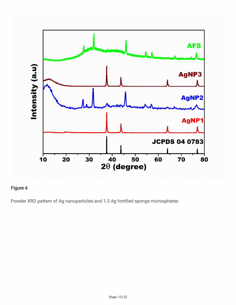
The crystalline phase and purity of the as-synthesized AgNP materials were examined by PXRD analysisand combined patterns are shown in Fig.4. for AgNP1 all the diffraction peaks can be indexed to anorthorhombic phase of Ag nano spheres and is good consistence with the standard JCPDS card no. [04-0783]. There are no other signi�cant impurities were identi�ed which revealed that successful formationpure AgNP1 but small hump in between the angles 10 and 20. This indicates the presence of bioactivematerials from marine sponge but not fully intermixed. Then the XRD pattern of AFS shows the presenceof organic molecules overlapped with Ag. The four high intense peaks can be attributed to the (111),(200), (220), and (311) planes of AgNP respectively. The average crystallite size was calculated byScherer’s formula12 and the size is to be nearly10-20 nm.
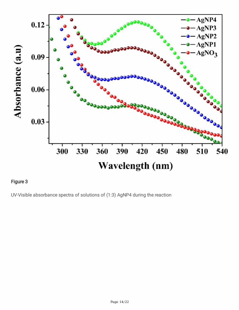
Fig. 3 shows that the complete surface Plasmon resonance (at 450 nm) detected in AFs while comparingto AgNPs in UV-Visible spectra of both. Fig.1 represents the FTIR spectrum of AFS and the peaks wereobserved at 995, 871 and 580 cm-1 which can be ascribed to metal nanoparticles. The band observed at1741 and 1649cm-1 were related the C=O and –OH bending modes, respectively. The bands at 1514 and1400 cm-1were corresponding to the C–C aromatic vibrations and C–H deformation vibrations. The sharpband at 1153 cm-1 can be attributed to a carbonyl group such as in aldehyde or ketone group.
The surface morphology of the as-synthesized AgNPs 1-4 was observed using Scanning electronmicroscopy (SEM) analysis and presented in Fig. 5 and 6. The SEM images displayed the sphere-likestructure of Ag nanoparticles in both AgNP (Fig. 5) and AFS (Fig 6) with relatively smooth surfaces. Theaverage diameter is ~20 nm and the diameter of sponge sphere is to be 200 nm. In Fig. 6, All AgNPssynthesised using various stoichiometric ratios of AgNO3 and extract II solutions were exhibiting sameshape but with slight differences in size and Ag particle distributions.
The elemental compositions were investigated by Energy dispersive x-ray studies (EDX) in Fig. 7 whichrevealed that the presence of Ag, C, N and O elements in AFS. No other signi�cant elements wereidenti�ed which proved the presence of bioactive organic compounds which stablishing the silvernanoparticles on its surface. Fig. 8 exhibits the elemental mapping of AgNP4 composite. The red colourimage was showing that the spectra of silver which was clearly spread over the sponge microsphere. Thegreen colour image shows the presence of carbon in equivalent quantity with silver. The blue and yellowcolour images were acquired for nitrogen and oxygen elements respectively. The presence of theelements carbon, oxygen and nitrogen shows the microsphere is derived from complete organicsubstances of marine sponge material. Moreover, the transmission electron microscopy with differentmagni�cations in Fig 9 (A & B) further con�rmed the successful formation of sphere-like AgNP2 andsuccessful Ag forti�cation on sponge sphere and formation as AFS.
3.2. Anti-Microbial assay
In this present investigation, we evaluated the anti-microbial activity of as-synthesized AFS with differentconcentrations, sodium molybdate and stem extracts in various organisms like Bacillus subtilis(B.subtilis), E. coli, Staphylococcusaureus (S.aureus) and Pseudomonas aeruginosa (P.aeruginosa)

Page 8/22
colonies on nutrient agar plates supplemented with AFS and the results are shown in Fig.8. The standardantibiotic drug streptomycin was used as a positive control. The zone of inhibitions with differentconcentrations of AFS and marine sponge powder extract was carried out and the results are shown inFig.5 and were tabulated in �g. 8.
The results demonstrated that the zone of bacterial inhibition by AFS prepared from marine spongeextract show maximum inhibition for gram positive B.Subtilis which may be concluded from the fact thatthe particles had the smallest diameter than those prepared from other methods, which in turn exhibitedequal antimicrobial property. From the above results, we can conclude that AFS exhibits excellent anti-bacterial activity than the pure sponge extract. Hence, the as-synthesized AFS shows remarkableantimicrobial performances towards the clinical isolates.
3.3. In vitro cytotoxicity studies
Cytotoxic effect of marine sponge and AFS on HELA cells was evaluated using MTT assay. As shown inFig. 11 marine sponge and AFS caused a dosage dependent inhibition of cell proliferation towards HELAcells. The cell viability was recorded as 94 % and 85% in marine sponge at 20 µg/ml and 40 µg/mlconcentrations and the maximum decrease in cell viability were measured as 45 % at 140 µg/ml at 24 h.Marine sponge show lesser toxic effect due to the biological origin. In contrast, AFS was recorded highcytotoxic at low concentration and the IC50 value was 94 µg/ml. The maximum decrease of 24 % cellviability was measured after treatment with 140 µg/ml of AFS. Combination of AFS and the presence ofmarine sponge components, the cytotoxic effects were increased at lower concentrations38.
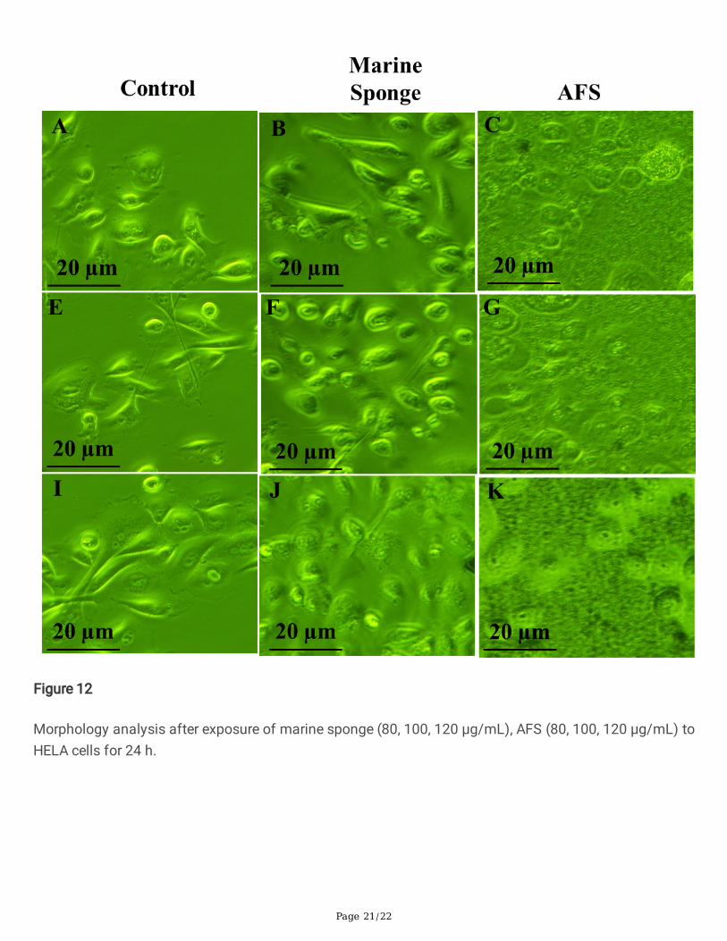
3.4. Morphological assessment
As shown in Fig. 12, the untreated HELA cells maintained their original morphology and attached properlyeven when the incubation was 24 h. In contrast, HELA cells lost their original shape at 24 h of 100 µg/mLmarine sponge treatment and it has not shown maximum damage at 80 µg/mL. The HELA cells had losttheir elongated spindle-shape morphology when it was treated with AFS at low concentration (80 µg/mL).When the AFS treatment of 100 µg/mL concentration, suspension cells (dead cells) were identi�ed andmore suspension cells were observed at 120 µg/mL. AFS were damage more cells in HELA cancer cellscompare to marine sponge.
3.5. Cellular uptake
The AgNPs cancer cells uptake was observed by �uorescent microscopy (Fig. 5). Silver nanoparticleuptake was found to be more particles in HELA cells based on the concentration. In initial concentrationof 1µM�uorescent intensity in the cancer cell was less over the time. AgNPs was penetrated into the cellvia passive diffusion and 20 nm size nanoparticles can be easily internalized deeply into tumor cells.FITC-AgNPs showed aincreasing green �uorescent signal for 3 µM and 4 µM and nuclear region wereconsist of smaller particles. It suggests the concentration-dependent cellular uptake directly localizematerial to nucleus.

Page 9/22
ConclusionIn conclusion, this is the �rst one pot phyto-synthesis of AFS using the marine sponge Clathriafrondiferaextract and as mentioned method is simple, rapid, stable and eco-friendly. The crystallinity andmorphologies were con�rmed by XRD, SEM, TEM and EDX analysis. The functional chemical groupsinvolved during the synthesis which was identi�ed through FTIR studies and further con�rmed with GC-MS data. The synthesized AFS showed an excellent antimicrobial e�cacy towards the both gram positiveand gram negative pathogens. AgNPs have higher cellular uptake, it induced a concentration-dependentcytotoxicity by increasing ROS level and damage the cells. In the recent progress and the on-going effortsin improving particle synthesis e�ciency and exploring their biomedical applications, it is hopeful that theexecution of the approach of ours on a large scale and their commercial applications in medicine andhealth care will be very much useful in the upcoming years. Hence, further rigorous studies are required toutilize these nanoparticles for their use in various formulations such as in bandages, ointments, gels,lotions and all other formulations in intradermal applications for problems arising from skin cancer orhealing process of the epidermal layers of the body.
References1. A. Shakeri and A. Sahebkar, Anti-Cancer Products from Marine Sponges: Progress and Promise,
Recent Patents on Drug Delivery & Formulation, 2015, 9(3), 1-2.
2. S. Ravichandran, K. Kathiresan and H. Balaram, Anti-malarials from marine sponges. Biotechnol MolBiol Rev 2007; 2(2): 03.
3. L. Fieseler, M. Horn, M. Wagner and U. Hentschel, Discovery of the novel candidate phylum“Poribacteria” in marine sponges. Appl Environ Microbiol 2004; 70(6): 3724-32.
4. A. Mollica, M. Locatelli, A. Stefanucci and F. Pinnen, Synthesis and bioactivity of secondarymetabolites from marine sponges containing dibrominated indolic systems, Molecules 2012, 17(5):6083-99.
5. Y. Sumii, N. Kotoku, A. Fukuda, T. Kawachi, M. Arai and M. Kobayashi, Structure-activity relationshipand in vivo antitumor evaluations of dictyoceratin-A and -C, hypoxia-selective growth inhibitors frommarine sponge, Marine Drugs, 2015, 13(12), 7419–7432.
�. D. T. A. Youssef, L. A. Shaala and K. Z. Alshali, “Bioactive hydantoin alkaloids from the Red Seamarine sponge Hemimycale arabica,” Marine Drugs, 2015, 13(11), 6609–6619.
7. D. Inbakandan, R. Venkatesan and S. Ajmal Khan, Colloid. Surf. B: Biointerface.,2010, 81, 634–639.
�. R. Hamed, M. H. Givianrad and A. M. Moradi, Orient. J. Chem., 2015, 31, 1961-1967.
9. M. R. Hamed, M. H. Givianrad and A. M. Moradi, Ind. J. Geo. Marine. Sci., 2017, 46, 125-130.
10. Y. N. Shkryl, G. N. Veremeichik, D. G. Kamenev, T. Y. Gorpenchenko, Y. A. Yugay, D. V. Mashtalyar, A. V.Nepomnyaschiy, T. V. Avramenko, A. A. Karabtsov, V. V. Ivanov, V. P. Bulgakov, S. V. Gnedenkov, Y. N.Kulchin and Y. N. Zhuravlev, Nanomed.Biotechnol., 2017, 46, 1646-1658.
11. G. Radhika, R. Venkatesan and S. Kathiroli, Ind. J. Marine. Sci., 2007, 36, 235-238.

Page 10/22
12. K. Saravanakumar, B. Ramkumar andV. Muthuraj, Int. J. Res. Phar. Chem., 2016, 6, 458-464.
13. S. H. Hassanpour and M. Dehghani, Review of cancer from perspective of molecular, Journal ofCancer Research and Practice, 2017, 4, 127-129
14. A. G. Waks and E. P. Winer, Breast Cancer Treatment, JAMA, 2019, 321(3), 288-300.
15. A. F. Santos, D. R. Q. Almeida, L. F. Terra, M. S. Baptista and L. Labriola, Photodynamic therapy incancer treatment – an update review, J Cancer Metastasis Treat, 2019, 5(25) 1-20.
1�. H. Yang, R. M. Villani, H. Wang, M. J. Simpson, M. S. Roberts, M. Tang and X. Liang, The role ofcellular reactive oxygen species in cancer chemotherapy, Journal of Experimental & Clinical CancerResearch, 2018, 37(266),
17. C. Thallinger, T. Fureder, M. Preusser, G. Heller, L. Mullauer, C. Holler, H. Prosch, N. Frank, R.Swierzewski, W. Berger, U. Jager and C. Zielinski, Review of cancer treatment with immunecheckpoint inhibitors, wiener klinische wochenschrift, 2018, 130, 85-91.
1�. R. Misra, S. Acharya, S. K. Sahoo, Cancer nanotechnology: application of nanotechnology in cancertherapy, Drug Discovery Today, 2010, 19-20, 842-850.
19. J. Shi, P. W. Kantoff, R. Wooster and O. C. Farokhzad, Cancer nanomedicine: progress, challenges andopportunities, Nature reviews: cancer, 2017, 17, 21-37.
20. V. P. Chauhan and R. K. Jain, Strategies for advancing cancer nanomedicine, Nature Materials, 2013,12,958–962.
21. A. Wicki, D. Witzigmann, V. Balasubramanian, J. Huwyler, Nanomedicine in cancer therapy:Challenges, opportunities, and clinical applications, Journal of Controlled Release, 2015, 200, 138-157.
22. P. Kumar, M. Govindaraju, S. Senthamilselvi and K. Premkumar, Colloid. Surf. B. Biointerface., 2013,103, 658– 66.
23. S. Y. Seo, G. H. Lee, S. G. Lee, S. Y. Jung, J. O. Lim and J. H. Choi, Carbohydr. Polym., 2012, 90, 109-115.
24. A. Aravinthan, M. Govarthanan, K. Selvam, L. Praburaman, T. Selvankumar, R. Balamurugan, S.Kamala-Kannan and J. Kim, Int. J. Nanomed., 2015, 10, 1977–1983.
25. N. Vigneshwaran, R. P. Nachane, R. H. Balasubramanya and P. V. Varadarajan, Carbohyd. Res., 2006,341, 2012–2018.
2�. S. Dinesh, S. Karthikeyan and P. Arumugam, Elixir Optical Mater., 2012, 44, 7364-7366.
27. P. Banerjee, M. Satapathy, A. Mukhopahayay and P. Das, Bioresources and Bioprocessing, 2014, 1, 1-10.
2�. S. Singh, J. P. Saikia and A. K. Buragohain, Colloid. Surf. B: Biointerface.,2013, 102, 83–85.
29. N. Asmathunisha and K. Kathiresan, Colloid. Surf. B,2013, 103, 283-287.
30. N. Vigneshwaranm, N. M. Ashtaputre, P. V. Varadarajan, R. P. Nachane, K. M. Paralikar and R. H.Balasubramanya, Mater. Lett., 2007, 61, 1413–1418.
Page 11/22
31. N. S. Shaligram, M. Blue, R. Bhambure, R. S. Singhal, S. K. Singh, G. Szakacs and A. Pandey, Proc.Biochem., 2009, 44, 939-943.
32. M. Vivek, P. S. Kumar, S. Ste�, S. Sudha, Avicenna J. Med. Biotechnol., 2011, 3, 143-148
33. M. H. Givianrad, T. Sadeghi, K. Larijani and S. E. Hosseini, J. Food Tech. Nutr., 2011, 30, 38-44.
34. R. Singh, S. K. Sahu and M. Thangaraj, J. Nanoparticle., 2014, 718240, 1-7.
35. N. Asmathunisha and K. Kathiresan, Colloid. Surf. B: Biointerface.,2013, 103, 283–287.
3�. K. Govindaraju, V. Kiruthiga, G. V. Kumar and G. Singaravelu, J. Nanosci. Nanotechnol., 2009, 9, 5497-5501.
37. D. Inbakandan, G. Sivaleela, D. Magesh Peter, R. Kiurbagaran, R. Venkatesan and S. Ajmal Khan,Mater. Lett.,2012, 87, 66–68.
3�. M. Jeyaraj, M. Rajesh, R. Arun, D. Mubarak Ali, G. Sathishkumar, G. Sivanandhan, G. Kapil Dev, M.Manickavasagam, K. Premkumar, N. Thajuddin and A. Ganapathi, Colloids Surf. B., 2013, 102, 708-717.
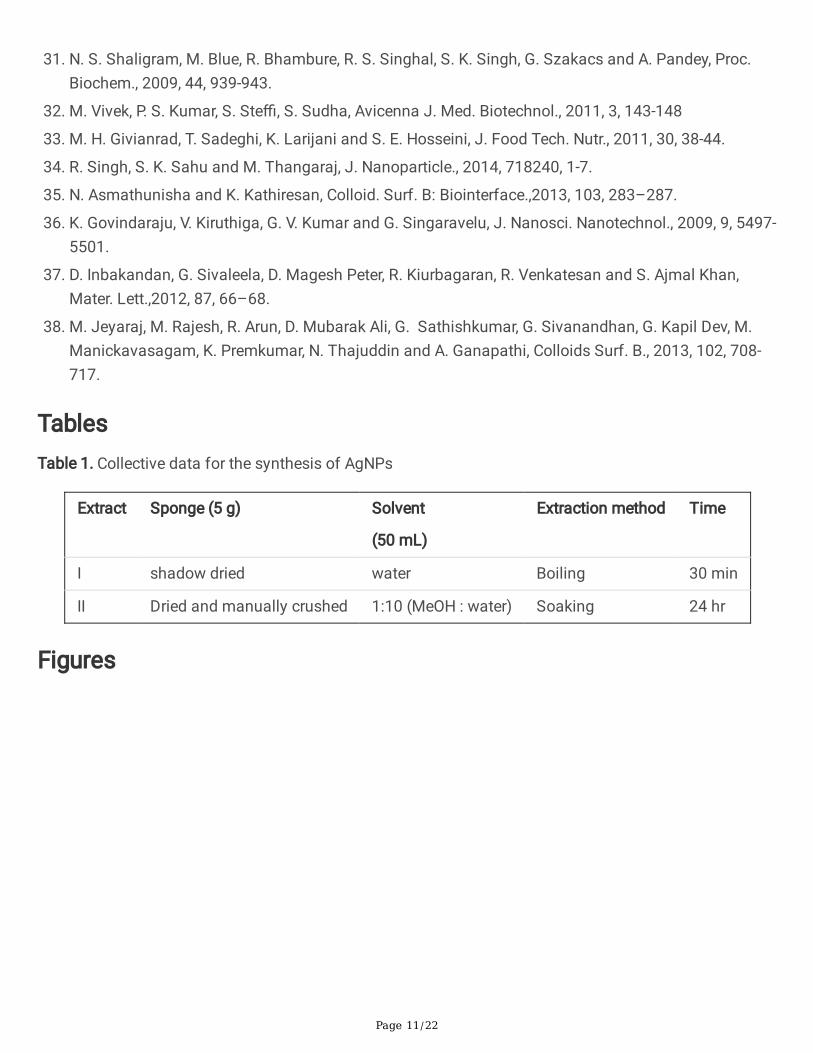
TablesTable 1. Collective data for the synthesis of AgNPs
Extract Sponge (5 g) Solvent
(50 mL)
Extraction method Time
I shadow dried water Boiling 30 min
II Dried and manually crushed 1:10 (MeOH : water) Soaking 24 hr
Figures

Page 12/22
Figure 1
FTIR spectrum of crushed and sieved �ne powder of Clathria frondifera marine sponge
Page 13/22
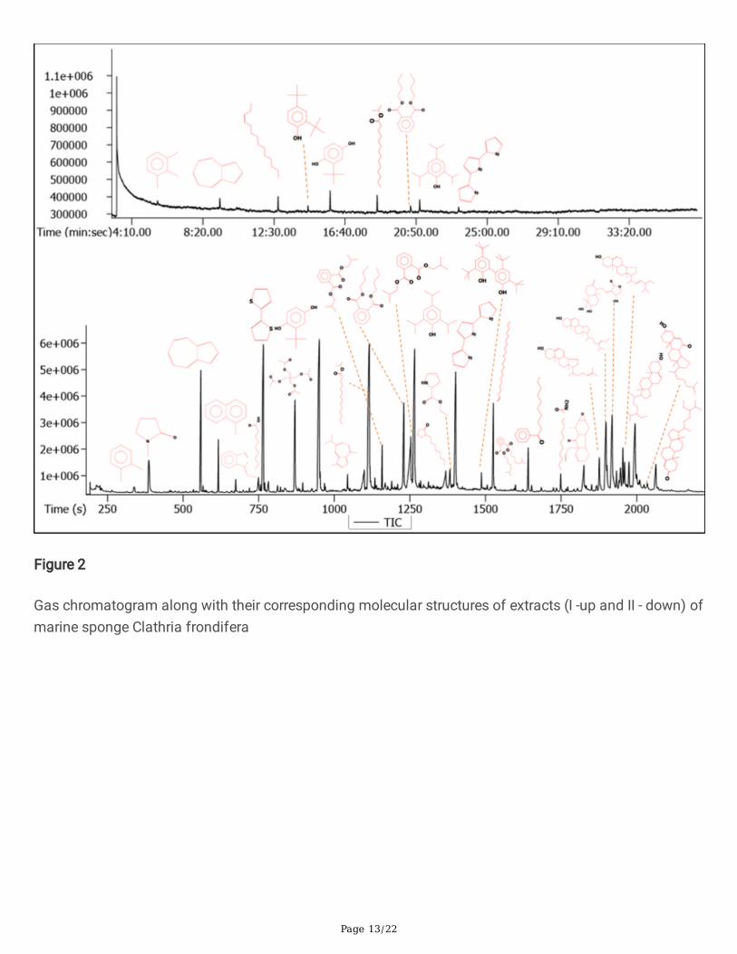
Figure 2
Gas chromatogram along with their corresponding molecular structures of extracts (I -up and II - down) ofmarine sponge Clathria frondifera
Page 14/22
Figure 3
UV-Visible absorbance spectra of solutions of (1:3) AgNP4 during the reaction
Page 15/22
Figure 4
Powder XRD pattern of Ag nanoparticles and 1:3 Ag forti�ed sponge microspheres

Page 16/22
Figure 5
SEM images of AgNP1 (a,b), AgNP2 (c,d), and AgNP3 (e,f)

Page 17/22
Figure 6
SEM images of silver forti�ed marine sponge microspheres (AgNP4) synthesized using variousstoichiometric ratios of AgNO3 and extract II solutions (a) 1:1, (b)1:2 (c, d)1:3 (e) 2:1 (f) 3:1

Page 18/22
Figure 7
EDAX spectrum of 1:3 Ag forti�ed marine sponge microspheres of marine sponge Clathria frondifera
Figure 8
Elemental mapping images of 1:3 Ag forti�ed marine sponge microspheres (silver - red, oxygen – yellow,carbon – green, nitrogen – blue)

Page 19/22
Figure 9
TEM images of 3:1 AgNP4 nanoparticles (a, b) and 1:3 Ag forti�ed sponge microspheres (c, d)
Figure 10

Page 20/22
Antimicrobial activity of AFS against (A)Escherichia coli, (B) Bacillus subtilis, (C) Staphylococcus aureusand (D) Pseudomonas aeruginosa clinical isolates
Figure 11
Cell viability studies after exposure of marine sponge and AFS to HELA cells for 24 h.
Page 21/22
Figure 12
Morphology analysis after exposure of marine sponge (80, 100, 120 µg/mL), AFS (80, 100, 120 µg/mL) toHELA cells for 24 h.

Page 22/22
Figure 13
Cellular uptake study of FITC-AgNPs(A) 1 µM (B) 2 µM (C) 3 µM (D) 4 µM
Supplementary Files
This is a list of supplementary �les associated with this preprint. Click to download.
Tableofcontent.docx


