Embed Size (px)
Citation preview
Molecules 2009, 14, 3662-3675; doi:10.3390/molecules14093662
molecules ISSN 1420-3049
www.mdpi.com/journal/molecules
Article
Adding Chemical Cross-Links to a Physical Hydrogel
Gaio Paradossi *, Ivana Finelli, Barbara Cerroni and Ester Chiessi
Dipartimento di Scienze e Tecnologie Chimiche, Università di Roma Tor Vergata and CNR – SOFT,
Italy; E-Mails: [email protected] (I.F.); [email protected] (B.C.);
[email protected] (E.C.)
* Author to whom correspondence should be addressed; E-mail: [email protected].
Received: 30 June 2009; in revised form: 27 August 2009 / Accepted: 16 September 2009 /
Published: 17 September 2009
Abstract: Synergistic hydrogels are often encountered in polysaccharide mixtures widely
used in food and biopharma products. The xanthan and konjac glucomannan pair provides
one of the most studied synergistic hydrogels. Recently we showed that the junction zones
stabilizing the 3D structure of this gel are present as macromolecular complexes in solution
formed by the partially depolymerised polysaccharidic chains. The non-covalent
interactions stabilizing the structure of the polysaccharidic complex cause the melting of
the ordered structure of the complex in the solution and of the hydrogels. Introduction of
chemical cross-links in the 3D structure of the synergistic hydrogel removes this
behaviour, adding new features to the swelling and to the viscoelastic properties of the
cured hydrogel. The use of epichlorohydrin as low molecular weight cross-linker does not
impact unfavourably on the viability of NIH 3T3 fibroblasts.
Keywords: hydrogels; swelling; biocompatibility; polysaccharides
1. Introduction
Xanthan, a microbial polysaccharide produced by the bacterium Xanthomonas campestris, has the
remarkable feature of forming physical hydrogels when it is mixed with gluco- or galactomannans
[1-3]. These hydrogels are usually called “synergistic gels” as their formation involves specific
interactions between these two types of polysaccharides, whereas the mixing of xanthan with other
polysaccharides at similar concentrations does not provide a gel phase.
This effect indicates that a specific interaction pattern occurs between the chains of xanthan with
either glucomannan, a copolymer of 1,4-linked -D-mannose and -D-glucose, or galactomannan, a
OPEN ACCESS

Molecules 2009, 14
3663
1,4-linked -D-mannose with α-D-galactose side-chains [2,4]. Investigations on hydrogels based on the
combined presence of xanthan, or differently modified xanthan, and several other (bio)polymers
demonstrate the interest in adding new features to materials with known performances.
Poly(acrylamide-grafted-xanthan)-based pH-sensitive hydrogel beads have been considered for enteric
delivery of ketoprofen [5]
In a previous paper [6], we have studied the solution behaviour of the depolymerised xanthan and
konjac glucomannan; hereafter named KGM, a complex where the involvement of side-chains
appeared as a relevant factor for the establishment of the macromolecular interactions. Similar
conclusions were drawn in an earlier former paper by Annable et al. [7] on the basis of DSC and EPR
experiments indicating in the reduction of the xanthan-water contacts the driving force stabilizing the
synergistic hydrogel.
In this work we describe the effect of introducing chemical cross-links in the synergistic hydrogels
in order to add new properties to the starting xanthan-KGM hydrogels. As expected based on rubber
elasticity theory [8], the swelling behaviour, the storage and loss moduli will be affected by such
modification, as well as the hampering of the hydrogel melting. Addition of chemical cross-links to the
xanthan–KGM physical network was accomplished using epichlorohydrin, a very reactive difunctional
molecule often recognized for its toxicity. However, it should be considered that epichlorohydrin is
presently used as additive in paper products for food industry and in water treatment [9]. We report
here on two xanthan/konjac glucomannan hydrogels, hereafter named XGE1 and XGE2 cured with
different epichlorohydrin concentrations, i.e., 0.6 M and 1.4 M, and the impact of epichlorohydrin used
as cross-linker on the viability of fibroblasts NIH 3T3 seeded on the chemically cross-linked hydrogels
was assessed.
2. Results and Discussion
The characterization of xanthan and KGM polysaccharides was performed on intact and partially
depolymerised samples. For the study in solution glucomannan was hydrolyzed with cellulase having
endo-activity for the (1→4)glucosic linkages. The chain degradation was carried out in acetate buffer
at pH 4.5 at 37 °C in the presence of 0.1% (w/v) enzyme and monitored by measuring the shear times
of the KGM solution in a Ubbelhode suspended flow capillary viscosimeter. After 1 hour the
degradation was completed, with a 3-fold drop in the relative viscosity, t/t0, as shown in Figure 1.
Figure 1. Enzymatic degradation of KGM by cellulase.
10 20 30 40 50 60 70 80 90 1001,0
1,5
2,0
2,5
3,0
3,5
t/t 0
degradation time (min)
Molecules 2009, 14
3664
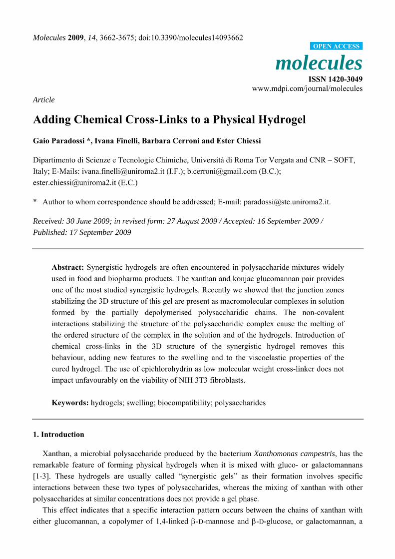
Whether the enzyme degradation affects the chemical structure of depolymerised KGM was
addressed by 13C-NMR. Figure 2 shows the 13C-NMR spectra of KGM samples degraded for 10
(spectrum a) and for 60 minutes (spectrum b). Assignment of the resonances was based on the
literature [6,10]. Integration of peaks at 101.7 and at 104 ppm, relative to non-reducing anomeric
carbons of mannose and glucose, respectively, gives a value of the mannose:glucose ratio of 1.7. As 13C-NMR is not commonly used for quantitative determinations, this result was validated by 1H-NMR
(not shown) giving similar results. The same mannose:glucose ratio was also found for native KGM.
Figure 2. 13C-NMR spectra of (a) KGM after 10 min degradation and (b) KGM after 60
min degradation.
ppm (t1)60708090100
-500
0
500
1000
1500
1.0
0
1.6
4
1.0
3
1.6
6
3.8
0
1.3
1
1.7
9
1.9
1
2.7
6
ppm (t1)60708090100
0
500
1000
15001
.00
1.7
4
0.8
8
1.5
7
4.0
3
1.2
3
1.9
2
1.9
8
2.8
6
Xanthan was depolymerised by high power sonication for four hours. Molecular parameters for
depolymerised xanthan and KGM polysaccharides are summarized in Table 1. The interaction between
xanthan and KGM in water was monitored by circular dichroism, CD, on partially depolymerised
(a)
(b)
Molecules 2009, 14
3665
chains, as the use of native (intact) polymers at the investigated concentrations is not suitable for CD
study due to sudden formation of the gel phase. The contribution of KGM to the total ellipticity is
negligible. As the carboxylic groups beared in the xanthan side-chains are the CD chromophores, the
spectrum reflects the conformational state of xanthan side-chains [6] complexing the glucomannan. A
melting curve with midpoint at about 45 °C shows an order ↔ disorder transition revealing that the
synergistic interaction is coupled with the ordering of xanthan side-chains (see Figure 3).
Table 1. Molecular parameters of Xanthan and KGM.
Xanthan sonicated 4h Native KGM KGM degraded 10min
Mw = 620,000 g/mol A2 = 6.7·10-04 cm3mol/g2 Rg = 98 nm dn/dc = 0.17 mL/g
Mw = 960,000 g/mol A2 = 3·10-05 cm3mol/g2 Rg = 100 nm dn/dc = 0.15 mL/g
Mw = 560,000 g/mol A2 = 4.3·10-05 cm3mol/g2 Rg = 70 nm dn/dc = 0.15 mL/g
Figure 3. (a) Circular dichroism spectra of partially depolymerised xanthan-KGM (1:1
w/w) aqueous solution (b) and ellipticity at 210 nm (b) as a function of temperature (step
10 °C).
-10
0
10
20
30
200 210 220 230 240 250 260 270 280
(nm)
20 °C
80 °C
-10
-5
0
5
10
15
20
25
10 20 30 40 50 60 70 80 90
T (°C)
(a)
(b)
Molecules 2009, 14
3666
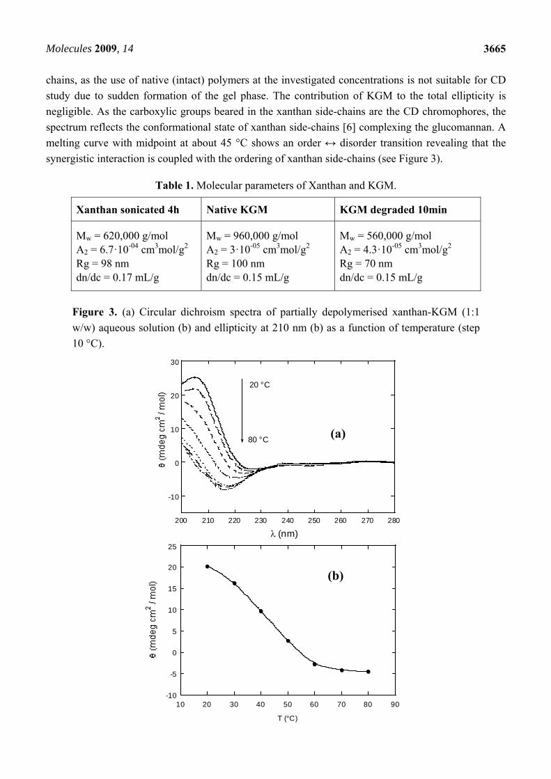
The gel formation was monitored by small strain oscillatory shear measurements, recording the
time dependence of G’ and G”. The time dependence of G’ in synergistic xanthan/KGM hydrogels
during the curing with epichlorohydrin was monitored at 1 Hz (see Figure 4). Storage modulus was
always higher than the loss modulus, a feature indicating clearly that the cross-linking reaction starts
and is carried out in an existing synergistic gel phase provided by the mixing of the two
polysaccharides. G’ shows the typical trend of a cross-linking reaction: An induction time, present for
any amount of epichlorohydrin, although the extent of this phase depends on the cross-linker
concentration, followed by a stage where G’ increases linearly, and a constant asymptotic behaviour at
long times.
Figure 4. Kinetics of the cross-linking of xanthan-KGM mixture by epichlorohydrin.
Initial cross-linker concentration: () 0.1 M; () 0.3 M; () 0.6 M; () 1.4 M.
0
100
200
300
400
500
0 4 8 12 16
G' (
Pa)
t (h)
The swelling and elastic properties of the synergistic hydrogels stabilized by chemical cross-links
depend on the amount of polymer effectively incorporated in the hydrogel contributing to the osmotic
(included Donnan) and elastic terms of water activity. The gel and the soluble amounts, Wgel and Wsol
(see Experimental section for definitions), account for the effective incorporation of the chains of the
two polysaccharides in the network.
Introduction of chemical cross-links adds new features to the hydrogels. The water weight fraction,
S, defined as (Ws – Wgel)/Wgel with Ws representing the weight of swollen hydrogel (see Experimental
section) was monitored at different times during the swelling/de-swelling process of the hydrogel in a
pH cycle, revealing the polyelectrolyte behaviour of xanthan chains in the hydrogel, as shown in
Figure 5. A responsive behaviour to ionic strength changes, as a consequence of the Debye screening
effect of the xanthan charges confined in the hydrogel, is also detected when the hydrogel is
equilibrated in 0.1 M NaCl.

Molecules 2009, 14
3667
Figure 5. Swelling/de-swelling behaviour: water weight fraction, S, in XGE hydrogels
stored sequencially in HCl 10-3 M, water, 0.1M NaCl. Empty symbols: XGE1; full symbols
XGE2.
0 5 10 15 20 25 30 3540
60
80
100
120
S
t(h)
Storage modulus, G’, of chemically cross-linked synergistic hydrogels was used to assess the
density of cross-links contributing to the elasticity of the system. According to modified rubber
elasticity theory [8,11] to take into account that the cross-linking reaction was carried-out in the
“nascent” state before the swelling process, the cross-links density in this state, e/V0, is related to G’
according to equation 1:
31
2
0
0
'
RT
G
Ve (1)
where φ0 and φ2 are the polymer volume fractions in the relaxed state, i.e. after cross-linking and
before the swelling, and after the swelling, respectively, and Q is the degree of swelling defined as
(1/φ2). Equation 1 holds for affine networks, i.e. ideal networks where all cross-links have the same
contribution to the macroscopic elasticity and chain deformations are the same as the macroscopically
imposed strain. In Table 2 the swelling features of the hydrogels are summarized in water, at pH 3 and
in NaCl 0.1 M.
Table 2. Swelling parameters of XGE hydrogels.
Hydrogel Wgel(g) W0(g) Wsol(g) Ws(g) φ0 φ2 Q XGE1 0.042 0.16 0.12 5.4 0.015 0.006 167 XGE2 0.055 0.31 0.26 3.3 0.020 0.013 77
XGE1 pH 3 0.042 0.16 0.12 4.4 0.015 0.007 143 XGE2 pH 3 0.055 0.31 0.26 3.0 0.020 0.014 71
XGE1 NaCl 0.1M 0.068 0.16 0.092 4.4 0.025 0.011 91 XGE2 NaCl 0.1M 0.073 0.31 0.24 3.1 0.026 0.018 56
pH 3 water
NaCl
Molecules 2009, 14
3668
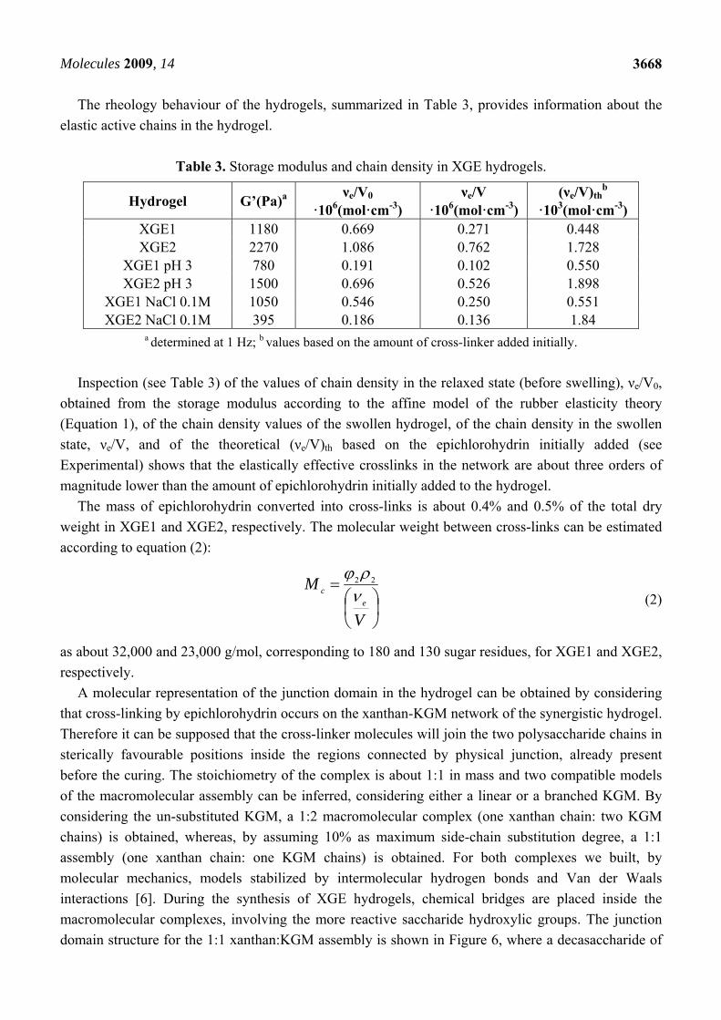
The rheology behaviour of the hydrogels, summarized in Table 3, provides information about the
elastic active chains in the hydrogel.
Table 3. Storage modulus and chain density in XGE hydrogels.
Hydrogel G’(Pa)a νe/V0 ·106(mol·cm-3)
νe/V ·106(mol·cm-3)
(νe/V)thb
·103(mol·cm-3) XGE1 1180 0.669 0.271 0.448 XGE2 2270 1.086 0.762 1.728
XGE1 pH 3 780 0.191 0.102 0.550 XGE2 pH 3 1500 0.696 0.526 1.898
XGE1 NaCl 0.1M 1050 0.546 0.250 0.551 XGE2 NaCl 0.1M 395 0.186 0.136 1.84
a determined at 1 Hz; b values based on the amount of cross-linker added initially.
Inspection (see Table 3) of the values of chain density in the relaxed state (before swelling), νe/V0,
obtained from the storage modulus according to the affine model of the rubber elasticity theory
(Equation 1), of the chain density values of the swollen hydrogel, of the chain density in the swollen
state, νe/V, and of the theoretical (νe/V)th based on the epichlorohydrin initially added (see
Experimental) shows that the elastically effective crosslinks in the network are about three orders of
magnitude lower than the amount of epichlorohydrin initially added to the hydrogel.
The mass of epichlorohydrin converted into cross-links is about 0.4% and 0.5% of the total dry
weight in XGE1 and XGE2, respectively. The molecular weight between cross-links can be estimated
according to equation (2):
V
Me
c 22
(2)
as about 32,000 and 23,000 g/mol, corresponding to 180 and 130 sugar residues, for XGE1 and XGE2,
respectively.
A molecular representation of the junction domain in the hydrogel can be obtained by considering
that cross-linking by epichlorohydrin occurs on the xanthan-KGM network of the synergistic hydrogel.
Therefore it can be supposed that the cross-linker molecules will join the two polysaccharide chains in
sterically favourable positions inside the regions connected by physical junction, already present
before the curing. The stoichiometry of the complex is about 1:1 in mass and two compatible models
of the macromolecular assembly can be inferred, considering either a linear or a branched KGM. By
considering the un-substituted KGM, a 1:2 macromolecular complex (one xanthan chain: two KGM
chains) is obtained, whereas, by assuming 10% as maximum side-chain substitution degree, a 1:1
assembly (one xanthan chain: one KGM chains) is obtained. For both complexes we built, by
molecular mechanics, models stabilized by intermolecular hydrogen bonds and Van der Waals
interactions [6]. During the synthesis of XGE hydrogels, chemical bridges are placed inside the
macromolecular complexes, involving the more reactive saccharide hydroxylic groups. The junction
domain structure for the 1:1 xanthan:KGM assembly is shown in Figure 6, where a decasaccharide of
Molecules 2009, 14
3669
KGM and a xanthan chain of five repeating units are bound by two epichlorohydrin bridges (yellow
atoms) between C6 carbon atoms of the KGM and of xanthan backbone chains. The complex
organizes in a double helical arrangement with antiparallel chains orientation and the average distance
between backbones is about 8 Å. The cross-linking degree shown in Figure 6 (one epichlorohydrin
every 18 sugar residues) does not reproduce the experimental picture as the image provides a
description of the junction with an atomic detail in a spatial domain of few nanometers. The presence
of chemical cross-links in XGE gels increases the hydrogel responsivity to sharp gradients of external
parameters as pH and ionic strength and allows a reversible behaviour of the system.
Figure 6. Molecular representation of the junction domain in XGE hydrogel.
Epichlorohydrin cross-links are evidenced in yellow. Xanthan and glucomannan chains are
shown in blue and red coloros, respectively. Hydrogen atoms are not shown.
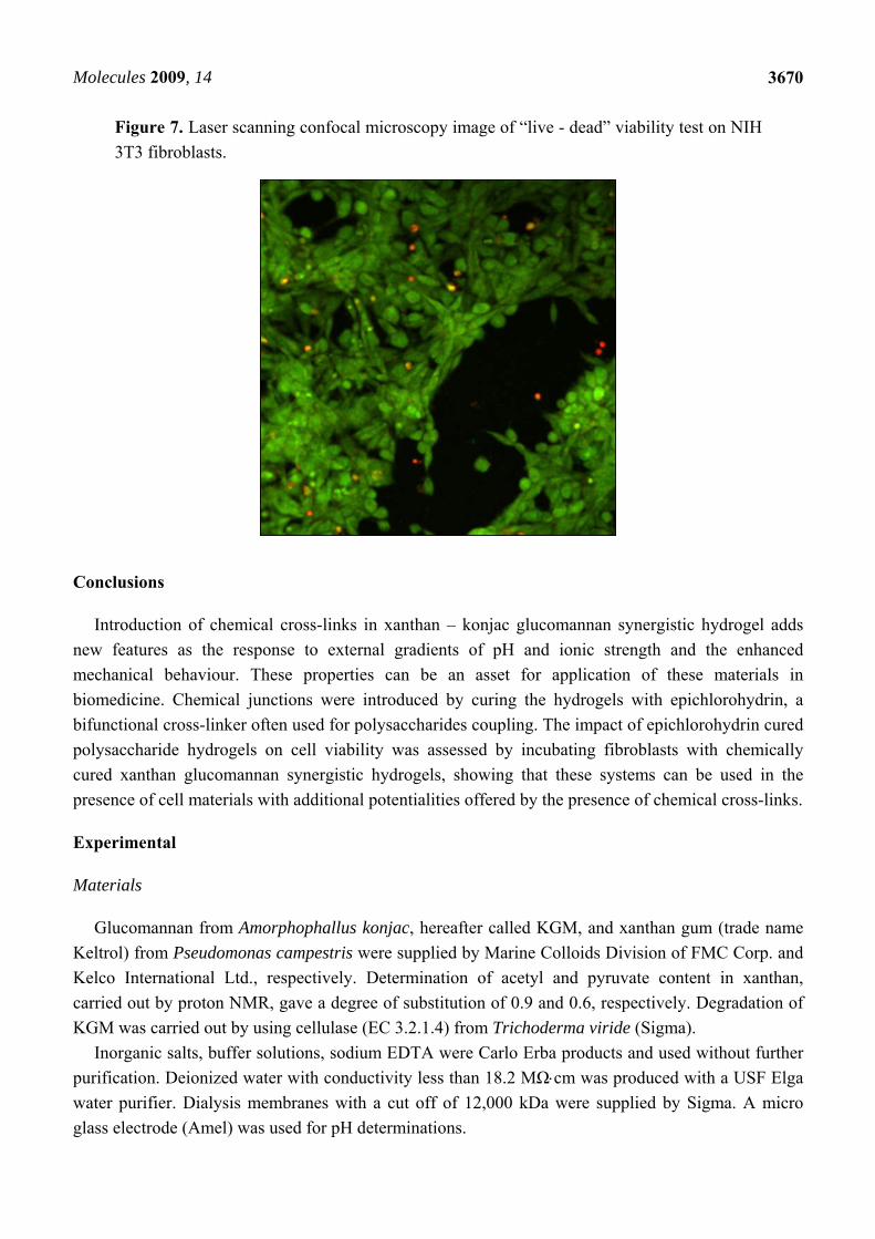
Cytotoxicity of xanthan/KGM chemically cross-linked synergistic hydrogels was evaluated by
seeding murine fibroblasts NIH 3T3 on the surface of XGE1, cured with 0.6 M epichlorohydrin. Cell
viability was tested with “Live-Dead” assay. In this test live cells display a green colour in
fluorescence microscopy mode, whereas red stained cells indicate dead cells. Figure 7 illustrates the
fibroblast viability after 12 h incubation on the surface of XGE1 hydrogels in the presence of a clear
predominance of live cells is detected with the typical morphology of adhered NIH 3T3 cells. It can be
concluded that the low epichlorohydrin content acting as cross-linker in the hydrogel, is not
unfavourable to cells viability as a very low cross-linking density is enough to add responsivity
features to physical synergic hydrogels.
Molecules 2009, 14
3670
Figure 7. Laser scanning confocal microscopy image of “live - dead” viability test on NIH
3T3 fibroblasts.
Conclusions
Introduction of chemical cross-links in xanthan – konjac glucomannan synergistic hydrogel adds
new features as the response to external gradients of pH and ionic strength and the enhanced
mechanical behaviour. These properties can be an asset for application of these materials in
biomedicine. Chemical junctions were introduced by curing the hydrogels with epichlorohydrin, a
bifunctional cross-linker often used for polysaccharides coupling. The impact of epichlorohydrin cured
polysaccharide hydrogels on cell viability was assessed by incubating fibroblasts with chemically
cured xanthan glucomannan synergistic hydrogels, showing that these systems can be used in the
presence of cell materials with additional potentialities offered by the presence of chemical cross-links.
Experimental
Materials
Glucomannan from Amorphophallus konjac, hereafter called KGM, and xanthan gum (trade name
Keltrol) from Pseudomonas campestris were supplied by Marine Colloids Division of FMC Corp. and
Kelco International Ltd., respectively. Determination of acetyl and pyruvate content in xanthan,
carried out by proton NMR, gave a degree of substitution of 0.9 and 0.6, respectively. Degradation of
KGM was carried out by using cellulase (EC 3.2.1.4) from Trichoderma viride (Sigma).
Inorganic salts, buffer solutions, sodium EDTA were Carlo Erba products and used without further
purification. Deionized water with conductivity less than 18.2 MΩcm was produced with a USF Elga
water purifier. Dialysis membranes with a cut off of 12,000 kDa were supplied by Sigma. A micro
glass electrode (Amel) was used for pH determinations.

Molecules 2009, 14
3671
Xanthan solutions: Native xanthan was degraded by sonication using a VCX 400 sonicator (Sonics and
Materials Inc.) with a maximum output power of 400 W at a frequency of 20 kHz equipped with a
macrotip (TI-6AL-4V). The sonication was carried out with a sequence of 1 s pulse and 0.3 s standby
at approximately 80% of the maximum power. Five hundred mL batches of 0.6% (w/v) solution of
xanthan were added with 1% (v/v) of acetone as radical scavenger and solid NaCl to a concentration of
0.1 M. Sonications were carried out in an external water/ice bath in order to avoid overheating. After
sonication, solutions were centrifuged at 10,000 rpm at 2 °C for 1 h with a Beckman J2-21
ultracentrifuge equipped with a J14 rotor in order to separate titanium particles from tip erosion and
cell debris. Solutions were dialyzed repeatedly for one day against 0.03 M EDTA and then
exhaustively for a week against Milli-Q grade water at 4 °C. Xanthan solutions were stored at 4 °C at a
concentration of 5% (w/v). Before mixing with glucomannan solutions, xanthan was always heated at
60 °C for 1 hour.
Glucomannan solutions: Konjac glucomannan, hereafter indicated as KGM, was dispersed at a
concentration of 0.5 (w/v) in 0.1 M sodium acetate/acetic acid buffer at pH 4.5 under vigorous
mechanical stirring and autoclaved for 20 min at 120 °C.
Enzymatic degradation of KGM
To characterize the sugar composition of KGM, an enzymatic degradation was carried out as the
viscosity of the sonicated samples made impracticable the analysis by 13C-NMR spectroscopy. The
hydrolytic activity of cellulase on the β(1→4) glycosidic linkages of nearest neighbor glucose units
allowed a degradation of KGM polymer chains to oligosaccharides. Some β(1→4) mannanase activity
was also observed in this enzyme preparation. A typical enzymatic degradation was carried out as
follows: 0.4% (w/v) KGM in sodium acetate/acetic acid buffer 0.1 M at pH 4.5 was added at 37 °C
with an amount of enzyme equal to 0.1 of the polysaccharide mass. Enzyme was incubated at 37 °C for
1 h before addition to the polysaccharide solution. Enzymatic digestion was carried out for 10 min and
for 1 h and stopped by adding 2-propanol to a concentration of 60% (v/v) in order to precipitate the
saccharide moiety. The precipitated xanthan was washed with 2-propanol and dried. 13C-NMR determination was carried out with a Brüker AM400 working at 100.63 MHz on 5%
(w/v) solutions of degraded KGM in deuterated DMSO. Chemical shifts were determined with respect
to dimethylsulfoxide. The information directly obtained from the C1 region of the degraded KGM
spectra is based on the assignments available in the literature for glucomannan from cell walls of
endosperm of A. officinalis [7]. The resonances grouped around 104 ppm and around 101.6 ppm were
assigned to the nonreducing anomeric carbons of α-D-glucose residues and of the α-D-mannose
residues, respectively. C1 resonances of reducing glucose and mannose units ranged from 93.4 to
97.4 ppm. On this basis it was possible to evaluate the ratio mannose/glucose of KGM.
Circular dichroism
The spectra were recorded with a JASCO J600 spectropolarimeter in the UV range 200-280 nm
with quartz cells with an optical path of 0.5 and 0.1 cm. Temperature was controlled with a LAUDA
M3 thermostat.

Molecules 2009, 14
3672
Light scattering
A BIC200 photometer (Brookhaven, USA) equipped with a solid state laser emitting at 532 nm
was used in an angular range from 20 to 154 degrees. Decalin was used as refractive index matching
liquid. Measurements were carried out in 0.1 M NaCl with polymer concentrations ranging from
0.1 mg/mL to 7 mg/mL for sonicated xanthan, from 0.2 mg/mL to 3.5 mg/mL for native KGM and
from 1 mg/ml to 10 mg/mL for degraded KGM. Toluene was used as reference liquid using a Rayleigh
ratio, RT = 2.6 × 10⁻⁵ cm-1 at 532 nm. The polymer solution Rayleigh ratio, Rθ, was determined as: 2
00 )(
T
T
T n
nR
I
IIR
(3)
where Iθ is the scattering intensity of the sample at angle θ, I0 is the scattering intensity of the solvent,
IT is the scattering intensity of toluene with the refractive indexes of the solvent and of toluene, n0 and
nT, respectively.
The optical constant 422
0
2 /4 ANdcdnnK , where λ is the laser radiation wavelength, was
determined by measuring the differential refractive index, (dn/dc), by means of a BIC DNDC
(Brookhaven, USA) calibrated with KCl aqueous solutions at 532 nm. The molecular parameters
reported in Table 1 were extracted according to the data treatment suggested by Zimm [12]:
cA
RnM
R
Kc
g
w
2
2
22
0
2
2sin3
41
1
(4)
where c is the polymer concentration (w/v) and Mw, Rg and A2 are the weight average molecular
weight, the radius of gyration and second virial coefficient of the polymer, respectively.
Rheology measurements
Capillary viscosimetry measurements were carried out at 25 °C by using a suspended-flow
viscosimeter (Ubbelhode type) with a flow time for water of 40 s. Dynamic viscoelastic measurements
were performed with a AR 2000 Advanced Rheometer (TA Instruments) using a parallel plates
geometry (diameter 20 mm, gap 2 mm) with the fixed plate equilibrated at 25 °C. Hydrogels were
poured directly onto the plate of the instrument.
Two types of viscoelastic measurements were carried out:
1. deformation sweeps at a constant frequency (1Hz) to determine the maximum deformation
attainable by a sample in the linear viscoelastic range.
2. storage and loss moduli, G’ and G”, on swollen hydrogels were obtained in small strain
oscillatory shear. An amplitude sweep (G’ vs. strain) was performed in order to assure that the
measurements were carried out in the linear viscoelastic regime. The mechanical spectra were
obtained recording the dynamic moduli G’and G” in requency sweeps at a constant
deformation, 1% strain (limit of linear viscoelastic strain was about 10%).

Molecules 2009, 14
3673
Formation and characterization of chemical hydrogels
Hydrogels were synthesized with native xanthan gum and KGM by mixing a 1:1 (w/w) ratio of
polysaccharides to a total polymer concentration of 2 wt% in an aqueous solution containing 1.5 M
NaOH and 3.3 M urea. The mixture was stirred at 25 °C for 1h and then was stored at -4 °C for 18h.
The frozen solid was thawed and stirred at room temperature for 30 min. A given amount of
epichlorohydrin (0.6 M and 1.4 M), used as crosslinker, was added dropwise into the solution. The
mixture was stirred for 10 min and stored at room temperature for 72 h. The reaction mixture was then
neutralized with acetic acid, washing exhaustively with 1:3 (v/v) water/acetone and finally with water.
The amount of polymer participating to the network formation, Wgel, was determined by drying the
hydrogel after exhaustive washings. Correspondingly, Wsol, was evaluated according to Wsol = Wgel – W0,
where W0 is the initial mass of polysaccharides used in the chemical cross-linking.
Swelling degree was evaluated according to Q= Ws/Wgel where Ws is the weight of the swollen gel.
The theoretical values of the chain density,(νe/V)th, based on the epichlorohydrin initially added,
were worked out according to the equation:
gels
water
th
e
WW
dVcrosslin
V
0ker][2
where dwater is the density of water and V0 is the volume of the gel before the cross-linking reaction,
typically 2 mL.
Swelling/deswelling cycles were carried out by equilibrating the hydrogels, typically 5 g, in a large
volume reservoir containing the aqueous medium with required characteristics of pH or ionic strength
under gentle stirring, in the sequence: water, pH 3, water, 0.1 M NaCl.. Weight of the swollen gel, Ws,
was determined at different times during the cycles.
Molecular modelling
A molecular model of the cross-linked domain was obtained by the molecular viewer software
package VMD [13], starting from an energy minimized structure of the xanthan-KGM 1:1 complex [6]
and by adding epichlorohydrin bridges in sterically allowed positions.
Cell cultures
NIH 3T3 mouse fibroblasts from the Istituto Sperimentale Zooprofilattico della Lombardia e
dell’Emilia Romagna (Italy), were cultured in Dulbecco’s Modified Eagle Medium (DMEM) high
glucose, supplemented with 10% fetal bovine serum, 20 mM of L-Glutamine, 100 U/mL penicillin and
100 µg/mL streptomycin at 37 °C in a 5% CO2.
Hydrogels XGE1 and XGE2 were inserted in multi-well plate (12 wells) and covered with DMEM
to permit the exchange between water and cell medium. DMEM was replaced twice and then gels were
sterilized by 30 min UV exposure.
Approximately 700,000 NIH 3T3 cells were seeded on gels and allowed to settle 12 hours prior
observation by light microscopy (Nikon Eclipse TE 2000-S) to check if cells were able to adhere on
the surface of XGE1 and XGE2 hydrogels.
Molecules 2009, 14
3674
Cell viability studies
Live or dead assay: Live/Dead® Reduced Biohazard Viability/Cytotoxicity Kit (Molecular Probes) was
used to assess cell viability and cytotoxicity. This assay permitted us to distinguish metabolically
active cells from injured and dead cells. After washing cells with HBSS solution (135 mM NaCl,
5 mM KCl, 1 mM MgSO4, 1.8 mM CaCl2, 10 mM HEPES, pH 7.4), they were incubated for 15 min at
room temperature protected from light with a solution containing 1:500 dilution of each probe
provided in the kit, SYTO 10 green fluorescent nucleic acid stain and DEAD Red (ethidium
homodimer-2) nucleic acid stain. After incubation, cells were washed with HBSS and fixed with 4%
glutaraldehyde in HBSS for 1h at room temperature, in the dark. Excess of glutaraldehyde was
removed and it was replaced by HBSS. Green and red stained cells were observed under a confocal
laser scanning microscope, Nikon Eclipse TE 2000-S equipped with a He-Ne and a Ar+ lasers.
Acknowledgement
This work was partially funded by PRIN 20077LCNTW_003. I.F. kindly acknowledges the MIUR
project “Giovani Ricercatori” supporting her Ph.D. fellowship.
References
1. Morris, E.R. Mixed Gels. In Food Gels; Harris, P., Ed.; Elsevier: New York, NY, USA, 1990;
pp. 291-359.
2. Richter, S.; Brand, T.; Berger, S. Gelation studies, 3 comparative monitoring of the gelation
process of a thermoreversible gelling system made of xanthan gum and locust bean gum by
dynamic light scattering and 1H NMR spectroscopy. Macromol. Rapid. Commun. 2005, 26,
548–553.
3. Sandolo, C; Coviello, T.; Matricardi, P.; Alhaique, F. Characterization of polysaccharide
hydrogels for modified drug delivery. Eur. Biophys. J. 2007, 36, 693–700.
4. Goycoolea, F.M.; Richardson, R.K.; Morris, E.R.; Gildey, M.J. Stoichiometry and Conformation
of Xanthan in Synergistic Gelation with Locust Bean Gum or Konjac Glucomannan: Evidence for
Heterotypic Binding. Macromolecules 1995, 28, 8308–8320.
5. Kulkarni, R.V.; Sa, B. Enteric Delivery of Ketoprofen through Functionally Modified
Poly(acrylamide-grafted-xanthan)-based pH-sensitive Hydrogel Beads: Preparation, in Vitro and
in Vivo Evaluation. J. Drug Targeting 2008, 16, 167-177.
6. Paradossi, G.; Chiessi, E.; Barbiroli, A.; Fessas, D. Xanthan and Glucomannan Mixtures:
Synergistic Interactions and Gelation. Biomacromolecules 2002, 3, 498–504.
7. Annable, P.; Williams, P.A.; Nishinari, K. Interaction in Xanthan-Glucomannan Mixtures and the
Influence of Electrolyte. Macromolecules 1994, 27, 4204–4211.
8. Flory, P.J.; Rehner, R., Jr. Statistical Mechanics of Cross-linked Polymer Networks. J. Chem.
Phys. 1943, 11, 521–526.
9. http://www.epa.gov/safewater/contaminants/dw_contamfs/epichlor.html, accessed September 2009.
10. Goldberg, R.; Gillou, L.; Prat, R.; Herve du Penhoat, C.; Michon, V. Structural Features of the
Cell-wall Polysaccharides of Asparagus officinalis Seeds. Carbohydr. Res. 1991, 210, 263–276.
Molecules 2009, 14
3675
11. Peppas, N.A.; Barr-Howell, B.D. Characterization of the Cross-linked Structure of Hydrogels. In
Hydrogels in Medicine and Pharmacy; Peppas, N.A., Ed.; CRC Press: Boca Raton, FL, USA,
1986; Volume 1, pp. 27-55.
12. Zimm, B.H. Apparatus and Methods for Measurement and Interpreatation of the Angular
Variation of Light Scattering: Preliminary Results on Polystyrene Solutions. J. Chem. Phys. 1948,
16, 1099-1116.
13. Humphrey, W.; Dalke, A.; Schulten, K. VMD - Visual Molecular Dynamics. J. Molec. Graphics,
1996, 14, 33-38.
Sample Availability: Samples of xanthan, KGM and XGE hydrogels are available from the authors.
© 2009 by the authors; licensee Molecular Diversity Preservation International, Basel, Switzerland.
This article is an open-access article distributed under the terms and conditions of the Creative
Commons Attribution license (http://creativecommons.org/licenses/by/3.0/).


