Embed Size (px)
Citation preview

Seediscussions,stats,andauthorprofilesforthispublicationat:https://www.researchgate.net/publication/280247431
3Dbioprintingofphotocrosslinkablehydrogelconstructs
ArticleinJournalofAppliedPolymerScience·July2015
DOI:10.1002/app.42458
CITATIONS
2
READS
324
2authors,including:
RúbenPereira
InstitutoPolitécnicodeLeiria
23PUBLICATIONS127CITATIONS
SEEPROFILE
Allin-textreferencesunderlinedinbluearelinkedtopublicationsonResearchGate,
lettingyouaccessandreadthemimmediately.
Availablefrom:RúbenPereira
Retrievedon:22July2016

3D bioprinting of photocrosslinkable hydrogel constructs
R�uben F. Pereira,1,2,3,4 Paulo J. B�artolo5,6
1Centre for Rapid and Sustainable Product Development (CDRsp), Polytechnic Institute of Leiria, Marinha Grande 2430-028,Portugal2Instituto de Investigac~ao e Inovac~ao em Sa�ude, (I3S) Universidade do Porto, Porto 4200-393, Portugal3Instituto Nacional de Engenharia Biom�edica (INEB), Universidade do Porto, Porto 4150-180, Portugal4Instituto de Ciencias Biom�edicas Abel Salazar (ICBAS), Universidade do Porto, Porto 4050-313, Portugal5School of Mechanical, Aerospace and Civil Engineering, University of Manchester, Manchester M13 9PL, United Kingdom6Manchester Institute of Biotechnology, University of Manchester, Manchester M1 7DN, United KingdomCorrespondence to: P. J. B�artolo (E - mail: [email protected])
ABSTRACT: Three-dimensional (3D) bioprinting comprises a group of biofabrication technologies for the additive manufacturing of
3D constructs by precisely printing biocompatible materials, cells and biochemicals in predesigned spatial positions. These technolo-
gies have been successfully applied to fabricate biodegradable 3D constructs with intricate architectures and heterogeneous composi-
tion, assuming a pivotal role in the field of tissue engineering. However, the full implementation of bioprinting strongly depends on
the development of novel biomaterials exhibiting fast crosslinking schemes and appropriate printability, cell-compatibility and biome-
chanical properties. Photocrosslinkable hydrogels are attractive materials for bioprinting as they provide fast polymerization under
cell-compatible conditions and exceptional spatiotemporal control over the gelation process. Photopolymerization can also be per-
formed during the bioprinting to promote the instantaneous formation of hydrogel with high well-defined architecture and structural
stability. In this review paper, we summarize the most recent developments on bioprinting of photocrosslinkable biodegradable hydro-
gels for tissue engineering, focusing on the chemical modification strategies and the combination of photocrosslinking reactions with
other gelation modalities. VC 2015 Wiley Periodicals, Inc. J. Appl. Polym. Sci. 2015, 132, 42458.
KEYWORDS: biodegradable; biomaterials; manufacturing; photopolymerization
Received 16 March 2015; accepted 30 April 2015DOI: 10.1002/app.42458
INTRODUCTION
The ultimate goal of tissue engineering (TE) relies on the devel-
opment of clinically effective strategies to promote the full res-
toration of injured or dysfunctional tissues and organs.1,2 The
classical approach in TE involves the fabrication of porous scaf-
folds through either traditional or additive biofabrication tech-
niques, followed by the seeding of cells onto such scaffolds, and
subsequent implantation into the defect.3–7 This approach
allows the fabrication of three-dimensional (3D) scaffolds capa-
ble of promoting the in vivo regeneration of certain tissues like
bone,8,9 though it faces important limitations on the effective
regeneration of highly complex and heterogeneous tissues due
to the difficulty in precisely seeding scaffolds with multiple cells,
heterogeneous distribution of cells throughout the scaffold, lim-
ited cell densities, and insufficient vascularization.1,10 As
scaffold-based therapies do not consider the incorporation of
cells and biological entities within the scaffold during biofabri-
cation, they are unable to adequately reproduce the cellular
microenvironment in vivo, in which cells are surrounded by a
highly hydrated extracellular matrix (ECM) composed of insolu-
ble macromolecules, soluble signals and cell-adhesive pro-
teins.11,12 To address these shortcomings, and better recapitulate
the architectural and compositional features of natural ECM,
recent approaches in TE evolved from the seeding of cells onto
pre-fabricated solid scaffolds towards the fabrication of highly
organized cellular constructs. The basic concept underlying this
approach lies on the ability of cells to undergo self-assembly
and self-organization when placed in appropriate positions
within a suitable microenvironment, forming new tissues with-
out need of rigid porous scaffolds.10,13 In recent years, these
principles have been applied to engineer multicellular and mul-
timaterial 3D constructs showing promising results in the in
vivo regeneration of multilayered and complex tissues, such as
the skin.14–16
Among the wide range of biofabrication techniques currently
available to generate cellular constructs for TE, 3D bioprinting
VC 2015 Wiley Periodicals, Inc.
WWW.MATERIALSVIEWS.COM J. APPL. POLYM. SCI. 2015, DOI: 10.1002/APP.4245842458 (1 of 15)
REVIEW
is one of the most attractive owing its ability to print multiple
biomaterials, cells and biochemicals (termed as “bioinks”) in
precise spatial locations with high resolution, accuracy and
reproducibility.4,17,18 Bioprinting technologies enable the auto-
mated biofabrication of cell-laden constructs through the layer-
by-layer deposition of bioinks in both in vitro and in vivo.19–23
In addition, these technologies are controlled by computer and
can be combined with medical image systems (e.g., computed
tomography, magnetic resonance imaging) and computer-aided
design and computer-aided manufacturing (CAD/CAM) tools
to generate personalized constructs organized at different length
scales.17,24 In contrast to the classical TE approach, bioprinting
allows the direct fabrication of complex 3D constructs contain-
ing spatial variations of biomaterials, cells and biochemicals in
the same structure, which significantly increases the level of bio-
mimicry regarding the compositional, architectural and bio-
chemical characteristics of the cell niche within the body. The
increased complexity of the generated constructs is relevant not
only for applications in tissue regeneration, but also for the
development of in vitro models for cell biology, drug develop-
ment and study of diseases.25–28
Despite the numerous advantages of bioprinting, the specificity
of the printing process together with the processing of cells and
sensitive biomolecules, result in new challenges regarding the
biomaterial’s properties, crosslinking pathways and printing
fidelity. Hydrogels are gold standard materials for bioprinting as
they provide an elastic and hydrated crosslinked network similar
to the natural ECM, in which cells can be viable and func-
tional.1,17,18,29 An important feature of these materials is the
ability to be rapidly formed in situ and in the presence of cells
through a variety of physical and chemical crosslinking meth-
ods, including photopolymerization, ionic interaction or
Michael addition reactions.30–33 The crosslinking scheme is of
prime importance for bioprinting as it determines the mechani-
cal properties of the constructs, the gelation kinetics, the shape
fidelity post-printing, and the viability of encapsulated cells.
Photopolymerization is emerging as a promising crosslinking
reaction for bioprinting because it enables the rapid formation
of hydrogels immediately after printing through the incidence
of light energy at appropriate wavelengths.18,29,31,34,35 By simply
adjusting the light intensity, exposure time and the illuminated
area, photopolymerization provides an exceptional control over
the spatiotemporal formation of the hydrogel and its network
properties, including the crosslinking density and matrix
stiffness.36,37 This paper describes the recent progress on the
bioprinting of photocrosslinkable hydrogels for tissue regenera-
tion. A brief overview about the operating principles and main
characteristics of 3D bioprinting technologies is provided,
paying special attention to the printing requisites of each
modality. Finally, the photocrosslinking biomaterials explored
for bioprinting are presented, highlighting the progress in the
field.
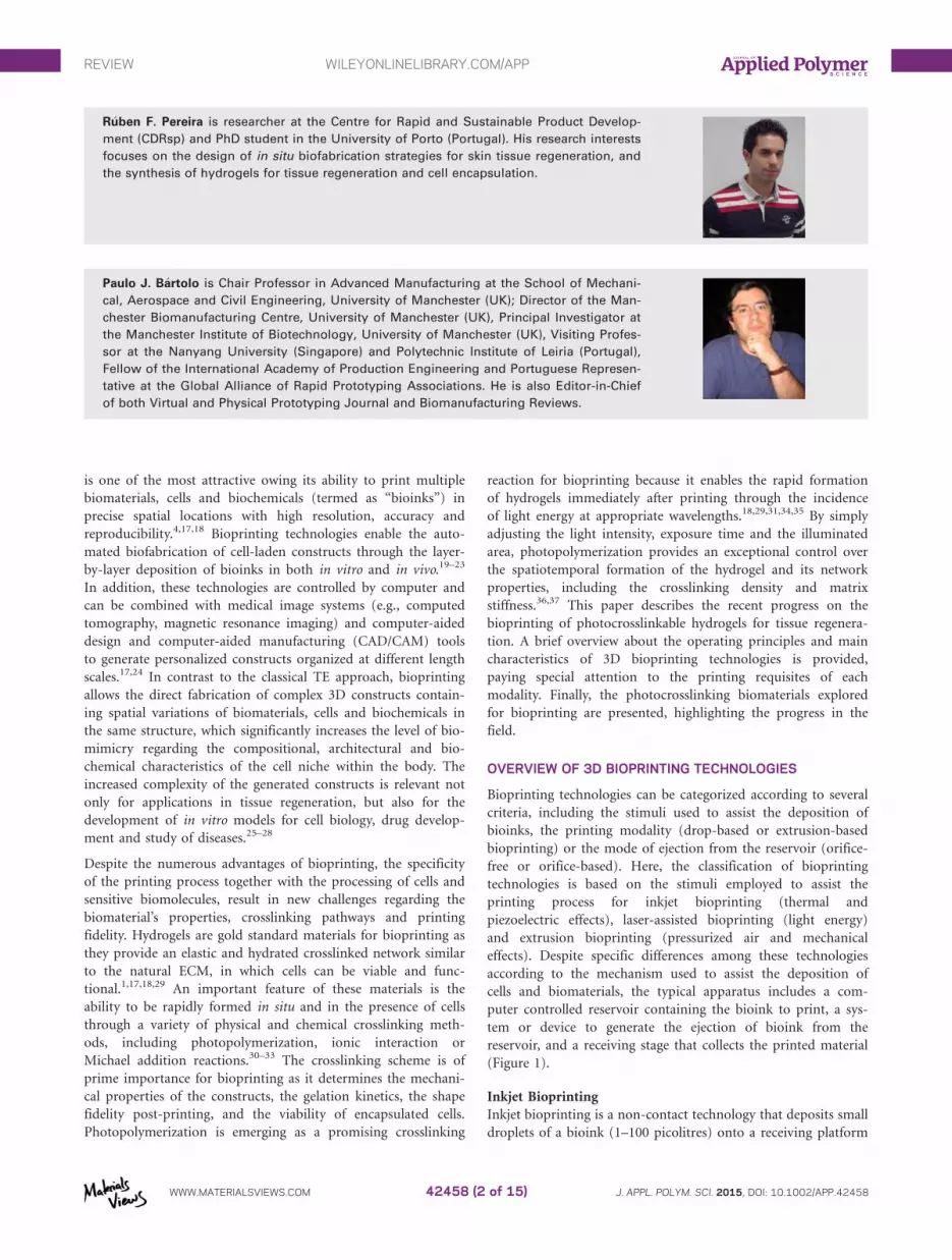
OVERVIEW OF 3D BIOPRINTING TECHNOLOGIES
Bioprinting technologies can be categorized according to several
criteria, including the stimuli used to assist the deposition of
bioinks, the printing modality (drop-based or extrusion-based
bioprinting) or the mode of ejection from the reservoir (orifice-
free or orifice-based). Here, the classification of bioprinting
technologies is based on the stimuli employed to assist the
printing process for inkjet bioprinting (thermal and
piezoelectric effects), laser-assisted bioprinting (light energy)
and extrusion bioprinting (pressurized air and mechanical
effects). Despite specific differences among these technologies
according to the mechanism used to assist the deposition of
cells and biomaterials, the typical apparatus includes a com-
puter controlled reservoir containing the bioink to print, a sys-
tem or device to generate the ejection of bioink from the
reservoir, and a receiving stage that collects the printed material
(Figure 1).
Inkjet Bioprinting
Inkjet bioprinting is a non-contact technology that deposits small
droplets of a bioink (1–100 picolitres) onto a receiving platform
R�uben F. Pereira is researcher at the Centre for Rapid and Sustainable Product Develop-
ment (CDRsp) and PhD student in the University of Porto (Portugal). His research interests
focuses on the design of in situ biofabrication strategies for skin tissue regeneration, and
the synthesis of hydrogels for tissue regeneration and cell encapsulation.
Paulo J. B�artolo is Chair Professor in Advanced Manufacturing at the School of Mechani-
cal, Aerospace and Civil Engineering, University of Manchester (UK); Director of the Man-
chester Biomanufacturing Centre, University of Manchester (UK), Principal Investigator at
the Manchester Institute of Biotechnology, University of Manchester (UK), Visiting Profes-
sor at the Nanyang University (Singapore) and Polytechnic Institute of Leiria (Portugal),
Fellow of the International Academy of Production Engineering and Portuguese Represen-
tative at the Global Alliance of Rapid Prototyping Associations. He is also Editor-in-Chief
of both Virtual and Physical Prototyping Journal and Biomanufacturing Reviews.
REVIEW WILEYONLINELIBRARY.COM/APP
WWW.MATERIALSVIEWS.COM J. APPL. POLYM. SCI. 2015, DOI: 10.1002/APP.4245842458 (2 of 15)
with micrometer resolution.38 In drop-on-demand printing,
droplets with an average diameter of 10–50 lm are quickly gener-
ated through the action of either thermal or piezoelectric effects,
and subsequently ejected through a tiny orifice placed in the
extremity of the reservoir.18,39 Thermal inkjet uses a heating ele-
ment to induce the vaporization of a small volume of bioink
inside the reservoir, with the consequent formation and ejection
of a small droplet. As a result, this method subjects the cells to
high temperatures (�3008C) for few microseconds (�2 ls) dur-
ing the printing process, which may result in the formation of
transient pores within the cell membrane.17,40 However, several
works established that thermal heating has minimal deleterious
effects in the viability of several cell types.40–42 In piezoelectric
inkjet, the droplet generation and ejection is achieved through the
mechanical deformation of a piezoelectric transducer controlled
by the application of external voltage, which precludes the tem-
perature increase to supraphysiological levels.39,43,44 Even though
inkjet bioprinting has been successfully applied to generate 2D/
3D patterns of mammalian cells with minimal reduction on cell
viability,22,40 its orifice nature severely restricts the processing
window. To prevent clogging issues, bioinks for inkjet bioprinting
must exhibit low viscosity (1–10 mPa/s) and limited cell densities
(typically <106 cells/mL), which causes additional constrains in
the printing process, such as cell settling and sedimentation, drop-
let spreading, loss of resolution and limited fabrication of 3D con-
structs with controlled architectures.17,18,45,46
Laser-Assisted Bioprinting
Laser-assisted bioprinting (LAB) evolved from the conventional
laser-induced forward transfer process originally developed to
the direct writing of metals.47 Its operation principle involves
the application of a high-energetic pulsed laser (often a near
infra-red laser) onto a donor ribbon coated with the bioink to
be printed to generate the local ejection of small droplets. The
incident laser light is primarily focused on a laser-transparent
substrate (e.g., glass or quartz substrate), which is coated with a
thin metal (e.g., gold and titanium) layer that absorbs the light
energy and promotes its transfer to the bioink. At this moment,
a high-pressure bubble is generated, and a small droplet is pro-
pelled towards a receiving platform.17,23,48 The mechanisms of
droplet formation and the effects of biofabrication parameters
on the printing resolution and cell functions are well docu-
mented in the literature.14,23,49–51 Deleterious effects to the cells
mainly result from the thermal heating, interaction with laser
light and impact with the receiving substrate.48,49 LAB is one of
the most promising bioprinting modalities for TE due to its
unique resolution (10–100 lm), high-throughput, and ability to
produce heterogeneous tissue constructs containing high cell
densities. Contrary to the inkjet printing, LAB is an orifice-free
technology, which precludes clogging issues and affords the dep-
osition of bioinks with a large range of viscosities (1–300 mPa/s)
and cell concentration (108 cells/mL).17 However, to print 3D
constructs retaining the prescribed resolution in the placement of
cells and biomaterials, LAB requires the development of biomate-
rials exhibiting fast crosslinking mechanisms compatible with the
light-wavelengths emitted by the laser sources. The biofabrication
of clinically relevant constructs through LAB takes long periods
of time and eventually the preparation of multiple ribbons con-
taining multiple materials and cell types.
Extrusion Bioprinting
Extrusion bioprinting is the most popular method to fabricate
3D cell-laden constructs for tissue regeneration. In a typical
setup, cells suspended in prepolymer solutions are loaded within
disposable medical grade syringes or reservoirs, and subse-
quently printed onto a platform driven by pressurized air or
mechanical forces generated by either a piston or a rotating
screw.17,18 Temperature controlled modules are often employed
to adjust the temperature of both the bioink and the construc-
tion platform during fabrication, which is particularly relevant
to control the bioink viscosity and to induce the in situ gelation
of temperature-sensitive polymers.52 Light sources and spraying
nozzles can also be coupled to the bioprinting system to provide
additional crosslinking schemes, improving the printing fidelity
and the structural stability of tissue constructs.24,53,54 Compres-
sion forces and shear stresses generated during the printing are
the major sources of cell damage, requiring a careful optimiza-
tion of the processing parameters (e.g., cellular density, bioink
viscosity, temperature, and air pressure) to prevent cell apopto-
sis. Although there are several works indicating how these
effects can be controlled and optimized to increase the printing
accuracy and cell viability,55–58 there is a need for systematic
studies detailing the functionality of cells after printing. Instead
of printing small droplets onto a platform, extrusion bioprint-
ing produces 3D constructs through the sequential deposition
of hydrogel filaments with diameters in a range of 150–300 lm.
Figure 1. Illustration of 3D bioprinting processes and its main components. [Color figure can be viewed in the online issue, which is available at
wileyonlinelibrary.com.]
REVIEW WILEYONLINELIBRARY.COM/APP
WWW.MATERIALSVIEWS.COM J. APPL. POLYM. SCI. 2015, DOI: 10.1002/APP.4245842458 (3 of 15)

The great advantage of this approach lies in the ability to print
viscous biomaterials containing very high cell densities into 3D
constructs with clinically relevant dimensions, which is imprac-
ticable using inkjet bioprinting and LAB. On the other hand,
major limitations are related to the reduced resolution (approxi-
mately 200 mm), nozzle clogging, and difficulties in fabricating
3D constructs maintaining the predesigned shape and providing
a suitable environment for the cells.17,18 Actually, the printing
of 3D constructs with complex and intricate microarchitectures
still remains a huge challenge in bioprinting. Until now, the
most common approach to address this issue involves the use
of highly concentrated polymer solutions, which imposes severe
restrictions to the cell mobility within the polymer network and
might ultimately lead to poor cell adhesion, and even cell
death.59 More recently, alternative approaches based on the
combination of multiple crosslinking pathways (e.g., photocros-
slinking and thermal gelation),52,60 integration of bioprinting
with melt extrusion,61–64 and the use of partial crosslinked
hydrogels54,59 have been explored with promising outcomes.
PHOTOCROSSLINKABLE BIOMATERIALS FOR BIOPRINTING
The development of advanced biomaterials simultaneously
exhibiting appropriate printability, biomechanical properties,
and providing a suitable microenvironment within which
encapsulated cells can migrate, proliferate and synthetize new
ECM still remains the major challenge in bioprinting. Hydrogels
are the most commonly explored materials for the fabrication
of cell-laden constructs through bioprinting, as they provide a
soft and porous matrix for encapsulated cells and their proper-
ties can be easily tuned.12,18,29,65 For example, the biological and
degradation properties of hydrogels can be tailored by the
incorporation of cell-adhesion motifs (e.g., Arg-Gly-Asp-
RGD)66,67 and cell-proteolytic domains into the polymer back-
bone,68,69 whereas the biomechanical characteristics can be
adjusted by varying the polymer molecular weight or the cross-
linking density.70,71 The wide spectrum of crosslinking schemes
and the large range of network properties also give hydrogels
high versatility for application in different tissues, including car-
tilage, bone, cardiovascular and skin.33,72
The formation of 3D hydrogels through photopolymerization is
well documented in literature with several works focusing on
the effects of light parameters and hydrogel precursors on cell
fate and in vivo tissue regeneration.73–76 This crosslinking reac-
tion has been used in conventional mold-based approaches and
additive biofabrication techniques, such as single- or two-
photon stereolithography and micro stereo-thermal-lithography
for many years.34,77–79 However, over the past 5 years, the appli-
cation of photopolymerization reactions in bioprinting has been
receiving a great deal of interest due to its unique spatiotempo-
ral control over the hydrogel formation and fast polymerization
under cytocompatible conditions. Photopolymerization can be
employed during the bioprinting80 or just after the deposition56
to induce the rapid establishment of crosslinks between the
polymer chains, this way addressing current challenges in the
field, including the fabrication of structurally stable 3D con-
structs with intricate architectures, enhanced spatial resolution
and printing fidelity.
Photopolymerization Reactions for Bioprinting
Free radical photopolymerization is the most popular method
to create chemically crosslinked cellular hydrogels by bioprinting
through a multistep process of initiation, propagation and ter-
mination reactions. In the first step, a photosensitive system
containing unsaturated prepolymers, cells and photoinitiators is
irradiated with a sufficient dosage of light energy to excite the
photoinitiators and trigger the formation of free radicals. Then,
these reactive species propagate across vinyl moieties on prepol-
ymers in solution, resulting in both generation of new free radi-
cals and the establishment of crosslinks between the polymer
chains. As the reaction proceeds, the number of crosslinks in
the system increases and a highly crosslinked network structure
is obtained via a chain-growth mechanism.34,81 Despite free rad-
ical photopolymerization enables the rapid fabrication of natu-
ral and synthetic hydrogels under biocompatible conditions, it
suffers from drawbacks including the oxygen inhibition, lack of
control over the crosslinking kinetics, and the generation of het-
erogeneities within the hydrogel network.81,82 The presence of
inhomogeneities in the network resulting from the random
chain polymerization have a significant impact in the mechani-
cal integrity and swelling behavior of the constructs.83
Recently, photopolymerization reactions based on bio-
orthogonal click reactions are emerging as promising alterna-
tives to the free radical photopolymerization counterpart.84,85
The thiol-norbornene photopolymerization is one example of a
click reaction that is triggered by an external light source, pro-
ceeding in a step-growth mechanism with the formation of
structurally uniform hydrogels with minimal network defects,
which provides better control over the gel crosslinking density
and hydrogel properties.81,82 Under ultraviolet (UV) or visible
light irradiation and in the presence of low amounts of a pho-
toinitiator, thiol-ene reaction promotes the rapid radical-
mediated addition of thiols to carbon-carbon double ene bonds
of functionalized prepolymers, yielding thiol-ether bonds.82,86
Thiol-norbornene photopolymerization has numerous advan-
tages, as it is not inhibited by oxygen, proceeds in a faster mag-
nitude than chain-growth photopolymerization, and can be
performed in the presence of cells.81,86 This photopolymeriza-
tion reaction has successfully been applied for the fabrication of
cell-laden hydrogels using natural and synthetic polymers func-
tionalized with norbornene moieties.87–89 Photocrosslinkable
hydrogels can also be formed from mixed-mode polymeriza-
tions (e.g., thiol-acrylate).81,90
Major concerns with photopolymerization reactions for cell
encapsulation arise from three major issues: potential deleteri-
ous effects of UV light irradiation, cytotoxicity of radicals gen-
erated by the dissociation of photoinitiators, and local
inflammation due to unreacted double bonds. For cell encapsu-
lation applications, light sources emitting either in the UV-A
range or visible light are usually employed to reduce deleterious
effects on the biological systems.91 Despite several studies have
shown that the cell damage caused by UV irradiation can be
significantly reduced or even eliminated by choosing appropri-
ate light wavelengths, intensity and irradiation time, great
efforts are currently being done towards the development of
photosensitive systems (hydrogel precursors and photoinitiators)
REVIEW WILEYONLINELIBRARY.COM/APP
WWW.MATERIALSVIEWS.COM J. APPL. POLYM. SCI. 2015, DOI: 10.1002/APP.4245842458 (4 of 15)

capable of undergoing photocrosslinking upon exposure to safe
visible light wavelengths.90,92 In parallel to the light source, pho-
toinitiators are a key element for photopolymerization, as they
are responsible for the generation of free radicals that promote
the formation of crosslinks between the polymer chains. How-
ever, these radicals can react with cellular components during
the photopolymerization either via direct contact or the forma-
tion of reactive oxygen species, which may compromise the cell
viability and ultimately induce DNA damage.73,74 To prevent
cell damage and ensure enough cell viability for new tissue for-
mation, the wavelength of emitted light must overlap the
absorption spectra of photoinitiator, and its concentration must
be carefully optimized, taking into account the compromise
between the crosslinking time and cell viability. Water soluble
type I initiators such as Irgacure 2959 (1-[4-(2-Hydroxyethoxy)-
phenyl]-2-hydroxy-2-methyl-1-propane-1-one), LAP (lithium
phenyl-2,4,6-trimethylbenzoylphosphinate) and VA-086 (2,20-Azobis[2-methyl-N-(2-hydroxyethyl)propionamide]) are the
most commonly used for cell encapsulation because of their
superior initiation efficiency and cytocompatibility.56,93 The
selection of the most appropriated photoinitiator can be par-
tially determined by the number and the reactivity of functional
groups in the polymer backbone. The functionalization of natu-
ral and synthetic polymers with reactive side groups such as
acrylates, methacrylates, fumarates and vinyl esters, is an indis-
pensable requisite for the photopolymerization. These reactive
groups are introduced in the polymer chain through chemical
reactions with functional groups already present in the native
polymer structure in either homogeneous93 or heterogeneous94
conditions. The type and the number of reactive groups intro-
duced in the polymer backbone is often determined by the
cytotoxicity and reactivity of the functional group (reactivity:
acrylate> vinyl ester> vinyl carbonate> vinyl carbama-
te>methacrylate> fumarate), being very important to ensure a
maximal consumption of reactive groups during the photopoly-
merization to prevent local inflammation and non-specific side
reactions with surrounding proteins.81
Photocrosslinkable Bioinks for Bioprinting
In addition to the classical properties that biomaterials should
present in the context of TE,4,95 bioinks for bioprinting of bio-
logical substitutes must fulfill additional requirements such as
the printing under cell-compatible conditions, exhibit fast cross-
linking reaction, allow the material to maintain its structural
integrity after the deposition process, and provide an interactive
microenvironment for the encapsulated cells.17,18,59 Rather than
simply provide a suitable environment that maintains the cells
viable and support their functions, advanced bioinks must be
capable of providing specific spatiotemporal cues to the embed-
ded cells towards direct the cell adhesion, proliferation, differen-
tiation and morphogenesis. As modern biomaterials are already
modified to incorporate cell-adhesion sites for cell anchorage,
and proteolytically sensitive domains for cell-mediated matrix
remodeling,12,65,96 these approaches can now be applied for the
design of advanced bioinks. Despite the bioprinting field is still
in the beginning, recently there has been great advances in the
development of novel bioinks addressing some of the current
challenges. Several natural and synthetic hydrogels have been
specifically modified to fulfill the printing requisites of each
bioprinting process, undergoing crosslinking through a variety
of crosslinking pathways, including ionic interactions, photopo-
lymerization and thermal gelation.21,30,32,37,45,52,56,59,60,97–104
Naturally Derived Bioinks. Natural polymers are widely used
to engineer bioinks for bioprinting due to their inherent bio-
chemical similarities with the natural ECM, biodegradability
and biological recognition.72,105 These materials are also readily
available in nature and contain a myriad of functional groups
in their chemical structure, which affords several functionaliza-
tion strategies for photopolymerization. However, the main con-
cerns with their use are related to the batch-to-batch variability,
potential immunogenicity, narrow processing window, and lim-
ited mechanical properties.34,105 Even though there is a wide
range of natural polymers currently available for biomedical
applications, photocrosslinkable bioinks for bioprinting are
mainly developed using the hyaluronic acid (HA) and gelatin.
Hyaluronic Acid. Hyaluronic acid or hyaluronan is an anionic,
non-sulfated, linear polysaccharide consisting of alternating
disaccharide units of D-glucuronic acid and N-acetyl-D-glucosa-
mine linked together by b-1,4 and b-1,3 glycosidic bonds.72 It
can be extracted from animal products or produced by bacterial
fermentation (e.g., Bacillus subtilis) to obtain a reproducible
and controlled molecular weight. HA is an important glycos-
aminoglycan in synovial fluid and natural ECM, being a major
constituent of connective, epithelial, and neural tissues.106 It is a
biocompatible and hydrophilic polymer that forms viscous solu-
tions at relatively low concentrations, playing a key role in
wound healing by promoting cell motility and proliferation.
Moreover, HA is biodegradable in mammals by the enzyme hya-
luronidase and reactive oxygen species, yielding the formation
of low molecular weight hyaluronic acid and oligosaccharides.
The useful ability of HA to interact with several cell surface
receptors like CD44, CD54 and CD168 also makes this material
very appealing for biomedical applications, including TE,
wound healing and controlled delivery.106,107 The main limita-
tions of HA rely on slow degradation and the inability to bind
key adhesion receptors such as integrins.12 To address these pit-
falls, HA-based hydrogels are usually decorated with cell-
adhesion ligands and matrix metalloproteinase (MMP)-sensitive
sites, which allow enhanced control over the cell adhesion and
degradation rate.
An important characteristic of HA for photopolymerization lies
on the presence of many free sites in its native chemical structure
[glucuronic acid carboxylic acid, primary and secondary hydroxyl
groups, N-acetyl group (following deamidation)],107 which afford
a myriad of functionalization strategies to render HA crosslink-
able upon the light exposure in presence of a photoinitiator. Pho-
tocrosslinkable HA hydrogels can be obtained by reacting the
polymer with functional groups, like methacrylates and norbor-
nenes, under homogeneous (e.g., water) or heterogeneous (e.g.,
water/DMSO) reaction conditions, resulting in the covalent deri-
vatization of the carboxylic acid or hydroxyl groups.60,87,94 To
improve the modification efficiency and reduce the hydrolysis of
functionalizing agents, HA can be converted into a tetrabutylam-
monium (TBA) salt to allow dissolution in organic solvents.60,108
REVIEW WILEYONLINELIBRARY.COM/APP
WWW.MATERIALSVIEWS.COM J. APPL. POLYM. SCI. 2015, DOI: 10.1002/APP.4245842458 (5 of 15)
Chemically hydrogels exhibiting tunable mechanical, swelling and
biodegradation properties can be obtained by varying the degree
of substitution achieved during the chemical functionalization,
the polymer concentration or the light parameters.94,109
Photocrosslinkable HA-based hydrogels have been widely inves-
tigated for the bioprinting of 3D cell-laden hydrogels. Skardal
et al.59 synthetized a new biocompatible, photocrosslinkable
hydrogel consisting of methacrylated HA (here referred as MA-
HA) and gelatin ethanolamide methacrylate (gelatin methacry-
late is herein termed as gelMA) for the fabrication of tubular
constructs by extrusion bioprinting. MA-HA was synthetized by
reaction with an excess of methacrylic anhydride, while gelMA
was obtained through a two-step procedure encompassing the
conversion of the carboxylic acid groups to ethanolamide deriv-
atives, followed by the methacrylation of primary hydroxyl
groups with methacrylic anhydride. Before bioprinting, the MA-
HA:gelMA (4:1) blend containing HepG2 C3A cells (25 3 106
cells/mL) was partially crosslinked by exposure to UV light irra-
diation for 120 seconds to facilitate the deposition. The printing
procedure of tubular constructs was carried out in a Fab@Home
printing system through the sequential deposition of a central
cell-free MA-HA hydrogel, followed by a cell-containing MA-
HA:gelMA hydrogel, and finally an outer cell-free MA-HA
hydrogel. After the printing procedure, the constructs were fur-
ther crosslinked with UV light to consolidate the polymer net-
work. After 3 weeks of in vitro culture, cells were adhered only
in the hydrogels containing gelatin, remodeled the printed ECM
and secreted collagen. Recently, Duan et al.109 developed photo-
crosslinkable hydrogel formulations based on MA-HA and
gelMA to print heart valve conduits containing encapsulated
human aortic valvular interstitial cells. The composition of the
bioink was firstly optimized regarding the matrix stiffness, vis-
cosity, cell spreading and printing accuracy. The most promising
polymer formulation (4% MA-HA/10% gelMA) containing the
photoinitiator Irgacure 2959 (0.05 wt %) was loaded in syringes
of a Fab@Home printing device, and extruded into a receiving
platform to produce a 3D cellular trileaflet heart valve model.
After photocrosslinking with UV light, the constructs main-
tained the structural integrity and supported high cell viability
(92.1 6 2.5%) for up 7 days of in vitro culture. It was also
found that encapsulated cells also remodeled the initial matrix by
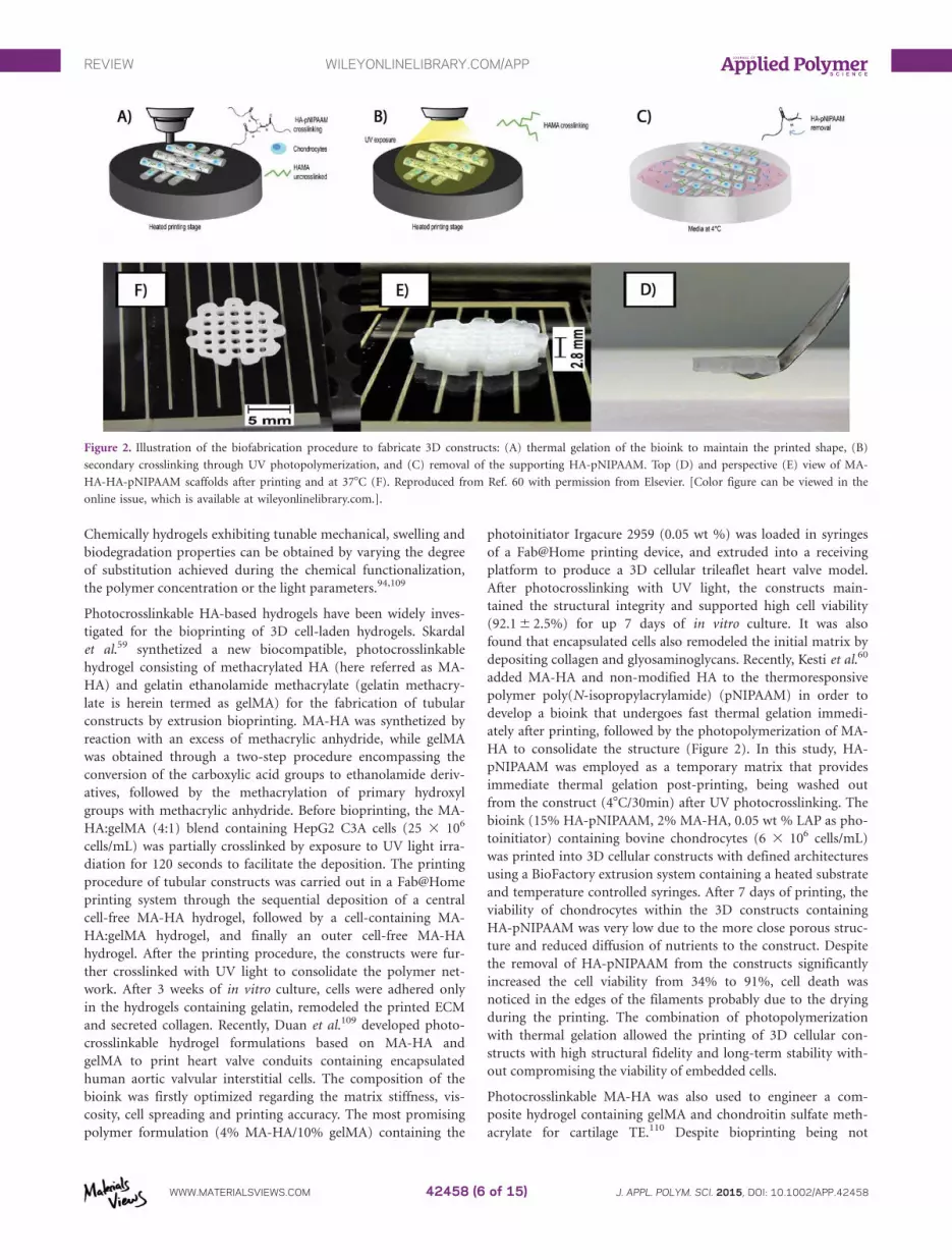
depositing collagen and glyosaminoglycans. Recently, Kesti et al.60
added MA-HA and non-modified HA to the thermoresponsive
polymer poly(N-isopropylacrylamide) (pNIPAAM) in order to
develop a bioink that undergoes fast thermal gelation immedi-
ately after printing, followed by the photopolymerization of MA-
HA to consolidate the structure (Figure 2). In this study, HA-
pNIPAAM was employed as a temporary matrix that provides
immediate thermal gelation post-printing, being washed out
from the construct (48C/30min) after UV photocrosslinking. The
bioink (15% HA-pNIPAAM, 2% MA-HA, 0.05 wt % LAP as pho-
toinitiator) containing bovine chondrocytes (6 3 106 cells/mL)
was printed into 3D cellular constructs with defined architectures
using a BioFactory extrusion system containing a heated substrate
and temperature controlled syringes. After 7 days of printing, the
viability of chondrocytes within the 3D constructs containing
HA-pNIPAAM was very low due to the more close porous struc-
ture and reduced diffusion of nutrients to the construct. Despite
the removal of HA-pNIPAAM from the constructs significantly
increased the cell viability from 34% to 91%, cell death was
noticed in the edges of the filaments probably due to the drying
during the printing. The combination of photopolymerization
with thermal gelation allowed the printing of 3D cellular con-
structs with high structural fidelity and long-term stability with-
out compromising the viability of embedded cells.
Photocrosslinkable MA-HA was also used to engineer a com-
posite hydrogel containing gelMA and chondroitin sulfate meth-
acrylate for cartilage TE.110 Despite bioprinting being not
Figure 2. Illustration of the biofabrication procedure to fabricate 3D constructs: (A) thermal gelation of the bioink to maintain the printed shape, (B)
secondary crosslinking through UV photopolymerization, and (C) removal of the supporting HA-pNIPAAM. Top (D) and perspective (E) view of MA-
HA-HA-pNIPAAM scaffolds after printing and at 378C (F). Reproduced from Ref. 60 with permission from Elsevier. [Color figure can be viewed in the
online issue, which is available at wileyonlinelibrary.com.].
REVIEW WILEYONLINELIBRARY.COM/APP
WWW.MATERIALSVIEWS.COM J. APPL. POLYM. SCI. 2015, DOI: 10.1002/APP.4245842458 (6 of 15)

demonstrated in this work, the addition of MA-HA improved
the chondrogenesis and increased the quantity and distribution
of new ECM, which provides new perspectives for the formula-
tion of novel bioinks for bioprinting. In another strategy, non-
modified HA was combined with hydroxyethyl-methacrylate-
derivatized dextran to obtain a biocompatible formulation with
appropriate viscosity and viscoelastic properties for extrusion
bioprinting. The bioink was printed into 3D acellular scaffolds
through a Bioscaffolder system and photocrosslinked by expo-
sure to UV light (10 min, 6 mW/cm2), yielding the formation
of a semi-interpenetrating hydrogel network.102
Gelatin. Gelatin is a mixture of peptide sequences obtained by
the denaturation of collagen through acid or alkaline treatment,
which results in gelatins with different negative charges.72 The
acid treatment does not significantly affect the amide groups,
whereas the basic treatment hydrolyzes the amide groups of
asparagine and glutamine into carboxyl groups.111 Gelatin is a
water-soluble and thermoresponsive protein that undergoes a
reversible sol-gel transition when cooled upper its critical solu-
tion temperature (25–358C), which is below the temperature of
the human body.37 One efficient approach to prevent the disso-
lution of gelatin at physiological temperature involves the for-
mation of chemical crosslinks through photopolymerization.56
Gelatin has been widely explored for TE applications owing its
biocompatibility, biodegradability and ability to support cell
adhesion and spreading due to the presence of cell adhesion
domains (e.g., RGD). However, as gelatin is derived from colla-
gen, the main component of the ECM and most abundant pro-
tein in humans, it is a potential source of disease transmission
and immunogenic reactions.12 Another drawback of gelatin is
the weak mechanical strength, which limits the range of applica-
tions. One strategy to address this issue, while preserving its
inherent cell-adhesive properties, relies on the combination of
gelatin with melt-extruded polymers. This approach was
recently explored to engineer mechanically robust 3D constructs
for cartilage TE through the alternated deposition of either
gelMA or gelMA/HA hydrogels between the strands of melt
extruded e-caprolactone scaffolds, followed by the UV irradia-
tion of gelMA to promote the formation of chemical cross-
links.37 In a similar work, gelMA was deposited within the
pores of thermoplastic scaffolds composed of poly(hydroxyme-
thylglycolide-co-e-caprolactone)/poly(e-caprolactone) function-
alized with methacrylate groups (pMHMGCL/PCL). Covalent
crosslinks established between the pMHMGCL/PCL and gelMA
resulted in a significant increase in the interface-binding
strength, while the chondrocytes within the hydrogel were capa-
ble of synthetizing glycosaminoglycans and collagen type II both
in vitro and in vivo.112
Photocrosslinkable gelatin hydrogels are usually formed by the
modification of primary amines with methacrylate groups
Figure 3. Macroscopic images of the printed 3D gelMA construct (A–C); Cross-section of the heating mantle around the tip to ensure stable printing
(D); Light microscopy image of the porous scaffold (E); Illustration of the theoretical construct dimensions (F); Microcomputed tomography reconstruc-
tions of the hydrated scaffold representing the gelMA portion (green) and the scaffold pore network (yellow) (G). Reproduced from Ref. 56 with permis-
sion from Elsevier. [Color figure can be viewed in the online issue, which is available at wileyonlinelibrary.com.]
REVIEW WILEYONLINELIBRARY.COM/APP
WWW.MATERIALSVIEWS.COM J. APPL. POLYM. SCI. 2015, DOI: 10.1002/APP.4245842458 (7 of 15)

(often methacrylic anhydride), yielding a light-sensitive polymer
that undergoes free radical polymerization in the presence of a
photoinitiator upon exposure to UV light. Recently, a great
number of gelatin-based bioinks have been developed for 3D
bioprinting, enabling the fabrication of cell-laden 3D constructs
with reproducible architectures.37,54,56,101,103,113 The extrusion
bioprinting of photocrosslinkable gelatin was elegantly demon-
strated by Billiet et al.,56 who formulated and optimized a bio-
ink composed of gelMA, VA-086 as photoinitiator, and
hepatocarcinoma cell line (HepG2, 1.5 3 106 cells/mL). They
started by carrying out a systematic evaluation about the effects
of the bioink properties (e.g., viscosity, concentration, cell den-
sity, photoinitiator, curing kinetics) and the operating parame-
ters (e.g., pressure, plotting speed, needle type and diameter) on
the architecture of bioprinted scaffold, including the filament
diameter, pore geometry and structural integrity. After the
establishment of optimal parameters, the bioink was printed
into 3D cell-laden constructs with interconnected pores (Figure
3) and excellent cell viability after 14 days post-printing
(98.92%). Encapsulated cells maintained the proliferative
capacity and the ability to express liver specific functions, as
observed by hematoxylin and eosin staining. In a different bio-
printing approach, Bertassoni et al.54 developed a three-step
biofabrication strategy to print cell-laden gelMA constructs
encompassing the (i) aspiration of the hydrogel precursor, fol-
lowed by (ii) the photocrosslinking inside a glass capillary, and
(iii) the dispensing via mechanical extrusion. They firstly
accessed the influence of polymer concentration, UV irradiation
and cell concentration on the printability and mechanical prop-
erties of gelMA hydrogels. Then, they showed the ability of their
strategy to print gelMA hydrogels with different architectures,
containing HepG2 cells that remained viable (>80%) for at
least 8 days after the bioprinting process. This strategy allows
the fabrication of hydrogels with complex architectures without
clogging due to the deposition of pre-polymerized cell-laden
gels from a glass capillary, though the length of each deposited
filament is limited to 65mm, which prevents the fabrication of
constructs with higher dimensions.
Hoch et al.45 developed a photocrosslinkable gelatin bioink to
fulfill the requisites for inkjet bioprinting of mammalian cells.
Gelatin was rendered photocrosslinkable through the reaction
of methacrylic anhydride and the effects of the methacrylation
degree, viscosity and printability of gelatin bioink were care-
fully accessed to preclude nozzle clogging. The gelMA was fur-
ther modified through the acetylation of free amino groups to
reduce the solution viscosity and prevent the formation of
physical gels during the printing procedure. A piezoelectric ink-
jet bioprinting system was used to print porcine articular chon-
drocytes embedded in gelMA (1 3 106 cell/mL) at 258C, as
this temperature led to reduced evaporation, nozzle clogging,
and better bioink stability. Both the bioink and the printing
Figure 4. Inkjet bioprinting of porcine articular chondrocytes embedded in gelMA onto swollen gelMA hydrogel substrates (A); Live/dead staining show-
ing the cell viability and morphology of printed cells after 3 hours, (C) 24 hours, and (D) 72 hours post-printing. Reproduced from Ref. 45 with permis-
sion from RCS Publishing. [Color figure can be viewed in the online issue, which is available at wileyonlinelibrary.com.]
REVIEW WILEYONLINELIBRARY.COM/APP
WWW.MATERIALSVIEWS.COM J. APPL. POLYM. SCI. 2015, DOI: 10.1002/APP.4245842458 (8 of 15)

parameters demonstrated to be cytocompatible, allowing the
printing of hydrogels capable of supporting cell adhesion and
proliferation (Figure 4). In another work, Gurkan et al.27 used
a valve-based droplet ejector system to print nanoliter droplets
of gelMA loaded with human mesenchymal stem cells
(hMSCs), bone morphogenetic protein 2 (BMP-2), and trans-
forming growth factor b1 (TGF-b1) to engineer an anisotropic
biomimetic fibrocartilage microenvironment. The authors dem-
onstrated the capabilities of the bioprinting process to generate
biochemical gradients by printing two cell-laden gelatin bio-
inks, one containing BMP-2 and the other loaded with TGF-
b1, and carried out a comprehensive phenotypic evaluation of
the tissue model.
Melchels et al.101 combined gelMA with high-molecular weight
gellan gum to develop a novel bioink that affords the fabrica-
tion of reproducible 3D constructs through a custom-made bio-
printer. The addition of small concentrations of gellan gum and
variable salt (Na1, K1) concentrations to the gelMA resulted on
the formation of ionic crosslinks between the gellan chains,
which allowed the formulation of a bioink exhibiting suitable
rheological properties (pseudo-plasticity and yield stress) for
bioprinting. The gelMA/gellan gum bioink was printed in 3D
constructs that retained the shape after fabrication and were
further stabilized by the photocrosslinking of gelMA. To dem-
onstrate the cell printing, a 3D cellular construct was fabricated
from a bioink composed of 1.0% gellan gum, 10% gelMA and
chondrocytes (0.5 3 106 cells/mL) in isotonic mannose, and
subsequently crosslinked by UV irradiation for 15 minutes. Nei-
ther the printing process nor the bioink composition was cyto-
toxic to the cells, though the resuspension of cells at a
supraphysiological temperature (508C) led to a significant
decrease on cell viability (�50%). In a similar work, 2.4% HA
was added to a solution of 20% gelMA to promote an increase
the viscosity and allow the reproducible fabrication of 3D scaf-
folds with interconnected pores and dimensions of 20 mm 3
20 mm 3 1.2 mm (Figure 5).37
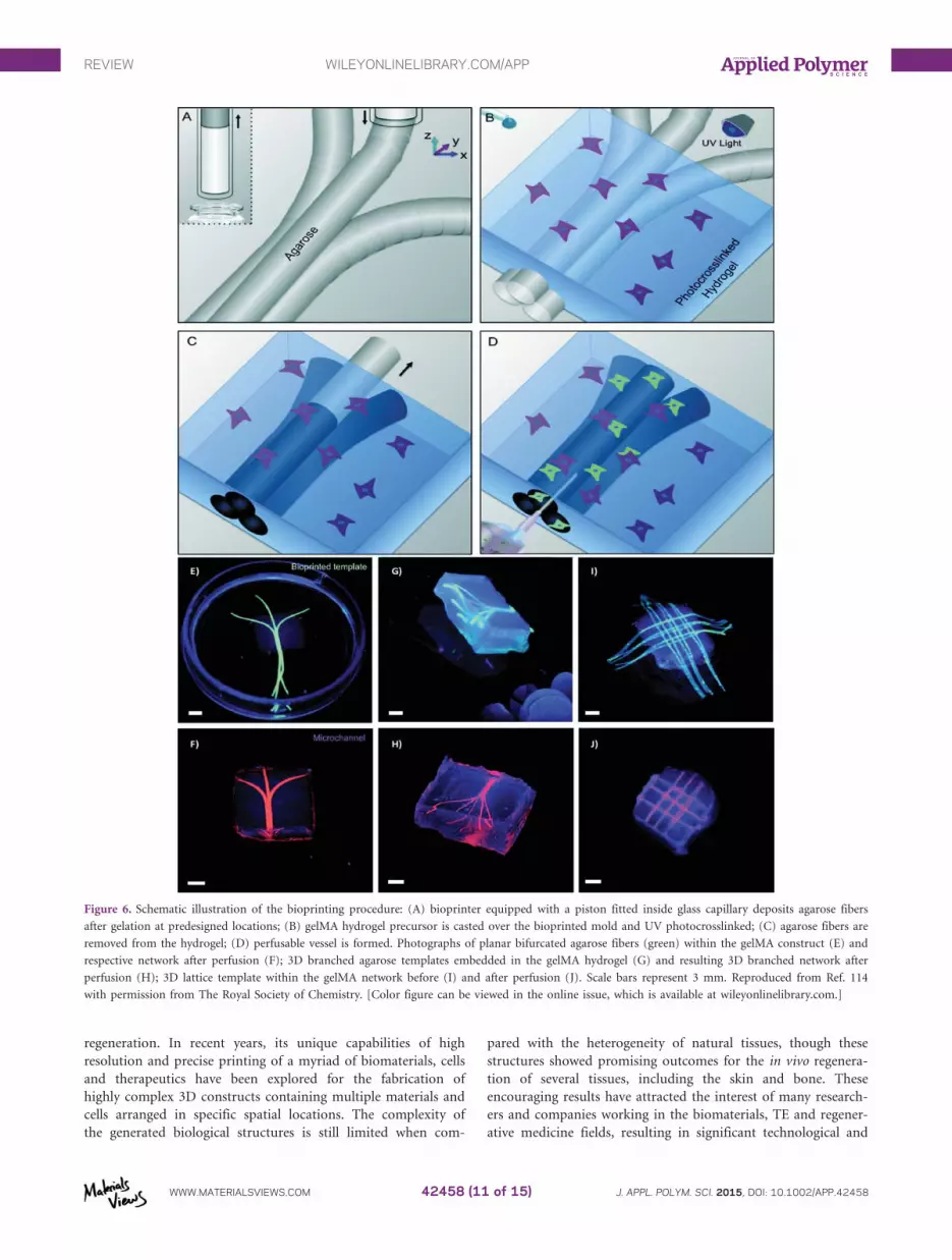
3D bioprinting technologies, in particular extrusion bioprinting,
together with photopolymerization are also assuming a pivotal
role in the fabrication of functional vessel networks within
hydrogels to supply the embedded cells with nutrients and
remove the waste products, which is essential for cell survival
and new tissue formation.114–116 Bertassoni et al.114 used an
extrusion bioprinting system to create perfusable vessels within
3D gelMA hydrogels. In this approach, perfusable microvascular
vessels were created by printing agarose template fibers, fol-
lowed by casting gelMA to cover the deposited filaments (Figure
6). After gelatin photocrosslinking, the filaments were removed
from the construct, leaving functional and perfusable vessels
capable of improving the viability and differentiation of encap-
sulated osteogenic cells.
Figure 5. Extrusion bioprinting of gelMA bioink forming droplets at the nozzle (A) and printed in flat strands that spread out on the surface (C). Addi-
tion of HA to gelMA allows the formation of strands that could be printed from the nozzle (B), allowing the fabrication of a construct composed of 4
layers (D). Scale bars represent 5 mm in A–C and 2 mm in D. Reproduced from Ref. 37 with permission from Wiley-VCH.
REVIEW WILEYONLINELIBRARY.COM/APP
WWW.MATERIALSVIEWS.COM J. APPL. POLYM. SCI. 2015, DOI: 10.1002/APP.4245842458 (9 of 15)

Synthetic-Derived Bioinks. Synthetic polymers are assuming a
pivotal role in the design of functional biomaterials for tissue
regeneration due to their superior mechanical properties, repro-
ducible composition and controlled properties.11,105,117
Although they often exhibit limited biocompatibility, biodegrad-
ability and cell adhesiveness, synthetic polymers can be modi-
fied using several functionalization strategies to provide cell-
mediated degradation and greater control over the cell func-
tions, including adhesion, proliferation and morphogenesis.11,117
In the context of 3D bioprinting, Polyethylene glycol (PEG) is
the most commonly used synthetic polymer because of its
hydrophilicity, presence of functional groups in the polymer
backbone for chemical modification and adjustable properties.
Polyethylene Glycol. PEG is a non-ionic polyester that has been
widely used for several biomedical applications owing its hydro-
philicity, low immunogenicity and antigenicity.81,117,118 Its basic
chemical structure is PEG diol with two hydroxyl end groups,
but it can be modified in an easy and reproducible manner into
other functional groups, such as acrylate, amine, thiol, azide or
vinyl sulfone.117 PEG is usually referred as a “blank slate” mate-
rial due to the absence of cell-adhesion domains, lack of protein
binding sites and low degradability.12 However, these properties
can be easily modified in a reproducible and controlled manner
by a variety of chemistries, including the co-polymerization
with other macromolecules and the introduction of functional
moieties to allow the cell-anchorage, cell-proteolysis and photo-
degradation.81,119–121 PEG macromolecules can also be easily
functionalized through the hydroxyl end groups to yield a vari-
ety of hydrogels precursors for photopolymerization. PEG acryl-
ates are the most used macromers for photopolymerization,
including PEG diacrylate (PEGDA), PEG dimethacrylate
(PEGDMA) and multiarmPEG(n-PEG). The later has been
functionalized with norbornene terminal groups for photopoly-
merization through a step-growth mechanism.73,80,117,120
Photocrosslinkable PEG has been widely used in bioprinting
either alone or combined with other polymers, providing a soft
and hydrophilic environment for the encapsulated cells. Censi
et al.97 developed a biodegradable triblock copolymer hydrogel
for bioprinting based on PEG and methacrylate-modified
poly(N-(2-hydroxypropyl)methacry-lamide lactate) that under-
goes crosslinking by means of thermal gelation and UV light
exposure. The hydrogel formulation solidifies by thermal gela-
tion after printing, and is then consolidated through UV photo-
polymerization. The bioink was successfully deposited in
predesigned patterns by extrusion bioprinting, affording the
additive biofabrication of stable 3D constructs with reproducible
vertical porosity, internal design and dimensional stability,
though the transversal pores suffered fusion during the printing.
3D constructs exhibited an elastic modulus of 119 6 4 kPa and
were completely degraded in approximately 190 days of incuba-
tion in phosphate buffer at pH 7.4 and 378C. Cell encapsulation
studies also show a good viability of chondrocytes (85 6 7%)
embedded within photocrosslinked solid constructs after 3 days
of in vitro culture, which reveals promising properties for 3D
bioprinting. Hockaday et al.24 combined PEGDA with alginate
to fabricate heterogeneous aortic valve hydrogel scaffolds
through extrusion bioprinting with simultaneous photopolyme-
rization (Figure 7). Alginate was added to the PEGDA to
achieve suitable viscosity for bioprinting, being washed out
upon photocrosslinking. Micro-CT analysis revealed that the
printing strategy allows the fabrication of 3D constructs with
high geometric precision, but the accuracy decreases with the
reduced size. For cell studies, collagen I was added to the
PEGDA bioink in an attempt to enhance cell adhesion and
spreading. Results showed that porcine aortic valve interstitial
cells seeded onto the 3D constructs populated the scaffold sur-
face and remained viable throughout 3 weeks, though collagen
did not significantly affected the cell behavior. Although this
work showed the capabilities of bioprinting combined with
photopolymerization to generate anatomically complex 3D valve
conduits at relevant scale lengths, the functionality of the
printed construct was not yet demonstrated.
Rather than forming composite bioinks by the addition of other
polymers, PEGDMA was also used as a sole biomaterial to
encapsulate human articular chondrocytes for the fabrication of
cellular constructs for cartilage TE.80,122 Cui et al.80 used a
modified thermal inkjet bioprinter to print a PEGDMA bioink
containing chondrocytes (5 3 106 cells/mL) in a 4-mm-
diameter full-thickness cartilage lesion created in the center of
an osteochondral plugs harvested from bovine femoral condyles,
serving as a biopaper. Cells printed with simultaneous photopo-
lymerization maintained the initial positions, exhibiting high
viability (89.2–3.6%, 24 hours) and ability to express collagen
type II and aggrecan after 6 weeks of in vitro culture. Results
also showed firmly integration between the printed construct
and the native cartilage with the production of proteoglycans at
the interface. Recently, the same research group demonstrated
the one-step inkjet bioprinting of acrylated peptides (RGD and
MMP-sensitive), acrylated-PEG and hMSCs to promote the
conjugation of peptides to the modified PEG hydrogel through
photopolymerization during the printing procedure. The pep-
tide conjugation occurred in biocompatible conditions and sig-
nificantly enhanced the bone and cartilage differentiation.123 In
one of the few examples about the bioprinting of biological sub-
stitutes for bone tissue, PEGDMA and bone marrow-derived
human mesenchymal stem cells (hMSCs) were printed together
with nanoparticles of bioactive glass (BG) and hydroxyapatite
(HA) to stimulate the osteogenesis of hMSCs.124 A thermal ink-
jet bioprinter was employed to produce 3D cell-laden bone con-
structs through the layer-by-layer printing of the composite
bioink with simultaneous photopolymerization to promote the
chemical crosslinking of PEGDMA. The presence of BG nano-
particles led to a reduced viability of hMSCs (63.80 6 7.54%)
encapsulated within the hydrogels when compared with the HA
nanoparticles (86.62 6 6.02%), which indicates possible cytotox-
icity. Printed HA nanoparticles also resulted in increased expres-
sion of ECM, collagen production and alkaline phosphatase
activity, indicating the osteogenic differentiation of encapsulated
hMSCs.
CONCLUSIONS AND FUTURE PERSPECTIVES
3D bioprinting is assuming a key role in the fabrication of
advanced 3D cellular constructs for tissue repair and
REVIEW WILEYONLINELIBRARY.COM/APP
WWW.MATERIALSVIEWS.COM J. APPL. POLYM. SCI. 2015, DOI: 10.1002/APP.4245842458 (10 of 15)
regeneration. In recent years, its unique capabilities of high
resolution and precise printing of a myriad of biomaterials, cells
and therapeutics have been explored for the fabrication of
highly complex 3D constructs containing multiple materials and
cells arranged in specific spatial locations. The complexity of
the generated biological structures is still limited when com-
pared with the heterogeneity of natural tissues, though these
structures showed promising outcomes for the in vivo regenera-
tion of several tissues, including the skin and bone. These
encouraging results have attracted the interest of many research-
ers and companies working in the biomaterials, TE and regener-
ative medicine fields, resulting in significant technological and
Figure 6. Schematic illustration of the bioprinting procedure: (A) bioprinter equipped with a piston fitted inside glass capillary deposits agarose fibers
after gelation at predesigned locations; (B) gelMA hydrogel precursor is casted over the bioprinted mold and UV photocrosslinked; (C) agarose fibers are
removed from the hydrogel; (D) perfusable vessel is formed. Photographs of planar bifurcated agarose fibers (green) within the gelMA construct (E) and
respective network after perfusion (F); 3D branched agarose templates embedded in the gelMA hydrogel (G) and resulting 3D branched network after
perfusion (H); 3D lattice template within the gelMA network before (I) and after perfusion (J). Scale bars represent 3 mm. Reproduced from Ref. 114
with permission from The Royal Society of Chemistry. [Color figure can be viewed in the online issue, which is available at wileyonlinelibrary.com.]
REVIEW WILEYONLINELIBRARY.COM/APP
WWW.MATERIALSVIEWS.COM J. APPL. POLYM. SCI. 2015, DOI: 10.1002/APP.4245842458 (11 of 15)

scientific advances regarding the commercialization of dedicated
bioprinting technologies and the development of novel biomate-
rials for bioprinting. Most of the bioprinting systems currently
used in laboratory evolved from technical modifications of
available technologies, however, there are now several bio-
printers in the market that were specifically designed to print
biological materials under sterile conditions, following best
manufacturing practices. In parallel, researchers have been
investigating the applicability of several biomaterials for bio-
printing to solve the lack of suitable bioinks for the fabrication
of larger 3D constructs in some extent similar to the natural tis-
sue organization. Most of these materials are inappropriate due
to the narrow rheological, mechanical and biological boundaries
of bioprinting, nevertheless they allowed a better comprehension
about the printing requisites. Based on this knowledge, several
photocrosslinkable biomaterials have been explored for bio-
printing to address the current need for bioinks exhibiting fast
crosslinking schemes, appropriate printability, biomechanical
properties, structural stability after printing, and suitable micro-
environment in which embedded cells can reside and synthetize
new tissue. Photocrosslinkable bioinks fulfill the major physical,
mechanical and biological requisites of bioprinting, allowing the
rapid formation of hydrogels during the printing procedure
upon the exposure to light energy, which allows the fabrication
of 3D constructs with complex internal architectures, structural
stability and printing accuracy, the major pitfalls of other cross-
linking pathways under investigation for bioprinting. In addi-
tion, cells and biochemical entities (e.g., growth factors) can be
Figure 7. (I) Illustration of the bioprinting setup (A); axisymmetric valve STL file designed by a CAD software (B); micro-CT scan of the porcine aortic
valve (C); representative micro-CT image of the valve with leaflet and root regions thresholded based on tissue density (D); thresholded regions were
reconstructed into printable STL geometries (E); printing software with sliced the geometries (F) (scale bar represents 1 cm). (II) Printing of heterogene-
ous valve constructs at different scales: porcine aortic valve model (A) and respective constructs (B, C); axisymmetric valve model (D) and respective
constructs (E, F) (scale bar represents 1 cm). Reproduced from Ref. 24 with permission from IOP Publishing. [Color figure can be viewed in the online
issue, which is available at wileyonlinelibrary.com.]
REVIEW WILEYONLINELIBRARY.COM/APP
WWW.MATERIALSVIEWS.COM J. APPL. POLYM. SCI. 2015, DOI: 10.1002/APP.4245842458 (12 of 15)

added to photocrosslinkable bioink and printed into 3D hydro-
gels with simultaneous photopolymerization without inducing
significant reduction on the viability of encapsulated cells. Even
so photopolymerization has emerged as a promising crosslink-
ing scheme for bioprinting, systematic studies detailing the
combined effects of functionalized biomaterials, printing proce-
dure, photoinitiators and light parameters on the function of
printed cells are necessary to elucidate and prove the safety of
this biofabrication strategy. Efforts should also be concentrated
in the research of more efficient and cytocompatible photoini-
tiators to promote the formation of hydrogels under UV light
and visible light. Simultaneously, in vivo studies should also be
conducted to evaluate the interaction between the biological tis-
sues and the bioprinted photocrosslinked hydrogels, and their
efficacy in promoting the repair and regeneration of complex
tissues. The potential of photocrosslinkable hydrogels for LAB
should also be investigated to access their feasibility to generate
3D constructs with relevant resolution for clinical applications.
ACKNOWLEDGMENTS
The authors thank the support of the Portuguese Foundation for
Science and Technology (FCT) through the strategic project UID/
Multi/04044/2013 and the Portuguese Foundation for Science and
Technology (FCT) for the doctoral grant SFRH/BD/91151/2012
(to R.F.P.).
REFERENCES
1. Melchels, F. P. W.; Domingos, M. A. N.; Klein, T. J.; Malda,
J.; Bartolo, P. J.; Hutmacher, W. Prog. Polym. Sci. 2012, 37,
1079.
2. Sousa, I.; Mendes, A.; Pereira, R. F.; B�artolo, P. J. Mater.
Lett. 2014, 134, 263.
3. Domingos, M.; Intranuovo, F.; Gloria, A.; Gristina, R.;
Ambrosio, L.; B�artolo, P. J.; Favia, P. Acta Biomater. 2013,
9, 5997.
4. Bajaj, P.; Schweller, R. M.; Khademhosseini, A.; West, J. L.;
Bashir, R. Ann. Rev. Biomed. Eng. 2014, 16, 247.
5. Hutmacher, D. W.; Sittinger, M.; Risbud, M. V. Trends Bio-
technol. 2004, 22, 354.
6. Bartolo, P.; Kruth, J.-P.; Silva, J.; Levy, G.; Malshe, A.;
Rajurkar, K.; Mitsuishi, M.; Ciurana, J.; Leu, M. CIRP Ann.
Manufact. Technol. 2012, 61, 635.
7. B�artolo, P. J.; Chua, C. K.; Almeida, H. A.; Chou, S. M.;
Lim, A. S. C. Virt. Phys. Prototyp. 2009, 4, 203.
8. Lee, J. W.; Kang, K. S.; Lee, S. H.; Kim, J.-Y.; Lee, B.-K.;
Cho, D.-W. Biomaterials 2011, 32, 744.
9. Simmons, C. A.; Alsberg, E.; Hsiong, S.; Kim, W. J.;
Mooney, D. J. Bone 2004, 35, 562.
10. Mironov, V.; Visconti, R. P.; Kasyanov, V.; Forgacs, G.;
Drake, C. J.; Markwald, R. R. Biomaterials 2009, 30, 2164.
11. Lutolf, M. P.; Hubbell, J. A. Nat Biotech 2005, 23, 47.
12. Rice, J. J.; Martino, M. M.; De Laporte, L.; Tortelli, F.;
Briquez, P. S.; Hubbell, J. A. Adv. Healthcare Mater. 2013,
2, 57.
13. Nichol, J. W.; Khademhosseini, A. Soft Matter 2009, 5, 1312.
14. Koch, L.; Deiwick, A.; Schlie, S.; Michael, S.; Gruene, M.;
Coger, V.; Zychlinski, D.; Schambach, A.; Reimers, K.; Vogt,
P. M.; Chichkov, B. Biotechnol. Bioeng. 2012, 109, 1855.
15. Michael, S.; Sorg, H.; Peck, C.-T.; Koch, L.; Deiwick, A.;
Chichkov, B.; Vogt, P. M; Reimers, K. PLoS ONE 2013, 8,
e57741.
16. Pereira, R. F.; Barrias, C. C.; Granja, P. L.; Bartolo, P. J.
Nanomedicine 2013, 8, 603.
17. Murphy, S. V.; Atala, A. Nat. Biotech. 2014, 32, 773.
18. Malda, J.; Visser, J.; Melchels, F. P.; J€ungst, T.; Hennink, W.
E.; Dhert, W. J. A.; Groll, J.; Hutmacher, D. W. Adv. Mater.
2013, 25, 5011.
19. Skardal, A.; Mack, D.; Kapetanovic, E.; Atala, A.; Jackson,
J. D.; Yoo, J.; Soker, S. Stem Cells Transl. Med. 2012, 1, 792.
20. Virginie, K.; Fabien, G.; Isabelle, A.; Bertrand, G.; Sylvain,
M.; Jo€elle, A.; Jean-Christophe, F; Sylvain, C. Biofabrication
2010, 2, 014101.
21. Das, S.; Pati, F.; Choi, Y.-, J.; Rijal, G.; Shim, J.-H.; kim, S.
W.; Ray, A. R.; Cho, D.-W; Ghosh, S. Acta Biomater. 2015,
11, 233.
22. Xu, T.; Zhao, W.; Zhu, J.-M.; Albanna, M. Z.; Yoo, J. J.;
Atala, A. Biomaterials 2013, 34, 130.
23. Guillemot, F.; Souquet, A.; Catros, S.; Guillotin, B.; Lopez,
J.; Faucon, M.; Pippenger, B.; Bareille, R.; R�emy, M.;
Bellance, S.; Chabassier, P.; Fricain, J. C.; Am�ed�ee, J. Acta
Biomater. 2010, 6, 2494.
24. Hockaday, L. A.; Kang, K. H.; Colangelo, N. W.; Cheung,
P. Y. C.; Duan, B.; Malone, E.; Wu, J.; Girardi, L. N.;
Bonassar, L. J.; Lipson, H.; Chu, C. C.; Butcher, J, T. Bio-
fabrication 2012, 4, 035005.
25. Dolatshahi-Pirouz, A.; Nikkhah, M.; Gaharwar, A. K.;
Hashmi, B.; Guermani, E.; Aliabadi, H.; Camci-Unal, G.;
Ferrante, T.; Foss, M.; Ingber, D. E.; Khademhosseini, A.
Sci. Rep. 2014, 4. 3896.
26. Horv�ath, L.; Umehara, Y.; Jud, C.; Blank, F.; Petri-Fink, A.;
Rothen-Rutishauser, B. Sci. Rep. 2015, 5. 7974.
27. Gurkan, U. A.; El Assal, R.; Yildiz, S. E.; Sung, Y.;
Trachtenberg, A. J.; Kuo, W. P.; Demirci, U. Mol. Pharm.
2014, 11, 2151.
28. Gruene, M.; Pflaum, M.; Hess, C.; Diamantouros, S.;
Schlie, S.; Deiwick, A.; Koch, L.; Wilhelmi, M.;
Jockenhoevel, S.; Haverich, A.; Chichkov, B. Tissue Eng.
Part C Methods 2011, 17, 973.
29. Skardal, A.; Atala, A. Ann Biomed Eng 2014, DOI: 10.1007/
s10439-014-1207-1, p 1.
30. Neufurth, M.; Wang, X.; Schr€oder, H. C.; Feng, Q.; Diehl-
Seifert, B.; Ziebart, T.; Steffen, R.; Wang., S.; M€uller, W. E.
G. Biomaterials 2014, 35, 8810.
31. Murphy, S. V.; Skardal, A.; Atala, A. J. Biomed. Mater. Res.
Part A 2013, 101A, 272.
32. Skardal, A.; Zhang, J.; Prestwich, G. D. Biomaterials 2010,
31, 6173.
33. Yang, J.-A.; Yeom, J.; Hwang, B. W.; Hoffman, A. S.; Hahn,
S. K. Prog. Polym. Sci. 2014, 39, 1973.
REVIEW WILEYONLINELIBRARY.COM/APP
WWW.MATERIALSVIEWS.COM J. APPL. POLYM. SCI. 2015, DOI: 10.1002/APP.4245842458 (13 of 15)

34. R�uben, F. P.; Paulo, J. B. In Hot Topics in Biomaterials,
Future Science Ltd., London, UK 2014, DOI: 10.4155/
ebo.13.650, p 6.
35. Nguyen, K. T.; West, J. L. Biomaterials 2002, 23, 4307.
36. Sunyer, R.; Jin, A. J.; Nossal, R.; Sackett, D. L. PLoS ONE
2012, 7, e46107.
37. Schuurman, W.; Levett, P. A.; Pot, M. W.; van Weeren, P.
R.; Dhert, W. J. A.; Hutmacher, D. W.; Melchels, F. P. W.;
Klein, T. J.; Malda, J. Macromol. Biosci. 2013, 13, 551.
38. Saunders, R. E.; Derby, B. Int. Mater. Rev. 2014, 59, 430.
39. Derby, B. Ann. Rev. Mater. Res. 2010, 40, 395.
40. Cui, X.; Dean, D.; Ruggeri, Z. M.; Boland, T. Biotechnol.
Bioeng. 2010, 106, 963.
41. Cui, X.; Boland, T.; Biomaterials 2009, 30, 6221.
42. Xu, T.; Jin, J.; Gregory, C.; Hickman, J. J.; Boland, T. Bio-
materials 2005, 26, 93.
43. Saunders, R. E.; Gough, J. E.; Derby, B. Biomaterials 2008,
29, 193.
44. Barbara, L.; Wen-Kai, H.; Ian, M. H.; Keith, R. M. Biofabri-
cation 2014, 6, 015001.
45. Hoch, E.; Hirth, T.; Tovar, G. E. M.; Borchers, K. J. Mater.
Chem. B 2013, 1, 5675.
46. Chahal, D.; Ahmadi, A; Cheung, K. C. Biotechnol. Bioeng.
2012, 109, 2932.
47. Arnold, C. B.; Serra, P.; Piqu�e, A. MRS Bulletin 2007, 32, 23.
48. Guillemot, F.; Souquet, A.; Catros, S.; Guillotin, B. Nano-
medicine 2010, 5, 507.
49. Catros, S.; Guillotin, B.; Bac�akov�a, M.; Fricain, J.-C.;
Guillemot, F. Appl. Surf. Sci. 2011, 257, 5142.
50. Sylvain, C.; Jean-Christophe, F.; Bertrand, G.; Benjamin, P.;
Reine, B.; Murielle, R.; Eric, L.; Bernard, D.; Jo€elle, A.;
Fabien, G. Biofabrication 2011, 3, 025001.
51. Ovsianikov, A.; Gruene, M.; Pflaum, M.; Koch, L.;
MaioranaF., Wilhelmi, M.; Haverich, A; Chichkov, B. Bio-
fabrication 2010, 2, 014104.
52. W€ust, S.; Godla, M. E.; M€uller, R.; Hofmann, S. Acta Bio-
mater. 2014, 10, 630.
53. Ahn, S.; Lee, H.; Puetzer, J.; Bonassar, L. J.; Kim, G. J.
Mater. Chem. 2012, 22, 18735.
54. Luiz, E. B.; Juliana, C. C.; Vijayan, M.; Ana, L. C.; Nupura,
S. B.; Wesleyan, A. A.; Pinar, Z.; Nihal, E. V.; Amir, M. G.;
Mehmet, R. D.; Ali, K. Biofabrication 2014, 6, 024105.
55. Fedorovich, N. E.; Schuurman, W.; Wijnberg, H. M.; Prins,
H.-J.; van Weeren, P. R.; Malda, J.; Alblas, J.; Dhert, W. J.
A. Tissue Eng. Part C Method. 2011, 18, 33.
56. Billiet, T.; Gevaert, E.; De Schryver, T.; Cornelissen, M.;
Dubruel, P. Biomaterials 2014, 35, 49.
57. Chang, R.; Nam, J.; Sun, W. Tissue Eng. Part A 2008, 14,
41.
58. Nair, K.; Gandhi, M.; Khalil, S.; Yan, K. C.; Marcolongo,
M.; Barbee, K.; Sun, W. Biotechnol. J. 2009, 4, 1168.
59. Skardal, A.; Zhang, J.; McCoard, L.; Xu, X.;
Oottamasathien, S.; Prestwich, G. D. Tissue Eng. Part A
2010, 16, 2675.
60. Kesti, M.; M€uller, M.; Becher, J.; Schnabelrauch, M.;
D’Este, M.; Eglin, D.; Zenobi-Wong, M. Acta Biomater.
2015, 11, 162.
61. Lee, H.; Ahn, S.; Bonassar, L. J.; Kim, G. Macromol. Rapid
Commun. 2013, 34, 142.
62. Pati, F.; Jang, J.; Ha, D.-H.; Won Kim, S.; Rhie, J.-W.; Shim,
J.-H.; Kim, D.-H; Cho, D.-W. Nat. Commun. 2014, 5.
63. Schiele, N. R.; Chrisey, D. B.; Corr, D. T. Tissue Eng. Part
C Methods 2011, 17, 289.
64. Schuurman, W.; Khristov, V.; Pot, M. W.; Weeren, P. R. v.;
Dhert, W. J. A.; Malda, J. Biofabrication 2011, 3, 021001.
65. Fonseca, K. B.; Granja, P. L.; Barrias, C. C. Prog. Polym.
Sci. 2014, 39, 2010.
66. Neves, S. C.; Gomes, D. B.; Sousa, A.; Bidarra, S. J.;
Petrini, P.; Moroni, L.; Barrias, C. C.; Granja, P. L. J. Mater.
Chem. B 2015, 3, 2096–2108 DOI: 10.1039/C4TB00885E.
67. Phelps, E. A.; Enemchukwu, N. O.; Fiore, V. F.; Sy, J. C.;
Murthy, N.; Sulchek, T. A.; Barker, T. H.; Garc�ıa, A. J. Adv.
Mater. 2012, 24, 64.
68. Patterson, J.; Hubbell, J. A. Biomaterials 2010, 31, 7836.
69. Fonseca, K. B.; Gomes, D. B.; Lee, K.; Santos, S. G.; Sousa,
A.; Silva, E. A.; Mooney, D. J.; Granja, P. L.; Barrias, C. C.
Biomacromolecules 2013, 15, 380.
70. Tokuda, E. Y.; Leight, J. L.; Anseth, K. S. Biomaterials 2014,
35, 4310.
71. Zhang, G.; Drinnan, C. T.; Geuss, L. R.; Suggs, L. J. Acta
Biomater. 2010, 6, 3395.
72. Van Vlierberghe, S.; Dubruel, P.; Schacht, E. Biomacromole-
cules 2011, 12, 1387.
73. Mironi-Harpaz, I.; Wang, D. Y.; Venkatraman, S.; Seliktar,
D. Acta Biomater. 2012, 8, 1838.
74. Fedorovich, N. E.; Oudshoorn, M. H.; van Geemen, D.;
Hennink, W. E.; Alblas, J.; Dhert, W. J. A. Biomaterials
2009, 30, 344.
75. Williams, C. G.; Malik, A. N.; Kim, T. K.; Manson, P. N.;
Elisseeff, J. H. Biomaterials 2005, 26, 1211.
76. Rossi, C. A.; Flaibani, M.; Blaauw, B.; Pozzobon, M.;
Figallo, E.; Reggiani, C.; Vitiello, L.; Elvassore, N.; De
Coppi, P. FASEB J. 2011, 25, 2296.
77. Patr�ıcio, T.; Pereira, R.; Oliveira, L.; B�artolo, P. Adv. Mater.
Res. 2013, 749, 87.
78. Pereira, R.; B�artolo, P. In Tissue Engineering; Fernandes, P.
R.; Bartolo, P. J., Eds.; Springer: Netherlands, 2014; Vol. 31,
Chapter 8, p 149.
79. Ovsianikov, A.; Mironov, V.; Stampfl, J.; Liska, R. Expert
Rev. Med. Dev. 2012, 9, 613.
80. Cui, X.; Breitenkamp, K.; Finn, M. G.; Lotz, M; D’Lima, D.
D. Tissue Eng. Part A 2012, 18, 1304.
81. Lin, C.-C.; Anseth, K. Pharm. Res. 2009, 26, 631.
82. Lin, C.-C.; Ki, C. S.; Shih., H. J. Appl. Polym. Sci. 2015,
132.
83. Tibbitt, M. W.; Kloxin, A. M.; Sawicki, L. A.; Anseth, K. S.
Macromolecules 2013, 46, 2785.
REVIEW WILEYONLINELIBRARY.COM/APP
WWW.MATERIALSVIEWS.COM J. APPL. POLYM. SCI. 2015, DOI: 10.1002/APP.4245842458 (14 of 15)

84. Azagarsamy, M. A.; Anseth, K. S. ACS Macro Lett. 2012, 2,
5.
85. Jiang, Y.; Chen, J.; Deng, C.; Suuronen, E. J.; Zhong, Z.
Biomaterials 2014, 35, 4969.
86. Hoyle, C. E.; Bowman, C. N. Angew. Chem. Int. Ed. 2010,
49, 1540.
87. Gramlich, W. M.; Kim, I. L.; Burdick, J. A. Biomaterials
2013, 34, 9803.
88. Shih, H.; Lin, C.-C. Macromol. Rapid Commun. 2013, 34,
269.
89. Munoz, Z.; Shih, H.; Lin, C.-C. Biomater. Sci. 2014, 2,
1063.
90. Hao, Y.; Shih, H.; Mu�noz, Z.; Kemp, A.; Lin, C.-C. Acta
Biomater. 2014, 10, 104.
91. Gupta, A.; Avci, P.; Dai, T.; Huang, Y.-Y.; Hamblin, M. R.
Adv. Wound Care 2013, 2, 422.
92. Hu, J.; Hou, Y.; Park, H.; Choi, B.; Hou, S.; Chung, A.;
Lee, M. Acta Biomater. 2012, 8, 1730.
93. Rouillard, A. D.; Berglund, C. M.; Lee, J. Y.; Polacheck, W.
J.; Tsui, Y.; Bonassar, L. J.; Kirby, B. J. Tissue Eng. Part C
Method. 2010, 17, 173.
94. Bencherif, S. A.; Srinivasan, A.; Horkay, F.; Hollinger, J. O.;
Matyjaszewski, K.; Washburn., N. R. Biomaterials 2008, 29,
1739.
95. Kharkar, P. M.; Kiick, K. L.; Kloxin, A. M. Chem. Soc. Rev.
2013, 42, 7335.
96. Maia, F. R.; Fonseca, K. B.; Rodrigues, G.; Granja, P. L.;
Barrias, C. C. Acta Biomater. 2014, 10, 3197.
97. Censi, R.; Schuurman, W.; Malda, J.; di Dato, G.; Burgisser,
P. E.; Dhert, W. J. A.; van Nostrum, C. F.; di Martino, P.;
Vermonden, T.; Hennink, W. E. Adv. Funct. Mater. 2011,
21, 1833.
98. Chung, J. H. Y.; Naficy, S.; Yue., Z.; Kapsa, R.; Quigley, A.;
Moulton, S. E.; Wallace, G. G. Biomaterials Sci. 2013, 1,
763.
99. Irvine, S.; Agrawal, A.; Lee, B.; Chua, H.; Low, K.; Lau, B.;
Machluf, M; Venkatraman, S. Biomed Microdev. 2015, 17, 1.
100. Jia, J.; Richards, D. J.; Pollard, S.; Tan, Y.; Rodriguez, J.;
Visconti, R. P.; Trusk, T. C.; Yost, M. J.; Yao, H.; Markwald,
R. R.; Mei, Y. Acta Biomater. 2014, 10, 4323.
101. Melchels, F. P. W.; Dhert, W. J. A.; Hutmacher, D. W.;
Malda, J. J. Materials Chem. B 2014, 2, 2282.
102. Pescosolido, L.; Schuurman, W.; Malda, J.; Matricardi, P.;
Alhaique, F; .,; Coviello, T.; van Weeren, P. R.; Dhert, W. J.
A.; Hennink, W. E.; Vermonden, T. Biomacromolecules
2011, 12, 1831.
103. Rutz, A. L.; Hyland, K. E.; Jakus, A. E.; Burghardt, W. R.;
Shah, R. N. Adv Mater. 2015, DOI: 10.1002/
adma.201405076.
104. Skardal, A.; Zhang, J.; McCoard, L.; Oottamasathien, S.;
Prestwich, G. D. Adv. Mater. 2010, 22, 4736.
105. Annabi, N.; Tamayol, A.; Uquillas, J. A.; Akbari, M.;
Bertassoni, L. E.; Cha, C.; Camci-Unal, G.; Dokmeci, M. R.;
Peppas, N. A.; Khademhosseini, A. Adv. Mater 2014, 26, 85.
106. Collins, M. N.; Birkinshaw, C. Carbohydr. Polym. 2013, 92,
1262.
107. Prestwich, G. D. J. Contr. Rel. 2011, 155, 193.
108. Oudshoorn, M. H. M.; Rissmann, R.; Bouwstra, J. A.;
Hennink, W. E. Polymer 2007, 48, 1915.
109. Duan, B.; Kapetanovic, E.; Hockaday, L. A.; Butcher, J. T.
Acta Biomater. 2014, 10, 1836.
110. Levett, P. A.; Melchels, F. P. W.; Schrobback, K.;
Hutmacher, D. W.; Malda, J; Klein, T. J. Acta Biomater.
2014, 10, 214.
111. Samal, S. K.; Dash, M.; Van Vlierberghe, S.; Kaplan, D. L.;
Chiellini, E.; van Blitterswijk, C.; Moroni, L.; Dubruel, P.
Chem. Soc. Rev. 2012, 41, 7147.
112. Boere, K. W. M.; Visser, J.; Seyednejad, H.; Rahimian, S.;
Gawlitta, D.; van Steenbergen, M. J.; Dhert, W. J. A.;
Hennink, W. E.; Vermonden, T.; Malda, J. Acta Biomater.
2014, 10, 2602.
113. Riccardo, L.; Jetze, V.; Josep, A. P.; Elisabeth, E.; Jos, M.;
Miguel, A. M. T. Biofabrication 2014, 6, 035020.
114. Bertassoni, L. E.; Cecconi, M.; Manoharan, V.; Nikkhah,
M.; Hjortnaes, J.; Cristino, A. L.; Barabaschi, G.; Demarchi,
D.; Dokmeci, M. R.; Yang, Y.; Khademhosseini, A. Lab
Chip 2014, 14, 2202.
115. Kolesky, D. B.; Truby, R. L.; Gladman, A. S.; Busbee, T. A.;
Homan, K. A.; Lewis, J. A. Adv. Mater. 2014, 26, 3124.
116. Wu, W.; DeConinck, A.; Lewis, J. A. Adv. Mater. 2011, 23,
H178.
117. Zhu, J. Biomaterials 2010, 31, 4639.
118. Knop, K.; Hoogenboom, R.; Fischer, D.; Schubert, U. S.
Angew. Chem. Int. Ed. 2010, 49, 6288.
119. DeForest, C. A.; Anseth, K. S. Nat. Chem. 2011, 3, 925.
120. Fairbanks, B. D.; Schwartz, M. P.; Halevi, A. E.; Nuttelman,
C. R.; Bowman, C. N.; Anseth, K. S. Adv. Mater. 2009, 21,
5005.
121. Mosiewicz, K. A.; Kolb, L.; van der Vlies, A. J.; Martino,
M. M.; Lienemann, P. S.; Hubbell, J. A.; Ehrbar, M.; Lutolf,
M. P. Nat. Mater. 2013, 12, 1072.
122. Cui, X.; Breitenkamp, K.; Lotz, M.; D’Lima, D. Biotechnol.
Bioeng. 2012, 109, 2357.
123. Gao, G; Yonezawa, T.; Hubbell, K.; Dai, G.; Cui, X. Biotech-
nol. J. 2015, DOI: 10.1002/biot.201400635.
124. Gao, G.; Schilling, A. F.; Yonezawa, T.; Wang, J.; Dai, G.;
Cui, X. Biotechnol. J. 2014, 9, 1304.
REVIEW WILEYONLINELIBRARY.COM/APP
WWW.MATERIALSVIEWS.COM J. APPL. POLYM. SCI. 2015, DOI: 10.1002/APP.4245842458 (15 of 15)


