Embed Size (px)
Citation preview

ARTICLE
Received 29 May 2013 | Accepted 25 Jul 2013 | Published 19 Aug 2013
Patterned prevascularised tissue constructs byassembly of polyelectrolyte hydrogel fibresMeng Fatt Leong1, Jerry K.C. Toh1, Chan Du1, Karthikeyan Narayanan1, Hong Fang Lu1, Tze Chiun Lim1,
Andrew C.A. Wan1 & Jackie Y. Ying1
The in vivo efficacy of engineered tissue constructs depends largely on their integration with
the host vasculature. Prevascularisation has been noted to facilitate integration of the con-
structs via anastomosis of preformed microvascular networks. Here we report a technique to
fabricate aligned, spatially defined prevascularised tissue constructs with endothelial vessels
by assembling individually tailored cell-laden polyelectrolyte hydrogel fibres. Stable, aligned
endothelial vessels form in vitro within these constructs in 24 h, and these vessels anasto-
mose with the host circulation in a mouse subcutaneous model. We create vascularised
adipose and hepatic tissues by co-patterning the respective cell types with the preformed
endothelial vessels. Our study indicates that the formation of aligned endothelial vessels in a
hydrogel is an efficient prevascularisation approach in the engineering of tissue constructs.
DOI: 10.1038/ncomms3353
1 Institute of Bioengineering and Nanotechnology, 31 Biopolis Way, The Nanos, Singapore 138669. Correspondence and requests for materials should beaddressed to A.C.A.W. (email: [email protected]).
NATURE COMMUNICATIONS | 4:2353 | DOI: 10.1038/ncomms3353 | www.nature.com/naturecommunications 1
& 2013 Macmillan Publishers Limited. All rights reserved.

Cell survival within implanted engineered constructsdepends on the timely formation of distributed, anasto-mosed vasculature1–3. Timely integration of the constructs
with the host vasculature is important for providing oxygen andessential nutrients and for the removal of metabolic wasteproducts2. Viable cells also require blood circulation within closeproximity as the diffusion limit isB100–200mm2–4. There is thusa need to create prevascularised tissue-engineered constructs thatfacilitate rapid and directed anastomosis.
The creation of a preformed microvascular network within atissue construct has been successful in promoting vascularintegration with the host1,4,5. These networks have been shownto rapidly anastomose with host vasculature, overcoming theproblems posed by the slow ingrowth of physiological vascu-lature. Prevascularisation is generally achieved by seeding orencapsulating endothelial cells (ECs) with other cell types.However, in many of these approaches, the distribution ofvasculature within a thick construct relies on in vitro cellularinfiltration and self-organization of the cell mixture. Thesecellular processes are slow and the resultant vascularisation of thetissue construct is not uniform. Furthermore, the need for acritical concentration of ECs for the vascular self-assembly tooccur in vitro severely limits the number of other cells that can beco-cultured6.
One approach is to co-pattern ECs with other cell types in ahydrogel. The patterning of ECs in a prevascularised constructwould serve to guide the distribution of the newly formedvasculature while simultaneously allowing the localization ofECs in defined regions at the critical density, leaving in the restof the tissue construct space available for other cell types.Moreover, placing cells within hydrogels instead of creatingcompletely cell-dense tissue constructs would provide an initialreservoir of nutrients to the encapsulated cells while allowingfurther diffusion of the required nutrients. However, co-patterning multiple cell types within a hydrogel is not a simpletask. Cells have been patterned in three-dimensional (3D)hydrogels using techniques such as dielectrophoretic force cellpatterning7, laser-guided direct writing8, micromolding of cell-laden hydrogels9 and organ printing10. However, these techniquesare limited by slow cell aggregation, the need for large volumesof cell suspension, complicated multistep processes and expensiveinstrumentation11. As a result, large-scale production of im-plantable 3D cell-patterned constructs at high resolution remainsa challenge. Furthermore, these approaches have, to date, notsuccessfully addressed the problem of vascularisation and masstransport through thick engineered tissue constructs12. Thus, ourgoal is to develop a scalable technique to build 3D cell-patternedconstructs for creating vascularised tissues.
An attractive encapsulation technique to create cell-laden hydrogelfibres is interfacial polyelectrolyte complexation (IPC)13,14. Theadvantage of this encapsulation technique is that the fibresare produced under aqueous conditions at room temperature13.The fibres are therefore amenable to the incorporation of bio-logics, including cells and proteins14. Subsequently, each cell-ladenfibre can be used as a building block to create complex tissueconstructs.
In the present study, we develop an IPC fibre assemblytechnique to create an aligned endothelial vessel network thatanastomoses with the host vasculature in a mouse model. Cell-laden fibres can also be assembled into highly patterned multi-cellular constructs. Patterned constructs of ECs with adipocytesand hepatocytes anastomose with the host, leading to vascularisedtissues. This technique provides an easily adaptable platform tocreate 3D cell-patterned hierarchical structures that are spatiallywell defined and supported by vasculature, leading ultimately tofunctionally enhanced tissues.
ResultsAssembly of cell-laden polyelectrolyte fibres. Individual fibreswere drawn in parallel from polyionic droplets arranged on atemplate and then assembled into higher-order structures(Fig. 1a). Fusion of these fibres was attained via secondarycomplexation, resulting in a continuous yet heterogeneousmatrix (Supplementary Fig. S1) that could be easily handled(Supplementary Movie 1). The importance of being able to createindividual niches at high resolution in a 3D environment hasbeen recognized15,16, and we have achieved this by incorporatingbiologics in the polyionic solutions prior to drawing them into thefibres (Fig. 1b,c). Viability of encapsulated cells using this tech-nique is generally high, with cells able to migrate and proliferatewithin the fibres (see Supplementary Fig. S2 and SupplementaryMovies 2 and 3).
Alignment of cells in fibre constructs. To create an organizednetwork of endothelial vessels within the hydrogel, we firstinvestigated the processes of cell alignment and self-assembly toform tubes within the fibres. To demonstrate the use of fibresto align cells, we assembled IPC fibre constructs from thecomplexation of water-soluble chitin (WSC) and rat methylatedcollagen (RMC) with sodium alginate to form a continuoushydrogel with fibrous features and a uniform distribution of RMC(Supplementary Fig. S3a,b). We showed the alignment of ECsalong the fibre axis. Similar observations were made with severalother cell types. (Supplementary Fig. S3b–e and SupplementaryMovie 3). We have also studied neural stem cell (NSC) alignmentand differentiation in our constructs. Fibre constructs withencapsulated NSCs were cultured in neural differentiation med-ium. After 10 days, we observed parallel alignment of NSCs.Additionally, positive staining for neurofilaments indicated in situdifferentiation of NSCs within the construct (SupplementaryFig. S3f). In a further study, we observed tube morphogenesis ofencapsulated Madin–Darby canine kidney cells when coculturedwith NIH3T3 fibroblasts (Supplementary Fig. S4a). This beha-viour was not observed in a control Madin–Darby canine kidneymonoculture system (Supplementary Fig. S4b). The formation ofparallel tube tracts indicated that the aligned collagen componentwithin the matrix had directed the tube formation along thefibre axis.
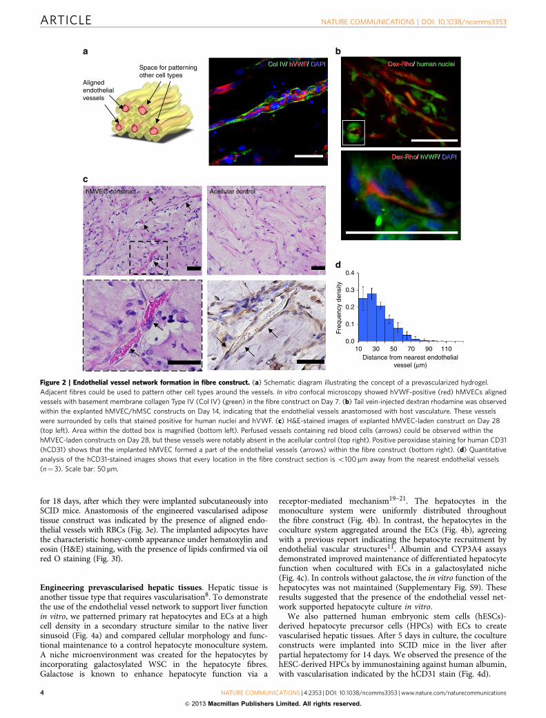
Formation of endothelial vessel network in IPC hydrogel. Wehypothesized that by assembly of EC-laden IPC fibres, we wouldbe able to achieve patterning of an aligned vascular networkwithin a gel matrix. We proceeded to investigate fibre constructsmade up of repeating units that contained a central human EC-laden fibre for the ability to develop and maintain an endothelialvessel network in vitro and in vivo (Fig. 2a). Human mesenchy-mal stem cells (hMSCs) were used as mural cells to stabilize theendothelial vessels17,18. The formation of aligned endothelialtubes was observed within 24 h when cultured in vitro, whichwas verified at Day 7 by immunofluorescence staining for humanvon Willebrand factor (hVWF)-positive endothelial structureswithin our construct flanked by basement membrane protein—collagen type IV (Col IV) (Fig. 2a and Supplementary Movies 4and 5). These endothelial tubes, when co-encapsulated withhMSCs, remained stable for up to 56 days (Supplementary Fig. S5).
Next, we implanted these endothelial constructs subcuta-neously into mice with severe combined immunodeficiency(SCID) after 24 h of in vitro culture. To demonstrate that theendothelial vessels anastomosed with the host vasculature,dextran rhodamine dye was injected into the tail vein of themouse on Day 14. Aligned vessels containing dextran rhodaminedye were observed in the explanted tissue construct (Fig. 2b). The
ARTICLE NATURE COMMUNICATIONS | DOI: 10.1038/ncomms3353
2 NATURE COMMUNICATIONS | 4:2353 | DOI: 10.1038/ncomms3353 | www.nature.com/naturecommunications
& 2013 Macmillan Publishers Limited. All rights reserved.

presence of human nuclei- and hVWF-positive cells aroundthese vessels indicated that the engineered vessels in the fibre-assembled hydrogel had integrated with the host vasculature(Fig. 2b). Histological analysis of constructs explanted on Day 28showed that the acellular control construct exhibited minimumcell infiltration and an absence of vessel-like structures (Fig. 2c).In contrast, the endothelial construct demonstrated preservationof endothelial vessel structures with the infiltration of redblood cells (RBCs) (Fig. 2c). Uniform distribution of these RBCscontaining endothelial vessels was observed within the implantedconstructs (Supplementary Fig. S6). The presence of the im-planted ECs lining the vessels in our explanted EC-ladenconstructs was confirmed by human-specific CD31 staining(Fig. 2c). Moreover, patterning cells around the aligned endo-thelial tubes in a secondary structure guaranteed the closeproximity of each cell to a vascular element below the 200-mmthreshold distance associated with mass transfer limitations(Fig. 2d and Supplementary Fig. S7). Hence, patterning EC
within a hydrogel led to the formation of a prevascularised bedwith patterned, perfused blood vessels.
Engineering prevascularised adipose tissues. We evaluated theability of the fibre constructs to support the differentiation of3T3-L1 adipocyte precursor cells over the course of 18 days, andthe maintenance of EC-derived vascular structures during this timeframe. We observed the formation of lipid vesicles in the differ-entiated 3T3-L1 cells encapsulated within the fibres (Fig. 3a,b).When co-patterned with ECs, adiponectin-positive cells aggre-gated around hVWF-positive endothelial tubes (Fig. 3c,d). Wealso embedded these coculture constructs within permissive RGD-QGel matrices and observed the sprouting of tubular structuresinto the gel, indicating the permissiveness of the fibre construct foreventual construct integration and anastomosis (SupplementaryFig. S8). For implantation, 3T3-L1 cells were encapsulated withinEC-laden constructs. The coculture constructs were differentiated
Cell
Drawingof fiber
PolycationInterface Polyanion
s.s.
Cell A Cell B
Biologics
Primary Secondary Tertiary
a
b
c
Figure 1 | Engineering of hierarchical tissue structures by fibre assembly. (a) Schematic of the process flow to achieve the hierarchical tissue structure at
each step. Polyionic solutions containing biologics are first localized on a template. IPC fibres are drawn in parallel and brought to fuse at a single
point by rotation of the template. Continuous upward drawing coupled with fusion by a deposited droplet of polyionic solution (either polycation or
polyanion) forms the secondary structure (s.s.). The secondary structure is subsequently assembled to tertiary structure by spooling and layering
secondary polyelectrolyte complexation. (b) Micropatterned niche environment can be created by assembling primary biostructural units containing
different cell types. Each secondary structure consists of one central fibre surrounded by other fibres. This micropattern is repeated in the eventual tertiary
construct. (c) Confocal microscopy images of the biostructural units. Labelled cells are encapsulated at high density in chitin–alginate polyelectrolyte fibres,
demonstrating primary, secondary and tertiary structures. Scale bars: 100 mm.
NATURE COMMUNICATIONS | DOI: 10.1038/ncomms3353 ARTICLE
NATURE COMMUNICATIONS | 4:2353 | DOI: 10.1038/ncomms3353 | www.nature.com/naturecommunications 3
& 2013 Macmillan Publishers Limited. All rights reserved.
for 18 days, after which they were implanted subcutaneously intoSCID mice. Anastomosis of the engineered vascularised adiposetissue construct was indicated by the presence of aligned endo-thelial vessels with RBCs (Fig. 3e). The implanted adipocytes havethe characteristic honey-comb appearance under hematoxylin andeosin (H&E) staining, with the presence of lipids confirmed via oilred O staining (Fig. 3f).
Engineering prevascularised hepatic tissues. Hepatic tissue isanother tissue type that requires vascularisation8. To demonstratethe use of the endothelial vessel network to support liver functionin vitro, we patterned primary rat hepatocytes and ECs at a highcell density in a secondary structure similar to the native liversinusoid (Fig. 4a) and compared cellular morphology and func-tional maintenance to a control hepatocyte monoculture system.A niche microenvironment was created for the hepatocytes byincorporating galactosylated WSC in the hepatocyte fibres.Galactose is known to enhance hepatocyte function via a
receptor-mediated mechanism19–21. The hepatocytes in themonoculture system were uniformly distributed throughoutthe fibre construct (Fig. 4b). In contrast, the hepatocytes in thecoculture system aggregated around the ECs (Fig. 4b), agreeingwith a previous report indicating the hepatocyte recruitment byendothelial vascular structures11. Albumin and CYP3A4 assaysdemonstrated improved maintenance of differentiated hepatocytefunction when cocultured with ECs in a galactosylated niche(Fig. 4c). In controls without galactose, the in vitro function of thehepatocytes was not maintained (Supplementary Fig. S9). Theseresults suggested that the presence of the endothelial vessel net-work supported hepatocyte culture in vitro.
We also patterned human embryonic stem cells (hESCs)-derived hepatocyte precursor cells (HPCs) with ECs to createvascularised hepatic tissues. After 5 days in culture, the cocultureconstructs were implanted into SCID mice in the liver afterpartial hepatectomy for 14 days. We observed the presence of thehESC-derived HPCs by immunostaining against human albumin,with vascularisation indicated by the hCD31 stain (Fig. 4d).
Alignedendothelialvessels
Space for patterningother cell types
Col IV/ hVWF/ DAPICol IV/ hVWF/ DAPI Dex-Rho/ human nucleiDex-Rho/ human nuclei
Dex-Rho/ hVWF/ DAPIDex-Rho/ hVWF/ DAPI
hMVEC construct Acellular control
0.4
0.3
0.2
0.1
0.010 30 50 70 90 110
Distance from nearest endothelialvessel (µm)
Freq
uenc
y de
nsity
a b
c
d
Figure 2 | Endothelial vessel network formation in fibre construct. (a) Schematic diagram illustrating the concept of a prevascularized hydrogel.
Adjacent fibres could be used to pattern other cell types around the vessels. In vitro confocal microscopy showed hVWF-positive (red) hMVECs aligned
vessels with basement membrane collagen Type IV (Col IV) (green) in the fibre construct on Day 7. (b) Tail vein-injected dextran rhodamine was observed
within the explanted hMVEC/hMSC constructs on Day 14, indicating that the endothelial vessels anastomosed with host vasculature. These vessels
were surrounded by cells that stained positive for human nuclei and hVWF. (c) H&E-stained images of explanted hMVEC-laden construct on Day 28
(top left). Area within the dotted box is magnified (bottom left). Perfused vessels containing red blood cells (arrows) could be observed within the
hMVEC-laden constructs on Day 28, but these vessels were notably absent in the acellular control (top right). Positive peroxidase staining for human CD31
(hCD31) shows that the implanted hMVEC formed a part of the endothelial vessels (arrows) within the fibre construct (bottom right). (d) Quantitative
analysis of the hCD31-stained images shows that every location in the fibre construct section is o100 mm away from the nearest endothelial vessels
(n¼ 3). Scale bar: 50mm.
ARTICLE NATURE COMMUNICATIONS | DOI: 10.1038/ncomms3353
4 NATURE COMMUNICATIONS | 4:2353 | DOI: 10.1038/ncomms3353 | www.nature.com/naturecommunications
& 2013 Macmillan Publishers Limited. All rights reserved.

DiscussionOur approach of assembling fibres in a spatially defined manner,which allows for patterning of the biologics within a continuoushydrogel matrix, is different from other hydrogel techniquesthat encapsulate cells in a homogeneous matrix. Each fibrecan support a specific set of biologics and/or cell type,constituting the biostructural unit14,22 (Fig. 1b). This primarystructure can be built up into secondary structures consisting ofone central fibre surrounded by other fibres, and the lattermicropattern can be further repeated to form the eventual tertiaryconstruct.
In this study, prevascularised tissue constructs were formed byfibre assembly. Here an endothelial unit was surrounded byadjacent fibres encapsulating the parenchymal cells, which is inturn combined to form a construct with the aligned vasculature.The alignment of EC was achieved by presenting attachmentmoieties on submicron nuclear fibres within IPC fibres. Thesenuclear fibres aligned along the fibre axis during the drawingprocess13. RMC complexed with the nuclear fibres of the IPCfibre because of its net positive charge23. The presence of the
aligned collagen component within the fibres helped to promotecell–matrix interaction and align cells without the use of externalstimuli (Supplementary Fig. S3)23.
We applied these endothelial constructs towards engineeringadipose tissue. Adipose tissue is known to be highly metabolicand susceptible to necrosis because of the lack of an oxygenatedblood supply after implantation24–26. By co-patterning EC with3T3-L1 cells in the fibre construct, we could ensure that theimplanted adipose cells were in close proximity to the perfusedvessels, thereby improving cell viability.
Besides forming vasculature, ECs have also been shown toregulate tissue function8,17,27–30 and organogenesis31–33. Thus, thefunctionality of 3D structures would be aided by interaction withECs in a multicellular environment. In the case of the adiposeconstruct, proliferation and tubulogenesis of ECs and differentiationof 3T3-L1 cells are known to be enhanced by coculture28,33.Paracrine signalling from the 3T3-L1 cells via the secretion of pro-angio-genic adipocyte-derived microvesicles further aids theformation and stabilization of microvasculature34. ECs have alsobeen shown to help preserve hepatocyte function when cocultured
hVWF/ Adiponectin/ DAPIhVWF/ Adiponectin/ DAPI
a b
c d
e f
Figure 3 | Engineering of prevascularised adipose tissue. The accumulation of lipid vesicles after 18 days in both (a) tissue culture plate control
and (b) within the fibres was used as an indicator for differentiation. (c) Tubulogenesis of EC in the constructs was evaluated using a central fibre laden with
EC and 3T3-L1 in a 5:1 ratio surrounded by blank fibres. Tubulogenesis was observed after 2 days. (d) Immunostaining of human hVWF (red) with
adiponectin (green) and DAPI (blue) confirmed the preservation of the endothelial vessels with closely associated clusters of differentiated 3T3-L1 after 18
days in culture. (e) Endothelial vessel formation within 3T3-L1/ EC coculture. H&E staining shows aligned patent blood vessels in close association
with adipocytes within the fibre matrix, with arrows indicating the blood vessels. (f) Oil red O staining confirmed the presence of lipids. Scale bars: 100mm.
NATURE COMMUNICATIONS | DOI: 10.1038/ncomms3353 ARTICLE
NATURE COMMUNICATIONS | 4:2353 | DOI: 10.1038/ncomms3353 | www.nature.com/naturecommunications 5
& 2013 Macmillan Publishers Limited. All rights reserved.

in vitro. Patterning hepatocytes with ECs in a biomimetic sinusoid-like structure would also allow control of the geometric interactionof the hepatocytes and endothelial vessels8,29, providing abetter representation of native cellular response than a randomcoculture30. The liver sinusoid-like structure thus presents a usefulbuilding block in the engineering of liver tissue. Although adipocytesand hepatocytes were the chosen parenchymal cells in the currentstudy, a wide range of other cell types could be cultured with goodviability in our tissue constructs (see Supplementary Figs S2, S3 andS4), and thus used in conjunction with the vascular bed.
We have developed a simple and scalable technique forderiving macroscopic tissue constructs with microscopic features.This simple technique does not require specialized microfabrica-tion equipment and can be readily adapted by most researchers.
The patterned structure is obtained by simply spooling the repeatunit (secondary structure). The resolution of the construct,determined by the fibre diameter, is tunable by varying processingparameters such as the fibre draw rate, droplet size and solutionconcentration13. Moreover, sizeable constructs can be obtainedfrom small volumes of cell suspension, with little dead volume,and within a short timespan. In this study, the generationof a single acellular construct (10� 1� 0.5mm3) from 50 ml ofpolyionic solutions typically took o15min. Larger constructswere obtained by fusing several tertiary structures together(Supplementary Fig. S1e). Although chitin and alginate were usedas the polyelectrolyte pair in this study, other combinations ofpolyelectrolytes are possible and may potentially improve tissuefunctionality.
hVWF/ rAlb/ DAPIhVWF/ rAlb/ DAPI
120
100
80
60
40
20
01 2 3 4 5 6 7
Day
Alb
umin
sec
retio
n (%
of d
ay 1
) 40
35
30
25
20
15
10
5
03D Hep 3D CC
CY
P3A
4 ac
tivity
(R
LU p
er n
gD
NA
per
ml)
120
100
80
60
40
20
01 2 3 4 5 6 7
Day
Ure
a sy
nthe
sis
(% o
f day
1) 3D single culture
3D co-culture*
* **
*
hCD31/ hAlb/ DAPIhCD31/ hAlb/ DAPI
a b
c
d
Figure 4 | Engineering of prevascularised hepatic tissue. (a) Bright-field microscopic image of a hepatocyte-laden fibre construct showing high cell
density. (b) Immunostaining of hepatocyte monoculture (left) and coculture (right) with HUVEC after 24 h in fibre construct. Regions of aligned sinusoid-
like structures were observed for the coculture. Hepatocytes and HUVEC were stained positive with rat serum albumin (rAlb) and hVWF, respectively. (c)
Functional characterization of hepatocyte cultures. Better maintenance of albumin secretion function and higher CYP3A4 activity were observed for the
co-culture construct. Urea synthesis function for both constructs remained relatively stable over 7 days. * denotes intergroup significant difference
(Po0.01) at the specific time point (n¼ 3, mean±s.d.). (d) H&E-stained and immunostained images of explanted hESC-derived hepatocyte constructs.
Immunostaining indicates the presence of human serum albumin (hAlb)-positive cells in close association with hCD31-positive endothelial cells. Tissue
scaffold boundary is indicated by white dotted line. Scale bars: 100 mm.
ARTICLE NATURE COMMUNICATIONS | DOI: 10.1038/ncomms3353
6 NATURE COMMUNICATIONS | 4:2353 | DOI: 10.1038/ncomms3353 | www.nature.com/naturecommunications
& 2013 Macmillan Publishers Limited. All rights reserved.

In conclusion, our study shows that the assembly of cell-ladenpolyelectrolyte hydrogel fibres is an effective prevascularisationmethod for creating tissue constructs. The technique enables 3Dmicropatterning of different cell types, in particular, thepatterning of ECs to create an endothelial vessel network tosupport and interact with other parenchymal cells within thehydrogel. Future work will focus on applying and adapting thetechnology towards recapitulating the specific cell arrangementsand microenvironment of various tissue types, with the eventualgoal of achieving functional tissue for therapeutic applications.
MethodsFibre assembly into higher-order structures. IPC fibres were drawn aspreviously described13. Five microlitre of each of WSC (1w/v% in phosphate-buffered saline (PBS)) and alginate (1w/v% in deionized water) solutions weredispensed on opposite sides of a pipette tip and brought into contact with eachother. Fibres were then drawn by an upward motion of the tip with a linear motorat a speed of 0.3mm s� 1. One linear motor and one rotation motor were used toassemble the fibres. Nine pairs of polyelectrolyte solution were localized on atemplate from which nine fibres (one central fibre with eight outer fibres) weredrawn in parallel in a humidified chamber to prevent drying. Once the fibresreached a height of 5 cm, the linear motor was stopped and the base plate wasrotated at 20 r.p.m. for five revolutions to allow the fibres to meet at a point. A 5-mldroplet of alginate solution (0.5 w/v%) was then applied on the fibre fusion point.Subsequently, the linear motor was reactivated to draw the fibres upwards while thedroplet travelled down the nascent construct to fuse the fibres via secondarycomplexation. The resulting secondary structure was spooled and packed on a two-pronged collection rod, and adjacent segments of the secondary structure werefused using layer-by-layer polyelectrolyte complexation. This was performed bydipping the construct into the WSC solution (0.5 w/v%), PBS, alginate solution(0.5 w/v%) and PBS sequentially twice. The assembled tertiary structures were thenremoved from the collecting rod and placed in the culture medium.
Encapsulation of cells and incorporation of biologics into IPC fibres. For cell-laden fibres, the cells were first trypsinized and centrifuged at 500 r.p.m. for 3minto obtain a cell pellet. The cells were taken from the pellet and mixed in the WSCsolution at a cell pellet/WSC solution volume ratio of 3:7 to form a workingpolycation solution. This working solution was used to form IPC cell-laden fibres,and the resulting constructs were incubated in the appropriate culture medium.Biologics were incorporated by mixing in either the WSC or alginate solution. Incases where cellular alignment was required, RMC (0.25 w/v%) was added to theWSC solution (Supplementary Methods).
Encapsulation of labelled cells. HepG2 cells were subcultured in DMEM sup-plemented with 10 v/v% fetal bovine serum (FBS) and 1 v/v% penicillin/strepto-mycin (P/S). Cells were trypsinized and the cell suspension was aliquoted into twoportions. Both cell suspensions were centrifuged and the supernatant was removed.Subsequently, the cells were incubated with either CellTracker Green CMFDA(10 mM) or CellTracker Orange CMTMR (10 mM) for 30min. The supernatant wasthen removed and the cells were resuspended in the culture medium, followed bycentrifugation to form the cell pellet. Cells stained with CellTracker Orange wereencapsulated in the central fibre, whereas cells stained with CellTracker Green wereencapsulated in the eight outer fibres. The tertiary construct was formed andimaged under the confocal microscope after 24 h of culture (Fig. 1c).
Subcutaneous implantation of endothelial construct in SCID mice. The animalstudies were conducted in accordance to the Agency for Science, Technology andResearch (A*STAR) Biological Resource Centre IACUC guidelines (Protocol No.:090498). Human microvascular ECs (hMVECs) (third passage) and hMSC (fourthpassage) (ratio 4:1) were encapsulated in the central fibre surrounded by eight cell-free fibres. The two ends of the construct were cut with a scalpel, and the constructswere then cultured in EGM2-MV medium (Lonza) for 24 h. Subsequently, weimplanted the constructs (B10� 5� 2mm3) into mice with SCID. Mice weighingbetween 25 and 30 g were used without gender differentiation for the experiments.The constructs were washed thoroughly with PBS. The animals were anesthetizedwith ketamine (150mg kg� 1) and xylazine (10mg kg� 1) by intraperitonealadministration. The dorsum was shaved and cleansed with alternate alcohol andbetadine swabs (three times). A single 0.5-cm midline incision was made usingsterile surgical scalpel. Two subcutaneous pockets were created on either side of theincision. One endothelial construct and one cell-free construct (control) wereimplanted into each animal. The incision was closed with interrupted 4/0 sutures.For perfusion experiments, 100ml of 5 v/v% dextran rhodamine solution (LifeTechnologies, USA) were injected into the tail vein of animals implanted with theconstructs on Day 14 (refs 18,35). The animal was euthanized with the help ofcarbon dioxide inhalation after 15min. For histological examination, the animalswere killed on Days 14 and 28. For explantation, the dorsum was shaved and the
previous incision was reopened. All scaffolds were retrieved with the surroundingtissue and fixed in neutral buffered formalin (10 v/v%) for processing andhistological sectioning. Sections were stained with human nuclei and hVWFantibodies and counterstained with 40 ,6-diamidino-2-phenylindole (DAPI).
Engineering prevascularised adipose tissue constructs in vivo. Fibre constructsconsisting of a central endothelial fibre surrounded by eight fibres with 3T3-L1adipose precursor cells were used for implantation. After 18 days of in vitro culturein DMEM supplemented with 20% FBS, the constructs were cut with a scalpel toexpose the ends of the fibre construct and washed thoroughly with PBS. Theconstructs were embedded in QGel, then implanted subcutaneously into SCIDmice for 28 days. The explanted tissues were embedded in paraffin wax andsectioned lengthwise. Sections of cocultures were stained with H&E and oil red O.
Evaluation of adipogenic differentiation in vitro. Fibre constructs consisting ofnine adipocyte-laden fibres were formed by suspending 3 ml of 3T3-L1 cell pelletwith 7 ml of WSC, and drawing into a fibre with alginate. For coculture, the centrefibre was replaced with 3 ml of a 5:1 HUVEC:3T3-L1 cell mixture. The fibreconstructs, together with a 2D tissue-culture plate control, were cultured understatic culture conditions with DMEM supplemented with 10 v/v% FBS and 1 v/v%P/S for 4 days. DMEM supplemented with 20 v/v% FBS and 1 v/v% P/S was thenfed to the 3T3-L1 cells for 18 days to induce adipogenesis. Differentiation of3T3-L1 cells was indicated by the accumulation of lipid droplets. The constructswere fixed in 10% neutral buffered formalin and stained for hVWF and adipo-nectin to indicate the presence of ECs and differentiated adipocytes, respectively(Supplementary Table S1).
Engineering prevascularised hepatic tissue constructs in vivo. hESCs weredifferentiated with a three-stage differentiation medium to obtain HPCs(Supplementary Methods). HUVECs (fifth passage) were encapsulated in thecentral fibre surrounded by eight fibres laden with HPCs. These HPC constructswere implanted at the liver injury site after partial hepatectomy in which 70% of thehost liver was removed. The animals were killed on Day 14 and the explantedtissues were examined histologically. Sections of the constructs were stained withH&E and immunostained with human albumin and hCD31 antibodies(Supplementary Table S1).
Evaluation of primary hepatocyte constructs in vitro. HUVECs (fifth passage)were encapsulated in the central fibre surrounded by eight fibres laden with pri-mary rat hepatocytes. Hepatocyte monoculture constructs (control) were similarlycreated with cell-free central fibres. Hepatocyte-laden fibres were formed frompolycation solution containing WSC (1w/v%), galactosylated WSC (0.25 w/v%)and RMC (0.25w/v%). Constructs were maintained in EndoGro-LS (Millipore)and hepatozyme-SFM (Invitrogen) (1:1 mixture) for 7 days. The medium wascollected every day for albumin and urea production. Urea production was inducedby incubating the constructs with culture medium supplemented with 1mM ofammonium chloride (NH4Cl) for 2 h, after which the supplemented medium wasremoved and replaced with fresh medium. Functional maintenance of hepatocyteswas assessed by measuring albumin and urea secretion over a period of 7 days(n¼ 3). Data for each individual construct were normalized with the Day 1 value toobtain the maintenance profiles for albumin and urea. The normalized percentageswere averaged to obtain the final values. A separate experiment was conducted,whereby the hepatocyte-laden fibres did not contain galactosylated WSC.
Statistical analysis. All data collected were presented as mean±standarddeviation (s.d.) of three samples. A Student’s t-test was used to compare the datasets. A P-value of 0.01 or less was considered statistically significant.
References1. Levenberg, S. et al. Engineering vascularized skeletal muscle tissue.
Nat. Biotechnol. 23, 879–884 (2005).2. Jain, R. K., Au, P., Tam, J., Duda, D. G. & Fukumura, D. Engineering
vascularized tissue. Nat. Biotechnol. 23, 821–823 (2005).3. Laschke, M. W., Vollmar, B. & Menger, M. D. Inosculation: connecting the life-
sustaining pipelines. Tissue Eng. Part B-Rev. 15, 455–465 (2009).4. Asakawa, N. et al. Pre-vascularization of in vitro three-dimensional tissues
created by cell sheet engineering. Biomaterials 31, 3903–3909 (2010).5. Alajati, A. et al. Spheroid-based engineering of a human vasculature in mice.
Nat. Methods 5, 439–445 (2008).6. Schechner, J. S. et al. In vivo formation of complex microvessels lined by
human endothelial cells in an immunodeficient mouse. Proc. Natl Acad. Sci.USA 97, 9191–9196 (2000).
7. Albrecht, D. R., Underhill, G. H., Wassermann, T. B., Sah, R. L. & Bhatia, S. N.Probing the role of multicellular organization in three-dimensionalmicroenvironments. Nat. Methods 3, 369–375 (2006).
NATURE COMMUNICATIONS | DOI: 10.1038/ncomms3353 ARTICLE
NATURE COMMUNICATIONS | 4:2353 | DOI: 10.1038/ncomms3353 | www.nature.com/naturecommunications 7
& 2013 Macmillan Publishers Limited. All rights reserved.
8. Nahmias, Y. & Odde, D. J. Micropatterning of living cells by laser-guided directwriting: application to fabrication of hepatic endothelial sinusoid-likestructures. Nat. Protoc. 1, 2288–2296 (2006).
9. Yeh, J. et al. Micromolding of shape-controlled, harvestable cell-ladenhydrogels. Biomaterials 27, 5391–5398 (2006).
10. Mironov, V., Boland, T., Trusk, T., Forgacs, G. & Markwald, R. R. Organprinting: computer-aided jet-based 3D tissue engineering. Trends. Biotechnol.21, 157–161 (2003).
11. Tsang, V. L. & Bhatia, S. N. Three-dimensional tissue fabrication. Adv. Drug.Deliv. Rev. 56, 1635–1647 (2004).
12. Griffith, L. G. & Naughton, G. Tissue engineering - current challenges andexpanding opportunities. Science 295, 1009–1014 (2002).
13. Wan, A. C. A., Liao, I. C., Yim, E. K. F. & Leong, K. W. Mechanism of fibreformation by interfacial polyelectrolyte complexation. Macromolecules 37,7019–7025 (2004).
14. Wan, A. C. A., Yim, E. K. F., Liao, I. C., Le Visage, C. & Leong, K. W.Encapsulation of biologics in self-assembled fibres as biostructural units fortissue engineering. J. Biomed. Mater. Res. A 71A, 586–595 (2004).
15. Lutolf, M. P. & Hubbell, J. A. Synthetic biomaterials as instructive extracellularmicroenvironments for morphogenesis in tissue engineering. Nat. Biotechnol.23, 47–55 (2005).
16. Bhatia, S. N., Balis, U. J., Yarmush, M. L. & Toner, M. Effect of cell-cellinteractions in preservation of cellular phenotype: cocultivation of hepatocytesand nonparenchymal cells. FASEB J. 13, 1883–1900 (1999).
17. Red-Horse, K., Crawford, Y., Shojaei, F. & Ferrara, N. Endothelium-microenvironment interactions in the developing embryo and in the adult.Dev. Cell. 12, 181–194 (2007).
18. Au, P., Tam, J., Fukumura, D. & Jain, R. K. Bone marrow-derived mesenchymalstem cells facilitate engineering of long-lasting functional vasculature. Blood111, 4551–4558 (2008).
19. Ng, S. et al. Optimization of 3-D hepatocyte culture by controlling the physicaland chemical properties of the extra-cellular matrices. Biomaterials 26,3153–3163 (2005).
20. Du, Y. et al. 3D hepatocyte monolayer on hybrid RGD/galactose substratum.Biomaterials 27, 5669–5680 (2006).
21. Cho, C. S. et al. Galactose-carrying polymers as extracellular matrices for livertissue engineering. Biomaterials 27, 576–585 (2006).
22. Wan, A. C. A., Leong, M. F., Toh, J. K. C., Zheng, Y. G. & Ying, J. Y.Multicomponent fibers by multi-interfacial polyelectrolyte complexation.Adv. Healthcare Mater. 1, 101–105 (2012).
23. Yow, S. Z., Quek, C. H., Yim, E. K. F., Lim, C. T. & Leong, K. W. Collagen-based fibrous scaffold for spatial organization of encapsulated and seededhuman mesenchymal stem cells. Biomaterials 30, 1133–1142 (2009).
24. Kirn, T. G., Park, S.-H., Chung, H. J., Yang, D.-Y. & Park, T. G. Hierarchicallyassembled mesenchymal stem cell spheroids using biomimicking nanofilamentsand microstructured scaffolds for vascularized adipose tissue engineering.Adv. Funct. Mater. 20, 2303–2309 (2010).
25. Stosich, M. S. et al. Vascularized adipose tissue grafts from humanmesenchymal stem cells with bioactive cues and microchannel conduits.Tiss. Eng. 13, 2881–2890 (2007).
26. Patrick, C. W., Zheng, B., Johnston, C. & Reece, G. P. Long-term implantationof preadipocyte-seeded PLGA scaffold. Tiss. Eng. 8, 283–293 (2002).
27. Narmoneva, D. A., Vukmirovic, R., Davis, M. E., Kamm, R. D. & Lee, R. T.Endothelial cells promote cardiac myocyte survival and spatial reorganization—Implications for cardiac regeneration. Circulation 110, 962–968 (2004).
28. Lai, N., Jayaraman, A. & Lee, K. Enhanced proliferation of human umbilicalvein endothelial cells and differentiation of 3T3-L1 adipocytes in coculture.Tissue Eng. Part A 15, 1053–1061 (2009).
29. Nahmias, Y., Schwartz, R. E., Hu, W. S., Verfaillie, C. M. & Odde, D. J.Endothelium-mediated hepatocyte recruitment in the establishment of liver-like tissue in vitro. Tiss. Eng. 12, 1627–1638 (2006).
30. Khetani, S. R. & Bhatia, S. N. Microscale culture of human liver cells for drugdevelopment. Nat. Biotechnol. 26, 120–126 (2008).
31. Matsumoto, K., Yoshitomi, H., Rossant, J. & Zaret, K. S. Liver organogenesispromoted by endothelial cells prior to vascular function. Science 294, 559–563(2001).
32. Lammert, E., Cleaver, O. & Melton, D. Induction of pancreatic differentiationby signals from blood vessels. Science 294, 564–567 (2001).
33. Montesano, R., Pepper, M. S. & Orci, L. Paracrine induction of angiogenesisin vitro by Swiss 3T3 fibroblasts. J. Cell Sci. 105, 1013–1024 (1993).
34. Aoki, N. et al. Adipocyte-derived microvesicles are associated with multipleangiogenic factors and induce angiogenesis in vivo and in vitro. Endocrinology151, 2567–2576 (2010).
35. Wang, Z. Z. et al. Endothelial cells derived from human embryonic stem cellsform durable blood vessels in vivo. Nat. Biotechnol. 25, 317–318 (2007).
AcknowledgementsWe thank W.X. Kwang and S. Wang for supplying the neural stem cells; Y. Zheng forsupplying the quantum dots; K. Rogers and his associates for assistance with the his-tochemical characterization of the subcutaneous explants; J.S.K. Chan for his assistancewith fibre assembly and the staff of the Biopolis Shared Facilities for confocal microscopy.Funding was provided by the Institute of Bioengineering and Nanotechnology (Bio-medical Research Council, A*STAR, Singapore). J.K.C. Toh would like to thank A*STARGraduate Academy for his graduate scholarship.
Author contributionsJ.Y.Y., A.C.A.W., M.F.L. and J.K.C.T. led the project, conducted the data analysis andwrote the paper. M.F.L., J.K.C.T., C.D., K.N., H.F.L., T.C.L. and A.C.A.W. performed theexperiments and collected the data.
Additional informationSupplementary Information accompanies this paper at http://www.nature.com/naturecommunications
Competing financial interests: The authors declare no competing financial interests.
Reprints and permission information is available online at http://npg.nature.com/reprintsandpermissions/
How to cite this article: Leong, M. F. et al. Patterned prevascularised tissue constructsby assembly of polyelectrolyte hydrogel fibres. Nat. Commun. 4:2353 doi: 10.1038/ncomms3353 (2013).
ARTICLE NATURE COMMUNICATIONS | DOI: 10.1038/ncomms3353
8 NATURE COMMUNICATIONS | 4:2353 | DOI: 10.1038/ncomms3353 | www.nature.com/naturecommunications
& 2013 Macmillan Publishers Limited. All rights reserved.


