Embed Size (px)
Citation preview
Paul K Buxton
ABCOF
DERMATOLOGYFOURTH EDITION
Includes
CD Rom

ABC OF DERMATOLOGY
Fourth Edition
ABC of Dermatology CD Rom
FeaturesABC of Dermatology PDF eBook● Bookmarked and hyperlinked for instant access to all headings and topics● Fully indexed and searchable text—just click the “Search Text” button
Artwork slideshow● Every diagram and photograph from the book, organised by chapter● Hover over a image thumbnail and the caption will appear in a pop-up window● Click on the image thumbnail to view at full-screen size, then use the left and right cursor keys to view the previous or next figure
PDA Edition sample chapter● A chapter from ABC of Dermatology, adapted for use on handheld devices such as Palm and Pocket PC● Click on the underlined text to view an image (or images) relevant to the text concerned● Uses Mobipocket Reader technology, compatible with all PDA devices and also available for Windows● Follow the on-screen instructions on the relevant part of the CD Rom to install Mobipocket for your device● Full title available in this format for purchase as a download from http://www.pda.bmjbooks.com
BMJ Books catalogue● Instant access to BMJ Books full catalogue, including an order form
Instructions for useThe CD Rom should start automatically upon insertion, on all Windows systems. The menu screen will appear and you can thennavigate by clicking on the headings. If the CD Rom does not start automatically upon insertion, please browse using “WindowsExplorer” and double-click the file “BMJ_Books.exe”.
TipsTo minimise the bookmarks pane so that you can zoom the page to full screen width, simply click on the “Bookmarks” tab on theleft of your screen. The bookmarks can be accessed again at any time by simply clicking this tab again.To search the text simply click on “Search Text”, then type into the window provided. You can stop the search at any time byclicking “Stop Search”, and can then navigate directly to a search result by double-clicking on the specific result in the Search pane.By clicking your left mouse button once on a page in the PDF ebook window, you “activate” the window. You can now scrollthrough pages uses the scroll-wheel on your mouse, or by using the cursor keys on your keyboard.
Note : the ABC of Dermatology PDF eBook is for search and reference only and cannot be printed. A printable PDF version as wellas the full PDA edition can be purchased from http://www.bmjbookshop.com
TroubleshootingIf any problems are experienced with use of the CD Rom, we can give you access to all content* via the internet. Please send yourCD Rom with proof of purchase to the following address, with a letter advising your email address and the problem you haveencountered:
ABC of Dermatology eBook accessBMJ BookshopBMA HouseTavistock SquareLondonWC1H 9JR
*Unfortunately, due to technical limitations, this offer currently excludes the artwork slideshow
ABC OF DERMATOLOGY
Fourth Edition
PAUL K BUXTONConsultant Dermatologist
Royal Infirmary, Edinburgh

© BMJ Publishing Group Ltd 1988, 1993, 1998, 1999, 2003
All rights reserved. No part of this publication may be reproduced, stored in a retrieval system, or transmitted, in any form or by any means, electronic, mechanical, photocopying, recording and/or otherwise, without the prior written permission of the publishers
First published by the BMJ Publishing Group Ltd in 1988Second edition 1993Third edition 1998
Hot Climates edition 1999Fourth edition 2003
BMJ Publishing Group Ltd, BMA House, Tavistock Square, London WC1H 9JR
British Library Cataloguing in Publication DataA catalogue record for this book is available from the British Library
ISBN 0-7279-1696-3
Typeset by Newgen Imaging Systems (P) Ltd., Chennai, IndiaPrinted and bound in Malaysia by Times Offset
Cover picture is a light micrograph of a vertical section through a human skull showing several hair follicles. With permission of
Dr Clive Kocher/Science Photo Library
Contents
CD Rom instructions ii
Contributors vi
Acknowledgements vii
Preface viii
1 Introduction 1
2 Psoriasis 8
3 Treatment of psoriasis 13
4 Eczema and dermatitis 17
5 Treatment of eczema and inflammatory dermatoses 25
6 Rashes with epidermal changes 27
7 Rashes arising in the dermis 35
8 Blisters and pustules 39
9 Leg ulcers 43
10 Acne and rosacea 47
11 The hair and scalp 51D Kemmett
12 Diseases of the nails 57AL Wright
13 Lumps and bumps 61
14 The sun and the skin 65R StC Barnetson
15 Black spots in the skin 68
16 The skin and systemic disease—Genetics and skin disease 72( JA Savin)
17 Cutaneous immunology—Autoimmune disease and the skin 82( DJ Gawkrodger)
18 Bacterial infection 87RJ Hay
19 Viral infections 92
20 AIDS and the skin 98MA Waugh
21 Fungal and yeast infections 101RJ Hay
22 Insect bites and infestations 105
23 Tropical dermatology 109B Leppard
24 Practical procedures and where to use them 115DWS Harris
25 Dermatology in general practice 121R Balfour, E Crawford
26 Formulary 124
Appendix: Patient support groups 129
Index 130
v

R BalfourGeneral Practitioner, Edinburgh
R StC BarnetsonProfessor of Dermatology, Department of Dermatology, Prince Albert Hospital, Camperdown, Australia
E CrawfordGeneral Practitioner, Edinburgh
DJ GawkrodgerConsultant Dermatologist, Royal Hallamshire Hospital, Sheffield
DWS HarrisConsultant Dermatologist, Whittington Hospital, London
RJ HayDean, Faculty of Medicine and Health Sciencesand Professor of Dermatology, Queens University, Belfast
D KemmettConsultant Dermatologist, Lothian University NHS Trust,Edinburgh
B LeppardProfessor, Regional Dermatology Training Centre, Moshi,Tanzania
JA SavinConsultant Dermatologist, Lothian University NHS Trust,Edinburgh
MA WaughConsultant in Genitourinary Medicine, Leeds Teaching Hospitals NHS Trust, Leeds
AL WrightConsultant Dermatologist, Bradford Royal Infirmary
vi
Contributors
vii
Professor R StC Barnetson, University of Sydney, Australia, wrote the original chapter on the sun and the skin, which is included inthis edition. Professor Barbara Leppard, Regional Dermatology Training Centre, Moshi, Tanzania, has contributed a chapter on tropicaldermatology with her own illustrations and some from Professor Barnetson. Professor R Hay, St Johns Institute of Dermatology,UMDS, Guy’s Hospital, London, extensively revised the section on bacterial and fungal infections and provided some illustrations. Dr JA Savin, Lothian University NHS Trust, Edinburgh, rewrote the section on genetics and skin disease. Dr MA Waugh, consultantin GU medicine, The Leeds Teaching Hospitals NHS Trust, provided material and illustrations on AIDS. Dr Robin Balfour and Dr Ewan Crawford, general practitioners in Edinburgh, provided contributions on dermatology in general practice.
Material from contributors to earlier editions has been retained, particularly that supplied by Dr DJ Gawkrodger, consultantdermatologist, Royal Hallamshire Hospital, Sheffield (autoimmunity), Dr DWS Harris, consultant dermatologist, WhittingtonHospital, London (practical procedures), Dr D Kemmett, consultant dermatologist, Lothian University NHS Trust, Edinburgh(diseases of hair and scalp), Dr AL Wright, consultant dermatologist, Bradford Royal Infirmary (diseases of nails).
The illustrations come from the Fife hospitals, the Royal Infirmary Edinburgh and the author’s own collection. Some specificillustrations have been donated by Dr JA Savin (flea bites on the ankle); Dr Peter Ball (rubella); Professor CV Ruckley (varicoseveins); Dr GB Colver (spider naevus); Dr MA Waugh and Dr M Jones (AIDS); Dr PMW Copemen (dermatoses in black skin). Miss Julie Close made the diagrams of the nail and types of immune response. The illustrations for dermatology in general practicewere produced by Sister Sheila Robertson, Dermatology Liaison Nurse in Fife and Julie Close. The text of the third edition, on whichthis one is based, was typed by Mrs Mary Henderson. I would also like to thank Pat Croucher, who proofread the third edition, forcopy-editing the script for this edition with perception and patience. Sally Carter and the editorial staff at BMJ Books gave great helpand support.
Finally thanks are due to all the hospital staff—and particularly the patients—without whom dermatology could not be practisedat all.
Acknowledgements
viii
Preface
The remit for the first edition of the ABC of Dermatology in 1987 was that it should concentrate on common conditions and give down to earth advice. The ABC format proved well suited for this and there has been a steady demand for the book since then. Inthis edition the same approach is maintained while taking into account advances in diagnosis and treatment. Research in geneticsand immunology is providing ever-increasing insights into the mechanisms that underlie clinical changes, and has led to moreaccurate diagnosis and more rational treatment. Specialised techniques that may not be relevant to common conditions can be ofthe greatest importance to an individual patient with a rare disease. In epidermolysis bullosa, for example, the ability to differentiateaccurately between the different types with electronmicroscopy and immunohistochemistry is of considerable significance. Generallyresearch increases our understanding of how diseases arise, but we have to admit to ourselves and our patients that why they occurremains as elusive as ever.
In recent years the management of inflammatory skin conditions has become both more effective and less demanding for thepatient. In addition there is greater recognition of the impact of skin diseases on the patient’s life. Major advances in treatmentinclude more effective and safer phototherapy and the use of immunosuppressive drugs that enable inflammatory dermatoses to bemanaged without the need to attend for dressings or admission to hospital. This is just as well, since dermatology inpatient beds areno longer available in many hospitals. As a consequence, more dermatology patients are managed in the community with a greaterrole for the community nurse and general practitioner or family doctor. Dermatology liaison nurses play a very important part inmaking sure that the patients are using their treatment effectively at home and in maintaining the link between the hospitaldepartment, the home situation, and the general practitioner. Self-help groups are a valuable resource of support for patients, andthere is now much more information available to the public on the recognition and management of skin disease.
Progress has been made in increasing the awareness of the general public and the politicians (who control the resources forhealth care) of the importance of skin diseases. In countries with minimal medical services there are immense challenges—particularly the need for training medical workers in the community who can recognise and treat the most important conditions.This has a major impact on the suffering and disability from skin diseases. The International Foundation for Dermatology and thepioneering Regional Dermatology Training Centre in Moshi, Tanzania, have set an important lead in this regard.
All the chapters have been revised for this new edition and a number of new illustrations included. A new chapter on tropicaldermatology, which was previously included in the “hot climates” Australasian edition, is incorporated. In addition, there is a chapteron dermatology in general practice. Colleagues with special areas of expertise have been generous in giving advice and suggestionsfor this edition, which I trust will be a means of introducing the reader to a fascinating clinical discipline, covering all age groupsand relevant to all areas of medicine.
Edinburgh, 2003, Paul Buxton
The object of this book is to provide the non-dermatologistwith a practical guide to the diagnosis and treatment of skinconditions. One advantage of dealing with skin conditions isthat the lesions are easily examined and can be interpretedwithout the need for complex investigations, although a biopsymay be required to make or confirm the diagnosis. Anunderstanding of the microscopic changes underlying theclinical presentation makes this interpretation easier and moreinteresting.
In the early chapters the relationship between the clinicalpresentation and the underlying pathological changes isdiscussed for a few important conditions, such as psoriasis.These are then used as a model for comparison with other skindiseases. This approach is suitable for skin conditions thatpresent with characteristic lesions.
In other disorders a variety of causes may produce the sametype of lesion. In this case it is more helpful to describe thecharacteristic clinical pattern that results. For example, similarinflamatory changes may result from drug allergy, autoimmunedisease, or infection.
Tumours, acne, and leg ulcers are covered as separatesubjects, as are diseases of the hair and nails.
The same condition is sometimes dealt with in more thanone section, for example, fungal infections are discussed under“Rashes with epidermal changes” and again under “Fungal andyeast infections”, giving different perspectives of the samedisorder.
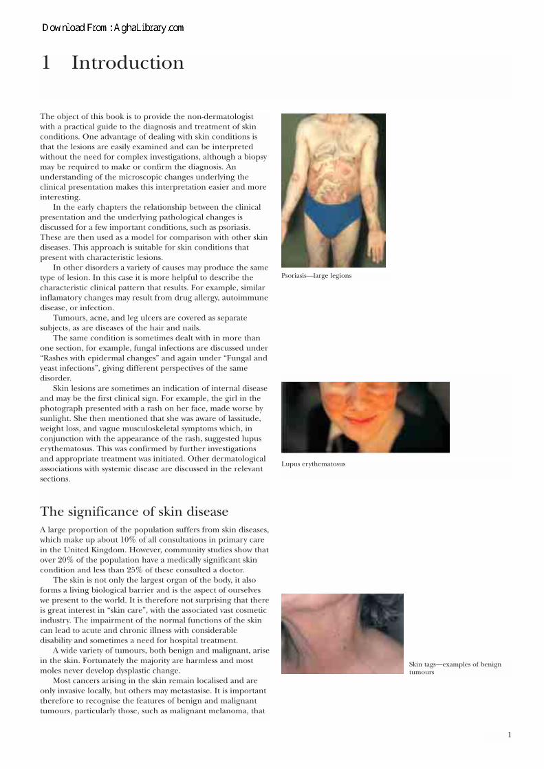
Skin lesions are sometimes an indication of internal diseaseand may be the first clinical sign. For example, the girl in thephotograph presented with a rash on her face, made worse bysunlight. She then mentioned that she was aware of lassitude,weight loss, and vague musculoskeletal symptoms which, inconjunction with the appearance of the rash, suggested lupuserythematosus. This was confirmed by further investigationsand appropriate treatment was initiated. Other dermatologicalassociations with systemic disease are discussed in the relevantsections.
The significance of skin diseaseA large proportion of the population suffers from skin diseases,which make up about 10% of all consultations in primary carein the United Kingdom. However, community studies show thatover 20% of the population have a medically significant skincondition and less than 25% of these consulted a doctor.
The skin is not only the largest organ of the body, it alsoforms a living biological barrier and is the aspect of ourselveswe present to the world. It is therefore not surprising that thereis great interest in “skin care”, with the associated vast cosmeticindustry. The impairment of the normal functions of the skincan lead to acute and chronic illness with considerabledisability and sometimes a need for hospital treatment.
A wide variety of tumours, both benign and malignant, arisein the skin. Fortunately the majority are harmless and mostmoles never develop dysplastic change.
Most cancers arising in the skin remain localised and areonly invasive locally, but others may metastasise. It is importanttherefore to recognise the features of benign and malignanttumours, particularly those, such as malignant melanoma, that
1
1 Introduction
Lupus erythematosus
Psoriasis—large legions
Skin tags—examples of benigntumours
can develop widespread metastases. Recognition of typicalbenign tumours saves the patient unneccessary investigationsand the anxiety involved in waiting for results.
Although a wide range of internal diseases produce physicalsigns in the skin, most skin diseases do not themselves haveserious physical effects. However there can be significantpsychological effects and problems with personal relationships,employment, and sporting activity. It is therefore important touse what Dr Papworth called “wide angle lenses” in assessingthe patient and their disease. So, in addition to concentratingon the skin changes, the overall health and demeanour of thepatient should be taken into account. This also means makingsure that there are no other signs, such as involvement of thenails, mucous membranes, or other parts of the skin. Thegeneral physical condition and psychological state of thepatient should be assessed, with more specific examination if indicated.
Descriptive termsAll specialties have their own common terms, and familiaritywith a few of those used in dermatology is a great help. Themost important are defined below.
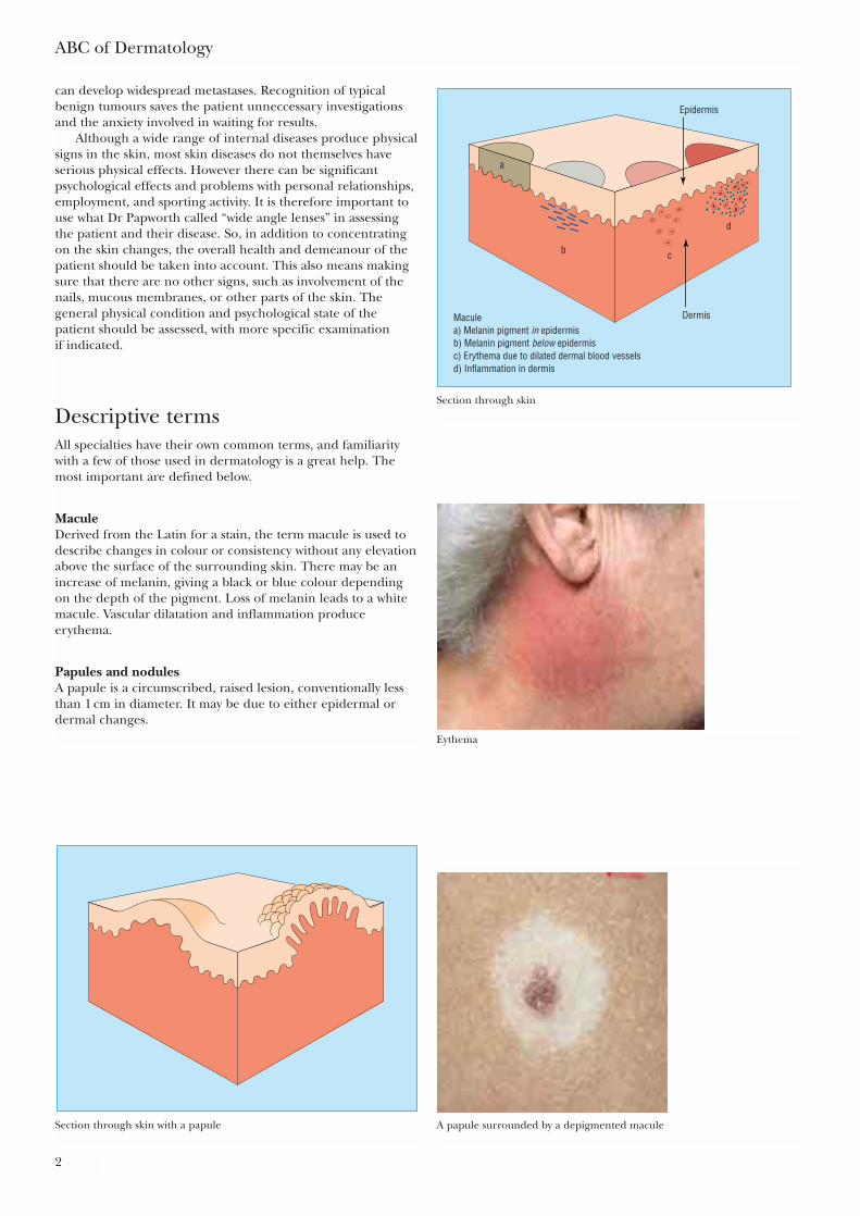
MaculeDerived from the Latin for a stain, the term macule is used todescribe changes in colour or consistency without any elevationabove the surface of the surrounding skin. There may be anincrease of melanin, giving a black or blue colour dependingon the depth of the pigment. Loss of melanin leads to a whitemacule. Vascular dilatation and inflammation produceerythema.
Papules and nodulesA papule is a circumscribed, raised lesion, conventionally lessthan 1 cm in diameter. It may be due to either epidermal ordermal changes.
ABC of Dermatology
2
Epidermis
a
b c
d
DermisMaculea) Melanin pigment in epidermisb) Melanin pigment below epidermisc) Erythema due to dilated dermal blood vesselsd) Inflammation in dermis
Section through skin
Eythema
Section through skin with a papule A papule surrounded by a depigmented macule
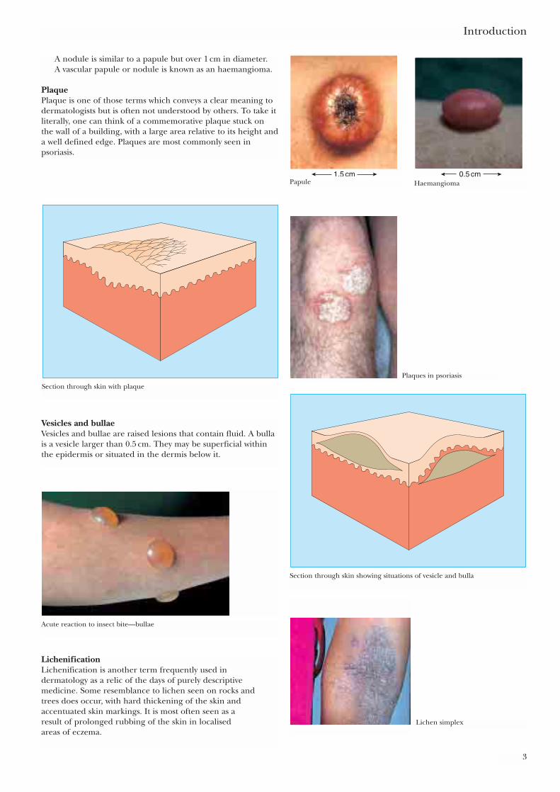
A nodule is similar to a papule but over 1 cm in diameter.A vascular papule or nodule is known as an haemangioma.
PlaquePlaque is one of those terms which conveys a clear meaning todermatologists but is often not understood by others. To take itliterally, one can think of a commemorative plaque stuck onthe wall of a building, with a large area relative to its height anda well defined edge. Plaques are most commonly seen inpsoriasis.
Introduction
3
1.5 cmPapule
Plaques in psoriasis
0.5 cmHaemangioma
Section through skin with plaque
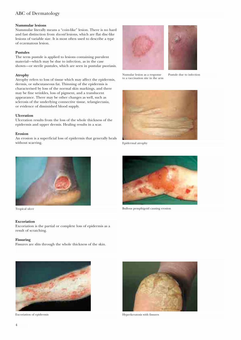
Vesicles and bullaeVesicles and bullae are raised lesions that contain fluid. A bullais a vesicle larger than 0.5 cm. They may be superficial withinthe epidermis or situated in the dermis below it.
Section through skin showing situations of vesicle and bulla
Acute reaction to insect bite—bullae
LichenificationLichenification is another term frequently used in dermatology as a relic of the days of purely descriptivemedicine. Some resemblance to lichen seen on rocks and trees does occur, with hard thickening of the skin and accentuated skin markings. It is most often seen as a result of prolonged rubbing of the skin in localised areas of eczema.
Lichen simplex
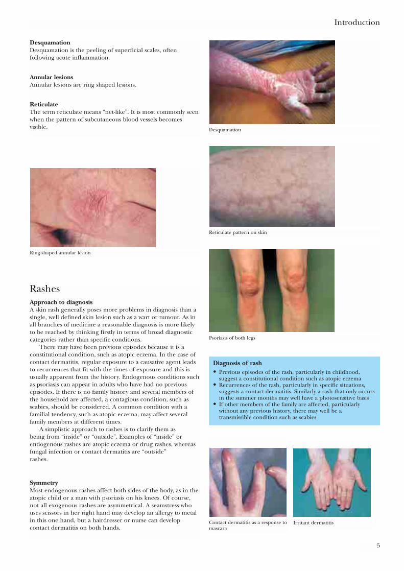
Nummular lesionsNummular literally means a “coin-like” lesion. There is no hardand fast distinction from discoid lesions, which are flat disc-likelesions of variable size. It is most often used to describe a typeof eczematous lesion.
PustulesThe term pustule is applied to lesions containing purulentmaterial—which may be due to infection, as in the caseshown—or sterile pustules, which are seen in pustular psoriasis.
AtrophyAtrophy refers to loss of tissue which may affect the epidermis,dermis, or subcutaneous fat. Thinning of the epidermis ischaracterised by loss of the normal skin markings, and theremay be fine wrinkles, loss of pigment, and a translucentappearance. There may be other changes as well, such assclerosis of the underlying connective tissue, telangiectasia, or evidence of diminished blood supply.
UlcerationUlceration results from the loss of the whole thickness of theepidermis and upper dermis. Healing results in a scar.
ErosionAn erosion is a superficial loss of epidermis that generally healswithout scarring.
ABC of Dermatology
4
Numular lesion as a responseto a vaccination site in the arm
Pustule due to infection
Epidermal atrophy
Tropical ulcer Bullous pemphigoid causing erosion
ExcoriationExcoriation is the partial or complete loss of epidermis as aresult of scratching.
FissuringFissures are slits through the whole thickness of the skin.
Excoriation of epidermis Hyperkeratosis with fissures
DesquamationDesquamation is the peeling of superficial scales, oftenfollowing acute inflammation.
Annular lesionsAnnular lesions are ring shaped lesions.
ReticulateThe term reticulate means “net-like”. It is most commonly seenwhen the pattern of subcutaneous blood vessels becomesvisible.
Introduction
5
Desquamation
Ring-shaped annular lesion
Reticulate pattern on skin
Psoriasis of both legs
RashesApproach to diagnosisA skin rash generally poses more problems in diagnosis than asingle, well defined skin lesion such as a wart or tumour. As inall branches of medicine a reasonable diagnosis is more likelyto be reached by thinking firstly in terms of broad diagnosticcategories rather than specific conditions.
There may have been previous episodes because it is aconstitutional condition, such as atopic eczema. In the case ofcontact dermatitis, regular exposure to a causative agent leadsto recurrences that fit with the times of exposure and this isusually apparent from the history. Endogenous conditions suchas psoriasis can appear in adults who have had no previousepisodes. If there is no family history and several members ofthe household are affected, a contagious condition, such asscabies, should be considered. A common condition with afamilial tendency, such as atopic eczema, may affect severalfamily members at different times.
A simplistic approach to rashes is to clarify them as being from “inside” or “outside”. Examples of “inside” orendogenous rashes are atopic eczema or drug rashes, whereasfungal infection or contact dermatitis are “outside” rashes.
SymmetryMost endogenous rashes affect both sides of the body, as in theatopic child or a man with psoriasis on his knees. Of course,not all exogenous rashes are asymmetrical. A seamstress whouses scissors in her right hand may develop an allergy to metalin this one hand, but a hairdresser or nurse can developcontact dermatitis on both hands.
Contact dermatitis as a response tomascara
Irritant dermatitis
Diagnosis of rash• Previous episodes of the rash, particularly in childhood,
suggest a constitutional condition such as atopic eczema• Recurrences of the rash, particularly in specific situations,
suggests a contact dermatitis. Similarly a rash that only occursin the summer months may well have a photosensitive basis
• If other members of the family are affected, particularlywithout any previous history, there may well be atransmissible condition such as scabies
DistributionIt is useful to be aware of the usual sites of common skinconditions. These are shown in the appropriate chapters.Eruptions that appear only on areas exposed to sun may beentirely or partially due to sunlight. Some are due to asensitivity to sunlight alone, such as polymorphous lighteruption, or a photosensitive allergy to topically appliedsubstances or drugs taken internally.
MorphologyThe appearance of the skin lesion may give clues to theunderlying pathological process.
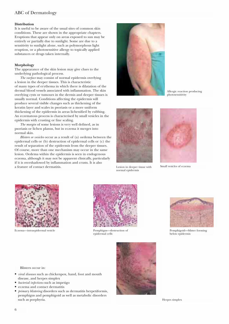
The surface may consist of normal epidermis overlying a lesion in the deeper tissues. This is characteristic of many types of erythema in which there is dilatation of thedermal blood vessels associated with inflammation. The skinoverlying cysts or tumours in the dermis and deeper tissues isusually normal. Conditions affecting the epidermis willproduce several visible changes such as thickening of thekeratin layer and scales in psoriasis or a more uniformthickening of the epidermis in areas lichenified by rubbing. An eczematous process is characterised by small vesicles in theepidermis with crusting or fine scaling.
The margin of some lesions is very well defined, as inpsoriasis or lichen planus, but in eczema it merges into normal skin.
Blisters or vesicles occur as a result of (a) oedema between theepidermal cells or (b) destruction of epidermal cells or (c) theresult of separation of the epidermis from the deeper tissues.Of course, more than one mechanism may occur in the samelesion. Oedema within the epidermis is seen in endogenouseczema, although it may not be apparent clinically, particularlyif it is overshadowed by inflammation and crusts. It is also a feature of contact dermatitis.
ABC of Dermatology
6
Allergic reaction producingphotosensitivity
Lesion in deeper tissue withnormal epidermis
Small vesicles of eczema
Eczema—intraepidermal vesicle Pemphigus—destruction ofepidermal cells
Pemphigoid—blister formingbelow epidermis
Blisters occur in:
• viral diseases such as chickenpox, hand, foot and mouthdisease, and herpes simplex
• bacterial infections such as impetigo • eczema and contact dermatitis• primary blistering disorders such as dermatitis herpetiformis,
pemphigus and pemphigoid as well as metabolic disorderssuch as porphyria. Herpes simplex
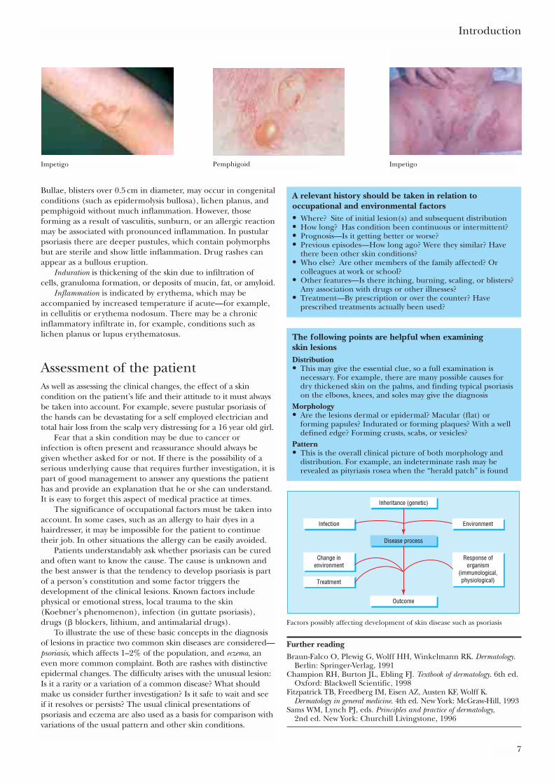
Bullae, blisters over 0.5 cm in diameter, may occur in congenitalconditions (such as epidermolysis bullosa), lichen planus, andpemphigoid without much inflammation. However, thoseforming as a result of vasculitis, sunburn, or an allergic reactionmay be associated with pronounced inflammation. In pustularpsoriasis there are deeper pustules, which contain polymorphsbut are sterile and show little inflammation. Drug rashes canappear as a bullous eruption.
Induration is thickening of the skin due to infiltration of cells, granuloma formation, or deposits of mucin, fat, or amyloid.
Inflammation is indicated by erythema, which may beaccompanied by increased temperature if acute—for example,in cellulitis or erythema nodosum. There may be a chronicinflammatory infiltrate in, for example, conditions such aslichen planus or lupus erythematosus.
Assessment of the patientAs well as assessing the clinical changes, the effect of a skincondition on the patient’s life and their attitude to it must alwaysbe taken into account. For example, severe pustular psoriasis ofthe hands can be devastating for a self employed electrician andtotal hair loss from the scalp very distressing for a 16 year old girl.
Fear that a skin condition may be due to cancer or infection is often present and reassurance should always begiven whether asked for or not. If there is the possibility of aserious underlying cause that requires further investigation, it ispart of good management to answer any questions the patienthas and provide an explanation that he or she can understand.It is easy to forget this aspect of medical practice at times.
The significance of occupational factors must be taken intoaccount. In some cases, such as an allergy to hair dyes in ahairdresser, it may be impossible for the patient to continuetheir job. In other situations the allergy can be easily avoided.
Patients understandably ask whether psoriasis can be curedand often want to know the cause. The cause is unknown andthe best answer is that the tendency to develop psoriasis is partof a person’s constitution and some factor triggers thedevelopment of the clinical lesions. Known factors includephysical or emotional stress, local trauma to the skin(Koebner’s phenomenon), infection (in guttate psoriasis),drugs (� blockers, lithium, and antimalarial drugs).
To illustrate the use of these basic concepts in the diagnosisof lesions in practice two common skin diseases are considered—psoriasis, which affects 1–2% of the population, and eczema, aneven more common complaint. Both are rashes with distinctiveepidermal changes. The difficulty arises with the unusual lesion:Is it a rarity or a variation of a common disease? What shouldmake us consider further investigation? Is it safe to wait and seeif it resolves or persists? The usual clinical presentations ofpsoriasis and eczema are also used as a basis for comparison withvariations of the usual pattern and other skin conditions.
Introduction
7
Impetigo Pemphigoid
A relevant history should be taken in relation tooccupational and environmental factors• Where? Site of initial lesion(s) and subsequent distribution• How long? Has condition been continuous or intermittent?• Prognosis—Is it getting better or worse?• Previous episodes—How long ago? Were they similar? Have
there been other skin conditions?• Who else? Are other members of the family affected? Or
colleagues at work or school?• Other features—Is there itching, burning, scaling, or blisters?
Any association with drugs or other illnesses?• Treatment—By prescription or over the counter? Have
prescribed treatments actually been used?
The following points are helpful when examining skin lesionsDistribution• This may give the essential clue, so a full examination is
necessary. For example, there are many possible causes fordry thickened skin on the palms, and finding typical psoriasison the elbows, knees, and soles may give the diagnosis
Morphology• Are the lesions dermal or epidermal? Macular (flat) or
forming papules? Indurated or forming plaques? With a welldefined edge? Forming crusts, scabs, or vesicles?
Pattern• This is the overall clinical picture of both morphology and
distribution. For example, an indeterminate rash may berevealed as pityriasis rosea when the “herald patch” is found
Inheritance (genetic)
Outcome
Disease process
EnvironmentInfection
Treatment
Response oforganism
(immunological,physiological)
Change inenvironment
Factors possibly affecting development of skin disease such as psoriasis
Further readingBraun-Falco O, Plewig G, Wolff HH, Winkelmann RK. Dermatology.
Berlin: Springer-Verlag, 1991Champion RH, Burton JL, Ebling FJ. Textbook of dermatology. 6th ed.
Oxford: Blackwell Scientific, 1998Fitzpatrick TB, Freedberg IM, Eisen AZ, Austen KF, Wolff K.
Dermatology in general medicine. 4th ed. New York: McGraw-Hill, 1993Sams WM, Lynch PJ, eds. Principles and practice of dermatology,
2nd ed. New York: Churchill Livingstone, 1996
Impetigo

8
The familiar pink or red lesions with a scaling surface and welldefined edge are easily recognised. These changes can berelated to the histological appearance:
2 Psoriasis
Increasedthickness ofepidermis
Thick keratinscale
Polymorphs
Dilated tortuousblood vessels
Increased epidermal proliferation—nuclei found … throughout the epidermis
Pitting of the nail
Plaques Plaques
Large lesions
Small lesions
• The increased thickness of the epidermis, presence of nucleiabove the basal layer, and thick keratin are related toincreased epidermal turnover.
• Because the epidermis is dividing it does not differentiateadequately into normal keratin scales. These are readilyremoved to reveal the tortuous blood vessels beneath,appearing clinically as “Auspitz sign”. The psoriatic plaque canbe likened to a brick wall badly built by a workman in toomuch of a hurry—it may be high but it is easily knocked down.
• The polymorphs that migrate into the epidermis form sterilepustules in pustular psoriasis. These are most commonly seenon the palms and soles.
• The dilated blood vessels can be a main feature, giving theclinical picture of intense erythema.
The equivalent changes in the nail cause thickening and “pits”0.5–1.0 mm in diameter on the surface; these are thought to bedue to small areas of psoriatic changes in the upper layer of thenail plate that then fall out. Onycholysis, in which the nail plateis raised up, also occurs in psoriasis.
Clinical appearanceThe main characteristics of psoriatic lesions, which reflect thepathological processes listed above, follow.
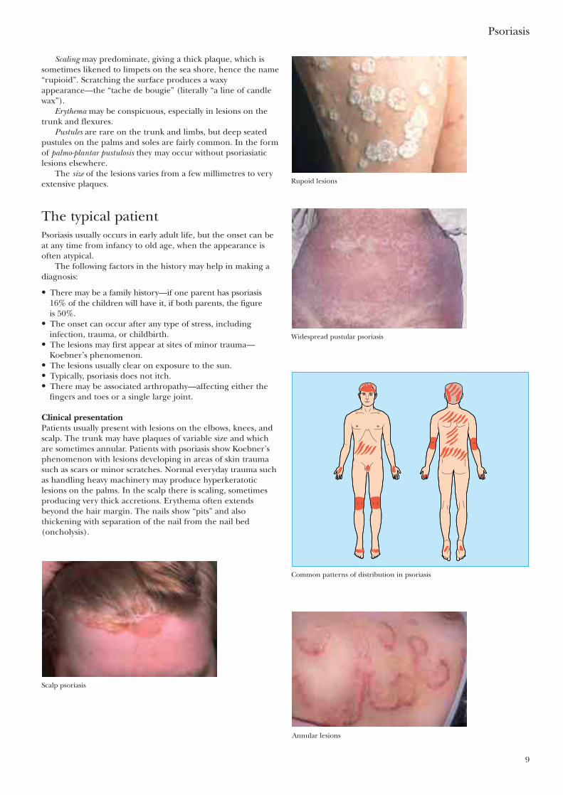
Plaques consisting of well defined raised areas of psoriasis.These may be few or numerous, covering large areas of thetrunk and limbs. Sometimes there are large confluent lesions.
Scaling may predominate, giving a thick plaque, which issometimes likened to limpets on the sea shore, hence the name“rupioid”. Scratching the surface produces a waxyappearance—the “tache de bougie” (literally “a line of candlewax”).
Erythema may be conspicuous, especially in lesions on thetrunk and flexures.
Pustules are rare on the trunk and limbs, but deep seatedpustules on the palms and soles are fairly common. In the formof palmo-plantar pustulosis they may occur without psoriasiaticlesions elsewhere.
The size of the lesions varies from a few millimetres to veryextensive plaques.
The typical patientPsoriasis usually occurs in early adult life, but the onset can beat any time from infancy to old age, when the appearance isoften atypical.
The following factors in the history may help in making adiagnosis:
• There may be a family history—if one parent has psoriasis16% of the children will have it, if both parents, the figure is 50%.
• The onset can occur after any type of stress, includinginfection, trauma, or childbirth.
• The lesions may first appear at sites of minor trauma—Koebner’s phenomenon.
• The lesions usually clear on exposure to the sun.• Typically, psoriasis does not itch.• There may be associated arthropathy—affecting either the
fingers and toes or a single large joint.
Clinical presentationPatients usually present with lesions on the elbows, knees, andscalp. The trunk may have plaques of variable size and whichare sometimes annular. Patients with psoriasis show Koebner’sphenomenon with lesions developing in areas of skin traumasuch as scars or minor scratches. Normal everyday trauma suchas handling heavy machinery may produce hyperkeratoticlesions on the palms. In the scalp there is scaling, sometimesproducing very thick accretions. Erythema often extendsbeyond the hair margin. The nails show “pits” and alsothickening with separation of the nail from the nail bed(oncholysis).
Psoriasis
9
Rupoid lesions
Widespread pustular psoriasis
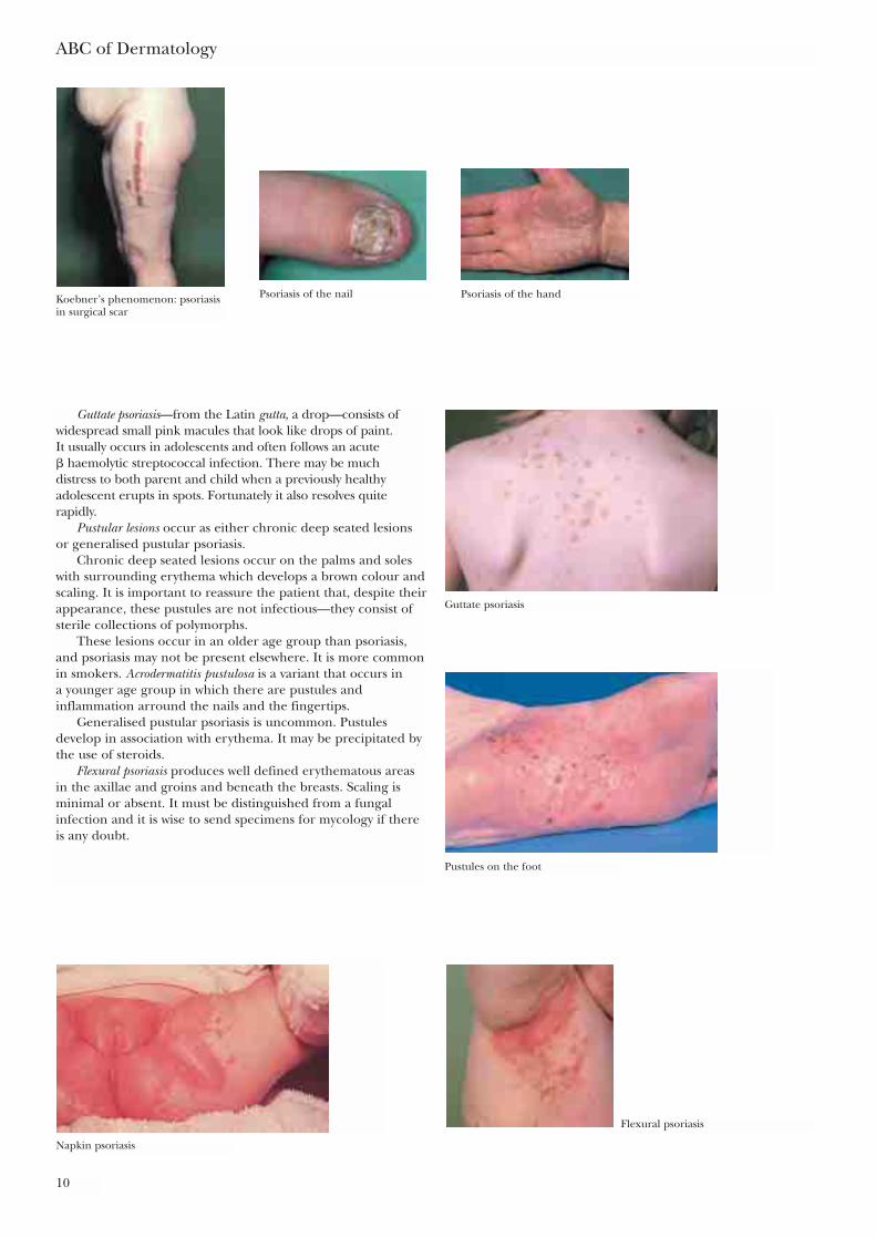
Common patterns of distribution in psoriasis
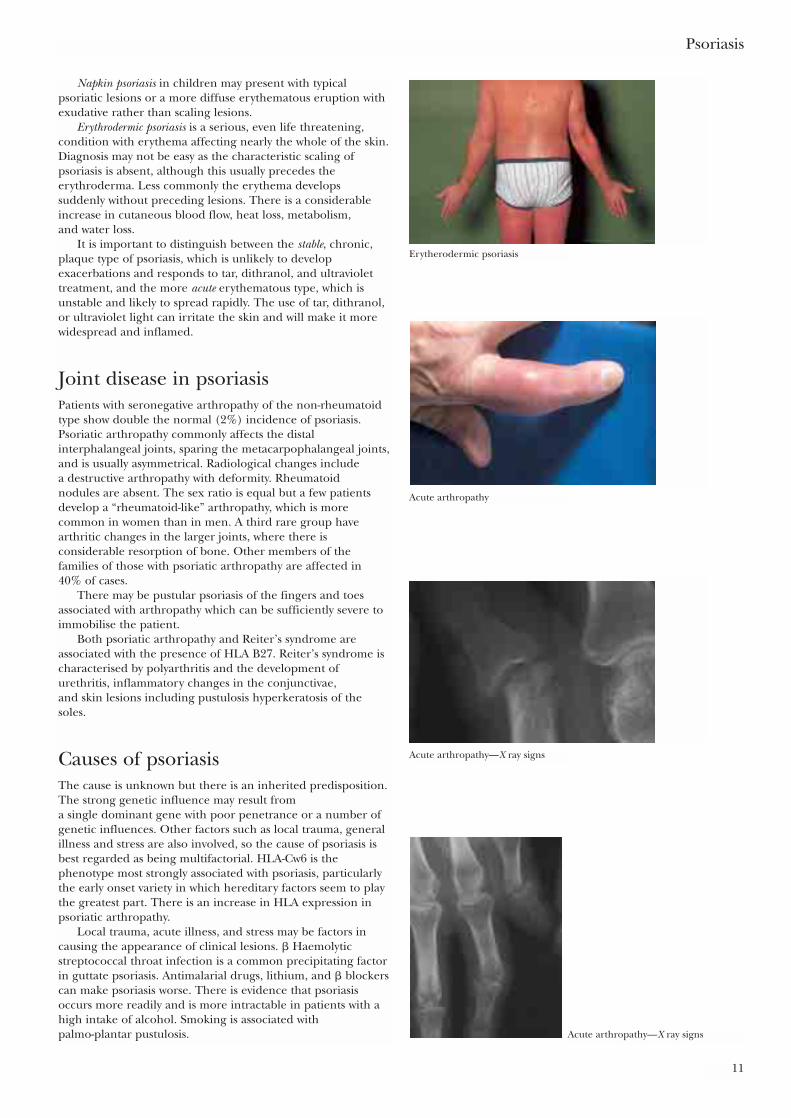
Annular lesions
Scalp psoriasis
Guttate psoriasis—from the Latin gutta, a drop—consists ofwidespread small pink macules that look like drops of paint. It usually occurs in adolescents and often follows an acute � haemolytic streptococcal infection. There may be much distress to both parent and child when a previously healthyadolescent erupts in spots. Fortunately it also resolves quiterapidly.
Pustular lesions occur as either chronic deep seated lesionsor generalised pustular psoriasis.
Chronic deep seated lesions occur on the palms and soleswith surrounding erythema which develops a brown colour andscaling. It is important to reassure the patient that, despite theirappearance, these pustules are not infectious—they consist ofsterile collections of polymorphs.
These lesions occur in an older age group than psoriasis,and psoriasis may not be present elsewhere. It is more commonin smokers. Acrodermatitis pustulosa is a variant that occurs in a younger age group in which there are pustules andinflammation arround the nails and the fingertips.
Generalised pustular psoriasis is uncommon. Pustulesdevelop in association with erythema. It may be precipitated bythe use of steroids.
Flexural psoriasis produces well defined erythematous areasin the axillae and groins and beneath the breasts. Scaling isminimal or absent. It must be distinguished from a fungalinfection and it is wise to send specimens for mycology if thereis any doubt.
ABC of Dermatology
10
Koebner’s phenomenon: psoriasisin surgical scar
Psoriasis of the nail Psoriasis of the hand
Guttate psoriasis
Pustules on the foot
Flexural psoriasis
Napkin psoriasis
Napkin psoriasis in children may present with typicalpsoriatic lesions or a more diffuse erythematous eruption withexudative rather than scaling lesions.
Erythrodermic psoriasis is a serious, even life threatening,condition with erythema affecting nearly the whole of the skin.Diagnosis may not be easy as the characteristic scaling ofpsoriasis is absent, although this usually precedes theerythroderma. Less commonly the erythema develops suddenly without preceding lesions. There is a considerableincrease in cutaneous blood flow, heat loss, metabolism, and water loss.
It is important to distinguish between the stable, chronic,plaque type of psoriasis, which is unlikely to developexacerbations and responds to tar, dithranol, and ultraviolettreatment, and the more acute erythematous type, which isunstable and likely to spread rapidly. The use of tar, dithranol,or ultraviolet light can irritate the skin and will make it morewidespread and inflamed.
Joint disease in psoriasisPatients with seronegative arthropathy of the non-rheumatoidtype show double the normal (2%) incidence of psoriasis.Psoriatic arthropathy commonly affects the distalinterphalangeal joints, sparing the metacarpophalangeal joints,and is usually asymmetrical. Radiological changes include a destructive arthropathy with deformity. Rheumatoid nodules are absent. The sex ratio is equal but a few patientsdevelop a “rheumatoid-like” arthropathy, which is morecommon in women than in men. A third rare group havearthritic changes in the larger joints, where there isconsiderable resorption of bone. Other members of thefamilies of those with psoriatic arthropathy are affected in 40% of cases.
There may be pustular psoriasis of the fingers and toesassociated with arthropathy which can be sufficiently severe toimmobilise the patient.
Both psoriatic arthropathy and Reiter’s syndrome areassociated with the presence of HLA B27. Reiter’s syndrome ischaracterised by polyarthritis and the development ofurethritis, inflammatory changes in the conjunctivae, and skin lesions including pustulosis hyperkeratosis of the soles.
Causes of psoriasisThe cause is unknown but there is an inherited predisposition.The strong genetic influence may result from a single dominant gene with poor penetrance or a number ofgenetic influences. Other factors such as local trauma, generalillness and stress are also involved, so the cause of psoriasis isbest regarded as being multifactorial. HLA-Cw6 is thephenotype most strongly associated with psoriasis, particularlythe early onset variety in which hereditary factors seem to playthe greatest part. There is an increase in HLA expression inpsoriatic arthropathy.
Local trauma, acute illness, and stress may be factors incausing the appearance of clinical lesions. � Haemolyticstreptococcal throat infection is a common precipitating factorin guttate psoriasis. Antimalarial drugs, lithium, and � blockerscan make psoriasis worse. There is evidence that psoriasisoccurs more readily and is more intractable in patients with ahigh intake of alcohol. Smoking is associated with palmo-plantar pustulosis.
Psoriasis
11
Erytherodermic psoriasis
Acute arthropathy
Acute arthropathy—X ray signs
Acute arthropathy—X ray signs
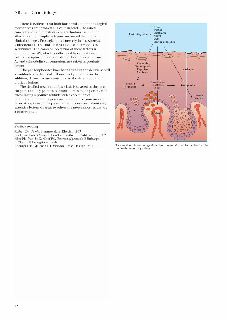
There is evidence that both hormonal and immunologicalmechanisms are involved at a cellular level. The raisedconcentrations of metabolites of arachodonic acid in theaffected skin of people with psoriasis are related to the clinical changes. Prostaglandins cause erythema, whereasleukotrienes (LTB4 and 12 HETE) cause neutrophils toaccumulate. The common precursor of these factors isphospholipase A2, which is influenced by calmodulin, a cellular receptor protein for calcium. Both phospholipase A2 and calmodulin concentrations are raised in psoriaticlesions.
T helper lymphocytes have been found in the dermis as wellas antibodies to the basal cell nuclei of psoriatic skin. Inaddition, dermal factors contribute to the development ofpsoriatic lesions.
The detailed treatment of psoriasis is covered in the nextchapter. The only point to be made here is the importance ofencouraging a positive attitude with expectation ofimprovement but not a permanent cure, since psoriasis canrecur at any time. Some patients are unconcerned about veryextensive lesions whereas to others the most minor lesions are a catastrophe.
ABC of Dermatology
12
StressInfectionLocal traumaAlcoholDrugsGenetic predisposition
Precipitating factors
Increasedproliferation Prostaglandins
Vasculardilatation
Leukotrienes
CalmodulinPhospholipase C
PolyaminesProteinases
T lymphocytesLeukotrienes
12 HETE
NeutrophilsNeutrophilsNeutrophils
Hormonal and immunological mechanisms and dermal factors involved inthe development of psoriasis
Further readingFarber EM. Psoriasis. Amsterdam: Elsevier, 1987Fry L. An atlas of psoriasis. London: Parthenon Publications, 1992Mier PD, Van de Kerkhof PC. Textbook of psoriasis. Edinburgh:
Churchill Livingstone, 1986Roenigk HH, Maibach HI. Psoriasis. Basle: Dekker, 1991
It vanished quite slowly, beginning with the end of the tail, and endingwith the grin, which remained some time after the rest of it had gone.
Lewis Carroll, Alice in Wonderland
To ignore the impact of the condition on the patient’s life is tofail in treating psoriasis. Like the Cheshire cat that Alice met, ittends to clear slowly and the last remaining patches are oftenthe hardest to clear. This is frustrating enough, but there is also the knowledge that it will probably recur and need furthertedious courses of treatment, so encouragement and supportare an essential part of treatment.
In an attempt to quantify the impact of psoriasis on the lifeof the individual patient the Psoriasis Disability Index (PDI) hasbeen developed. This takes the form of a questionnaire andcovers all aspects of the patient’s work, personal relationships,domestic situation, and recreational activities. It can be helpfulin assessing the effectiveness of treatment as perceived by thepatient.
Patients understandably ask whether psoriasis can be curedand often want to know the cause. The cause is unknown andthe best answer is that the tendency to develop psoriasis is partof an affected person’s constitution and some factor triggersthe development of the clinical lesions. Known factors includephysical or emotional stress, local trauma to the skin(Koebner’s phenomenon), infection (in guttate psoriasis),drugs (� blockers, lithium, and antimalarial drugs).
Treatment comprises ointments and pastes, systemic drugs,or various forms of ultraviolet light. The treatment should suitthe type of psoriasis. The age and health of the patient, socialand occupational factors need to be taken into consideration.The motivation of the individual patient is also important.
The preparations mentioned in the text are listed in theformulary in chapter 26. It is estimated that 80% of patientswith psoriasis do not consult a doctor, as the lesions areminimal.
13
3 Treatment of psoriasis
Treatment of psoriasis
Type of psoriasis Treatment Alternative treatmentStable plaque psoriasis Tar preparations Short contact dithranol
Calcipotriol � topical steroidsTacalcitolUltraviolet B (TL 01)
Extensive stable plaques As above. If not responding: Methotrexate Ultraviolet B (TL01) Ciclosporin Apsoralen with ultraviolet A � etretinate Tacrolimus
Widespread small plaque Ultraviolet B Tar
Guttate psoriasis Emollients then ultraviolet B Weak tar preparations
Facial psoriasis 1% hydrocortisone ointment
Flexural psoriasis Local mild to moderate strength steroids � antifungal
Pustular psoriasis of hands and feet Moderate to potent strength topical Acitretinsteroids
Acute erythrodermic, unstable, or Inpatient treatment Methotrexategeneral pustular psoriasis Short term local
steroids for acutely inflamed lesionsAcitretinCiclosporin or other immunosuppressants
Preparation applied to affected area (left). Application ofstockinette (right)
Bandages being applied to larger areas (left). Patient now preparedfor contact treatment (right)
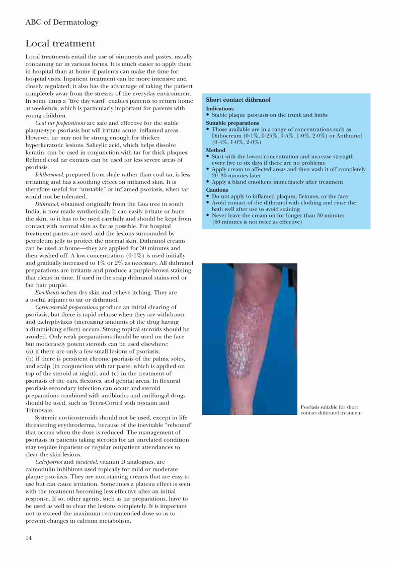
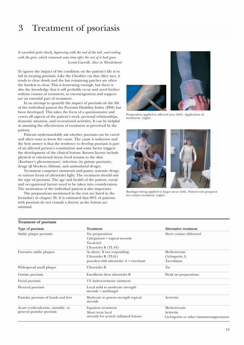
Local treatmentLocal treatments entail the use of ointments and pastes, usuallycontaining tar in various forms. It is much easier to apply themin hospital than at home if patients can make the time forhospital visits. Inpatient treatment can be more intensive andclosely regulated; it also has the advantage of taking the patientcompletely away from the stresses of the everyday environment.In some units a “five day ward” enables patients to return homeat weekends, which is particularly important for parents withyoung children.
Coal tar preparations are safe and effective for the stableplaque-type psoriasis but will irritate acute, inflamed areas.However, tar may not be strong enough for thickerhyperkeratotic lesions. Salicylic acid, which helps dissolvekeratin, can be used in conjunction with tar for thick plaques.Refined coal tar extracts can be used for less severe areas ofpsoriasis.
Ichthammol, prepared from shale rather than coal tar, is lessirritating and has a soothing effect on inflamed skin. It istherefore useful for “unstable” or inflamed psoriasis, when tarwould not be tolerated.
Dithranol, obtained originally from the Goa tree in southIndia, is now made synthetically. It can easily irritate or burnthe skin, so it has to be used carefully and should be kept fromcontact with normal skin as far as possible. For hospitaltreatment pastes are used and the lesions surrounded bypetroleum jelly to protect the normal skin. Dithranol creamscan be used at home—they are applied for 30 minutes andthen washed off. A low concentration (0·1%) is used initiallyand gradually increased to 1% or 2% as necessary. All dithranolpreparations are irritants and produce a purple-brown stainingthat clears in time. If used in the scalp dithranol stains red orfair hair purple.
Emollients soften dry skin and relieve itching. They are a useful adjunct to tar or dithranol.
Corticosteroid preparations produce an initial clearing ofpsoriasis, but there is rapid relapse when they are withdrawnand tachyphylaxis (increasing amounts of the drug having a diminishing effect) occurs. Strong topical steroids should beavoided. Only weak preparations should be used on the facebut moderately potent steroids can be used elsewhere: (a) if there are only a few small lesions of psoriasis; (b) if there is persistent chronic psoriasis of the palms, soles,and scalp (in conjunction with tar paste, which is applied ontop of the steroid at night); and (c) in the treatment ofpsoriasis of the ears, flexures, and genital areas. In flexuralpsoriasis secondary infection can occur and steroidpreparations combined with antibiotics and antifungal drugsshould be used, such as Terra-Cortril with nystatin andTrimovate.
Systemic corticosteroids should not be used, except in lifethreatening erythroderma, because of the inevitable “rebound”that occurs when the dose is reduced. The management ofpsoriasis in patients taking steroids for an unrelated conditionmay require inpatient or regular outpatient attendances toclear the skin lesions.
Calcipotriol and tacalcitol, vitamin D analogues, arecalmodulin inhibitors used topically for mild or moderateplaque psoriasis. They are non-staining creams that are easy touse but can cause irritation. Sometimes a plateau effect is seenwith the treatment becoming less effective after an initialresponse. If so, other agents, such as tar preparations, have tobe used as well to clear the lesions completely. It is importantnot to exceed the maximum recommended dose so as toprevent changes in calcium metabolism.
ABC of Dermatology
14
Short contact dithranolIndications• Stable plaque psoriasis on the trunk and limbsSuitable preparations• Those available are in a range of concentrations such as
Dithocream (0·1%, 0·25%, 0·5%, 1·0%, 2·0%) or Anthranol(0·4%, 1·0%, 2·0%)
Method• Start with the lowest concentration and increase strength
every five to six days if there are no problems• Apply cream to affected areas and then wash it off completely
20–30 minutes later• Apply a bland emollient immediately after treatmentCautions• Do not apply to inflamed plaques, flexures, or the face• Avoid contact of the dithranol with clothing and rinse the
bath well after use to avoid staining• Never leave the cream on for longer than 30 minutes
(60 minutes is not twice as effective)
Psoriasis suitable for shortcontact dithranol treatment
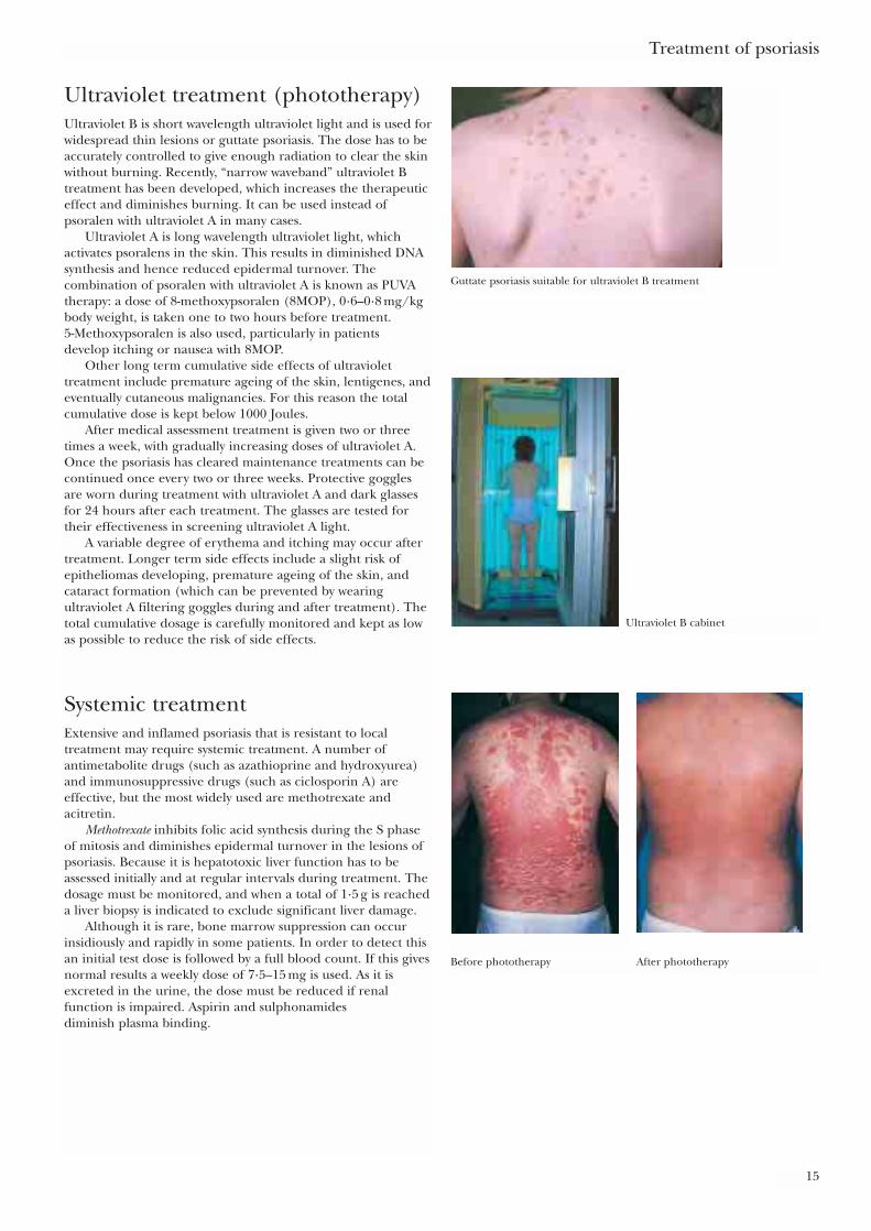
Ultraviolet treatment (phototherapy)Ultraviolet B is short wavelength ultraviolet light and is used forwidespread thin lesions or guttate psoriasis. The dose has to beaccurately controlled to give enough radiation to clear the skinwithout burning. Recently, “narrow waveband” ultraviolet Btreatment has been developed, which increases the therapeuticeffect and diminishes burning. It can be used instead ofpsoralen with ultraviolet A in many cases.
Ultraviolet A is long wavelength ultraviolet light, whichactivates psoralens in the skin. This results in diminished DNAsynthesis and hence reduced epidermal turnover. Thecombination of psoralen with ultraviolet A is known as PUVAtherapy: a dose of 8-methoxypsoralen (8MOP), 0·6–0·8 mg/kgbody weight, is taken one to two hours before treatment. 5-Methoxypsoralen is also used, particularly in patients develop itching or nausea with 8MOP.
Other long term cumulative side effects of ultraviolettreatment include premature ageing of the skin, lentigenes, andeventually cutaneous malignancies. For this reason the totalcumulative dose is kept below 1000 Joules.
After medical assessment treatment is given two or threetimes a week, with gradually increasing doses of ultraviolet A.Once the psoriasis has cleared maintenance treatments can becontinued once every two or three weeks. Protective gogglesare worn during treatment with ultraviolet A and dark glassesfor 24 hours after each treatment. The glasses are tested fortheir effectiveness in screening ultraviolet A light.
A variable degree of erythema and itching may occur aftertreatment. Longer term side effects include a slight risk ofepitheliomas developing, premature ageing of the skin, andcataract formation (which can be prevented by wearingultraviolet A filtering goggles during and after treatment). Thetotal cumulative dosage is carefully monitored and kept as lowas possible to reduce the risk of side effects.
Systemic treatmentExtensive and inflamed psoriasis that is resistant to localtreatment may require systemic treatment. A number ofantimetabolite drugs (such as azathioprine and hydroxyurea)and immunosuppressive drugs (such as ciclosporin A) areeffective, but the most widely used are methotrexate andacitretin.
Methotrexate inhibits folic acid synthesis during the S phaseof mitosis and diminishes epidermal turnover in the lesions ofpsoriasis. Because it is hepatotoxic liver function has to beassessed initially and at regular intervals during treatment. Thedosage must be monitored, and when a total of 1·5 g is reacheda liver biopsy is indicated to exclude significant liver damage.
Although it is rare, bone marrow suppression can occurinsidiously and rapidly in some patients. In order to detect thisan initial test dose is followed by a full blood count. If this givesnormal results a weekly dose of 7·5–15 mg is used. As it isexcreted in the urine, the dose must be reduced if renalfunction is impaired. Aspirin and sulphonamides diminish plasma binding.
Treatment of psoriasis
15
Guttate psoriasis suitable for ultraviolet B treatment
Ultraviolet B cabinet
Before phototherapy After phototherapy
Methotrexate may interact with barbiturates, para-aminobenzoic acid, phenytoin, probenecid, phenylbutazone,oral contraceptives, and colchicine.
Acitretin is a vitamin A derivative that can be prescribed onlyin hospital in the United Kingdom. It is useful in pustularpsoriasis and has some effect on other types of psoriasis.However, the effect is increased when combined with PUVA.Minor side effects include drying of the mucous membranes,crusting in the nose, itching, thinning of the hair, anderythema of the palms and nail folds. These are usually notsevere and settle when treatment stops. More serious sideeffects include hepatotoxicity and raised lipid concentrations.Liver function tests and serum lipid (cholesterol andtriglyceride) concentrations have to be carefully monitored.Etretinate is teratogenic and should only be taken by womenduring reproductive years if effective contraception is usedduring treatment and for two years afterwards, as the half life is 70–100 days.
Ciclosporin A is an immunosuppressant widely used followingorgan transplantation. It is effective in suppressing theinflammatory types of psoriasis. Blood tests should be carriedout before starting treatment, particularly serum creatinine,urea, and electrolytes, as ciclosporin A can interfere with renalfunction.
ABC of Dermatology
16
Further readingLowe NJ. Managing your psoriasis. London: Master Media, 1993Lowe NJ. Practical psoriasis therapy, 2nd ed. St Louis: Mosby, 1992
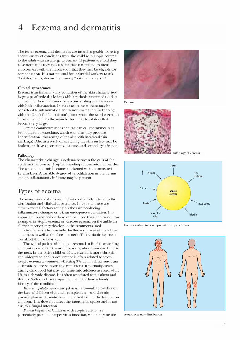
Psoriasis of the scalpThis condition can be very difficult to clear, particularly if there are thick scales
• 3% salicylic acid in a suitable base and left on for four to six hours or overnight and then washed out with a tar shampoo
• Dithranol preparations are effective but will tint blonde or red hair purple
• Steroid preparations can be used to control itching
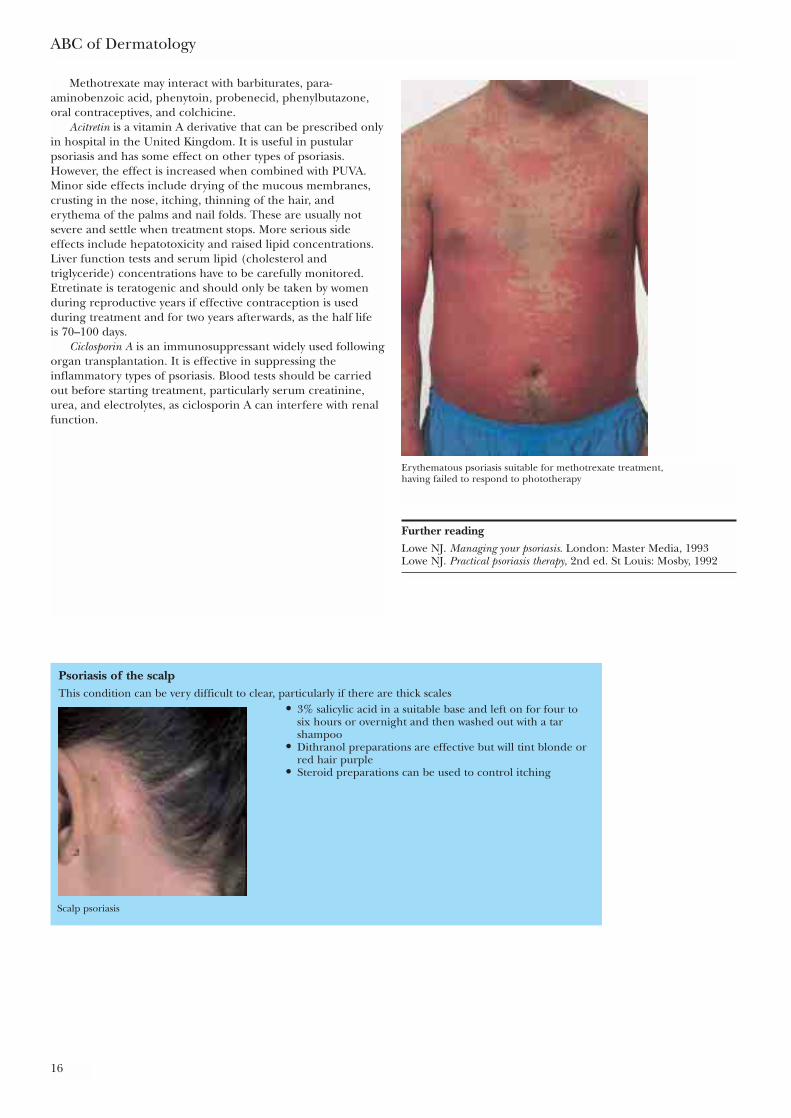
Erythematous psoriasis suitable for methotrexate treatment,having failed to respond to phototherapy
Scalp psoriasis
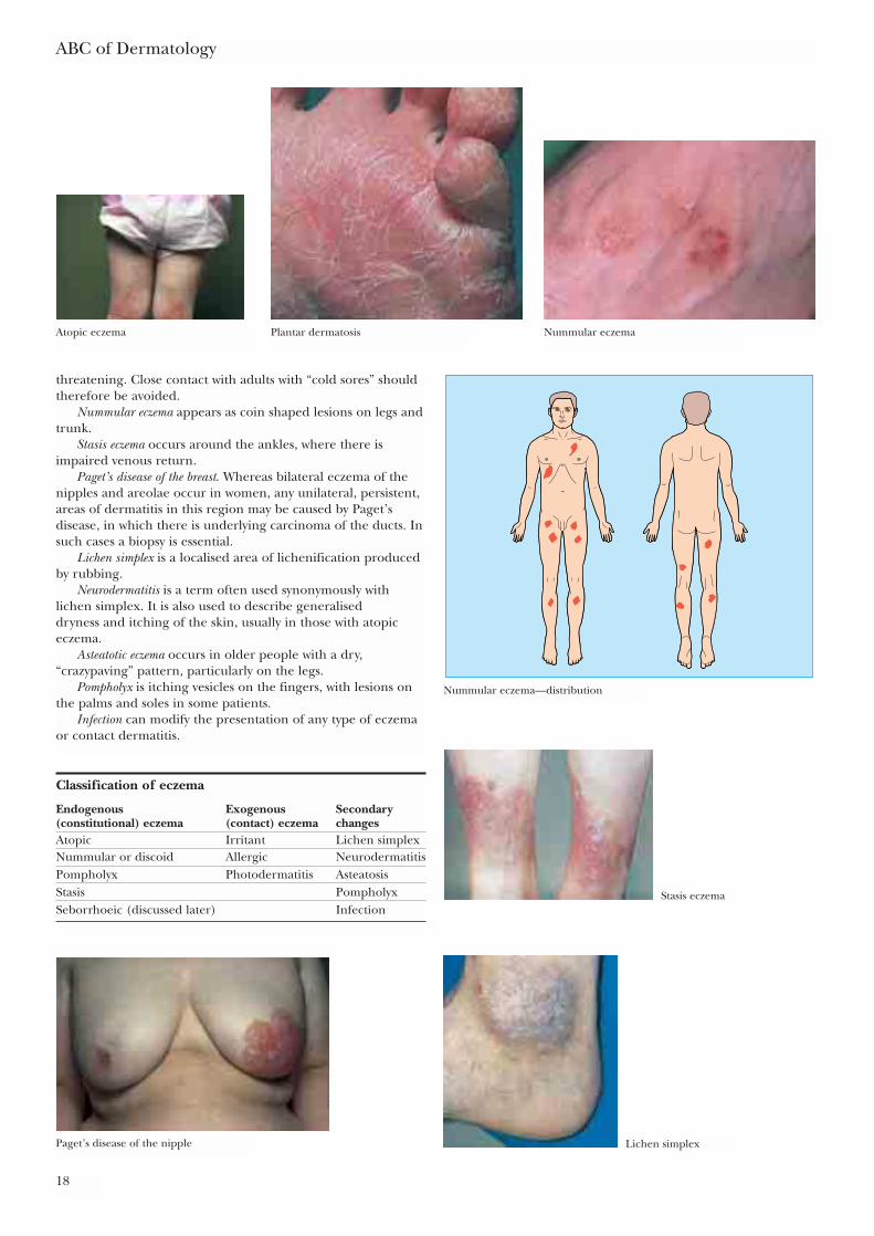
The terms eczema and dermatitis are interchangeable, coveringa wide variety of conditions from the child with atopic eczemato the adult with an allergy to cement. If patients are told theyhave dermatitis they may assume that it is related to theiremployment with the implication that they may be eligible forcompensation. It is not unusual for industrial workers to ask “Is it dermatitis, doctor?”, meaning “is it due to my job?”
Clinical appearanceEczema is an inflammatory condition of the skin characterisedby groups of vesicular lesions with a variable degree of exudateand scaling. In some cases dryness and scaling predominate,with little inflammation. In more acute cases there may beconsiderable inflammation and vesicle formation, in keepingwith the Greek for “to boil out”, from which the word eczema isderived. Sometimes the main feature may be blisters thatbecome very large.
Eczema commonly itches and the clinical appearance may be modified by scratching, which with time may producelichenification (thickening of the skin with increased skinmarkings). Also as a result of scratching the skin surface may bebroken and have excoriations, exudate, and secondary infection.
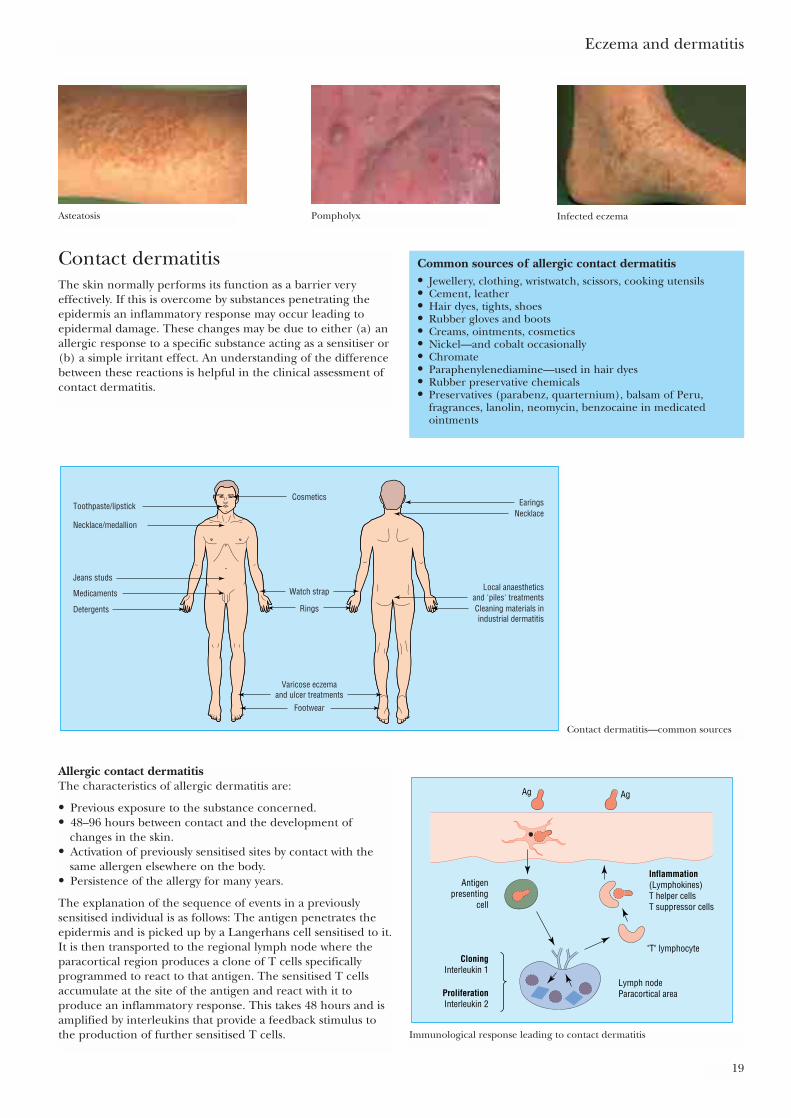
PathologyThe characteristic change is oedema between the cells of theepidermis, known as spongiosus, leading to formation of vesicles.The whole epidermis becomes thickened with an increasedkeratin layer. A variable degree of vasodilatation in the dermisand an inflammatory infiltrate may be present.
Types of eczemaThe many causes of eczema are not consistently related to thedistribution and clinical appearance. In general there areeither external factors acting on the skin producinginflammatory changes or it is an endogenous condition. It isimportant to remember there can be more than one cause—forexample, in atopic eczema or varicose eczema on the ankle anallergic reaction may develop to the treatments used.
Atopic eczema affects mainly the flexor surfaces of the elbowsand knees as well as the face and neck. To a variable degree itcan affect the trunk as well.
The typical patient with atopic eczema is a fretful, scratchingchild with eczema that varies in severity, often from one hour tothe next. In the older child or adult, eczema is more chronicand widespread and its occurrence is often related to stress.Atopic eczema is common, affecting 3% of all infants, and runsa chronic course with variable remissions. It normally clearsduring childhood but may continue into adolescence and adultlife as a chronic disease. It is often associated with asthma andrhinitis. Sufferers from atopic eczema often have a familyhistory of the condition.
Variants of atopic eczema are pityriasis alba—white patches onthe face of children with a fair complexion—and chronicjuvenile plantar dermatosis—dry cracked skin of the forefoot inchildren. This does not affect the interdigital spaces and is notdue to a fungal infection.
Eczema herpeticum. Children with atopic eczema areparticularly prone to herpes virus infection, which may be life
17
4 Eczema and dermatitis
Eczema
Pathology of eczema
Stress
Atopiceczema
Skinirritation
Inoculations
Infection
Foods
Climate
Sweating
House dustmite
Factors leading to development of atopic eczema
Atopic eczema—distribution
threatening. Close contact with adults with “cold sores” shouldtherefore be avoided.
Nummular eczema appears as coin shaped lesions on legs andtrunk.
Stasis eczema occurs around the ankles, where there isimpaired venous return.
Paget’s disease of the breast. Whereas bilateral eczema of thenipples and areolae occur in women, any unilateral, persistent,areas of dermatitis in this region may be caused by Paget’sdisease, in which there is underlying carcinoma of the ducts. Insuch cases a biopsy is essential.
Lichen simplex is a localised area of lichenification producedby rubbing.
Neurodermatitis is a term often used synonymously withlichen simplex. It is also used to describe generalised dryness and itching of the skin, usually in those with atopiceczema.
Asteatotic eczema occurs in older people with a dry,“crazypaving” pattern, particularly on the legs.
Pompholyx is itching vesicles on the fingers, with lesions onthe palms and soles in some patients.
Infection can modify the presentation of any type of eczemaor contact dermatitis.
ABC of Dermatology
18
Classification of eczema
Endogenous Exogenous Secondary (constitutional) eczema (contact) eczema changesAtopic Irritant Lichen simplexNummular or discoid Allergic NeurodermatitisPompholyx Photodermatitis AsteatosisStasis PompholyxSeborrhoeic (discussed later) Infection
Atopic eczema Plantar dermatosis Nummular eczema
Nummular eczema—distribution
Stasis eczema
Paget’s disease of the nipple Lichen simplex
Contact dermatitisThe skin normally performs its function as a barrier veryeffectively. If this is overcome by substances penetrating theepidermis an inflammatory response may occur leading toepidermal damage. These changes may be due to either (a) anallergic response to a specific substance acting as a sensitiser or(b) a simple irritant effect. An understanding of the differencebetween these reactions is helpful in the clinical assessment ofcontact dermatitis.
Eczema and dermatitis
19
Asteatosis Pompholyx Infected eczema
Common sources of allergic contact dermatitis• Jewellery, clothing, wristwatch, scissors, cooking utensils• Cement, leather• Hair dyes, tights, shoes• Rubber gloves and boots• Creams, ointments, cosmetics• Nickel—and cobalt occasionally• Chromate• Paraphenylenediamine—used in hair dyes• Rubber preservative chemicals• Preservatives (parabenz, quarternium), balsam of Peru,
fragrances, lanolin, neomycin, benzocaine in medicatedointments
Toothpaste/lipstick
Necklace/medallion
Jeans studs
Medicaments
Detergents
EaringsNecklace
Local anaestheticsand 'piles' treatmentsCleaning materials inindustrial dermatitis
Footwear
Rings
Watch strap
Cosmetics
Varicose eczemaand ulcer treatments
Contact dermatitis—common sources
Allergic contact dermatitisThe characteristics of allergic dermatitis are:
• Previous exposure to the substance concerned.• 48–96 hours between contact and the development of
changes in the skin.• Activation of previously sensitised sites by contact with the
same allergen elsewhere on the body.• Persistence of the allergy for many years.
The explanation of the sequence of events in a previouslysensitised individual is as follows: The antigen penetrates theepidermis and is picked up by a Langerhans cell sensitised to it.It is then transported to the regional lymph node where theparacortical region produces a clone of T cells specificallyprogrammed to react to that antigen. The sensitised T cellsaccumulate at the site of the antigen and react with it toproduce an inflammatory response. This takes 48 hours and isamplified by interleukins that provide a feedback stimulus tothe production of further sensitised T cells.
Ag
"T" lymphocyte
Lymph nodeParacortical area
Inflammation(Lymphokines)T helper cellsT suppressor cells
CloningInterleukin 1
ProliferationInterleukin 2
Ag
Antigenpresenting
cell
Immunological response leading to contact dermatitis
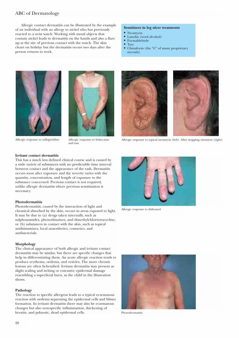
Allergic contact dermatitis can be illustrated by the exampleof an individual with an allergy to nickel who has previouslyreacted to a wrist watch. Working with metal objects thatcontain nickel leads to dermatitis on the hands and also a flareup at the site of previous contact with the watch. The skinclears on holiday but the dermatitis recurs two days after theperson returns to work.
ABC of Dermatology
20
Sensitisers in leg ulcer treatments• Neomycin• Lanolin (wool alcohol)• Formaldehyde• Tars• Chinaform (the “C” of many proprietary
steroids)
Allergic response to sulfapyridine Allergic response to Solarcaine and sun
Allergic response to topical neomycin (left). After stopping ointment (right)
Irritant contact dermatitisThis has a much less defined clinical course and is caused by a wide variety of substances with no predictable time intervalbetween contact and the appearance of the rash. Dermatitisoccurs soon after exposure and the severity varies with thequantity, concentration, and length of exposure to thesubstance concerned. Previous contact is not required, unlike allergic dermatitis where previous sensitisation isnecessary.
PhotodermatitisPhotodermatitis, caused by the interaction of light andchemical absorbed by the skin, occurs in areas exposed to light.It may be due to (a) drugs taken internally, such assulphonamides, phenothiazines, and dimethylchlortetracycline,or (b) substances in contact with the skin, such as topicalantihistamines, local anaesthetics, cosmetics, and antibacterials.
MorphologyThe clinical appearance of both allergic and irritant contactdermatitis may be similar, but there are specific changes thathelp in differentiating them. An acute allergic reaction tends toproduce erythema, oedema, and vesicles. The more chroniclesions are often lichenified. Irritant dermatitis may present asslight scaling and itching or extensive epidermal damageresembling a superficial burn, as the child in the illustrationshows.
PathologyThe reaction to specific allergens leads to a typical eczematousreaction with oedema separating the epidermal cells and blisterformation. In irritant dermatitis there may also be eczematouschanges but also non-specific inflammation, thickening ofkeratin, and pyknotic, dead epidermal cells. Photodermatitis
Allergic response to dithranol
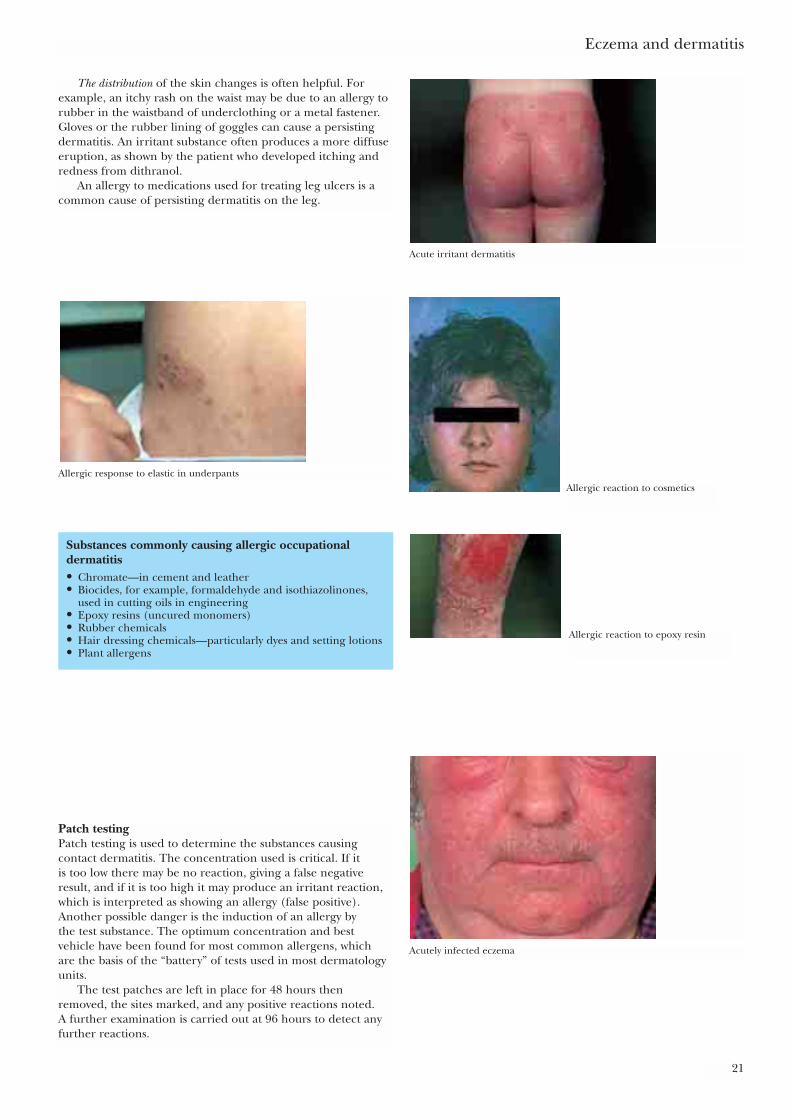
The distribution of the skin changes is often helpful. Forexample, an itchy rash on the waist may be due to an allergy torubber in the waistband of underclothing or a metal fastener.Gloves or the rubber lining of goggles can cause a persistingdermatitis. An irritant substance often produces a more diffuseeruption, as shown by the patient who developed itching andredness from dithranol.
An allergy to medications used for treating leg ulcers is acommon cause of persisting dermatitis on the leg.
Eczema and dermatitis
21
Acute irritant dermatitis
Substances commonly causing allergic occupationaldermatitis• Chromate—in cement and leather• Biocides, for example, formaldehyde and isothiazolinones,
used in cutting oils in engineering• Epoxy resins (uncured monomers)• Rubber chemicals• Hair dressing chemicals—particularly dyes and setting lotions• Plant allergens
Allergic response to elastic in underpants
Patch testingPatch testing is used to determine the substances causingcontact dermatitis. The concentration used is critical. If it is too low there may be no reaction, giving a false negativeresult, and if it is too high it may produce an irritant reaction,which is interpreted as showing an allergy (false positive).Another possible danger is the induction of an allergy by the test substance. The optimum concentration and bestvehicle have been found for most common allergens, which are the basis of the “battery” of tests used in most dermatologyunits.
The test patches are left in place for 48 hours thenremoved, the sites marked, and any positive reactions noted. A further examination is carried out at 96 hours to detect anyfurther reactions.
Allergic reaction to epoxy resin
Allergic reaction to cosmetics
Acutely infected eczema
It is most important not to put a possible causativesubstance on the skin in a random manner without properdilution and without control patches. The results will bemeaningless and irritant reactions, which are unpleasant forthe patient, may occur.
ABC of Dermatology
22
Test patches in place
Patches being removed after 48 hours
Positive reactions marked
Occupational dermatitisDermatitis, which is simply inflammation of the skin, can ariseas a result of:
In the workplace, all three factors may contribute to dermatitis.For example, a student nurse or trainee hairdresser is exposedto water, detergents, and other factors that will exacerbate anypre-existing eczema. In addition, there may be specific allergiesand, as a result of the broken skin, secondary infection canoccur making the situation even worse. The following pointsare helpful in determining the role of occupational causes.
• If the dermatitis first occurred during employment or with achange of employment and had not been present before,then occupational factors are more likely.
• If the condition generally clears during holidays and whenaway from the workplace, this suggests an occupational cause,but chronic irritant dermatitis may persist when the patient isaway from the workplace.
• If there is exposure to substances that are known to inducedermatitis and protective measures are inadequate at theworkplace then an occupational cause is likely.
• If secondary infection is present, this can keep a dermatitisactive even when away from the workplace and sometimesallergen exposure continues at home; for example, an allergy
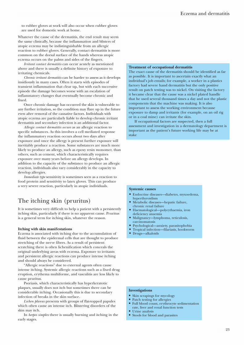
Contact with irritant Time
Contact with irritant
Acute
Damage Repair
"Failure" ofrepair mechanism
Skin clinicallynormal but
physiologicallyabnormal
Skin clinicallyabnormal
Chronic
Impa
irmen
t in
func
tion
of s
tratu
m c
orne
umIm
pairm
ent i
n fu
nctio
nof
stra
tum
cor
neum
Progression of acute and chronic dermatitis
• Inherited tendency to eczema (atopy)• Contact dermatitis, which may be either irritant or allergic• Infection.
to rubber gloves at work will also occur when rubber glovesare used for domestic work at home.
Whatever the cause of the dermatitis, the end result may seemthe same clinically, because the inflammation and blisters ofatopic eczema may be indistinguishable from an allergicreaction to rubber gloves. Generally, contact dermatitis is morecommon on the dorsal surface of the hands whereas atopiceczema occurs on the palms and sides of the fingers.
Irritant contact dermatitis can occur acutely as mentionedabove and there is usually a definite history of exposure toirritating chemicals.
Chronic irritant dermatitis can be harder to assess as it developsinsidiously in many cases. Often it starts with episodes oftransient inflammation that clear up, but with each successiveepisode the damage becomes worse with an escalation ofinflammatory changes that eventually become chronic andfixed.
Once chronic damage has occurred the skin is vulnerable toany further irritation, so the condition may flare up in the futureeven after removal of the causative factors. Individuals withatopic eczema are particularly liable to develop chronic irritantdermatitis and secondary infection is an additional factor.
Allergic contact dermatitis occurs as an allergic reaction tospecific substances. As this involves a cell mediated responsethe inflammatory reaction occurs about two days after exposure and once the allergy is present further exposure willinevitably produce a reaction. Some substances are much morelikely to produce an allergy, such as epoxy resin monomer, thanothers, such as cement, which characteristically requiresexposure over many years before an allergy develops. Inaddition to the capacity of the substance to produce an allergicreaction, individuals also vary considerably in the capacity todevelop allergies.
Immediate type sensitivity is sometimes seen as a reaction tofood protein and sensitivity to latex gloves. This can produce a very severe reaction, particularly in atopic individuals.
The itching skin (pruritus)It is sometimes very difficult to help a patient with a persistentlyitching skin, particularly if there is no apparent cause. Pruritusis a general term for itching skin, whatever the reason.
Itching with skin manifestationsEczema is associated with itching due to the accumulation offluid between the epidermal cells that are thought to producestretching of the nerve fibres. As a result of persistentscratching there is often lichenification which conceals theoriginal underlying areas with eczema. Exposure to irritantsand persistent allergic reactions can produce intense itchingand should always be considered.
“Allergic reactions” due to external agents often causeintense itching. Systemic allergic reactions such as a fixed drugeruption, erythema multiforme, and vasculitis are less likely tocause pruritus.
Psoriasis, which characteristically has hyperkeratoticplaques, usually does not itch but sometimes there can beconsiderable itching. Occasionally this is due to secondaryinfection of breaks in the skin surface.
Lichen planus presents with groups of flat-topped papuleswhich often cause an intense itch. Blistering disorders of theskin may itch.
In herpes simplex there is usually burning and itching in theearly stages.
Eczema and dermatitis
23
Systemic causes• Endocrine diseases—diabetes, myxoedema,
hyperthyroidism• Metabolic diseases—hepatic failure,
chronic renal failure• Haematological—polycythaemia, iron
deficiency anaemia• Malignancy—lymphoma, reticulosis,
carcinomatosis• Psychological—anxiety, parasitophobia• Tropical infection—filariasis, hookworm• Drugs—alkaloids
Treatment of occupational dermatitisThe exact cause of the dermatitis should be identified as faras possible. It is important to ascertain exactly what anindividual’s job entails; for example, a worker in a plasticsfactory had severe hand dermatitis but the only positiveresult on patch testing was to nickel. On visiting the factoryit became clear that the cause was a nickel plated handlethat he used several thousand times a day and not the plasticcomponents that the machine was making. It is alsoimportant to assess the working environment becauseexposure to damp and irritants (for example, on an oil rigor in a coal mine) can irritate the skin.
If occupational factors are suspected, then a fullassessment and investigation in a dermatology department isimportant as the patient’s future working life may be at stake
Investigations• Skin scrapings for mycology• Patch testing for allergies• Full blood count, erythrocyte sedimentation
rate, liver and renal function tests• Urine analysis• Stools for blood and parasites
In herpes zoster there may be a variable degree of itching, butthis is overshadowed by the pain and discomfort of the fullydeveloped lesions.
By contrast, bullous impetigo causes few symptoms, althoughthere may be extensive blisters. Itching is usually not present.
Dermatitis herpetiformis is characterised by intense persistentand severe itching that patients often describe as beingunendurable. Usual measures such as topical steroids andantihistamines have little if any effect.
By contrast, the blisters of pemphigoid do not itch althoughthe earlier inflamed lesions can be irritating.
Parasites. Fleas and mites cause pruritic papules in groups.The patient may not realise that they may have been acquiredafter a walk in the country or encountering a dog or cat.Nodular prurigo may develop after insect bites and ischaracterised by persistent itching, lichenified papules, andnodules over the trunk and limbs. The patient attacks themvigorously and promotes a persisting “itch–scratch–itch” cyclewhich is very difficult to break.
Parasitophobia is characterised by the patient reporting thepresence of small insects burrowing into the skin which persistsdespite all forms of treatment. The patient will produce smallflakes of skin, fibres of clothing, and pieces of dust, usually incarefully folded pieces of paper, for examination. These shouldalways be examined and the patient gently informed that noinsect could be found but this will not be believed. Treatment istherefore very difficult and sometimes recourse has to be hadto psychotropic drugs (see page 106).
Infestations with lice cause irritation and a scabies mite cancause widespread persistent pruritus, even though only a dozenor so active scabies burrows are present. It is always acquired byclose human contact and the diagnosis may be missed unless anadequate history of personal contacts and a thorough clinicalexamination is carried out. However, a speculative diagnosis ofscabies should be avoided.
Itching with no skin lesionsIf no dermatological lesions are present generalised pruritus oritchy skin may indicate an underlying internal cause. In elderlypatients, however, the skin may itch simply because it is dry.Hodgkin’s disease may present with pruritus as a sign of theinternal malignancy long before any other manifestations. A 35 year old ambulance driver attended the dermatologyclinic with intense itching but a normal skin and no history ofskin disease. His general health was good and both physicalexamination and all blood tests were normal. However, a chestx ray examination showed a mediastinal shadow that was foundto be due to Hodgkin’s lymphoma. Fortunately this was easilytreated. Other forms of carcinoma rarely cause pruritus.
Metabolic and endocrine diseaseBiliary obstruction and chronic renal disease cause intense pruritus.Thyroid disease can be associated with an itching skin. Inhyperthyroidism the skin seems normal but in hypothyroidismthere is dryness of the skin causing pruritus.
Blood diseases. Polycythaemia and iron deficiency aresometimes associated with itching skin.
ABC of Dermatology
24
TreatmentTreatment of the cause must be carried out when possible.Calamine lotion cools the skin with 0·5% menthol or 1%phenol in aqueous cream. Camphor-containing preparationsand crotamiton (Eurax) are also helpful. Topical steroidointments and occlusive dressings may help to preventscratching and may help to break the itch–scratch–itch cycle.Emollients should be used for dry skin.
Topical local anaesthetics may give relief but intolerancedevelops and they can cause allergic reactions. Sedativeantihistamines at night may be helpful. In liver failurecholestyramine powder may help to relieve the intensepruritus, as this is thought to be due to bile salts in the skin.
Antihistamines can be helpful both for their antipruriticeffect and because many are sedative and enable the itchingpatient to sleep.
Pruritus ani is a common troublesome condition and thefollowing points may be helpful:
• Advise gentle cleaning once daily and patients should beadvised to avoid excessive washing.
• Avoid harsh toilet paper, especially if coloured, becausecheap dyes irritate and cause allergies. Olive oil and cottonwool can be used instead.
• Weak topical steroids will help to reduce inflammation, withzinc cream or ointment as a protective layer on top.
• Anal leakage from an incompetent sphincter, skin tags, orhaemorrhoids may require surgical treatment.
• There may be an anxiety or depression and prutitus ani itselfcan lead to irritability and depression.
Pruritus vulvae is a persistent irritation of the vulva whichcan be most distressing and is most common inpostmenopausal women. It is important to eliminate any factors that may be preventing resolution. These include:
• Secondary infection with pyogenic bacteria or yeasts• Eczema or contact dermatitis• Lichen sclerosus atrophicus.
The adjacent vaginal mucosa should be examined to excludean intraepithelial neoplasm or lichen planus. Treatmentincludes suitable antiseptic preparations such as 2% eosin,regular but not excessive washing, emollients, and topicalsteroids, bearing in mind the possibility of infection.
Further readingAdams RM. Occupational skin disease, 2nd ed. Philadelphia:
Saunders, 1990Arndt KA. Manual of dermatological therapeutics, 5th ed. New York:
Little, 1995Atherton DJ. Eczema in childhood. The facts. Oxford: Oxford
University Press, 1994Cronin E. Contact dermatitis. Edinburgh: Churchill Livingstone,
1980Fisher AA. Contact dermatitis, 3rd ed. Baltimore: Williams and
Wilkins, 1986Foussereau J, Benezra JE, Maibach H. Occupational contact dermatitis.
Copenhagen: Munksgaard, 1982Schwanitz HJ. Atopic palmoplantar eczema. Berlin: Springer-Verlag,
1988
25
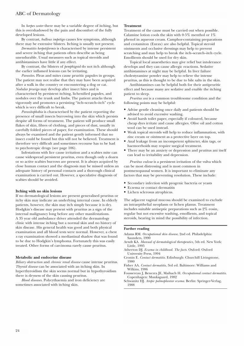
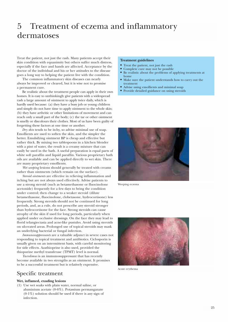
Treat the patient, not just the rash. Many patients accept theirskin condition with equanimity but others suffer much distress,especially if the face and hands are affected. Acceptance by thedoctor of the individual and his or her attitudes to the diseasegoes a long way to helping the patient live with the condition.
The common inflammatory skin diseases can nearly always be improved or cleared, but it is wise not to promise a permanent cure.
Be realistic about the treatment people can apply in their ownhomes. It is easy to unthinkingly give patients with a widespreadrash a large amount of ointment to apply twice daily, which ishardly used because: (a) they have a busy job or young childrenand simply do not have time to apply ointment to the whole skin;(b) they have arthritic or other limitations of movement and canreach only a small part of the body; (c) the tar or other ointmentis smelly or discolours their clothes. Most of us have been guilty offorgetting these factors at one time or another.
Dry skin tends to be itchy, so advise minimal use of soap.Emollients are used to soften the skin, and the simpler thebetter. Emulsifying ointment BP is cheap and effective butrather thick. By mixing two tablespoons in a kitchen blenderwith a pint of water, the result is a creamy mixture that caneasily be used in the bath. A useful preparation is equal parts ofwhite soft paraffin and liquid paraffin. Various proprietary bathoils are available and can be applied directly to wet skin. Thereare many proprietary emollients.
Wet weeping lesions should generally be treated with creamsrather than ointments (which remain on the surface).
Steroid ointments are effective in relieving inflammation anditching but are not always used effectively. Advise patients touse a strong steroid (such as betamethasone or fluocinoloneacetonide) frequently for a few days to bring the conditionunder control; then change to a weaker steroid (dilutebetamethasone, fluocinolone, clobetasone, hydrocortisone) lessfrequently. Strong steroids should not be continued for longperiods, and, as a rule, do not prescribe any steroid strongerthan hydrocortisone for the face. Strong steroids can causeatrophy of the skin if used for long periods, particularly whenapplied under occlusive dressings. On the face they may lead toflorid telangiectasia and acne-like pustules. Avoid using steroidson ulcerated areas. Prolonged use of topical steroids may maskan underlying bacterial or fungal infection.
Immunosuppressants are a valuable adjunct in severe cases notresponding to topical treatment and antibiotics. Ciclosporin isusually given on an intermittent basis, with careful monitoringfor side effects. Azathioprine is also used, provided thethiopurine methyl transferase (TPMT) level is normal.
Tacrolimus is an immunosuppressant that has recentlybecome available in two strengths as an ointment. It promisesto be a successful treatment but is relatively expensive.
Specific treatmentWet, inflamed, exuding lesions(1) Use wet soaks with plain water, normal saline, or
aluminium acetate (0·6%). Potassium permanganate(0·1%) solution should be used if there is any sign ofinfection.
5 Treatment of eczema and inflammatorydermatoses
Treatment guidelines• Treat the patient, not just the rash• Complete cure may not be possible• Be realistic about the problems of applying treatments at
home• Make sure the patient understands how to carry out the
treatment• Advise using emollients and minimal soap• Provide detailed guidance on using steroids
Weeping eczema
Acute erythema
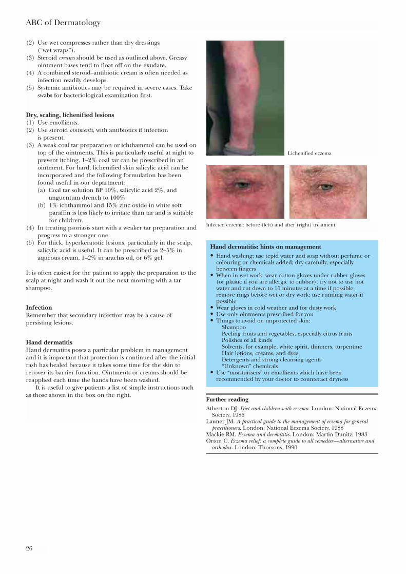
ABC of Dermatology
26
(2) Use wet compresses rather than dry dressings (“wet wraps”).
(3) Steroid creams should be used as outlined above. Greasyointment bases tend to float off on the exudate.
(4) A combined steroid–antibiotic cream is often needed asinfection readily develops.
(5) Systemic antibiotics may be required in severe cases. Takeswabs for bacteriological examination first.
Dry, scaling, lichenified lesions(1) Use emollients.(2) Use steroid ointments, with antibiotics if infection
is present.(3) A weak coal tar preparation or ichthammol can be used on
top of the ointments. This is particularly useful at night toprevent itching. 1–2% coal tar can be prescribed in anointment. For hard, lichenified skin salicylic acid can beincorporated and the following formulation has beenfound useful in our department:(a) Coal tar solution BP 10%, salicylic acid 2%, and
unguentum drench to 100%.(b) 1% ichthammol and 15% zinc oxide in white soft
paraffin is less likely to irritate than tar and is suitablefor children.
(4) In treating psoriasis start with a weaker tar preparation andprogress to a stronger one.
(5) For thick, hyperkeratotic lesions, particularly in the scalp,salicylic acid is useful. It can be prescribed as 2–5% inaqueous cream, 1–2% in arachis oil, or 6% gel.
It is often easiest for the patient to apply the preparation to thescalp at night and wash it out the next morning with a tarshampoo.
InfectionRemember that secondary infection may be a cause ofpersisting lesions.
Hand dermatitisHand dermatitis poses a particular problem in managementand it is important that protection is continued after the initialrash has healed because it takes some time for the skin torecover its barrier function. Ointments or creams should bereapplied each time the hands have been washed.
It is useful to give patients a list of simple instructions suchas those shown in the box on the right.
Hand dermatitis: hints on management• Hand washing: use tepid water and soap without perfume or
colouring or chemicals added; dry carefully, especiallybetween fingers
• When in wet work: wear cotton gloves under rubber gloves(or plastic if you are allergic to rubber); try not to use hotwater and cut down to 15 minutes at a time if possible;remove rings before wet or dry work; use running water ifpossible
• Wear gloves in cold weather and for dusty work• Use only ointments prescribed for you• Things to avoid on unprotected skin:
ShampooPeeling fruits and vegetables, especially citrus fruitsPolishes of all kindsSolvents, for example, white spirit, thinners, turpentineHair lotions, creams, and dyesDetergents and strong cleansing agents“Unknown” chemicals
• Use “moisturisers” or emollients which have beenrecommended by your doctor to counteract dryness
Further readingAtherton DJ. Diet and children with eczema. London: National Eczema
Society, 1986Launer JM. A practical guide to the management of eczema for general
practitioners. London: National Eczema Society, 1988Mackie RM. Eczema and dermatitis. London: Martin Dunitz, 1983Orton C. Eczema relief: a complete guide to all remedies—alternative and
orthodox. London: Thorsons, 1990
Lichenified eczema
Infected eczema: before (left) and after (right) treatment
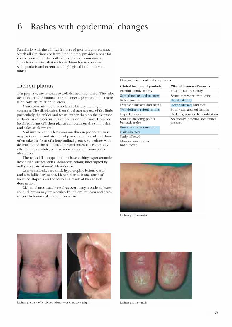
27
Familiarity with the clinical features of psoriasis and eczema,which all clinicians see from time to time, provides a basis forcomparison with other rather less common conditions. The characteristics that each condition has in common with psoriasis and eczema are highlighted in the relevant tables.
Lichen planusLike psoriasis, the lesions are well defined and raised. They alsooccur in areas of trauma—the Koebner’s phenomenon. Thereis no constant relation to stress.
Unlike psoriasis, there is no family history. Itching iscommon. The distribution is on the flexor aspects of the limbs,particularly the ankles and wrists, rather than on the extensorsurfaces, as in psoriasis. It also occurs on the trunk. However,localised forms of lichen planus can occur on the shin, palm,and soles or elsewhere.
Nail involvement is less common than in psoriasis. Theremay be thinning and atrophy of part or all of a nail and theseoften take the form of a longitudinal groove, sometimes withdestruction of the nail plate. The oral mucosa is commonlyaffected with a white, net-like appearance and sometimesulceration.
The typical flat topped lesions have a shiny hyperkeratoticlichenified surface with a violaceous colour, interrupted bymilky white streaks—Wickham’s striae.
Less commonly, very thick hypertrophic lesions occur and also follicular lesions. Lichen planus is one cause oflocalised alopecia on the scalp as a result of hair follicledestruction.
Lichen planus usually resolves over many months to leaveresidual brown or grey macules. In the oral mucosa and areassubject to trauma ulceration can occur.
6 Rashes with epidermal changes
Characteristics of lichen planus
Clinical features of psoriasis Clinical features of eczemaPossible family history Possible family history
Sometimes worse with stressItching—rareExtensor surfaces and trunk
Poorly demarcated lesionsHyperkeratosis Oedema, vesicles, lichenificationScaling, bleeding points Secondary infection sometimes beneath scales present
Scalp affectedMucous membranes not affected
Lichen planus—wrist
Lichen planus (left). Lichen planus—oral mucosa (right) Lichen planus—nails
Sometimes related to stressUsually itchingFlexor surfaces and face
Well defined, raised lesions
Koebner’s phenomenonNails affected
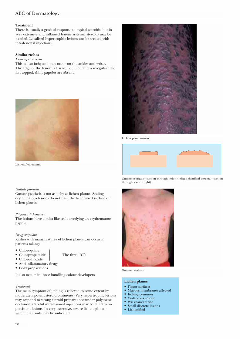
ABC of Dermatology
28
TreatmentThere is usually a gradual response to topical steroids, but invery extensive and inflamed lesions systemic steroids may beneeded. Localised hypertrophic lesions can be treated withintralesional injections.
Similar rashesLichenified eczemaThis is also itchy and may occur on the ankles and wrists. The edge of the lesion is less well defined and is irregular. Theflat topped, shiny papules are absent.
Lichen planus—skin
Lichenified eczema
Guttate psoriasis—section through lesion (left); lichenified eczema—sectionthrough lesion (right)
Guttate psoriasis
Lichen planus• Flexor surfaces• Mucous membranes affected• Itching common• Violaceous colour• Wickham’s striae• Small discrete lesions• Lichenified
Guttate psoriasisGuttate psoriasis is not as itchy as lichen planus. Scalingerythematous lesions do not have the lichenified surface oflichen planus.
Pityriasis lichenoidesThe lesions have a mica-like scale overlying an erythematouspapule.
Drug eruptionsRashes with many features of lichen planus can occur inpatients taking:
• Chloroquine• Chlorpropamide } The three “C”s• Chlorothiazide• Anti-inflammatory drugs• Gold preparations
It also occurs in those handling colour developers.
TreatmentThe main symptom of itching is relieved to some extent bymoderately potent steroid ointments. Very hypertrophic lesionsmay respond to strong steroid preparations under polytheneocclusion. Careful intralesional injections may be effective inpersistent lesions. In very extensive, severe lichen planussystemic steroids may be indicated.
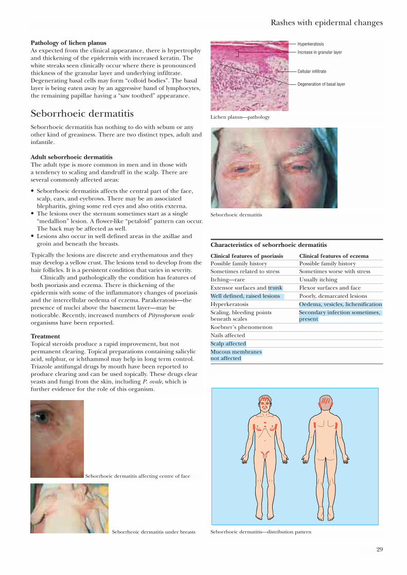
Rashes with epidermal changes
29
Pathology of lichen planusAs expected from the clinical appearance, there is hypertrophyand thickening of the epidermis with increased keratin. Thewhite streaks seen clinically occur where there is pronouncedthickness of the granular layer and underlying infiltrate.Degenerating basal cells may form “colloid bodies”. The basallayer is being eaten away by an aggressive band of lymphocytes,the remaining papillae having a “saw toothed” appearance.
Seborrhoeic dermatitisSeborrhoeic dermatitis has nothing to do with sebum or anyother kind of greasiness. There are two distinct types, adult andinfantile.
Adult seborrhoeic dermatitisThe adult type is more common in men and in those with a tendency to scaling and dandruff in the scalp. There areseveral commonly affected areas:
• Seborrhoeic dermatitis affects the central part of the face,scalp, ears, and eyebrows. There may be an associatedblepharitis, giving some red eyes and also otitis externa.
• The lesions over the sternum sometimes start as a single“medallion” lesion. A flower-like “petaloid” pattern can occur.The back may be affected as well.
• Lesions also occur in well defined areas in the axillae andgroin and beneath the breasts.
Typically the lesions are discrete and erythematous and theymay develop a yellow crust. The lesions tend to develop from thehair follicles. It is a persistent condition that varies in severity.
Clinically and pathologically the condition has features ofboth psoriasis and eczema. There is thickening of theepidermis with some of the inflammatory changes of psoriasisand the intercellular oedema of eczema. Parakeratosis—thepresence of nuclei above the basement layer—may benoticeable. Recently, increased numbers of Pityrosporum ovaleorganisms have been reported.
TreatmentTopical steroids produce a rapid improvement, but notpermanent clearing. Topical preparations containing salicylicacid, sulphur, or ichthammol may help in long term control.Triazole antifungal drugs by mouth have been reported toproduce clearing and can be used topically. These drugs clearyeasts and fungi from the skin, including P. ovale, which isfurther evidence for the role of this organism.
Hyperkeratosis
Increase in granular layer
Cellular infiltrate
Degeneration of basal layer
Lichen planus—pathology
Seborrhoeic dermatitis
Seborrhoeic dermatitis—distribution pattern
Seborrhoeic dermatitis affecting centre of face
Seborrheoic dermatitis under breasts
Characteristics of seborrhoeic dermatitis
Clinical features of psoriasis Clinical features of eczemaPossible family history Possible family historySometimes related to stress Sometimes worse with stressItching—rare Usually itchingExtensor surfaces and Flexor surfaces and face
Poorly, demarcated lesionsHyperkeratosisScaling, bleeding points beneath scalesKoebner’s phenomenonNails affected
trunkWell defined, raised lesions
Oedema, vesicles, lichenification
Scalp affected
not affectedMucous membranes
Secondary infection sometimes,present
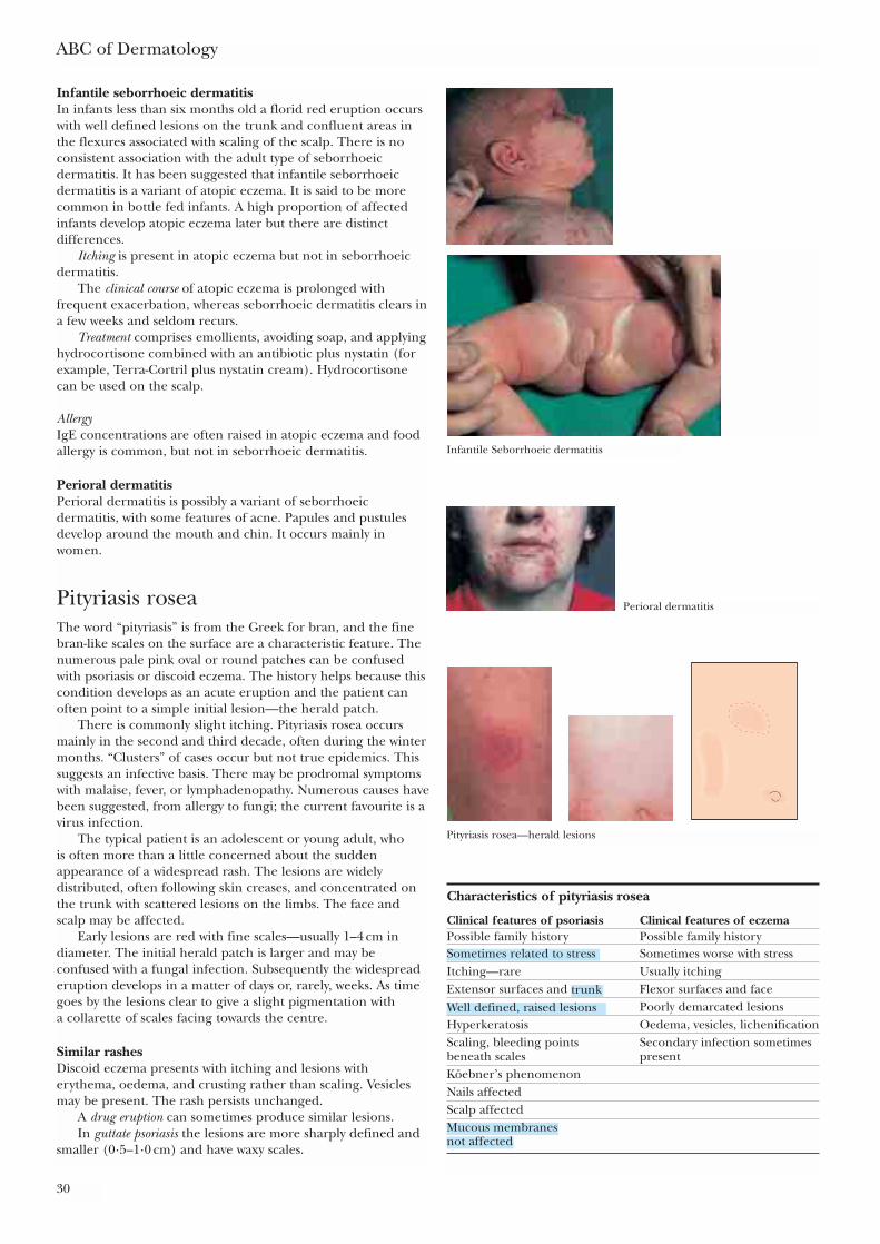
ABC of Dermatology
30
Infantile seborrhoeic dermatitisIn infants less than six months old a florid red eruption occurswith well defined lesions on the trunk and confluent areas inthe flexures associated with scaling of the scalp. There is noconsistent association with the adult type of seborrhoeicdermatitis. It has been suggested that infantile seborrhoeicdermatitis is a variant of atopic eczema. It is said to be morecommon in bottle fed infants. A high proportion of affectedinfants develop atopic eczema later but there are distinctdifferences.
Itching is present in atopic eczema but not in seborrhoeicdermatitis.
The clinical course of atopic eczema is prolonged withfrequent exacerbation, whereas seborrhoeic dermatitis clears ina few weeks and seldom recurs.
Treatment comprises emollients, avoiding soap, and applyinghydrocortisone combined with an antibiotic plus nystatin (forexample, Terra-Cortril plus nystatin cream). Hydrocortisonecan be used on the scalp.
AllergyIgE concentrations are often raised in atopic eczema and foodallergy is common, but not in seborrhoeic dermatitis.
Perioral dermatitisPerioral dermatitis is possibly a variant of seborrhoeicdermatitis, with some features of acne. Papules and pustulesdevelop around the mouth and chin. It occurs mainly inwomen.
Pityriasis roseaThe word “pityriasis” is from the Greek for bran, and the finebran-like scales on the surface are a characteristic feature. Thenumerous pale pink oval or round patches can be confusedwith psoriasis or discoid eczema. The history helps because thiscondition develops as an acute eruption and the patient canoften point to a simple initial lesion—the herald patch.
There is commonly slight itching. Pityriasis rosea occursmainly in the second and third decade, often during the wintermonths. “Clusters” of cases occur but not true epidemics. Thissuggests an infective basis. There may be prodromal symptomswith malaise, fever, or lymphadenopathy. Numerous causes havebeen suggested, from allergy to fungi; the current favourite is avirus infection.
The typical patient is an adolescent or young adult, who is often more than a little concerned about the suddenappearance of a widespread rash. The lesions are widelydistributed, often following skin creases, and concentrated onthe trunk with scattered lesions on the limbs. The face andscalp may be affected.
Early lesions are red with fine scales—usually 1–4 cm indiameter. The initial herald patch is larger and may beconfused with a fungal infection. Subsequently the widespreaderuption develops in a matter of days or, rarely, weeks. As timegoes by the lesions clear to give a slight pigmentation with a collarette of scales facing towards the centre.
Similar rashesDiscoid eczema presents with itching and lesions witherythema, oedema, and crusting rather than scaling. Vesiclesmay be present. The rash persists unchanged.
A drug eruption can sometimes produce similar lesions.In guttate psoriasis the lesions are more sharply defined and
smaller (0·5–1·0 cm) and have waxy scales.
Infantile Seborrhoeic dermatitis
Perioral dermatitis
Pityriasis rosea—herald lesions
Characteristics of pityriasis rosea
Clinical features of psoriasis Clinical features of eczemaPossible family history Possible family history
Sometimes worse with stressItching—rare Usually itchingExtensor surfaces and Flexor surfaces and face
Poorly demarcated lesionsHyperkeratosis Oedema, vesicles, lichenificationScaling, bleeding points Secondary infection sometimesbeneath scales presentKöebner’s phenomenonNails affectedScalp affected
Sometimes related to stress
trunkWell defined, raised lesions
not affectedMucous membranes
Rashes with epidermal changes
31
PathologyHistological changes are non-specific, showing slightinflammatory changes in the dermis, oedema, and slighthyperkeratosis.
Pityriasis lichenoidesPityriasis lichenoides is a less common condition occurring inacute and chronic forms.
The acute form presents with widespread pink papuleswhich itch and form crusts, sometimes with vesicle formationsuggestive of chickenpox. There may be ulceration. The lesionsmay develop in crops and resolve over a matter of weeks.
The chronic form presents as reddish brown papules—oftenwith a “mica”-like scale that reveals a smooth, red surfaceunderneath, unlike the bleeding points of psoriasis. In lichenplanus there is no superficial scale and blistering is unusual.
The distribution is over the trunk, thighs, and arms, usuallysparing the face and scalp.
The underlying pathology—vascular dilatation and alymphocytic infiltrate with a keratotic scale—is in keeping withthe clinical appearance. The cause is unknown.
Treatment is with topical steroids. Ultraviolet light treatmentis also helpful.
Characteristics of pityriasis lichenoides
Clinical features of psoriasis Clinical features of eczemaPossible family history Possible family historySometimes related to stress Sometimes worse with stressItching—rareExtensor surfaces and Flexor surfaces and face
Poorly demarcated lesionsHyperkeratosisScaling, bleeding points Secondary infection sometimes beneath scales presentKoebner’s phenomenonNails affectedScalp affectedMucous membranes not affected
Pityriasis lichenoides showing acuteerythematous rash
A mica scale pityriasis lichenoides
Pityriasis versicolor skin lesions
Pityriasis lichenoides—distribution pattern
Pityriasis versicolorPityriasis versicolor is a skin eruption that usually develops aftersun exposure with white macules on the tanned skin but palebrown patches on the covered areas, hence the nameversicolor, or variable colour. The lesions are: (a) flat; (b) only partially depigmented—areas of vitiligo are totallywhite; and (c) do not show inflammation or vesicles.
The causative organism is a yeast, Pityrosporum orbiculare, thattakes advantage of some unknown change in the epidermis anddevelops a proliferative, stubby, mycelial form, Malassezia furfur.This otherwise incidental information can be simply put topractical use by taking a superficial scraping from a lesion on toa microscope slide—add a drop of potassium hydroxide orwater with a coverslip. The organisms are readily seen underthe microscope as spherical yeast forms and mycelial rods,resembling “grapes and bananas” (“spaghetti and meatballs” inthe United States).
Treatment is simple: selenium sulphide shampoo appliedregularly with ample water while showering or bathing will clearthe infection. The colour change may take some time to clear.
Usually itching
Well defined, raised lesionsOedema, vesicles, lichenification
trunk
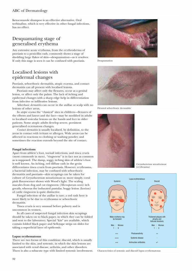
ABC of Dermatology
32
Ketoconazole shampoo is an effective alternative. Oralterbinafine, which is very effective in other fungal infections,has no effect.
Desquamating stage of generalised erythemaAny extensive acute erythema, from the erythroderma ofpsoriasis to a penicillin rash, commonly shows a stage ofshedding large flakes of skin—desquamation—as it resolves. If only this stage is seen it can be confused with psoriasis.
Localised lesions with epidermal changesPsoriasis, seborrhoeic dermatitis, atopic eczema, and contactdermatitis can all present with localised lesions.
Psoriasis may affect only the flexures, occur as a genitallesion, or affect only the palms. The lack of itching andepidermal changes with a sharp edge help in differentiationfrom infective or infiltrative lesions.
Seborrhoeic dermatitis can occur in the axillae or scalp with nolesions of other areas.
In atopic eczema the “classical” sites in children—flexures ofthe elbows and knees and the face—may be modified in adultsto localised vesicular lesions on the hands and feet in olderpatients. Some atopic adults develop severe, persistentgeneralised eczematous changes.
Contact dermatitis is usually localised, by definition, to theareas in contact with irritant or allergen. Wide areas can beaffected in reactions to clothing or washing powder, andsometimes the reaction extends beyond the site of contact.
Fungal infectionsApart from athlete’s foot, toenail infections, and tinea cruris(most commonly in men), “ringworm” is in fact not as commonas is supposed. The damp, soggy, itching skin of athlete’s foot is well known. An itching, red diffuse rash in the groindifferentiates tinea cruris from psoriasis. However, erythrasma,a bacterial infection, may be confused with seborrhoeicdermatitis and psoriasis—skin scrapings can be taken forculture of Corynebacterium minutissimum or, more simply, coralpink fluorescence shown with Wood’s light. The scalingmacules from dog and cat ringworm (Microsporum canis) itchgreatly, whereas the indurated pustular, boggy lesion (kerion)of cattle ringworm is quite distinctive.
Fungal infection of the axillae is rare; a red rash here ismore likely to be due to erythrasma or seborrhoeic dermatitis.
Tinea cruris is very unusual before puberty and isuncommon in women.
In all cases of suspected fungal infection skin scrapingsshould be taken on to black paper, in which they can be foldedand sent to the laboratory. Special “kits” are available, whichcontain folded black paper and Sellotape strips on slides fortaking a superficial layer of epidermis.
Lupus erythematosus There are two forms of this condition: discoid, which is usuallylimited to the skin, and systemic, in which the skin lesions areassociated with renal disease, arthritis, and other disorders.There is also a subacute type with limited systemic involvement.
Desquamation
Flexural seborrhoeic dermatitis
Corynebacterium minutissimum(erythrasma)
Lupus erythematosusSystemic
Men WomenMen Women
Photosensitivity
Systemic disease
Antinuclear antibodies
+++
++++
+++
++
-
+/-
1 31 9 ::::
Discoid
Malar erythema maybe transient
Thickened plaques withatrophy and
follicular keratosis
Characteristics of systemic and discoid lupus erythematosus
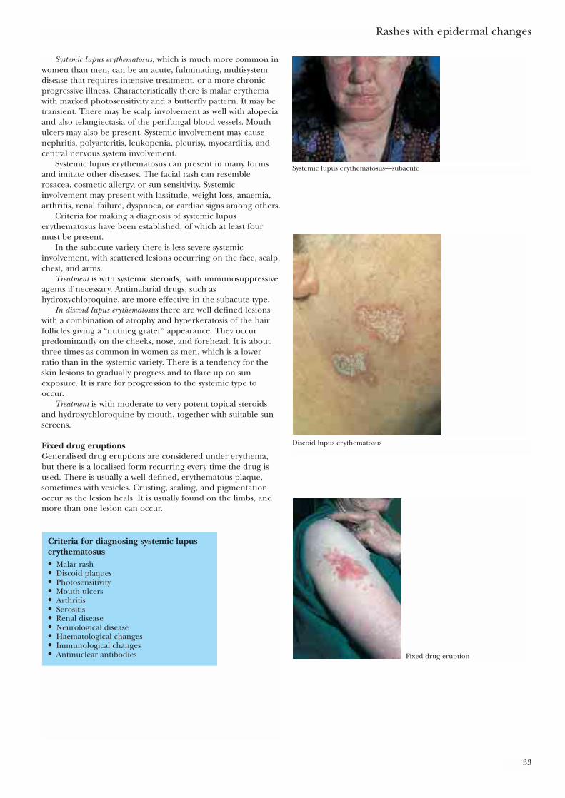
Systemic lupus erythematosus, which is much more common inwomen than men, can be an acute, fulminating, multisystemdisease that requires intensive treatment, or a more chronicprogressive illness. Characteristically there is malar erythemawith marked photosensitivity and a butterfly pattern. It may betransient. There may be scalp involvement as well with alopeciaand also telangiectasia of the perifungal blood vessels. Mouthulcers may also be present. Systemic involvement may causenephritis, polyarteritis, leukopenia, pleurisy, myocarditis, andcentral nervous system involvement.
Systemic lupus erythematosus can present in many formsand imitate other diseases. The facial rash can resemblerosacea, cosmetic allergy, or sun sensitivity. Systemicinvolvement may present with lassitude, weight loss, anaemia,arthritis, renal failure, dyspnoea, or cardiac signs among others.
Criteria for making a diagnosis of systemic lupuserythematosus have been established, of which at least fourmust be present.
In the subacute variety there is less severe systemicinvolvement, with scattered lesions occurring on the face, scalp,chest, and arms.
Treatment is with systemic steroids, with immunosuppressiveagents if necessary. Antimalarial drugs, such ashydroxychloroquine, are more effective in the subacute type.
In discoid lupus erythematosus there are well defined lesionswith a combination of atrophy and hyperkeratosis of the hairfollicles giving a “nutmeg grater” appearance. They occurpredominantly on the cheeks, nose, and forehead. It is aboutthree times as common in women as men, which is a lowerratio than in the systemic variety. There is a tendency for theskin lesions to gradually progress and to flare up on sunexposure. It is rare for progression to the systemic type tooccur.
Treatment is with moderate to very potent topical steroidsand hydroxychloroquine by mouth, together with suitable sunscreens.
Fixed drug eruptionsGeneralised drug eruptions are considered under erythema,but there is a localised form recurring every time the drug isused. There is usually a well defined, erythematous plaque,sometimes with vesicles. Crusting, scaling, and pigmentationoccur as the lesion heals. It is usually found on the limbs, andmore than one lesion can occur.
Rashes with epidermal changes
33
Systemic lupus erythematosus—subacute
Discoid lupus erythematosus
Fixed drug eruption
Criteria for diagnosing systemic lupuserythematosus• Malar rash• Discoid plaques• Photosensitivity• Mouth ulcers• Arthritis• Serositis• Renal disease• Neurological disease• Haematological changes• Immunological changes• Antinuclear antibodies
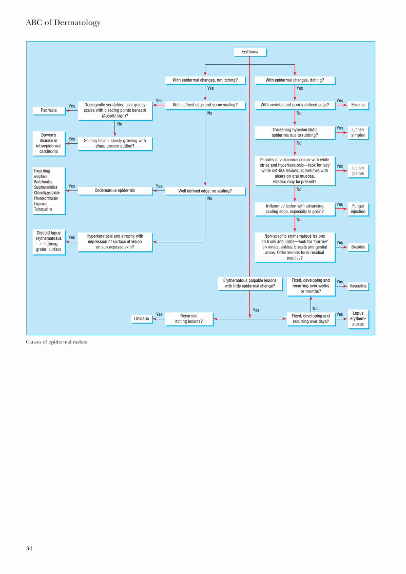
ABC of Dermatology
34
Erythema
With epidermal changes, itching?
With vesicles and poorly defined edge?
Oedematous epidermis
EczemaPsoriasis
Thickening hyperkeratoticepidermis due to rubbing?
Solitary lesion, slowly growing withsharp uneven outline?
Lichensimplex
Yes
Lichenplanus
Yes
Fungalinjection
Fixed, developing andrecurring over days?
Recurrentitching lesions?
Yes
YesYes
Yes
Yes
Bowen'sdisease or
intraepidermalcarcinoma
Yes
Discoid lupuserythematosus
– 'nutmeggrater' surface
Yes
Fixed drugeruption:BarbituratesSulphonamidesChlordiazepoxidePhenolphthaleinDapsoneTetracycline
Yes
Yes
No
Yes
No
No
No
No
No
ScabiesYes
Vasculitis
UrticariaYes
Fixed, developing andrecurring over weeks
or months?
Lupuserythem-atosus
Yes
YesYes
Inflammed lesion with advancingscaling edge, especially in groin?
No
Papules of violaceous colour with whitestriae and hyperkeratosis—look for lacywhite net-like lesions, sometimes with
ulcers on oral mucosa.Blisters may be present?
Non-specific erythematous lesionson trunk and limbs—look for "burrow"on wrists, ankles, breasts and genital
areas. Older lesions form residualpapules?
With epidermal changes, not itching?
Well defined edge, no scaling?
Well defined edge and some scaling?
No
Does gentle scratching give greasyscales with bleeding points beneath
(Auspitz sign)?
Hyperkeratosis and atrophy withdepression of surface of lesion
on sun exposed skin?
Erythematous palpable lesionswith little epidermal change?
Causes of epidermal rashes
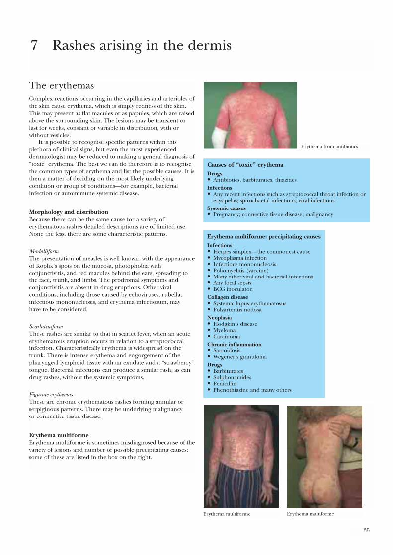
35
The erythemasComplex reactions occurring in the capillaries and arterioles ofthe skin cause erythema, which is simply redness of the skin.This may present as flat macules or as papules, which are raisedabove the surrounding skin. The lesions may be transient orlast for weeks, constant or variable in distribution, with orwithout vesicles.
It is possible to recognise specific patterns within thisplethora of clinical signs, but even the most experienceddermatologist may be reduced to making a general diagnosis of“toxic” erythema. The best we can do therefore is to recognisethe common types of erythema and list the possible causes. It isthen a matter of deciding on the most likely underlyingcondition or group of conditions—for example, bacterialinfection or autoimmune systemic disease.
Morphology and distributionBecause there can be the same cause for a variety oferythematous rashes detailed descriptions are of limited use.None the less, there are some characteristic patterns.
MorbilliformThe presentation of measles is well known, with the appearanceof Koplik’s spots on the mucosa, photophobia withconjunctivitis, and red macules behind the ears, spreading tothe face, trunk, and limbs. The prodromal symptoms andconjunctivitis are absent in drug eruptions. Other viralconditions, including those caused by echoviruses, rubella,infectious mononucleosis, and erythema infectiosum, may have to be considered.
ScarlatiniformThese rashes are similar to that in scarlet fever, when an acuteerythematous eruption occurs in relation to a streptococcalinfection. Characteristically erythema is widespread on thetrunk. There is intense erythema and engorgement of thepharyngeal lymphoid tissue with an exudate and a “strawberry”tongue. Bacterial infections can produce a similar rash, as candrug rashes, without the systemic symptoms.
Figurate erythemasThese are chronic erythematous rashes forming annular orserpiginous patterns. There may be underlying malignancy or connective tissue disease.
Erythema multiformeErythema multiforme is sometimes misdiagnosed because of thevariety of lesions and number of possible precipitating causes;some of these are listed in the box on the right.
7 Rashes arising in the dermis
Causes of “toxic” erythemaDrugs• Antibiotics, barbiturates, thiazidesInfections• Any recent infections such as streptococcal throat infection or
erysipelas; spirochaetal infections; viral infectionsSystemic causes• Pregnancy; connective tissue disease; malignancy
Erythema multiforme: precipitating causesInfections• Herpes simplex—the commonest cause• Mycoplasma infection• Infectious mononucleosis• Poliomyelitis (vaccine)• Many other viral and bacterial infections• Any focal sepsis• BCG inoculatonCollagen disease• Systemic lupus erythematosus• Polyarteritis nodosaNeoplasia• Hodgkin’s disease• Myeloma• CarcinomaChronic inflammation• Sarcoidosis• Wegener’s granulomaDrugs• Barbiturates• Sulphonamides• Penicillin• Phenothiazine and many others
Erythema from antibiotics
Erythema multiforme Erythema multiforme
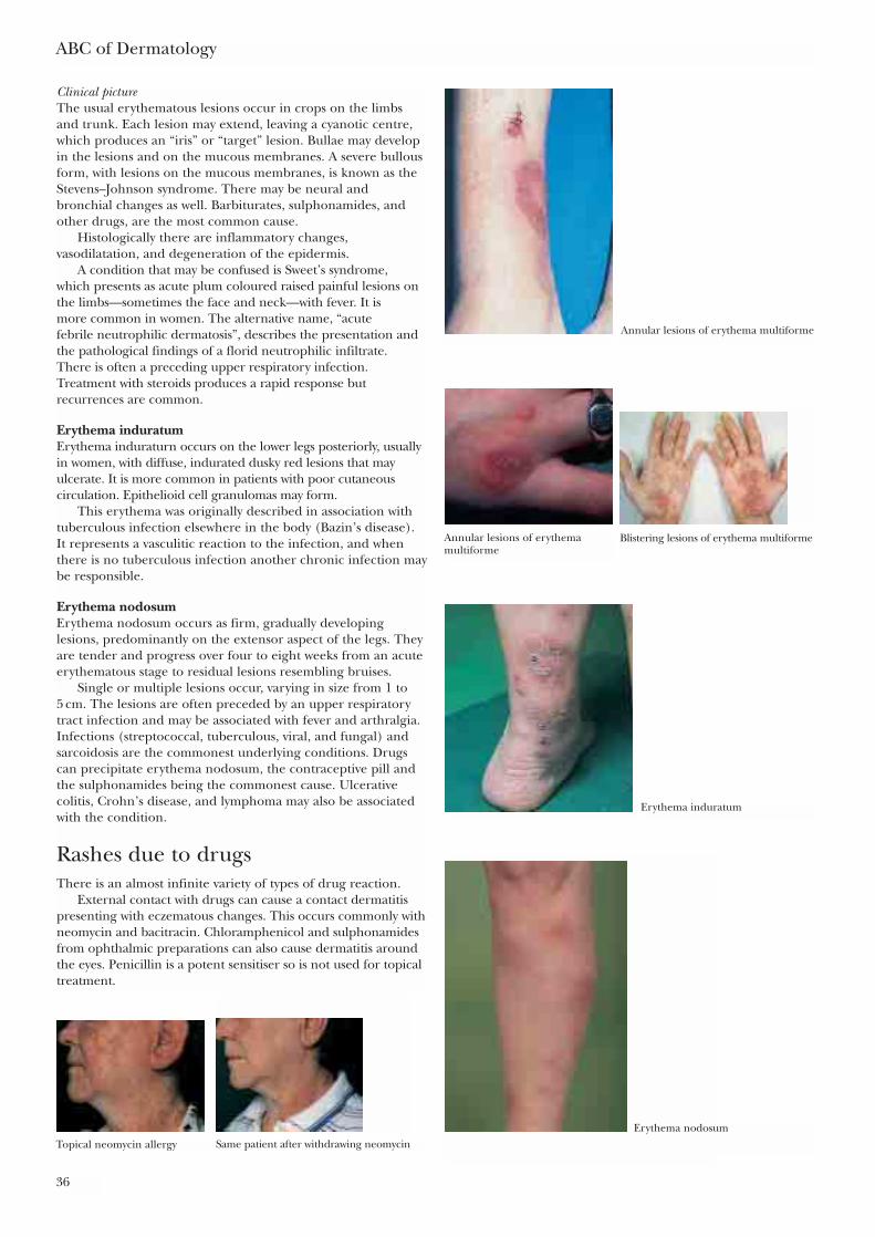
ABC of Dermatology
36
Clinical pictureThe usual erythematous lesions occur in crops on the limbsand trunk. Each lesion may extend, leaving a cyanotic centre,which produces an “iris” or “target” lesion. Bullae may developin the lesions and on the mucous membranes. A severe bullousform, with lesions on the mucous membranes, is known as theStevens–Johnson syndrome. There may be neural andbronchial changes as well. Barbiturates, sulphonamides, andother drugs, are the most common cause.
Histologically there are inflammatory changes,vasodilatation, and degeneration of the epidermis.
A condition that may be confused is Sweet’s syndrome, which presents as acute plum coloured raised painful lesions onthe limbs—sometimes the face and neck—with fever. It is more common in women. The alternative name, “acute febrile neutrophilic dermatosis”, describes the presentation andthe pathological findings of a florid neutrophilic infiltrate. There is often a preceding upper respiratory infection.Treatment with steroids produces a rapid response butrecurrences are common.
Erythema induratumErythema induraturn occurs on the lower legs posteriorly, usuallyin women, with diffuse, indurated dusky red lesions that mayulcerate. It is more common in patients with poor cutaneouscirculation. Epithelioid cell granulomas may form.
This erythema was originally described in association withtuberculous infection elsewhere in the body (Bazin’s disease).It represents a vasculitic reaction to the infection, and whenthere is no tuberculous infection another chronic infection maybe responsible.
Erythema nodosumErythema nodosum occurs as firm, gradually developinglesions, predominantly on the extensor aspect of the legs. Theyare tender and progress over four to eight weeks from an acuteerythematous stage to residual lesions resembling bruises.
Single or multiple lesions occur, varying in size from 1 to 5 cm. The lesions are often preceded by an upper respiratorytract infection and may be associated with fever and arthralgia.Infections (streptococcal, tuberculous, viral, and fungal) andsarcoidosis are the commonest underlying conditions. Drugscan precipitate erythema nodosum, the contraceptive pill andthe sulphonamides being the commonest cause. Ulcerativecolitis, Crohn’s disease, and lymphoma may also be associatedwith the condition.
Rashes due to drugsThere is an almost infinite variety of types of drug reaction.
External contact with drugs can cause a contact dermatitispresenting with eczematous changes. This occurs commonly withneomycin and bacitracin. Chloramphenicol and sulphonamidesfrom ophthalmic preparations can also cause dermatitis aroundthe eyes. Penicillin is a potent sensitiser so is not used for topicaltreatment.
Annular lesions of erythema multiforme
Annular lesions of erythemamultiforme
Blistering lesions of erythema multiforme
Erythema induratum
Erythema nodosum
Topical neomycin allergy Same patient after withdrawing neomycin
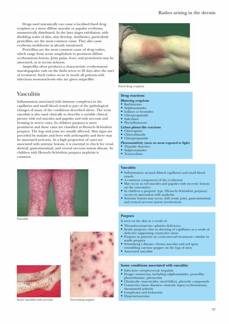
Rashes arising in the dermis
37
Drugs used systemically can cause a localised fixed drugeruption or a more diffuse macular or papular erythema,symmetrically distributed. In the later stages exfoliation, withshedding scales of skin, may develop. Antibiotics, particularlypenicillins, are the most common cause. They also causeerythema multiforme as already mentioned.
Penicillins are the most common cause of drug rashes,which range from acute anaphylaxis to persistent diffuseerythematous lesions. Joint pains, fever, and proteinuria may beassociated, as in serum sickness.
Ampicillin often produces a characteristic erythematousmaculopapular rash on the limbs seven to 20 days after the startof treatment. Such rashes occur in nearly all patients withinfectious mononucleosis who are given ampicillin.
VasculitisInflammation associated with immune complexes in thecapillaries and small blood vessels is part of the pathologicalchanges of many of the conditions described above. The termvasculitis is also used clinically to describe a variable clinicalpicture with red macules and papules and with necrosis andbruising in severe cases. In children purpura is moreprominent and these cases are classified as Henoch–Schönleinpurpura. The legs and arms are usually affected. Skin signs arepreceded by malaise and fever with arthropathy and there maybe associated urticaria. As a high proportion of cases areassociated with systemic lesions, it is essential to check for renal,skeletal, gastrointestinal, and central nervous system disease. Inchildren with Henoch–Schönlein purpura nephritis iscommon.
Fixed drug eruption
Drug reactions Blistering eruptions• Barbiturates• Sulphonamides• Iodines or bromides• Chlorpropamide• Salicylates• PhenylbutazoneLichen planus like reactions• Chloroquine• Chlorothiazide• ChlorpropamidePhotosensitivity (seen on areas exposed to light)• Thiazide diuretics• Sulphonamides• Tetracyclines
Vasculitis• Inflammation around dilated capillaries and small blood
vessels• A common component of the erythemas• May occur as red macules and papules with necrotic lesions
on the extremities• In children a purpuric type (Henoch–Schönlein purpura)
occurs in association with nephritis• Systemic lesions may occur, with renal, joint, gastrointestinal,
and central nervous system involvement
PurpuraIs seen on the skin as a result of:• Thrombocytopenia—platelet deficiency• Senile purpura—due to shearing of capillaries as a result of
defective supporting connective tissue• Purpura in patients on corticosteroid treatment—similar to
senile purpura• Schamberg’s disease—brown macules and red spots
resembling cayenne pepper on the legs of men• Associated vasculitis
Some conditions associated with vasculitis• Infection—streptococcal, hepatitis• Drugs—numerous, including sulphonamides, penicillin,
phenothiazine, phenacitin• Chemicals—insecticides, weed killers, phenolic compounds• Connective tissue diseases—systemic lupus erythematosus,
rheumatoid arthritis• Lymphoma and leukaemia• Dysproteinaemias
Vasculitis
Acute vasculitis with necrosis Necrotising angiitis
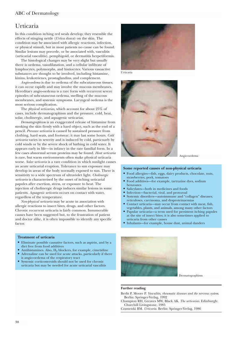
UrticariaIn this condition itching red weals develop; they resemble theeffects of stinging nettle (Urtica dioica) on the skin. Thecondition may be associated with allergic reactions, infection,or physical stimuli, but in most patients no cause can be found.Similar lesions may precede, or be associated with, vasculitis(urticarial vasculitis), pemphigoid, or dermatitis herpetiformis.
The histological changes may be very slight but usuallythere is oedema, vasodilatation, and a cellular infiltrate oflymphocytes, polymorphs, and histiocytes. Various vasoactivesubstances are thought to be involved, including histamine,kinins, leukotrienes, prostaglandins, and complement.
Angio-oedema is due to oedema of the subcutaneous tissues;it can occur rapidly and may involve the mucous membranes.Hereditary angio-oedema is a rare form with recurrent severeepisodes of subcutaneous oedema, swelling of the mucousmembranes, and systemic symptoms. Laryngeal oedema is themost serious complication.
The physical urticarias, which account for about 25% ofcases, include dermatographism and the pressure, cold, heat,solar, cholinergic, and aquagenic urticarias.
Dermatographism is an exaggerated release of histamine fromstroking the skin firmly with a hard object, such as the end of apencil. Pressure urticaria is caused by sustained pressure fromclothing, hard seats, and footwear; it may last some hours. Coldurticaria varies in severity and is induced by cold, particuarly bycold winds or by the severe shock of bathing in cold water. Itappears early in life—in infancy in the rare familial form. In afew cases abnormal serum proteins may be found. Heat urticariais rare, but warm environments often make physical urticariaworse. Solar urticaria is a rare condition in which sunlight causesan acute urticarial eruption. Tolerance to sun exposure maydevelop in areas of the body normally exposed to sun. There issensitivity to a wide spectrum of ultraviolet light. Cholinergicurticaria is characterised by the onset of itching urticarialpapules after exertion, stress, or exposure to heat. Theinjection of cholinergic drugs induces similar lesions in somepatients. Aquagenic urticaria occurs on contact with water,regardless of the temperature.
Non-physical urticaria may be acute in association withallergic reactions to insect bites, drugs, and other factors.Chronic recurrent urticaria is fairly common. Innumerablecauses have been suggested but, to the frustration of patientand doctor alike, it is often impossible to identify any specificfactor.
ABC of Dermatology
38
Urticaria
Some reported causes of non-physical urticaria• Food allergies—fish, eggs, dairy products, chocolate, nuts,
strawberries, pork, tomatoes• Food additives—for example, tartrazine dyes, sodium
benzoates• Salicylates—both in medicines and foods• Infection—bacterial, viral, and protozoal• Systemic disorders—autoimmune and “collagen” diseases;
reticuloses, carcinoma, and dysproteinaemias• Contact urticaria—may occur from contact with meat, fish,
vegetables, plants, and animals, among many other factors• Papular urticaria—a term used for persistent itching papules
at the site of insect bites; it is also sometimes applied tourticaria from other causes
• Inhalants—for example, house dust, animal danders
Treatment of urticaria• Eliminate possible causative factors, such as aspirin, and by a
diet free from food additives• Antihistamines. Also, H2 blockers, for example, cimetidine• Adrenaline can be used for acute attacks, particularly if there
is angio-oedema of the respiratory tract• Systemic corticosteroids should not be used for chronic
urticaria but may be needed for acute urticarial vasculitis
Angio-oedema
Dermatographism
Further readingBerlit P, Moore P. Vasculitis, rheumatic diseases and the nervous system.
Berlin: Springer-Verlag, 1992Champion RH, Greaves MW, Black AK. The urticarias. Edinburgh:
Churchill Livingstone, 1985Czametzki BM. Urticaria. Berlin: Springer-Verlag, 1986
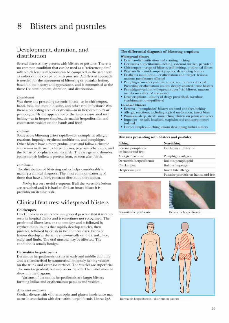
39
Development, duration, anddistributionSeveral diseases may present with blisters or pustules. There isno common condition that can be used as a “reference point”with which less usual lesions can be compared in the same wayas rashes can be compared with psoriasis. A different approachis needed for the assessment of blistering or pustular lesions,based on the history and appearance, and is summarised as thethree Ds: development, duration, and distribution.
DevelopmentWas there any preceding systemic illness—as in chickenpox,hand, foot, and mouth disease, and other viral infections? Wasthere a preceding area of erythema—as in herpes simplex orpemphigoid? Is the appearance of the lesions associated withitching—as in herpes simplex, dermatitis herpetiformis, andeczematous vesicles on the hands and feet?
DurationSome acute blistering arises rapidly—for example, in allergicreactions, impetigo, erythema multiforme, and pemphigus.Other blisters have a more gradual onset and follow a chroniccourse—as in dermatitis herpetiformis, pityriasis lichenoides, andthe bullae of porphyria cutanea tarda. The rare genetic disorderepidermolysis bullosa is present from, or soon after, birth.
DistributionThe distribution of blistering rashes helps considerably inmaking a clinical diagnosis. The most common patterns ofthose that have a fairly constant distribution are shown.
Itching is a very useful symptom. If all the accessible lesionsare scratched and it is hard to find an intact blister it isprobably an itching rash.
Clinical features: widespread blistersChickenpoxChickenpox is so well known in general practice that it is rarelyseen in hospital clinics and is sometimes not recognised. Theprodromal illness lasts one to two days and is followed byerythematous lesions that rapidly develop vesicles, thenpustules, followed by crusts in two to three days. Crops oflesions develop at the same sites—usually on the trunk, face,scalp, and limbs. The oral mucosa may be affected. Thecondition is usually benign.
Dermatitis herpetiformisDermatitis herpetiformis occurs in early and middle adult lifeand is characterised by symmetrical, intensely itching vesicleson the trunk and extensor surfaces. The vesicles are superficial.The onset is gradual, but may occur rapidly. The distribution isshown in the diagram.
Variants of dermatitis herpetiformis are larger blistersforming bullae and erythematous papules and vesicles.
Associated conditionsCoeliac disease with villous atrophy and gluten intolerance mayoccur in association with dermatitis herpetiformis. Linear IgA
8 Blisters and pustules
The differential diagnosis of blistering eruptionsWidespread blisters• Eczema—lichenification and crusting, itching• Dermatitis herpetiformis—itching, extensor surface, persistent• Chickenpox—crops of blisters, self limiting, prodromal illness• Pityriasis lichenoides—pink papules, developing blisters• Erythema multiforme—erythematous and “target” lesions,
mucous membranes affected• Pemphigoid—older patients, trunk, and flexures affected.
Preceding erythematous lesions, deeply situated, tense blisters• Pemphigus—adults, widespread superficial blisters, mucous
membranes affected (erosions)• Drug eruptions—history of drugs prescribed, overdose
(barbiturates, tranquillisers)Localised blisters• Eczema—“pompholyx” blisters on hand and feet, itching• Allergic reactions, including topical medication, insect bites• Psoriasis—deep, sterile, non-itching blisters on palms and soles• Impetigo—usually localised, staphylococci and streptococci
isolated• Herpes simplex—itching lesions developing turbid blisters
Diseases presenting with blisters and pustules
Itching Non-itchingEczema pompholyx Erythema multiformeon hands and feetAllergic reactions Pemphigus vulgarisDermatitis herpetiformis Bullous pemphigoidChickenpox Bullous impetigoHerpes simplex Insect bite allergy
Pustular psoriasis on hands and feet
Dermatitis herpetiformis Dermatitis herpetiformis
Dermatitis herpetiformis—distribution pattern
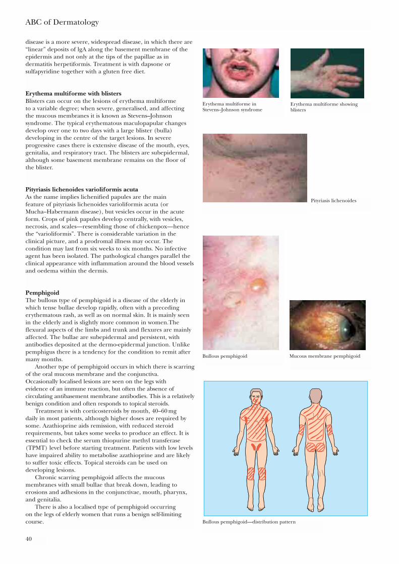
disease is a more severe, widespread disease, in which there are“linear” deposits of lgA along the basement membrane of theepidermis and not only at the tips of the papillae as indermatitis herpetiformis. Treatment is with dapsone orsulfapyridine together with a gluten free diet.
Erythema multiforme with blistersBlisters can occur on the lesions of erythema multiforme to a variable degree; when severe, generalised, and affectingthe mucous membranes it is known as Stevens–Johnsonsyndrome. The typical erythematous maculopapular changesdevelop over one to two days with a large blister (bulla)developing in the centre of the target lesions. In severeprogressive cases there is extensive disease of the mouth, eyes,genitalia, and respiratory tract. The blisters are subepidermal,although some basement membrane remains on the floor ofthe blister.
Pityriasis lichenoides varioliformis acutaAs the name implies lichenified papules are the main feature of pityriasis lichenoides varioliformis acuta (orMucha–Habermann disease), but vesicles occur in the acuteform. Crops of pink papules develop centrally, with vesicles,necrosis, and scales—resembling those of chickenpox—hencethe “varioliformis”. There is considerable variation in theclinical picture, and a prodromal illness may occur. Thecondition may last from six weeks to six months. No infectiveagent has been isolated. The pathological changes parallel theclinical appearance with inflammation around the blood vesselsand oedema within the dermis.
PemphigoidThe bullous type of pemphigoid is a disease of the elderly inwhich tense bullae develop rapidly, often with a precedingerythematous rash, as well as on normal skin. It is mainly seenin the elderly and is slightly more common in women.Theflexural aspects of the limbs and trunk and flexures are mainlyaffected. The bullae are subepidermal and persistent, withantibodies deposited at the dermo-epidermal junction. Unlikepemphigus there is a tendency for the condition to remit aftermany months.
Another type of pemphigoid occurs in which there is scarringof the oral mucous membrane and the conjunctiva.Occasionally localised lesions are seen on the legs with evidence of an immune reaction, but often the absence ofcirculating antibasement membrane antibodies. This is a relativelybenign condition and often responds to topical steroids.
Treatment is with corticosteroids by mouth, 40–60 mg daily in most patients, although higher doses are required bysome. Azathioprine aids remission, with reduced steroidrequirements, but takes some weeks to produce an effect. It isessential to check the serum thiopurine methyl transferase(TPMT) level before starting treatment. Patients with low levelshave impaired ability to metabolise azathioprine and are likelyto suffer toxic effects. Topical steroids can be used ondeveloping lesions.
Chronic scarring pemphigoid affects the mucousmembranes with small bullae that break down, leading toerosions and adhesions in the conjunctivae, mouth, pharynx,and genitalia.
There is also a localised type of pemphigoid occurring on the legs of elderly women that runs a benign self-limitingcourse.
ABC of Dermatology
40
Erythema multiforme inStevens–Johnson syndrome
Erythema multiforme showingblisters
Pityriasis lichenoides
Mucous membrane pemphigoidBullous pemphigoid
Bullous pemphigoid—distribution pattern
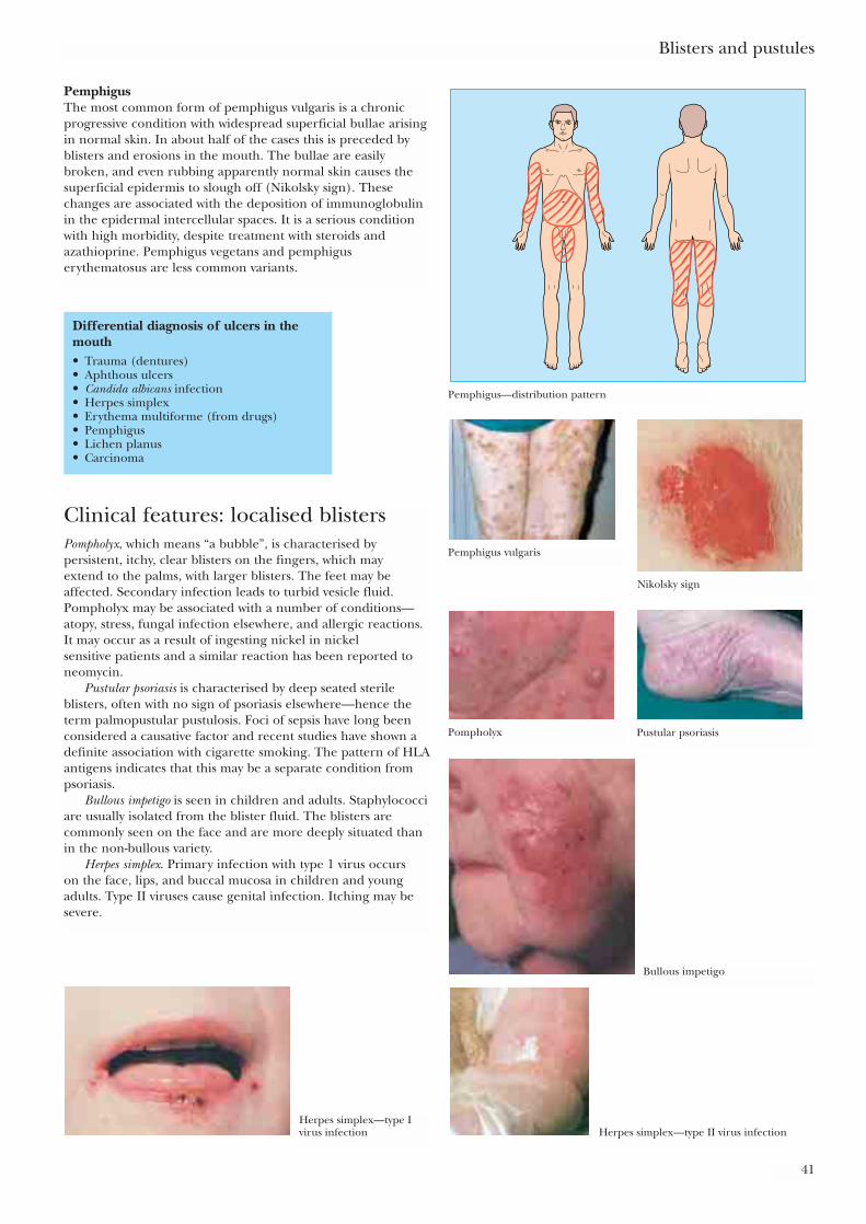
PemphigusThe most common form of pemphigus vulgaris is a chronicprogressive condition with widespread superficial bullae arisingin normal skin. In about half of the cases this is preceded byblisters and erosions in the mouth. The bullae are easilybroken, and even rubbing apparently normal skin causes thesuperficial epidermis to slough off (Nikolsky sign). Thesechanges are associated with the deposition of immunoglobulinin the epidermal intercellular spaces. It is a serious conditionwith high morbidity, despite treatment with steroids andazathioprine. Pemphigus vegetans and pemphiguserythematosus are less common variants.
Blisters and pustules
41
Pemphigus—distribution pattern
Pemphigus vulgaris
Nikolsky sign
Pompholyx Pustular psoriasis
Bullous impetigo
Herpes simplex—type II virus infectionHerpes simplex—type Ivirus infection
Differential diagnosis of ulcers in themouth• Trauma (dentures)• Aphthous ulcers• Candida albicans infection• Herpes simplex• Erythema multiforme (from drugs)• Pemphigus• Lichen planus• Carcinoma
Clinical features: localised blistersPompholyx, which means “a bubble”, is characterised bypersistent, itchy, clear blisters on the fingers, which may extend to the palms, with larger blisters. The feet may beaffected. Secondary infection leads to turbid vesicle fluid.Pompholyx may be associated with a number of conditions—atopy, stress, fungal infection elsewhere, and allergic reactions.It may occur as a result of ingesting nickel in nickel sensitive patients and a similar reaction has been reported toneomycin.
Pustular psoriasis is characterised by deep seated sterileblisters, often with no sign of psoriasis elsewhere—hence theterm palmopustular pustulosis. Foci of sepsis have long beenconsidered a causative factor and recent studies have shown adefinite association with cigarette smoking. The pattern of HLAantigens indicates that this may be a separate condition frompsoriasis.
Bullous impetigo is seen in children and adults. Staphylococciare usually isolated from the blister fluid. The blisters arecommonly seen on the face and are more deeply situated thanin the non-bullous variety.
Herpes simplex. Primary infection with type 1 virus occurs on the face, lips, and buccal mucosa in children and youngadults. Type II viruses cause genital infection. Itching may besevere.
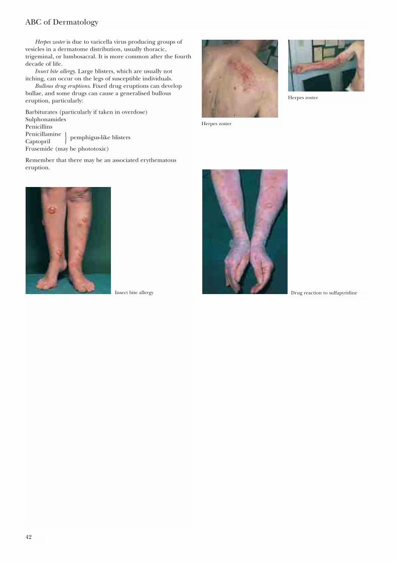
Herpes zoster is due to varicella virus producing groups ofvesicles in a dermatome distribution, usually thoracic,trigeminal, or lumbosacral. It is more common after the fourthdecade of life.
Insect bite allergy. Large blisters, which are usually notitching, can occur on the legs of susceptible individuals.
Bullous drug eruptions. Fixed drug eruptions can developbullae, and some drugs can cause a generalised bullouseruption, particularly:
Barbiturates (particularly if taken in overdose)SulphonamidesPenicillinsPenicillamine } pemphigus-like blistersCaptoprilFrusemide (may be phototoxic)
Remember that there may be an associated erythematouseruption.
ABC of Dermatology
42
Herpes zoster
Herpes zoster
Insect bite allergy Drug reaction to sulfapyridine
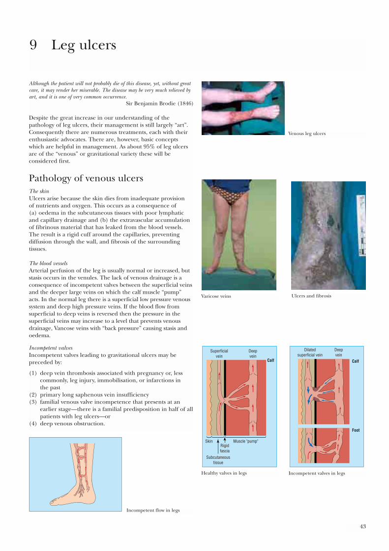
43
Although the patient will not probably die of this disease, yet, without greatcare, it may render her miserable. The disease may be very much relieved byart, and it is one of very common occurrence.
Sir Benjamin Brodie (1846)
Despite the great increase in our understanding of thepathology of leg ulcers, their management is still largely “art”.Consequently there are numerous treatments, each with theirenthusiastic advocates. There are, however, basic conceptswhich are helpful in management. As about 95% of leg ulcersare of the “venous” or gravitational variety these will beconsidered first.
Pathology of venous ulcersThe skinUlcers arise because the skin dies from inadequate provision of nutrients and oxygen. This occurs as a consequence of (a) oedema in the subcutaneous tissues with poor lymphaticand capillary drainage and (b) the extravascular accumulationof fibrinous material that has leaked from the blood vessels.The result is a rigid cuff around the capillaries, preventingdiffusion through the wall, and fibrosis of the surroundingtissues.
The blood vessels Arterial perfusion of the leg is usually normal or increased, butstasis occurs in the venules. The lack of venous drainage is aconsequence of incompetent valves between the superficial veinsand the deeper large veins on which the calf muscle “pump”acts. In the normal leg there is a superficial low pressure venoussystem and deep high pressure veins. If the blood flow fromsuperficial to deep veins is reversed then the pressure in thesuperficial veins may increase to a level that prevents venousdrainage, Vancose veins with “back pressure” causing stasis andoedema.
Incompetent valvesIncompetent valves leading to gravitational ulcers may bepreceded by:
(1) deep vein thrombosis associated with pregnancy or, lesscommonly, leg injury, immobilisation, or infarctions in the past
(2) primary long saphenous vein insufficiency(3) familial venous valve incompetence that presents at an
earlier stage—there is a familial predisposition in half of allpatients with leg ulcers—or
(4) deep venous obstruction.
9 Leg ulcers
Venous leg ulcers
Varicose veins Ulcers and fibrosis
Superficialvein
Skin
Subcutaneoustissue
Muscle "pump"Rigidfascia
Deepvein
Calf
Healthy valves in legs
Dilatedsuperficial vein
Deepvein
Calf
Foot
Incompetent valves in legs
Incompetent flow in legs
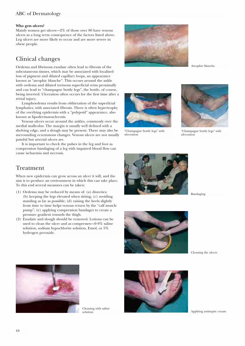
ABC of Dermatology
44
Who gets ulcers?Mainly women get ulcers—2% of those over 80 have venousulcers as a long term consequence of the factors listed above.Leg ulcers are more likely to occur and are more severe inobese people.
Clinical changesOedema and fibrinous exudate often lead to fibrosis of thesubcutaneous tissues, which may be associated with localisedloss of pigment and dilated capillary loops, an appearanceknown as “atrophic blanche”. This occurs around the anklewith oedema and dilated tortuous superficial veins proximallyand can lead to “champagne bottle legs”, the bottle, of course,being inverted. Ulceration often occurs for the first time after atrivial injury.
Lymphoedema results from obliteration of the superficiallymphatics, with associated fibrosis. There is often hypertrophyof the overlying epidermis with a “polypoid” appearance, alsoknown as lipodermatosclerosis.
Venous ulcers occur around the ankles, commonly over themedial malleolus. The margin is usually well defined with ashelving edge, and a slough may be present. There may also besurrounding eczematous changes. Venous ulcers are not usuallypainful but arterial ulcers are.
It is important to check the pulses in the leg and foot ascompression bandaging of a leg with impaired blood flow cancause ischaemia and necrosis.
TreatmentWhen new epidermis can grow across an ulcer it will, and theaim is to produce an environment in which this can take place.To this end several measures can be taken:
(1) Oedema may be reduced by means of: (a) diuretics; (b) keeping the legs elevated when sitting; (c) avoidingstanding as far as possible; (d) raising the heels slightlyfrom time to time helps venous return by the “calf musclepump”; (e) applying compression bandages to create apressure gradient towards the thigh.
(2) Exudate and slough should be removed. Lotions can beused to clean the ulcer and as compresses—0·9% salinesolution, sodium hypochlorite solution, Eusol, or 5%hydrogen peroxide.
Atrophie blanche
“Champagne bottle legs” withulceration
“Champagne bottle legs” withulceration
Bandaging
Cleaning the ulcers
Applying antiseptic creamCleaning with salinesolution
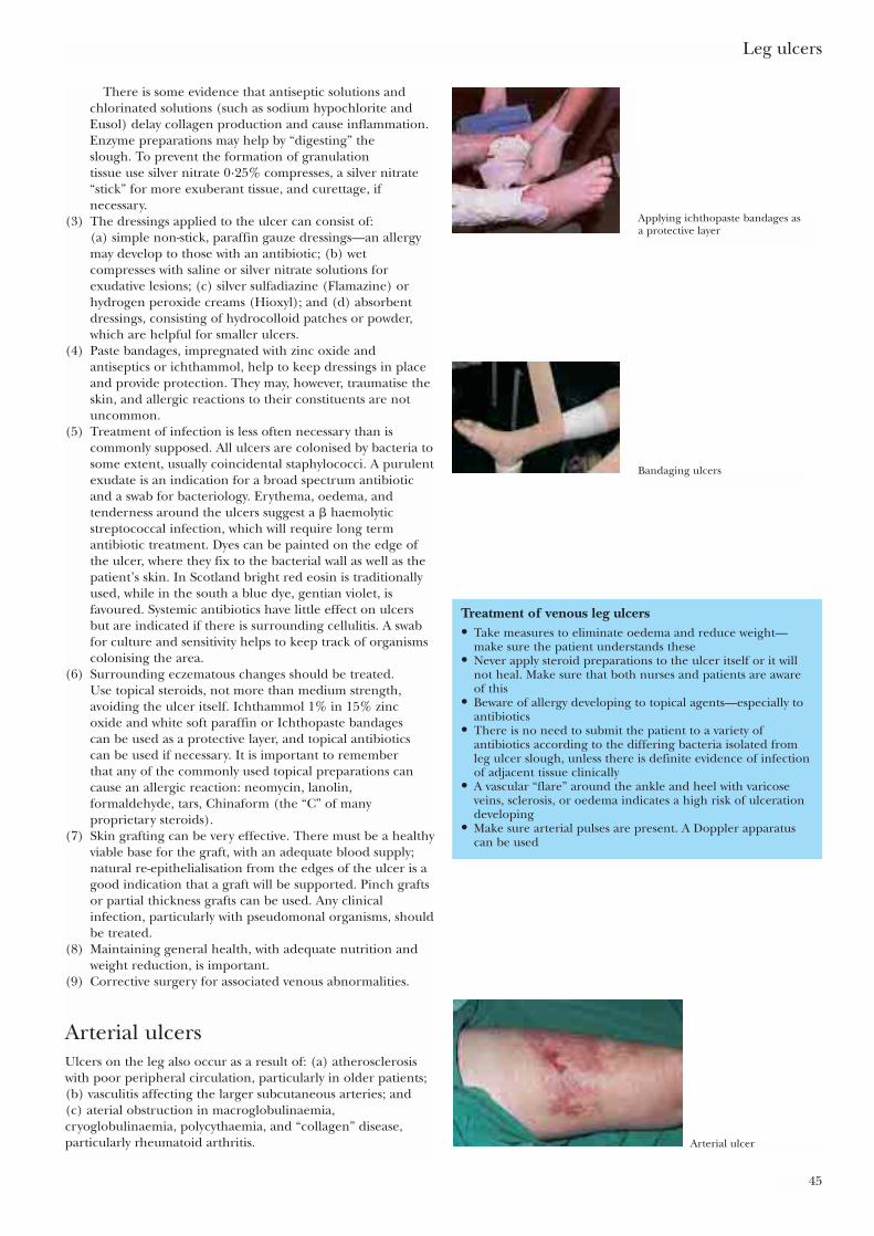
Leg ulcers
45
There is some evidence that antiseptic solutions andchlorinated solutions (such as sodium hypochlorite andEusol) delay collagen production and cause inflammation.Enzyme preparations may help by “digesting” the slough. To prevent the formation of granulation tissue use silver nitrate 0·25% compresses, a silver nitrate“stick” for more exuberant tissue, and curettage, ifnecessary.
(3) The dressings applied to the ulcer can consist of: (a) simple non-stick, paraffin gauze dressings—an allergymay develop to those with an antibiotic; (b) wetcompresses with saline or silver nitrate solutions forexudative lesions; (c) silver sulfadiazine (Flamazine) orhydrogen peroxide creams (Hioxyl); and (d) absorbentdressings, consisting of hydrocolloid patches or powder,which are helpful for smaller ulcers.
(4) Paste bandages, impregnated with zinc oxide andantiseptics or ichthammol, help to keep dressings in placeand provide protection. They may, however, traumatise theskin, and allergic reactions to their constituents are notuncommon.
(5) Treatment of infection is less often necessary than iscommonly supposed. All ulcers are colonised by bacteria tosome extent, usually coincidental staphylococci. A purulentexudate is an indication for a broad spectrum antibioticand a swab for bacteriology. Erythema, oedema, andtenderness around the ulcers suggest a � haemolyticstreptococcal infection, which will require long termantibiotic treatment. Dyes can be painted on the edge ofthe ulcer, where they fix to the bacterial wall as well as thepatient’s skin. In Scotland bright red eosin is traditionallyused, while in the south a blue dye, gentian violet, isfavoured. Systemic antibiotics have little effect on ulcersbut are indicated if there is surrounding cellulitis. A swabfor culture and sensitivity helps to keep track of organismscolonising the area.
(6) Surrounding eczematous changes should be treated. Use topical steroids, not more than medium strength,avoiding the ulcer itself. Ichthammol 1% in 15% zinc oxide and white soft paraffin or Ichthopaste bandages can be used as a protective layer, and topical antibiotics can be used if necessary. It is important to remember that any of the commonly used topical preparations cancause an allergic reaction: neomycin, lanolin,formaldehyde, tars, Chinaform (the “C” of manyproprietary steroids).
(7) Skin grafting can be very effective. There must be a healthyviable base for the graft, with an adequate blood supply;natural re-epithelialisation from the edges of the ulcer is agood indication that a graft will be supported. Pinch graftsor partial thickness grafts can be used. Any clinicalinfection, particularly with pseudomonal organisms, shouldbe treated.
(8) Maintaining general health, with adequate nutrition andweight reduction, is important.
(9) Corrective surgery for associated venous abnormalities.
Arterial ulcersUlcers on the leg also occur as a result of: (a) atherosclerosiswith poor peripheral circulation, particularly in older patients;(b) vasculitis affecting the larger subcutaneous arteries; and (c) aterial obstruction in macroglobulinaemia,cryoglobulinaemia, polycythaemia, and “collagen” disease,particularly rheumatoid arthritis.
Treatment of venous leg ulcers• Take measures to eliminate oedema and reduce weight—
make sure the patient understands these• Never apply steroid preparations to the ulcer itself or it will
not heal. Make sure that both nurses and patients are awareof this
• Beware of allergy developing to topical agents—especially toantibiotics
• There is no need to submit the patient to a variety ofantibiotics according to the differing bacteria isolated fromleg ulcer slough, unless there is definite evidence of infectionof adjacent tissue clinically
• A vascular “flare” around the ankle and heel with varicoseveins, sclerosis, or oedema indicates a high risk of ulcerationdeveloping
• Make sure arterial pulses are present. A Doppler apparatuscan be used
Arterial ulcer
Applying ichthopaste bandages as a protective layer
Bandaging ulcers
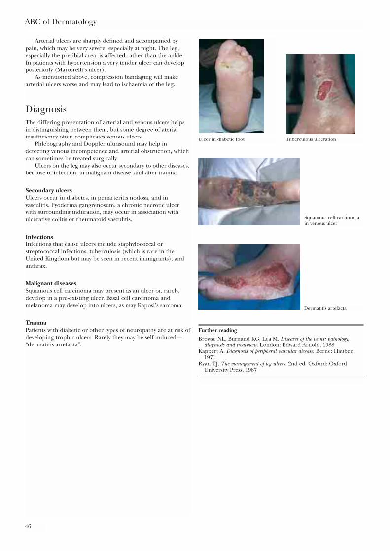
ABC of Dermatology
46
Arterial ulcers are sharply defined and accompanied bypain, which may be very severe, especially at night. The leg,especially the pretibial area, is affected rather than the ankle.In patients with hypertension a very tender ulcer can developposteriorly (Martorelli’s ulcer).
As mentioned above, compression bandaging will makearterial ulcers worse and may lead to ischaemia of the leg.
DiagnosisThe differing presentation of arterial and venous ulcers helpsin distinguishing between them, but some degree of aterialinsufficiency often complicates venous ulcers.
Phlebography and Doppler ultrasound may help indetecting venous incompetence and arterial obstruction, whichcan sometimes be treated surgically.
Ulcers on the leg may also occur secondary to other diseases,because of infection, in malignant disease, and after trauma.
Secondary ulcersUlcers occur in diabetes, in periarteritis nodosa, and invasculitis. Pyoderma gangrenosum, a chronic necrotic ulcerwith surrounding induration, may occur in association withulcerative colitis or rheumatoid vasculitis.
InfectionsInfections that cause ulcers include staphylococcal orstreptococcal infections, tuberculosis (which is rare in theUnited Kingdom but may be seen in recent immigrants), andanthrax.
Malignant diseasesSquamous cell carcinoma may present as an ulcer or, rarely,develop in a pre-existing ulcer. Basal cell carcinoma andmelanoma may develop into ulcers, as may Kaposi’s sarcoma.
TraumaPatients with diabetic or other types of neuropathy are at risk ofdeveloping trophic ulcers. Rarely they may be self induced—“dermatitis artefacta”.
Further reading
Browse NL, Burnand KG, Lea M. Diseases of the veins: pathology,diagnosis and treatment. London: Edward Arnold, 1988
Kappert A. Diagnosis of peripheral vascular disease. Berne: Hauber,1971
Ryan TJ. The management of leg ulcers, 2nd ed. Oxford: OxfordUniversity Press, 1987
Ulcer in diabetic foot Tuberculous ulceration
Squamous cell carcinomain venous ulcer
Dermatitis artefacta
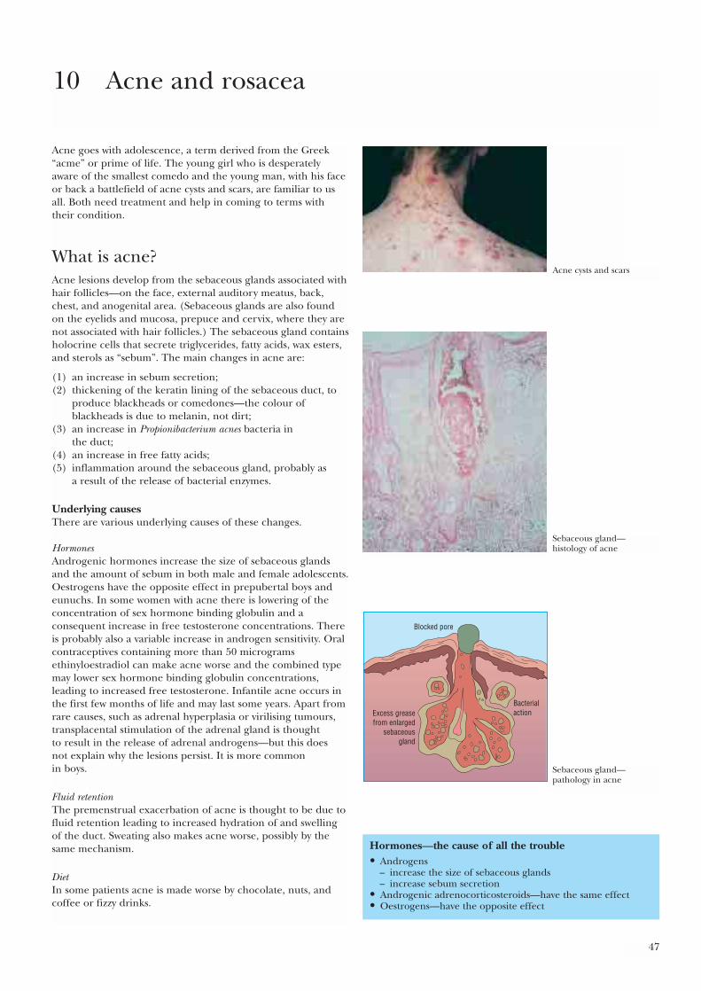
47
Acne goes with adolescence, a term derived from the Greek“acme” or prime of life. The young girl who is desperatelyaware of the smallest comedo and the young man, with his faceor back a battlefield of acne cysts and scars, are familiar to usall. Both need treatment and help in coming to terms withtheir condition.
What is acne?Acne lesions develop from the sebaceous glands associated withhair follicles—on the face, external auditory meatus, back,chest, and anogenital area. (Sebaceous glands are also foundon the eyelids and mucosa, prepuce and cervix, where they arenot associated with hair follicles.) The sebaceous gland containsholocrine cells that secrete triglycerides, fatty acids, wax esters,and sterols as “sebum”. The main changes in acne are:
(1) an increase in sebum secretion;(2) thickening of the keratin lining of the sebaceous duct, to
produce blackheads or comedones—the colour ofblackheads is due to melanin, not dirt;
(3) an increase in Propionibacterium acnes bacteria in the duct;
(4) an increase in free fatty acids;(5) inflammation around the sebaceous gland, probably as
a result of the release of bacterial enzymes.
Underlying causesThere are various underlying causes of these changes.
HormonesAndrogenic hormones increase the size of sebaceous glandsand the amount of sebum in both male and female adolescents.Oestrogens have the opposite effect in prepubertal boys andeunuchs. In some women with acne there is lowering of theconcentration of sex hormone binding globulin and aconsequent increase in free testosterone concentrations. Thereis probably also a variable increase in androgen sensitivity. Oralcontraceptives containing more than 50 microgramsethinyloestradiol can make acne worse and the combined typemay lower sex hormone binding globulin concentrations,leading to increased free testosterone. Infantile acne occurs inthe first few months of life and may last some years. Apart fromrare causes, such as adrenal hyperplasia or virilising tumours,transplacental stimulation of the adrenal gland is thought to result in the release of adrenal androgens—but this does not explain why the lesions persist. It is more common in boys.
Fluid retentionThe premenstrual exacerbation of acne is thought to be due tofluid retention leading to increased hydration of and swellingof the duct. Sweating also makes acne worse, possibly by thesame mechanism.
DietIn some patients acne is made worse by chocolate, nuts, andcoffee or fizzy drinks.
10 Acne and rosacea
Hormones—the cause of all the trouble• Androgens
– increase the size of sebaceous glands – increase sebum secretion
• Androgenic adrenocorticosteroids—have the same effect• Oestrogens—have the opposite effect
Acne cysts and scars
Sebaceous gland—histology of acne
Blocked pore
Excess greasefrom enlarged
sebaceousgland
Bacterialaction
Sebaceous gland—pathology in acne
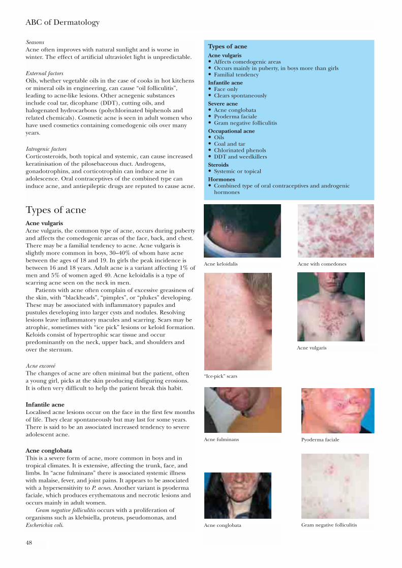
ABC of Dermatology
48
SeasonsAcne often improves with natural sunlight and is worse inwinter. The effect of artificial ultraviolet light is unpredictable.
External factorsOils, whether vegetable oils in the case of cooks in hot kitchensor mineral oils in engineering, can cause “oil folliculitis”,leading to acne-like lesions. Other acnegenic substancesinclude coal tar, dicophane (DDT), cutting oils, andhalogenated hydrocarbons (polychlorinated biphenols andrelated chemicals). Cosmetic acne is seen in adult women whohave used cosmetics containing comedogenic oils over manyyears.
Iatrogenic factorsCorticosteroids, both topical and systemic, can cause increasedkeratinisation of the pilosebaceous duct. Androgens,gonadotrophins, and corticotrophin can induce acne inadolescence. Oral contraceptives of the combined type caninduce acne, and antiepileptic drugs are reputed to cause acne.
Types of acneAcne vulgarisAcne vulgaris, the common type of acne, occurs during pubertyand affects the comedogenic areas of the face, back, and chest.There may be a familial tendency to acne. Acne vulgaris isslightly more common in boys, 30–40% of whom have acnebetween the ages of 18 and 19. In girls the peak incidence isbetween 16 and 18 years. Adult acne is a variant affecting 1% ofmen and 5% of women aged 40. Acne keloidalis is a type ofscarring acne seen on the neck in men.
Patients with acne often complain of excessive greasiness ofthe skin, with “blackheads”, “pimples”, or “plukes” developing.These may be associated with inflammatory papules andpustules developing into larger cysts and nodules. Resolvinglesions leave inflammatory macules and scarring. Scars may beatrophic, sometimes with “ice pick” lesions or keloid formation.Keloids consist of hypertrophic scar tissue and occurpredominantly on the neck, upper back, and shoulders andover the sternum.
Acne excoreéThe changes of acne are often minimal but the patient, often a young girl, picks at the skin producing disfiguring erosions. It is often very difficult to help the patient break this habit.
Infantile acneLocalised acne lesions occur on the face in the first few monthsof life. They clear spontaneously but may last for some years.There is said to be an associated increased tendency to severeadolescent acne.
Acne conglobataThis is a severe form of acne, more common in boys and intropical climates. It is extensive, affecting the trunk, face, andlimbs. In “acne fulminans” there is associated systemic illnesswith malaise, fever, and joint pains. It appears to be associatedwith a hypersensitivity to P. acnes. Another variant is pyodermafaciale, which produces erythematous and necrotic lesions andoccurs mainly in adult women.
Gram negative folliculitis occurs with a proliferation oforganisms such as klebsiella, proteus, pseudomonas, andEscherichia coli.
Types of acneAcne vulgaris• Affects comedogenic areas • Occurs mainly in puberty, in boys more than girls• Familial tendencyInfantile acne• Face only• Clears spontaneouslySevere acne• Acne conglobata• Pyoderma faciale• Gram negative folliculitisOccupational acne• Oils• Coal and tar• Chlorinated phenols• DDT and weedkillersSteroids• Systemic or topicalHormones• Combined type of oral contraceptives and androgenic
hormones
Acne keloidalis Acne with comedones
“Ice-pick” scars
Acne conglobata
Acne fulminans Pyoderma faciale
Acne vulgaris
Gram negative folliculitis
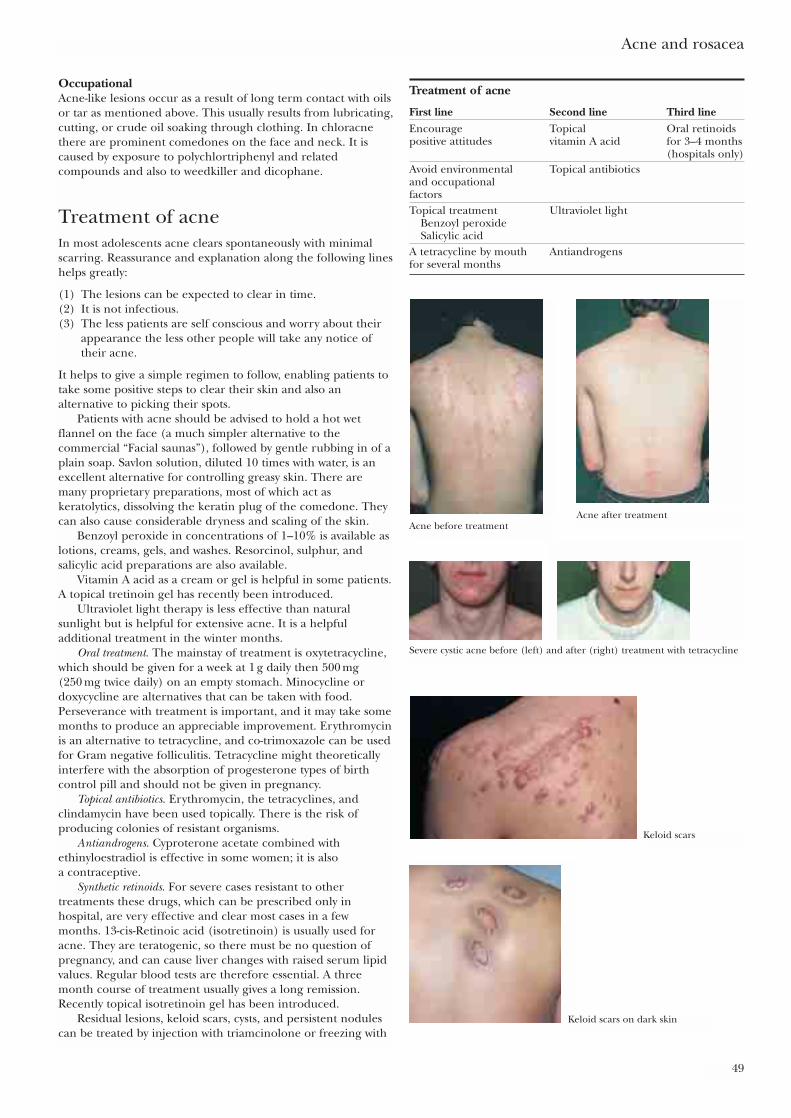
Acne and rosacea
49
OccupationalAcne-like lesions occur as a result of long term contact with oilsor tar as mentioned above. This usually results from lubricating,cutting, or crude oil soaking through clothing. In chloracnethere are prominent comedones on the face and neck. It iscaused by exposure to polychlortriphenyl and relatedcompounds and also to weedkiller and dicophane.
Treatment of acneIn most adolescents acne clears spontaneously with minimalscarring. Reassurance and explanation along the following lineshelps greatly:
(1) The lesions can be expected to clear in time.(2) It is not infectious.(3) The less patients are self conscious and worry about their
appearance the less other people will take any notice oftheir acne.
It helps to give a simple regimen to follow, enabling patients totake some positive steps to clear their skin and also analternative to picking their spots.
Patients with acne should be advised to hold a hot wetflannel on the face (a much simpler alternative to thecommercial “Facial saunas”), followed by gentle rubbing in of aplain soap. Savlon solution, diluted 10 times with water, is anexcellent alternative for controlling greasy skin. There aremany proprietary preparations, most of which act askeratolytics, dissolving the keratin plug of the comedone. Theycan also cause considerable dryness and scaling of the skin.
Benzoyl peroxide in concentrations of 1–10% is available aslotions, creams, gels, and washes. Resorcinol, sulphur, andsalicylic acid preparations are also available.
Vitamin A acid as a cream or gel is helpful in some patients.A topical tretinoin gel has recently been introduced.
Ultraviolet light therapy is less effective than naturalsunlight but is helpful for extensive acne. It is a helpfuladditional treatment in the winter months.
Oral treatment. The mainstay of treatment is oxytetracycline,which should be given for a week at 1 g daily then 500 mg (250 mg twice daily) on an empty stomach. Minocycline ordoxycycline are alternatives that can be taken with food.Perseverance with treatment is important, and it may take somemonths to produce an appreciable improvement. Erythromycinis an alternative to tetracycline, and co-trimoxazole can be usedfor Gram negative folliculitis. Tetracycline might theoreticallyinterfere with the absorption of progesterone types of birthcontrol pill and should not be given in pregnancy.
Topical antibiotics. Erythromycin, the tetracyclines, andclindamycin have been used topically. There is the risk ofproducing colonies of resistant organisms.
Antiandrogens. Cyproterone acetate combined withethinyloestradiol is effective in some women; it is also a contraceptive.
Synthetic retinoids. For severe cases resistant to othertreatments these drugs, which can be prescribed only inhospital, are very effective and clear most cases in a fewmonths. 13-cis-Retinoic acid (isotretinoin) is usually used foracne. They are teratogenic, so there must be no question ofpregnancy, and can cause liver changes with raised serum lipidvalues. Regular blood tests are therefore essential. A threemonth course of treatment usually gives a long remission.Recently topical isotretinoin gel has been introduced.
Residual lesions, keloid scars, cysts, and persistent nodulescan be treated by injection with triamcinolone or freezing with
Treatment of acne
First line Second line Third lineEncourage Topical Oral retinoidspositive attitudes vitamin A acid for 3–4 months
(hospitals only)Avoid environmental Topical antibioticsand occupational factorsTopical treatment Ultraviolet light
Benzoyl peroxideSalicylic acid
A tetracycline by mouth Antiandrogensfor several months
Acne before treatmentAcne after treatment
Severe cystic acne before (left) and after (right) treatment with tetracycline
Keloid scars
Keloid scars on dark skin
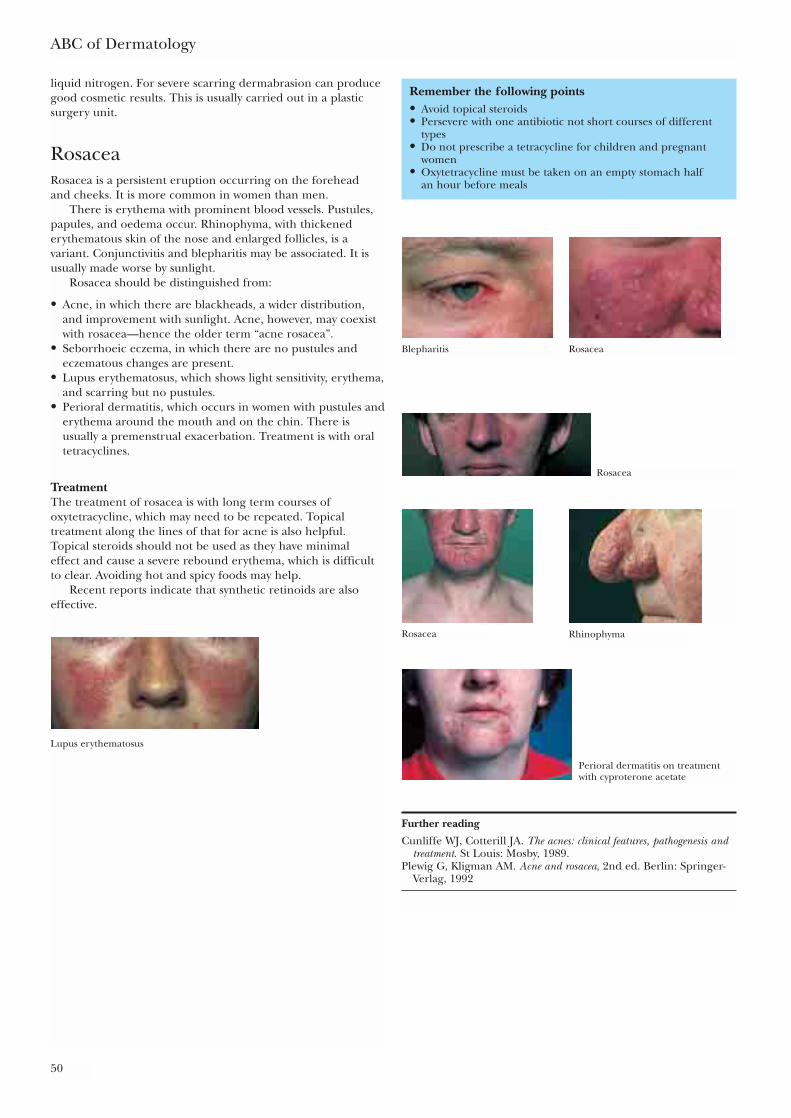
ABC of Dermatology
50
liquid nitrogen. For severe scarring dermabrasion can producegood cosmetic results. This is usually carried out in a plasticsurgery unit.
RosaceaRosacea is a persistent eruption occurring on the forehead and cheeks. It is more common in women than men.
There is erythema with prominent blood vessels. Pustules,papules, and oedema occur. Rhinophyma, with thickenederythematous skin of the nose and enlarged follicles, is avariant. Conjunctivitis and blepharitis may be associated. It isusually made worse by sunlight.
Rosacea should be distinguished from:
• Acne, in which there are blackheads, a wider distribution,and improvement with sunlight. Acne, however, may coexistwith rosacea—hence the older term “acne rosacea”.
• Seborrhoeic eczema, in which there are no pustules andeczematous changes are present.
• Lupus erythematosus, which shows light sensitivity, erythema,and scarring but no pustules.
• Perioral dermatitis, which occurs in women with pustules anderythema around the mouth and on the chin. There isusually a premenstrual exacerbation. Treatment is with oraltetracyclines.
TreatmentThe treatment of rosacea is with long term courses ofoxytetracycline, which may need to be repeated. Topicaltreatment along the lines of that for acne is also helpful.Topical steroids should not be used as they have minimal effect and cause a severe rebound erythema, which is difficultto clear. Avoiding hot and spicy foods may help.
Recent reports indicate that synthetic retinoids are alsoeffective.
Remember the following points• Avoid topical steroids• Persevere with one antibiotic not short courses of different
types• Do not prescribe a tetracycline for children and pregnant
women• Oxytetracycline must be taken on an empty stomach half
an hour before meals
Further reading
Cunliffe WJ, Cotterill JA. The acnes: clinical features, pathogenesis andtreatment. St Louis: Mosby, 1989.
Plewig G, Kligman AM. Acne and rosacea, 2nd ed. Berlin: Springer-Verlag, 1992
Blepharitis Rosacea
Rosacea
Rosacea Rhinophyma
Lupus erythematosus
Perioral dermatitis on treatmentwith cyproterone acetate
51
IntroductionHair, which has an essential physiological role in animals, ismainly of psychological importance in man. A good head ofhair provides some degree of warmth for the human head andalso protection from ultraviolet radiation, but its significance isotherwise in the eye of the beholder. In the form of wool, hairis of economic importance and considerable research has beencarried out into the cycles of growth and the structure of woolkeratin in sheep.
Too much hair, particularly on the face of women, is anembarrassment and cosmetic problem and loss of hair from thescalp is equally troublesome. Changes in hair growth are notonly of cosmetic significance but can also be associated withunderlying diseases. Diseases occurring in the skin of the scalpcan damage hair follicles leading to loss of hair. This chaptercovers: (a) the normal pattern of hair growth; (b) causes ofhair loss; (c) skin diseases involving the scalp; (d) causes of excess hair growth; (e) abnormalities of the hair itself; and (f) treatment.
The normal pattern of hair growthUnlike other epidermal structures which grow continuously,hair has a cyclical pattern of growth. The growing phase oranagen lasts an average of 1000 days on the scalp followed byan involutional phase known as catagen which is quite short,lasting only a few days. The hair then enters a resting phase,telogen, lasting about 100 days. In man, hair growth is normallyasynchronous, with each individual hair following its own cycleindependently of the others. The basal layer of the hair bulbfrom which the hair itself is produced is known as the matrixand contains melanocytes from which melanin pigment isincorporated into the hair. The type of melanin determines thecolour and in grey or white hair, pigment production isreduced or absent.
The body surface with the exception of the palms, soles, the lips, and the genitalia, is covered with fine vellus hairs thatdo not have a medulla and are not pigmented. These hairsdevelop into longer coarse, medullated, terminal hair on the scalp and eyebrows. At puberty a similar change occurs in the pubic area and the axillae, also on the face and trunk, in the male. These changes are androgen dependent, even infemales, but testicular androgen is required to produce beardgrowth and balding in men.
Racial characteristics and the genetic make-up of theindividual determine the type and colour of the hair. Straightblack oriental hair is clearly different from the nordic blonde type.
Hair lossThis is known as alopecia, said to be derived from the Latin“alopex”, a fox, presumably because of the bald patches ofmange seen in wild foxes.
Adult male pattern alopecia is so common as to beconsidered normal. Circulating levels of testosterone are not
11 The hair and scalpD Kemmett
85% of hair 15% of hair
Sebaceousgland
Dermalpapilla Cord of
epithelial cells
Anagen Catagen Telogen
3 years Few days 100 days
New anagen hair
Telogenclub hair
Diagrammatic cross-section of hair at various growth phases
Anagen 1000 days
Catagen
Telogen 100 days
Hair growth cycle
Male pattern baldness
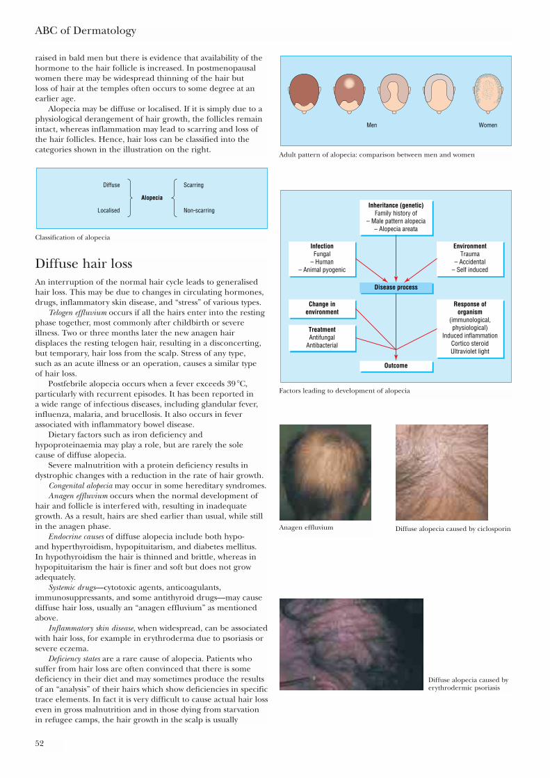
ABC of Dermatology
52
raised in bald men but there is evidence that availability of thehormone to the hair follicle is increased. In postmenopausalwomen there may be widespread thinning of the hair but loss of hair at the temples often occurs to some degree at anearlier age.
Alopecia may be diffuse or localised. If it is simply due to aphysiological derangement of hair growth, the follicles remainintact, whereas inflammation may lead to scarring and loss ofthe hair follicles. Hence, hair loss can be classified into thecategories shown in the illustration on the right.
Diffuse hair lossAn interruption of the normal hair cycle leads to generalisedhair loss. This may be due to changes in circulating hormones,drugs, inflammatory skin disease, and “stress” of various types.
Telogen effluvium occurs if all the hairs enter into the restingphase together, most commonly after childbirth or severeillness. Two or three months later the new anagen hairdisplaces the resting telogen hair, resulting in a disconcerting,but temporary, hair loss from the scalp. Stress of any type, such as an acute illness or an operation, causes a similar type of hair loss.
Postfebrile alopecia occurs when a fever exceeds 39 �C,particularly with recurrent episodes. It has been reported in a wide range of infectious diseases, including glandular fever,influenza, malaria, and brucellosis. It also occurs in feverassociated with inflammatory bowel disease.
Dietary factors such as iron deficiency andhypoproteinaemia may play a role, but are rarely the sole cause of diffuse alopecia.
Severe malnutrition with a protein deficiency results indystrophic changes with a reduction in the rate of hair growth.
Congenital alopecia may occur in some hereditary syndromes.Anagen effluvium occurs when the normal development of
hair and follicle is interfered with, resulting in inadequategrowth. As a result, hairs are shed earlier than usual, while stillin the anagen phase.
Endocrine causes of diffuse alopecia include both hypo- and hyperthyroidism, hypopituitarism, and diabetes mellitus. In hypothyroidism the hair is thinned and brittle, whereas inhypopituitarism the hair is finer and soft but does not growadequately.
Systemic drugs—cytotoxic agents, anticoagulants,immunosuppressants, and some antithyroid drugs—may causediffuse hair loss, usually an “anagen effluvium” as mentionedabove.
Inflammatory skin disease, when widespread, can be associatedwith hair loss, for example in erythroderma due to psoriasis orsevere eczema.
Deficiency states are a rare cause of alopecia. Patients whosuffer from hair loss are often convinced that there is somedeficiency in their diet and may sometimes produce the resultsof an “analysis” of their hairs which show deficiencies in specifictrace elements. In fact it is very difficult to cause actual hair losseven in gross malnutrition and in those dying from starvationin refugee camps, the hair growth in the scalp is usually
Men Women
Adult pattern of alopecia: comparison between men and women
Inheritance (genetic)Family history of
– Male pattern alopecia– Alopecia areata
Outcome
Disease process
EnvironmentTrauma
– Accidental– Self induced
InfectionFungal
– Human– Animal pyogenic
Response oforganism
(immunological,physiological)
Induced inflammationCortico steroidUltraviolet light
Change inenvironment
TreatmentAntifungal
Antibacterial
Factors leading to development of alopecia
Diffuse
Localised
Scarring
Alopecia
Non-scarring
Classification of alopecia
Anagen effluvium Diffuse alopecia caused by ciclosporin
Diffuse alopecia caused byerythrodermic psoriasis
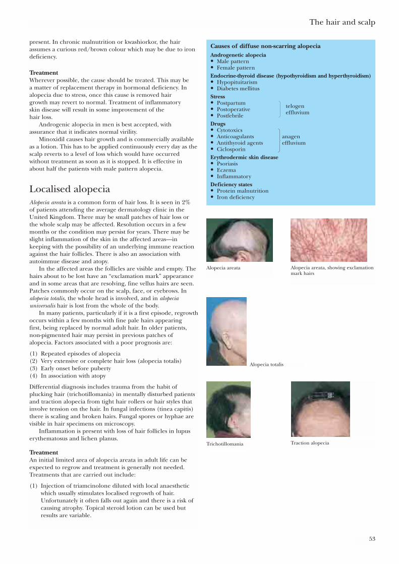
The hair and scalp
53
present. In chronic malnutrition or kwashiorkor, the hairassumes a curious red/brown colour which may be due to irondeficiency.
TreatmentWherever possible, the cause should be treated. This may be a matter of replacement therapy in hormonal deficiency. Inalopecia due to stress, once this cause is removed hair growth may revert to normal. Treatment of inflammatory skin disease will result in some improvement of the hair loss.
Androgenic alopecia in men is best accepted, withassurance that it indicates normal virility.
Minoxidil causes hair growth and is commercially availableas a lotion. This has to be applied continuously every day as thescalp reverts to a level of loss which would have occurredwithout treatment as soon as it is stopped. It is effective inabout half the patients with male pattern alopecia.
Localised alopeciaAlopecia areata is a common form of hair loss. It is seen in 2% of patients attending the average dermatology clinic in theUnited Kingdom. There may be small patches of hair loss orthe whole scalp may be affected. Resolution occurs in a fewmonths or the condition may persist for years. There may beslight inflammation of the skin in the affected areas—inkeeping with the possibility of an underlying immune reactionagainst the hair follicles. There is also an association withautoimmue disease and atopy.
In the affected areas the follicles are visible and empty. Thehairs about to be lost have an “exclamation mark” appearanceand in some areas that are resolving, fine vellus hairs are seen.Patches commonly occur on the scalp, face, or eyebrows. Inalopecia totalis, the whole head is involved, and in alopeciauniversalis hair is lost from the whole of the body.
In many patients, particularly if it is a first episode, regrowthoccurs within a few months with fine pale hairs appearing first, being replaced by normal adult hair. In older patients,non-pigmented hair may persist in previous patches of alopecia. Factors associated with a poor prognosis are:
(1) Repeated episodes of alopecia(2) Very extensive or complete hair loss (alopecia totalis)(3) Early onset before puberty(4) In association with atopy
Differential diagnosis includes trauma from the habit ofplucking hair (trichotillomania) in mentally disturbed patientsand traction alopecia from tight hair rollers or hair styles thatinvolve tension on the hair. In fungal infections (tinea capitis)there is scaling and broken hairs. Fungal spores or hyphae arevisible in hair specimens on microscopy.
Inflammation is present with loss of hair follicles in lupuserythematosus and lichen planus.
TreatmentAn initial limited area of alopecia areata in adult life can beexpected to regrow and treatment is generally not needed.Treatments that are carried out include:
(1) Injection of triamcinolone diluted with local anaestheticwhich usually stimulates localised regrowth of hair.Unfortunately it often falls out again and there is a risk ofcausing atrophy. Topical steroid lotion can be used butresults are variable.
Causes of diffuse non-scarring alopeciaAndrogenetic alopecia• Male pattern• Female patternEndocrine-thyroid disease (hypothyroidism and hyperthyroidism)• Hypopituitarism• Diabetes mellitusStress• Postpartum• Postoperative• PostfebrileDrugs• Cytotoxics• Anticoagulants anagen• Antithyroid agents effluvium• CiclosporinErythrodermic skin disease• Psoriasis• Eczema• InflammatoryDeficiency states• Protein malnutrition• Iron deficiency
telogeneffluvium
Alopecia areata Alopecia areata, showing exclamationmark hairs
Alopecia totalis
Trichotillomania Traction alopecia
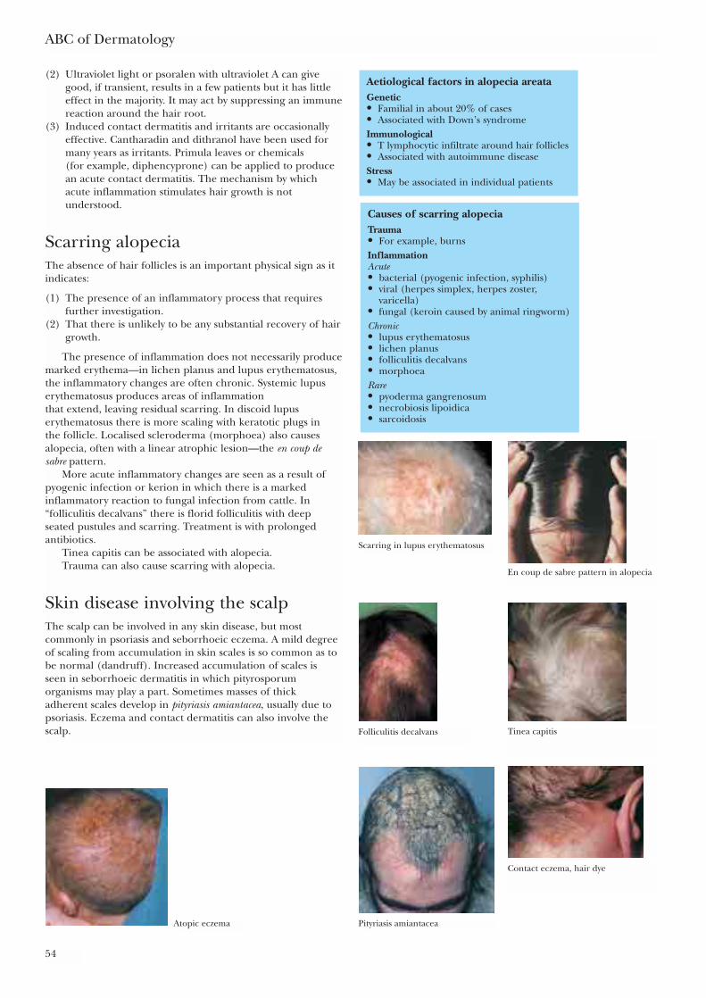
ABC of Dermatology
54
(2) Ultraviolet light or psoralen with ultraviolet A can givegood, if transient, results in a few patients but it has littleeffect in the majority. It may act by suppressing an immunereaction around the hair root.
(3) Induced contact dermatitis and irritants are occasionallyeffective. Cantharadin and dithranol have been used formany years as irritants. Primula leaves or chemicals (for example, diphencyprone) can be applied to producean acute contact dermatitis. The mechanism by whichacute inflammation stimulates hair growth is notunderstood.
Scarring alopeciaThe absence of hair follicles is an important physical sign as itindicates:
(1) The presence of an inflammatory process that requiresfurther investigation.
(2) That there is unlikely to be any substantial recovery of hairgrowth.
The presence of inflammation does not necessarily producemarked erythema—in lichen planus and lupus erythematosus,the inflammatory changes are often chronic. Systemic lupuserythematosus produces areas of inflammation that extend, leaving residual scarring. In discoid lupuserythematosus there is more scaling with keratotic plugs in the follicle. Localised scleroderma (morphoea) also causesalopecia, often with a linear atrophic lesion—the en coup desabre pattern.
More acute inflammatory changes are seen as a result ofpyogenic infection or kerion in which there is a markedinflammatory reaction to fungal infection from cattle. In“folliculitis decalvans” there is florid folliculitis with deep seated pustules and scarring. Treatment is with prolongedantibiotics.
Tinea capitis can be associated with alopecia.Trauma can also cause scarring with alopecia.
Skin disease involving the scalpThe scalp can be involved in any skin disease, but mostcommonly in psoriasis and seborrhoeic eczema. A mild degreeof scaling from accumulation in skin scales is so common as tobe normal (dandruff). Increased accumulation of scales is seen in seborrhoeic dermatitis in which pityrosporumorganisms may play a part. Sometimes masses of thick adherent scales develop in pityriasis amiantacea, usually due topsoriasis. Eczema and contact dermatitis can also involve thescalp.
Causes of scarring alopeciaTrauma• For example, burnsInflammationAcute• bacterial (pyogenic infection, syphilis)• viral (herpes simplex, herpes zoster,
varicella)• fungal (keroin caused by animal ringworm)Chronic• lupus erythematosus• lichen planus• folliculitis decalvans• morphoeaRare• pyoderma gangrenosum• necrobiosis lipoidica• sarcoidosis
Aetiological factors in alopecia areataGenetic• Familial in about 20% of cases• Associated with Down’s syndromeImmunological• T lymphocytic infiltrate around hair follicles• Associated with autoimmune diseaseStress• May be associated in individual patients
Scarring in lupus erythematosus
En coup de sabre pattern in alopecia
Folliculitis decalvans Tinea capitis
Pityriasis amiantacea
Contact eczema, hair dye
Atopic eczema
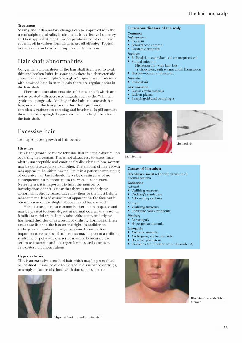
The hair and scalp
55
TreatmentScaling and inflammatory changes can be improved with theuse of sulphur and salicylic ointment. It is effective but messyand best applied at night. Tar preparations, oil of cade, andcoconut oil in various formulations are all effective. Topicalsteroids can also be used to suppress inflammation.
Hair shaft abnormalitiesCongenital abnormalities of the hair shaft itself lead to weak,thin and broken hairs. In some cases there is a characteristicappearance, for example “spun glass” appearance of pili tortiwith a twisted hair. In monilethrix there are regular nodes inthe hair shaft.
There are other abnormalities of the hair shaft which arenot associated with increased fragility, such as the Willi hairsyndrome, progressive kinking of the hair and uncombablehair, in which the hair grows in disorderly profusion,completely resistant to combing and brushing. In pili annulatithere may be a spangled appearance due to bright bands in the hair shaft.
Excessive hairTwo types of overgrowth of hair occur:
HirsutiesThis is the growth of coarse terminal hair in a male distributionoccurring in a woman. This is not always easy to assess sincewhat is unacceptable and emotionally disturbing to one womanmay be quite acceptable to another. The amount of hair growthmay appear to be within normal limits in a patient complainingof excessive hair but it should never be dismissed as of noconsequence if it is important to the woman concerned.Nevertheless, it is important to limit the number ofinvestigations once it is clear that there is no underlyingabnormality. Strong reassurance may then be the most helpfulmanagement. It is of course most apparent on the face but isoften present on the thighs, abdomen and back as well.
Hirsuties occurs most commonly after the menopause andmay be present to some degree in normal women as a result offamilial or racial traits. It may arise without any underlyinghormonal disorder or as a result of virilising hormones. Thesecauses are listed in the box on the right. In addition toandrogens, a number of drugs can cause hirsuties. It isimportant to remember that hirsuties may be part of a virilisingsyndrome or polycystic ovaries. It is useful to measure theserum testosterone and oestrogen level, as well as urinary 17 oxosteroid concentrations.
HypertrichosisThis is an excessive growth of hair which may be generalised or localised. It may be due to metabolic disturbance or drugs,or simply a feature of a localised lesion such as a mole.
Causes of hirsutismHereditary, racial with wide variation ofnormal patternEndocrineAdrenal• Virilising tumours• Cushing’s syndrome• Adrenal hyperplasiaOvarian• Virilising tumours• Polycystic ovary syndromePituitary• Acromegaly• HyperprolactinaemiaIatrogenic• Anabolic steroids• Androgens, corticosteroids• Danazol, phenytoin• Psoralens (in psoralen with ultraviolet A)
Cutaneous diseases of the scalpCommonInflammatory• Psoriasis• Seborrhoeic eczema• Contact dermatitisInfection• Folliculitis—staphylococcal or streptococcal• Fungal infection
Microsporum, with hair lossTrichophyton, with scaling and inflammation
• Herpes—zoster and simplexInfestation• PediculosisLess common• Lupus erythematosus• Lichen planus• Pemphigoid and pemphigus
Monilethrix
Monilethrix
Hirsuties due to virilisingtumour
Hypertrichosis caused by minoxidil

TreatmentIt is clearly important to treat any underlying cause and this will usually improve the condition. Otherwise treatment issymptomatic and includes:
• Removal of hair by shaving or hair removing creams.• Electrolysis and diathermy, which gives permanent
destruction of the hair follicle.• Antiandrogen drugs such as cyproterone can be used under
specialist supervision.
ABC of Dermatology
56
Further readingOrfanos CE, Happle R. Hair and hair disease. Berlin: Springer-
Verlag, 1990Rook A, Dawber R. Diseases of the hair and scalp, 3rd ed. Oxford:
Blackwell Scientific, 1997
Causes of hypertrichosis• Naevoid—in pigmented naevi and Becker’s naevus• Congenital (rare)• Acquired—porphyria, hyperthyroidism, anorexia nervosa
some developmental defects, for example, Hurler’ssyndrome, tumours (hypertrichosis lanuginosa), drugs(diazoxide, minoxidil, ciclosporin)
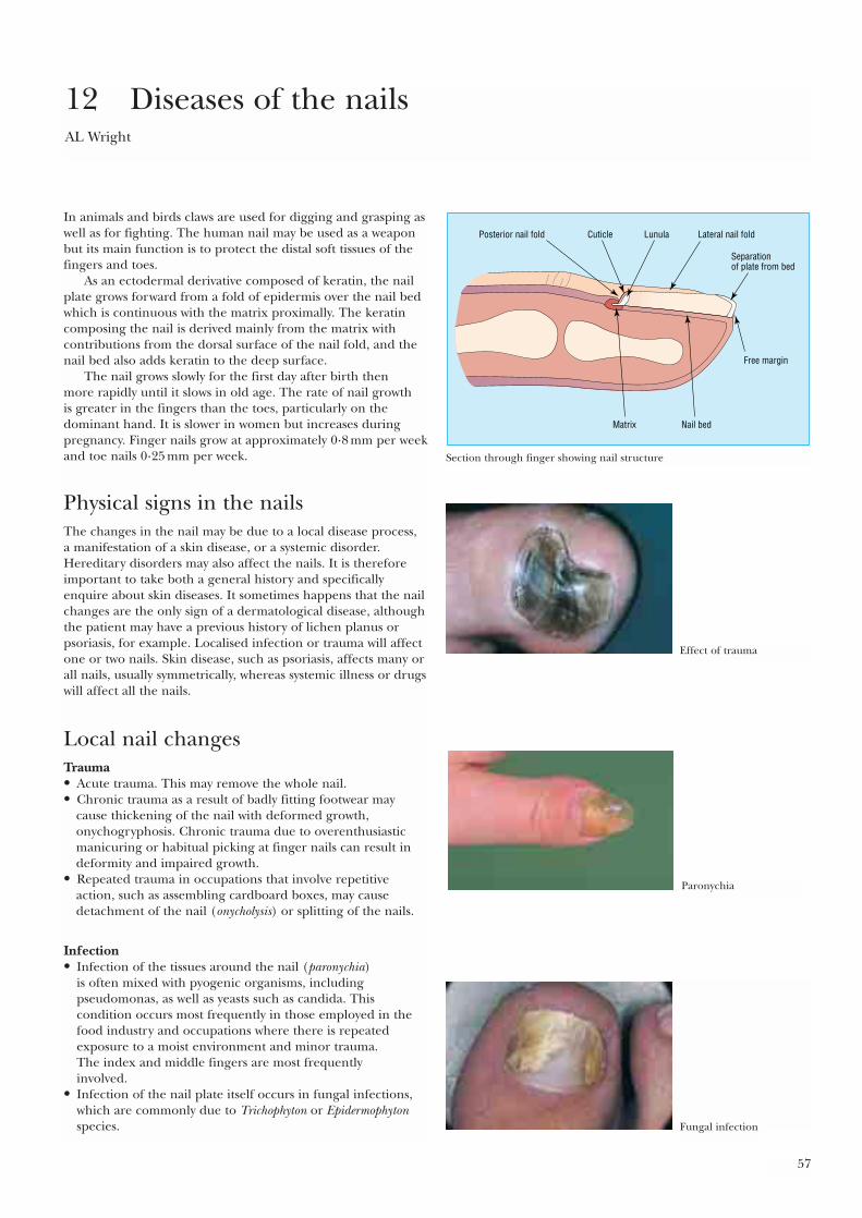
57
In animals and birds claws are used for digging and grasping aswell as for fighting. The human nail may be used as a weaponbut its main function is to protect the distal soft tissues of thefingers and toes.
As an ectodermal derivative composed of keratin, the nailplate grows forward from a fold of epidermis over the nail bedwhich is continuous with the matrix proximally. The keratincomposing the nail is derived mainly from the matrix withcontributions from the dorsal surface of the nail fold, and thenail bed also adds keratin to the deep surface.
The nail grows slowly for the first day after birth then more rapidly until it slows in old age. The rate of nail growth is greater in the fingers than the toes, particularly on thedominant hand. It is slower in women but increases duringpregnancy. Finger nails grow at approximately 0·8 mm per weekand toe nails 0·25 mm per week.
Physical signs in the nailsThe changes in the nail may be due to a local disease process, a manifestation of a skin disease, or a systemic disorder.Hereditary disorders may also affect the nails. It is thereforeimportant to take both a general history and specificallyenquire about skin diseases. It sometimes happens that the nailchanges are the only sign of a dermatological disease, althoughthe patient may have a previous history of lichen planus orpsoriasis, for example. Localised infection or trauma will affectone or two nails. Skin disease, such as psoriasis, affects many orall nails, usually symmetrically, whereas systemic illness or drugswill affect all the nails.
Local nail changesTrauma• Acute trauma. This may remove the whole nail. • Chronic trauma as a result of badly fitting footwear may
cause thickening of the nail with deformed growth,onychogryphosis. Chronic trauma due to overenthusiasticmanicuring or habitual picking at finger nails can result indeformity and impaired growth.
• Repeated trauma in occupations that involve repetitiveaction, such as assembling cardboard boxes, may causedetachment of the nail (onycholysis) or splitting of the nails.
Infection• Infection of the tissues around the nail (paronychia)
is often mixed with pyogenic organisms, includingpseudomonas, as well as yeasts such as candida. Thiscondition occurs most frequently in those employed in thefood industry and occupations where there is repeatedexposure to a moist environment and minor trauma. The index and middle fingers are most frequently involved.
• Infection of the nail plate itself occurs in fungal infections,which are commonly due to Trichophyton or Epidermophytonspecies.
12 Diseases of the nailsAL Wright
Posterior nail fold Lateral nail fold
Separationof plate from bed
Cuticle Lunula
Matrix Nail bed
Free margin
Section through finger showing nail structure
Effect of trauma
Paronychia
Fungal infection
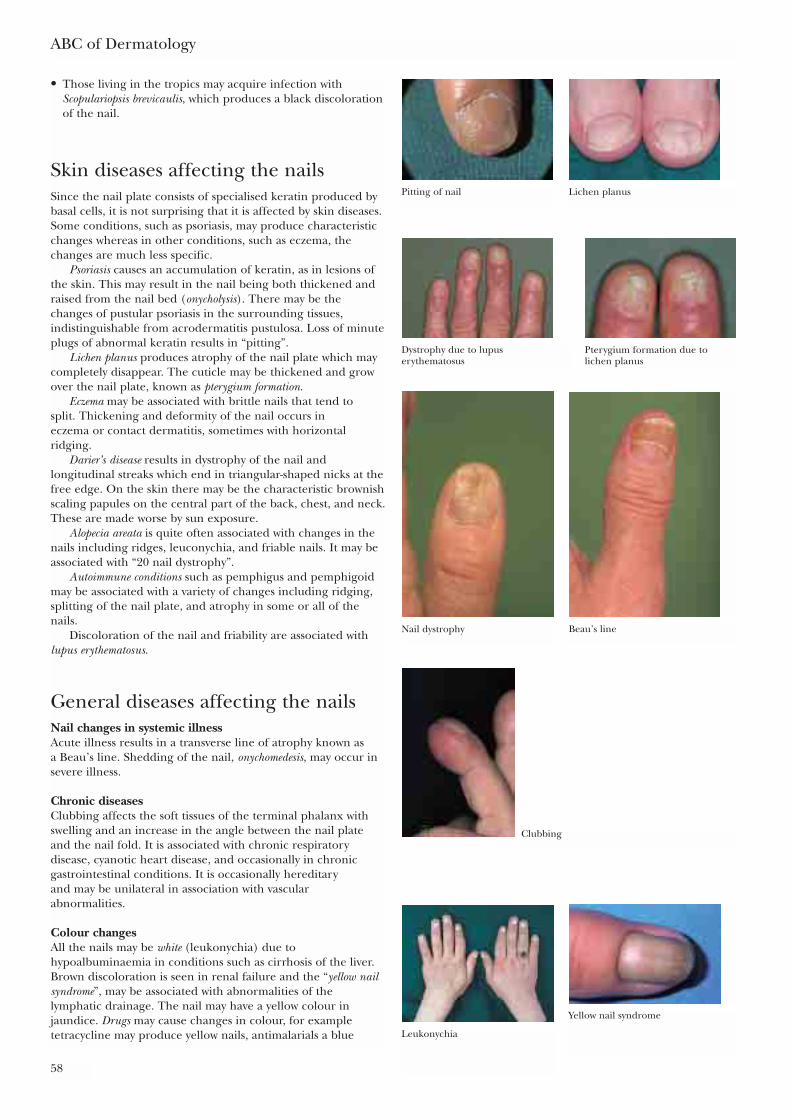
• Those living in the tropics may acquire infection withScopulariopsis brevicaulis, which produces a black discolorationof the nail.
Skin diseases affecting the nailsSince the nail plate consists of specialised keratin produced bybasal cells, it is not surprising that it is affected by skin diseases.Some conditions, such as psoriasis, may produce characteristicchanges whereas in other conditions, such as eczema, thechanges are much less specific.
Psoriasis causes an accumulation of keratin, as in lesions ofthe skin. This may result in the nail being both thickened andraised from the nail bed (onycholysis). There may be thechanges of pustular psoriasis in the surrounding tissues,indistinguishable from acrodermatitis pustulosa. Loss of minuteplugs of abnormal keratin results in “pitting”.
Lichen planus produces atrophy of the nail plate which maycompletely disappear. The cuticle may be thickened and growover the nail plate, known as pterygium formation.
Eczema may be associated with brittle nails that tend to split. Thickening and deformity of the nail occurs in eczema or contact dermatitis, sometimes with horizontalridging.
Darier’s disease results in dystrophy of the nail andlongitudinal streaks which end in triangular-shaped nicks at thefree edge. On the skin there may be the characteristic brownishscaling papules on the central part of the back, chest, and neck.These are made worse by sun exposure.
Alopecia areata is quite often associated with changes in thenails including ridges, leuconychia, and friable nails. It may beassociated with “20 nail dystrophy”.
Autoimmune conditions such as pemphigus and pemphigoidmay be associated with a variety of changes including ridging,splitting of the nail plate, and atrophy in some or all of thenails.
Discoloration of the nail and friability are associated withlupus erythematosus.
General diseases affecting the nailsNail changes in systemic illnessAcute illness results in a transverse line of atrophy known as a Beau’s line. Shedding of the nail, onychomedesis, may occur insevere illness.
Chronic diseasesClubbing affects the soft tissues of the terminal phalanx withswelling and an increase in the angle between the nail plateand the nail fold. It is associated with chronic respiratorydisease, cyanotic heart disease, and occasionally in chronicgastrointestinal conditions. It is occasionally hereditary and may be unilateral in association with vascularabnormalities.
Colour changesAll the nails may be white (leukonychia) due tohypoalbuminaemia in conditions such as cirrhosis of the liver.Brown discoloration is seen in renal failure and the “yellow nailsyndrome”, may be associated with abnormalities of thelymphatic drainage. The nail may have a yellow colour injaundice. Drugs may cause changes in colour, for exampletetracycline may produce yellow nails, antimalarials a blue
ABC of Dermatology
58
Pitting of nail Lichen planus
Dystrophy due to lupuserythematosus
Pterygium formation due tolichen planus
Nail dystrophy Beau’s line
Clubbing
Leukonychia
Yellow nail syndrome
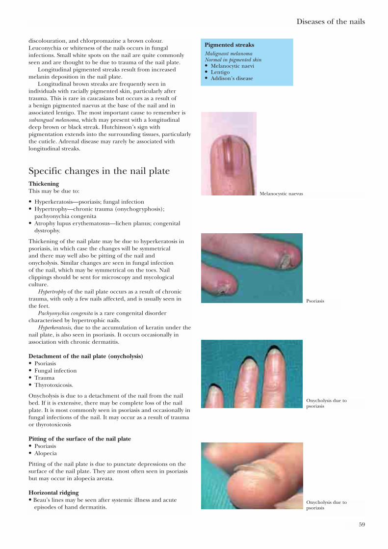
discolouration, and chlorpromazine a brown colour.Leuconychia or whiteness of the nails occurs in fungalinfections. Small white spots on the nail are quite commonlyseen and are thought to be due to trauma of the nail plate.
Longitudinal pigmented streaks result from increasedmelanin deposition in the nail plate.
Longitudinal brown streaks are frequently seen inindividuals with racially pigmented skin, particularly aftertrauma. This is rare in caucasians but occurs as a result of a benign pigmented naevus at the base of the nail and inassociated lentigo. The most important cause to remember issubungual melanoma, which may present with a longitudinaldeep brown or black streak. Hutchinson’s sign withpigmentation extends into the surrounding tissues, particularlythe cuticle. Adrenal disease may rarely be associated withlongitudinal streaks.
Specific changes in the nail plateThickeningThis may be due to:
• Hyperkeratosis—psoriasis; fungal infection• Hypertrophy—chronic trauma (onychogryphosis);
pachyonychia congenita• Atrophy lupus erythematosus—lichen planus; congenital
dystrophy.
Thickening of the nail plate may be due to hyperkeratosis inpsoriasis, in which case the changes will be symmetrical and there may well also be pitting of the nail and onycholysis. Similar changes are seen in fungal infection of the nail, which may be symmetrical on the toes. Nailclippings should be sent for microscopy and mycologicalculture.
Hypertrophy of the nail plate occurs as a result of chronictrauma, with only a few nails affected, and is usually seen in the feet.
Pachyonychia congenita is a rare congenital disordercharacterised by hypertrophic nails.
Hyperkeratosis, due to the accumulation of keratin under thenail plate, is also seen in psoriasis. It occurs occasionally inassociation with chronic dermatitis.
Detachment of the nail plate (onycholysis)• Psoriasis• Fungal infection• Trauma• Thyrotoxicosis.
Onycholysis is due to a detachment of the nail from the nailbed. If it is extensive, there may be complete loss of the nailplate. It is most commonly seen in psoriasis and occasionally infungal infections of the nail. It may occur as a result of traumaor thyrotoxicosis
Pitting of the surface of the nail plate• Psoriasis• Alopecia
Pitting of the nail plate is due to punctate depressions on thesurface of the nail plate. They are most often seen in psoriasisbut may occur in alopecia areata.
Horizontal ridging• Beau’s lines may be seen after systemic illness and acute
episodes of hand dermatitis.
Diseases of the nails
59
Pigmented streaksMalignant melanomaNormal in pigmented skin• Melanocytic naevi• Lentigo• Addison’s disease
Melanocystic naevus
Psoriasis
Onycholysis due topsoriasis
Onycholysis due topsoriasis
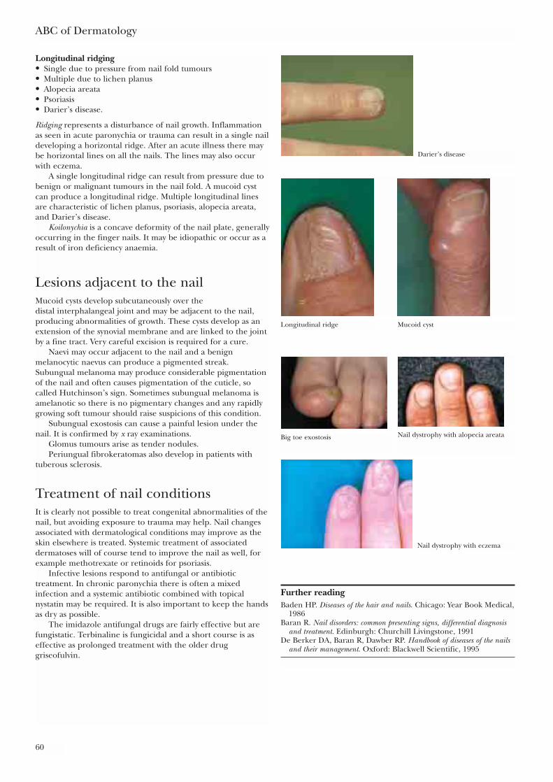
Longitudinal ridging• Single due to pressure from nail fold tumours• Multiple due to lichen planus• Alopecia areata• Psoriasis• Darier’s disease.
Ridging represents a disturbance of nail growth. Inflammationas seen in acute paronychia or trauma can result in a single naildeveloping a horizontal ridge. After an acute illness there maybe horizontal lines on all the nails. The lines may also occurwith eczema.
A single longitudinal ridge can result from pressure due tobenign or malignant tumours in the nail fold. A mucoid cystcan produce a longitudinal ridge. Multiple longitudinal linesare characteristic of lichen planus, psoriasis, alopecia areata,and Darier’s disease.
Koilonychia is a concave deformity of the nail plate, generallyoccurring in the finger nails. It may be idiopathic or occur as aresult of iron deficiency anaemia.
Lesions adjacent to the nailMucoid cysts develop subcutaneously over the distal interphalangeal joint and may be adjacent to the nail,producing abnormalities of growth. These cysts develop as anextension of the synovial membrane and are linked to the jointby a fine tract. Very careful excision is required for a cure.
Naevi may occur adjacent to the nail and a benignmelanocytic naevus can produce a pigmented streak.Subungual melanoma may produce considerable pigmentationof the nail and often causes pigmentation of the cuticle, socalled Hutchinson’s sign. Sometimes subungual melanoma isamelanotic so there is no pigmentary changes and any rapidlygrowing soft tumour should raise suspicions of this condition.
Subungual exostosis can cause a painful lesion under thenail. It is confirmed by x ray examinations.
Glomus tumours arise as tender nodules.Periungual fibrokeratomas also develop in patients with
tuberous sclerosis.
Treatment of nail conditionsIt is clearly not possible to treat congenital abnormalities of thenail, but avoiding exposure to trauma may help. Nail changesassociated with dermatological conditions may improve as theskin elsewhere is treated. Systemic treatment of associateddermatoses will of course tend to improve the nail as well, forexample methotrexate or retinoids for psoriasis.
Infective lesions respond to antifungal or antibiotictreatment. In chronic paronychia there is often a mixedinfection and a systemic antibiotic combined with topicalnystatin may be required. It is also important to keep the handsas dry as possible.
The imidazole antifungal drugs are fairly effective but arefungistatic. Terbinaline is fungicidal and a short course is aseffective as prolonged treatment with the older druggriseofulvin.
ABC of Dermatology
60
Further readingBaden HP. Diseases of the hair and nails. Chicago: Year Book Medical,
1986Baran R. Nail disorders: common presenting signs, differential diagnosis
and treatment. Edinburgh: Churchill Livingstone, 1991De Berker DA, Baran R, Dawber RP. Handbook of diseases of the nails
and their management. Oxford: Blackwell Scientific, 1995
Longitudinal ridge Mucoid cyst
Big toe exostosis Nail dystrophy with alopecia areata
Nail dystrophy with eczema
Darier’s disease
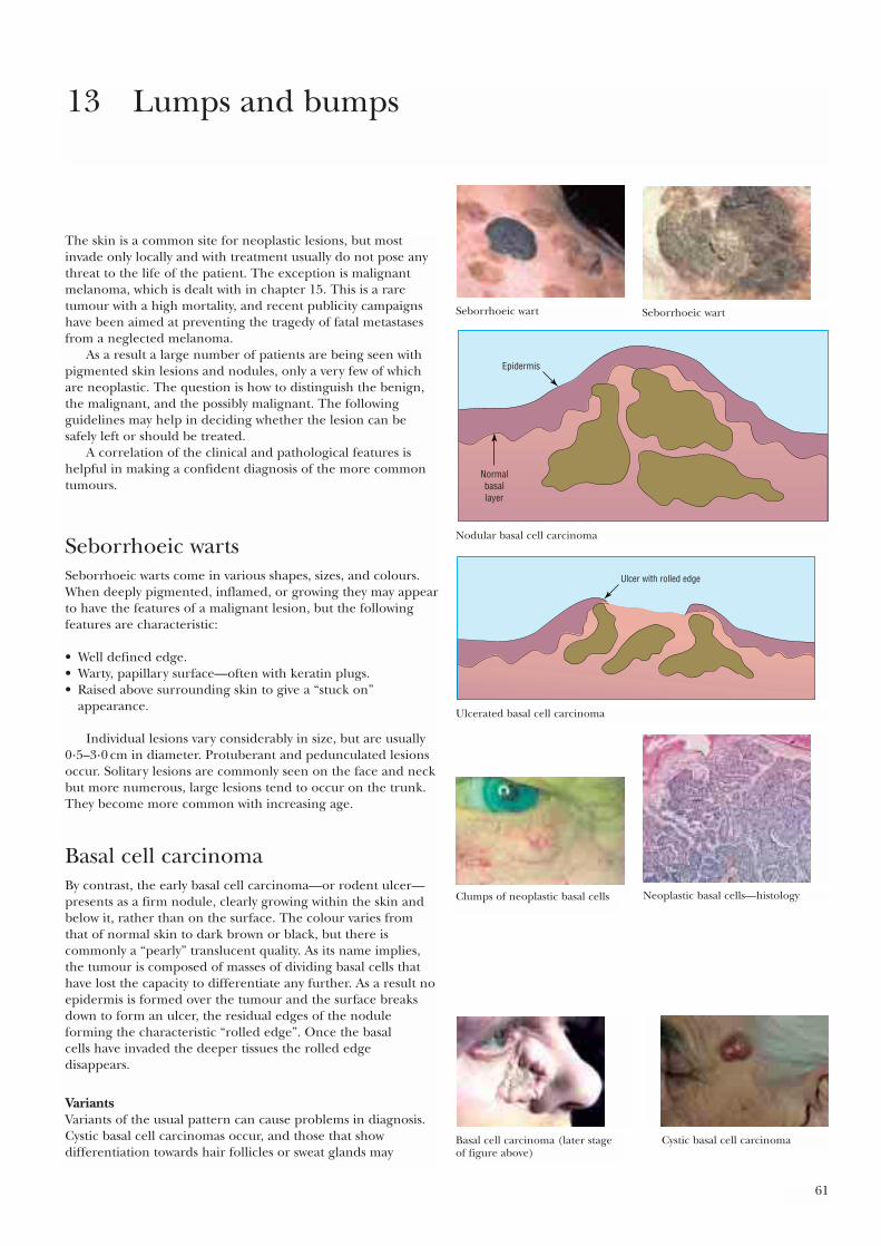
61
The skin is a common site for neoplastic lesions, but mostinvade only locally and with treatment usually do not pose anythreat to the life of the patient. The exception is malignantmelanoma, which is dealt with in chapter 15. This is a raretumour with a high mortality, and recent publicity campaignshave been aimed at preventing the tragedy of fatal metastasesfrom a neglected melanoma.
As a result a large number of patients are being seen withpigmented skin lesions and nodules, only a very few of whichare neoplastic. The question is how to distinguish the benign,the malignant, and the possibly malignant. The followingguidelines may help in deciding whether the lesion can besafely left or should be treated.
A correlation of the clinical and pathological features ishelpful in making a confident diagnosis of the more commontumours.
Seborrhoeic wartsSeborrhoeic warts come in various shapes, sizes, and colours.When deeply pigmented, inflamed, or growing they may appearto have the features of a malignant lesion, but the followingfeatures are characteristic:
• Well defined edge.• Warty, papillary surface—often with keratin plugs.• Raised above surrounding skin to give a “stuck on”
appearance.
Individual lesions vary considerably in size, but are usually0·5–3·0 cm in diameter. Protuberant and pedunculated lesionsoccur. Solitary lesions are commonly seen on the face and neckbut more numerous, large lesions tend to occur on the trunk.They become more common with increasing age.
Basal cell carcinomaBy contrast, the early basal cell carcinoma—or rodent ulcer—presents as a firm nodule, clearly growing within the skin andbelow it, rather than on the surface. The colour varies fromthat of normal skin to dark brown or black, but there iscommonly a “pearly” translucent quality. As its name implies,the tumour is composed of masses of dividing basal cells thathave lost the capacity to differentiate any further. As a result noepidermis is formed over the tumour and the surface breaksdown to form an ulcer, the residual edges of the noduleforming the characteristic “rolled edge”. Once the basal cells have invaded the deeper tissues the rolled edgedisappears.
VariantsVariants of the usual pattern can cause problems in diagnosis.Cystic basal cell carcinomas occur, and those that showdifferentiation towards hair follicles or sweat glands may
13 Lumps and bumps
Seborrhoeic wart Seborrhoeic wart
Epidermis
Normalbasallayer
Nodular basal cell carcinoma
Ulcer with rolled edge
Ulcerated basal cell carcinoma
Neoplastic basal cells—histologyClumps of neoplastic basal cells
Basal cell carcinoma (later stageof figure above)
Cystic basal cell carcinoma
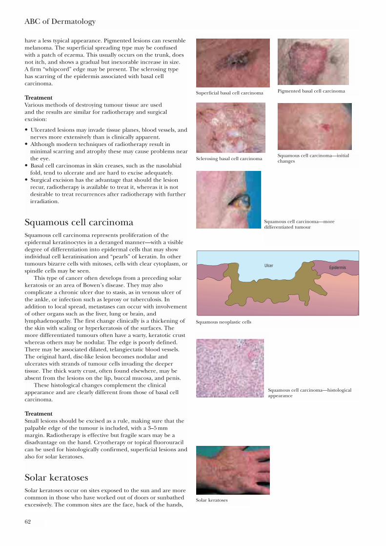
have a less typical appearance. Pigmented lesions can resemblemelanoma. The superficial spreading type may be confusedwith a patch of eczema. This usually occurs on the trunk, doesnot itch, and shows a gradual but inexorable increase in size. A firm “whipcord” edge may be present. The sclerosing typehas scarring of the epidermis associated with basal cellcarcinoma.
TreatmentVarious methods of destroying tumour tissue are used and the results are similar for radiotherapy and surgicalexcision:
• Ulcerated lesions may invade tissue planes, blood vessels, andnerves more extensively than is clinically apparent.
• Although modern techniques of radiotherapy result inminimal scarring and atrophy these may cause problems nearthe eye.
• Basal cell carcinomas in skin creases, such as the nasolabialfold, tend to ulcerate and are hard to excise adequately.
• Surgical excision has the advantage that should the lesionrecur, radiotherapy is available to treat it, whereas it is notdesirable to treat recurrences after radiotherapy with furtherirradiation.
Squamous cell carcinomaSquamous cell carcinoma represents proliferation of theepidermal keratinocytes in a deranged manner—with a visibledegree of differentiation into epidermal cells that may showindividual cell keratinisation and “pearls” of keratin. In othertumours bizarre cells with mitoses, cells with clear cytoplasm, orspindle cells may be seen.
This type of cancer often develops from a preceding solarkeratosis or an area of Bowen’s disease. They may alsocomplicate a chronic ulcer due to stasis, as in venous ulcer ofthe ankle, or infection such as leprosy or tuberculosis. Inaddition to local spread, metastases can occur with involvementof other organs such as the liver, lung or brain, andlymphadenopathy. The first change clinically is a thickening ofthe skin with scaling or hyperkeratosis of the surfaces. Themore differentiated tumours often have a warty, keratotic crustwhereas others may be nodular. The edge is poorly defined.There may be associated dilated, telangiectatic blood vessels.The original hard, disc-like lesion becomes nodular andulcerates with strands of tumour cells invading the deepertissue. The thick warty crust, often found elsewhere, may beabsent from the lesions on the lip, buccal mucosa, and penis.
These histological changes complement the clinicalappearance and are clearly different from those of basal cellcarcinoma.
TreatmentSmall lesions should be excised as a rule, making sure that thepalpable edge of the tumour is included, with a 3–5 mmmargin. Radiotherapy is effective but fragile scars may be adisadvantage on the hand. Cryotherapy or topical fluorouracilcan be used for histologically confirmed, superficial lesions andalso for solar keratoses.
Solar keratosesSolar keratoses occur on sites exposed to the sun and are morecommon in those who have worked out of doors or sunbathedexcessively. The common sites are the face, back of the hands,
ABC of Dermatology
62
Superficial basal cell carcinoma Pigmented basal cell carcinoma
Sclerosing basal cell carcinomaSquamous cell carcinoma—initialchanges
Squamous cell carcinoma—moredifferentiated tumour
Ulcer Epidermis
Squamous neoplastic cells
Squamous cell carcinoma—histologicalappearance
Solar keratoses
arms, and legs. They also develop on the scalp in bald men and on the lips, particularly in pipe smokers. They show alterations in keratinisation and have the potential to becomedysplastic and eventually develop into squamous cellcarcinoma, a change often preceded by inflammation. Theycan be regarded as squamous cell carcinoma grade 1/2.
The clinical appearance varies from a rough area of skin to a raised keratotic lesion. The edge is irregular and they are usually less than 1 cm in diameter. Inflammation andtenderness may be associated with progression to carcinoma.
TreatmentTreatment with cryotherapy, using liquid nitrogen or carbondioxide, repeated if necessary, is usually effective.
5-Fluorouracil cream is useful for larger or multiple lesions.It is applied twice daily for two weeks, which producesinflammation and necrosis. Simple dressings are applied for thenext two weeks. This process can be repeated if necessary. As itis a cytotoxic drug it should be handled with care and appliedsparingly with a cotton bud while wearing gloves.
Other conditionsBowen’s disease is characterised by a well defined, erythematousmacule with little induration and slight crusting. It is acondition of the middle aged and elderly, occurring commonlyon the trunk and limbs. It is an intraepidermal carcinoma,which has been reported to follow the ingestion of arsenic in“tonics” taken in years gone by or exposure to sheep dip,weedkiller, or industrial processes. After many years floridcarcinoma may develop with invasion of deeper tissues. It maybe confused with a patch of eczema or superficial basal cellcarcinoma. Lesions on covered areas may be associated withunderlying malignancy. Erythroplasia of Queyrat is a similarprocess occurring on the glans penis or prepuce.
Paget’s disease of the nipple presents with unilateral non-specific erythematous changes on the aureola and nipple,spreading to the surrounding skin. The cause is an underlyingadenocarcinoma of the ducts. It should be considered in anypatient with eczematous changes of one breast that fail torespond to simple treatment. Extramammary lesions occur.
Keratoacanthoma is a rapidly growing fleshy nodule thatdevelops a hard keratotic centre. Healing occurs with somescarring. Although benign, it may recur after being removedwith curette and cautery, particularly from the face, and is bestexcised.
Benign tumoursDermatofibroma is a simple, discrete firm nodule, arising in thedermis at the site of an insect bite or other trivial injury. Oftenthere is a brown or red vascular lesion initially, which thenbecomes fibrotic—a sclerosing haemangioma. The histiocytomais similar but composed of histiocytes.
Skin tags may be pigmented but rarely cause any diagnosticproblems unless inflamed. Some are in fact pedunculatedseborrhoeic warts and others simple papillomas (fibroepithelial polyps).
Lipomas are slow growing benign subcutaneous tumours.
Other benign tumoursA wide variety of tumours may develop from the hair follicleand sebaceous, exocrine (sweat), and apocrine glands. Themore common include syringomas—slowly growing, small,multiple nodules on the face of eccrine gland origin.
Lumps and bumps
63
Bowen’s disease
Paget’s disease of the nipple
Keratoacanthoma
Dermatofibroma
Skin tags
Syringoma
Lipoma
Naevus sebaceous is warty, well defined, varying in size from asmall nodule to one several centimetres in diameter. Lesionsoccur in the scalp of children, may be present at birth, andgradually increase in size. They may proliferate or develop intoa basal cell carcinoma in adult life and they are therefore bestremoved.
Verrucous epidermal naevi are probably a variant, found onthe trunk and limbs.
CystsThe familiar epidermoid cyst—also known as sebaceous cyst orwen—occurs as a soft, well defined, mobile swelling usually onthe face, neck, shoulder, or chest. It is not derived fromsebaceous glands but contains keratin produced by the liningwall.
Pilar cysts on the scalp are similar lesions derived from hairfollicles.
Milia are small keratin cysts consisting of small whitepapules found on the cheek and eyelids.
Vascular lesionsThe more common vascular naevi are described.
The port wine stain, or naevus flammeus, presents at birth asa flat red lesion, usually on the face, neck, or upper trunk.There is usually a sharp midline border on the more commonunilateral lesions. In time the affected area becomes raised andthickened because of proliferation of vascular and connectivetissue. If the area supplied by the ophthalmic or maxillarydivisions of the trigeminal nerve is affected there may beassociated angiomas of the underlying meninges withepilepsy—Sturge–Weber syndrome. Lesions of the limb may beassociated with arteriovenous fistulae.
Cavernous angioma—strawberry naevi—appear in the firstfew weeks of life or at birth. A soft vascular swelling is found,most commonly on the head and neck. The lesions resolvespontaneously in time and do not require treatment unlessinterfering with visual function.
Spider naevus consist of a central vascular papule with finelines radiating from it. They are more common in children andwomen. Large numbers in a man raise the possibility of liverdisease.
Campbell de Morgan spots are discrete red papules 1–5 mm indiameter. They are more common on the trunk.
Pyogenic granuloma is a lesion that contains no pus but is infact vascular and grows rapidly. It may arise at the site oftrauma. Distinction from amelanotic melanoma is important.
ABC of Dermatology
64
Further readingEnzinger F, Weiss S. Soft tissue tumours, 2nd ed. St Louis: Mosby,
1988Mackie RM. Skin cancer: an illustrated guide to the etiology, clinical
features, pathology and management of benign and malignant cutaneoustumours, 2nd ed. London: Martin Dunitz, 1996
Naevus sebaceous
Verrucous epidermal naevus Epidermoid cyst
Milia
Naevus flammeus Sturge–Weber syndrome
Cavernous angioma in a five month old baby
Patient five years later withouttreatment
Spider naevi Pyogenic granuloma
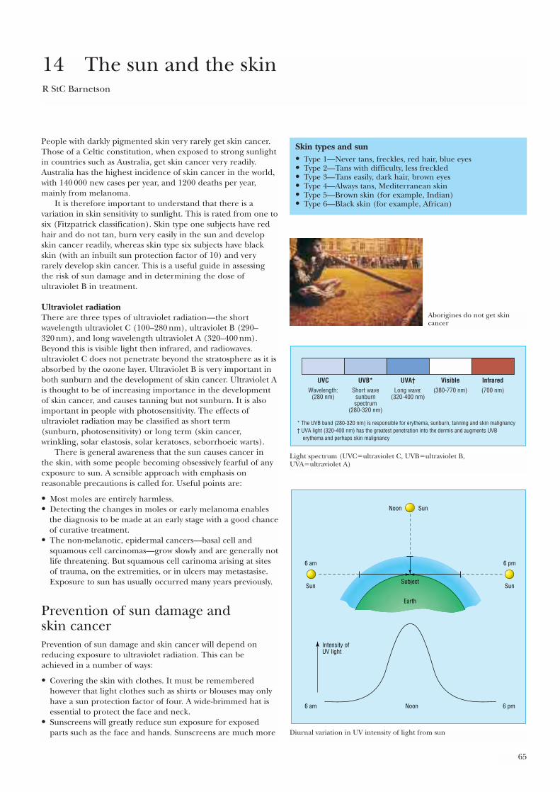
65
People with darkly pigmented skin very rarely get skin cancer.Those of a Celtic constitution, when exposed to strong sunlightin countries such as Australia, get skin cancer very readily.Australia has the highest incidence of skin cancer in the world,with 140 000 new cases per year, and 1200 deaths per year,mainly from melanoma.
It is therefore important to understand that there is avariation in skin sensitivity to sunlight. This is rated from one tosix (Fitzpatrick classification). Skin type one subjects have redhair and do not tan, burn very easily in the sun and developskin cancer readily, whereas skin type six subjects have blackskin (with an inbuilt sun protection factor of 10) and veryrarely develop skin cancer. This is a useful guide in assessingthe risk of sun damage and in determining the dose ofultraviolet B in treatment.
Ultraviolet radiationThere are three types of ultraviolet radiation—the shortwavelength ultraviolet C (100–280 nm), ultraviolet B (290–320 nm), and long wavelength ultraviolet A (320–400 nm).Beyond this is visible light then infrared, and radiowaves.ultraviolet C does not penetrate beyond the stratosphere as it isabsorbed by the ozone layer. Ultraviolet B is very important inboth sunburn and the development of skin cancer. Ultraviolet Ais thought to be of increasing importance in the developmentof skin cancer, and causes tanning but not sunburn. It is alsoimportant in people with photosensitivity. The effects ofultraviolet radiation may be classified as short term (sunburn, photosensitivity) or long term (skin cancer,wrinkling, solar elastosis, solar keratoses, seborrhoeic warts).
There is general awareness that the sun causes cancer in the skin, with some people becoming obsessively fearful of anyexposure to sun. A sensible approach with emphasis onreasonable precautions is called for. Useful points are:
• Most moles are entirely harmless.• Detecting the changes in moles or early melanoma enables
the diagnosis to be made at an early stage with a good chanceof curative treatment.
• The non-melanotic, epidermal cancers—basal cell andsquamous cell carcinomas—grow slowly and are generally notlife threatening. But squamous cell carinoma arising at sitesof trauma, on the extremities, or in ulcers may metastasise.Exposure to sun has usually occurred many years previously.
Prevention of sun damage and skin cancerPrevention of sun damage and skin cancer will depend onreducing exposure to ultraviolet radiation. This can beachieved in a number of ways:
• Covering the skin with clothes. It must be rememberedhowever that light clothes such as shirts or blouses may onlyhave a sun protection factor of four. A wide-brimmed hat isessential to protect the face and neck.
• Sunscreens will greatly reduce sun exposure for exposedparts such as the face and hands. Sunscreens are much more
14 The sun and the skinR StC Barnetson
Skin types and sun• Type 1—Never tans, freckles, red hair, blue eyes• Type 2—Tans with difficulty, less freckled• Type 3—Tans easily, dark hair, brown eyes• Type 4—Always tans, Mediterranean skin• Type 5—Brown skin (for example, Indian)• Type 6—Black skin (for example, African)
Aborigines do not get skincancer
UVC UVB* UVA† Visible InfraredWavelength:
(280 nm)
* The UVB band (280-320 nm) is responsible for erythema, sunburn, tanning and skin malignancy† UVA light (320-400 nm) has the greatest penetration into the dermis and augments UVB erythema and perhaps skin malignancy
Short wavesunburnspectrum
(280-320 nm)
Long wave:(320-400 nm)
(380-770 nm) (700 nm)
Light spectrum (UVC�ultraviolet C, UVB�ultraviolet B, UVA�ultraviolet A)
Sun
Subject
Noon
Earth
6 am
Intensity ofUV light
6 pmNoon
6 pm
Sun
6 am
Sun
Diurnal variation in UV intensity of light from sun
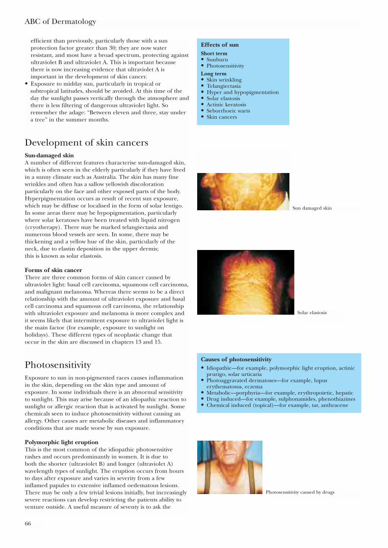
efficient than previously, particularly those with a sunprotection factor greater than 30; they are now waterresistant, and most have a broad spectrum, protecting againstultraviolet B and ultraviolet A. This is important becausethere is now increasing evidence that ultraviolet A isimportant in the development of skin cancer.
• Exposure to midday sun, particularly in tropical orsubtropical latitudes, should be avoided. At this time of theday the sunlight passes vertically through the atmosphere andthere is less filtering of dangerous ultraviolet light. Soremember the adage: “Between eleven and three, stay undera tree” in the summer months.
Development of skin cancersSun-damaged skinA number of different features characterise sun-damaged skin,which is often seen in the elderly particularly if they have livedin a sunny climate such as Australia. The skin has many finewrinkles and often has a sallow yellowish discolorationparticularly on the face and other exposed parts of the body.Hyperpigmentation occurs as result of recent sun exposure,which may be diffuse or localised in the form of solar lentigo.In some areas there may be hypopigmentation, particularlywhere solar keratoses have been treated with liquid nitrogen(cryotherapy). There may be marked telangiectasia andnumerous blood vessels are seen. In some, there may bethickening and a yellow hue of the skin, particularly of theneck, due to elastin deposition in the upper dermis; this is known as solar elastosis.
Forms of skin cancerThere are three common forms of skin cancer caused byultraviolet light: basal cell carcinoma, squamous cell carcinoma,and malignant melanoma. Whereas there seems to be a directrelationship with the amount of ultraviolet exposure and basalcell carcinoma and squamous cell carcinoma, the relationshipwith ultraviolet exposure and melanoma is more complex andit seems likely that intermittent exposure to ultraviolet light isthe main factor (for example, exposure to sunlight onholidays). These different types of neoplastic change that occur in the skin are discussed in chapters 13 and 15.
PhotosensitivityExposure to sun in non-pigmented races causes inflammationin the skin, depending on the skin type and amount ofexposure. In some individuals there is an abnormal sensitivityto sunlight. This may arise because of an idiopathic reaction tosunlight or allergic reaction that is activated by sunlight. Somechemicals seen to induce photosensitivity without causing anallergy. Other causes are metabolic diseases and inflammatoryconditions that are made worse by sun exposure.
Polymorphic light eruption This is the most common of the idiopathic photosensitiverashes and occurs predominantly in women. It is due to both the shorter (ultraviolet B) and longer (ultraviolet A)wavelength types of sunlight. The eruption occurs from hoursto days after exposure and varies in severity from a fewinflamed papules to extensive inflamed oedematous lesions.There may be only a few trivial lesions initially, but increasinglysevere reactions can develop restricting the patients ability toventure outside. A useful measure of seventy is to ask the
ABC of Dermatology
66
Effects of sunShort term• Sunburn• PhotosensitivityLong term• Skin wrinkling• Telangiectasia• Hyper and hypopigmentation• Solar elastosis• Actinic keratosis• Seborrhoeic warts• Skin cancers
Causes of photosensitivity• Idiopathic—for example, polymorphic light eruption, actinic
prurigo, solar urticaria• Photoaggravated dermatoses—for example, lupus
erythematosus, eczema• Metabolic—porphyria—for example, erythropoietic, hepatic• Drug induced—for example, sulphonamides, phenothiazines• Chemical induced (topical)—for example, tar, anthracene
Sun damaged skin
Solar elastosis
Photosensitivity caused by drugs

patient if they cross to the shady side of the street to avoid thesun. Treatment includes topical or systemic steroids for theacute rash and prevention by using sunscreens. Desensitisationby narrow waveband phototherapy before exposure is effective.
Solar urticariaThis is a much less common condition and may be induced bylonger wavelength (ultraviolet A) and visible radiation as well asultraviolet B. It is characterised by rapidly developing irritationand in the exposed skin is followed by urticarial wheals. It canoccur as part of a photoallergic reaction, in which caseavoidance of the relevant allergen will prevent the condition.
Treatment is with antihistamines and sunscreens. In somecases phototherapy with ultraviolet B, narrow waveband orpsoralen with ultraviolet A (PUVA), is helpful.
The sun and the skin
67
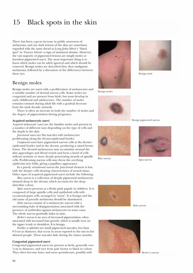
68
There has been a great increase in public awareness ofmelanoma, and any dark lesions of the skin are sometimesregarded with the same dread as Long John Silver’s “black spot” in Treasure Island—a sign of imminent demise. However,the vast majority of pigmented lesions are simply moles orharmless pigmented naevi. The most important thing is toknow which moles can be safely ignored and which should beremoved. Benign moles are described first, then malignantmelanoma, followed by a discussion of the differences betweenthese two.
Benign molesBenign moles are naevi with a proliferation of melanocytes anda variable number of dermal naevus cells. Some moles arecongenital and are present from birth, but most develop inearly childhood and adolescence. The number of molesremains constant during adult life with a gradual decrease from the sixth decade onwards.
There is often an increase in both the number of moles andthe degree of pigmentation during pregnancy.
Acquired melanocytic naeviAcquired melanocytic naevi are the familiar moles and present in a number of different ways depending on the type of cells andthe depth in the skin.
Junctional naevi are flat macules with melanocytesproliferating along the dermo-epidermal border.
Compound naevi have pigmented naevus cells at the dermo-epidermal border and in the dermis, producing a raised brownlesion. The dermal melanocytes may accumulate around theskin appendages and blood vessels and form a band of cellswithout melanin or more deeply penetrating strands of spindlecells. Proliferating naevus cells may throw the overlyingepidermis into folds, giving a papillary appearance.
In a purely intradermal naevus the junctional element is lost,with the deeper cells showing characteristics of neural tissue.Other types of acquired pigmented naevi include the following.
Blue naevus is a collection of deeply pigmented melanocytessituated deep in the dermis, which accounts for the deep slate-blue colour.
Spitz naevus presents as a fleshy pink papule in children. It iscomposed of large spindle cells and epitheloid cells withoccasional giant cells, arranged in “nests”. It is benign and theold name of juvenile melanoma should be abandoned.
Halo naevus consists of a melanocytic naevus with asurrounding halo of depigmentation associated with thepresence of antibodies against melanocytes in some cases. The whole naevus gradually fades in time.
Becker’s naevus is an area of increased pigmentation, oftenassociated with increased hair growth, which is usually seen onthe upper trunk or shoulders. It is benign.
Freckles or ephelides are small pigmented macules, less than 0·5 cm in diameter, that occur in areas exposed to the sun in fairskinned people. These macules fade during the winter months.
Congenital pigmented naeviCongenital pigmented naevi are present at birth, generally over1 cm in diameter, and vary from pale brown to black in colour.They often become hairy and more protuberant, possibly with
15 Black spots in the skin
Benign moles
Benign mole
Benign pigmented naevus
Blue naevus Spitz naevus
Halo naevus
Becker’s naevus
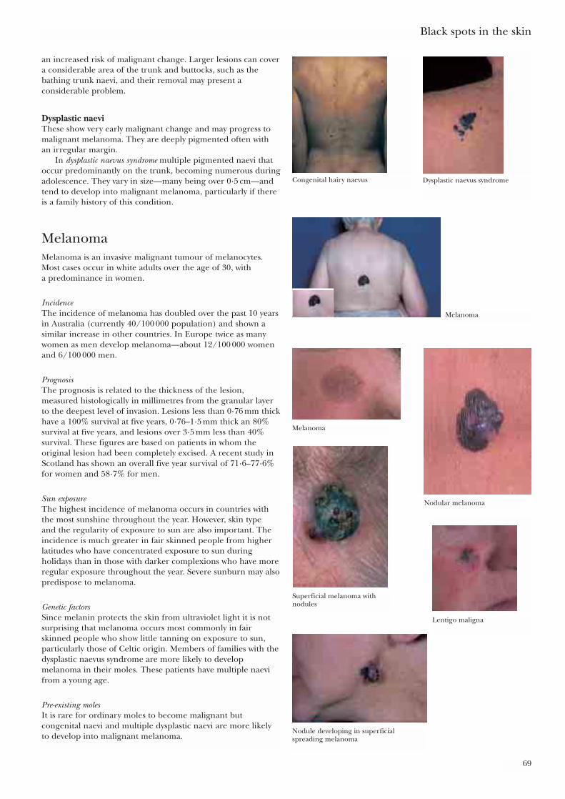
an increased risk of malignant change. Larger lesions can covera considerable area of the trunk and buttocks, such as thebathing trunk naevi, and their removal may present aconsiderable problem.
Dysplastic naeviThese show very early malignant change and may progress tomalignant melanoma. They are deeply pigmented often with an irregular margin.
In dysplastic naevus syndrome multiple pigmented naevi thatoccur predominantly on the trunk, becoming numerous duringadolescence. They vary in size—many being over 0·5 cm—andtend to develop into malignant melanoma, particularly if thereis a family history of this condition.
MelanomaMelanoma is an invasive malignant tumour of melanocytes.Most cases occur in white adults over the age of 30, with a predominance in women.
IncidenceThe incidence of melanoma has doubled over the past 10 yearsin Australia (currently 40/100 000 population) and shown asimilar increase in other countries. In Europe twice as manywomen as men develop melanoma—about 12/100 000 womenand 6/100 000 men.
PrognosisThe prognosis is related to the thickness of the lesion,measured histologically in millimetres from the granular layerto the deepest level of invasion. Lesions less than 0·76 mm thickhave a 100% survival at five years, 0·76–1·5 mm thick an 80%survival at five years, and lesions over 3·5 mm less than 40%survival. These figures are based on patients in whom theoriginal lesion had been completely excised. A recent study inScotland has shown an overall five year survival of 71·6–77·6%for women and 58·7% for men.
Sun exposureThe highest incidence of melanoma occurs in countries withthe most sunshine throughout the year. However, skin type and the regularity of exposure to sun are also important. Theincidence is much greater in fair skinned people from higherlatitudes who have concentrated exposure to sun duringholidays than in those with darker complexions who have moreregular exposure throughout the year. Severe sunburn may alsopredispose to melanoma.
Genetic factorsSince melanin protects the skin from ultraviolet light it is notsurprising that melanoma occurs most commonly in fairskinned people who show little tanning on exposure to sun,particularly those of Celtic origin. Members of families with thedysplastic naevus syndrome are more likely to developmelanoma in their moles. These patients have multiple naevifrom a young age.
Pre-existing moles It is rare for ordinary moles to become malignant butcongenital naevi and multiple dysplastic naevi are more likelyto develop into malignant melanoma.
Black spots in the skin
69
Congenital hairy naevus
Melanoma
Superficial melanoma withnodules
Lentigo maligna
Nodular melanoma
Nodule developing in superficialspreading melanoma
Melanoma
Dysplastic naevus syndrome
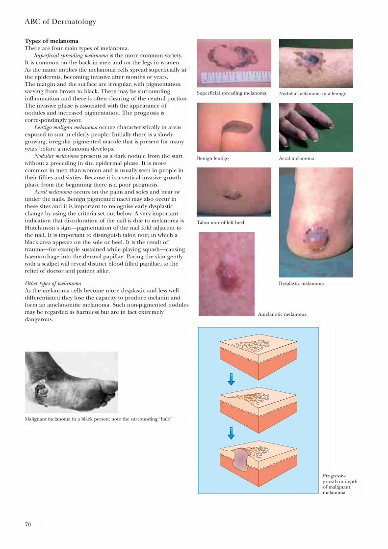
Types of melanomaThere are four main types of melanoma.
Superficial spreading melanoma is the more common variety. It is common on the back in men and on the legs in women. As the name implies the melanoma cells spread superficially inthe epidermis, becoming invasive after months or years. The margin and the surface are irregular, with pigmentationvarying from brown to black. There may be surroundinginflammation and there is often clearing of the central portion.The invasive phase is associated with the appearance of nodules and increased pigmentation. The prognosis iscorrespondingly poor.
Lentigo maligna melanoma occurs characteristically in areasexposed to sun in elderly people. Initially there is a slowlygrowing, irregular pigmented macule that is present for manyyears before a melanoma develops.
Nodular melanoma presents as a dark nodule from the startwithout a preceding in situ epidermal phase. It is morecommon in men than women and is usually seen in people intheir fifties and sixties. Because it is a vertical invasive growthphase from the beginning there is a poor prognosis.
Acral melanoma occurs on the palm and soles and near orunder the nails. Benign pigmented naevi may also occur inthese sites and it is important to recognise early dysplasticchange by using the criteria set out below. A very importantindication that discoloration of the nail is due to melanoma isHutchinson’s sign—pigmentation of the nail fold adjacent tothe nail. It is important to distinguish talon noir, in which ablack area appears on the sole or heel. It is the result oftrauma—for example sustained while playing squash—causinghaemorrhage into the dermal papillae. Paring the skin gentlywith a scalpel will reveal distinct blood filled papillae, to therelief of doctor and patient alike.
Other types of melanomaAs the melanoma cells become more dysplastic and less welldifferentiated they lose the capacity to produce melanin andform an amelanonitic melanoma. Such non-pigmented nodulesmay be regarded as harmless but are in fact extremelydangerous.
ABC of Dermatology
70
Superficial spreading melanoma Nodular melanoma in a lentigo
Benign lentigo
Talon noir of left heel
Dysplastic melanoma
Acral melanoma
Amelanotic melanoma
Progressivegrowth in depthof malignantmelanoma
Malignant melanoma in a black person; note the surrounding “halo”

PrognosisThis depends on the depth to which the melanoma haspenetrated below the base of the epidermis—lesions confinedto the epidermis having better prognosis than thosepenetrating into the dermis. The Clark classification describesthe depth of penetration as follows:
Level I—within the epidermisLevel II—few melanoma cells within the dermal papillaeLevel III—many melanoma cells in the papillary dermisLevel IV—invasion of the reticular dermisLevel V—invasion of the subcutaneous tissues
The Breslow classification is based on measurements of tumourthickness from the granular layer overlying epidermis. A depthof less than 1·5 mm is associated with a 90% five year survival,1·5–3·5 mm with a 75% five year survival, and greater than 3·5 mm with only a 50% five year survival.
In deeper tumours “sentinel lymph node” biopsy may becarried out to assess whether lymphatic spread has occurred.
How to tell the differenceBenign moles show little change and remain static for years.Any change may indicate that a mole is in fact a melanoma orthat a mole is becoming active. Size, shape, and colour are themain features and it is change in them that is most important.Patients with moles should have these changes explained tothem, in particular that they indicate activity of the cells, notnecessarily malignant change.
Black spots in the skin
71
Criteria for suspecting malignant changes in pigmentedlesions• Growth—Benign pigmented naevi continue to appear in
adolescents and young adults. Any mole increasing in size inan adult over the age of 30 may be a melanoma
• Shape—Moles usually have a symmetrical, even outline, anyindentations being quite regular; melanomas usually have anirregular edge with one part advancing more than the others
• Colour—Variation in colour of benign moles is even but amelanoma may be intensely black or show irregularcoloration varying from white to slate blue, with all shades ofblack and brown. Inflammation may give a red colour as well.The amelanotic melanoma shows little or no pigmentation
• Size—Apart from congenital pigmentation naevi most benignmoles are less than 1 cm in diameter. Any lesion growing toover 0·5 cm should be carefully checked
• Itching—Normally a mole does not itch but a melanoma may.Irritated seborrhoeic warts also itch
• Bleeding and crusting occur in an actively growing melanomaIf more than two of these features are present refer the patientfor specialist opinionA simple summary:A—Asymmetry of the lesion C—Variations in colourB—Irregularity of the border D—Diameter larger
than 0·5 cm
Further readingAckerman AB. Malignant melanoma and other melanocytic neoplasms.
Baltimore: Williams and Wilkins,1984Mackie R (ed). Primary and secondary prevention of malignant
melanoma. Basle: KargerRoses DF. Diagnosis and management of cutaneous malignant melanoma.
Philadelphia: Saunders, 1983Seigler HE. Clinical management of melanoma. The Hague: Nijhoff,
1982
72
When a man has on the skin of his body a swelling or an eruption or aspot … and the disease appears to be deeper than the skin it is a leprousdisease.
Leviticus 13: 2–3
In ancient times changes in the skin were taken to indicate that the whole body was diseased and although argumentscontinue about what the Old Testament writers understood by “leprous”, there was clearly an appreciation of theconnection between the skin and systemic illness. Clinical signs in the skin may give valuable diagnostic clues tounderlying disease. The cutaneous signs of systemic disease is a very large subject; and what follows is only an outline of the more common skin changes that may be associated with systemic illness.
A disease affecting internal organs may produce the samechanges in both the skin and other organs—as in the connectivetissue diseases. However, underlying conditions may beassociated with skin changes brought about by quite differentprocesses, as in acanthosis nigricans or dermatomyositis in whichthere is an underlying neoplasm with characteristic skin signs.Sometimes severe skin disease itself may be the cause ofgeneralised illness.
The skin is also a common site for allergic reactions todrugs, with a rash being the first clinical sign. The florid skinlesions of AIDS illustrate the results of infections when theimmune response is impaired.
Conditions affecting both the skin and the internal organsImmune reactionsAllergic reactions to drugs such as penicillin can occur. In this case the penicillin molecule attaches to serum protein.This compound acts as an antigen and may form a complexwith IgG antibody. It is this complex which attaches to bloodvessel walls to produce an inflammatory reaction. This presentsas a rash developing a few days to two weeks after treatment onthe skin, but if it occurs in the kidneys the resulting tissuedamage can have serious consequences. This is an example ofType III allergy with antigen–antibody complexes beingdeposited in the small blood vessels. Sometimes a much moreacute anaphylactic reaction develops A fixed drug eruption ischaracterised by a localised patch of erythema that flares upwhenever the drug is taken. Erythema multiforme can occur indrug reactions.
Connective tissue diseases involve complex immunologicalprocesses that affect both internal organs and the skin. Thismeans that it is particularly important to realise thesignificance of any associated skin changes.
Lupus erythematosusThis condition has been described as “a disease with athousand faces” because of the wide range of organs involved
16 The skin and systemic disease—Genetics andskin disease (JA Savin)
When to suspect an underlying systemic disease• An unusual rash which does not have the features of one of
the common primary inflammatory skin conditions• Evidence of systemic illness—weight loss, and other symptoms
such as breathlessness, altered bowel function or painfuljoints
• Erythema of the skin due to inflammation around the bloodvessels, usually without epidermal changes—reactiveerythema. Vasculitis, in which there are palpableerythematous lesions which may be painful or nodular
• Unusual changes in pigmentation or texture of the skin• Palpable dermal lesions that may be due to granuloma,
metastases, lymphoma, or deposits of fat or minerals
Rash from penicillin
Erythema multiforme

The skin and systemic disease—Genetics and skin disease
73
and the numerous ways in which it can present. In threequarters of the patients the skin is involved. There are fourmain types, with numerous variations.
In systemic lupus erythematosus (SLE) the commonest skinchange is an acute erythematous eruption occurring bilaterallyon the malar area of the face in a “butterfly” distribution.There may also be photosensitivity, hair loss, and areas ofvasculitis in the skin. There is often intolerance of sunlight. It ismore common in females with a female:male ratio of 8:1.
The systemic changes include fever, arthritis and renalinvolvement, but there may be involvement of a wide range oforgans. The criteria for diagnosing the condition include at least four of the features in the box on the right.
Subacute lupus erythematosus is a variant in about 10% ofpatients with lupus erythematosus that presents with non-scarring erythematosus plaques mainly on the face, handsand arms. Papulo squamous lesions also occur. They may beannular. Systemic involvement is less common and severe thanin SLE. It is associated with a high incidence of neonatal lupuserythematosus in children born to mothers with the condition.The antinuclear factor test is positive in 60% andanticytoplasmic antibodies are present in 80% of patients.
Discoid lupus erythematosus (DLE) is a condition in whichcirculating antinuclear antibodies are very rare. There are quitewell defined photosensitive inflammatory lesions, with somedegree of atrophy and hyperkeratosis of the follicles, giving a“nutmeg grater” feel. It occurs predominantly on the face orareas exposed to the sun, becoming worse in the summermonths. Scarring is common causing hair loss in lesions on thescalp.
Treatment of SLE with the threatened or actual involvementof other organs is important. Prednisolone is usually requiredand sometimes immunosuppressant drugs such as azathioprineas well. Treatment of DLE is generally with topical steroids.Hydroxychloroquine by mouth is also used, generally in a doseof 200mg daily. This drug can diminish visual acuity in higherdoses and this should be checked every few months. A simplechart, the Amsler Chart, is available for patients to use, consistingof a central dot with a grid which becomes blurred when held atarm’s length when there is any impairment of acutity.
DermatomyositisThis condition is associated in adults with underlyingcarcinoma—commonly of the breasts, lung, ovary, orgastrointestinal tract. It is characterised by localised erythemawith a purple hue (heliotrope), predominantly on the eyelids,cheeks, and forehead. There may be similar changes on thedorsal surface of the fingers, often with dilated nail foldcapillaries. These changes may precede the discovery of anunderlying tumour and may also fade away once it is removed.There is a variable association with muscle discomfort andweakness, mainly in the upper limb girdle. The finding ofmuscle weakness together with specific electromyographicchanges and an inflammatory infiltrate in the muscle meansthere is almost certainly an underlying malignancy, so suitableinvestigation is indicated.
Systemic sclerosisAs the name implies, there is extensive sclerosis of theconnective tissue of the lungs, gastrointestinal tract, kidneys,and heart. Endothelial cell damage in the capillaries results infibrosis and sclerosis of the organs concerned. The skinbecomes tethered to the subcutaneous tissues and immobile,leading to fixed claw like hands, constricted mouth withfurrowed lips, and beak-like nose. There are vascular changesproducing Raynaud’s phenomenon and telangiectasia around
Clinical variants of lupus erythematosus• Systemic• Subacute cutaneous• Discoid (neonatal)• Systemic sclerosis
Criteria for diagnosing systemic lupus erythematosus• Malar rash • Serositis• Discoid plaques • Neurological involvement• Photosensitivity • Haematological changes• Arthritis • Immunological changes• Mouth ulcers • Antinuclear antibodies• Renal changes
Systemic lupuserythematosus
Dermatomyositis
Persistent dermatomyositis
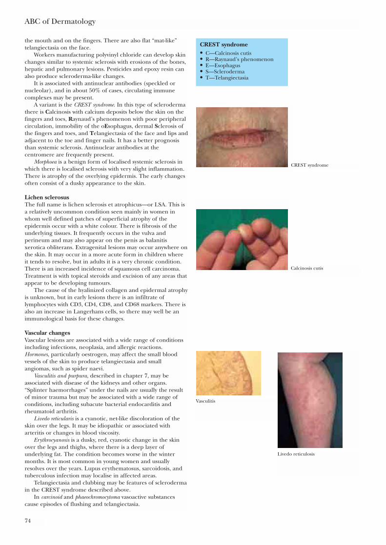
ABC of Dermatology
74
the mouth and on the fingers. There are also flat “mat-like”telangiectasia on the face.
Workers manufacturing polyvinyl chloride can develop skinchanges similar to systemic sclerosis with erosions of the bones,hepatic and pulmonary lesions. Pesticides and epoxy resin canalso produce scleroderma-like changes.
It is associated with antinuclear antibodies (speckled ornucleolar), and in about 50% of cases, circulating immunecomplexes may be present.
A variant is the CREST syndrome. In this type of sclerodermathere is Calcinosis with calcium deposits below the skin on thefingers and toes, Raynaud’s phenomenon with poor peripheralcirculation, immobility of the oEsophagus, dermal Sclerosis ofthe fingers and toes, and Telangiectasia of the face and lips andadjacent to the toe and finger nails. It has a better prognosisthan systemic sclerosis. Antinuclear antibodies at thecentromere are frequently present.
Morphoea is a benign form of localised systemic sclerosis inwhich there is localised sclerosis with very slight inflammation.There is atrophy of the overlying epidermis. The early changesoften consist of a dusky appearance to the skin.
Lichen sclerosusThe full name is lichen sclerosis et atrophicus—or LSA. This isa relatively uncommon condition seen mainly in women inwhom well defined patches of superficial atrophy of theepidermis occur with a white colour. There is fibrosis of theunderlying tissues. It frequently occurs in the vulva andperineum and may also appear on the penis as balanitisxerotica obliterans. Extragenital lesions may occur anywhere onthe skin. It may occur in a more acute form in children whereit tends to resolve, but in adults it is a very chronic condition.There is an increased incidence of squamous cell carcinoma.Treatment is with topical steroids and excision of any areas thatappear to be developing tumours.
The cause of the hyalinized collagen and epidermal atrophyis unknown, but in early lesions there is an infiltrate oflymphocytes with CD3, CD4, CD8, and CD68 markers. There isalso an increase in Langerhans cells, so there may well be animmunological basis for these changes.
Vascular changesVascular lesions are associated with a wide range of conditionsincluding infections, neoplasia, and allergic reactions.Hormones, particularly oestrogen, may affect the small bloodvessels of the skin to produce telangiectasia and smallangiomas, such as spider naevi.
Vasculitis and purpura, described in chapter 7, may beassociated with disease of the kidneys and other organs.“Splinter haemorrhages” under the nails are usually the resultof minor trauma but may be associated with a wide range ofconditions, including subacute bacterial endocarditis andrheumatoid arthritis.
Livedo reticularis is a cyanotic, net-like discoloration of theskin over the legs. It may be idiopathic or associated witharteritis or changes in blood viscosity.
Erythrocyanosis is a dusky, red, cyanotic change in the skinover the legs and thighs, where there is a deep layer ofunderlying fat. The condition becomes worse in the wintermonths. It is most common in young women and usuallyresolves over the years. Lupus erythematosus, sarcoidosis, andtuberculous infection may localise in affected areas.
Telangiectasia and clubbing may be features of sclerodermain the CREST syndrome described above.
In carcinoid and phaeochromocytoma vasoactive substancescause episodes of flushing and telangiectasia.
CREST syndrome• C—Calcinosis cutis• R—Raynaud’s phenomenon• E—Esophagus• S—Scleroderma• T—Telangiectasia
Calcinosis cutis
CREST syndrome
Vasculitis
Livedo reticulosis
The skin and systemic disease—Genetics and skin disease
75
In hereditary haemorrhagic telangiectasia thin walled ectaticblood vessels develop in the mucous membranes and the skin—generally on the upper half of the body and the nail beds.
ErythemasErythema is macular redness of the skin due to congestion inthe capillaries. It occurs as part of immunological reactions inthe skin as in drug allergies and specific patterns of viralinfections, such as measles. There are other types that show a specific pattern but are associated with a wide range ofunderlying conditions, such as erythema multiforme.
Erythema multiforme is associated with herpes and other viralinfections or streptococcal and various bacterial infections, butalso with many other conditions, particularly connective tissuedisease, sarcoidosis, and reactions to drugs such assulphonamides.
The lesions consist of erythematous macules becomingraised and typically developing into “target lesions” in whichthere is a dusky red or purpuric centre with a pale induratedzone surrounded by an outer ring of erythema. The lesionsmay be few or multiple and diffuse, often involving the hands,feet, elbows, and knees. Blisters may develop.
In the more severe forms there may be dermal changes andblister formation with involvement of the mucous membranes(Stevens–Johnson syndrome). There is often pyrexia withgastrointestinal and renal lesions. It can progress to toxicepidermal necrolysis, which some consider a form of erythemamultiforme.
Erythema annulare is a specific pattern in the skin with alarge number of reported associations, ranging from fungaland viral infections to sarcoidosis and carcinoma. It consists ofa small erythematous macule that enlarges to form anexpanding ring, usually on the trunk.
Erythema chronicum migrans is associated with Borreliainfection and Lyme disease—it is described on page 106.
There are many other types of “figurate erythemas”. Erythemagyratum repens is associated with underlying carcinoma anderythema marginatum, which is now rare, with rheumatic fever.
AngiomasSpider naevi, which show a central blood vessel with radiatingbranches, are frequently seen in women (especially duringpregnancy) and children. If they occur in large numbers,particularly in men, they may indicate liver failure. Palmarerythema and yellow nails may also be present.
Congenital angiomasEruptive angiomas may be associated with systemic angiomas of the liver, lung, and brain. Port wine stain due to abnormalityof the dermal capillaries commonly develops on the head andneck. It may be associated with congenital vascular abnormalitiesof the meningies and epilepsy. Vascular abnormalities of the eye,and also glaucoma, occur with lesions on the face.
Erythema of the nailbeds This may be associated with connective tissue disease, such aslupus erythematosus, scleroderma, and dermatomyositis.
Changes in pigmentationHypopigmentationHormonalA widespread partial loss of melanocyte functions with loss of skin colour is seen in hypopituitarism and is caused by anabsence of melanocyte stimulating hormone.
Erythemas associated with systemicconditions• Erythema multiforme• Erythema annulare• Erythema chronicum migrans• Erythema gyratum repens• Erythema marginatum
Figurate erythema
Erythema of nailbeds andclubbing
ABC of Dermatology
76
GeneticIn albinism, an autorecessive condition, there is little or noproduction of melanin with loss of pigment from the skin, hair,and eyes. Other genetic conditions with loss of skin pigmentinclude piebaldism, phenylketonuria, and tuberous sclerosis.
Localised depigmentation is most commonly seen invitiligo, in which a family history of the condition is found inone third of the patients. In the sharply demarcated,symmetrical macular lesions there is loss of melanocytes andmelanin. There is an increased incidence of organ specificantibodies and their associated diseases.
Other causes of hypopigmented macules include:postinflammatory conditions after psoriasis, eczema, lichenplanus, and lupus erythematosus; infections, for example, tineaversicolor and leprosy; chemicals, such as hydroquinones,hydroxychloroquine, and arsenicals, reactions to pigmentednaevi, seen in halo naevus; and genetic diseases, such astuberous sclerosis (“ash leaf” macules).
HyperpigmentationThere is wide variation in the pattern of normal pigmentationas a result of heredity and exposure to the sun. Darkening ofthe skin may be due to an increase in the normal pigmentmelanin or to the deposition of bile salts in liver disease, ironsalts (haemochromatosis), drugs, or metallic salts fromingestion. In agyria ingested silver salts are deposited in the skin.
Causes of hyperpigmentation include the following factors.
HormonalAn increase in circulating hormones that have melanocytestimulating activity occurs in hyperthyroidism, Addison’sdisease, and acromegaly. In women who are pregnant or takingoral contraceptives there may be an increase in melanocyticpigmentation of the face. This is known as melasma (or chloasma) and occurs mainly on the forehead and cheeks.It may fade slowly. Sometimes a premenstrual darkening of theface occurs.
Increased deposition of haemosiderin is generalised inhaemochromatosis. Localised red-brown discoloration of thelegs is seen with longstanding varicose veins. It also occurs in a specific localised pattern in Schamberg’s disease, when there is a “cayenne pepper” appearance of the legs and thighs.
NeoplasiaLymphomas may be associated with increased pigmentation.Acanthosis nigricans, characterised by darkening andthickening of the skin of the axillae, neck, nipples, andumbilicus, occurs with internal cancers, usuallyadenocarcinoma of the stomach. It is also seen in acromegaly.There is a benign juvenile type. Pseudoacanthosis nigricans ismuch more common, consisting of simple darkening of theskin in the flexures of obese individuals; it is not associated withmalignancy.
Autoimmune associations with vitiligo• Thyroid disease • Myasthenia gravis• Pernicious anaemia • Alopecia areata• Hypoparathyroidism • Halo naevus• Addison’s disease • Morphoea and • Diabetes lichen sclerosus
Vitiligo
Agyria (silver salts in skin)
Melasma
Acanthosis nigricans
Carcinoma left upper zone
Pseudoacanthosis nigricans Acanthosis nigricans
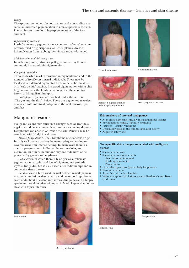
The skin and systemic disease—Genetics and skin disease
77
DrugsChlorpromazine, other phenothiazines, and minocycline maycause an increased pigmentation in areas exposed to the sun.Phenytoin can cause local hyperpigmentation of the face and neck.
Inflammatory reactionsPostinflammatory pigmentation is common, often after acuteeczema, fixed drug eruptions, or lichen planus. Areas oflichenification from rubbing the skin are usually darkened.
Malabsorption and deficiency statesIn malabsorption syndromes, pellagra, and scurvy there iscommonly increased skin pigmentation.
Congenital conditionsThere is clearly a marked variation in pigmentation and in thenumber of freckles in normal individuals. There may belocalised well defined pigmented areas in neurofibromatosiswith “cafe au lait” patches. Increased pigmentation with a bluetinge occurs over the lumbosacral region in the conditionknown as Mongolian blue spot.
Peutz–Jeghers syndrome is described under the section “The gut and the skin”, below. There are pigmented maculesassociated with intestinal polyposis in the oral mucosa, lips, and face.
Malignant lesionsMalignant lesions may cause skin changes such as acanthosisnigricans and dermatomyositis or produce secondary deposits.Lymphomas can arise in or invade the skin. Pruritus may beassociated with Hodgkin’s disease.
Mycosis fungoides is a T cell lymphoma of cutaneous origin.Initially well demarcated erythematous plaques develop oncovered areas with intense itching. In many cases there is agradual progression to infiltrated lesions, nodules, andulceration. In others the tumour may occur de novo or bepreceded by generalised erythema.
Poikiloderma, in which there is telangiectasia, reticulatepigmentation, atrophy, and loss of pigment, may precedemycosis fungoides, but it is also seen after radiotherapy and inconnective tissue diseases.
Parapsoriasisis a term used for well defined maculopapularerythematous lesions that occur in middle and old age. Somecases undoubtedly develop into mycosis fungoides and a biopsyspecimen should be taken of any such fixed plaques that do notclear with topical steroids.
Skin markers of internal malignancy• Acanthosis nigricans—usually intra-abdominal lesions• Erythematous rashes, “figurate erythema”• Pruritus—usually lymphoma• Dermatomyositis in the middle aged and elderly• Acquired ichthyosis
Non-specific skin changes associated with malignantdisease• Secondary deposits• Secondary hormonal effects
Acne (adrenal tumours)Flushing (carcinoid)Pigmentation
• Generalised pruritus (particularly lymphoma)• Figurate erythema• Superficial thrombophlebitis• Various eruptive skin lesions seen in Gardener’s and Bazex
syndromes
Neurofibromatosis Neurofibromatosis
Increased pigmentation inmalabsorption syndrome
Peutz–Jeghers syndrome
B cell lymphoma
Lymphoma
Poikiloderma
Parapsoriasis
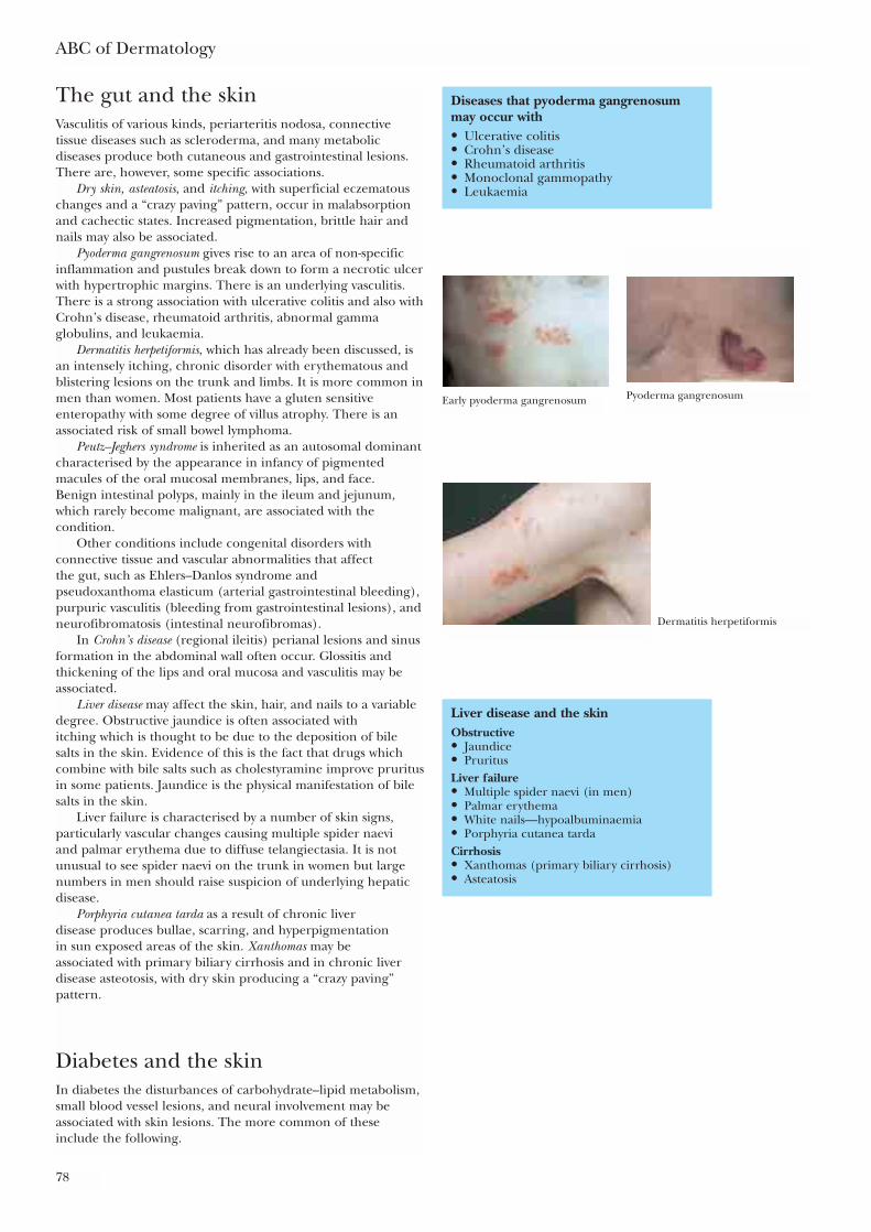
ABC of Dermatology
78
The gut and the skinVasculitis of various kinds, periarteritis nodosa, connectivetissue diseases such as scleroderma, and many metabolicdiseases produce both cutaneous and gastrointestinal lesions.There are, however, some specific associations.
Dry skin, asteatosis, and itching, with superficial eczematouschanges and a “crazy paving” pattern, occur in malabsorptionand cachectic states. Increased pigmentation, brittle hair andnails may also be associated.
Pyoderma gangrenosum gives rise to an area of non-specificinflammation and pustules break down to form a necrotic ulcerwith hypertrophic margins. There is an underlying vasculitis.There is a strong association with ulcerative colitis and also withCrohn’s disease, rheumatoid arthritis, abnormal gammaglobulins, and leukaemia.
Dermatitis herpetiformis, which has already been discussed, isan intensely itching, chronic disorder with erythematous andblistering lesions on the trunk and limbs. It is more common inmen than women. Most patients have a gluten sensitiveenteropathy with some degree of villus atrophy. There is anassociated risk of small bowel lymphoma.
Peutz–Jeghers syndrome is inherited as an autosomal dominantcharacterised by the appearance in infancy of pigmentedmacules of the oral mucosal membranes, lips, and face. Benign intestinal polyps, mainly in the ileum and jejunum,which rarely become malignant, are associated with thecondition.
Other conditions include congenital disorders withconnective tissue and vascular abnormalities that affect the gut, such as Ehlers–Danlos syndrome and pseudoxanthoma elasticum (arterial gastrointestinal bleeding),purpuric vasculitis (bleeding from gastrointestinal lesions), andneurofibromatosis (intestinal neurofibromas).
In Crohn’s disease (regional ileitis) perianal lesions and sinusformation in the abdominal wall often occur. Glossitis andthickening of the lips and oral mucosa and vasculitis may beassociated.
Liver disease may affect the skin, hair, and nails to a variabledegree. Obstructive jaundice is often associated with itching which is thought to be due to the deposition of bilesalts in the skin. Evidence of this is the fact that drugs whichcombine with bile salts such as cholestyramine improve pruritusin some patients. Jaundice is the physical manifestation of bilesalts in the skin.
Liver failure is characterised by a number of skin signs,particularly vascular changes causing multiple spider naevi and palmar erythema due to diffuse telangiectasia. It is notunusual to see spider naevi on the trunk in women but largenumbers in men should raise suspicion of underlying hepaticdisease.
Porphyria cutanea tarda as a result of chronic liver disease produces bullae, scarring, and hyperpigmentation in sun exposed areas of the skin. Xanthomas may be associated with primary biliary cirrhosis and in chronic liverdisease asteotosis, with dry skin producing a “crazy paving”pattern.
Diabetes and the skinIn diabetes the disturbances of carbohydrate–lipid metabolism,small blood vessel lesions, and neural involvement may beassociated with skin lesions. The more common of theseinclude the following.
Liver disease and the skinObstructive• Jaundice• PruritusLiver failure• Multiple spider naevi (in men)• Palmar erythema• White nails—hypoalbuminaemia• Porphyria cutanea tardaCirrhosis• Xanthomas (primary biliary cirrhosis)• Asteatosis
Diseases that pyoderma gangrenosummay occur with• Ulcerative colitis• Crohn’s disease• Rheumatoid arthritis• Monoclonal gammopathy• Leukaemia
Early pyoderma gangrenosum Pyoderma gangrenosum
Dermatitis herpetiformis
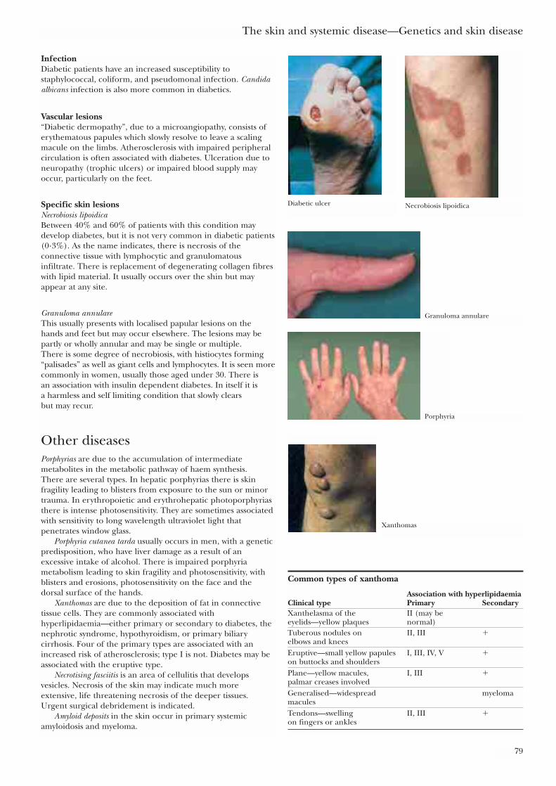
The skin and systemic disease—Genetics and skin disease
79
InfectionDiabetic patients have an increased susceptibility tostaphylococcal, coliform, and pseudomonal infection. Candidaalbicans infection is also more common in diabetics.
Vascular lesions“Diabetic dermopathy”, due to a microangiopathy, consists oferythematous papules which slowly resolve to leave a scalingmacule on the limbs. Atherosclerosis with impaired peripheralcirculation is often associated with diabetes. Ulceration due toneuropathy (trophic ulcers) or impaired blood supply mayoccur, particularly on the feet.
Specific skin lesionsNecrobiosis lipoidica Between 40% and 60% of patients with this condition maydevelop diabetes, but it is not very common in diabetic patients(0·3%). As the name indicates, there is necrosis of theconnective tissue with lymphocytic and granulomatousinfiltrate. There is replacement of degenerating collagen fibreswith lipid material. It usually occurs over the shin but mayappear at any site.
Granuloma annulare This usually presents with localised papular lesions on the hands and feet but may occur elsewhere. The lesions may bepartly or wholly annular and may be single or multiple. There is some degree of necrobiosis, with histiocytes forming“palisades” as well as giant cells and lymphocytes. It is seen morecommonly in women, usually those aged under 30. There is an association with insulin dependent diabetes. In itself it is a harmless and self limiting condition that slowly clears but may recur.
Other diseasesPorphyrias are due to the accumulation of intermediatemetabolites in the metabolic pathway of haem synthesis. There are several types. In hepatic porphyrias there is skinfragility leading to blisters from exposure to the sun or minortrauma. In erythropoietic and erythrohepatic photoporphyriasthere is intense photosensitivity. They are sometimes associatedwith sensitivity to long wavelength ultraviolet light thatpenetrates window glass.
Porphyria cutanea tarda usually occurs in men, with a geneticpredisposition, who have liver damage as a result of anexcessive intake of alcohol. There is impaired porphyriametabolism leading to skin fragility and photosensitivity, withblisters and erosions, photosensitivity on the face and thedorsal surface of the hands.
Xanthomas are due to the deposition of fat in connectivetissue cells. They are commonly associated withhyperlipidaemia—either primary or secondary to diabetes, thenephrotic syndrome, hypothyroidism, or primary biliarycirrhosis. Four of the primary types are associated with anincreased risk of atherosclerosis; type I is not. Diabetes may beassociated with the eruptive type.
Necrotising fasciitis is an area of cellulitis that developsvesicles. Necrosis of the skin may indicate much moreextensive, life threatening necrosis of the deeper tissues.Urgent surgical debridement is indicated.
Amyloid deposits in the skin occur in primary systemicamyloidosis and myeloma.
Common types of xanthoma
Association with hyperlipidaemiaClinical type Primary SecondaryXanthelasma of the II (may beeyelids—yellow plaques normal)Tuberous nodules on II, III �elbows and kneesEruptive—small yellow papules I, III, IV, V �on buttocks and shouldersPlane—yellow macules, I, III �palmar creases involvedGeneralised—widespread myelomamaculesTendons—swelling II, III �on fingers or ankles
Diabetic ulcer Necrobiosis lipoidica
Granuloma annulare
Porphyria
Xanthomas
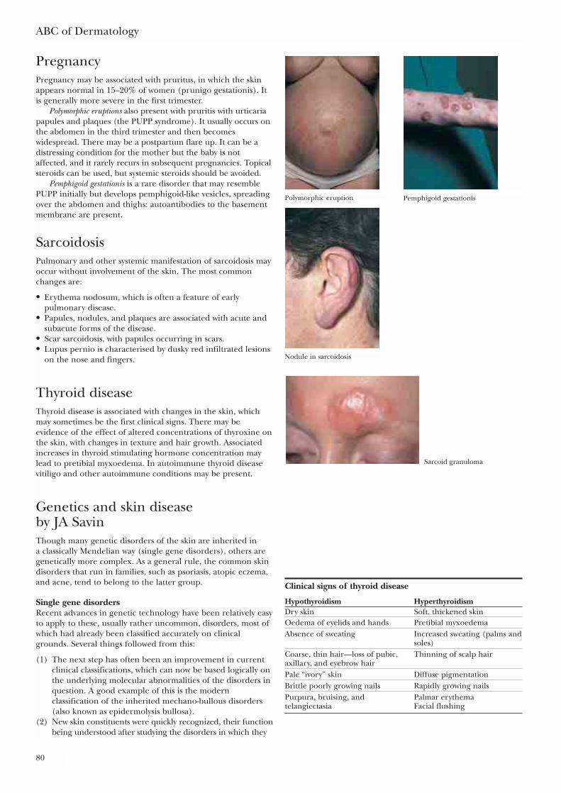
ABC of Dermatology
80
PregnancyPregnancy may be associated with pruritus, in which the skinappears normal in 15–20% of women (prunigo gestationis). Itis generally more severe in the first trimester.
Polymorphic eruptions also present with pruritis with urticariapapules and plaques (the PUPP syndrome). It usually occurs onthe abdomen in the third trimester and then becomeswidespread. There may be a postpartum flare up. It can be adistressing condition for the mother but the baby is notaffected, and it rarely recurs in subsequent pregnancies. Topicalsteroids can be used, but systemic steroids should be avoided.
Pemphigoid gestationis is a rare disorder that may resemblePUPP initially but develops pemphigoid-like vesicles, spreadingover the abdomen and thighs: autoantibodies to the basementmembrane are present.
SarcoidosisPulmonary and other systemic manifestation of sarcoidosis mayoccur without involvement of the skin. The most commonchanges are:
• Erythema nodosum, which is often a feature of earlypulmonary disease.
• Papules, nodules, and plaques are associated with acute andsubacute forms of the disease.
• Scar sarcoidosis, with papules occurring in scars.• Lupus pernio is characterised by dusky red infiltrated lesions
on the nose and fingers.
Thyroid diseaseThyroid disease is associated with changes in the skin, whichmay sometimes be the first clinical signs. There may beevidence of the effect of altered concentrations of thyroxine onthe skin, with changes in texture and hair growth. Associatedincreases in thyroid stimulating hormone concentration maylead to pretibial myxoedema. In autoimmune thyroid diseasevitiligo and other autoimmune conditions may be present.
Genetics and skin disease by JA SavinThough many genetic disorders of the skin are inherited in a classically Mendelian way (single gene disorders), others aregenetically more complex. As a general rule, the common skindisorders that run in families, such as psoriasis, atopic eczema,and acne, tend to belong to the latter group.
Single gene disordersRecent advances in genetic technology have been relatively easyto apply to these, usually rather uncommon, disorders, most ofwhich had already been classified accurately on clinicalgrounds. Several things followed from this:
(1) The next step has often been an improvement in currentclinical classifications, which can now be based logically onthe underlying molecular abnormalities of the disorders inquestion. A good example of this is the modernclassification of the inherited mechano-bullous disorders(also known as epidermolysis bullosa).
(2) New skin constituents were quickly recognized, their functionbeing understood after studying the disorders in which they
Clinical signs of thyroid disease
Hypothyroidism HyperthyroidismDry skin Soft, thickened skinOedema of eyelids and hands Pretibial myxoedemaAbsence of sweating Increased sweating (palms and
soles)Coarse, thin hair—loss of pubic, Thinning of scalp hairaxillary, and eyebrow hairPale “ivory” skin Diffuse pigmentationBrittle poorly growing nails Rapidly growing nailsPurpura, bruising, and Palmar erythematelangiectasia Facial flushing
Polymorphic eruption Pemphigoid gestationis
Nodule in sarcoidosis
Sarcoid granuloma
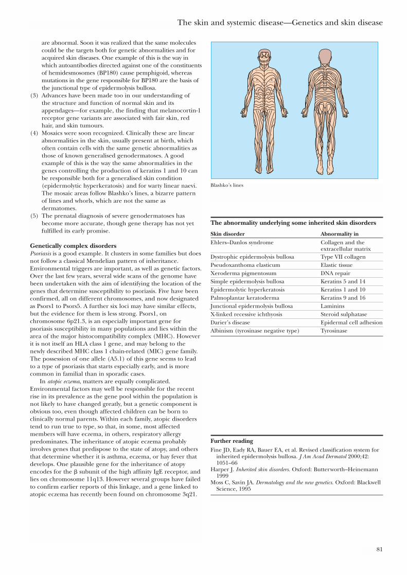
The skin and systemic disease—Genetics and skin disease
81
are abnormal. Soon it was realized that the same moleculescould be the targets both for genetic abnormalities and foracquired skin diseases. One example of this is the way inwhich autoantibodies directed against one of the constituentsof hemidesmosomes (BP180) cause pemphigoid, whereasmutations in the gene responsible for BP180 are the basis ofthe junctional type of epidermolysis bullosa.
(3) Advances have been made too in our understanding of the structure and function of normal skin and itsappendages—for example, the finding that melanocortin-1receptor gene variants are associated with fair skin, redhair, and skin tumours.
(4) Mosaics were soon recognized. Clinically these are linearabnormalities in the skin, usually present at birth, whichoften contain cells with the same genetic abnormalities asthose of known generalised genodermatoses. A goodexample of this is the way the same abnormalities in thegenes controlling the production of keratins 1 and 10 canbe responsible both for a generalised skin condition(epidermolytic hyperkeratosis) and for warty linear naevi.The mosaic areas follow Blashko’s lines, a bizarre patternof lines and whorls, which are not the same asdermatomes.
(5) The prenatal diagnosis of severe genodermatoses hasbecome more accurate, though gene therapy has not yetfulfilled its early promise.
Genetically complex disordersPsoriasis is a good example. It clusters in some families but doesnot follow a classical Mendelian pattern of inheritance.Environmental triggers are important, as well as genetic factors.Over the last few years, several wide scans of the genome havebeen undertaken with the aim of identifying the location of thegenes that determine susceptibility to psoriasis. Five have beenconfirmed, all on different chromosomes, and now designatedas Psors1 to Psors5. A further six loci may have similar effects,but the evidence for them is less strong. Psors1, onchromosome 6p21.3, is an especially important gene forpsoriasis susceptibility in many populations and lies within thearea of the major histocompatibility complex (MHC). Howeverit is not itself an HLA class 1 gene, and may belong to the newly described MHC class 1 chain-related (MIC) gene family.The possession of one allele (A5.1) of this gene seems to leadto a type of psoriasis that starts especially early, and is morecommon in familial than in sporadic cases.
In atopic eczema, matters are equally complicated.Environmental factors may well be responsible for the recentrise in its prevalence as the gene pool within the population isnot likely to have changed greatly, but a genetic component isobvious too, even though affected children can be born toclinically normal parents. Within each family, atopic disorderstend to run true to type, so that, in some, most affectedmembers will have eczema, in others, respiratory allergypredominates. The inheritance of atopic eczema probablyinvolves genes that predispose to the state of atopy, and othersthat determine whether it is asthma, eczema, or hay fever thatdevelops. One plausible gene for the inheritance of atopyencodes for the � subunit of the high affinity IgE receptor, andlies on chromosome 11q13. However several groups have failedto confirm earlier reports of this linkage, and a gene linked toatopic eczema has recently been found on chromosome 3q21.
The abnormality underlying some inherited skin disorders
Skin disorder Abnormality inEhlers–Danlos syndrome Collagen and the
extracellular matrixDystrophic epidermolysis bullosa Type VII collagenPseudoxanthoma elasticum Elastic tissueXeroderma pigmentosum DNA repairSimple epidermolysis bullosa Keratins 5 and 14Epidermolytic hyperkeratosis Keratins 1 and 10Palmoplantar keratoderma Keratins 9 and 16Junctional epidermolysis bullosa LamininsX-linked recessive ichthyosis Steroid sulphataseDarier’s disease Epidermal cell adhesionAlbinism (tyrosinase negative type) Tyrosinase
Further readingFine JD, Eady RA, Bauer EA, et al. Revised classification system for
inherited epidermolysis bullosa. J Am Acad Dermatol 2000;42:1051–66
Harper J. Inherited skin disorders. Oxford: Butterworth–Heinemann1999
Moss C, Savin JA. Dermatology and the new genetics. Oxford: BlackwellScience, 1995
Blashko’s lines
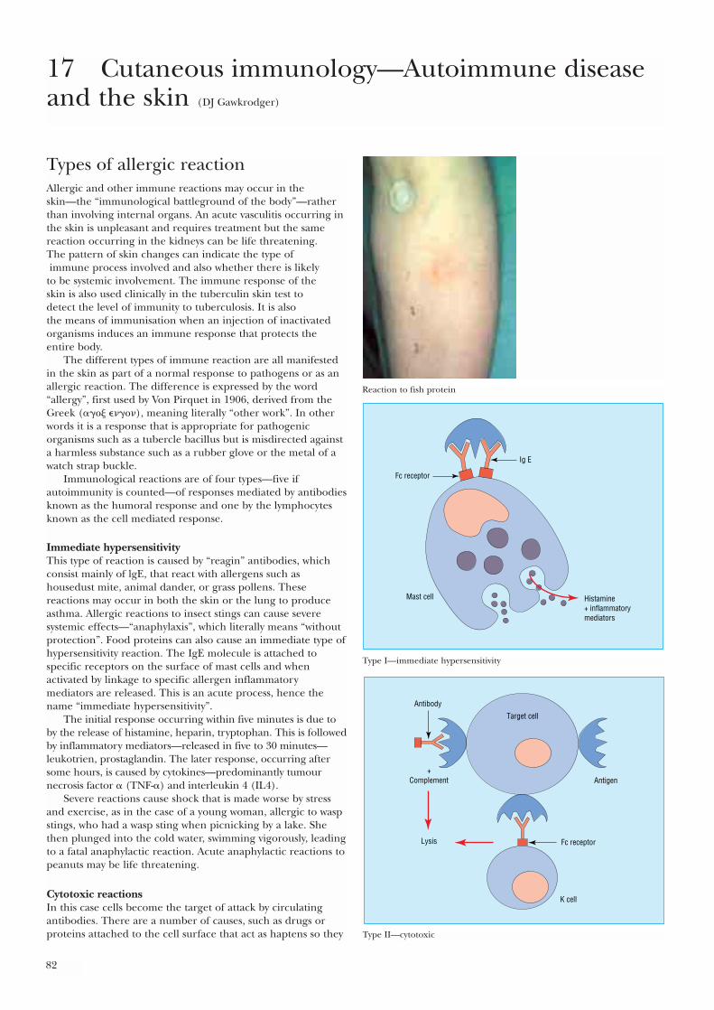
82
Types of allergic reactionAllergic and other immune reactions may occur in the skin—the “immunological battleground of the body”—ratherthan involving internal organs. An acute vasculitis occurring inthe skin is unpleasant and requires treatment but the samereaction occurring in the kidneys can be life threatening. The pattern of skin changes can indicate the type ofimmune process involved and also whether there is likely
to be systemic involvement. The immune response of the skin is also used clinically in the tuberculin skin test to detect the level of immunity to tuberculosis. It is also the means of immunisation when an injection of inactivatedorganisms induces an immune response that protects theentire body.
The different types of immune reaction are all manifestedin the skin as part of a normal response to pathogens or as anallergic reaction. The difference is expressed by the word“allergy”, first used by Von Pirquet in 1906, derived from theGreek (���� ��), meaning literally “other work”. In otherwords it is a response that is appropriate for pathogenicorganisms such as a tubercle bacillus but is misdirected againsta harmless substance such as a rubber glove or the metal of awatch strap buckle.
Immunological reactions are of four types—five ifautoimmunity is counted—of responses mediated by antibodiesknown as the humoral response and one by the lymphocytesknown as the cell mediated response.
Immediate hypersensitivityThis type of reaction is caused by “reagin” antibodies, whichconsist mainly of lgE, that react with allergens such ashousedust mite, animal dander, or grass pollens. Thesereactions may occur in both the skin or the lung to produceasthma. Allergic reactions to insect stings can cause severesystemic effects—“anaphylaxis”, which literally means “withoutprotection”. Food proteins can also cause an immediate type ofhypersensitivity reaction. The IgE molecule is attached tospecific receptors on the surface of mast cells and whenactivated by linkage to specific allergen inflammatorymediators are released. This is an acute process, hence thename “immediate hypersensitivity”.
The initial response occurring within five minutes is due toby the release of histamine, heparin, tryptophan. This is followedby inflammatory mediators—released in five to 30 minutes—leukotrien, prostaglandin. The later response, occurring aftersome hours, is caused by cytokines—predominantly tumournecrosis factor � (TNF-�) and interleukin 4 (IL4).
Severe reactions cause shock that is made worse by stressand exercise, as in the case of a young woman, allergic to waspstings, who had a wasp sting when picnicking by a lake. Shethen plunged into the cold water, swimming vigorously, leadingto a fatal anaphylactic reaction. Acute anaphylactic reactions topeanuts may be life threatening.
Cytotoxic reactions In this case cells become the target of attack by circulatingantibodies. There are a number of causes, such as drugs orproteins attached to the cell surface that act as haptens so they
17 Cutaneous immunology—Autoimmune diseaseand the skin (DJ Gawkrodger)
Reaction to fish protein
Fc receptor
Mast cell Histamine+ inflammatorymediators
Ig E
Type I—immediate hypersensitivity
Antibody
Antigen
K cell
+Complement
Lysis Fc receptor
Target cell
Type II—cytotoxic
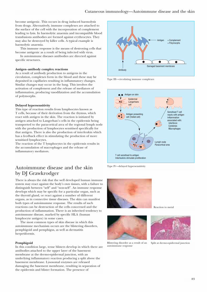
Cutaneous immunology—Autoimmune disease and the skin
83
become antigenic. This occurs in drug induced haemolysisfrom drugs. Alternatively, immune complexes are attached tothe surface of the cell with the incorporation of complementleading to lysis. In haemolytic anaemia and incompatible bloodtransfusions antibodies are formed against erythrocytes. Theymay also be destroyed by killer cells. A typical example ishaemolytic anaemia.
This immune response is the means of destroying cells thatbecome antigenic as a result of being infected with virus.
In autoimmune diseases antibodies are directed againstspecific structures.
Antigen–antibody complex reactionsAs a result of antibody production to antigens in thecirculation, complexes form in the blood and these may bedeposited in capillaries resulting in inflammatory changes.Similar changes may occur in the lung. This involves theactivation of complement and the release of mediators ofinflammation, producing vasodilatation and the accumulationof polymorphs.
Delayed hypersensitivityThis type of reaction results from lymphocytes known as T cells, because of their derivation from the thymus, whichreact with antigen in the skin. The reaction is initiated byantigen attached to Langerhan’s cells in the epidermis beingtransported to the paracortical area of the regional lymph nodewith the production of lymphocytes sensitised specifically forthat antigen. There is also the production of interleukin whichhas a feedback effect in stimulating the production of moresensitised lymphocytes. The reaction of the T lymphocytes in the epidermis results inthe accumulation of macrophages and the release ofinflammatory mediators.
Autoimmune disease and the skinby DJ GrawkrodgerThere is always the risk that the well developed human immunesystem may react against the body’s own tissues, with a failure todistinguish between “self” and “non-self”. An immune responsedevelops which may be specific for a particular organ, such asthe thyroid gland, or react against a number of differentorgans, as in connective tissue diseases. The skin can manifestboth types of autoimumme response. The results of suchreactions can be destruction of the cells concerned and theproduction of inflammation. There is an inherited tendency toautoimmune disease, marked by specific HLA (humanlymphocyte antigen) in some cases.
The most common types of skin disease in which thisautoimmune mechanism occurs are the blistering disorders,pemphigoid and pemphigus, as well as dermatitisherpetiformis.
PemphigoidIn this condition large, tense blisters develop in which there areantibodies attached to the upper layer of the basementmembrane at the dermo-epidermal junction, with anunderlying inflammatory reaction producing a split above thebasement membrane. Lysosomal enzymes are releaseddamaging the basement membrane, resulting in separation ofthe epidermis and blister formation. The presence of
Antigen + Complement+ Polymorphs
Antibody
Damaged basement membrane
Type III—circulating immune complexes
Lymph nodeParacortical area
Sensitised T cellreacts with antigenInflammationassociated with: Interferon Cytokines Macrophages
Antigen on skin
Antigen presentingcell (Veiled cell)
T cell sensitised to antigenInterleukins stimulate proliferation
EpidermalLangerhanscell
Type IV—delayed hypersensitivity
Reaction to metal
Blistering disorder as a result of anautoimmune response
Split at dermo-epidermal junction
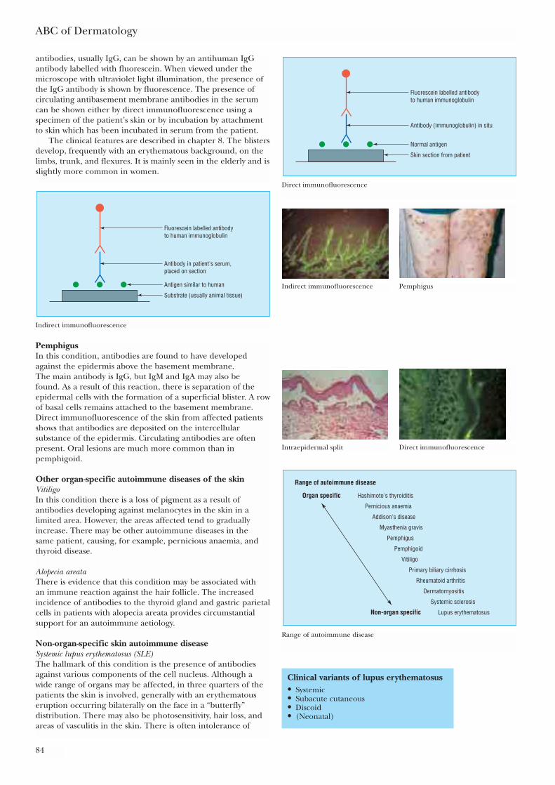
ABC of Dermatology
84
antibodies, usually IgG, can be shown by an antihuman IgGantibody labelled with fluorescein. When viewed under themicroscope with ultraviolet light illumination, the presence ofthe IgG antibody is shown by fluorescence. The presence ofcirculating antibasement membrane antibodies in the serumcan be shown either by direct immunofluorescence using aspecimen of the patient’s skin or by incubation by attachmentto skin which has been incubated in serum from the patient.
The clinical features are described in chapter 8. The blistersdevelop, frequently with an erythematous background, on thelimbs, trunk, and flexures. It is mainly seen in the elderly and isslightly more common in women.
Clinical variants of lupus erythematosus• Systemic• Subacute cutaneous• Discoid• (Neonatal)
Skin section from patient
Normal antigen
Antibody (immunoglobulin) in situ
Fluorescein labelled antibodyto human immunoglobulin
Direct immunofluorescence
Indirect immunofluorescence PemphigusSubstrate (usually animal tissue)
Antigen similar to human
Antibody in patient's serum,placed on section
Fluorescein labelled antibodyto human immunoglobulin
Indirect immunofluorescence
Intraepidermal split Direct immunofluorescence
Range of autoimmune disease
Hashimoto's thyroiditisOrgan specific
Pernicious anaemia
Addison's disease
Myasthenia gravis
Pemphigus
Pemphigoid
Vitiligo
Primary biliary cirrhosis
Rheumatoid arthritis
Dermatomyositis
Systemic sclerosis
Lupus erythematosusNon-organ specific
Range of autoimmune disease
PemphigusIn this condition, antibodies are found to have developedagainst the epidermis above the basement membrane. The main antibody is IgG, but IgM and IgA may also be found. As a result of this reaction, there is separation of theepidermal cells with the formation of a superficial blister. A rowof basal cells remains attached to the basement membrane.Direct immunofluorescence of the skin from affected patientsshows that antibodies are deposited on the intercellularsubstance of the epidermis. Circulating antibodies are oftenpresent. Oral lesions are much more common than inpemphigoid.
Other organ-specific autoimmune diseases of the skinVitiligoIn this condition there is a loss of pigment as a result ofantibodies developing against melanocytes in the skin in alimited area. However, the areas affected tend to graduallyincrease. There may be other autoimmune diseases in the same patient, causing, for example, pernicious anaemia, andthyroid disease.
Alopecia areataThere is evidence that this condition may be associated with an immune reaction against the hair follicle. The increasedincidence of antibodies to the thyroid gland and gastric parietalcells in patients with alopecia areata provides circumstantialsupport for an autoimmune aetiology.
Non-organ-specific skin autoimmune diseaseSystemic lupus erythematosus (SLE)The hallmark of this condition is the presence of antibodiesagainst various components of the cell nucleus. Although awide range of organs may be affected, in three quarters of thepatients the skin is involved, generally with an erythematouseruption occurring bilaterally on the face in a “butterfly”distribution. There may also be photosensitivity, hair loss, andareas of vasculitis in the skin. There is often intolerance of
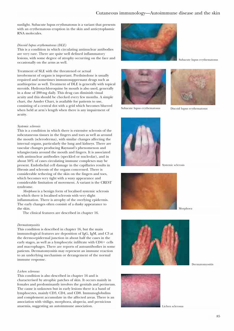
Cutaneous immunology—Autoimmune disease and the skin
85
sunlight. Subacute lupus erythmatosus is a variant that presentswith an erythematous eruption in the skin and anticytoplasmicRNA molecules.
Discoid lupus erythematosus (DLE)This is a condition in which circulating antinuclear antibodiesare very rare. There are quite well defined inflammatorylesions, with some degree of atrophy occurring on the face andoccasionally on the arms as well.
Treatment of SLE with the threatened or actual involvement of organs is important. Prednisolone is usuallyrequired and sometimes immunosuppressant drugs such asazathioprine as well. Treatment of DLE is generally with topicalsteroids. Hydroxychloroquine by mouth is also used, generallyin a dose of 200 mg daily. This drug can diminish visual acuity and this should be checked every few months. A simplechart, the Amsler Chart, is available for patients to use,consisting of a central dot with a grid which becomes blurredwhen held at arm’s length when there is any impairment ofacuity.
Systemic sclerosisThis is a condition in which there is extensive sclerosis of thesubcutaneous tissues in the fingers and toes as well as aroundthe mouth (scleroderma), with similar changes affecting theinternal organs, particularly the lung and kidneys. There arevascular changes producing Raynaud’s phenomenon andtelangiectasia around the mouth and fingers. It is associatedwith antinuclear antibodies (speckled or nucleolar), and inabout 50% of cases circulating immune complexes may bepresent. Endothelial cell damage in the capillaries results infibrosis and sclerosis of the organs concerned. There isconsiderable tethering of the skin on the fingers and toes,which becomes very tight with a waxy appearance andconsiderable limitation of movement. A variant is the CRESTsyndrome.
Morphoea is a benign form of localised systemic sclerosis in which there is localised sclerosis with very slightinflammation. There is atrophy of the overlying epidermis. The early changes often consisit of a dusky appearance to the skin.
The clinical features are described in chapter 16.
DermatomyositisThis condition is described in chapter 16, but the mainimmunological features are deposition of IgG, IgM, and C3 atthe dermo-epidermal junction in about half the cases in theearly stages, as well as a lymphocytic infiltrate with CD4� cellsand macrophages. There are reports of autoantibodies in somepatients. Dermatomyositis may represent an immune reactionto an underlying mechanism or derangement of the normalimmune response.
Lichen sclerosusThis condition is also described in chapter 16 and ischaracterised by atrophic patches of skin. It occurs mainly infemales and predominantly involves the genitals and perineum.The cause is unknown but in early lesions there is a band oflymphocytes, mainly CD3, CD4, and CD8. Immunoglobulinsand complement accumulate in the affected areas. There is anassociation with vitiligo, morphoea, alopecia, and perniciousanaemia, suggesting an autoimmune association.
Subacute lupus erythematosus
Subacute lupus erythematosus Discoid lupus erythematosus
Systemic sclerosis
Morphoea
Dermatomyositis
Lichen sclerosus
Graft versus host diseaseThis reaction occurs following bone marrow transplantation inimmunosuppressed patients. T lymphocytes produced by thegraft react against the body’s own tissues, producing a skineruption which may resemble measles. There is lysis of thebasement membrane with shedding of the skin and sometimeslichen planus-like eruption. In the more chronic form,localised lesions develop, with immunoglobulins deposited inthe walls of blood vessels with the activation of complement.
ABC of Dermatology
86
Further readingRoitt IM, Brostoff J, Male D. Immunology, 6th ed. St Louis: Mosby,
2001
Graft versus host disease
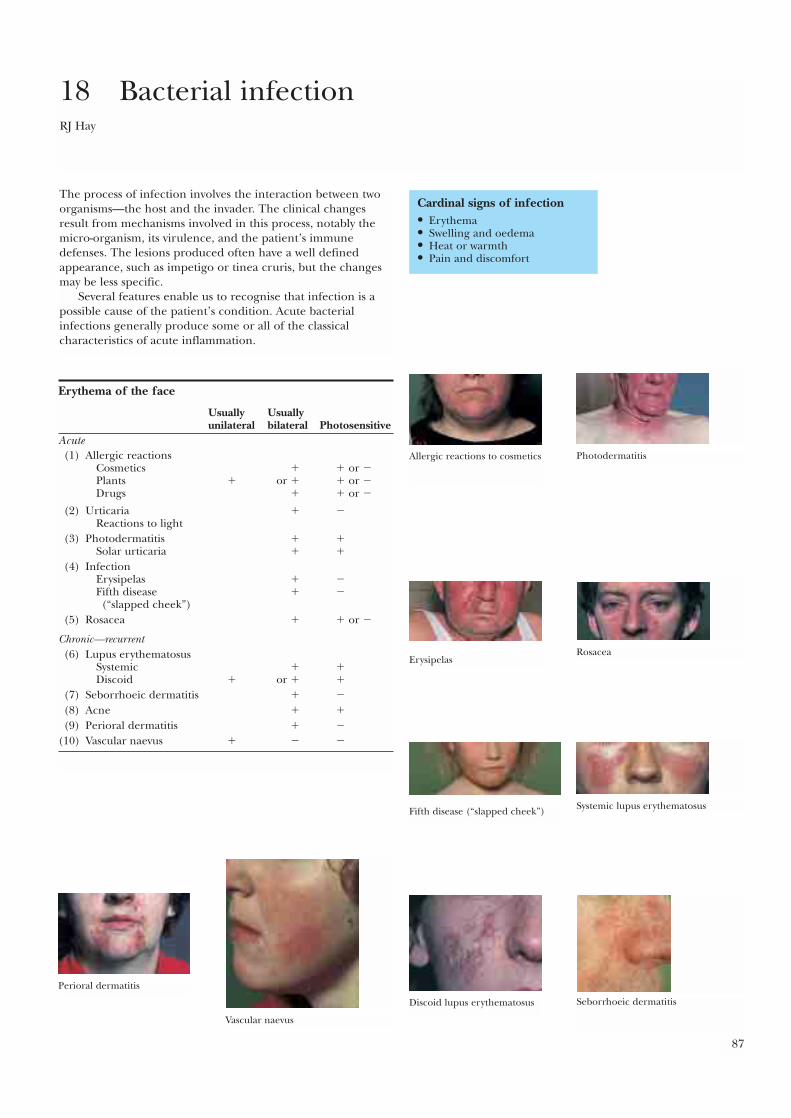
87
The process of infection involves the interaction between twoorganisms—the host and the invader. The clinical changesresult from mechanisms involved in this process, notably themicro-organism, its virulence, and the patient’s immunedefenses. The lesions produced often have a well definedappearance, such as impetigo or tinea cruris, but the changesmay be less specific.
Several features enable us to recognise that infection is apossible cause of the patient’s condition. Acute bacterialinfections generally produce some or all of the classicalcharacteristics of acute inflammation.
18 Bacterial infectionRJ Hay
Cardinal signs of infection• Erythema• Swelling and oedema• Heat or warmth• Pain and discomfort
Erythema of the face
Usually Usuallyunilateral bilateral Photosensitive
Acute(1) Allergic reactions
Cosmetics � � or �Plants � or � � or �Drugs � � or �
(2) Urticaria � �Reactions to light
(3) Photodermatitis � �Solar urticaria � �
(4) InfectionErysipelas � �Fifth disease � �(“slapped cheek”)
(5) Rosacea � � or �
Chronic—recurrent(6) Lupus erythematosus
Systemic � �Discoid � or � �
(7) Seborrhoeic dermatitis � �
(8) Acne � �
(9) Perioral dermatitis � �
(10) Vascular naevus � � �
Allergic reactions to cosmetics Photodermatitis
ErysipelasRosacea
Fifth disease (“slapped cheek”) Systemic lupus erythematosus
Discoid lupus erythematosus Seborrhoeic dermatitis
Perioral dermatitis
Vascular naevus
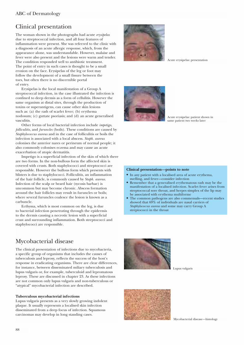
ABC of Dermatology
88
Clinical presentationThe woman shown in the photographs had acute erysipelasdue to streptococcal infection, and all four features ofinflammation were present. She was referred to the clinic witha diagnosis of an acute allergic response, which, from theappearance alone, was understandable. However, malaise andfever were also present and the lesions were warm and tender.The condition responded well to antibiotic treatment. The point of entry in such cases is thought to be a smallerosion on the face. Erysipelas of the leg or foot may follow the development of a small fissure between the toes, but often there is no discernible portal of entry.
Erysipelas is the local manifestation of a Group Astreptococcal infection, in the case illustrated the infection isconfined to deep dermis as a form of cellulitis. However thesame organism at distal sites, through the production of toxins or superantigens, can cause other skin lesions such as: (a) the rash of scarlet fever; (b) erythema nodosum; (c) guttate psoriasis; and (d) an acute generalisedvasculitis.
Other forms of local bacterial infection include impetigo,folliculitis, and furuncles (boils). These conditions are caused byStaphylococcus aureus and in the case of folliculitis or boils theinfection is associated with a local abscess. Staph. aureuscolonises the anterior nares or perineum of normal people; italso commonly colonises eczema and may cause an acuteexacerbation of atopic dermatitis.
Impetigo is a superficial infection of the skin of which thereare two forms. In the non-bullous form the affected skin iscovered with crusts. Both staphylococci and streptococci areresponsible. However the bullous form which presents withblisters is due to staphylococci. Folliculitis, an inflammation of the hair follicle, is commonly caused by Staph. aureus.Infection of the scalp or beard hair (sycosis barbae) isuncommon but may become chronic. Abscess formationaround the hair follicles may result in furuncles or boils; where several furuncles coalesce the lesion is known as acarbuncle.
Ecthyma, which is most common on the leg, is due to bacterial infection penetrating through the epidermis to the dermis causing a necrotic lesion with a superficial crust and surrounding inflammation. Both streptococci andstaphylococci are responsible.
Mycobacterial diseaseThe clinical presentation of infections due to mycobacteria, a specific group of organisms that includes the causes oftuberculosis and leprosy, reflects the success of the host’sresponse in eradicating organisms. There are clear differences,for instance, between disseminated miliary tuberculosis andlupus vulgaris or, for example, tuberculoid and lepromatousleprosy. These are discussed in chapter 23. As these infectionsare not common only lupus vulgaris and non-tuberculous or“atypical” mycobacterial infection are described.
Tuberculous mycobacterial infectionsLupus vulgaris presents as a very slowly growing indolentplaque. It usually represents a localised skin infectiondisseminated from a deep focus of infection. Squamouscarcinomas may develop in long standing cases.
Clinical presentation—points to note• In any patient with a localised area of acute erythema,
swelling, and fever—consider infection• Remember that a generalised erythematous rash may be the
manifestation of a localised infection. Scarlet fever arises fromstreptococcal sore throat, and herpes simplex of the lip maybe associated with erythema multiforme
• The common pathogens are also commensals—recent studiesshowed that 69% of individuals are nasal carriers ofStaphylococcus aureus and some may carry Group Astreptococci in the throat
Acute erysipelas: presentation
Acute erysipelas: patient shown in same patient two weeks later
Lupus vulgaris
Mycobacterial disease—histology
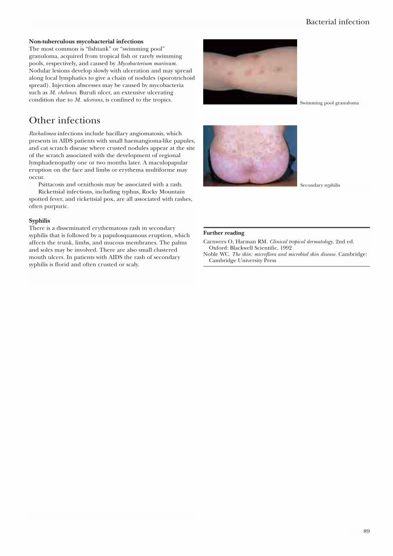
Bacterial infection
89
Non-tuberculous mycobacterial infectionsThe most common is “fishtank” or “swimming pool”granuloma, acquired from tropical fish or rarely swimmingpools, respectively, and caused by Mycobacterium marinum.Nodular lesions develop slowly with ulceration and may spreadalong local lymphatics to give a chain of nodules (sporotrichoidspread). Injection abscesses may be caused by mycobacteriasuch as M. chelonei. Buruli ulcer, an extensive ulceratingcondition due to M. ulcerans, is confined to the tropics.
Other infectionsRochalimea infections include bacillary angiomatosis, whichpresents in AIDS patients with small haemangioma-like papules,and cat scratch disease where crusted nodules appear at the siteof the scratch associated with the development of regionallymphadenopathy one or two months later. A maculopapulareruption on the face and limbs or erythema multiforme mayoccur.
Psittacosis and ornithosis may be associated with a rash.Rickettsial infections, including typhus, Rocky Mountain
spotted fever, and rickettsial pox, are all associated with rashes,often purpuric.
SyphilisThere is a disseminated erythematous rash in secondarysyphilis that is followed by a papulosquamous eruption, whichaffects the trunk, limbs, and mucous membranes. The palmsand soles may be involved. There are also small clusteredmouth ulcers. In patients with AIDS the rash of secondarysyphilis is florid and often crusted or scaly.
Further readingCarnwres O, Harman RM. Clinical tropical dermatology. 2nd ed.
Oxford: Blackwell Scientific, 1992Noble WC. The skin: microflora and microbial skin disease. Cambridge:
Cambridge University Press
Swimming pool granuloma
Secondary syphilis
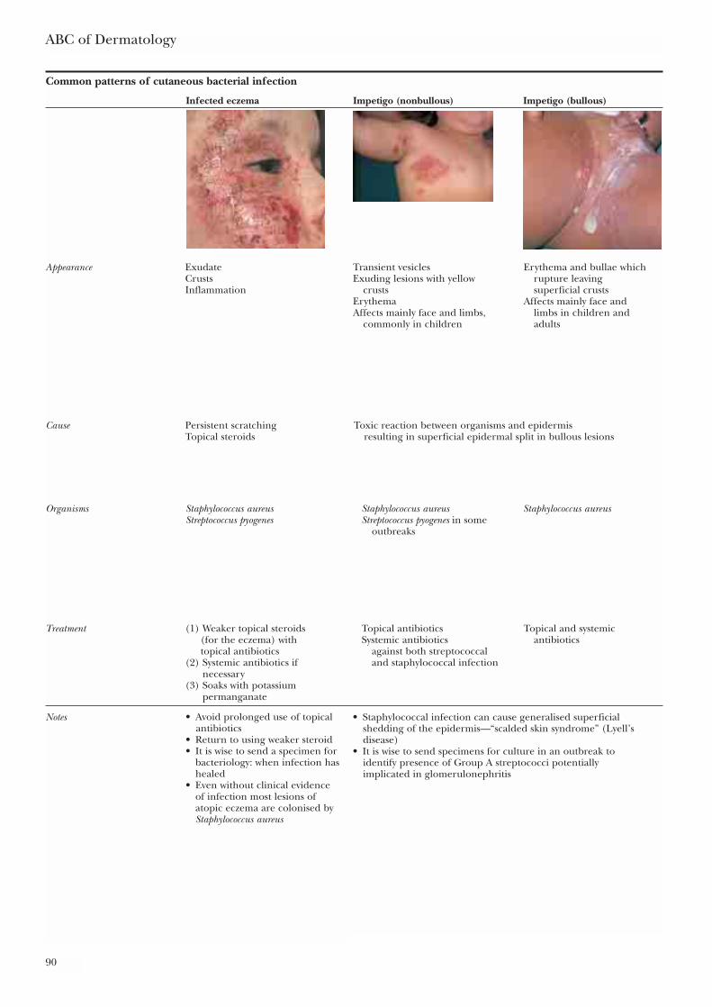
ABC of Dermatology
90
Common patterns of cutaneous bacterial infection
Infected eczema Impetigo (nonbullous) Impetigo (bullous)
Appearance Exudate Transient vesicles Erythema and bullae which Crusts Exuding lesions with yellow rupture leaving Inflammation crusts superficial crusts
Erythema Affects mainly face andAffects mainly face and limbs, limbs in children and
commonly in children adults
Cause Persistent scratching Toxic reaction between organisms and epidermisTopical steroids resulting in superficial epidermal split in bullous lesions
Organisms Staphylococcus aureus Staphylococcus aureus Staphylococcus aureusStreptococcus pyogenes Streptococcus pyogenes in some
outbreaks
Treatment (1) Weaker topical steroids Topical antibiotics Topical and systemic(for the eczema) with Systemic antibiotics antibioticstopical antibiotics against both streptococcal
(2) Systemic antibiotics if and staphylococcal infectionnecessary
(3) Soaks with potassium permanganate
Notes • Staphylococcal infection can cause generalised superficialshedding of the epidermis—“scalded skin syndrome” (Lyell’sdisease)
• It is wise to send specimens for culture in an outbreak to identify presence of Group A streptococci potentially implicated in glomerulonephritis
• Avoid prolonged use of topical antibiotics
• Return to using weaker steroid• It is wise to send a specimen for
bacteriology: when infection hashealed
• Even without clinical evidence of infection most lesions of atopic eczema are colonised byStaphylococcus aureus
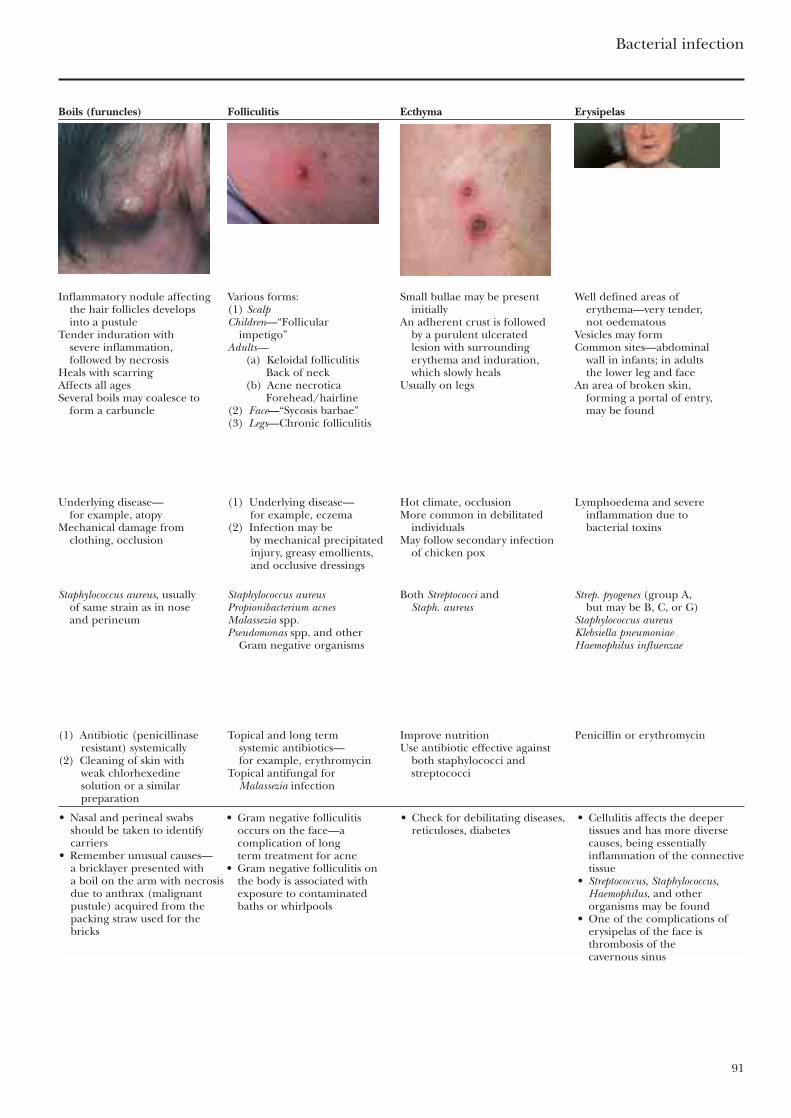
Bacterial infection
91
Boils (furuncles) Folliculitis Ecthyma Erysipelas
Inflammatory nodule affecting Various forms: Small bullae may be present Well defined areas ofthe hair follicles develops (1) Scalp initially erythema—very tender,into a pustule Children—“Follicular An adherent crust is followed not oedematous
Tender induration with impetigo” by a purulent ulcerated Vesicles may formsevere inflammation, Adults— lesion with surrounding Common sites—abdominalfollowed by necrosis (a) Keloidal folliculitis erythema and induration, wall in infants; in adults
Heals with scarring Back of neck which slowly heals the lower leg and faceAffects all ages (b) Acne necrotica Usually on legs An area of broken skin,Several boils may coalesce to Forehead/hairline forming a portal of entry,
form a carbuncle (2) Face—“Sycosis barbae” may be found(3) Legs—Chronic folliculitis
Underlying disease— (1) Underlying disease— Hot climate, occlusion Lymphoedema and severefor example, atopy for example, eczema More common in debilitated inflammation due to
Mechanical damage from (2) Infection may be individuals bacterial toxinsclothing, occlusion by mechanical precipitated May follow secondary infection
injury, greasy emollients, of chicken poxand occlusive dressings
Staphylococcus aureus, usually Staphylococcus aureus Both Streptococci and Strep. pyogenes (group A,of same strain as in nose Propionibacterium acnes Staph. aureus but may be B, C, or G)and perineum Malassezia spp. Staphylococcus aureus
Pseudomonas spp. and other Klebsiella pneumoniaeGram negative organisms Haemophilus influenzae
(1) Antibiotic (penicillinase Topical and long term Improve nutrition Penicillin or erythromycinresistant) systemically systemic antibiotics— Use antibiotic effective against
(2) Cleaning of skin with for example, erythromycin both staphylococci andweak chlorhexedine Topical antifungal for streptococcisolution or a similar Malassezia infectionpreparation
• Nasal and perineal swabsshould be taken to identifycarriers
• Remember unusual causes—a bricklayer presented with a boil on the arm with necrosisdue to anthrax (malignantpustule) acquired from thepacking straw used for thebricks
• Gram negative folliculitis occurs on the face—a complication of long term treatment for acne
• Gram negative folliculitis on the body is associated with exposure to contaminatedbaths or whirlpools
• Check for debilitating diseases,reticuloses, diabetes
• Cellulitis affects the deepertissues and has more diversecauses, being essentiallyinflammation of the connectivetissue
• Streptococcus, Staphylococcus,Haemophilus, and otherorganisms may be found
• One of the complications oferysipelas of the face isthrombosis of thecavernous sinus
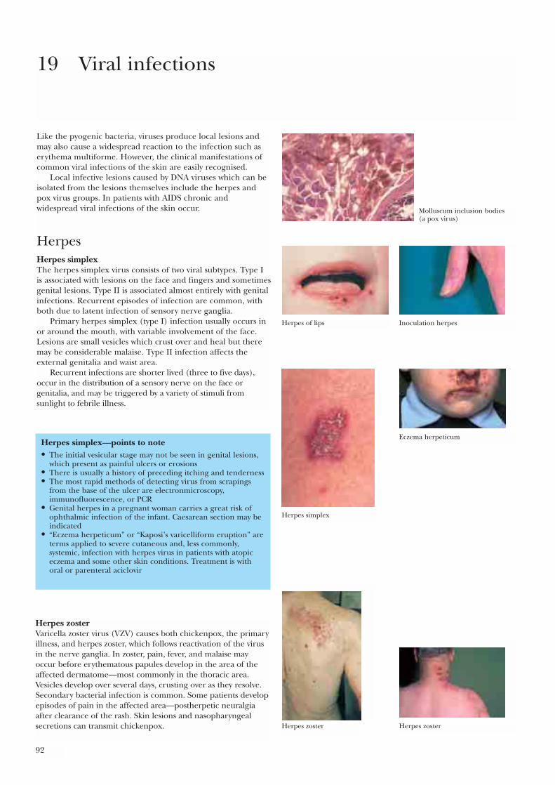
92
Like the pyogenic bacteria, viruses produce local lesions andmay also cause a widespread reaction to the infection such aserythema multiforme. However, the clinical manifestations ofcommon viral infections of the skin are easily recognised.
Local infective lesions caused by DNA viruses which can beisolated from the lesions themselves include the herpes andpox virus groups. In patients with AIDS chronic andwidespread viral infections of the skin occur.
HerpesHerpes simplexThe herpes simplex virus consists of two viral subtypes. Type Iis associated with lesions on the face and fingers and sometimesgenital lesions. Type II is associated almost entirely with genitalinfections. Recurrent episodes of infection are common, withboth due to latent infection of sensory nerve ganglia.
Primary herpes simplex (type I) infection usually occurs inor around the mouth, with variable involvement of the face.Lesions are small vesicles which crust over and heal but theremay be considerable malaise. Type II infection affects theexternal genitalia and waist area.
Recurrent infections are shorter lived (three to five days),occur in the distribution of a sensory nerve on the face orgenitalia, and may be triggered by a variety of stimuli fromsunlight to febrile illness.
19 Viral infections
Herpes simplex—points to note• The initial vesicular stage may not be seen in genital lesions,
which present as painful ulcers or erosions• There is usually a history of preceding itching and tenderness• The most rapid methods of detecting virus from scrapings
from the base of the ulcer are electronmicroscopy,immunofluorescence, or PCR
• Genital herpes in a pregnant woman carries a great risk ofophthalmic infection of the infant. Caesarean section may beindicated
• “Eczema herpeticum” or “Kaposi’s varicelliform eruption” areterms applied to severe cutaneous and, less commonly,systemic, infection with herpes virus in patients with atopiceczema and some other skin conditions. Treatment is withoral or parenteral aciclovir
Herpes zosterVaricella zoster virus (VZV) causes both chickenpox, the primaryillness, and herpes zoster, which follows reactivation of the virusin the nerve ganglia. In zoster, pain, fever, and malaise mayoccur before erythematous papules develop in the area of theaffected dermatome—most commonly in the thoracic area.Vesicles develop over several days, crusting over as they resolve.Secondary bacterial infection is common. Some patients developepisodes of pain in the affected area—postherpetic neuralgiaafter clearance of the rash. Skin lesions and nasopharyngealsecretions can transmit chickenpox.
Molluscum inclusion bodies(a pox virus)
Herpes of lips Inoculation herpes
Herpes simplex
Eczema herpeticum
Herpes zoster Herpes zoster
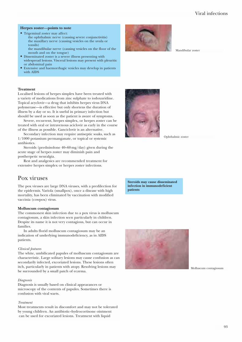
TreatmentLocalised lesions of herpes simplex have been treated with a variety of medications from zinc sulphate to iodoxuridine.Topical acyclovir—a drug that inhibits herpes virus DNApolymerase—is effective but only shortens the duration ofillness by a day or so. It is useful in primary infection butshould be used as soon as the patient is aware of symptoms.
Severe, recurrent, herpes simplex, or herpes zoster can betreated with oral or intravenous aciclovir as early in the courseof the illness as possible. Ganciclovir is an alternative.
Secondary infection may require antiseptic soaks, such as1/1000 potassium permanganate, or topical or systemicantibiotics.
Steroids (prednisolone 40–60 mg/day) given during theacute stage of herpes zoster may diminish pain andpostherpetic neuralgia.
Rest and analgesics are recommended treatment forextensive herpes simplex or herpes zoster infections.
Pox virusesThe pox viruses are large DNA viruses, with a predilection forthe epidermis. Variola (smallpox), once a disease with highmortality, has been eliminated by vaccination with modifiedvaccinia (cowpox) virus.
Molluscum contagiosumThe commonest skin infection due to a pox virus is molluscumcontagiosum, a skin infection seen particularly in children.Despite its name it is not very contagious, but can occur infamilies.
In adults florid molluscum contagiosum may be anindication of underlying immunodeficiency, as in AIDSpatients.
Clinical featuresThe white, umbilicated papules of molluscum contagiosum arecharacteristic. Large solitary lesions may cause confusion as cansecondarily infected, excoriated lesions. These lesions oftenitch, particularly in patients with atopy. Resolving lesions maybe surrounded by a small patch of eczema.
DiagnosisDiagnosis is usually based on clinical appearances ormicroscopy of the contents of papules. Sometimes there isconfusion with viral warts.
TreatmentMost treatments result in discomfort and may not be toleratedby young children. An antibiotic–hydrocortisone ointmentcan be used for excoriated lesions. Treatment with liquid
Viral infections
93
Herpes zoster—points to note• Trigeminal zoster may affect:
the ophthalmic nerve (causing severe conjunctivitis)the maxillary nerve (causing vesicles on the uvula ortonsils)the mandibular nerve (causing vesicles on the floor of themouth and on the tongue)
• Disseminated zoster is a severe illness presenting withwidespread lesions. Visceral lesions may present with pleuriticor abdominal pain
• Extensive and haemorrhagic vesicles may develop in patientswith AIDS
Steroids may cause disseminatedinfection in immunodeficientpatients
Mandibular zoster
Ophthalmic zoster
Molluscum contagiosum
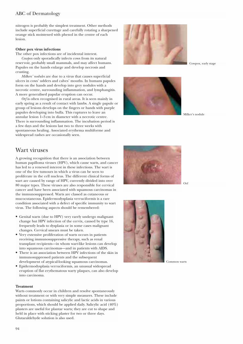
nitrogen is probably the simplest treatment. Other methodsinclude superficial curettage and carefully rotating a sharpenedorange stick moistened with phenol in the centre of eachlesion.
Other pox virus infectionsThe other pox infections are of incidental interest.
Cowpox only sporadically infects cows from its naturalreservoir, probably small mammals, and may affect humans.Papules on the hands enlarge and develop necrosis andcrusting.
Milkers’ nodules are due to a virus that causes superficialulcers in cows’ udders and calves’ mouths. In humans papulesform on the hands and develop into grey nodules with anecrotic centre, surrounding inflammation, and lymphangitis.A more generalised papular eruption can occur.
Orf is often recognised in rural areas. It is seen mainly inearly spring as a result of contact with lambs. A single papule orgroup of lesions develops on the fingers or hands with purplepapules developing into bulla. This ruptures to leave anannular lesion 1–3 cm in diameter with a necrotic centre. There is surrounding inflammation. The incubation period is a few days and the lesions last two to three weeks withspontaneous healing. Associated erythema multiforme andwidespread rashes are occasionally seen.
Wart virusesA growing recognition that there is an association betweenhuman papilloma viruses (HPV), which cause warts, and cancerhas led to a renewed interest in these infections. The wart isone of the few tumours in which a virus can be seen toproliferate in the cell nucleus. The different clinical forms ofwart are caused by range of HPV, currently divided into over 80 major types. These viruses are also responsible for cervicalcancer and have been associated with squamous carcinomas inthe immunosuppressed. Warts are classed as cutaneous ormucocutaneous. Epidermodysplasia verruciformis is a rarecondition associated with a defect of specific immunity to wartvirus. The following aspects should be remembered:
• Genital warts (due to HPV) very rarely undergo malignantchange but HPV infection of the cervix, caused by type 16,frequently leads to dysplasia or in some cases malignantchanges. Cervical smears must be taken.
• Very extensive proliferation of warts occurs in patientsreceiving immunosuppressive therapy, such as renaltransplant recipients—in whom wart-like lesions can developinto squamous carcinomas—and in patients with AIDS.
• There is an association between HPV infections of the skin inimmunosuppressed patients and the subsequentdevelopment of atypical-looking squamous carcinomas.
• Epidermodysplasia verruciformis, an unusual widespreaderuption of flat erythematous warty plaques, can also developinto carcinoma.
TreatmentWarts commonly occur in children and resolve spontaneouslywithout treatment or with very simple measures. These includepaints or lotions containing salicylic and lactic acids in variousproportions, which should be applied daily. Salicylic acid (40%)plasters are useful for plantar warts; they are cut to shape andheld in place with sticking plaster for two or three days.Glutaraldehyde solution is also used.
ABC of Dermatology
94
Cowpox, early stage
Milker’s nodule
Orf
Common warts
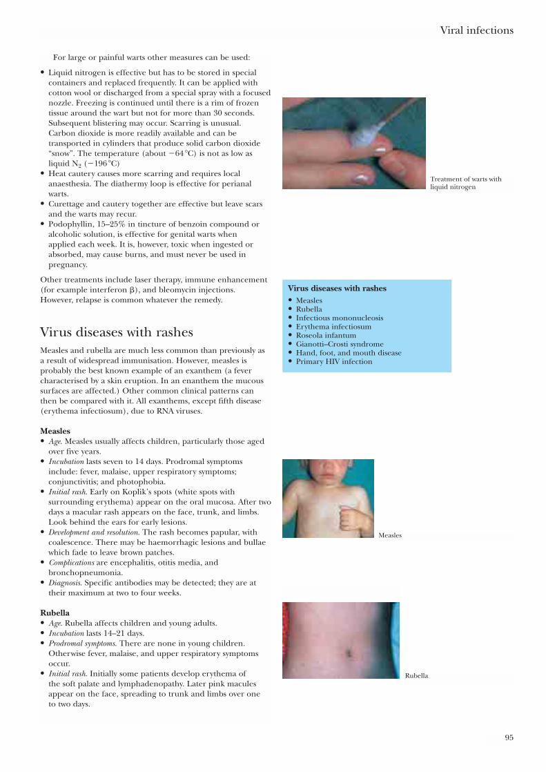
For large or painful warts other measures can be used:
• Liquid nitrogen is effective but has to be stored in specialcontainers and replaced frequently. It can be applied withcotton wool or discharged from a special spray with a focusednozzle. Freezing is continued until there is a rim of frozentissue around the wart but not for more than 30 seconds.Subsequent blistering may occur. Scarring is unusual. Carbon dioxide is more readily available and can betransported in cylinders that produce solid carbon dioxide“snow”. The temperature (about �64 �C) is not as low asliquid N2 (�196 �C)
• Heat cautery causes more scarring and requires localanaesthesia. The diathermy loop is effective for perianalwarts.
• Curettage and cautery together are effective but leave scarsand the warts may recur.
• Podophyllin, 15–25% in tincture of benzoin compound oralcoholic solution, is effective for genital warts when applied each week. It is, however, toxic when ingested orabsorbed, may cause burns, and must never be used inpregnancy.
Other treatments include laser therapy, immune enhancement(for example interferon �), and bleomycin injections. However, relapse is common whatever the remedy.
Virus diseases with rashesMeasles and rubella are much less common than previously as a result of widespread immunisation. However, measles isprobably the best known example of an exanthem (a fevercharacterised by a skin eruption. In an enanthem the mucoussurfaces are affected.) Other common clinical patterns canthen be compared with it. All exanthems, except fifth disease(erythema infectiosum), due to RNA viruses.
Measles• Age. Measles usually affects children, particularly those aged
over five years.• Incubation lasts seven to 14 days. Prodromal symptoms
include: fever, malaise, upper respiratory symptoms;conjunctivitis; and photophobia.
• Initial rash. Early on Koplik’s spots (white spots withsurrounding erythema) appear on the oral mucosa. After twodays a macular rash appears on the face, trunk, and limbs.Look behind the ears for early lesions.
• Development and resolution. The rash becomes papular, withcoalescence. There may be haemorrhagic lesions and bullaewhich fade to leave brown patches.
• Complications are encephalitis, otitis media, andbronchopneumonia.
• Diagnosis. Specific antibodies may be detected; they are attheir maximum at two to four weeks.
Rubella• Age. Rubella affects children and young adults.• Incubation lasts 14–21 days.• Prodromal symptoms. There are none in young children.
Otherwise fever, malaise, and upper respiratory symptomsoccur.
• Initial rash. Initially some patients develop erythema of the soft palate and lymphadenopathy. Later pink maculesappear on the face, spreading to trunk and limbs over one to two days.
Viral infections
95
Virus diseases with rashes• Measles• Rubella• Infectious mononucleosis• Erythema infectiosum• Roseola infantum• Gianotti–Crosti syndrome• Hand, foot, and mouth disease• Primary HIV infection
Treatment of warts withliquid nitrogen
Measles
Rubella
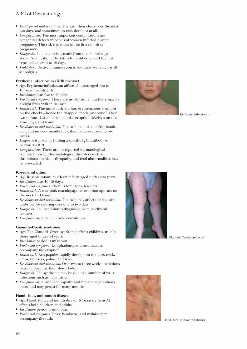
• Development and resolution. The rash then clears over the nexttwo days, and sometimes no rash develops at all.
• Complications. The most important complications arecongenital defects in babies of women infected duringpregnancy. The risk is greatest in the first month ofpregnancy.
• Diagnosis. The diagnosis is made from the clinical signsabove. Serum should be taken for antibodies and the testrepeated at seven to 10 days.
• Prophylaxis. Active immunisation is routinely available for allschoolgirls.
Erythema infectiosum (fifth disease)• Age. Erythema infectiosum affects children aged two to
10 years, mainly girls. • Incubation lasts five to 20 days.• Prodromal symptoms. There are usually none, but there may be
a slight fever with initial rash.• Initial rash. The initial rash is a hot, erythematous eruption
on the cheeks—hence the “slapped cheek syndrome”. Overtwo to four days a maculopapular eruption develops on thearms, legs, and trunk.
• Development and resolution. The rash extends to affect hands,feet, and mucous membranes, then fades over one to twoweeks.
• Diagnosis is made by finding a specific IgM antibody toparvovirus B19.
• Complications. There are no reported dermatologicalcomplications but haematological disorders such asthrombocytopenia, arthropathy, and fetal abnormalities maybe associated.
Roseola infantum• Age. Roseola infantum affects infants aged under two years. • Incubation lasts 10–15 days.• Prodromal symptoms. There is fever for a few days.• Initial rash. A rose pink maculopapular eruption appears on
the neck and trunk.• Development and resolution. The rash may affect the face and
limbs before clearing over one to two days.• Diagnosis. The condition is diagnosed from its clinical
features.• Complications include febrile convulsions.
Gianotti–Crosti syndrome• Age. The Gianotti–Crosti syndrome affects children, usually
those aged under 14 years.• Incubation period is unknown.• Prodromal symptoms. Lymphadenopathy and malaise
accompany the eruption.• Initial rash. Red papules rapidly develop on the face, neck,
limbs, buttocks, palms, and soles.• Development and resolution. Over two to three weeks the lesions
become purpuric then slowly fade.• Diagnosis. The syndrome may be due to a number of virus
infections such as hepatitis B.• Complications. Lymphadenopathy and hepatomegaly always
occur and may persist for many months.
Hand, foot, and mouth disease• Age. Hand, foot, and mouth disease (Coxsackie virus A)
affects both children and adults.• Incubation period is unknown.• Prodromal symptoms. Fever, headache, and malaise may
accompany the rash.
ABC of Dermatology
96
Erythema infectiosum
Gianotti–Crosti syndrome
Hand, foot, and mouth disease

• Initial rash. Initially there may be intense erythemasurrounding yellow-grey vesicles 1–1·5 mm in diameter. Theseare mainly distributed on the palms and soles and in themouth. Sometimes a more generalised eruption maydevelop.
• Development and resolution. Over three to five days the rashfades.
• Diagnosis. Coxsackie A (usually A16) virus is isolated fromlesions and stools. A specific antibody may be found in theserum.
• Complications are rare but include widespread vesicular rashesand erythema multiforme.
Other infectionsInfectious mononucleosisAs well as the erythematous lesions on the palate amaculopapular rash affecting the face and limbs can occur.
Viral infections
97
Further readingJoklik WK. Virology, 3rd ed. Norwalk: Appleton and Lange, 1988Mandell GL, Douglas RG, Bennett JF. Principles and practice of
infectious diseases, 3rd ed. New York: Churchill Livingstone, 1990Timbury MC. Notes on medical virology, 9th ed. Edinburgh: Churchill
Livingstone, 1991White DO, Fenner FJ. Medical virology, 4th ed. New York: Academic
Press, 1994
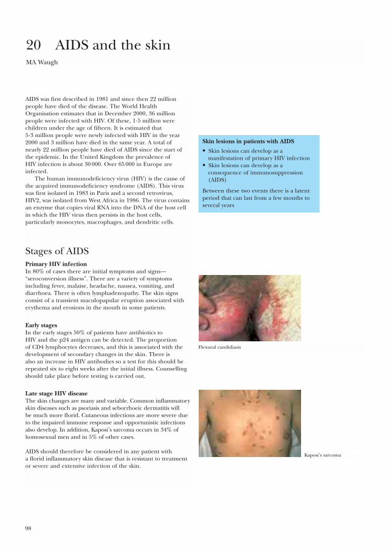
Skin lesions in patients with AIDS
• Skin lesions can develop as amanifestation of primary HIV infection
• Skin lesions can develop as aconsequence of immunosuppression(AIDS)
Between these two events there is a latentperiod that can last from a few months toseveral years
98
AIDS was first described in 1981 and since then 22 millionpeople have died of the disease. The World HealthOrganisation estimates that in December 2000, 36 millionpeople were infected with HIV. Of these, 1·5 million werechildren under the age of fifteen. It is estimated that 5·3 million people were newly infected with HIV in the year2000 and 3 million have died in the same year. A total of nearly 22 million people have died of AIDS since the start ofthe epidemic. In the United Kingdom the prevalence of HIV infection is about 30 000. Over 65 000 in Europe areinfected.
The human immunodeficiency virus (HIV) is the cause ofthe acquired immunodeficiency syndrome (AIDS). This viruswas first isolated in 1983 in Paris and a second retrovirus,HIV2, was isolated from West Africa in 1986. The virus containsan enzyme that copies viral RNA into the DNA of the host cellin which the HIV virus then persists in the host cells,particularly monocytes, macrophages, and dendritic cells.
Stages of AIDSPrimary HIV infectionIn 80% of cases there are initial symptoms and signs—“seroconversion illness”. There are a variety of symptomsincluding fever, malaise, headache, nausea, vomiting, anddiarrhoea. There is often lymphadenopathy. The skin signsconsist of a transient maculopapular eruption associated witherythema and erosions in the mouth in some patients.
Early stagesIn the early stages 50% of patients have antibiotics to HIV and the p24 antigen can be detected. The proportion of CD4 lymphocytes decreases, and this is associated with thedevelopment of secondary changes in the skin. There is also an increase in HIV antibodies so a test for this should berepeated six to eight weeks after the initial illness. Counsellingshould take place before testing is carried out.
Late stage HIV diseaseThe skin changes are many and variable. Common inflammatoryskin diseases such as psoriasis and seborrhoeic dermatitis will be much more florid. Cutaneous infections are more severe dueto the impaired immune response and opportunistic infectionsalso develop. In addition, Kaposi’s sarcoma occurs in 34% ofhomosexual men and in 5% of other cases.
AIDS should therefore be considered in any patient with a florid inflammatory skin disease that is resistant to treatmentor severe and extensive infection of the skin.
20 AIDS and the skinMA Waugh
Flexural candidiasis
Kaposi’s sarcoma

AIDS and the skin
99
Skin changes in AIDSSeborrhoeic eczemaThis is common and may be the only evidence of HIV infectioninitially. It is more extensive and inflamed than usual. The roleof Pityrosporum organisms is indicated by the response toimidazole antifungal drugs.
PsoriasisPsoriasis is more widespread, severe, and resistant to treatmentin patients with late HIV disease. The use of ultraviolet lightmay lead to an increased risk of Kaposi’s sarcoma.
InfectionsAny type of opportunistic infection is more likely in patientswith AIDS and will generally be more severe. An itching,inflammatory folliculitis occurs in many cases. The cause isunknown, but it is possible that Demodex spp. play a part.
Fungal infectionsSuperficial fungal infections are often much more extensiveand invade more deeply into the dermis than usual. There mayalso be granuloma formation.
Deep fungal infections that are not normally seen inhealthy individuals occur in AIDS patients as opportunisticinfections. Cryptococcus neoformans and Histoplasma capsulatummay cause inflammatory papular and necrotic lesions,particularly in the later stages of the disease.
Candidiasis is common and often associated with bacterialinfections. It occurs particularly in and around the mouth, onthe palate, and in the pharynx. It commonly causes severevulvovaginitis in infected women.
Pityrosporum organisms occur more frequently and mayproduce widespread pityriasis versicolor on the trunk orextensive folliculitis.
Bacterial infectionsImpetigo may be severe, with particularly large bullous lesionsoccurring.
Mycobacteria may produce widespread cutaneous andsystemic lesions. Varieties of mycobacteria that do not normallyinfect the skin may cause persistent necrotic papules or ulcers.
Viral infectionsBoth herpes simplex and herpes zoster infections may beunusually extensive, with large individual lesions. In the case of herpes zoster the affected area may extend beyondindividual dermatomes. Sometimes persisting ulcerated lesionsoccur.
Molluscum contagiosum lesions are frequently seen. They are much larger than usual and develop over quite large areas of skin. They are readily identified as small, firmpapules with an umbilicated centre. When very large individual molluscum lesions occur they may be due tolocalised fungal infection, particularly cryptococcus andhistoplasmosis.
Viral warts may be large and extensive. Perianal and genitalwarts due to the human papilloma virus (HPV) are commonand may be associated with intraepithelial neoplasia of thecervix and sometimes invasive perianal squamous cellcarcinoma. The warts tend to become smaller as the immune status of the patient improves with the treatment. It is not unusual for florid viral warts to develop in the mouth.
Skin changes in AIDS• Seborrhoeic eczema• Psoriasis• Fungal infections• Bacterial infections• Viral infections• Kaposi’s sarcoma• Drug rashes• Oral hairy leukoplakia
Seborrhoeic dermatitis
Pseudomembranous candida
Aciclovir–resistant perianal herpes simplex infection
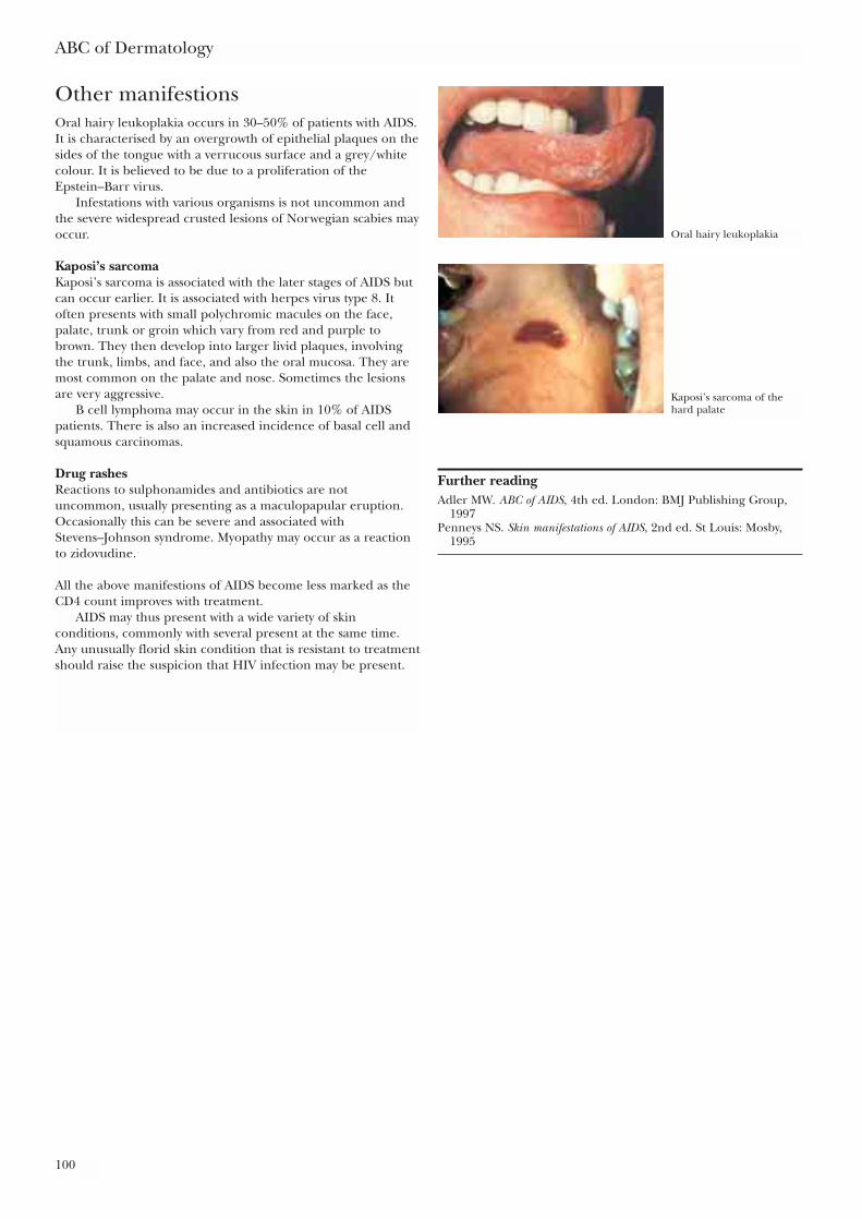
ABC of Dermatology
100
Other manifestionsOral hairy leukoplakia occurs in 30–50% of patients with AIDS.It is characterised by an overgrowth of epithelial plaques on thesides of the tongue with a verrucous surface and a grey/whitecolour. It is believed to be due to a proliferation of theEpstein–Barr virus.
Infestations with various organisms is not uncommon andthe severe widespread crusted lesions of Norwegian scabies mayoccur.
Kaposi’s sarcomaKaposi’s sarcoma is associated with the later stages of AIDS butcan occur earlier. It is associated with herpes virus type 8. Itoften presents with small polychromic macules on the face,palate, trunk or groin which vary from red and purple tobrown. They then develop into larger livid plaques, involvingthe trunk, limbs, and face, and also the oral mucosa. They aremost common on the palate and nose. Sometimes the lesionsare very aggressive.
B cell lymphoma may occur in the skin in 10% of AIDSpatients. There is also an increased incidence of basal cell andsquamous carcinomas.
Drug rashesReactions to sulphonamides and antibiotics are notuncommon, usually presenting as a maculopapular eruption.Occasionally this can be severe and associated withStevens–Johnson syndrome. Myopathy may occur as a reactionto zidovudine.
All the above manifestions of AIDS become less marked as theCD4 count improves with treatment.
AIDS may thus present with a wide variety of skinconditions, commonly with several present at the same time.Any unusually florid skin condition that is resistant to treatmentshould raise the suspicion that HIV infection may be present.
Further readingAdler MW. ABC of AIDS, 4th ed. London: BMJ Publishing Group,
1997Penneys NS. Skin manifestations of AIDS, 2nd ed. St Louis: Mosby,
1995
Oral hairy leukoplakia
Kaposi’s sarcoma of thehard palate
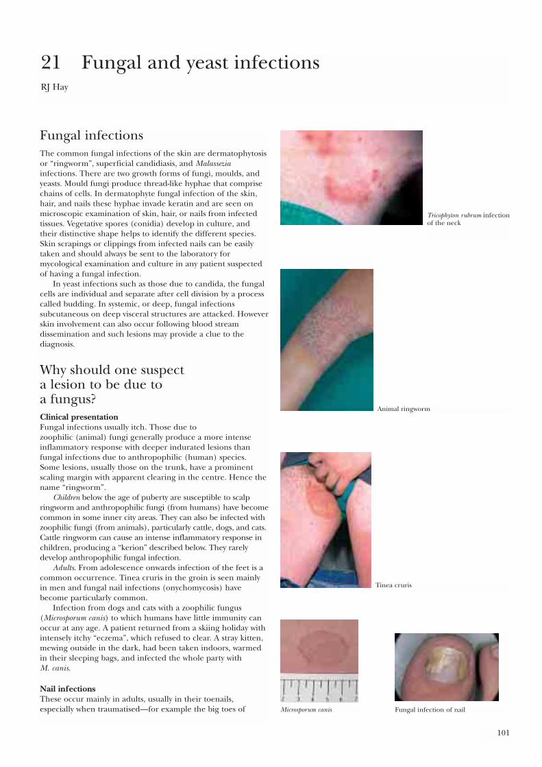
101
Fungal infectionsThe common fungal infections of the skin are dermatophytosisor “ringworm”, superficial candidiasis, and Malasseziainfections. There are two growth forms of fungi, moulds, andyeasts. Mould fungi produce thread-like hyphae that comprisechains of cells. In dermatophyte fungal infection of the skin,hair, and nails these hyphae invade keratin and are seen onmicroscopic examination of skin, hair, or nails from infectedtissues. Vegetative spores (conidia) develop in culture, andtheir distinctive shape helps to identify the different species.Skin scrapings or clippings from infected nails can be easilytaken and should always be sent to the laboratory formycological examination and culture in any patient suspectedof having a fungal infection.
In yeast infections such as those due to candida, the fungalcells are individual and separate after cell division by a processcalled budding. In systemic, or deep, fungal infectionssubcutaneous on deep visceral structures are attacked. Howeverskin involvement can also occur following blood streamdissemination and such lesions may provide a clue to thediagnosis.
Why should one suspect a lesion to be due to a fungus?Clinical presentationFungal infections usually itch. Those due to zoophilic (animal) fungi generally produce a more intenseinflammatory response with deeper indurated lesions thanfungal infections due to anthropophilic (human) species.Some lesions, usually those on the trunk, have a prominentscaling margin with apparent clearing in the centre. Hence thename “ringworm”.
Children below the age of puberty are susceptible to scalpringworm and anthropophilic fungi (from humans) have becomecommon in some inner city areas. They can also be infected withzoophilic fungi (from animals), particularly cattle, dogs, and cats.Cattle ringworm can cause an intense inflammatory response inchildren, producing a “kerion” described below. They rarelydevelop anthropophilic fungal infection.
Adults. From adolescence onwards infection of the feet is acommon occurrence. Tinea cruris in the groin is seen mainlyin men and fungal nail infections (onychomycosis) havebecome particularly common.
Infection from dogs and cats with a zoophilic fungus(Microsporum canis) to which humans have little immunity canoccur at any age. A patient returned from a skiing holiday withintensely itchy “eczema”, which refused to clear. A stray kitten,mewing outside in the dark, had been taken indoors, warmedin their sleeping bags, and infected the whole party with M. canis.
Nail infections These occur mainly in adults, usually in their toenails,especially when traumatised—for example the big toes of
21 Fungal and yeast infectionsRJ Hay
Tricophyton rubrum infectionof the neck
Animal ringworm
Tinea cruris
Microsporum canis Fungal infection of nail
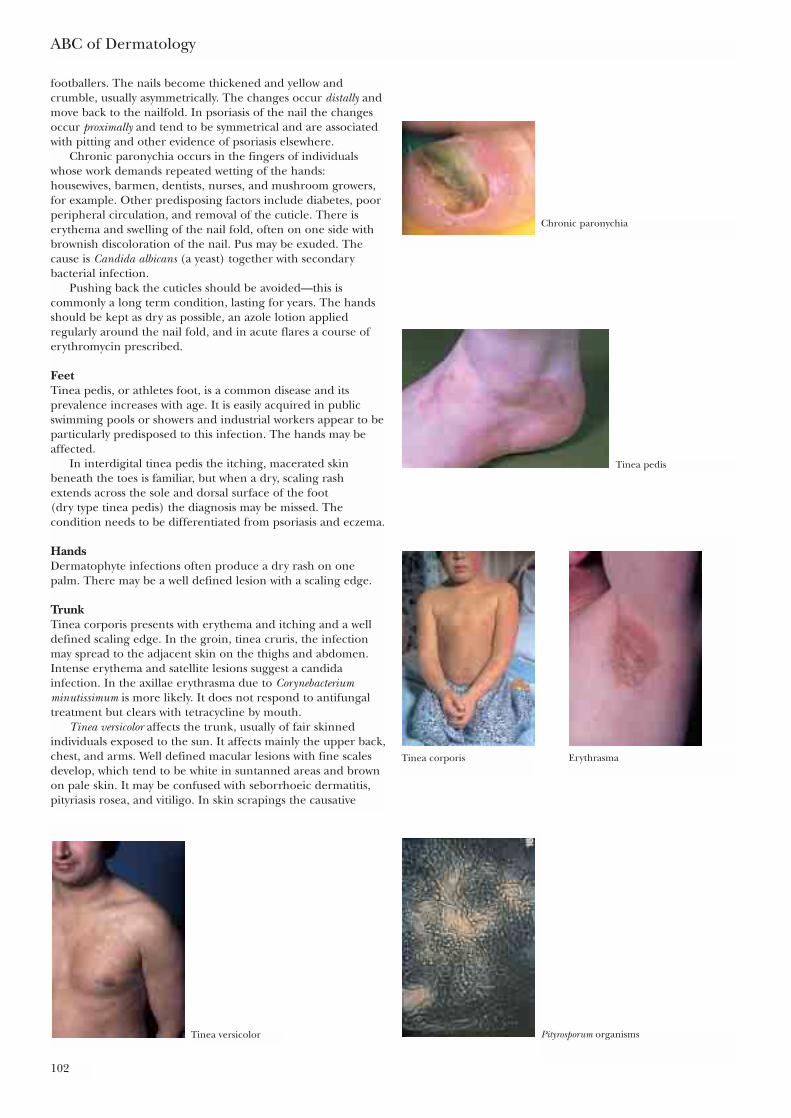
footballers. The nails become thickened and yellow andcrumble, usually asymmetrically. The changes occur distally andmove back to the nailfold. In psoriasis of the nail the changesoccur proximally and tend to be symmetrical and are associatedwith pitting and other evidence of psoriasis elsewhere.
Chronic paronychia occurs in the fingers of individualswhose work demands repeated wetting of the hands:housewives, barmen, dentists, nurses, and mushroom growers,for example. Other predisposing factors include diabetes, poorperipheral circulation, and removal of the cuticle. There iserythema and swelling of the nail fold, often on one side withbrownish discoloration of the nail. Pus may be exuded. Thecause is Candida albicans (a yeast) together with secondarybacterial infection.
Pushing back the cuticles should be avoided—this iscommonly a long term condition, lasting for years. The handsshould be kept as dry as possible, an azole lotion appliedregularly around the nail fold, and in acute flares a course oferythromycin prescribed.
FeetTinea pedis, or athletes foot, is a common disease and itsprevalence increases with age. It is easily acquired in publicswimming pools or showers and industrial workers appear to beparticularly predisposed to this infection. The hands may beaffected.
In interdigital tinea pedis the itching, macerated skinbeneath the toes is familiar, but when a dry, scaling rashextends across the sole and dorsal surface of the foot (dry type tinea pedis) the diagnosis may be missed. Thecondition needs to be differentiated from psoriasis and eczema.
HandsDermatophyte infections often produce a dry rash on onepalm. There may be a well defined lesion with a scaling edge.
TrunkTinea corporis presents with erythema and itching and a welldefined scaling edge. In the groin, tinea cruris, the infectionmay spread to the adjacent skin on the thighs and abdomen.Intense erythema and satellite lesions suggest a candidainfection. In the axillae erythrasma due to Corynebacteriumminutissimum is more likely. It does not respond to antifungaltreatment but clears with tetracycline by mouth.
Tinea versicolor affects the trunk, usually of fair skinnedindividuals exposed to the sun. It affects mainly the upper back,chest, and arms. Well defined macular lesions with fine scalesdevelop, which tend to be white in suntanned areas and brownon pale skin. It may be confused with seborrhoeic dermatitis,pityriasis rosea, and vitiligo. In skin scrapings the causative
ABC of Dermatology
102
Chronic paronychia
Tinea pedis
Tinea corporis Erythrasma
Tinea versicolor Pityrosporum organisms
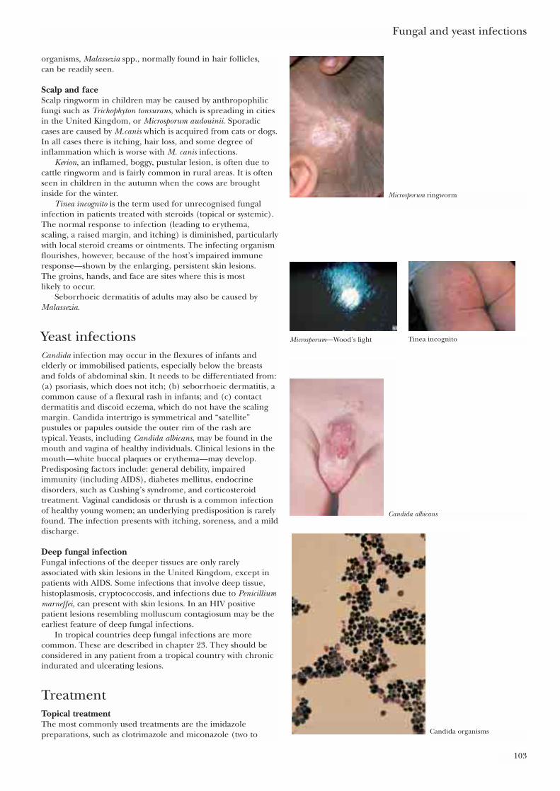
organisms, Malassezia spp., normally found in hair follicles, can be readily seen.
Scalp and faceScalp ringworm in children may be caused by anthropophilicfungi such as Trichophyton tonsurans, which is spreading in citiesin the United Kingdom, or Microsporum audouinii. Sporadiccases are caused by M.canis which is acquired from cats or dogs.In all cases there is itching, hair loss, and some degree ofinflammation which is worse with M. canis infections.
Kerion, an inflamed, boggy, pustular lesion, is often due tocattle ringworm and is fairly common in rural areas. It is oftenseen in children in the autumn when the cows are broughtinside for the winter.
Tinea incognito is the term used for unrecognised fungalinfection in patients treated with steroids (topical or systemic).The normal response to infection (leading to erythema,scaling, a raised margin, and itching) is diminished, particularlywith local steroid creams or ointments. The infecting organismflourishes, however, because of the host’s impaired immuneresponse—shown by the enlarging, persistent skin lesions. The groins, hands, and face are sites where this is most likely to occur.
Seborrhoeic dermatitis of adults may also be caused byMalassezia.
Yeast infectionsCandida infection may occur in the flexures of infants andelderly or immobilised patients, especially below the breastsand folds of abdominal skin. It needs to be differentiated from:(a) psoriasis, which does not itch; (b) seborrhoeic dermatitis, acommon cause of a flexural rash in infants; and (c) contactdermatitis and discoid eczema, which do not have the scalingmargin. Candida intertrigo is symmetrical and “satellite”pustules or papules outside the outer rim of the rash aretypical. Yeasts, including Candida albicans, may be found in themouth and vagina of healthy individuals. Clinical lesions in themouth—white buccal plaques or erythema—may develop.Predisposing factors include: general debility, impairedimmunity (including AIDS), diabetes mellitus, endocrinedisorders, such as Cushing’s syndrome, and corticosteroidtreatment. Vaginal candidosis or thrush is a common infectionof healthy young women; an underlying predisposition is rarelyfound. The infection presents with itching, soreness, and a milddischarge.
Deep fungal infectionFungal infections of the deeper tissues are only rarelyassociated with skin lesions in the United Kingdom, except inpatients with AIDS. Some infections that involve deep tissue,histoplasmosis, cryptococcosis, and infections due to Penicilliummarneffei, can present with skin lesions. In an HIV positivepatient lesions resembling molluscum contagiosum may be theearliest feature of deep fungal infections.
In tropical countries deep fungal infections are morecommon. These are described in chapter 23. They should beconsidered in any patient from a tropical country with chronicindurated and ulcerating lesions.
TreatmentTopical treatmentThe most commonly used treatments are the imidazolepreparations, such as clotrimazole and miconazole (two to
Fungal and yeast infections
103
Microsporum ringworm
Microsporum—Wood’s light Tinea incognito
Candida albicans
Candida organisms
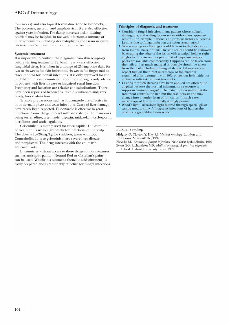
four weeks) and also topical terbinafine (one to two weeks). The polyenes, nystatin, and amphotericin B are also effectiveagainst yeast infection. For damp macerated skin dustingpowders may be helpful. In toe web infections a mixture ofmicro-organisms including dermatophytes and Gram negativebacteria may be present and both require treatment.
Systemic treatmentIt is important to confirm the diagnosis from skin scrapingsbefore starting treatment. Terbinafine is a very effectivefungicidal drug. It is taken in a dosage of 250 mg once daily fortwo to six weeks for skin infections, six weeks for finger nail orthree months for toenail infections. It is only approved for usein children in some countries. Blood monitoring is only advisedin patients with liver disease or impaired renal function.Pregnancy and lactation are relative contraindications. Therehave been reports of headaches, taste disturbances and, veryrarely, liver dysfunction.
Triazole preparations such as itraconazole are effective inboth dermatophyte and yeast infections. Cases of liver damagehave rarely been reported. Fluconazole is effective in yeastinfections. Some drugs interact with azole drugs, the main onesbeing terfenadine, astemizole, digoxin, midazolam, cyclosporin,tacrolimus, and anticoagulants.
Griseofulvin is mainly used for tinea capitis. The duration of treatment is six to eight weeks for infections of the scalp.The dose is 10–20 mg/kg for children, taken with food.Contraindications to griseofulvin are severe liver disease and porphyrias. The drug interacts with the coumarinanticoagulants.
In countries without access to these drugs simple measuressuch as antiseptic paints—Neutral Red or Castellan’s paint—can be used. Whitfield’s ointment (benzoic acid ointment) iseasily prepared and is reasonably effective for fungal infections.
ABC of Dermatology
104
Further readingMidgley G, Clayton Y, Hay RJ. Medical mycology. London and
St Louis: Mosby-Wolfe, 1997Elewski BE. Cutaneous fungal infections. New York: lgaku-Shoin, 1992Evans EG, Richardson MD. Medical mycology: A practical approach.
Oxford: Oxford University Press, 1989
Principles of diagnosis and treatment• Consider a fungal infection in any patient where isolated,
itching, dry, and scaling lesions occur without any apparentreason—for example, if there is no previous history of eczema.Lesions due to fungal infection are often asymmetrical
• Skin scrapings or clippings should be sent to the laboratoryfrom lesions, nails, or hair. The skin scales should be removedby scraping the edge of the lesion with a scalpel held at rightangles to the skin on to a piece of dark paper—transportpacks are available commercially. Clippings can be taken fromthe nails and as much material as possible should be takenfrom the nail including subungual debris. Laboratories willreport first on the direct microscopy of the materialexamined after treatment with 10% potassium hydroxide butculture results take at least two weeks
• Lesions to which steroids have been applied are often quiteatypical because the normal inflammatory response issuppressed—tinea incognito. The patient often states that thetreatment controls the itch but the rash persists and maychange into a tender form of folliculitis. In such casesmicroscopy of lesions is usually strongly positive
• Wood’s light (ultraviolet light filtered through special glass) can be used to show Microsporum infections of hair, as theyproduce a green-blue fluorescence
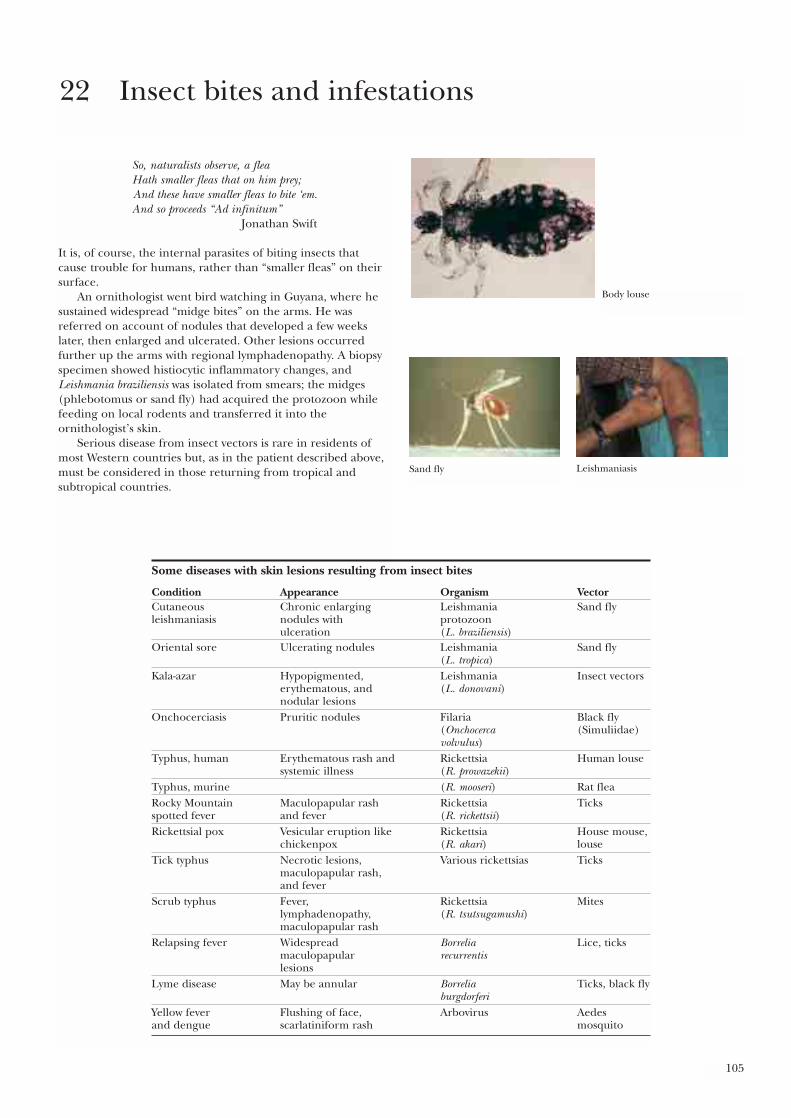
105
So, naturalists observe, a fleaHath smaller fleas that on him prey;And these have smaller fleas to bite ‘em.And so proceeds “Ad infinitum”
Jonathan Swift
It is, of course, the internal parasites of biting insects that cause trouble for humans, rather than “smaller fleas” on theirsurface.
An ornithologist went bird watching in Guyana, where hesustained widespread “midge bites” on the arms. He wasreferred on account of nodules that developed a few weekslater, then enlarged and ulcerated. Other lesions occurredfurther up the arms with regional lymphadenopathy. A biopsyspecimen showed histiocytic inflammatory changes, andLeishmania braziliensis was isolated from smears; the midges(phlebotomus or sand fly) had acquired the protozoon whilefeeding on local rodents and transferred it into theornithologist’s skin.
Serious disease from insect vectors is rare in residents ofmost Western countries but, as in the patient described above,must be considered in those returning from tropical andsubtropical countries.
22 Insect bites and infestations
Some diseases with skin lesions resulting from insect bites
Condition Appearance Organism VectorCutaneous Chronic enlarging Leishmania Sand flyleishmaniasis nodules with protozoon
ulceration (L. braziliensis)Oriental sore Ulcerating nodules Leishmania Sand fly
(L. tropica)Kala-azar Hypopigmented, Leishmania Insect vectors
erythematous, and (L. donovani)nodular lesions
Onchocerciasis Pruritic nodules Filaria Black fly(Onchocerca (Simuliidae)volvulus)
Typhus, human Erythematous rash and Rickettsia Human lousesystemic illness (R. prowazekii)
Typhus, murine (R. mooseri) Rat fleaRocky Mountain Maculopapular rash Rickettsia Ticksspotted fever and fever (R. rickettsii)Rickettsial pox Vesicular eruption like Rickettsia House mouse,
chickenpox (R. akari) louseTick typhus Necrotic lesions, Various rickettsias Ticks
maculopapular rash,and fever
Scrub typhus Fever, Rickettsia Miteslymphadenopathy, (R. tsutsugamushi)maculopapular rash
Relapsing fever Widespread Borrelia Lice, ticksmaculopapular recurrentislesions
Lyme disease May be annular Borrelia Ticks, black flyburgdorferi
Yellow fever Flushing of face, Arbovirus Aedesand dengue scarlatiniform rash mosquito
Body louse
Sand fly Leishmaniasis
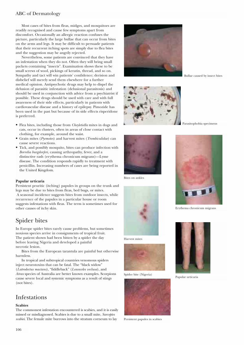
ABC of Dermatology
106
Most cases of bites from fleas, midges, and mosquitoes arereadily recognised and cause few symptoms apart fromdiscomfort. Occasionally an allergic reaction confuses thepicture, particularly the large bullae that can occur from biteson the arms and legs. It may be difficult to persuade patientsthat their recurrent itching spots are simply due to flea bitesand the suggestion may be angrily rejected.
Nevertheless, some patients are convinced that they have an infestation when they do not. Often they will bring smallpackets containing “insects”. Examination shows these to besmall screws of wool, pickings of keratin, thread, and so on.Sympathy and tact will win patients’ confidence; derision anddisbelief will merely send them elsewhere for a further medical opinion. Antipsychotic drugs may help to dispel thedelusion of parasitic infestation (delusional parasitosis) andshould be used in conjunction with advice from a psychiatrist ifpossible. These drugs should be used with care and with fullawareness of their side effects, particularly in patients withcardiovascular disease and a history of epilepsy. Pimozide hasbeen used in the past but because of its side effects risperidoneis preferred.
• Flea bites, including those from Cheyletiella mites in dogs andcats, occur in clusters, often in areas of close contact withclothing, for example, around the waist.
• Grain mites (Pyemotes) and harvest mites (Trombiculidae) cancause severe reactions.
• Tick, and possibly mosquito, bites can produce infection withBorrelia burgdorferi, causing arthropathy, fever, and adistinctive rash (erythema chronicum migrans)—Lymedisease. The condition responds rapidly to treatment withpenicillin. Increasing numbers of cases are being reported inthe United Kingdom.
Papular urticariaPersistent pruritic (itching) papules in groups on the trunk andlegs may be due to bites from fleas, bed bugs, or mites. A seasonal incidence suggests bites from outdoor insects, while
recurrence of the papules in a particular house or roomsuggests infestations with fleas. The term is sometimes used forother causes of itchy skin.
Spider bitesIn Europe spider bites rarely cause problems, but sometimesnoxious species arrive in consignments of tropical fruit. The patient shown had been bitten by a spider the day before leaving Nigeria and developed a painful necrotic lesion.
Bites from the European tarantula are painful but otherwiseharmless.
In tropical and subtropical countries venomous spiders inject neurotoxins that can be fatal. The “black widow”(Latrodectus mactans), “fiddleback” (Loxosceles veclusa), and Atrax species of Australia are better known examples. Scorpionscause severe local and systemic symptoms as a result of stings(not bites).
InfestationsScabiesThe commonest infestation encountered is scabies, and it is easilymissed or misdiagnosed. Scabies is due to a small mite, Sarcoptesscabiei. The female mite burrows into the stratum corneum to lay
Bullae caused by insect bites
Parasitophobia specimens
Bites on ankles
Erythema chronicum migrans
Harvest mites
Papular urticaria
Persistent papules in scabies
Spider bite (Nigeria)

Insect bites and infestations
107
her eggs; the male dies after completing his role of fertilisation,and the developing eggs hatch into larvae within a few days.Intense itching occurs some two weeks later, during which time extensive colonisation may have occurred. The infestation isacquired only by close contact with infected people.
DiagnosisFinding a burrow—the small (5–10 mm long) ridge, often Sshaped—can be difficult as it is often obscured by excoriationfrom scratching. Without finding a burrow, however, thediagnosis remains uncertain. Isolation of an acarus with aneedle or scalpel blade and its demonstration under themicroscope convinces the most sceptical patient. Always askwhether there are others in the patient’s household and if anyof them are itching.
Treatment10% sulphur in yellow soft paraffin is traditional, effective, andsafe. There are several more modern treatments, including25% benzyl benzoate emulsion, 0·5% malathion cream, 1%gamma benzene hexachloride (lindane) lotion, and 1%permethrin. In children benzyl benzoate should be diluted to10% and used with care as toxicity results from absorption. Ininfants over two months old permethrin or 2·5% sulphurointment can be used. Gamma benzene hexachloride shouldnot be given to children under 10 years or pregnant women inthe first trimester. Important points are:
(1) The patient should wash well: a hot bath was formerlyadvocated but it is now known that this may increaseabsorption through the skin.
(2) The lotion should be applied from the neck down,concentrating on affected areas and making sure that theaxillae, wrists, ankles, and pubic areas are included. If thereis any doubt about the thoroughness of application theprocess should be repeated in a few days.
(3) All contacts and members of the patient’s householdshould be treated at the same time.
(4) Residual papules may persist for many weeks. Topicalsteroids can be used to relieve the itching.
(5) Secondary infection as a result of scratching may need tobe treated.
DemodexDemodex folliculorum is a small mite that inhabits the human hair follicle, the eggs being deposited in the sebaceous gland. It is found on the central area of the face, chest, and neck of adults. It may have a role in the pathogenesis of rosacea, in which it may be found in large numbers. It may beassociated with a pustular eruption round the mouth andblepharitis.
Larva migransThe patient in the illustration had been on holiday at a tropicalcoastal town and regularly visited a beach frequented by dogs.Two weeks after returning to Britain he started itching on thebuttocks and subsequently noticed a linear, raised area—acondition known as larva migrans, due to the larvae of thehookworm of dogs and cats, Ancylostoma caninum. The ova are shed in the faeces and in a warm moist environment hatch into larvae that invade “dead end” hosts. They do not develop any further, so systemic disease does not occur.
Scabies—points to note• There may be very few burrows, though the patient has
widespread itching• The distribution of the infestation is characteristically the
fingers, wrists, nipples, abdomen, genitalia, buttocks, andankles
• Close personal contact is required for infestation to occur, for example, within a family, through infants in playgroups,and through regular nursing of elderly patients
• Itching may persist even after all mites have been eliminated;itching papules on the scrotum and penis are particularlypersistent
Burrows of scabies
Larva migrans
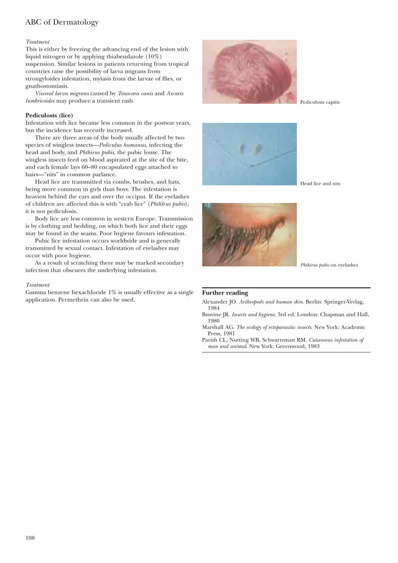
ABC of Dermatology
108
TreatmentThis is either by freezing the advancing end of the lesion withliquid nitrogen or by applying thiabendazole (10%)suspension. Similar lesions in patients returning from tropicalcountries raise the possibility of larva migrans fromstrongyloides infestation, myiasis from the larvae of flies, orgnathostomiasis.
Visceral larva migrans caused by Toxocara canis and Ascarislumbricoides may produce a transient rash.
Pediculosts (lice)Infestation with lice became less common in the postwar years,but the incidence has recently increased.
There are three areas of the body usually affected by twospecies of wingless insects—Pediculus humanus, infecting thehead and body, and Phthirus pubis, the pubic louse. Thewingless insects feed on blood aspirated at the site of the bite,and each female lays 60–80 encapsulated eggs attached tohairs—”nits” in common parlance.
Head lice are transmitted via combs, brushes, and hats,being more common in girls than boys. The infestation isheaviest behind the ears and over the occiput. If the eyelashesof children are affected this is with “crab lice” (Phthlrus pubis);it is not pediculosis.
Body lice are less common in western Europe. Transmissionis by clothing and bedding, on which both lice and their eggsmay be found in the seams. Poor hygiene favours infestation.
Pubic lice infestation occurs worldwide and is generallytransmitted by sexual contact. Infestation of eyelashes mayoccur with poor hygiene.
As a result of scratching there may be marked secondaryinfection that obscures the underlying infestation.
TreatmentGamma benzene hexachloride 1% is usually effective as a singleapplication. Permethrin can also be used.
Further readingAlexander JO. Arthropods and human skin. Berlin: Springer-Verlag,
1984Busvine JR. Insects and hygiene, 3rd ed. London: Chapman and Hall,
1980Marshall AG. The ecology of ectoparasitic insects. New York: Academic
Press, 1981Parish CL, Nutting WB, Schwartzman RM. Cutaneous infestation of
man and animal. New York: Greenwood, 1983
Pediculosis capitis
Head lice and nits
Phthirus pubis on eyelashes
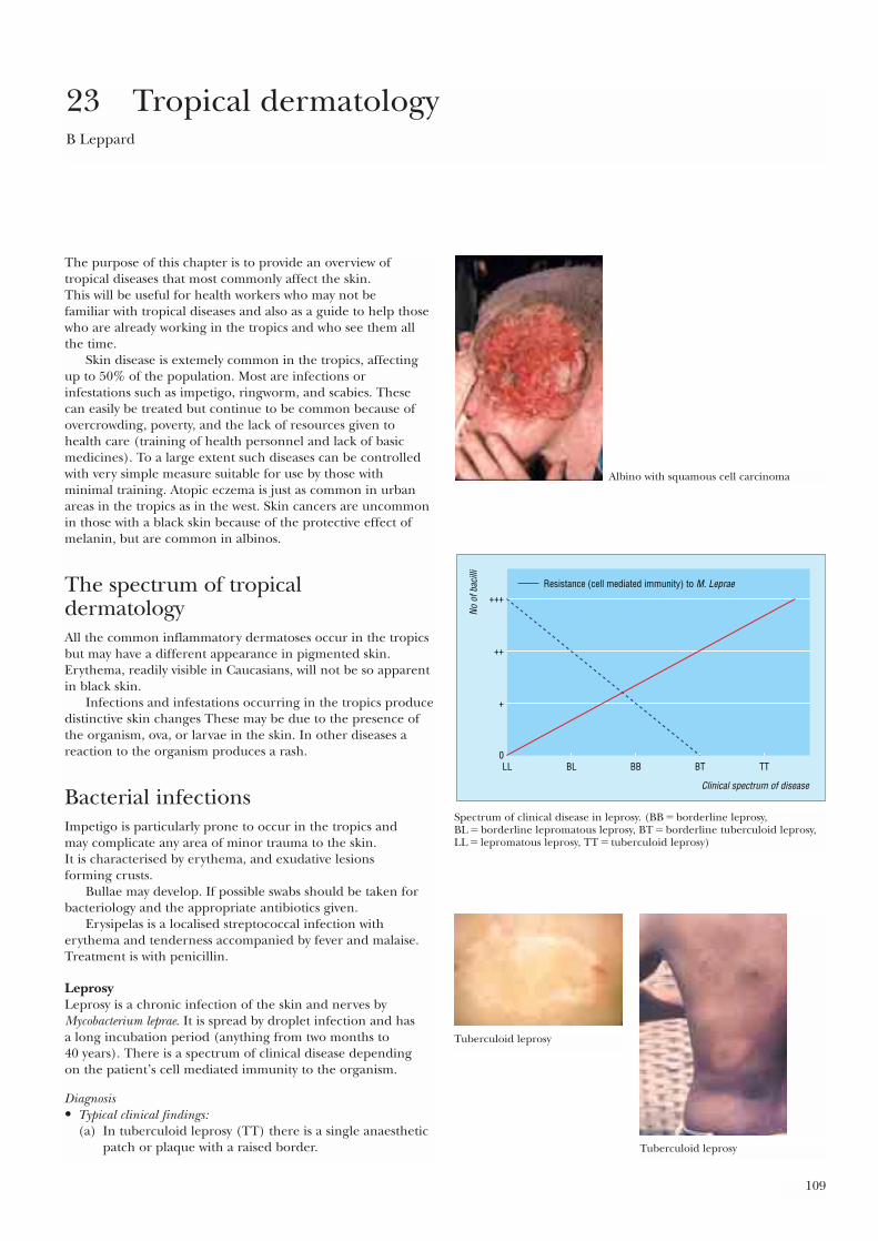
109
The purpose of this chapter is to provide an overview oftropical diseases that most commonly affect the skin. This will be useful for health workers who may not be familiar with tropical diseases and also as a guide to help thosewho are already working in the tropics and who see them allthe time.
Skin disease is extemely common in the tropics, affectingup to 50% of the population. Most are infections orinfestations such as impetigo, ringworm, and scabies. These can easily be treated but continue to be common because ofovercrowding, poverty, and the lack of resources given tohealth care (training of health personnel and lack of basicmedicines). To a large extent such diseases can be controlledwith very simple measure suitable for use by those withminimal training. Atopic eczema is just as common in urbanareas in the tropics as in the west. Skin cancers are uncommonin those with a black skin because of the protective effect ofmelanin, but are common in albinos.
The spectrum of tropical dermatologyAll the common inflammatory dermatoses occur in the tropicsbut may have a different appearance in pigmented skin.Erythema, readily visible in Caucasians, will not be so apparentin black skin.
Infections and infestations occurring in the tropics producedistinctive skin changes These may be due to the presence ofthe organism, ova, or larvae in the skin. In other diseases areaction to the organism produces a rash.
Bacterial infectionsImpetigo is particularly prone to occur in the tropics and may complicate any area of minor trauma to the skin. It is characterised by erythema, and exudative lesions forming crusts.
Bullae may develop. If possible swabs should be taken forbacteriology and the appropriate antibiotics given.
Erysipelas is a localised streptococcal infection witherythema and tenderness accompanied by fever and malaise.Treatment is with penicillin.
LeprosyLeprosy is a chronic infection of the skin and nerves byMycobacterium leprae. It is spread by droplet infection and has a long incubation period (anything from two months to 40 years). There is a spectrum of clinical disease depending on the patient’s cell mediated immunity to the organism.
Diagnosis• Typical clinical findings:
(a) In tuberculoid leprosy (TT) there is a single anaestheticpatch or plaque with a raised border.
23 Tropical dermatologyB Leppard
Albino with squamous cell carcinoma
Clinical spectrum of disease
No o
f bac
illi
LL BL BB
Resistance (cell mediated immunity) to M. Leprae
BT TT0
++
+++
+
Spectrum of clinical disease in leprosy. (BB � borderline leprosy,BL � borderline lepromatous leprosy, BT � borderline tuberculoid leprosy,LL � lepromatous leprosy, TT � tuberculoid leprosy)
Tuberculoid leprosy
Tuberculoid leprosy
ABC of Dermatology
110
(b) In lepromatous leprosy (LL) there are widespreadsymmetrical shiny papules, nodules, and plaques whichare not anaesthetic.
(c) In borderline leprosy (BT, BB, BL) there are varyingnumbers of lesions, few in BT and numerous in BL.They may be widespread but are asymmetrical.
(d) Palpably enlarged cutaneous nerves (great auricularnerve in the neck, the superficial branch of the radialnerve at the wrist, the ulnar nerve at the elbow, thelateral popliteal nerve at the knee, and the sural nerveon the lower leg).
(e) Glove and stocking sensory loss causing blisters, ulcers orboth on anaesthetic fingers or toes.
(f) Deformity due to invasion of the peripheral nerves withleprosy bacilli, a leprosy reaction or recurrent trauma toanaesthetic limbs.
• Slit skin smears measure the numbers of bacilli in the skin(Bacterial Index (BI)) and the % of these that are living(Morphological Index (MI)).
TreatmentPaucibacillary leprosy (BI of 0 or 1�):
Rifampicin 600 mg once a month (supervised) for Dapsone 100 mg daily six months
Multibacillary leprosy (BI of 2� or more):Rifampicin 600 mg once a month (supervised)Clofazamine 300 mg once a month
(supervised) for two yearsClofazamine 50 mg/dayDapsone 100 mg/day
Cutaneous leishmaniasisCutaneous leishmaniasis is due to the protozoa Leishmaniatropica, and is transmitted by the bite of a sandfly. There is a spectrum of disease depending on the patient’s immunity.
Acute leishmaniasisA red nodule like a boil occurs at the site of the bite. It enlarges, may or may not ulcerate, and heals spontaneouslyafter about one year leaving a cribriform scar.
Chronic leishmaniasisIn a patient with good cell mediated immunity, after the acuteleishmaniasis has healed, new granulomata appear at the edgeof the scar; these do not heal spontaneously.
Diffuse cutaneous leishmaniasisThis is leishmaniasis in a patient with no immunity to theorganism (equivalent to lepromatous leprosy). Extensive skinnodules occur that are full of organisms.
TreatmentIntramuscular injections of sodium stibogluconate 10 mg/kgbody weight daily until healing occurs.
Superficial fungal infectionsThe same fungal infections of the epidermis occur in the tropicsas in temperate climates only more so—heat and occlusion ofclothing leading to maceration of the skin in which fungi thrive,so expect to see more florid lesions. There are also many fungal infections that are specifically found in the tropics. These include tinea imbricata, tinea nigra, piedra, and favus.
Bacterial Index (BI)0 � No bacilli seen
1� � 1–10 bacilli in 100 oil immersion fields2� � 1–10 bacilli in 10 oil immersion fields3� � 1–10 bacilli in 1 oil immersion field4� � 10–100 bacilli in an average oil immersion field5� � 100–1000 bacilli in an average oil immersion field6� � �1000 bacilli in an average oil immersion field
Lepromatous leprosy Borderline leprosy
Borderline tuberculoid leprosy
Acute leishmaniasis Chronic leishmaniasis
Diffuse cutaneous leishmaniasis
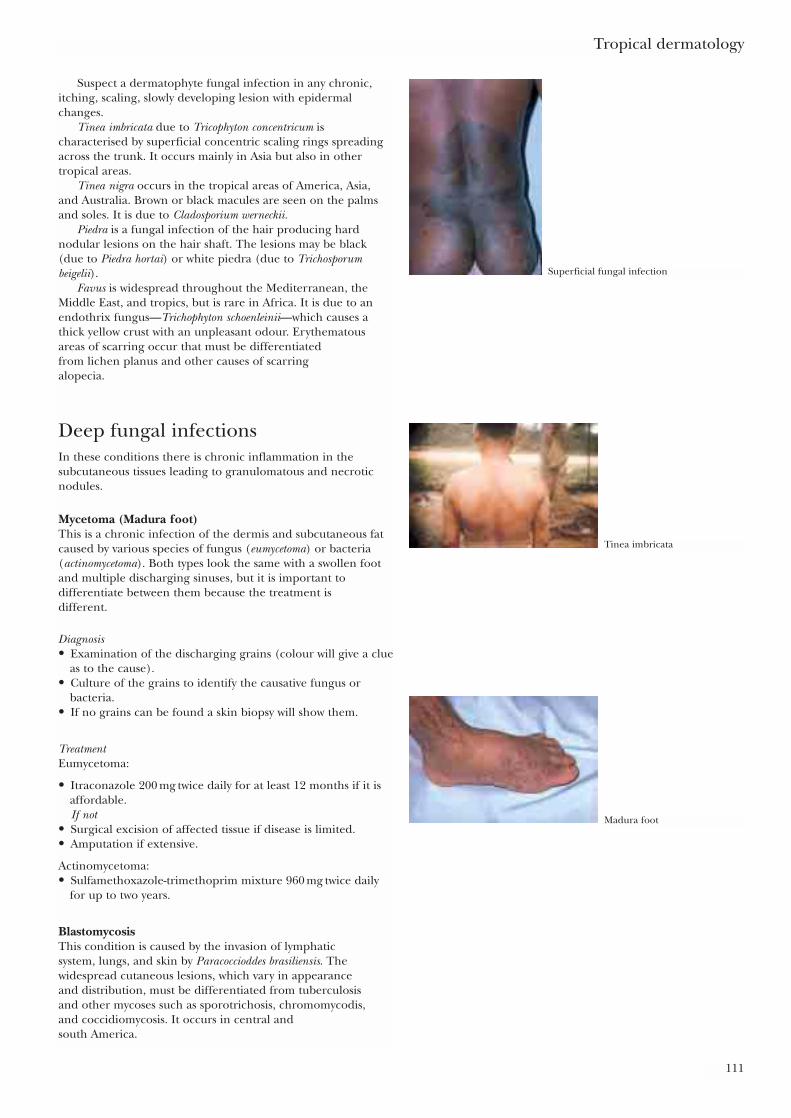
Tropical dermatology
111
Suspect a dermatophyte fungal infection in any chronic,itching, scaling, slowly developing lesion with epidermalchanges.
Tinea imbricata due to Tricophyton concentricum ischaracterised by superficial concentric scaling rings spreadingacross the trunk. It occurs mainly in Asia but also in othertropical areas.
Tinea nigra occurs in the tropical areas of America, Asia,and Australia. Brown or black macules are seen on the palmsand soles. It is due to Cladosporium werneckii.
Piedra is a fungal infection of the hair producing hardnodular lesions on the hair shaft. The lesions may be black(due to Piedra hortai) or white piedra (due to Trichosporumbeigelii).
Favus is widespread throughout the Mediterranean, theMiddle East, and tropics, but is rare in Africa. It is due to anendothrix fungus—Trichophyton schoenleinii—which causes athick yellow crust with an unpleasant odour. Erythematousareas of scarring occur that must be differentiated from lichen planus and other causes of scarring alopecia.
Deep fungal infectionsIn these conditions there is chronic inflammation in thesubcutaneous tissues leading to granulomatous and necroticnodules.
Mycetoma (Madura foot)This is a chronic infection of the dermis and subcutaneous fatcaused by various species of fungus (eumycetoma) or bacteria(actinomycetoma). Both types look the same with a swollen footand multiple discharging sinuses, but it is important todifferentiate between them because the treatment is different.
Diagnosis• Examination of the discharging grains (colour will give a clue
as to the cause).• Culture of the grains to identify the causative fungus or
bacteria.• If no grains can be found a skin biopsy will show them.
TreatmentEumycetoma:
• Itraconazole 200 mg twice daily for at least 12 months if it isaffordable.If not
• Surgical excision of affected tissue if disease is limited.• Amputation if extensive.
Actinomycetoma:• Sulfamethoxazole-trimethoprim mixture 960 mg twice daily
for up to two years.
BlastomycosisThis condition is caused by the invasion of lymphatic system, lungs, and skin by Paracoccioddes brasiliensis. Thewidespread cutaneous lesions, which vary in appearance and distribution, must be differentiated from tuberculosis and other mycoses such as sporotrichosis, chromomycodis, and coccidiomycosis. It occurs in central and south America.
Superficial fungal infection
Tinea imbricata
Madura foot
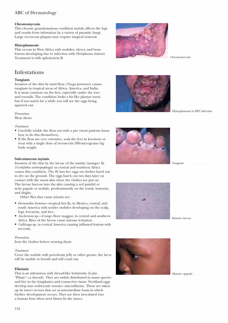
ABC of Dermatology
112
ChromomycosisThis chronic granulomatous condition mainly affects the legsand results from infestation by a variety of parasitic fungi. Large verrucous plaques may require surgical removal.
HistoplasmosisThis occurs in West Africa with nodules, ulcers, and bonelesions developing due to infection with Histoplasma duboisii.Treatment is with aphotericin B.
InfestationsTungiasisInvasion of the skin by sand fleas (Tunga penetrans) causestungiasis in tropical areas of Africa, America, and India. It is most common on the feet, especially under the toes and toenails. The condition looks a bit like plantar warts, but if you watch for a while you will see the eggs being squirted out.
PreventionWear shoes.
Treatment• Carefully winkle the fleas out with a pin (most patients know
how to do this themselves).• If the fleas are very extensive, soak the feet in kerosene or
treat with a single dose of ivermectin 200 micrograms/kgbody weight.
Subcutaneous myiasisInvasion of the skin by the larvae of the tumbu (mango) fly(Cordylobia anthropophaga) in central and southern Africa causes this condition. The fly lays her eggs on clothes layed outto dry on the ground. The eggs hatch out two days later oncontact with the warm skin when the clothes are put on. The larvae burrow into the skin causing a red painful or itchy papule or nodule, predominantly on the trunk, buttocks,and thighs.
Other flies that cause miyasis are:
• Dermatobia hominis—tropical bot fly, in Mexico, central, andsouth America with tender nodules developing on the scalp,legs, forearms, and face.
• Aucheronia sp.—Congo floor maggot, in central and southernAfrica. Bites of the larvae cause intense irritation.
• Callitroga sp. in central America causing inflamed lesions withnecrosis.
PreventionIron the clothes before wearing them.
TreatmentCover the nodule with petroleum jelly or other grease; the larvawill be unable to breath and will crawl out.
FilariasisThis is an infestation with thread-like helminths (Latin“Filum”—a thread). They are widely distributed in many speciesand live in the lymphatics and connective tissue. Fertilised eggsdevelop into embryonic worms—microfilariae. These are takenup by insect vectors that act as intermediate hosts in whichfurther development occurs. They are then inoculated into a human host when next bitten by the insect.
Chromomycosis
Histoplasmosis in HIV infection
Tungiasis
Myiasis—larvae
Myiasis—papule
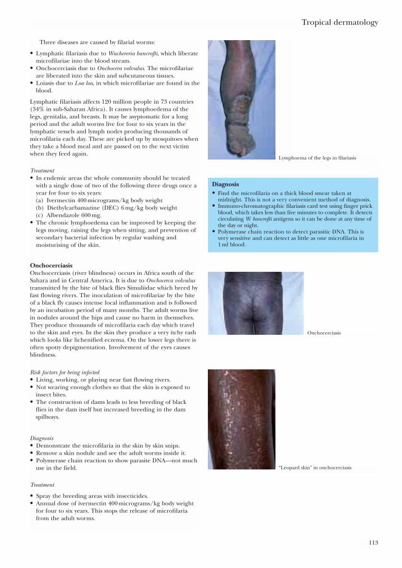
Tropical dermatology
113
Three diseases are caused by filarial worms:
• Lymphatic filariasis due to Wuchereria bancrofti, which liberatemicrofilariae into the blood stream.
• Onchocerciasis due to Onchocera volvulus. The microfilariaeare liberated into the skin and subcutaneous tissues.
• Loiasis due to Loa loa, in which microfilariae are found in theblood.
Lymphatic filariasis affects 120 million people in 73 countries(34% in sub-Saharan Africa). It causes lymphoedema of thelegs, genitalia, and breasts. It may be asyptomatic for a longperiod and the adult worms live for four to six years in thelymphatic vessels and lymph nodes producing thousands ofmicrofilaria each day. These are picked up by mosquitoes whenthey take a blood meal and are passed on to the next victimwhen they feed again.
Treatment• In endemic areas the whole community should be treated
with a single dose of two of the following three drugs once ayear for four to six years:(a) Ivermectin 400 micrograms/kg body weight(b) Diethylcarbamazine (DEC) 6 mg/kg body weight(c) Albendazole 600 mg.
• The chronic lymphoedema can be improved by keeping thelegs moving, raising the legs when sitting, and prevention ofsecondary bacterial infection by regular washing andmoisturising of the skin.
OnchocerciasisOnchocerciasis (river blindness) occurs in Africa south of theSahara and in Central America. It is due to Onchocerca volvulustransmitted by the bite of black flies Simuliidae which breed byfast flowing rivers. The inoculation of microfilariae by the biteof a black fly causes intense local inflammation and is followedby an incubation period of many months. The adult worms livein nodules around the hips and cause no harm in themselves.They produce thousands of microfilaria each day which travelto the skin and eyes. In the skin they produce a very itchy rashwhich looks like lichenified eczema. On the lower legs there isoften spotty depigmentation. Involvement of the eyes causesblindness.
Risk factors for being infected• Living, working, or playing near fast flowing rivers.• Not wearing enough clothes so that the skin is exposed to
insect bites.• The construction of dams leads to less breeding of black
flies in the dam itself but increased breeding in the damspillways.
Diagnosis• Demonstrate the microfilaria in the skin by skin snips.• Remove a skin nodule and see the adult worms inside it.• Polymerase chain reaction to show parasite DNA—not much
use in the field.
Treatment
• Spray the breeding areas with insecticides.• Annual dose of ivermectin 400 micrograms/kg body weight
for four to six years. This stops the release of microfilariafrom the adult worms.
Diagnosis• Find the microfilaria on a thick blood smear taken at
midnight. This is not a very convenient method of diagnosis.• Immuno-chromatographic filariasis card test using finger prick
blood, which takes less than five minutes to complete. It detectscirculating W. bancrofti antigens so it can be done at any time ofthe day or night.
• Polymerase chain reaction to detect parasitic DNA. This isvery sensitive and can detect as little as one microfilaria in 1 ml blood.
Lymphoema of the legs in filariasis
Onchocerciasis
“Leopard skin” in onchocerciasis
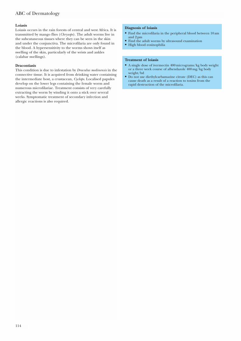
ABC of Dermatology
114
LoiasisLoiasis occurs in the rain forests of central and west Africa. It istransmitted by mango flies (Chrysops). The adult worms live inthe subcutaneous tissues where they can be seen in the skinand under the conjunctiva. The microfilaria are only found inthe blood. A hypersensitivity to the worms shows itself asswelling of the skin, particularly of the wrists and ankles(calabar swellings).
DracontiasisThis condition is due to infestation by Draculus medinensis in theconnective tissue. It is acquired from drinking water containingthe intermediate host, a crustacean, Cyclops. Localised papulesdevelop on the lower legs containing the female worm andnumerous microfilariae. Treatment consists of very carefullyextracting the worm by winding it onto a stick over severalweeks. Symptomatic treatment of secondary infection andallergic reactions is also required.
Diagnosis of loiasis • Find the microfilaria in the peripheral blood between 10 am
and 2 pm• Find the adult worms by ultrasound examination• High blood eosinophilia
Treatment of loiasis • A single dose of ivermectin 400 micrograms/kg body weight
or a three week course of albendazole 400 mg/kg bodyweight/bd
• Do not use diethylcarbamazine citrate (DEC) as this cancause death as a result of a reaction to toxins from therapid destruction of the microfilaria.
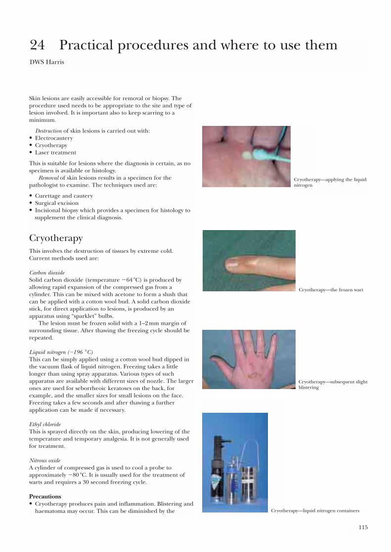
115
Skin lesions are easily accessible for removal or biopsy. Theprocedure used needs to be appropriate to the site and type oflesion involved. It is important also to keep scarring to aminimum.
Destruction of skin lesions is carried out with:• Electrocautery• Cryotherapy• Laser treatment
This is suitable for lesions where the diagnosis is certain, as nospecimen is available or histology.
Removal of skin lesions results in a specimen for thepathologist to examine. The techniques used are:
• Curettage and cautery• Surgical excision• Incisional biopsy which provides a specimen for histology to
supplement the clinical diagnosis.
CryotherapyThis involves the destruction of tissues by extreme cold.Current methods used are:
Carbon dioxideSolid carbon dioxide (temperature �64 �C) is produced byallowing rapid expansion of the compressed gas from acylinder. This can be mixed with acetone to form a slush thatcan be applied with a cotton wool bud. A solid carbon dioxidestick, for direct application to lesions, is produced by anapparatus using “sparklet” bulbs.
The lesion must be frozen solid with a 1–2 mm margin ofsurrounding tissue. After thawing the freezing cycle should berepeated.
Liquid nitrogen (�196 �C)This can be simply applied using a cotton wool bud dipped inthe vacuum flask of liquid nitrogen. Freezing takes a littlelonger than using spray apparatus. Various types of suchapparatus are available with different sizes of nozzle. The largerones are used for seborrheoic keratoses on the back, forexample, and the smaller sizes for small lesions on the face.Freezing takes a few seconds and after thawing a furtherapplication can be made if necessary.
Ethyl chlorideThis is sprayed directly on the skin, producing lowering of thetemperature and temporary analgesia. It is not generally usedfor treatment.
Nitrous oxideA cylinder of compressed gas is used to cool a probe toapproximately �80 �C. It is usually used for the treatment ofwarts and requires a 30 second freezing cycle.
Precautions• Cryotherapy produces pain and inflammation. Blistering and
haematoma may occur. This can be diminished by the
24 Practical procedures and where to use themDWS Harris
Cryotherapy—applying the liquidnitrogen
Cryotherapy—the frozen wart
Cryotherapy—subsequent slightblistering
Cryotherapy—liquid nitrogen containers
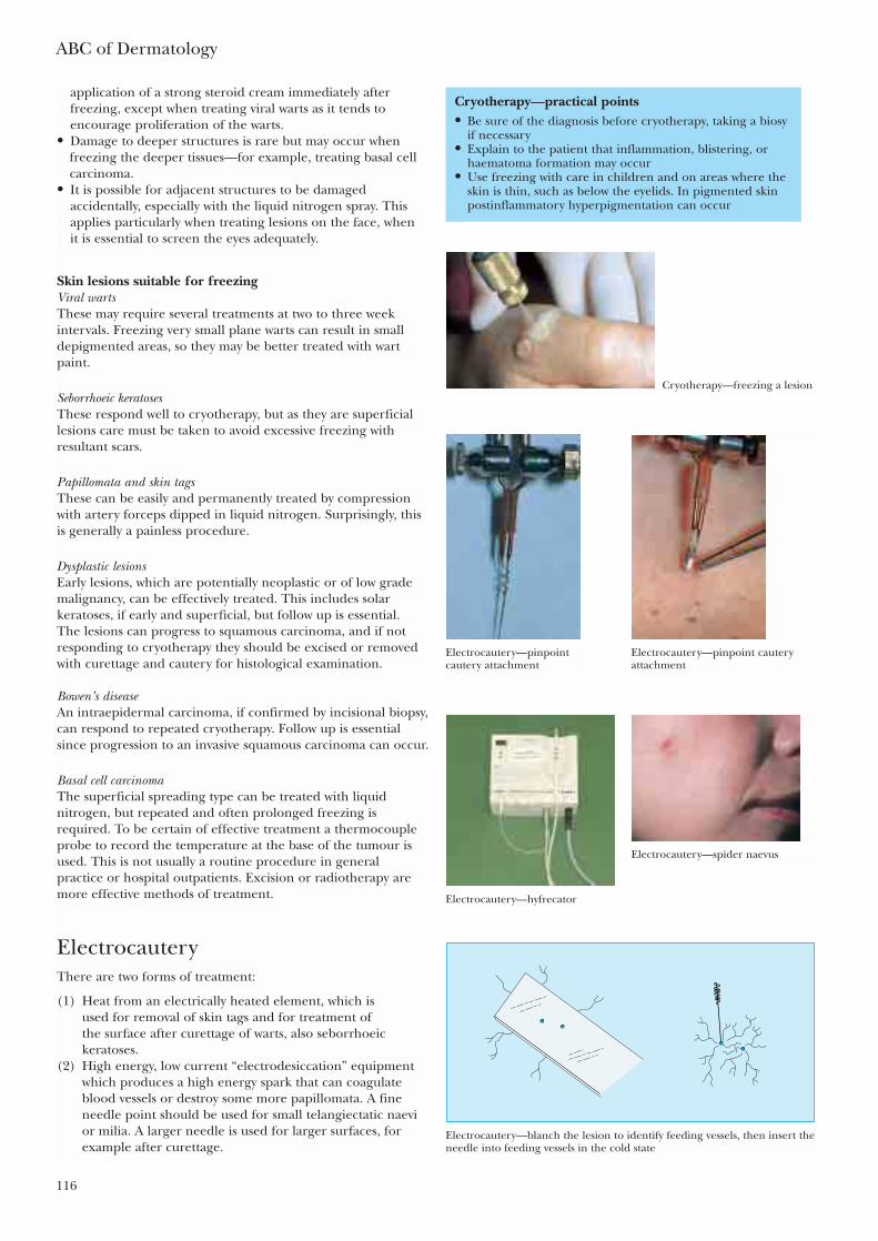
ABC of Dermatology
116
application of a strong steroid cream immediately afterfreezing, except when treating viral warts as it tends toencourage proliferation of the warts.
• Damage to deeper structures is rare but may occur whenfreezing the deeper tissues—for example, treating basal cellcarcinoma.
• It is possible for adjacent structures to be damagedaccidentally, especially with the liquid nitrogen spray. Thisapplies particularly when treating lesions on the face, when it is essential to screen the eyes adequately.
Skin lesions suitable for freezingViral wartsThese may require several treatments at two to three weekintervals. Freezing very small plane warts can result in smalldepigmented areas, so they may be better treated with wartpaint.
Seborrhoeic keratosesThese respond well to cryotherapy, but as they are superficiallesions care must be taken to avoid excessive freezing withresultant scars.
Papillomata and skin tagsThese can be easily and permanently treated by compressionwith artery forceps dipped in liquid nitrogen. Surprisingly, thisis generally a painless procedure.
Dysplastic lesionsEarly lesions, which are potentially neoplastic or of low grademalignancy, can be effectively treated. This includes solarkeratoses, if early and superficial, but follow up is essential. The lesions can progress to squamous carcinoma, and if notresponding to cryotherapy they should be excised or removedwith curettage and cautery for histological examination.
Bowen’s diseaseAn intraepidermal carcinoma, if confirmed by incisional biopsy,can respond to repeated cryotherapy. Follow up is essentialsince progression to an invasive squamous carcinoma can occur.
Basal cell carcinomaThe superficial spreading type can be treated with liquidnitrogen, but repeated and often prolonged freezing isrequired. To be certain of effective treatment a thermocoupleprobe to record the temperature at the base of the tumour isused. This is not usually a routine procedure in generalpractice or hospital outpatients. Excision or radiotherapy aremore effective methods of treatment.
ElectrocauteryThere are two forms of treatment:
(1) Heat from an electrically heated element, which is used for removal of skin tags and for treatment of the surface after curettage of warts, also seborrhoeickeratoses.
(2) High energy, low current “electrodesiccation” equipmentwhich produces a high energy spark that can coagulateblood vessels or destroy some more papillomata. A fineneedle point should be used for small telangiectatic naevior milia. A larger needle is used for larger surfaces, forexample after curettage.
Cryotherapy—freezing a lesion
Electrocautery—pinpointcautery attachment
Electrocautery—pinpoint cauteryattachment
Electrocautery—hyfrecator
Electrocautery—spider naevus
Electrocautery—blanch the lesion to identify feeding vessels, then insert theneedle into feeding vessels in the cold state
Cryotherapy—practical points• Be sure of the diagnosis before cryotherapy, taking a biosy
if necessary• Explain to the patient that inflammation, blistering, or
haematoma formation may occur• Use freezing with care in children and on areas where the
skin is thin, such as below the eyelids. In pigmented skinpostinflammatory hyperpigmentation can occur
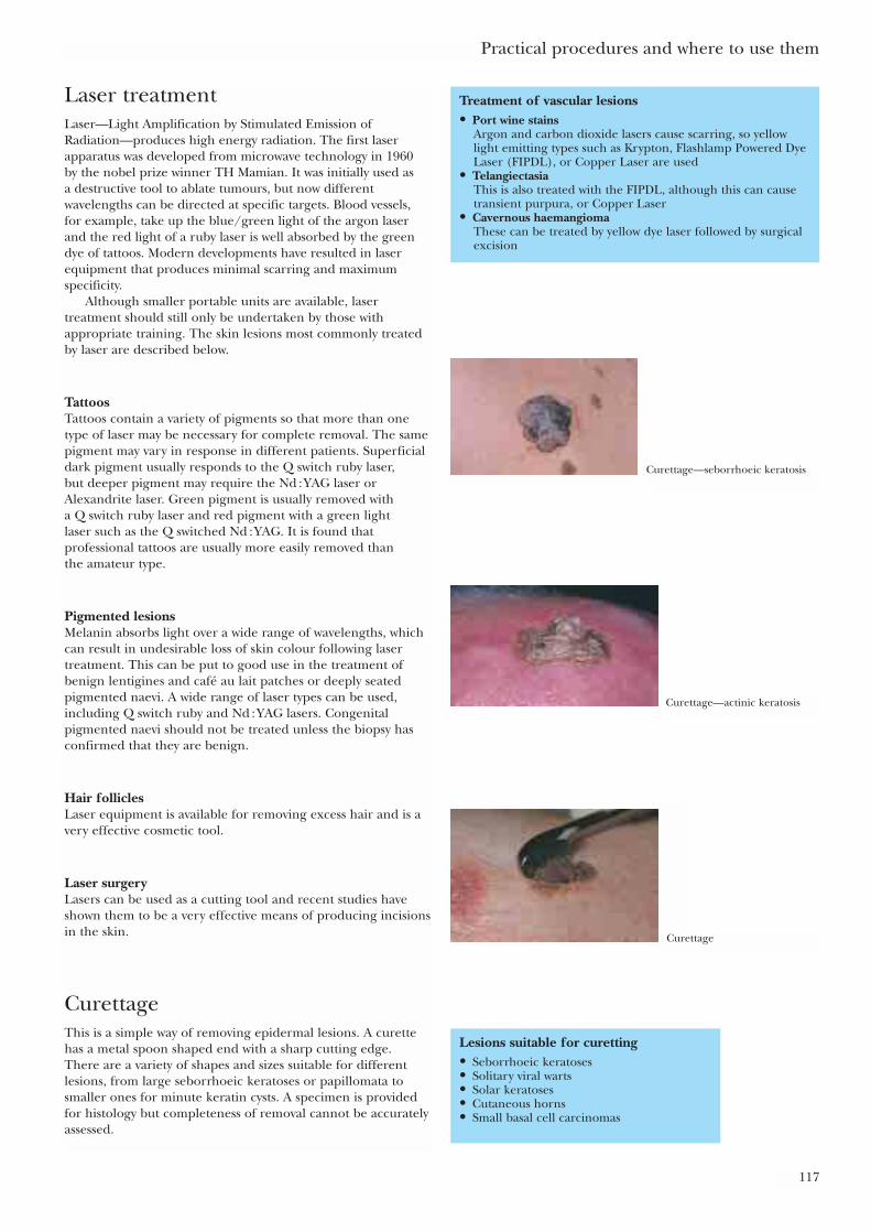
Practical procedures and where to use them
117
Laser treatmentLaser—Light Amplification by Stimulated Emission ofRadiation—produces high energy radiation. The first laserapparatus was developed from microwave technology in 1960 by the nobel prize winner TH Mamian. It was initially used as a destructive tool to ablate tumours, but now differentwavelengths can be directed at specific targets. Blood vessels, for example, take up the blue/green light of the argon laserand the red light of a ruby laser is well absorbed by the greendye of tattoos. Modern developments have resulted in laserequipment that produces minimal scarring and maximumspecificity.
Although smaller portable units are available, lasertreatment should still only be undertaken by those withappropriate training. The skin lesions most commonly treatedby laser are described below.
TattoosTattoos contain a variety of pigments so that more than onetype of laser may be necessary for complete removal. The samepigment may vary in response in different patients. Superficialdark pigment usually responds to the Q switch ruby laser, but deeper pigment may require the Nd : YAG laser orAlexandrite laser. Green pigment is usually removed with a Q switch ruby laser and red pigment with a green light laser such as the Q switched Nd : YAG. It is found thatprofessional tattoos are usually more easily removed than the amateur type.
Pigmented lesionsMelanin absorbs light over a wide range of wavelengths, whichcan result in undesirable loss of skin colour following lasertreatment. This can be put to good use in the treatment ofbenign lentigines and café au lait patches or deeply seatedpigmented naevi. A wide range of laser types can be used,including Q switch ruby and Nd : YAG lasers. Congenitalpigmented naevi should not be treated unless the biopsy hasconfirmed that they are benign.
Hair folliclesLaser equipment is available for removing excess hair and is avery effective cosmetic tool.
Laser surgeryLasers can be used as a cutting tool and recent studies haveshown them to be a very effective means of producing incisionsin the skin.
CurettageThis is a simple way of removing epidermal lesions. A curettehas a metal spoon shaped end with a sharp cutting edge. There are a variety of shapes and sizes suitable for differentlesions, from large seborrhoeic keratoses or papillomata tosmaller ones for minute keratin cysts. A specimen is providedfor histology but completeness of removal cannot be accuratelyassessed.
Treatment of vascular lesions• Port wine stains
Argon and carbon dioxide lasers cause scarring, so yellowlight emitting types such as Krypton, Flashlamp Powered DyeLaser (FIPDL), or Copper Laser are used
• TelangiectasiaThis is also treated with the FIPDL, although this can causetransient purpura, or Copper Laser
• Cavernous haemangiomaThese can be treated by yellow dye laser followed by surgicalexcision
Curettage—seborrhoeic keratosis
Curettage—actinic keratosis
Curettage
Lesions suitable for curetting• Seborrhoeic keratoses• Solitary viral warts• Solar keratoses• Cutaneous horns• Small basal cell carcinomas
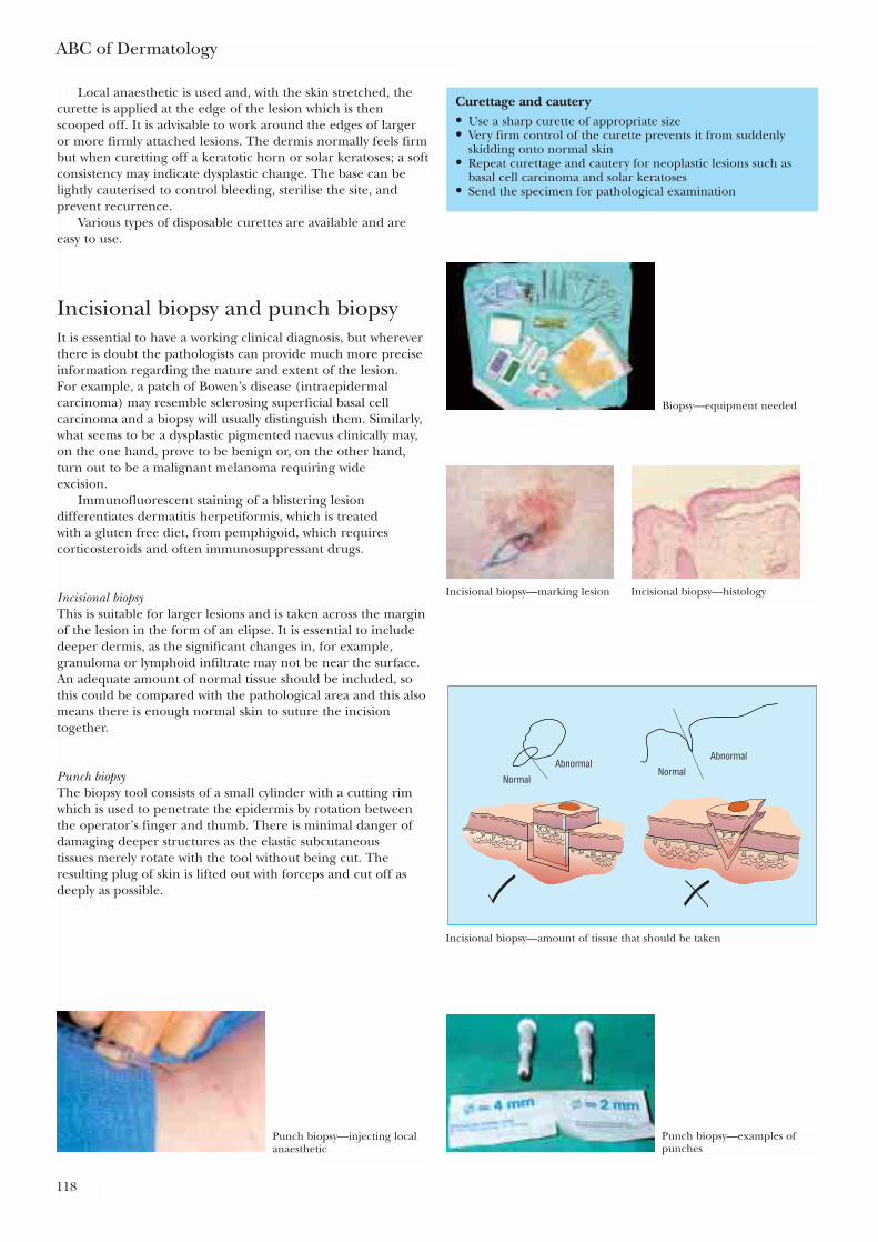
ABC of Dermatology
118
Local anaesthetic is used and, with the skin stretched, thecurette is applied at the edge of the lesion which is thenscooped off. It is advisable to work around the edges of largeror more firmly attached lesions. The dermis normally feels firmbut when curetting off a keratotic horn or solar keratoses; a softconsistency may indicate dysplastic change. The base can belightly cauterised to control bleeding, sterilise the site, andprevent recurrence.
Various types of disposable curettes are available and areeasy to use.
Incisional biopsy and punch biopsyIt is essential to have a working clinical diagnosis, but whereverthere is doubt the pathologists can provide much more preciseinformation regarding the nature and extent of the lesion. For example, a patch of Bowen’s disease (intraepidermalcarcinoma) may resemble sclerosing superficial basal cellcarcinoma and a biopsy will usually distinguish them. Similarly,what seems to be a dysplastic pigmented naevus clinically may,on the one hand, prove to be benign or, on the other hand,turn out to be a malignant melanoma requiring wide excision.
Immunofluorescent staining of a blistering lesiondifferentiates dermatitis herpetiformis, which is treated with a gluten free diet, from pemphigoid, which requirescorticosteroids and often immunosuppressant drugs.
Incisional biopsyThis is suitable for larger lesions and is taken across the marginof the lesion in the form of an elipse. It is essential to includedeeper dermis, as the significant changes in, for example,granuloma or lymphoid infiltrate may not be near the surface.An adequate amount of normal tissue should be included, sothis could be compared with the pathological area and this alsomeans there is enough normal skin to suture the incisiontogether.
Punch biopsy The biopsy tool consists of a small cylinder with a cutting rimwhich is used to penetrate the epidermis by rotation betweenthe operator’s finger and thumb. There is minimal danger ofdamaging deeper structures as the elastic subcutaneous tissues merely rotate with the tool without being cut. Theresulting plug of skin is lifted out with forceps and cut off asdeeply as possible.
Curettage and cautery• Use a sharp curette of appropriate size• Very firm control of the curette prevents it from suddenly
skidding onto normal skin• Repeat curettage and cautery for neoplastic lesions such as
basal cell carcinoma and solar keratoses• Send the specimen for pathological examination
Biopsy—equipment needed
Incisional biopsy—marking lesion Incisional biopsy—histology
Normal
AbnormalNormal
Abnormal
Incisional biopsy—amount of tissue that should be taken
Punch biopsy—examples ofpunches
Punch biopsy—injecting localanaesthetic
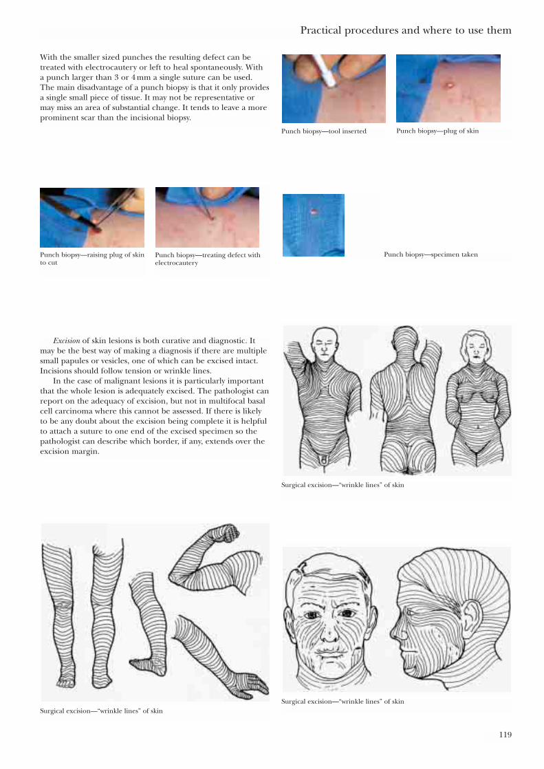
Practical procedures and where to use them
119
With the smaller sized punches the resulting defect can betreated with electrocautery or left to heal spontaneously. With a punch larger than 3 or 4 mm a single suture can be used. The main disadvantage of a punch biopsy is that it only providesa single small piece of tissue. It may not be representative ormay miss an area of substantial change. It tends to leave a moreprominent scar than the incisional biopsy.
Punch biopsy—tool inserted Punch biopsy—plug of skin
Punch biopsy—raising plug of skinto cut
Punch biopsy—treating defect withelectrocautery
Punch biopsy—specimen taken
Excision of skin lesions is both curative and diagnostic. Itmay be the best way of making a diagnosis if there are multiplesmall papules or vesicles, one of which can be excised intact.Incisions should follow tension or wrinkle lines.
In the case of malignant lesions it is particularly importantthat the whole lesion is adequately excised. The pathologist canreport on the adequacy of excision, but not in multifocal basalcell carcinoma where this cannot be assessed. If there is likelyto be any doubt about the excision being complete it is helpfulto attach a suture to one end of the excised specimen so thepathologist can describe which border, if any, extends over theexcision margin.
Surgical excision—“wrinkle lines” of skin
Surgical excision—“wrinkle lines” of skinSurgical excision—“wrinkle lines” of skin
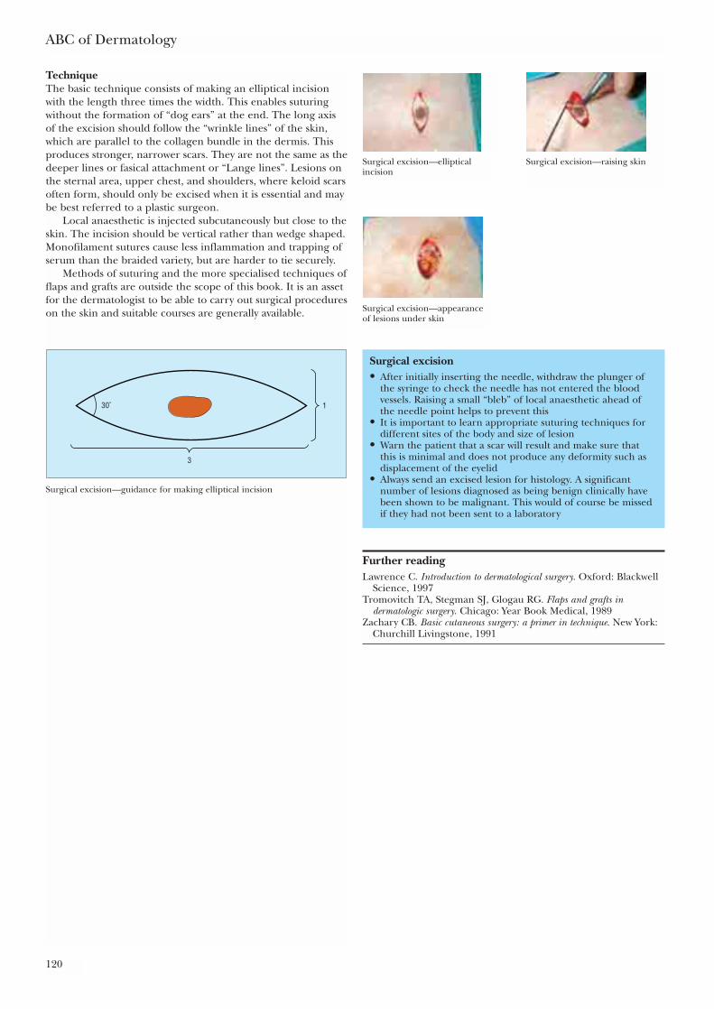
TechniqueThe basic technique consists of making an elliptical incisionwith the length three times the width. This enables suturingwithout the formation of “dog ears” at the end. The long axisof the excision should follow the “wrinkle lines” of the skin,which are parallel to the collagen bundle in the dermis. Thisproduces stronger, narrower scars. They are not the same as thedeeper lines or fasical attachment or “Lange lines”. Lesions onthe sternal area, upper chest, and shoulders, where keloid scarsoften form, should only be excised when it is essential and maybe best referred to a plastic surgeon.
Local anaesthetic is injected subcutaneously but close to theskin. The incision should be vertical rather than wedge shaped.Monofilament sutures cause less inflammation and trapping ofserum than the braided variety, but are harder to tie securely.
Methods of suturing and the more specialised techniques offlaps and grafts are outside the scope of this book. It is an assetfor the dermatologist to be able to carry out surgical procedureson the skin and suitable courses are generally available.
ABC of Dermatology
120
Further readingLawrence C. Introduction to dermatological surgery. Oxford: Blackwell
Science, 1997Tromovitch TA, Stegman SJ, Glogau RG. Flaps and grafts in
dermatologic surgery. Chicago: Year Book Medical, 1989Zachary CB. Basic cutaneous surgery: a primer in technique. New York:
Churchill Livingstone, 1991
Surgical excision• After initially inserting the needle, withdraw the plunger of
the syringe to check the needle has not entered the bloodvessels. Raising a small “bleb” of local anaesthetic ahead ofthe needle point helps to prevent this
• It is important to learn appropriate suturing techniques fordifferent sites of the body and size of lesion
• Warn the patient that a scar will result and make sure thatthis is minimal and does not produce any deformity such asdisplacement of the eyelid
• Always send an excised lesion for histology. A significantnumber of lesions diagnosed as being benign clinically havebeen shown to be malignant. This would of course be missedif they had not been sent to a laboratory
30˚
3
1
Surgical excision—guidance for making elliptical incision
Surgical excision—ellipticalincision
Surgical excision—raising skin
Surgical excision—appearanceof lesions under skin
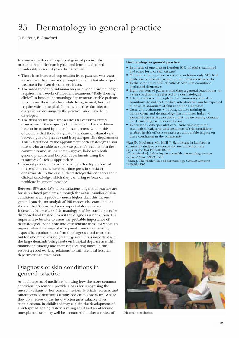
121
In common with other aspects of general practice themanagement of dermatological problems has changedconsiderably in recent years. In particular:
• There is an increased expectation from patients, who wantan accurate diagnosis and prompt treatment but also expecttreatment for even the smallest lesion.
• The management of inflammatory skin conditions no longerrequires many weeks of inpatient treatment. “Daily dressingclinics” in hospital dermatology departments enable patientsto continue their daily lives while being treated, but stillrequire visits to hospital. In many practices facilities forcarrying out dressings by the practice nurse have beendeveloped.
• The demand for specialist services far outstrips supply.Consequently the majority of patients with skin conditionshave to be treated by general practitioners. One positiveoutcome is that there is a greater emphasis on shared carebetween general practice and hospital specialist departments.This is facilitated by the appointment of dermatology liaisonnurses who are able to supervise patient’s treatment in thecommunity and, as the name suggests, liaise with bothgeneral practice and hospital departments using theresources of each as appropriate.
• General practitioners are increasingly developing specialinterests and many have part-time posts in specialistdepartments. In the case of dermatology this enhances theirclinical knowledge, which they can bring to bear on theproblems in general practice.
Between 10% and 15% of consultations in general practice arefor skin related problems, although the actual number of skinconditions seen is probably much higher than this. In onegeneral practice an analysis of 100 consecutive consultationsshowed that 38 involved some aspect of dermatology.Increasing knowledge of dermatology enables conditions to bediagnosed and treated. Even if the diagnosis is not known it isimportant to be able to assess the probable importance ofdermatological conditions and differentiate those for whom anurgent referral to hospital is required from those needing a specialist opinion to confirm the diagnosis and treatment but for whom there is no great urgency. This is important withthe large demands being made on hospital departments withdiminished funding and increasing waiting times. In thisrespect a good working relationship with the local hospitaldepartment is a great asset.
Diagnosis of skin conditions ingeneral practiceAs in all aspects of medicine, knowing how the more commonconditions present will provide a basis for recognising theunusual variants or less common lesions. Psoriasis, eczema, andother forms of dermatitis usually present no problems. Wherethey do a review of the history often gives valuable clues.Atopic eczema in childhood may explain the development of a widespread itching rash in a young adult and an otherwiseunexplained rash may well be accounted for after a review of
25 Dermatology in general practiceR Balfour, E Crawford
Dermatology in general practice• In a study of one area of London 55% of adults examined
had some form of skin disease*• Of those with moderate or severe conditions only 24% had
made use of medical facilities in the previous six months• In the same study 30% of patients with skin conditions
medicated themselves• Eight per cent of patients attending a general practitioner for
a skin condition are referred to a dermatologist†• A large reservoir of people in the community with skin
conditions do not seek medical attention but can be expectedto do so as awareness of skin conditions increases‡
• General practitioners with postgraduate training indermatology and dermatology liaison nurses linked tospecialist centres are needed so that the increasing demandfor dermatology services can be met
• In countries with specialist care, basic training in theessentials of daignosis and treatment of skin conditionsenables health officers to make a considerable impact onthese conditions in the community
*Rea JN, Newhouse ML, Halil T. Skin disease in Lambeth: acommunity study of prevalence and use of medical care. Br J Prev Soc Med 1976;30:107-14†Carmichael AJ. Achieving an accessible dermatology service.Dermatol Pract 1995;3:13-16‡Savin J. The hidden face of dermatology. Clin Exp Dermatol1999;18:393-5
Hospital consultation
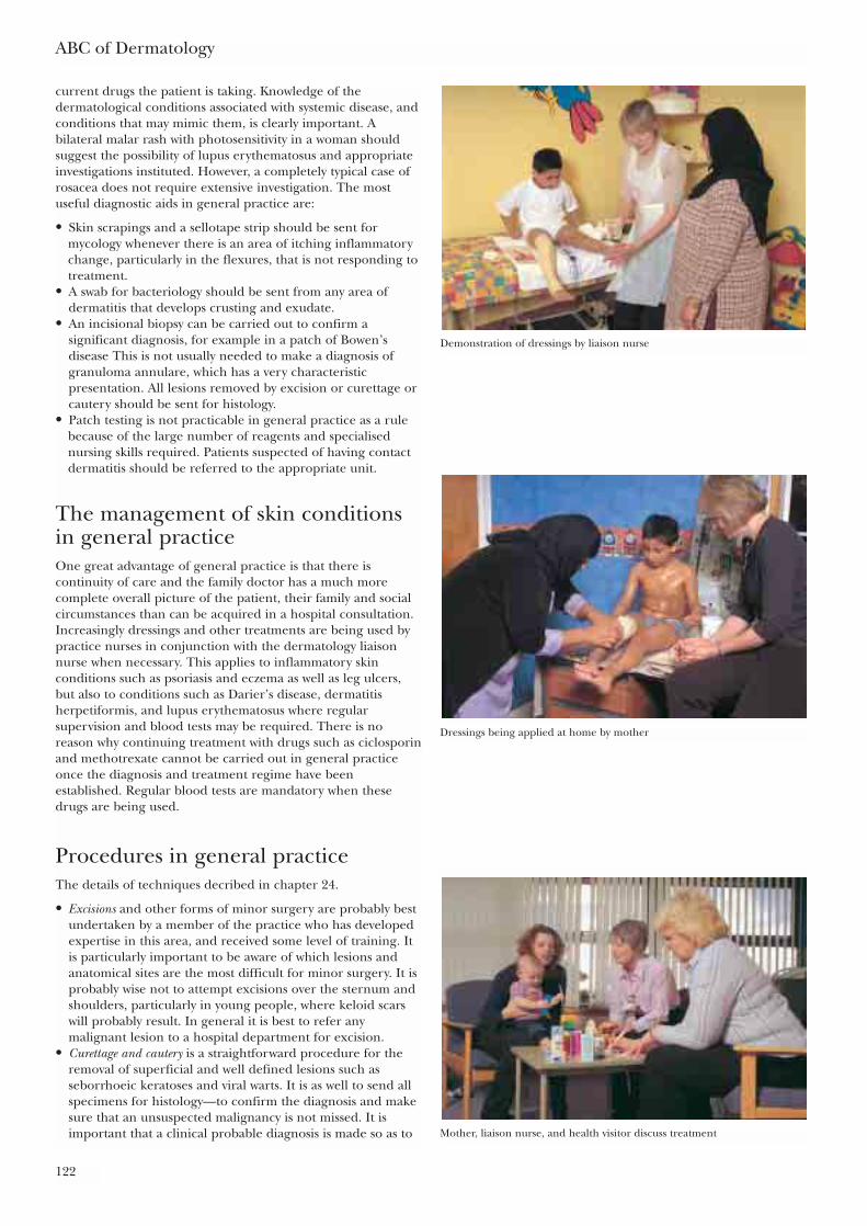
current drugs the patient is taking. Knowledge of thedermatological conditions associated with systemic disease, andconditions that may mimic them, is clearly important. Abilateral malar rash with photosensitivity in a woman shouldsuggest the possibility of lupus erythematosus and appropriateinvestigations instituted. However, a completely typical case ofrosacea does not require extensive investigation. The mostuseful diagnostic aids in general practice are:
• Skin scrapings and a sellotape strip should be sent formycology whenever there is an area of itching inflammatorychange, particularly in the flexures, that is not responding totreatment.
• A swab for bacteriology should be sent from any area ofdermatitis that develops crusting and exudate.
• An incisional biopsy can be carried out to confirm asignificant diagnosis, for example in a patch of Bowen’sdisease This is not usually needed to make a diagnosis ofgranuloma annulare, which has a very characteristicpresentation. All lesions removed by excision or curettage orcautery should be sent for histology.
• Patch testing is not practicable in general practice as a rulebecause of the large number of reagents and specialisednursing skills required. Patients suspected of having contactdermatitis should be referred to the appropriate unit.
The management of skin conditionsin general practiceOne great advantage of general practice is that there iscontinuity of care and the family doctor has a much morecomplete overall picture of the patient, their family and socialcircumstances than can be acquired in a hospital consultation.Increasingly dressings and other treatments are being used bypractice nurses in conjunction with the dermatology liaisonnurse when necessary. This applies to inflammatory skinconditions such as psoriasis and eczema as well as leg ulcers,but also to conditions such as Darier’s disease, dermatitisherpetiformis, and lupus erythematosus where regularsupervision and blood tests may be required. There is noreason why continuing treatment with drugs such as ciclosporinand methotrexate cannot be carried out in general practiceonce the diagnosis and treatment regime have beenestablished. Regular blood tests are mandatory when thesedrugs are being used.
Procedures in general practiceThe details of techniques decribed in chapter 24.
• Excisions and other forms of minor surgery are probably bestundertaken by a member of the practice who has developedexpertise in this area, and received some level of training. Itis particularly important to be aware of which lesions andanatomical sites are the most difficult for minor surgery. It isprobably wise not to attempt excisions over the sternum andshoulders, particularly in young people, where keloid scarswill probably result. In general it is best to refer anymalignant lesion to a hospital department for excision.
• Curettage and cautery is a straightforward procedure for theremoval of superficial and well defined lesions such asseborrhoeic keratoses and viral warts. It is as well to send allspecimens for histology—to confirm the diagnosis and makesure that an unsuspected malignancy is not missed. It isimportant that a clinical probable diagnosis is made so as to
ABC of Dermatology
122
Dressings being applied at home by mother
Mother, liaison nurse, and health visitor discuss treatment
Demonstration of dressings by liaison nurse
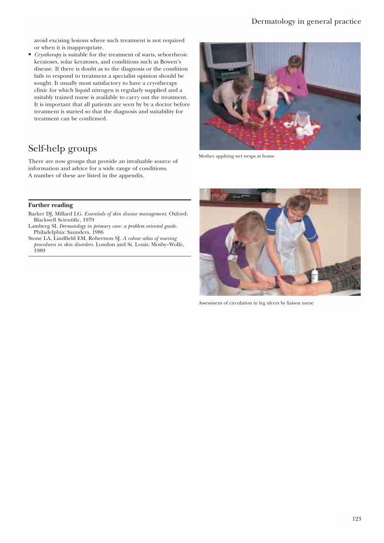
avoid excising lesions where such treatment is not requiredor when it is inappropriate.
• Cryotherapy is suitable for the treatment of warts, seborrheoickeratoses, solar keratoses, and conditions such as Bowen’sdisease. If there is doubt as to the diagnosis or the conditionfails to respond to treatment a specialist opinion should besought. It usually most satisfactory to have a cryotherapyclinic for which liquid nitrogen is regularly supplied and asuitably trained nurse is available to carry out the treatment.It is important that all patients are seen by by a doctor beforetreatment is started so that the diagnosis and suitability fortreatment can be confirmed.
Self-help groupsThere are now groups that provide an invaluable source ofinformation and advice for a wide range of conditions. A number of these are listed in the appendix.
Dermatology in general practice
123
Mother applying wet wraps at home
Assessment of circulation in leg ulcers by liaison nurse
Further readingBarker DJ, Millard LG. Essentials of skin disease management. Oxford:
Blackwell Scientific, 1979Lamberg SI. Dermatology in primary care: a problem oriented guide.
Philadelphia: Saunders, 1986Stone LA, Lindfield EM, Robertson SJ. A colour atlas of nursing
procedures in skin disorders. London and St. Louis: Mosby–Wolfe,1989
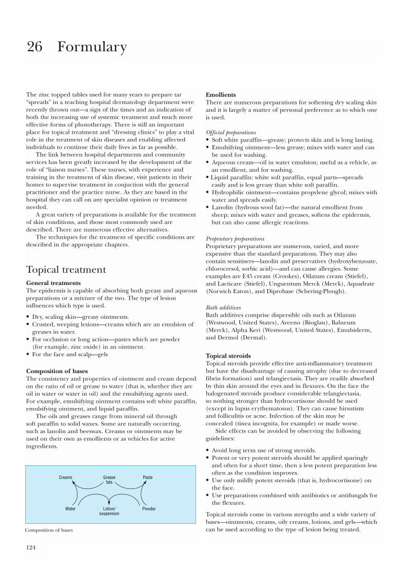
124
The zinc topped tables used for many years to prepare tar“spreads” in a teaching hospital dermatology department wererecently thrown out—a sign of the times and an indication ofboth the increasing use of systemic treatment and much moreeffective forms of phototherapy. There is still an importantplace for topical treatment and “dressing clinics” to play a vitalrole in the treatment of skin diseases and enabling affectedindividuals to continue their daily lives as far as possible.
The link between hospital departments and communityservices has been greatly increased by the development of therole of “liaison nurses”. These nurses, with experience andtraining in the treatment of skin disease, visit patients in theirhomes to supervise treatment in conjuction with the generalpractitioner and the practice nurse. As they are based in thehospital they can call on any specialist opinion or treatmentneeded.
A great variety of preparations is available for the treatmentof skin conditions, and those most commonly used aredescribed. There are numerous effective alternatives.
The techniques for the treatment of specific conditions aredescribed in the appropriate chapters.
Topical treatmentGeneral treatmentsThe epidermis is capable of absorbing both greasy and aqueouspreparations or a mixture of the two. The type of lesioninfluences which type is used.
• Dry, scaling skin—greasy ointments.• Crusted, weeping lesions—creams which are an emulsion of
greases in water.• For occlusion or long action—pastes which are powder
(for example, zinc oxide) in an ointment.• For the face and scalp—gels
Composition of basesThe consistency and properties of ointment and cream dependon the ratio of oil or grease to water (that is, whether they areoil in water or water in oil) and the emulsifying agents used.For example, emulsifying ointment contains soft white paraffin,emulsifying ointment, and liquid paraffin.
The oils and greases range from mineral oil through soft paraffin to solid waxes. Some are naturally occurring, such as lanolin and beeswax. Creams or ointments may be used on their own as emollients or as vehicles for activeingredients.
EmollientsThere are numerous preparations for softening dry scaling skinand it is largely a matter of personal preference as to which oneis used.
Official preparations• Soft white paraffin—greasy; protects skin and is long lasting.• Emulsifying ointment—less greasy; mixes with water and can
be used for washing.• Aqueous cream—oil in water emulsion; useful as a vehicle, as
an emollient, and for washing.• Liquid paraffin: white soft paraffin, equal parts—spreads
easily and is less greasy than white soft paraffin.• Hydrophilic ointment—contains propylene glycol; mixes with
water and spreads easily.• Lanolin (hydrous wool fat)—the natural emollient from
sheep; mixes with water and greases, softens the epidermis,but can also cause allergic reactions.
Proprietary preparationsProprietary preparations are numerous, varied, and moreexpensive than the standard preparations. They may alsocontain sensitisers—lanolin and preservatives (hydroxybenzoate,chlorocresol, sorbic acid)—and can cause allergies. Someexamples are E45 cream (Crookes), Oilatum cream (Stiefel),and Lacticare (Stiefel), Unguentum Merck (Merck), Aquadrate(Norwich Eaton), and Diprobase (Schering-Plough).
Bath additivesBath additives comprise dispersible oils such as Oilatum(Westwood, United States), Aveeno (Bioglan), Balneum(Merck), Alpha Keri (Westwood, United States), Emulsiderm,and Dermol (Dermal).
Topical steroidsTopical steroids provide effective anti-inflammatory treatmentbut have the disadvantage of causing atrophy (due to decreasedfibrin formation) and telangiectasis. They are readily absorbedby thin skin around the eyes and in flexures. On the face thehalogenated steroids produce considerable telangiectasia, so nothing stronger than hydrocortisone should be used(except in lupus erythematosus). They can cause hirsutism and folliculitis or acne. Infection of the skin may be concealed (tinea incognita, for example) or made worse.
Side effects can be avoided by observing the followingguidelines:
• Avoid long term use of strong steroids.• Potent or very potent steroids should be applied sparingly
and often for a short time, then a less potent preparation lessoften as the condition improves.
• Use only mildly potent steroids (that is, hydrocortisone) onthe face.
• Use preparations combined with antibiotics or antifungals forthe flexures.
Topical steroids come in various strengths and a wide variety ofbases—ointments, creams, oily creams, lotions, and gels—whichcan be used according to the type of lesion being treated.
26 Formulary
Greasefats
PasteCreams
Lotion/suspension
PowderWater
Composition of bases
Formulary
125
Their pharmacological activity varies and they are classifiedaccording to their potency, the synthetic halogenated steroidsbeing much stronger than hydrocortisone:
• Mildly potent—hydrocortisone 0·5%, 1%, and 2·5%• Moderately potent—Eumovate (GSK), Stiedex LP (Stiefel)• Potent—Betnovate (GSK), Cutivate (GSK), Locoid
(Brocades), Synalar (Zeneca)• Very potent—Dermovate (GSK).
In Britain a full list showing relative potencies appears in MIMS. Combinations with antiseptics and antifungals, arelisted below:
Mildly potent• Vioform HC (Zyma)• Terra-Cortril ointment (Pfizer), containing oxytetracycline
and hydrocortisone• Fucidin H cream or ointment (Leo), containing fucidic acid
and hydrocortisone• Canesten HC (Baypharm)• Daktacort cream (Janssen)
Moderately potent• Betnovate N (betamethasone and neomycin) (GSK)• Synalar N (neomycin; Zeneca)• Trimovate cream (clobetasone butyrate, nystatin, and
oxytetracyclin; GSK)• Fucibet (betamethasone, fucidic acid; Leo)
Very potent• Dermovate-NN (clobetasone, with neomycin and nystatin;
GSK).
Antiseptics and cleaning lotionsSimple antiseptics are very useful for cleaning infected,weeping lesions and leg ulcers.
Potassium permanganate can be used by dropping four or fivecrystals in a litre of water or in an 0·1% solution that is dilutedto 0·01% for use as a soak. It will stain the skin temporarily andplastic containers permanently.
Silver nitrate 0·25% is a simple, safe antiseptic solution that,applied as a wet compress, is useful for cleaning ulcers.
Flamazine (Smith and Nephew) is silver sulfadiazine cream,used for leg ulcers, pressure sores, and burns.
Hydrogen peroxide (6%) helps remove slough but tends to bepainful. Hioxyl (Quinoderm) is a proprietary cream fordesloughing.
Iodine (2·5%) is an old fashioned, effective preparation as a tincture in alcohol and Betadine (Napp) is a proprietaryequivalent.
Shampoos. Ceanel concentrate (Quinoderm) containscetrimide 10%. Ionil T (Galderma) has benzalkonium chlorideand coal tar solution, and Betadine (SSL) contains povidoneiodine. Shampoos containing selenium sulphide (Selsun,Abbot) and ketokonazole (Nizoral, Janssen) can be used for seborrhoeic dermatitis and also for pityriasis versicolor ofthe skin.
There are numerous other antiseptic, cleansing, anddesloughing agents such as cetrimide, chlorhexidine,benzalkonium chloride, benzoic acid, and enzyme preparationssuch as Varidase (Lederle), a streptokinase and streptodornasepreparation.
Tar preparationsThese are mainly used for treating psoriasis as described inchapter 3.
Tar has an anti-inflammatory effect and seems to suppressthe epidermal turnover in lesions of psoriasis. The various tarpastes are generally too messy to use at home and are mostsuitable for dermatology treatment centres. Standard tar pastecontains a strong solution of coal tar 7·5% in 25 g of zinc oxide,25 g of starch, and 50 g of white soft paraffin.
There are numerous proprietary preparations that are lessmessy and do not stain but are not so effective. They are usefulfor treating less severe psoriasis at home. Examples are:Alphosyl cream (Stafford-Miller), Pragmatar (Bioglan),Psoriderm (Dermal); alphosyl HC (Stafford-Miller) and Carbo-Cort (Lagap) contain hydrocortisone as well.
Dithranol can be used in a paste containing salicylic acid, zincoxide starch, and soft white paraffin. It has to be appliedcarefully avoiding contact with the surrounding skin, as it can cause severe irritation. It is best to start with a lowconcentration.
For short contact treatment relatively clean preparations ina range of concentrations are available, such as Dithrocream(Dermal), Anthranol (Stiefel), and Psoradrate cream (Stafford-Miller).
Ichthammol is a useful soothing extract of shale tar. It can bemade up as a 1% paste in yellow soft paraffin with 15% zincoxide.
Bath preparations are useful for dry skin and widespreadpsoriasis. Coal tar solution (20%) can be used or PolytarEmollient (Stiefel) or Psoriderm.
Tar shampoos are useful for treating psoriasis of the scalp.Polytar (Stiefel), T-Gel (Neutrogena), Capasal (Dermal), andAlphosyl (Stafford-Miller) are some examples.
Keratolytics These can be used for hyperkeratotic lesions. They soften andhelp remove excess keratin. If used for extensive areas or ininfants systemic absorption can occur. A useful preparation issalicylic acid 2–4% in aqueous cream. Salicylic acid withbetamethasone ointment (Diprosalic ointment, Schering-Plough) can be used for hyperkeratotic lesions whereinflammation is present.
AntipruriticsUseful anti-pruritics for persistent itching include menthol(0·5%) or phenol 1% in aqueous cream, and calamine lotion,which contains arachis oil.
Barrier and protective preparationsThese preparations protect against softening and macerationfrom moisture in flexures, for example the groins. They also have an occlusive effect. They are essentially bases withzinc oxide or silicone (as dimethicone). There are manypreparations; some of the most commonly used are:
• Zinc cream BP, contains zinc oxide, arachis oil, and lanolin• Zinc and castor oil ointment BP• Conotrane (Yamanoouchi) and Siopel (Bioglan), contain
dimeticone (dimethicone)• Metanium (Roche), contains titanium dioxide• Sudocrem (Forest) and Drapolene (Pfizer), contain
lanolin.
Treatment for specific situationsSunscreensThese give a degree of protection—mainly to ultraviolet B butalso to ultraviolet A. They depend on their effect on a physicalbarrier (usually titanium dioxide) and chemicals that combinewith epidermal cells, usually esters of PABA or oxybenzone.
ABC of Dermatology
126
CamouflageScars, congenital naevi and other blemishes that cannot beremoved can be covered with suitable creams. Proprietarypreparations are available.
AntiperspirantsAluminium chloride for hyperhidrosis: aluminium chloride20% (Driclor, Stiefel, or Anhydrol, Dermal Laboratories).
Depigmenting agents2% hydroquinone cream is available without prescription as“fade-out”. Preparations containing corticosteroids are alsoprescribed but not available as proprietary preparations.
Antimitotic agents5-Flourouracil cream is useful for treating incipientmalignancies—that is, solar keratoses, but not actualcarcinomas. It is available as Efudix cream (Roche), which isapplied daily for one to two weeks. It produces a variabledegree of inflammation that is allowed to subside before thetreatment is repeated.
Infestations(1) Scabies. The correct procedure for treatment is more
important than the preparation used. Benzyl benzoate 25%application BP is still available and is cheap but tends toirritate the skin. Malathion is available as Derbac (SSL),Prioderm (SSL), and Quellada M (Stafford-Miller), andPermethrin as Lyclear cream (Kestrel) preparations aremore effective and less likely to irritate. 6% sulphur inwhite soft paraffin or permethrin are recommended foryoung children and pregnant or lactating women. Theprocedures for treatment set out on page 107 should befollowed and clearly explained to the patient. For resistantcases ivermectin (Mectizan, MSD) by mouth is available ona named patient basis.
(2) Pediculosis. Preparations containing malathion, carbaryl,and permethrin are used either as shampoos or lotions.Lotions are most effective and should be left on the skinfor 12 hours before washing off. The same preparations areavailable as for treating scabies, with the addition of 0·5%malathion lotion as Suleo-M (SSL). Recently a lotion ofphenothrin (Full Marks, SSL) has become available fortreating head and pubic lice.
Preparations for the mouthSteroids—Adcortyl in Orabase (Squibb) or Corlan pellets (Evans). Both these preparations containcorticosteroids.
Antifungals—Daktarin (Janssen) or Fungilin lozenges(Squibb); Nystan (nystatin suspension, Squibb). Corsoidyl(chlorhexidine, GSK), and Difflam (3m Riker) are usefulmouthwashes.
Topical immunosuppressantsTacrolimus (Protopic, Fujisawa) has recently become availableas an ointment in two strengths, 0.03% and 0.1%. It has notbeen evaluated in children under the age of two or in pregnantwomen. It is recommended that it is only used bydermatologists or those with considerable experience intreating eczema. Although the exact mode of action isunknown it does diminish T cell stimulation by Langerhan cellsand diminishes the production of inflammatory mediators frommast cells. It should be used in moderate to severe atopiceczema that has not responded to either treatment. Skinirritation with burning, erythema, and pruritis are the mostcommon side effects. In view of its immunosuppressive activity
any infection should be treated first and it should be used withcaution if there is a risk of viral infection or if inoculationsusing attenuated or live organisms are being used.
Pimecrolimus (Elidel, Steeple Novartis) is a similarpreparation recommended for intermittent treatment ofeczema can also be used as an initial treatment for any flare upof eczema. It diminishes cytokine activity long term relievingboth the erythema and pruritis of eczema.
In common with topical steroids any immunosuppressivedrug should be used with caution as viral infections are likely tobe present or the patient is undergoing inoculation with live orattenuated organisms.
Systemic treatmentAntibiotics are probably the most commonly used systemictreatment. Long term antibiotics are needed for acne andcellulitis. Antifungal and antiviral drugs are indicated if topical treatment is ineffective, particularly in theimmunosuppressed, and when the infection has beenconfirmed by laboratory tests.
Immunosuppressant drugs have had a considerable impacton the treatment of autoimmune and connective tissue diseasesand diminished the need for systemic steroids—previously theonly treatment available. They are increasingly used forextensive and persistently inflamed dermatoses, particularlypsoriasis and eczema.
Antibacterial drugsAll penicillins may cause allergic rashes, which may be severe,and the broad spectrum penicillins, amoxicillin, ampicillin, and co-amoxiclav, are particularly likely to cause an intense rash in patients with glandular fever. They tend to accumulatein patients with renal failure and may reduce the excretion ofmethotrexate which is used in the treatment of psoriasis.
Phenoxymethylpenicillin (penicillin V) is useful in Grampositive infections and erysipelas.
Flucloxacillin is used to treat infections due to penicillinase producing organisms. It is used in impetigo andcellulitis.
Amoxicillin and ampicillin are broad spectrum antibioticsbut are destroyed by penicillinase. Co-amoxiclav is acombination of amoxacillin and clavullinic acid. It is effectiveagainst a wide range of organisms and beta lactamaseproducing staphylococci as well.
Cephalosporins are not affected by penicillinase and areeffective against both Gram positive and Gram negativeinfections.
Ciprofloxacin is used for infections with both Gram positiveand Gram negative organisms such as pseudomonas.
Erythromycin is used for the treatment of acne and is usefulin Gram positive infections. Resistant strains of staphylococcusare appearing.
Metronidazole is useful for treating anaerobic infectionsand trichomonas infections. It is useful for rosacea that is notresponding to conventional treatment.
Antifungal drugsTopical treatment is usually effective but for fungal infection of the nails and intractable infections of the skin systemictreatment may be required.
Griseofulvin (500 mg daily) is a well established treatmentfor fungal infections of the skin, hair, and nails. Although itshould not be used in pregnancy, it can be used in children. Itcan cause lupus erythematosus to flare up.
Formulary
127
Terbinafine (250 mg daily) is an effective systemicantifungal drug that does not affect the liver. It is used for bothnail and skin infections.
Imidazole and triazole drugs include itraconazole andketoconazole, which are effective for dermatophyte infectionsof the skin and pityriasis versicolor.
Antiviral drugsDiscovery of drugs that inhibit viral DNA polymerase andinhibit their proliferation in vivo means that effective treatmentfor herpes simplex and zoster is now possible. They areeffective at the early stages of infection and should be started assoon as symptoms appear. Aciclovir (Zovirax, GSK) is availableas a cream.
Aciclovir is effective against both herpes simplex and zoster.The standard dose is 200 mg five times daily for five days. In varicella infections and herpes zoster 800 mg is given fivetimes daily for seven days. It can also be given by intravenousinfusion, and should be applied as soon as symptoms appear. In addition, it can be used for prophylaxis, particularly in theimmunocompromised patients and atopics who are liable tofulminating infection.
Famciclovir and valaciclovir are similar and are recommendedfor treating herpes zoster.
AntihistaminesThese drugs are used in urticaria and acute allergic (type Iimmediate hypersensitivity) reactions. The newer long actingand non-sedating antihistamines are useful for treatmentduring the day and can be combined with one of the sedatingtype at night if pruritus is preventing sleep.
Non-sedating antihistamines only cross the blood–brainbarrier to a slight extent. They may cause arrhythmias,particularly terfenadine.
• Acrivastine (Semprex, GSK) 8 mg three times daily• Cetirizine (Zirtek, UCB Pharma) 10 mg once daily• Fexofenadine (Telfast, Hoechst) 120 or 180 mg once daily• Loratadine (Clarityn, Schering-Plough) 10 mg once daily.
Sedating antihistaminesThere are many available and which is used is largely a matterof personal preference. The sedating effect, which is enhancedby alcohol, means that they are best taken at night. They alsopotentiate CNS depressants and anticholinergic drugs. Theytend to have anticholinergic effects, causing dry mouth,blurred vision, tachycardia, and urinary retention. Thosecommonly used are:
• Chlorphenamine (Piriton Stafford-Miller 4 mg daily)• Cyproheptadine (Periactin (MSD) 4 mg up to four times
daily)• Hydroxyzine (Atarax (Pfizer) 10–25 mg at night; can be used
during the day if drowsiness is not a problem)• Promethazine (10 or 25 mg at night or twice daily)• Trimeprazine (Vallergan (Castlemead) 10 mg two to three
times daily).
CorticosteroidsIn addition to topical preparations, systemic steroids may berequired for the treatment of severe inflammatory skinconditions such as erythroderma developing from psoriasis oreczema. They are also used in vasculitis and erythemamultiforme as well as connective tissue diseases. They are oftenrequired for the treatment of pemphigoid and pemphigustogether with immunosuppressant drugs.
The side effects must be borne in mind, particularly for anylong term treatment. Most important are given below.
Water and electrolytesSodium and water retention with loss of potassium.
MusculoskeletalOsteoporosis, aseptic necrosis of the femoral head, growthretardation in children, and muscle wasting.
Ophthalmic effectCataract formation and increased tendency to glaucoma.
Other effectsIncrease in blood pressure, peptic ulceration and fatredistribution, and impaired glucose intolerance.
RetinoidsThese vitamin A derivatives have proved very effective in thetreatment of psoriasis and acne but are not without risk of sideeffects. The most serious is that they are teratogenic and mustbe discontinued for at least three months after stoppingtreatment in the case of isotretinoin and five years after takingacitretin.
All patients should be warned of possible side effects andwomen of childbearing age must be using an effective form ofcontraception, which must have been used for at least a monthbefore treatment has started as well as having a pregnancy testcarried out. Liver function tests and fasting cholesterol andtriglycerides should be carried out on all patients. Afterprolonged treatment in adolescence, radiological tests shouldbe carried out to ensure that there is no extraosseouscalcification. The most important side effects are:
• Abnormal liver function tests• An increase in cholesterol and triglycerides• Occasional increases in electron spin resonance and lowered
white count.
Clinical side effectsDrying and roughening of the skin and mucous membranes,particularly the lips, can occur. There may also be thinning ofthe hair and nails. Photosensitivity eruptions can develop.Occasionally muscle and joint pains occur.
AcitretinThis drug is used for severe psoriasis including pustulosis of thehands and feet. It has also been used in other forms ofkeratosis such as Darier’s disease and pityriasis rubra pilaris.
IsotretinoinThis drug is used for severe acne vulgaris that has notresponded to antibiotics or other treatments. It is thereforeoften used in adolescence and it is important to be aware of themusculoskeletal effects and possible mood changes.
ImmunosuppressantsMethotrexateThis drug is useful in severe psoriasis that is not responding totopical treatment. The main disadvantage is its adverse effecton the liver, which precludes its use in those who have alcoholicliver disease but who are often those most needing systemictreatments. Idiopathic immunosuppression can occur so a testdose must always be given and a full blood count carried out 48 hours later before treatment has started. There may begastrointestinal upsets and osteometitis as well.
ABC of Dermatology
128
Methotrexate interacts with anti-inflammatory andantiepileptic drugs. A full list of drug interactions should beconsulted before treatment is started.
After a 2·5 mg test dose and full blood count 48 hours later, the regular dose is 5–15 mg by mouth once a week. Full blood count and liver function tests should becarried out once a week for the first six weeks and thereafteronce a month during treatment. Folinic acid should be given at the same time, as this prevents bone marrow depression. In many centres a liver biopsy is considered mandatory before treatment is started since blood tests will remain normal for some time during the development of hepaticfibrosis.
AzathioprineThis drug is used for systemic lupus erythematosus, pemphigus, and bullous pemphigoid. It enables the dosage of systemic steroids to be reduced. The most serious side effect is bone marrow suppression. This may occur quite rapidly, particularly in those with diminished abilityto metabolise the drug. This is carried out by thiopurinemethyl transferase (TMT). The level of this enzyme shouldtherefore be determined before treatment is started and thoseat low levels given a lower dosage. Those who inherit highactivity may require higher doses. Other side effects includegastrointestinal upset, liver toxicity, and an increased tendencyto infection.
CiclosporinThis drug has proved helpful in severe psoriasis withininflammatory lesions and, secondly, in the treatment of severeatopic dermatitis. There are a number of drug interactions andit is important to check renal function and monitor both bloodurea and serum creatinine.
Other drugsDapsoneThis drug was originally developed for treating leprosy but wasproved very effective in dermatitis pityformis and some otherconditions, such as pyoderma gangrenosum. It may causehaemolytic anaemia, and other side effects include bonemarrow suppression, hepatitis, and peripheral neuropathy.Regular blood checks are essential.
HydroxychloroquineThis drug is used in both systemic and discoid lupuserythematosus as well polymorphic light eruption andporphyria cutanea tarda. The most serious side effect isretinopathy but this does not occur if the dose does not exceed6·5 mg/kg lean body weight.
PsoralensThese drugs are used in conjunction with long wavelengthultraviolet light as psoralen with ultraviolet A (PUVA) therapydescribed on page 67. It is used for the treatment of severepsoriasis. It has also proved effective in some cases of atopiceczema, T cell lymphoma of the skin, and occasionally in lichenplanus. There is a risk of cataract formation, and a full bloodcount as well as antinuclear factor tests should be carried out.
Preparations for treating acne and varicose ulcers aredescribed in the appropriate sections.
Further readingArndt KA. Manual of dermatological therapeutics, 5th ed. NewYork:
Little, 1995

129
Acne Support Group (0870) 870 2263Behcet’s Syndrome Society (01488) 71116Allergy UK (020) 8303 8583Cancer BACUP (0808) 800 1234British Association of Skin Camouflage (01625) 267880British Red Cross Skin Camouflage Service (020) 7201 5172British Leprosy Relief Association (01206) 562286Congenital Melanocytic Naevus Support Group (0151) 281 9716Darier’s Disease Support Group (01646) 695055Dystrophic Epidermolysis Bullosa Research Association (01344) 771971Ectodermal Dysplasia Society (01242) 261332Ehlers–Danlos Support Group (01252) 690940Hairline International (01564) 775281Herpes Viruses Association (020) 7609 9061Ichthyosis Support Group (020) 7461 0356Latex Allergy Support Group (07071) 225838Lupus UK (01708) 731251Lymphoma Association (LA) (0808) 808 5555Marfan Association UK (01252) 810472Myositis Support Group (023) 8044 9708National Eczema Society (0870) 241 3604Neurofibromatosis Association (020) 8547 1636Pemphigus Vulgaris Network (020) 8690 6462Primary Immunodeficiency Association (PIA) (010) 7976 7640Pseudoxanthoma Elasticum (PXE) Support Group (01628) 476687Psoriatic Arthropathy Alliance (01923) 672837Psoriasis Association (01604) 711129Raynaud’s and Scleroderma Association Trust (01270) 872776Scleroderma Society (020) 8961 4912Shingles Support Society (020) 7607 9061Telangiectasia Self Help Group (01494) 528047Tuberous Sclerosis Association (01527) 871898Vitiligo Society (020) 7840 0855
Appendix: Patient support groups

130
abcesses 88acanthosis nigricans 76aciclovir 93, 126acitretin 128
adverse effects 16psoriasis treatment 16
acne 47–50causes 47, 47–8
external factors 48cystic 47, 49infantile 47, 48occupational 48, 49scars 47treatment 49, 49–50, 50types 48, 48–9
acne conglobata 48, 48acne excoreé 48acne fulminans 48acne keloidalis 48acne vulgaris 48, 48acquired immunodeficiency syndrome see AIDSacquired melanocytic naevi 68acral melanoma 70acrodermatitis pustulosa 10, 58actinic keratosis see solar (actinic) keratosesactinomycetoma 111acute erythrodermic psoriasis 11acute febrile neutrophilic dermatosis 36acyclovir 93, 126adenoma sebaceum 64AIDS 98–100
B cell lymphoma 100drug eruptions 100infections 99
viral 93, 94, 99Kaposi’s sarcoma 100skin changes 99stages 98syphilis with 89see also HIV infection
albinism 76squamous cell carcinoma 109
allergic contact dermatitis 19, 19–22, 23, 83allergic reactions 6, 24, 82
causes and process 82cosmetics 87, 87dithranol 20, 21drugs 20, 72, 87fish protein 82latex 21, 23neomycin 20, 36, 41nickel 20, 23, 41plants 87types 82–3
alopeciaadult pattern 52aetiology and pathogenesis 52, 52androgenic 51–2, 53classification 52congenital 52diffuse 52
non-scarring, causes 53treatment 53
drug-induced 52lichen planus 27localised 53–4male pattern 51, 53postfebrile 52scarring 54, 54traction 53
alopecia areata 53–4aetiology 54autoimmune aetiology 84differential diagnosis 53nail changes 58, 59, 60treatment 53–4
alopecia totalis 53alopecia universalis 53amelanotic melanoma 70ampicillin, rashes due to 37Amsler Chart 73, 85amyloidosis 79anagen 51anagen effluvium 52anaphylactic reactions 72, 82Ancylostoma caninum 107androgenic alopecia 51–2, 53androgens
acne association 47, 47hair growth 51
angiitis, necrotising 37angiomas 75
cavernous 64congenital 75eruptive 75spider naevi 75
angio-oedema 38animal ringworm 101, 103annular lesions 5, 9anthrax 91antiandrogens 56
acne treatment 49antibiotics 126
acne treatment 49erythema due to 35rashes due to 36, 37, 72
antifungal drugs 103–4, 126nail infections 60oral use 126seborrhoeic dermatitis treatment 29
antigen-antibody complex reactions 83, 83antihistamines 24, 127
non-sedating 127sedating 127
antimitotic agents 126antiperspirants 126anti-pruritics 125antiseptic paints 104antiseptics 125antiviral drugs 126aquagenic urticaria 38argyria 76
IndexPage numbers printed in italics refer to boxed material

Index
131
arterial ulcers 45–6arthropathy 11Ascaris lumbricoides 108asteatosis 19, 78asteatotic eczema 18athlete’s foot 32, 102atopic eczema 17
diagnosis in general practice 121–2distribution 17genetic basis 81localised lesions 32scalp 54variants 17
atopy, inheritance 81atrophie blanche 44atrophy 4Auspitz sign 8autoantibodies 81autoimmune disease 83–6
nail involvement 58range 84vitiligo association 76
azathioprine 127azole drugs 104, 126
bacillary angiomatosis 89Bacterial Index (BI), leprosy 110bacterial infections 87–91
AIDS-related 99clinical presentation 88, 88common patterns 90–1mycobacterial 88–9signs 87tropical 109–10
balanitis xerotica obliterans 74barrier preparations 125basal cell(s), neoplastic 61basal cell carcinoma 61–2
cystic 61nodular 61pigmented 62sclerosing 62sun exposure and 66superficial 62treatment 62
cryotherapy 116ulcerated 46, 61, 62
basal cell epithelioma 61bases, topical preparations 124bath additives 124bath preparations, tar/coal tar 125Bazin’s disease 36BCC see basal cell carcinomaB cell lymphoma 77
AIDS 100Beau’s lines 58, 59Becker’s naevus 68benzoyl peroxide 49biliary obstruction, pruritus 24biopsy 118–20
incisional 118, 122blackheads 48Blashko’s lines 81blastomycosis 111blepharitis 50blistering eruptions, drug-induced 37blisters 39–42
autoimmune response 83development, duration and distribution 39differential diagnosis 39diseases presenting with 6, 39erythema multiforme 36, 40
pemphigoid 6, 7, 40pityriasis lichenoides varioliformis acuta 40see also bullae
blue naevus 68body lice 105, 108boils 88, 91borderline leprosy 110Borrelia infections 75, 106Bowen’s disease 34, 63
cryotherapy treatment 116squamous cell carcinoma developing 62
breast, Paget’s disease 18, 63Breslow classification, melanoma 71bullae 3
insect bites 106see also blisters
bullous impetigo 24, 41bullous pemphigoid 4, 40Buruli ulcer 89“butterfly” rash 73, 84, 87
“cafe au lait” patches 77Calamine lotion 24calcipotriol 14camouflage preparations 126Campbell de Morgan spots 64Candida albicans 102, 103Candida intertrigo 103candidiasis 101, 103
AIDS-related 99diabetes-related 79flexural 98pseudomembranous 98
cantharadin 54carbon dioxide, solid 115
wart treatment 95carbon dioxide cryotherapy 115carbuncle 88, 91carcinoid 74carcinoma see basal cell carcinoma; squamous cell carcinomacatagen 51cat scratch disease 89cautery 118
see also electrocautery; heat cauterycavernous angioma 64cavernous haemangioma 117cellulitis 88, 91cervix, intraepithelial neoplasia 99champagne bottle legs 44chickenpox 39, 92chloasma 76chloracne 49chlorpromazine 77cholestyramine 78cholinergic urticaria 38chromomycosis 112ciclosporin 127
alopecia due to 52psoriasis treatment 16
Clark classification, melanoma 71cleaning lotions 125clubbing, of nails 58, 75coal tar preparations 14, 26, 125coeliac disease 39“cold sores” (herpes simplex virus type I) 41, 92cold urticaria 38comedones 48compound naevi 68compression bandages, venous ulcer treatment 44congenital angiomas 75congenital hairy naevus 69congenital pigmented naevus 68–9

Index
132
connective tissue diseases 72–4contact dermatitis 5, 6, 19–22
allergic 19, 19–22, 23, 83alopecia treatment 54drug-related 36hair dye causing 54immunological response 19irritant see irritant contact dermatitis localised lesions 32morphology 20occupational 7, 20, 22–3
causative substances 21patch testing 21–2pathology 20–1
corticosteroid preparations 127see also steroid(s)
Corynebacterium minutissimum 32, 102cosmetic acne 48cosmetic allergy 87, 87cowpox 94crab lice 108“crazy paving” pattern 18, 78CREST syndrome 74, 74, 85Crohn’s disease 78cryotherapy 115–16, 116
in general practice 123indications 115–16
curettage 117–18in general practice 122–3suitable lesions 118warts 95
cutaneous leishmaniasis 105, 110cyclosporin see ciclosporincyproterone acetate 49cystic acne 47, 49cystic basal cell carcinoma 61–2cysts 64cytotoxic reactions 82, 82–3
dapsone 128Darier’s disease 58, 60deep vein obstruction 43deficiency states
alopecia due to 52hyperpigmentation association 77
delayed hypersensitivity 83, 83delusional parasitosis 106Demodex folliculorum 107dengue 105depigmentation, localised 76depigmenting agents 126dermatitis
contact see contact dermatitis hand 26, 26irritant see irritant contact dermatitis occupational see occupational dermatitis perioral 30, 50, 87seborrhoeic see seborrhoeic dermatitis see also eczema
dermatitis artefacta 46dermatitis herpetiformis 7, 24, 39, 78dermatofibroma 63dermatographism 38dermatomyositis 73, 85dermatophyte infections 101, 102
see also ringwormdermo-epidermal junction 83desquamation 5diabetes mellitus 78–9diabetic dermopathy 79diabetic ulcers 46, 79diagnosis of skin conditions 121–2
diet, acne and 47direct immunofluorescence 84discoid eczema 4, 18, 30discoid lupus erythematosus 32–3, 73
clinical features 32, 33, 34, 73, 87diagnosis 34immunology 85scarring alopecia 54treatment 73, 85
dithranol 125allergic reactions 20, 21psoriasis treatment 14, 16short contact 14
dracontiasis 114dressings, leg ulcers 44, 45drug eruptions 28
AIDS-associated 100bullous 42fixed see fixed drug eruptions
drugsacne association 48allergic reactions 87alopecia due to 52blistering eruptions due to 37contact dermatitis association 36hirsuties due to 54hyperpigmentation due to 77lichen planus-like reactions 37nail colour changes due to 58–9photosensitivity due to 37photosensitivity induced by 66rashes due to 36–7
dry skin 78treatment 25
dysplastic lesions, cryotherapy 116dysplastic melanoma 70dysplastic naevi 69dysplastic naevus syndrome 68, 69
ecthyma 88, 91eczema
asteatotic 18atopic see atopic eczema classification 18clinical features 17, 29diagnosis 34discoid 4, 30infected 26, 90intraepidermal vesicles 6itching (pruritus) 23–4lichenified 26, 28nail changes 58nummular 4, 18pathology 17seborrhoeic see seborrhoeic dermatitis stasis 18treatment 25–6
guidelines 25types 17–18weeping 25see also dermatitis
eczema herpeticum 17–18, 92Ehlers–Danlos syndrome 78elastic, allergic reaction 21electrocautery 116
equipment 116technique 116
electrodesiccation 116elliptical excision 119emollients 14, 26, 124
eczema treatment 25proprietary preparations 124

Index
133
emulsifying ointment BP 25en coup de sabre pattern 54endocrine disorders
alopecia 52see also individual disorders
environmental factors, history-taking 7enzyme preparations, venous leg ulcers 45ephelides 68epidermal thickening, psoriasis 8epidermodysplasia verruciformis 94epidermoid cyst 64epidermolysis bullosa 80
genetic basis 81Epidermophyton spp. 57epithelioma, basal cell 61erosion 4erysipelas 87, 88, 91, 109erythema 2, 7, 34, 35–6, 75
acute 25from antibiotics 35desquamating stage 32facial 87figurate 35, 75malar 33nailbed 75in systemic disease 72toxic, causes 35see also rashes
erythema annulare 75erythema chronicum migrans 75, 106erythema gyratum repens 75erythema induratum 36erythema infectiosum 87, 95, 96erythema marginatum 75erythema multiforme 35–6, 72, 75
annular lesions 36blisters 36, 40causes 35, 75target lesions 75
erythema nodosum 36, 80erythrasma 32, 102erythrocyanosis 74erythrodermic psoriasis 11, 52
treatment 13, 16erythroplasia of Queyrat 63ethyl chloride, cryotherapy 115etretinate, psoriasis treatment 16eumycetoma 111examination, clinical 7excision, surgical 120
in general practice 122–3excoriation 4exostosis, subungual 60eyes, blepharitis 50
facial erythema 87facial psoriasis, treatment 13fasciitis, necrotising 79favus 111fibrokeratoma, periungual 60fifth disease 87, 95, 96figurate erythema 35, 75filariasis 112–13
diagnosis 113lymphoedema 113
fish protein, allergic reaction 82“fishtank” granuloma 89fissuring 4Fitzpatrick classification, skin types 65fixed drug eruptions 33, 34, 37, 37, 42, 72
diagnostic criteria 33flamazine 125
flea bites 106flexural candidiasis 98flexural psoriasis 10, 13fluid retention, acne and 475-fluorouracil cream 63, 126folliculitis 88, 91
Gram negative 91folliculitis decalvans 54formulary 124–8
systemic agents 126–8topical agents 124–6
freckles 68fungal infections 32, 101–3
AIDS-related 99clinical presentation 101deep 103
tropical 111–12diagnosis 34, 104localised lesions 32nails 57, 101–2superficial 101–3
tropical 110, 111treatment 34, 103–4, 104tropical skin disease 110–12
furuncles 88, 91
ganciclovir 93gastrointestinal disease, skin lesions 78general practice, and skin conditions 121, 121–3
diagnosis 121–2management 122procedures used 122–3
genetics and skin disease 80–1, 81complex disorders 81single gene disorders 80–1
genital herpes 92genital warts 94, 98, 99genodermatoses 80–1, 81German measles 95–6Gianotti–Crosti syndrome 96graft versus host disease 86grain mites 106Gram negative folliculitis 48granuloma
“fishtank” 89pyogenic 64sarcoid 80“swimming pool” 89
granuloma annulare 79gravitational ulcers 43griseofulvin 104guttate psoriasis 10, 28
lesions 30treatment 13, 15
haemangioma 3cavernous 117sclerosing 63
haemolytic anaemia 83haemosiderin deposition 76hair 51–6
excessive 55–6laser treatment 117
exclamation mark appearance 53growth pattern 51loss see alopecia psychological significance 51shaft abnormalities 55
hairy naevus, congenital 69halo naevus 68hand, foot and mouth disease 96–7, 97hand dermatitis, treatment 26, 26

Index
134
hands, dermatophyte infections 102harvest mites 106head lice 108heat cautery 95heat urticaria 38heliotrope rash 73helminth infestations, tropical 112–13Henoch–Schönlein purpura 37hepatic porphyrias 79herald lesions 30hereditary angio-oedema 38hereditary haemorrhagic telangiectasia 75herpes simplex virus infections 92, 92
atopic eczema 17–18blisters 41itching 24of lips 41, 92treatment 93
herpes virus infections 92–3herpes zoster 24, 42, 92–3
disseminated 93hirsuties 55
causes 55histoplasmosis 112history-taking 7HIV infection 98
early stages 98histoplasmosis 112late stage disease 98“seroconversion illness” 98see also AIDS
Hodgkin’s disease, pruritus 24hormones
acne association 47, 47hirsuties due to 54hypopigmentation due to 75–6vascular lesions due to 74
human immunodeficiency virus (HIV) see HIV infectionhuman papilloma viruses (HPV) 94, 99Hutchinson’s sign 59, 60, 70hydrogen peroxide 125hydroxychloroquine 128
lupus erythematosus treatment 33, 73, 85hyperkeratosis 4
epidermolytic 81keratolytics for 125nail plate 59
hyperlipidaemia 79hyperpigmentation 76–7
sun exposure 66hypersensitivity
type I (immediate) 23, 82, 82type II (cytotoxic) 82–3type III (immune complex) 72, 83, 83type IV (delayed) 83, 83
hyperthyroidism 76, 80pruritus 24
hypertrichosis 55–6causes 56treatment 56
hypoalbuminaemia 58hypopigmentation 75–6hypopituitarism 52, 75hypothyroidism 80
pruritus 24
ice-pick scars 48ichthammol 125
eczema treatment 26psoriasis treatment 14
IgA disease, linear 39–40immediate hypersensitivity 23, 82, 82
immune complexes 83, 83circulating 83
immune reactions 72cell-mediated 82contact dermatitis 19humoral 82psoriasis pathogenesis 12
immunofluorescence 84, 84immunology 82–6
autoimmune disease 83–6hypersensitivity reactions see hypersensitivity
immunosuppressants 25, 126, 127–8impetigo 7, 88, 90
bullous 24, 41, 90“follicular” 91non-bullous 90in tropics 109
incisional biopsy 118, 122indirect immunofluorescence 84induration of skin 7infantile acne 47, 48infantile seborrhoeic dermatitis 30infections
AIDS 98bacterial see bacterial infections diabetes mellitus 79in eczema 18, 19fungal see fungal infections psoriasis pathogenesis 13tropical see tropical skin diseases viral see viral infections
infectious mononucleosis 97infestations 106–8
treatments 126tropical skin diseases 112–14
inflammation 7inflammatory skin disease, alopecia due to 52inoculation herpes 92insect bites 3, 24, 105–6
allergic reaction 42diseases due to 105
intradermal naevus 68intraepidermal carcinoma 34iodine solution 125iron deficiency 53
anaemia 60pruritus 24
irritant contact dermatitis 5, 18, 20, 21acute 21chronic 21, 23occupational 22, 23
isotretinoin 127, 128acne 49
itching see pruritusitch–scratch–itch cycle 24
jaundice 78junctional naevi 68juvenile plantar dermatosis 17, 18
kala-azar 105Kaposi’s sarcoma 46, 98, 99, 100Kaposi’s varicelliform eruption 92keloid scars 49keratoacanthoma 63keratolytics 125keratoses
seborrhoeic see seborrhoeic keratoses solar see solar (actinic) keratoses
kerion 103ketoconazole shampoo 32Koebner’s phenomenon 5, 9, 10, 13
lichen planus 27

Index
135
koilonychia 60Koplik’s spots 35, 95kwashiorkor, hair growth in 53
larva migrans 107–8visceral 108
laser treatment 117latex, allergic reaction 21, 23legs, healthy v incompetent valves 43leg ulcers 43–6
arterial 45–6clinical features 44diagnosis 46incompetent valves 43treatment 44–5, 45
sensitisers 20venous see venous ulcers
leishmaniasis, cutaneous 105, 110lentigo, benign 70lentigo maligna melanoma 69, 70“leopard skin” 113lepromatous leprosy 110leprosy 109–10
Bacterial Index (BI) 110spectrum of clinical disease 109types 110
leukonychia 58, 59leukoplakia, oral hairy 100lice infestation 105, 108lichenification 3lichenified eczema 26, 28lichen planus 27–9, 28, 28
characteristics 27diagnosis 34drug reactions related to 37nail involvement 27, 58pathology 29pruritus 24treatment 28
lichen sclerosus 85lichen sclerosus et atrophicus 74lichen simplex 3, 18, 34linear IgA disease 39–40lipodermatosclerosis 44lipoma 62liquid nitrogen 95, 115livedo reticularis 74liver disease 78
associated skin changes 78nail involvement 58
liver failure, skin signs 78Loa Loa 113loiasis 113, 114lupus erythematosus 1, 72–3
clinical variants 73, 84discoid see discoid lupus erythematosus hair loss 53nail changes 58
dystrophy 58pterygium formation 58
neonatal 73rosacea v 50scarring alopecia 54subacute 33, 73, 85systemic see systemic lupus erythematosus treatment 33, 73
lupus pernio 80lupus vulgaris 88Lyme disease 75, 105lymphatic filariasis 113lymphoedema
in filariasis 113
venous ulcers 44lymphoma 77
AIDS 100Hodgkin’s 24
macules 2Madura foot 111major histocompatibility complex (MHC) genes 81malabsorption syndromes, skin changes 77, 78malar rash 73, 84, 87Malassezia furfur 31malignancy
hyperpigmentation 76–7internal, skin markers 77skin see skin cancer
malignant lesions, surgical excision 119malignant melanoma see melanomamalnutrition, alopecia due to 52mandibular zoster 93Martorelli’s ulcer 46measles 95melanin 2melanocytic naevus
acquired (moles) 68nail 59
melanoma 61, 69–71acral 70amelanotic 70diagnostic criteria 71dysplastic 70dysplastic naevi relationship 69nodular 69, 70prognosis 69, 71risk factors 69subungual 59, 60sun exposure and 66, 69superficial spreading 69, 70types 69–70ulceration 46
melasma 76methotrexate 127
adverse effects 16, 127psoriasis treatment 16
microfilaria 112, 113Microsporum, ringworm 103Microsporum audouinii 103Microsporum canis 101milia 64milkers’ nodules 94minocycline, hyperpigmentation due to 77minor surgery, in general practice 122–3minoxidil, hair loss treatment 53moles
benign 65, 68–9melanoma diagnosis 71, 71
molluscum contagiosum 93–4, 99molluscum inclusion bodies 92Mongolian blue spot 77monilethrix 55morbilliform rashes 35morphoea 74, 85mosaics 81mouth ulcers, differential diagnosis 41Mucha–Habermann disease 40mucoid cyst 60mucous membrane, pemphigoid 40mycetoma 111mycobacterial disease 88–9
AIDS-related 99histology 88non-tuberculous 89tuberculous 88

Index
136
Mycobacterium chelonei 89Mycobacterium leprae 109Mycobacterium marinum 89Mycobacterium ulcerans 89mycosis fungoides 77myeloma 79myiasis 112
naevus (naevi)Becker’s 68blue 68compound 68congenital hairy 69congenital pigmented 68–9halo 68junctional 68melanocytic see melanocytic naevus spider 64, 75
treatment 116strawberry 64vascular 87verrucous epidermal 64
naevus flammeus see port wine stainnaevus sebaceous 64nailbed, erythema 75nail plate
changes 59–60detachment see onycholysis infections 57
nails 57–60Beau’s lines 58, 59clubbing 58colour changes 58–9dystrophy 58, 60in eczema 60fungal infection 57, 101–2general diseases involving 58–9growth rate 57lesions adjacent to 60lichen planus 27, 58local changes 57–8melanoma under 59, 60physical signs 57pigmented streaks 59pitting 8, 9, 58, 59psoriasis 8, 9, 10, 58, 59, 102ridging 59–60
horizontal 59longitudinal 60
skin diseases affecting 58streaks 59structure 57trauma 57treatment 60
napkin psoriasis 10, 11necrobiosis lipoidica 79necrotising angiitis 37necrotising fasciitis 79neomycin, allergic reaction 20, 36, 41neoplasms see malignancy; skin tumoursneurodermatitis 18neurofibromatosis 77, 78nickel allergy 20, 23, 41Nikolsky sign 41nitrous oxide, cryotherapy 115“nits” 108nodular melanoma 69, 70nodular prurigo 24nodules 2–3nummular eczema 4, 18
occupational acne 48, 49occupational conditions, systemic sclerosis 74
occupational dermatitis 7, 17, 20, 22–3causative substances 21contact dermatitis see contact dermatitis irritant contact 22, 23progression 22treatment 23
oedema 6legs 43management 44
oestrogens, acne association 47, 47oil folliculitis 48Onchocerca volvulus 113onchocerciasis 105, 113onychogryphosis 57onycholysis 8, 9, 57, 59
in psoriasis 58, 59onychomedesis 58onychomycosis 101ophthalmic zoster 93oral hairy leukoplakia 100orf 94oriental sore 105ornithosis 89oxytetracycline 50
acne 49rosacea 50
pachyonychia congenita 59Paget’s disease of nipple 18, 63palmo-plantar pustulosis 9, 41papilloma 63
cryotherapy 116papules 2–3parakeratosis 29parapsoriasis 77parasites 24parasitophobia 24, 106parasitosis, delusional 106paronychia 57, 60
chronic 102paste bandages, venous ulcer treatment 45patch testing 21–2, 122patient assessment 7patient support groups 123, 129pediculosis 108
treatment 126pediculosis capitis 108pellagra 77pemphigoid 24, 40
blisters 6, 7, 40, 83–4bullous 4, 40chronic scarring 40genetic basis 81immune basis 83–4mucous membrane 40nail changes 58
pemphigoid gestationis 80pemphigus 41
acantholysis 6blisters 6immune basis 84nail changes 58
pemphigus erythematosus 41pemphigus vegetans 41pemphigus vulgaris 41penicillin
allergic reaction 72rashes due to 36, 37, 72
Penicillium marneffei 103perianal squamous cell carcinoma 99perioral dermatitis 30, 50, 87periungual fibrokeratoma 60

Index
137
permethrin 107Peutz–Jeghers syndrome 77, 78phaeochromocytoma 74phenytoin, hyperpigmentation due to 77photodermatitis 20, 87photosensitivity 6, 66–7
causes 66drug-induced 20, 37, 66polymorphic light eruptions 66–7solar urticaria 38, 67
phototherapy, psoriasis treatment 15Phthirus pubis 108piedra 111pigmentation changes 75–7
hyperpigmentation 76–7hypopigmentation 75–6postinflammatory 77
pigmented lesions 68benign 68–9laser treatment 117malignant see melanoma see also melanocytic naevus
pilar cysts 64pinch grafts, venous leg ulcers 45pityriasis alba 17pityriasis amiantacea 54pityriasis lichenoides 28, 31
characteristics 31distribution pattern 31
pityriasis lichenoides varioliformis acuta 40pityriasis rosea 30–1
characteristics 30herald lesions 30pathology 31
pityriasis versicolor 31–2lesions 31, 32treatment 31–2
Pityrosporum 102in AIDS 98
Pityrosporum orbiculare 31Pityrosporum ovale 29plantar dermatosis, juvenile 17, 18plants, allergic reactions 87plaque 3plaque psoriasis 3, 8
stable, treatment 13, 14unstable, treatment 13
podophyllin 95poikiloderma 77polycythaemia, pruritus 24polymorphic light eruptions 66–7
PUPP syndrome 80polyvinyl chloride workers 74pompholyx 18, 19, 41porphyria 79
types 79porphyria cutanea tarda 78, 79port wine stain 64, 75
laser treatment 117, 117postfebrile alopecia 52postherpetic neuralgia 92postinflammatory pigmentation 77potassium permanganate 25, 125pox viruses 92, 93–4pregnancy 80premalignant lesions 62, 63pressure urticaria 38Propionibacterium acnes 47prurigo gestationis 80pruritus 23–4
blisters 39dry skin 25, 78
investigations 23pregnancy associated 80skin lesions absent 24skin manifestations with 23–4systemic causes 23treatment 24
pruritus ani 24pruritus vulvae 24pseudoacanthosis nigricans 76pseudomembranous candida 98pseudoxanthoma elasticum 78psittacosis 89psoralens 128
psoriasis treatment 15psoriasis
acrodermatitis pustulosa 10acute v chronic 11AIDS 99appearance 3, 8–9causes 7, 7, 11–12clinical features 9–11, 29, 34
pruritus 24epidermal thickening 8erythrodermic 11, 16, 52
treatment 13flexural 10, 13genetic basis 81guttate see guttate psoriasis impact on patient 13joint disease 11Koebner’s phenomenon 5, 10localised lesions 32local treatment 14nails 8, 9, 10, 58, 59, 102napkin 10, 11patient profile 9pustular see pustular psoriasis scalp 9, 16treatment 13–16triggers 7, 12, 13
Psoriasis Disability Index (PDI) 13pterygium formation 58pubic lice 108punch biopsy 118–19PUPP syndrome 80purpura 37, 74
senile 37purpuric vasculitis 78pustular psoriasis 9, 10, 11, 41
treatment 13pustules 4, 39–42
development, duration and distribution 39diseases presenting with 39
pustulosis, palmo-plantar 9, 41PUVA therapy 128
alopecia 54psoriasis treatment 15
pyoderma faciale 48pyoderma gangrenosum 78
conditions with 78pyogenic granuloma 64
rashesdiagnosis 5distribution 6morbilliform 35morphology 6scarlatiniform 35symmetry 5viral 95, 95–7
rashes arising in the dermis 35–8drug-induced 36–7, 72

Index
138
rashes arising in the dermis – Continuederythema induratum 36erythema multiforme see erythema multiforme erythema nodosum 36
rashes with epidermal changes 27–34, 34localised lesions 32–4
Raynaud’s phenomenon 85Reiter’s syndrome 11relapsing fever 105renal disease, pruritus 24reticulate pattern 5retinoids 127
acne treatment 49side effects 127
rhinophyma 50rickettsial infections 89, 105ringworm 32, 101, 102
animal 101, 103scalp 103see also entries beginning tinea
river blindness see onchocerciasisRochalimea infections 89Rocky Mountain spotted fever 105rodent ulcer see basal cell carcinomarosacea 50, 50, 87
differential diagnosis 50treatment 50
roseola infantum 96rubella 95–6rupioid lesions 9
salicylic acid 14, 16, 26wart treatment 94
sand fly 105saphenous vein insufficiency 43sarcoid granuloma 80sarcoidosis 80scabies 24, 106–7
diagnosis and treatment 34, 107, 107Norwegian (crusted) 100treatment 126
scales, mica-like, pityriasis lichenoides 31scalp, skin diseases involving 54–5, 55
psoriasis 9, 16ringworm 103
scarlatiniform rashes 35scarring alopecia 54, 54scars, keloid 49scar sarcoidosis 80Schamberg’s disease 37, 76scleroderma see systemic sclerosissclerosing haemangioma 63Scopulariopsis brevicaulis 58scrub typhus 105scurvy 77sebaceous cyst 64sebaceous gland 47
histology 47seborrhoeic dermatitis (eczema) 87
adult 29AIDS 99flexural 32infantile 30localised lesions 32treatment 29, 30
seborrhoeic keratosescryotherapy 116curettage 117
seborrhoeic warts 61selenium sulphide shampoo 31–2self-help groups 123, 129senile purpura 37
sentinel lymph node biopsy 71shampoos 125
tar-based 125shingles see herpes zostersilver nitrate solution 125silver salts, in skin 76skin cancer 61–2
basal cell carcinoma see basal cell carcinoma development 66increasing incidence 65prevention 65–6squamous cell carcinoma see squamous cell carcinoma sun exposure relationship 66, 69
skin diseasecommunity studies 1diagnosis 7occupational/environmental factors 7significance 1–2tropical see tropical skin diseases
skin grafts, venous leg ulcers 45skin tags 63
cryotherapy 116skin tumours
benign 63–4malignant see skin cancer
skin types 65“slapped cheek” disease (fifth disease) 87, 95, 96smallpox 93Solarcaine, allergic response 20solar elastosis 66solar (actinic) keratoses 62–3, 66
curettage 117squamous cell carcinoma development 62, 63
solar lentigo 66solar urticaria 38, 67spider bites 106spider naevi 64, 75
electrocautery treatment 116Spitz naevus 68splinter haemorrhage 74spongiosus 17squamous cell carcinoma 62, 109
albinism 109histology 62perianal 99sun exposure and 66treatment 62in venous ulcer 46
squamous cells 62staphylococcal infections 88, 90stasis eczema 18steroid(s) 126, 127
disseminated viral infections associated 93lichen planus 28oral use 126seborrhoeic dermatitis treatment 29topical 124–5
guidelines 124ointments, for eczema 25potency categories 124, 125psoriasis treatment 14, 16
Stevens–Johnson syndrome 36, 40, 75, 100stinging nettles, weals 38strawberry naevi 64streptococcal infections 88, 90stress 13Sturge–Weber syndrome 64subacute lupus erythematosus 73subcutaneous myiasis 112subungual exostosis 60subungual melanoma 59, 60sulfapyridine

Index
139
adverse reaction 42allergic reaction 20
sun damage 65–7prevention 65–6skin features 66
sun exposure, skin cancer relationship 66, 69sunscreens 65–6, 125superficial spreading melanoma 69, 70surgery, in general practice 122–3surgical excision 120Sweet’s syndrome 36“swimming pool” granuloma 89sycosis barbae 88, 91syphilis, secondary 89syringoma 63systemic corticosteroids 14systemic disease 72–81
conditions involving skin and organs 72–5cutaneous signs 72diabetes mellitus 78–9gastrointestinal and skin lesions 78malignant lesions 77pigmentation changes 75–7
systemic lupus erythematosus 32–3, 73, 84–5, 87characteristics 32clinical features 33diagnostic criteria 33, 73scarring alopecia 54subacute form 33, 73, 85treatment 33, 73, 85
systemic sclerosis (scleroderma) 73–4, 85alopecia 54
tacalcitol 14tache de bougie 9tacrolimus, eczema treatment 25talon noir 70tar paste 125tar preparations 14, 26, 125tattoos, laser treatment 117T cell(s)
contact dermatitis 19delayed hypersensitivity 83, 83graft versus host disease 86
T cell lymphoma 77telangiectasia 74
hereditary haemorrhagic 75laser treatment 117
telogen 51telogen effluvium 52teratogenic drugs 16terbinafine 104, 126thrush, vaginal 103thyroid disease 80
clinical signs 80pruritus 24
tick bites 106tick typhus 105tinea capitis 53, 54, 104tinea corporis 102tinea cruris 32, 101tinea imbricata 111tinea incognito 103tinea nigra 111tinea pedis (athlete’s foot) 32, 102tinea versicolor 102T lymphocytes see T cell(s)topical treatments 124–6
steroids see steroid(s) toxic epidermal necrolysis 75Toxocara canis 108traction alopecia 53
triamcinolone, alopecia areata treatment 53Trichophyton, nail infections 57Trichophyton concentricum 111Trichophyton rubrum infection 101Trichophyton schoenleinii 111Trichophyton tonsurans 103trichotillomania 53trigeminal zoster 93tropical skin diseases 109–10
bacterial 109–10fungal 110–12infestations 112–14
tropical ulcers 4tuberculoid leprosy 109tuberculous mycobacterial infections 88–9tuberous sclerosis 76tumours see malignancy; skin tumourstungiasis 112typhus 105
ulcers 4arterial 45–6diabetic 46, 79gravitational 43leg see leg ulcers; venous ulcers mouth 41secondary 46tuberous 46
ultraviolet A 65psoriasis treatment 15
ultraviolet B treatment 65narrow waveband 15
ultraviolet radiation 65diurnal variation 65effects on skin 65, 66light spectrum 65
ultraviolet treatment 65acne 49adverse effects 15alopecia 54psoriasis 15
urticaria 38non-physical, causes 38, 38papular 106physical 38solar 38, 67treatment 38
urticarial vasculitis 38
varicella zoster virus (VZV) 92varicose veins 43variola 93vascular lesions 64, 74–5
diabetes mellitus 79naevus 87
vasculitis 37, 37, 74, 82acute, with necrosis 37conditions associated 37diagnosis 34gastrointestinal disease 78purpuric 78urticarial 38
vellus hair 51venous ulcers 43–5
clinical features 44diagnosis 46infections 45pathology 43–4secondary causes 46treatment 44–5, 45
venous valves, incompetence 43verrucous epidermal naevi 64

Index
140
vesicles 3in rashes 6see also blisters
viral infections 92–7AIDS-related 99
see also HIV infection herpes viruses 92–3pox viruses 92, 93–4with rashes 95, 95–7wart viruses 94–5see also individual viruses
virilising syndrome 54vitamin A acid 49vitamin D analogues 14vitiligo 76, 84
warts, seborrhoeic 60warts, viral 94–5
AIDS patients 99treatment 95
cryotherapy 115, 116
wen (sebaceous cyst) 64wet soaks, eczema treatment 25Whitfield’s ointment 104Wickham’s striae 27Willi hair syndrome 55“wrinkle lines,” surgical excision 119Wuchereria bancrofti 113
xanthomas 78, 79common types 79
yeast infections 101, 103–4diagnosis and treatment 103–4, 104see also candidiasis
yellow fever 105yellow nail syndrome 58
zinc oxide 26


